Embed Size (px)
Citation preview
Management of lymphangioleiomyomatosisAngelo M. Taveira-DaSilva* and Joel Moss
Address: Cardiovascular and Pulmonary Branch, NHLBI, NIH Building 10, Room 6D03, MSC 1590, Bethesda, Maryland 20892-1590, USA
*Corresponding author: Angelo M. Taveira-DaSilva ([email protected])
F1000Prime Reports 2014, 6:116 (doi:10.12703/P6-116)
All F1000Prime Reports articles are distributed under the terms of the Creative Commons Attribution-Non Commercial License(http://creativecommons.org/licenses/by-nc/3.0/legalcode), which permits non-commercial use, distribution, and reproduction in any medium,provided the original work is properly cited.
The electronic version of this article is the complete one and can be found at: http://f1000.com/prime/reports/m/6/116
Abstract
Lymphangioleiomyomatosis (LAM), a multisystem disease affecting almost exclusively women, ischaracterized by cystic lung destruction and presents with dyspnea, recurrent pneumothoraxes, chylouseffusions, lymphangioleiomyomas, and angiomyolipomas. It is caused by the proliferation of a cancer-likeLAM cell that possesses a mutation in either the tuberous sclerosis complex (TSC)1 or TSC2 genes. Thisarticle reviews current therapies and new potential treatments that are currently undergoinginvestigation. The major development in the treatment of LAM is the discovery of two mammaliantarget of rapamycin (mTOR) inhibitors, sirolimus and everolimus, as effective drugs. However, inhibitionof mTOR increases autophagy, which may lead to enhanced LAM cell survival. Use of autophagyinhibitors, for example, hydroxychloroquine, in combination with sirolimus is now the subject of anongoing drug trial (SAIL trial). Another consequence of mTOR inhibition by sirolimus is an increase inRho activity, resulting in reduced programmed cell death. From these data, the concept evolved that acombination of sirolimus with disruption of Rho activity with statins (e.g. simvastatin) may increaseTSC-null cell death and reduce LAM cell survival. A combined trial of sirolimus with simvastatin is underinvestigation (SOS trial). Since LAM occurs primarily in women and TSC-null cell survival and tumorgrowth is promoted by estrogens, the inhibition of aromatase to block estrogen synthesis is currentlyundergoing study (TRAIL trial). Other targets, for example, estrogen receptors, mitogen-activatedprotein kinase inhibitors, vascular endothelial growth factor-D signaling pathway, and Src kinase, are alsobeing studied in experimental model systems. As in the case of cancer, combination therapy may becomethe treatment of choice for LAM.
IntroductionIn this review we discuss the treatment of LAM, amultisystem orphan disease affecting almost exclusivelywomen, which is associated with cystic lung destructionand extra-pulmonary abnormalities consisting ofabdominal tumors (e.g. angiomyolipomas), lymphatictumors (e.g. lymphangioleiomyomas), and chylouseffusions (Table 1 and Figure 1) [1–4]. The pathologicalfeatures of LAM result from proliferation of a neoplasticLAM cell that has characteristics both of smooth musclecells and melanocytes [3]. Lung lesions consist ofinfiltrates of LAM cells in the walls of cysts and alongblood vessels, lymphatics and bronchioles, leading toairway obstruction, vascular wall thickening, lymphaticdamage, and venous occlusion [2,3]. LAM lesions
comprise two types of cells: spindle-shaped and epithe-lioid [2,3]. Both cell types react with antibodies againstsmooth muscle antigens, for example, a-actin, vimentinand desmin. The epithelioid cells react with humanmelanin black antibody (HMB-45), a monoclonal anti-body that recognizes a premelanosomal protein (gp100)that is encoded by the Pmel17 gene [2,3]. In the properclinical setting, positive reaction to HMB-45 is virtuallydiagnostic of LAM [2–4].
LAM presents with dyspnea, recurrent pneumothoraxes,pleural effusions, ascites, and bleeding angiomyolipo-mas [4,5]. In most women, dyspnea and recurrentpneumothoraxes dominate the clinical picture, being amajor cause of morbidity. In some cases, lung disease
Page 1 of 16(page number not for citation purposes)
Published: 01 December 2014© 2014 Faculty of 1000 Ltd
progresses slowly, with decline in lung function leadingto respiratory failure [5,6]. In others, usually youngerwomen, LAM tends to run a more rapid course. Lungfunction abnormalities consist of decreased expiratoryflow expressed as a reduction in forced expiratoryvolume in the first second (FEV1), and decreased lungdiffusion capacity (DLCO), leading to a reduction in
breathing capacity and hypoxemia during exercise or atrest [1,5–7].
Two forms of LAM have been described. The inheritedform of LAM is reported to occur in up to 81% of womenwith tuberous sclerosis complex (TSC) [8], an autosomaldominant disorder characterized by hamartomatous
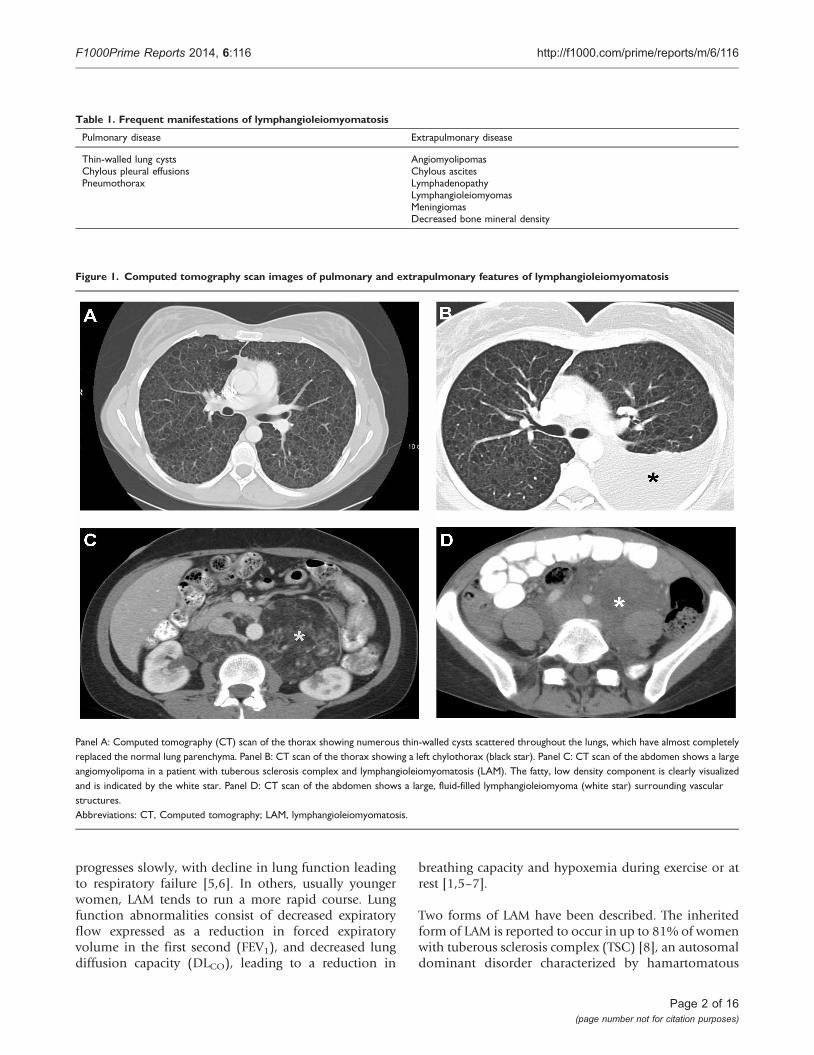
Table 1. Frequent manifestations of lymphangioleiomyomatosis
Pulmonary disease Extrapulmonary disease
Thin-walled lung cysts AngiomyolipomasChylous pleural effusions Chylous ascitesPneumothorax Lymphadenopathy
LymphangioleiomyomasMeningiomasDecreased bone mineral density
Figure 1. Computed tomography scan images of pulmonary and extrapulmonary features of lymphangioleiomyomatosis
Panel A: Computed tomography (CT) scan of the thorax showing numerous thin-walled cysts scattered throughout the lungs, which have almost completelyreplaced the normal lung parenchyma. Panel B: CT scan of the thorax showing a left chylothorax (black star). Panel C: CT scan of the abdomen shows a largeangiomyolipoma in a patient with tuberous sclerosis complex and lymphangioleiomyomatosis (LAM). The fatty, low density component is clearly visualizedand is indicated by the white star. Panel D: CT scan of the abdomen shows a large, fluid-filled lymphangioleiomyoma (white star) surrounding vascularstructures.Abbreviations: CT, Computed tomography; LAM, lymphangioleiomyomatosis.
Page 2 of 16(page number not for citation purposes)
F1000Prime Reports 2014, 6:116 http://f1000.com/prime/reports/m/6/116
tumors involving the central nervous system, skin, liver,heart and eyes, and associated with mental retardation,seizures and autism [9]. The sporadic form of LAM hasbeen reported to occur in 3.3–7.7 million women [10].In either form, LAM is caused by mutations in thetuberous sclerosis complex 1 (TSC1) or tuberoussclerosis complex 2 (TSC2) genes [11–13] that encodetwo proteins, hamartin and tuberin.
LAM is considered to be a low-grade malignancy. Data areconsistent with a metastatic model. Identical TSC2mutations have been found in the lungs and kidneys ofthe same patient with sporadic LAM [11,12]. Loss ofheterozygosity of TSC2 has been demonstrated in LAMcells isolated from lung, angiomyolipomas, blood, chyle,and urine from patients with sporadic LAM and TSC-LAM[11–15].
LAM cells have been detected in donor lungs of patientswho had lung transplantation [16,17]. These findingssupport the possibility that migration to the lungs ofcells from other sites, such as the kidney, lymphaticsystem, or uterus may occur [16–18].
Hamartin and tuberin together inhibit the mammaliantarget of rapamycin (mTOR) signaling pathway, a majorregulator of cell size and proliferation [19]. mTORinhibitors, sirolimus and everolimus, have been proveneffective in stabilizing lung function and reducing thesize of chylous effusions, lymphangioleiomyomas andangiomyolipomas [20–22].
The severity of lung disease and its rate of progression isbest assessed by clinical symptoms, histological grading oflung biopsy tissue, pulmonary function tests, computedtomography imaging, six-minute walk tests and cardio-pulmonary exercise tests [5]. The clinical data are used todetermine the need to treat patients with mTORinhibitors.
In this article, we will focus on targeted therapies shownto be effective in LAM, and other agents that appear to bepromising and are currently undergoing either pre-clinical or clinical testing. We will discuss how thesepotential treatments are expected to complement theactions of mTOR inhibitors. Finally, we will discuss thetreatment of pneumothoraxes, chylous effusions, andangiomyolipomas as well as issues related to pregnancyand lung transplantation.
General principles of managementPatients should be told that LAM is a chronic diseasewith a median transplant-free survival time of approxi-mately 29 years from the onset of symptoms and a
10-year transplant-free survival of 86% and that, as muchas possible, they should lead a normal life [5,23,24].Patients should be encouraged to lose excess weight,engage in physical activities, and exercise regularly.Levels of exercise should be limited only by the severityof lung disease. Sports involving physical contact andmartial arts should be avoided because of the potentialfor bleeding in patients who have angiomyolipomas.Patients should be allowed to travel by land or air, exceptto high-altitude locations, depending on disease severityand risk of pneumothorax. The risk of a life-threateningpneumothorax associated with air travel is minor [25].However, sudden onset of breathlessness or chest painsuggesting the presence of a pneumothorax should beinvestigated and ruled out prior to air travel. Arterialblood gases assist in determining whether a patient maytravel by air without supplemental oxygen. A six-minutewalk test or a cardiopulmonary exercise test to uncoverexercise-induced hypoxemia and determine the need forsupplemental oxygen is recommended [5,7]. Because ofthe potential risks of estrogens in the pathogenesis ofLAM, a disease found primarily in women, patientsshould be advised against using estrogen-containingcontraceptives and foods.
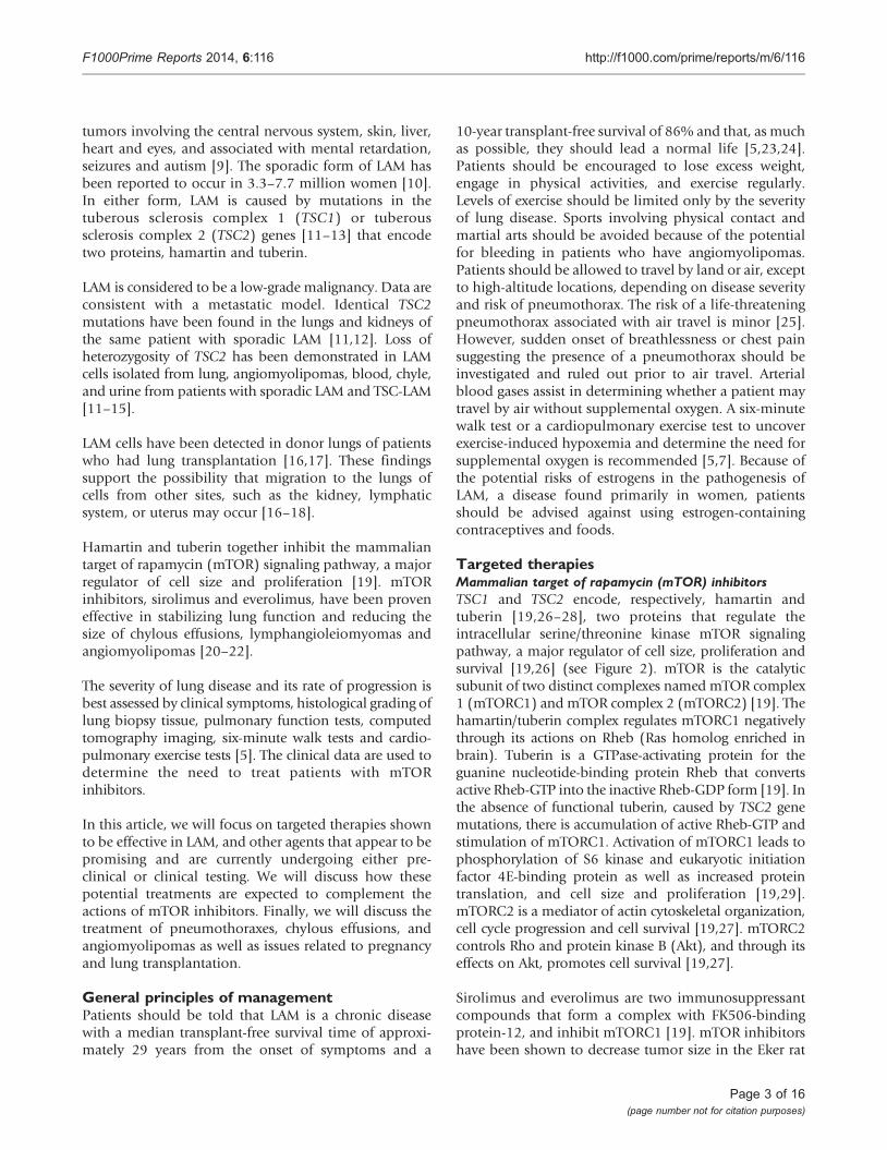
Targeted therapiesMammalian target of rapamycin (mTOR) inhibitorsTSC1 and TSC2 encode, respectively, hamartin andtuberin [19,26–28], two proteins that regulate theintracellular serine/threonine kinase mTOR signalingpathway, a major regulator of cell size, proliferation andsurvival [19,26] (see Figure 2). mTOR is the catalyticsubunit of two distinct complexes named mTOR complex1 (mTORC1) and mTOR complex 2 (mTORC2) [19]. Thehamartin/tuberin complex regulates mTORC1 negativelythrough its actions on Rheb (Ras homolog enriched inbrain). Tuberin is a GTPase-activating protein for theguanine nucleotide-binding protein Rheb that convertsactive Rheb-GTP into the inactive Rheb-GDP form [19]. Inthe absence of functional tuberin, caused by TSC2 genemutations, there is accumulation of active Rheb-GTP andstimulation of mTORC1. Activation of mTORC1 leads tophosphorylation of S6 kinase and eukaryotic initiationfactor 4E-binding protein as well as increased proteintranslation, and cell size and proliferation [19,29].mTORC2 is a mediator of actin cytoskeletal organization,cell cycle progression and cell survival [19,27]. mTORC2controls Rho and protein kinase B (Akt), and through itseffects on Akt, promotes cell survival [19,27].
Sirolimus and everolimus are two immunosuppressantcompounds that form a complex with FK506-bindingprotein-12, and inhibit mTORC1 [19]. mTOR inhibitorshave been shown to decrease tumor size in the Eker rat
Page 3 of 16(page number not for citation purposes)
F1000Prime Reports 2014, 6:116 http://f1000.com/prime/reports/m/6/116
Figure 2. Simplified diagram of the mammalian target of rapamycin (mTOR) signaling pathway and its relationship with autophagy andapoptosis
Tuberous sclerosis complex (TSC)1/2 integrates multiple signals to control cell size and proliferation. TSC1/2 regulates mammalian target of rapamycin(mTOR) complex 1 mTORC1 negatively through its actions on Rheb. Activation of mTORC1 leads to protein translation, cell growth and proliferation.mTORC1 is a major regulator of autophagy. Blockade of mTORC1 by sirolimus augments autophagy, leading to increased cell survival. This effect can beinhibited by hydroxychloroquine. mTOR complex 2 (mTORC2) regulates the actin cytoskeleton through Rho GTPases, which affect cell migration,morphogenesis and apoptosis. Simvastatin reduces Rheb and Rho activities and promotes apoptosis. Combined therapy with sirolimus, hydroxychloroquineor simvastatin may act synergically to inhibit lymphangioleiomyomatosis (LAM) cell growth and promote apoptosis.Abbreviations: Akt, protein kinase B; mTOR, mammalian target of rapamycin; mTORC1, mTOR complex 1; mTORC2, mTOR complex 2; Rac, small GTPasebinding protein of the Rho family; Rheb, Ras homolog enriched in brain; S6K1, S6 kinase 1; 4E-BP1, factor 4E binding protein 1; TSC, tuberous sclerosiscomplex.
Page 4 of 16(page number not for citation purposes)
F1000Prime Reports 2014, 6:116 http://f1000.com/prime/reports/m/6/116
model of TSC [30], decrease the growth of renalcystadenomas and liver hemangiomas in Tsc2 +/- mice,and decrease tumor growth and mortality in a mousemodel with Tsc2 +/- tumors [31]. Several clinical studieshave demonstrated the efficacy of mTORC1 inhibitors inthe treatment of LAM [20–22]. A double-blinded,placebo-controlled study (MILES trial) testing the effectof sirolimus on pulmonary function was undertaken in89 women with LAM. Forty-six patients were treated withsirolimus and 43 with placebo for 12 months [20].Patients were followed for a year after discontinuation oftherapy [20]. Compared to the placebo group, thesirolimus group had improvements from baseline invital capacity, FEV1, quality of life, and functional
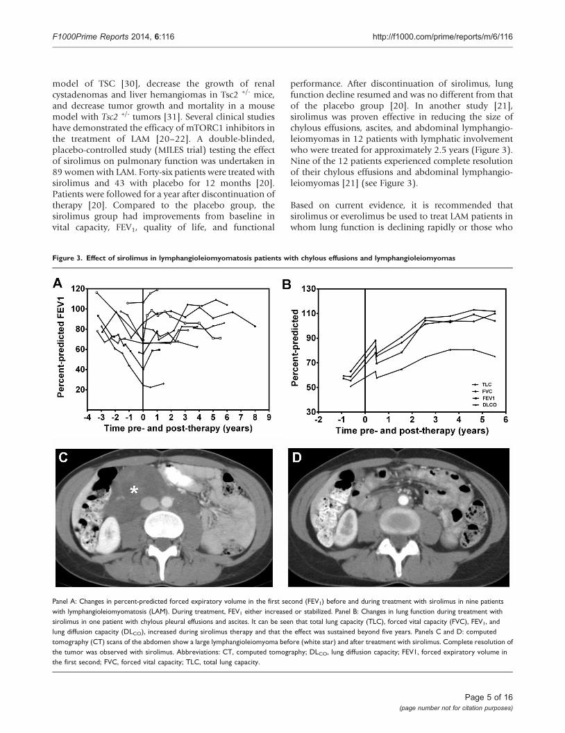
performance. After discontinuation of sirolimus, lungfunction decline resumed and was no different from thatof the placebo group [20]. In another study [21],sirolimus was proven effective in reducing the size ofchylous effusions, ascites, and abdominal lymphangio-leiomyomas in 12 patients with lymphatic involvementwho were treated for approximately 2.5 years (Figure 3).Nine of the 12 patients experienced complete resolutionof their chylous effusions and abdominal lymphangio-leiomyomas [21] (see Figure 3).
Based on current evidence, it is recommended thatsirolimus or everolimus be used to treat LAM patients inwhom lung function is declining rapidly or those who
Figure 3. Effect of sirolimus in lymphangioleiomyomatosis patients with chylous effusions and lymphangioleiomyomas
Panel A: Changes in percent-predicted forced expiratory volume in the first second (FEV1) before and during treatment with sirolimus in nine patientswith lymphangioleiomyomatosis (LAM). During treatment, FEV1 either increased or stabilized. Panel B: Changes in lung function during treatment withsirolimus in one patient with chylous pleural effusions and ascites. It can be seen that total lung capacity (TLC), forced vital capacity (FVC), FEV1, andlung diffusion capacity (DLCO), increased during sirolimus therapy and that the effect was sustained beyond five years. Panels C and D: computedtomography (CT) scans of the abdomen show a large lymphangioleiomyoma before (white star) and after treatment with sirolimus. Complete resolution ofthe tumor was observed with sirolimus. Abbreviations: CT, computed tomography; DLCO, lung diffusion capacity; FEV1, forced expiratory volume inthe first second; FVC, forced vital capacity; TLC, total lung capacity.
Page 5 of 16(page number not for citation purposes)
F1000Prime Reports 2014, 6:116 http://f1000.com/prime/reports/m/6/116
have symptomatic lymphangioleiomyomas, chylouspleural effusions or ascites [20,21,32]. The role ofsirolimus in patients with normal or stable lungfunction, or very slow rates of decline, is unclear. Thestarting dose of sirolimus should be 1 mg per day.Sirolimus serum levels must be monitored and dosageadjusted to attain serum trough levels between 5 and 15nanograms per ml [20–22]. The most frequent adverseevents associated with sirolimus therapy include stoma-titis, hypercholesterolemia, upper respiratory tract infec-tions, diarrhea, peripheral edema, acne, hypertension,headaches, leukopenia, delayed wound healing, throm-bocytopenia, and proteinuria [20]. Close patient mon-itoring is necessary [20]. Laboratory tests includingblood cells count, chemistries, urinalysis, urine protein/creatinine ratio, and sirolimus blood levels should beperformed. Pulmonary function studies should beperformed at least every six months. Interaction betweensirolimus and other drugs and some foods, such asgrapefruit, must be monitored carefully and adjustmentsto the dose of sirolimus made when appropriate.Currently, it is not known whether treatment must becontinued for life or whether resistance to sirolimuseventually develops. The optimal dose of sirolimus thatshould be employed and when it is most efficacious toinitiate treatment is not known. Dosage and blood levelscurrently used are based on experience with sirolimus inthe prevention of graft rejection in patients who haveundergone organ transplantation. It is possible thatlower sirolimus levels may be equally effective, especiallyin the treatment of lymphatic disease [33].
StatinsActivation of mTORC1 and mTORC2, and increased Rhoactivity, are necessary for TSC2-dependent cell prolifera-tion and survival [34]. Absence of TSC2 causes tuberindeficiency that results in increased Rheb and RhoAactivity and enhanced cell survival [34]. In TSC2-deficient, rat-derived, TSC2-null ELT3 cells, down-regulation of RhoA increases apoptosis, suggesting thatinhibition of RhoA, which is regulated by mTORC2, mayreduce cell survival [34]. Since sirolimus and everolimusonly suppress mTORC1, there is a rationale for therapiestargeting mTORC2 signaling [34–36].
Statins are 3-hydroxy-3-methyl-glutaryl-coenzyme-A(HMG-CoA) reductase inhibitors that inhibit geranylger-anylation of Rho GTPases, and farnesylation of the smallGTPases Ras and Rheb [37]. Atorvastatin was found toinhibit the growth of Tsc2-/- uterine-derived leiomyoma(ELT-3) and mouse embryonic fibroblasts by reducingRheb activity [38]. Simvastatin, another HMG-CoAreductase inhibitor, was shown to inhibit RhoA activity(see Figure 4) and the proliferation of TSC-null cells and
TSC2-null tumor growth in mice, and to promoteapoptosis [34]. Combined treatment with sirolimus andsimvastatin prevented recurrence of the tumors even afterdiscontinuation of both drugs [34]. This effect was specificfor simvastatin; atorvastatin did not reduce the size of liverand renal tumors in a mouse model of TSC [39].Simvastatin was also shown to reduce alveolar spaceenlargement in a mouse model of LAM [40]. Further,combined with sirolimus, simvastatin blocked matrixmetalloproteinase up-regulation and prevented alveolardestruction [40].
There are no data regarding the potential efficacy ofsimvastatin in the treatment of LAM. In one study, nocorrelation between statin use and angiomyolipomaresponse to sirolimus in patients with TSC or sporadicLAM was demonstrated [22]. In a retrospective study, therate of decline in lung diffusion capacity in LAM patientstreated with statins for hypercholesterolemia was greaterthan that of their matched, off-statin controls [41].However, in this study, the number of patients treatedwith simvastatin was small. The effect of simvastatincombined with sirolimus or everolimus in the treatmentof LAM is being investigated (NCT02061397).
Anti-estrogen therapyBecause LAM is predominantly a disease of pre-menopau-sal women, estrogens have been, from early on, implicatedin its pathogenesis [4–6]. Oophorectomy, progesteroneand gonadotrophin-releasing hormone (GnRH) analo-gues have all been used to treat LAM [6,42–46]. A numberof case reports and uncontrolled studies have suggestedbeneficial effects of anti-estrogen therapies [42]. Othersfound no benefit from oophorectomy or progesteronetherapy [43]. A reduced rate of decline in lung function inpre-menopausal patients treated with progesterone wasreported [44], but a large retrospective study involving275 patients reported no difference in disease progressionbetween patients treated or not treated with progesterone[6]. Studies that tested the effect of GnRH analogues havealso been inconclusive [45–46].
In vitro and experimental animal studies have provided arationale for anti-estrogen therapy in LAM [47–50] (seeFigure 4). LAM cells express estrogen receptors [51,52],and estradiol increases the proliferation of Eker rat-derivedTsc2-null, uterine ELT3 leiomyoma cells and the growth ofxenograph subcutaneous tumors in vivo [48,49]. Further,estradiol also increases the number of circulating tumorcells, the survival of injected ELT3 cells, and the number ofpulmonary metastasis of TSC-null cells injected subcuta-neously in oophorectomized mice [49]. This effect ofestradiol is associated with the activation of a mitogen-activated protein kinase (MAPK) signaling pathway that
Page 6 of 16(page number not for citation purposes)
F1000Prime Reports 2014, 6:116 http://f1000.com/prime/reports/m/6/116
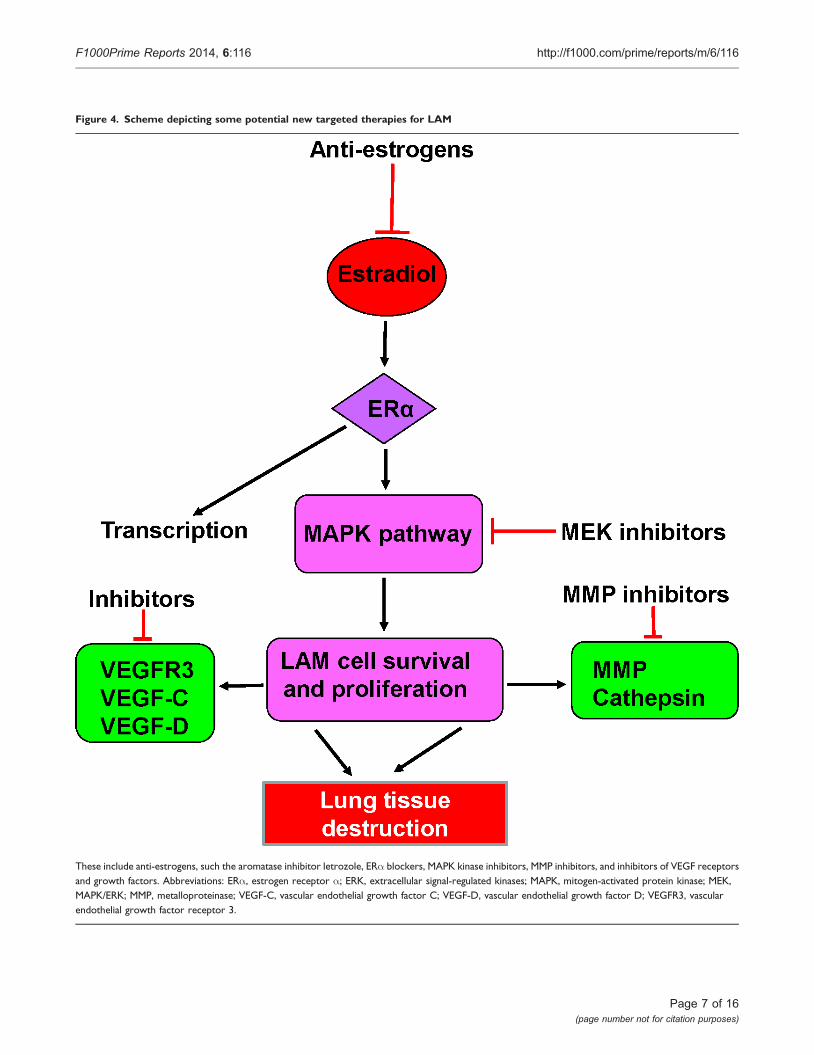
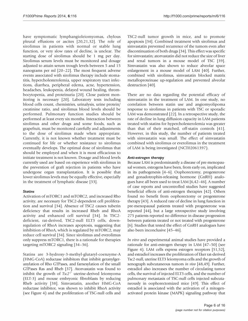
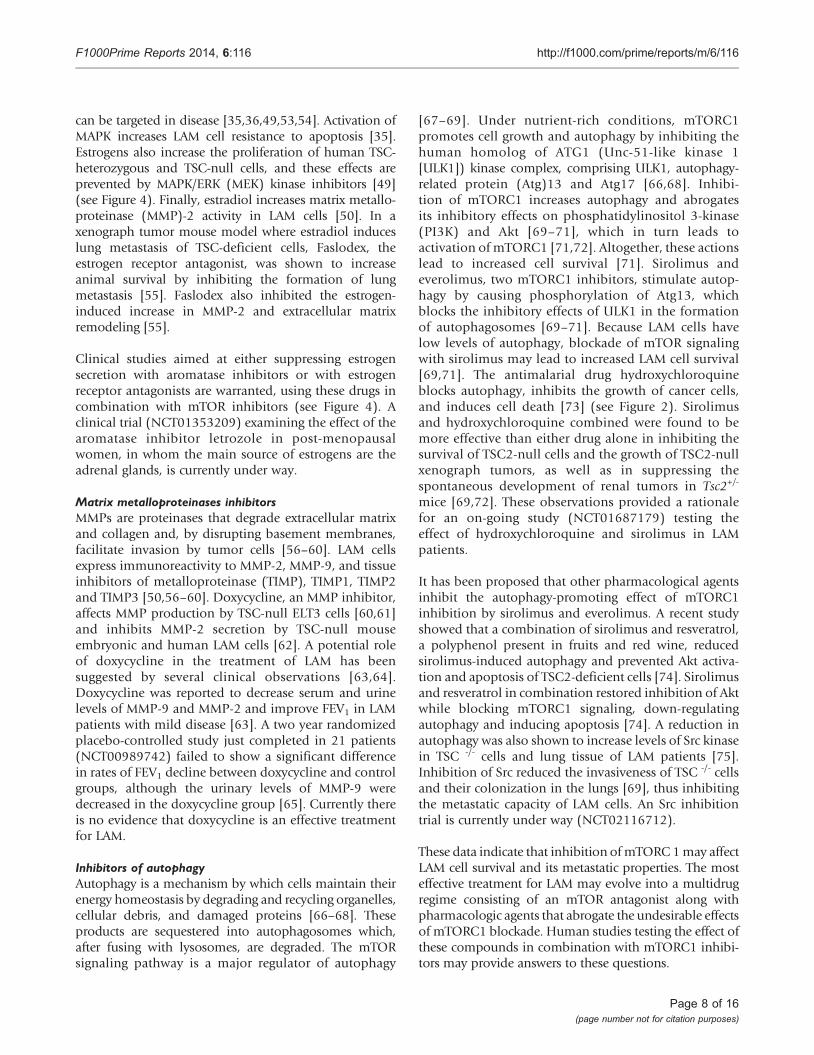
Figure 4. Scheme depicting some potential new targeted therapies for LAM
These include anti-estrogens, such the aromatase inhibitor letrozole, ERa blockers, MAPK kinase inhibitors, MMP inhibitors, and inhibitors of VEGF receptorsand growth factors. Abbreviations: ERa, estrogen receptor a; ERK, extracellular signal-regulated kinases; MAPK, mitogen-activated protein kinase; MEK,MAPK/ERK; MMP, metalloproteinase; VEGF-C, vascular endothelial growth factor C; VEGF-D, vascular endothelial growth factor D; VEGFR3, vascularendothelial growth factor receptor 3.
Page 7 of 16(page number not for citation purposes)
F1000Prime Reports 2014, 6:116 http://f1000.com/prime/reports/m/6/116
can be targeted in disease [35,36,49,53,54]. Activation ofMAPK increases LAM cell resistance to apoptosis [35].Estrogens also increase the proliferation of human TSC-heterozygous and TSC-null cells, and these effects areprevented by MAPK/ERK (MEK) kinase inhibitors [49](see Figure 4). Finally, estradiol increases matrix metallo-proteinase (MMP)-2 activity in LAM cells [50]. In axenograph tumor mouse model where estradiol induceslung metastasis of TSC-deficient cells, Faslodex, theestrogen receptor antagonist, was shown to increaseanimal survival by inhibiting the formation of lungmetastasis [55]. Faslodex also inhibited the estrogen-induced increase in MMP-2 and extracellular matrixremodeling [55].
Clinical studies aimed at either suppressing estrogensecretion with aromatase inhibitors or with estrogenreceptor antagonists are warranted, using these drugs incombination with mTOR inhibitors (see Figure 4). Aclinical trial (NCT01353209) examining the effect of thearomatase inhibitor letrozole in post-menopausalwomen, in whom the main source of estrogens are theadrenal glands, is currently under way.
Matrix metalloproteinases inhibitorsMMPs are proteinases that degrade extracellular matrixand collagen and, by disrupting basement membranes,facilitate invasion by tumor cells [56–60]. LAM cellsexpress immunoreactivity to MMP-2, MMP-9, and tissueinhibitors of metalloproteinase (TIMP), TIMP1, TIMP2and TIMP3 [50,56–60]. Doxycycline, an MMP inhibitor,affects MMP production by TSC-null ELT3 cells [60,61]and inhibits MMP-2 secretion by TSC-null mouseembryonic and human LAM cells [62]. A potential roleof doxycycline in the treatment of LAM has beensuggested by several clinical observations [63,64].Doxycycline was reported to decrease serum and urinelevels of MMP-9 and MMP-2 and improve FEV1 in LAMpatients with mild disease [63]. A two year randomizedplacebo-controlled study just completed in 21 patients(NCT00989742) failed to show a significant differencein rates of FEV1 decline between doxycycline and controlgroups, although the urinary levels of MMP-9 weredecreased in the doxycycline group [65]. Currently thereis no evidence that doxycycline is an effective treatmentfor LAM.
Inhibitors of autophagyAutophagy is a mechanism by which cells maintain theirenergy homeostasis by degrading and recycling organelles,cellular debris, and damaged proteins [66–68]. Theseproducts are sequestered into autophagosomes which,after fusing with lysosomes, are degraded. The mTORsignaling pathway is a major regulator of autophagy
[67–69]. Under nutrient-rich conditions, mTORC1promotes cell growth and autophagy by inhibiting thehuman homolog of ATG1 (Unc-51-like kinase 1[ULK1]) kinase complex, comprising ULK1, autophagy-related protein (Atg)13 and Atg17 [66,68]. Inhibi-tion of mTORC1 increases autophagy and abrogatesits inhibitory effects on phosphatidylinositol 3-kinase(PI3K) and Akt [69–71], which in turn leads toactivation of mTORC1 [71,72]. Altogether, these actionslead to increased cell survival [71]. Sirolimus andeverolimus, two mTORC1 inhibitors, stimulate autop-hagy by causing phosphorylation of Atg13, whichblocks the inhibitory effects of ULK1 in the formationof autophagosomes [69–71]. Because LAM cells havelow levels of autophagy, blockade of mTOR signalingwith sirolimus may lead to increased LAM cell survival[69,71]. The antimalarial drug hydroxychloroquineblocks autophagy, inhibits the growth of cancer cells,and induces cell death [73] (see Figure 2). Sirolimusand hydroxychloroquine combined were found to bemore effective than either drug alone in inhibiting thesurvival of TSC2-null cells and the growth of TSC2-nullxenograph tumors, as well as in suppressing thespontaneous development of renal tumors in Tsc2+/-
mice [69,72]. These observations provided a rationalefor an on-going study (NCT01687179) testing theeffect of hydroxychloroquine and sirolimus in LAMpatients.
It has been proposed that other pharmacological agentsinhibit the autophagy-promoting effect of mTORC1inhibition by sirolimus and everolimus. A recent studyshowed that a combination of sirolimus and resveratrol,a polyphenol present in fruits and red wine, reducedsirolimus-induced autophagy and prevented Akt activa-tion and apoptosis of TSC2-deficient cells [74]. Sirolimusand resveratrol in combination restored inhibition of Aktwhile blocking mTORC1 signaling, down-regulatingautophagy and inducing apoptosis [74]. A reduction inautophagy was also shown to increase levels of Src kinasein TSC -/- cells and lung tissue of LAM patients [75].Inhibition of Src reduced the invasiveness of TSC -/- cellsand their colonization in the lungs [69], thus inhibitingthe metastatic capacity of LAM cells. An Src inhibitiontrial is currently under way (NCT02116712).
These data indicate that inhibition ofmTORC 1may affectLAM cell survival and its metastatic properties. The mosteffective treatment for LAM may evolve into a multidrugregime consisting of an mTOR antagonist along withpharmacologic agents that abrogate the undesirable effectsof mTORC1 blockade. Human studies testing the effect ofthese compounds in combination with mTORC1 inhibi-tors may provide answers to these questions.
Page 8 of 16(page number not for citation purposes)
F1000Prime Reports 2014, 6:116 http://f1000.com/prime/reports/m/6/116
Potential future therapiesSeveral potential therapies for LAM are currently under-going clinical trials. In addition, in vitro or animal datahave suggested a potential beneficial role of other agentsin the treatment of LAM [76–79].
Vascular endothelial growth factor receptor (VEGFR)-3 is amajor regulator of lymphangiogenesis [54,80,81]. Immu-noreactivity for vascular endothelial growth factors(VEGFs)-C and -D is present in LAM lesions and LAMcells [80]. In addition, because of the important role oflymphangiogenesis in the pathogenesis of LAM [54], andevidence that increased VEGF-D levels in the serum ofLAM patients correlate with disease severity and clinicalcourse [82], blockade of VEGFRs or anti-VEGF-D therapiesare being considered as potential treatments [54,80,81]. Inaccordance, in a subcutaneous Tsc2 -/- tumor mousemodel, sorafenib (a Raf kinase and VEGF receptor pathwayinhibitor) and sirolimus together decreased tumor volumeand increased survival more effectively than sirolimusalone [76]. A member of the collagen IV family, namedlamstatin [83], was recently reported to have anti-lymphangiogenic properties. Levels of lamstatin werefound to be reduced in the lungs of LAM patients [83].Further, in vitro and in vivo studies showed that lamstatin,combined with a 17-aminoacid peptide (CP17), inhibitedproliferation and migration of human lung lymphaticendothelial cells, and decreased dysplasia of the tumor-associated lymphatic network in a lung adenocarcinomaxenograph mouse model [83].
Alterations in the interferon gamma (IFN-g) signalingpathway were suggested by the finding that expression ofIFN-g is not present in angiomyolipoma from patientswith TSC or LAM [77]. Treatment of Tsc1- or Tsc2-nullcells with IFN-g was shown to induce apoptosis [78].There was evidence of synergism between IFN-g andsirolimus in inducing apoptosis of these cells [78].However, sirolimus and IFN-g together were not moreeffective than sirolimus alone against TSC-relatedkidney tumors in Tsc2 +/- mice [76]. A combination ofan mTORC1 receptor antagonist and IFN-g was moreeffective than either single agent in decreasing theseverity of kidney cystadenomas and liver hemangio-mas in both Tsc +/- and Tsc -/- mouse models [31]. Bothdrugs together were also shown to be more effectivethan a single agent in reducing tumor growth in amouse model of TSC [79]. Although IFN-g appears notto directly inhibit the proliferation of Tsc2-null cells, orincrease the inhibitory effects of sirolimus, there aredata suggesting that increased expression of signaltransducer and activator of transcription (STAT) 3 ispresent in LAM lungs and may possibly be a target fornew therapies [84].
Finally, increased AMP-activated protein kinase (AMPK)activity has been reported in TSC tumors and Tsc-null cellsand this may contribute to increased LAM cell survival[85]. Since Rheb controls AMPK activity independent ofmTORC1 signaling [86,87] and Rheb depletion decreasestumorigenesis [87], targeting Rheb may have a role incombination therapy of LAM or TSC [87].
Treatment of complicationsPneumothoraxOnce a patient has had a pneumothorax, the chances ofhaving a recurrence are greater than 70% [1,88–90].A small pneumothorax may be treated by chest tubedrainage. If the air leak persists or the pneumothoraxrecurs, pleurodesis is recommended. A chemical or surgicalpleurectomy by video-assisted thoracoscopy should beconsidered [88,90]. Chemical sclerosis with doxycycline,pleurectomy, mechanical abrasion and talc poudrage arealso effective. Talc pleurodesis may result in considerablepleural scarring that may be associated with chronic painas well as bleeding complications during removal of thenative lungs at the time of transplantation [88,90].
Of great concern to patients with LAM is the risk ofpneumothorax associated with air travel [25,91]. Sincecabins of commercial aircraft are pressurized only to abarometric pressure equivalent to an altitude of 1,500 to2,500 meters, during the airplane’s ascent or descent thecabin pressure falls or increases according to Boyle’s law[91], leading to gas expansion or contraction within bodycavities, such as lung cysts or non-functioning bullae [91].Retrospective studies indicate that the risk of developing apneumothorax during air travel is small, ranging from2–4 % [25,91,92]. There is no evidence that pre-existingchronic loculated pneumothoraxes are associated with anincreased risk of additional pneumothorax or expansionof a pre-existing pneumothorax during air travel [25].Patients should be advised to take direct flights and toabstain from air travel if they are experiencing chest painand/or increased dyspnea, until the presence of apneumothorax is ruled out [25,92,93].
Another potential risk associated with flying is hypox-emia [91]. An arterial blood gas obtained while breath-ing room air may assist in deciding whether the patientmay fly without supplemental oxygen. More precisely,arterial blood gases may be measured while they breathea low oxygen concentration that produces an inspiredoxygen pressure similar to that present in pressurizedcabins of commercial airplanes.
Chylous effusions and lymphangioleiomyomasChylous pleural effusions, ascites, and lymphangioleio-myomas may compromise respiratory function and
Page 9 of 16(page number not for citation purposes)
F1000Prime Reports 2014, 6:116 http://f1000.com/prime/reports/m/6/116
cause abdominal pain, urinary frequency, obstipation,and peripheral edema [5]. Not uncommonly, patientsmay be misdiagnosed with a lymphoma or ovariancancer [94–97]. The symptomatology associated withlymphatic disease in patients with LAMmay cause a greatdeal of distress and poses difficult therapeutic problems.The least recommended therapeutic approach is continuoustube drainage of chylous pleural effusions and as-cites, as this approach results in nutritional comprom-ise, protein loss, lymphopenia, increased risk ofinfections, and weight loss [21,88,90,98]. Further, oncea thoracostomy tube is placed in the pleural cavity,drainage may become continuous and pleural symphysismay become difficult. It is preferable to performtherapeutic thoracentesis when patients become sympto-matic. Low fat diet, pleuro-peritoneal or peritoneal-venousshunts, treatment with somatostatin and octreotide havebeen employed but there is little experience with thesetherapies in LAM [99–102].
Surgical pleurodesis may be effective in reducing the sizeof the effusion, but this will not ameliorate abdominalsymptoms associated with ascites and lymphangioleio-myomas. To be effective, surgical pleurodesis must beperformed under conditions of reduced chyle flow. Toaccomplish this, patients must be placed on parenteralnutrition with a fat-free solution before, during, andafter pleurodesis. The thoracostomy tube should beremoved only when daily drainage volume decreases to200 ml or less.
A major advance in the treatment of lymphatic disease inLAM was the discovery that the mTOR inhibitorsirolimus decreased the volume of chylous effusions,the size of lymphangioleiomyomas, and ascites, clearedlung infiltrates caused by lung lymph accumulation anddramatically improved lung function [21,103]. Con-sequently, sirolimus is now considered to be the treatmentof choice (see Figure 3). Although volume reduction orcomplete resolution of the lymphangioleiomyomasgenerally occurs within a few months, resolution ofchylous effusions may take many months to more than ayear [21]. While on sirolimus, therapeutic thoracentesismay be performed periodically if patients experiencesignificant respiratory symptoms. Abdominal lymphan-gioleiomyomas must not be surgically removed orpartially resected as this approach may result in ascitesand disabling pleural effusions.
AngiomyolipomasAngiomyolipomas occur primarily in the kidney and liver[104]. The major risk of angiomyolipomas is bleeding.Arterial embolization is recommended for the treatmentof acute bleeding, severe pain, or prophylactically in
patients with angiomyolipomas larger than 3–4 cm indiameter [104,105]. Resection of the kidney should beavoided unless the angiomyolipoma has an atypicalradiologic appearance that raises the question of malig-nancy. In this case, a biopsy should be performed or thetumor should be resected [104,106,107]. mTORC1inhibitors are effective in decreasing the size of renalangiomyolipomas in patients with TSC or sporadic LAM,with tumor size being reduced in about 44 to 50% ofpatients [22,108–110]. In one study, 42% of 79 patientswith angiomyolipomas treated with everolimusresponded with a 50% reduction in tumor size after24 weeks of therapy [110]. Following discontinuation ofthe drugs, the angiolipomas tended to return to their initialsize. Treatment with mTOR inhibitors should probably bethe initial approach for the treatment of large angiomyo-lipomas. Arterial embolization should be reserved foracute bleeding or for patients who do not tolerate mTORinhibitors [104–110]. Once mTOR inhibitor therapy hasbegun, it must be continued because its discontinuationappears to return the tumor to its original size.
Management of pregnancyBy the time a young woman is diagnosed with LAM shemay already have experienced one or more pregnancies.Not infrequently, a pneumothorax or increased dyspneamay uncover the presence of LAM [1,111,112]. Despitethese events, of 353 pregnancies recorded in the LAMregistry, 67% resulted in live birth and only about 17%had spontaneous abortion [1]. Of those who had beenpregnant, 22% experienced worsening of respiratorysymptoms [1]. Patients who were diagnosed with LAMduring pregnancy appear to have had more prematurebirths, and higher frequency of dyspnea and pneu-mothorax than patients diagnosed either before or afterpregnancy [112]. These data, along with reports ofworsening symptoms during pregnancy, such as dys-pnea, pneumothorax, chylous effusions or hemorrhagefrom angiomyolipomas [111,113–115], have raised thequestion as to whether LAM patients should be advisednot to become pregnant.
Pregnancy should be discouraged in patients withmoderate to severe disease or those in whom lungfunction is rapidly declining. The rationale for thisrecommendation is that these patients should be treatedwith sirolimus rather than risking further deterioration inlung function and a delay in starting therapy, becausesirolimus must be discontinued prior to, and duringpregnancy. Patients with mild disease who strongly desireto become pregnant should be told about its potentialrisks (e.g. pneumothorax, decline in lung function). Theyshould be advised that with close medical and obstetricalsupervision, others have tolerated pregnancy and
Page 10 of 16(page number not for citation purposes)
F1000Prime Reports 2014, 6:116 http://f1000.com/prime/reports/m/6/116
delivered a normal child. However, at present, there is nomethod of predicting the outcome in terms of frequency ofcomplications such as pneumothorax or chylothorax, andthe magnitude of potential decline in lung function.
Lung transplantationExcept in advanced stages, dyspnea at rest is not a majorfeature of LAM [5]. Patients with an FEV1 of less than oneliter and a DLCO less than 30% predicted, who arereceiving supplemental oxygen, might be comfortable atrest. However, exercise and hypoxemia requiring supple-mental oxygen are major factors affecting the quality oflife, namely the ability of patients to conduct activities ofdaily living. In one study [116], preoperative FEV1 andDLCO prior to transplantation were, respectively, 20±8and 23±9% predicted and there was also restinghypoxemia. The average 6-minute walk test distancewas 250 meters. The five-year post-transplant survival ofLAM patients undergoing lung transplantation wasaround 69% [116–119]. The European experience withlung transplantation in LAM is similar to that of the USA[120], but others have reported five-year survivals ofaround 75% [121].
Because patients with very low FEV1 and DLCO onsupplemental oxygen may live for many years [23,24],lung function needs to be severely compromised beforelung transplantation is considered. We suggest that lungtransplantation be discussed with the patient when FEV1
and DLCO are about 30% predicted, the patient is oncontinuous supplemental oxygen, is unable to carry outactivities of daily living and has resting pulmonaryhypertension. Importantly, the patient should rate herquality of life as being so poor that she wishes to undergothe additional risks associated with lung transplantation.
Summary statement and conclusionsLAM is a disease affecting women, which is associatedwithcystic lung destruction, and extrapulmonary manifesta-tions consisting of abdominal angiomyolipomas andlymphangioleiomyomas. LAM presents with dyspnea,recurrent pneumothoraxes, and hemorrhages from angio-myolipomas. LAM is characterized by a reduction inbreathing capacity, hypoxemia during exercise or at rest,and respiratory failure.
Not long ago, LAM was defined as a fatal disease ofyoung women for which there was no effective therapyand the only treatment option was lung transplantation.The establishment of the National Heart, Lung, andBlood Institute (NHLBI) LAM registry and other registriesworldwide have led to major progress in the character-ization of the clinical features and natural history ofLAM. The finding that LAM is caused by mutations of
TSC1 or TSC2 genes that encode hamartin and tuberin,two proteins with a major role in control of the mTORsignaling pathway, led to therapies targeting mTOR. TwomTORC1 inhibitors, sirolimus and everolimus, havebeen shown to be effective in stabilizing lung function,and reducing the size of chylous effusions, lymphangio-leiomyomas, and angiomyolipomas.
Treatment with these pharmacological agents, which isgenerally tolerated by patients, is now the standardtherapy for patients showing compromised lung func-tion or those who have symptomatic chylous effusionsand lymphangioleiomyomas or large angiomyolipomas.
In the case of angiomyolipomas, discontinuation ofmTORC1 inhibitor therapy results in a return to pre-therapy tumor size, suggesting that to sustain thetherapeutic effects of mTORC1 inhibitors, treatment hasto be continued. mTORC1 inhibition results in increasedautophagy and possibly enhanced LAM cell survival, thusreducing the beneficial effects of sirolimus or everolimus.Inhibition of autophagy with hydroxychloroquine hasbeen suggested as a new treatment for LAM, complement-ing inhibition of mTORC1 with sirolimus or everolimus.Such a therapeutic regimen is now undergoing clinicaltesting.
Deficiency of tuberin due to TSC2 mutations, as occursin LAM, results in increased RhoA GTPase activity andincreased cell survival. This effect is mediated throughmTORC2 signaling. Since sirolimus and everolimus onlyaffect the activity of mTORC1, there is a rationale fortherapies targeting RhoA GTPases. Statins inhibit RhoGTPases and promote apoptosis. Simvastatin combinedwith sirolimus is effective in preventing lung destructionin a mouse model of LAM. Simvastatin combined withsirolimus or everolimus is currently undergoing phase 1clinical testing in patients with LAM.
Other treatments that are being investigated are estrogenreceptor blockers, aromatase inhibitors, MAPK/ERK(MEK) inhibitors and VEGFR antagonists. Blockade ofVEGF receptors or anti-VEGF therapies may be of value inthe treatments of LAM in view of the role of lymphangio-genesis in its pathogenesis and evidence that increasedlevels of VEGF-D in the serum of LAM patients correlateswith disease severity and clinical course. Preclinical studieshave shown that a VEGF pathway inhibitor and sirolimustogether decreased tumor volume in an animal model ofLAM and increased survival more effectively thansirolimus alone.
We conclude that, as in the case of cancer, LAM (which isconsidered a low grade malignancy) may be best treated
Page 11 of 16(page number not for citation purposes)
F1000Prime Reports 2014, 6:116 http://f1000.com/prime/reports/m/6/116

with multiple drugs targeting signaling pathways con-sidered important in the pathogenesis of LAM.
AbbreviationsAkt, protein kinase B; AMPK, AMP-activated proteinkinase; Atg, autophagy-related protein; DLCO, lungdiffusion capacity; FEV1, forced expiratory volume inthe first second; GnRH, gonadotrophin-releasing hor-mone; HMB, human melanin black antibody; IFN-g,interferon gamma; LAM, lymphangioleiomyomatosis;MAPK, mitogen-activated protein kinase; MMP, metal-loproteinase; mTOR, mammalian target of rapamycin;mTORC1, mTOR complex 1; mTORC2, mTOR complex2; PI3K, phosphoinositide-3-kinase; Rheb, Ras homologenriched in brain GTPase-activating protein; TIMP, tissueinhibitor of metalloproteinase; TSC, tuberous sclerosiscomplex; ULK1, Unc-51-like kinase 1; VEGF, vascularendothelial growth factor; VEGFR, vascular endothelialgrowth factor receptor.
DisclosuresThe authors declare that they have no disclosures.
AcknowledgmentsThe authors thank Gustavo Pacheco-Rodriguez PhD forhis assistance with the preparation of the figures. Authorswere supported by the Intramural Research Program,National Institutes of Health, National Heart, Lung andBlood Institute.
References1. Ryu JH, Moss J, Beck GJ, Lee J, Brown KK, Chapman JT, Finlay GA,
Olson EJ, Ruoss SJ, Maurer JR, Raffin TA, Peavy HH, McCarthy K,Taveira-Dasilva A, McCormack FX, Avila NA, Decastro RM, Jacobs SS,Stylianou M, Fanburg BL: The NHLBI lymphangioleiomyoma-tosis registry: characteristics of 230 patients at enrollment.Am J Respir Crit Care Med 2006, 173:105-11.
2. Matsui K, Tatsuguchi A, Valencia J, Yu Zx, Bechtle J, Beasley MB,Avila N, Travis WD, Moss J, Ferrans VJ: Extrapulmonarylymphangioleiomyomatosis (LAM): clinicopathologic featuresin 22 cases. Hum Pathol 2000, 31:1242-8.
3. Ferrans VJ, Yu Zx, Nelson WK, Valencia JC, Tatsuguchi A, Avila NA,Riemenschn W, Matsui K, Travis WD, Moss J: Lymphangioleio-myomatosis (LAM): a review of clinical and morphologicalfeatures. J Nippon Med Sch 2000, 67:311-29.
4. McCormack FX: Lymphangioleiomyomatosis: a clinical update.Chest 2008, 133:507-16.
5. Taveira-DaSilva AM, Pacheco-Rodriguez G, Moss J: The naturalhistory of lymphangioleiomyomatosis: markers of severity,rate of progression and prognosis. Lymphat Res Biol 2010,8:9-19.
6. Taveira-DaSilva AM, Stylianou MP, Hedin CJ, Hathaway O, Moss J:Decline in lung function in patients with lymphangioleiomyo-matosis treated with or without progesterone. Chest 2004,126:1867-74.
7. Taveira-DaSilva AM, Stylianou MP, Hedin CJ, Kristof AS, Avila NA,Rabel A, Travis WD, Moss J: Maximal oxygen uptake andseverity of disease in lymphangioleiomyomatosis. Am J RespirCrit Care Med 2003, 168:1427-31.
8. Cudzilo CJ, Szczesniak RD, Brody AS, Rattan MS, Krueger DA,Bissler JJ, Franz DN, McCormack FX, Young LR: Lymphangioleio-myomatosis screening in women with tuberous sclerosis.Chest 2013, 144:578-85.
9. Curatolo P, Bombardieri R, Jozwiak S: Tuberous sclerosis. Lancet2008, 372:657-68.
10. Harknett EC, Chang, WYC, Byrnes S, Johnson J, Lazor R, Cohen MM,Gray B, Geiling S, Telford H, Tattersfield AE, Hubbard RB,Johnson SR: Use of variability in national and regional datato estimate the prevalence of lymphangioleiomyomatosis.QJM 2011, 104:971-9.
11. Smolarek TA, Wessner LL, McCormack FX, Mylet JC, Menon AG,Henske EP: Evidence that lymphangiomyomatosis is caused byTSC2 mutations: chromosome 16p13 loss of heterozygosityin angiomyolipomas and lymph nodes from women withlymphangiomyomatosis. Am J Hum Genet 1998, 62:810-5.
12. Carsillo T, Astrinidis A, Henske EP: Mutations in the tuberoussclerosis complex gene TSC2 are a cause of sporadicpulmonary lymphangioleiomyomatosis. Proc Natl Acad Sci USA2000, 97:6085-90.
13. Yu J, Astrinidis A, Henske EP: Chromosome 16 loss ofheterozygosity in tuberous sclerosis and sporadic lymphan-giomyomatosis. Am J Respir Crit Care Med 2001, 164:1537-40.
14. Crooks DM, Pacheco-Rodriguez G, Decastro RM, McCoy JP, Wang J,Kumaki F, Darling T, Moss J: Molecular and genetic analysis ofdisseminated neoplastic cells in lymphangioleiomyomatosis.Proc Natl Acad Sci USA 2004, 101:17462-7.
15. Cai X, Pacheco-Rodriguez G, Fan Q, Haughey M, Samsel L, El-Chemaly S, Wu H, McCoy JP, Steagall WK, Lin J, Darling TN, Moss J:Phenotypic characterization of disseminated cells with TSC2loss of heterozygosity in patients with lymphangioleiomyo-matosis. Am J Respir Crit Care Med 2010, 182:1410-8.
16. Bittmann I, Rolf B, Amann G, Löhrs U: Recurrence of lymphan-gioleiomyomatosis after single lung transplantation: newinsights into pathogenesis. Hum Pathol 2003, 34:95-8.
17. Karbowniczek M, Astrinidis A, Balsara BR, Testa JR, Lium JH, Colby TV,McCormack FX, Henske EP: Recurrent lymphangiomyomatosisafter transplantation: genetic analyses reveal a metastaticmechanism. Am J Respir Crit Care Med 2003, 167:976-82.
18. Mitani K, Kumasaka T, Takemura H, Hayashi T, Gunji Y, Kunogi M,Akiyoshi T, Takahashi K, Suda K, Seyama K: Cytologic, immuno-cytochemical and ultrastructural characterization of lym-phangioleiomyomatosis cell clusters in chylous effusions ofpatients with lymphangioleiomyomatosis. Acta Cytol 2009,53:402-9.
19. Zoncu R, Efeyan A, Sabatini DM: mTOR: from growth signalintegration to cancer, diabetes and ageing. Nat Rev Mol Cell Biol2011, 12:21-35.
20. McCormack FX, Inoue Y, Moss J, Singer LG, Strange C, Nakata K,Barker AF, Chapman JT, Brantly ML, Stocks JM, Brown KK, Lynch JP,Goldberg HJ, Young LR, Kinder BW, Downey GP, Sullivan EJ,Colby TV, McKay RT, Cohen MM, Korbee L, Taveira-DaSilva AM,Lee H, Krischer JP, Trapnell BC: Efficacy and safety of sirolimus inlymphangioleiomyomatosis. N Engl J Med 2011, 364:1595-606.
21. Taveira-DaSilva AM, Hathaway O, Stylianou M, Moss J: Changes inlung function and chylous effusions in patients with lymphan-gioleiomyomatosis treated with sirolimus. Ann Intern Med 2011,154:797-805, W-292-3.
22. Bissler JJ, McCormack FX, Young LR, Elwing JM, Chuck G,Leonard JM, Schmithorst VJ, Laor T, Brody AS, Bean J, Salisbury S,
Page 12 of 16(page number not for citation purposes)
F1000Prime Reports 2014, 6:116 http://f1000.com/prime/reports/m/6/116
Franz DN: Sirolimus for angiomyolipoma in tuberous sclerosiscomplex or lymphangioleiomyomatosis. N Engl J Med 2008,358:140-51.
23. Cohen MM, Pollock-BarZiv S, Johnson SR: Emerging clinicalpicture of lymphangioleiomyomatosis. Thorax 2005, 60:875-9.
24. Oprescu N, McCormack FX, Byrnes S, Kinder BW: Clinicalpredictors of mortality and cause of death in lymphangioleio-myomatosis: a population-based registry. Lung 2013, 191:35-42.
25. Taveira-DaSilva AM, Burstein D, Hathaway OM, Fontana JR,Gochuico BR, Avila NA, Moss J: Pneumothorax after air travelin lymphangioleiomyomatosis, idiopathic pulmonary fibrosis,and sarcoidosis. Chest 2009, 136:665-70.
26. Rosner M, Hanneder M, Siegel N, Valli A, Hengstschläger M: Thetuberous sclerosis gene products hamartin and tuberin aremultifunctional proteins with a wide spectrum of interactingpartners. Mutat Res 2008, 658:234-46.
27. Sarbassov DD, Ali SM, Kim D, Guertin DA, Latek RR, Erdjument-Bromage H, Tempst P, Sabatini DM: Rictor, a novel bindingpartner of mTOR, defines a rapamycin-insensitive andraptor-independent pathway that regulates the cytoskeleton.Curr Biol 2004, 14:1296-302.
28. Huang J, Manning BD: A complex interplay between Akt, TSC2and the twomTOR complexes. Biochem Soc Trans 2009, 37:217-22.
29. Krymskaya VP, Goncharova EA:PI3K/mTORC1activation in hamar-toma syndromes: therapeutic prospects. Cell Cycle 2009, 8:403-13.
30. Kenerson H, Dundon TA, Yeung RS: Effects of rapamycin in theEker rat model of tuberous sclerosis complex. Pediatr Res 2005,57:67-75.
31. Lee L, Sudentas P, Donohue B, Asrican K, Worku A, Walker V, Sun Y,Schmidt K, Albert MS, El-Hashemite N, Lader AS, Onda H, Zhang H,Kwiatkowski DJ, Dabora SL: Efficacy of a rapamycin analog (CCI-779) and IFN-gamma in tuberous sclerosis mouse models.Genes Chromosomes Cancer 2005, 42:213-27.
32. Neurohr C, Hoffmann AL, Huppmann P, Herrera VA, Ihle F,Leuschner S, Wulffen W von, Meis T, Baezner C, Leuchte H,Baumgartner R, Zimmermann G, Behr J: Is sirolimus a therapeuticoption for patients with progressive pulmonary lymphangio-leiomyomatosis? Respir Res 2011, 12:66.
33. Ando K, Kurihara M, Kataoka H, Ueyama M, Togo S, Sato T, Doi T,Iwakami S, Takahashi K, Seyama K, Mikami M: Efficacy and safety oflow-dose sirolimus for treatment of lymphangioleiomyoma-tosis. Respir Investig 2013, 51:175-83.
34. Goncharova EA, Goncharov DA, Li H, Pimtong W, Lu S, Khavin I,Krymskaya VP: mTORC2 is required for proliferation andsurvival of TSC2-null cells. Mol Cell Biol 2011, 31:2484-98.
35. Hammes SR, Krymskaya VP: Targeted approaches towardunderstanding and treating pulmonary lymphangioleiomyo-matosis (LAM). Horm Cancer 2013, 4:70-7.
36. Henske EP, McCormack FX: Lymphangioleiomyomatosis - a wolfin sheep’s clothing. J Clin Invest 2012, 122:3807-16.
37. Atochina-Vasserman EN, Goncharov DA, Volgina AV, Milavec M,James ML, Krymskaya VP: Statins in lymphangioleiomyomatosis.Simvastatin and atorvastatin induce differential effects ontuberous sclerosis complex 2-null cell growth and signaling.Am J Respir Cell Mol Biol 2013, 49:704-9.
38. Finlay GA, Malhowski AJ, Liu Y, Fanburg BL, Kwiatkowski DJ,Toksoz D: Selective inhibition of growth of tuberous sclerosiscomplex 2 null cells by atorvastatin is associated withimpaired Rheb and Rho GTPase function and reducedmTOR/S6 kinase activity. Cancer Res 2007, 67:9878-86.
39. Finlay GA, Malhowski AJ, Polizzi K, Malinowska-Kolodziej I,Kwiatkowski DJ: Renal and liver tumors in Tsc2(+/-) mice, amodel of tuberous sclerosis complex, do not respond totreatment with atorvastatin, a 3-hydroxy-3-methylglutarylcoenzyme A reductase inhibitor. Mol Cancer Ther 2009,8:1799-807.
40. Goncharova EA, Goncharov DA, Fehrenbach M, Khavin I, Ducka B,Hino O, Colby TV, Merrilees MJ, Haczku A, Albelda SM,Krymskaya VP: Prevention of alveolar destruction and airspaceenlargement in a mouse model of pulmonary lymphangio-leiomyomatosis (LAM). Sci Transl Med 2012, 4:154ra134.
41. El-Chemaly S, Taveira-DaSilva A, Stylianou MP, Moss J: Statins inlymphangioleiomyomatosis: a word of caution. Eur Respir J2009, 34:513-4.
42. Eliasson AH, Phillips YY, Tenholder MF: Treatment of lymphan-gioleiomyomatosis. A meta-analysis. Chest 1989, 96:1352-5.
43. Taylor JR, Ryu J, Colby TV, Raffin TA: Lymphangioleiomyoma-tosis. Clinical course in 32 patients. N Engl J Med 1990,323:1254-60.
44. Johnson SR, Tattersfield AE: Decline in lung function inlymphangioleiomyomatosis: relation to menopause andprogesterone treatment. Am J Respir Crit Care Med 1999,160:628-33.
45. Harari S, Cassandro R, Chiodini I, Chiodini J, Taveira-DaSilva AM,Moss J: Effect of a gonadotrophin-releasing hormone analogueon lung function in lymphangioleiomyomatosis. Chest 2008,133:448-54.
46. Baldi BG, Medeiros Junior P, Pimenta SP, Lopes RI, Kairalla RA,Carvalho, Carlos Roberto Ribeiro: Evolution of pulmonaryfunction after treatment with goserelin in patients withlymphangioleiomyomatosis. J Bras Pneumol 2011, 37:375-9.
47. Howe SR, Gottardis MM, Everitt JI, Walker C: Estrogen stimula-tion and tamoxifen inhibition of leiomyoma cell growth invitro and in vivo. Endocrinology 1995, 136:4996-5003.
48. Yu J, Astrinidis A, Howard S, Henske EP: Estradiol and tamoxifenstimulate LAM-associated angiomyolipoma cell growth andactivate both genomic and nongenomic signaling pathways.Am J Physiol Lung Cell Mol Physiol 2004, 286:L694-700.
49. Yu JJ, Robb VA, Morrison TA, Ariazi EA, Karbowniczek M,Astrinidis A, Wang C, Hernandez-Cuebas L, Seeholzer LF,Nicolas E, Hensley H, Jordan VC, Walker CL, Henske EP: Estrogenpromotes the survival and pulmonary metastasis of tuberin-null cells. Proc Natl Acad Sci USA 2009, 106:2635-40.
50. Glassberg MK, Elliot SJ, Fritz J, Catanuto P, Potier M, Donahue R,Stetler-Stevenson W, Karl M: Activation of the estrogenreceptor contributes to the progression of pulmonarylymphangioleiomyomatosis via matrix metalloproteinase-induced cell invasiveness. J Clin Endocrinol Metab 2008, 93:1625-33.
51. Berger U, Khaghani A, Pomerance A, Yacoub MH, Coombes RC:Pulmonary lymphangioleiomyomatosis and steroid recep-tors. An immunocytochemical study Am J Clin Pathol 1990, 93:609-14.
52. Ohori NP, Yousem SA, Sonmez-Alpan E, Colby TV: Estrogen andprogesterone receptors in lymphangioleiomyomatosis,epithelioid hemangioendothelioma, and sclerosing heman-gioma of the lung. Am J Clin Pathol 1991, 96:529-35.
Page 13 of 16(page number not for citation purposes)
F1000Prime Reports 2014, 6:116 http://f1000.com/prime/reports/m/6/116
53. El-Chemaly S, Henske EP: Towards personalised therapy forlymphangioleiomyomatosis: lessons from cancer. Eur RespirRev 2014, 23:30-5.
54. Yu J, Henske EP: mTOR activation, lymphangiogenesis, andestrogen-mediated cell survival: the “perfect storm” of pro-metastatic factors in LAM pathogenesis. Lymphat Res Biol 2010,8:43-9.
55. Li C, Zhou X, Sun Y, Zhang E, Mancini JD, Parkhitko A, Morrison TA,Silverman EK, Henske EP, Yu JJ: Faslodex inhibits estradiol-induced extracellular matrix dynamics and lung metastasis ina model of lymphangioleiomyomatosis. Am J Respir Cell Mol Biol2013, 49:135-42.
56. Matsui K, Takeda K, Yu Zx, Travis WD, Moss J, Ferrans VJ: Role foractivation of matrix metalloproteinases in the pathogenesisof pulmonary lymphangioleiomyomatosis. Arch Pathol Lab Med2000, 124:267-75.
57. Krymskaya VP, Shipley JM: Lymphangioleiomyomatosis: a com-plex tale of serum response factor-mediated tissue inhibitorof metalloproteinase-3 regulation. Am J Respir Cell Mol Biol 2003,28:546-50.
58. Papakonstantinou E, Dionyssopoulos A, Aletras AJ, Pesintzaki C,Minas A, Karakiulakis G: Expression of matrix metalloprotei-nases and their endogenous tissue inhibitors in skin lesionsfrom patients with tuberous sclerosis. J Am Acad Dermatol 2004,51:526-33.
59. Zhe X, Yang Y, Jakkaraju S, Schuger L: Tissue inhibitor ofmetalloproteinase-3 downregulation in lymphangioleiomyo-matosis: potential consequence of abnormal serum responsefactor expression. Am J Respir Cell Mol Biol 2003, 28:504-11.
60. Odajima N, Betsuyaku T, Nasuhara Y, Inoue H, Seyama K,Nishimura M: Matrix metalloproteinases in blood frompatients with LAM. Respir Med 2009, 103:124-9.
61. Chang, William YC, Clements D, Johnson SR: Effect of doxycyclineon proliferation, MMP production, and adhesion in LAM-related cells. Am J Physiol Lung Cell Mol Physiol 2010, 299:L393-400.
62. Moir LM, Ng HY, Poniris MH, Santa T, Burgess JK, Oliver, BGG,Krymskaya VP, Black JL: Doxycycline inhibits matrixmetalloproteinase-2 secretion from TSC2-null mouseembryonic fibroblasts and lymphangioleiomyomatosiscells. Br J Pharmacol 2011, 164:83-92.
63. Moses MA, Harper J, Folkman J: Doxycycline treatment forlymphangioleiomyomatosis with urinary monitoring forMMPs. N Engl J Med 2006, 354:2621-2.
64. Pimenta SP, Baldi BG, Acencio, Milena Marques Pagliarelli, Kairalla RA,Carvalho, Carlos Roberto Ribeiro: Doxycycline use in patientswith lymphangioleiomyomatosis: safety and efficacy inmetalloproteinase blockade. J Bras Pneumol 2011, 37:424-30.
65. Chang, William YC, Cane JL, Kumaran M, Lewis S, Tattersfield AE,Johnson SR: A 2-year randomised placebo-controlled trial ofdoxycycline for lymphangioleiomyomatosis. Eur Respir J 2014,43:1114-23.
66. Yang ZJ, Chee CE, Huang S, Sinicrope FA: The role of autophagy incancer: therapeutic implications. Mol Cancer Ther 2011,10:1533-41.
67. Pattingre S, Espert L, Biard-Piechaczyk M, Codogno P: Regulation ofmacroautophagy by mTOR and Beclin 1 complexes. Biochimie2008, 90:313-23.
68. Jiang P, Mizushima N: Autophagy and human diseases. Cell Res2014, 24:69-79.
69. Yu J, Parkhitko AA, Henske EP: Mammalian target of rapamycinsignaling and autophagy: roles in lymphangioleiomyomatosistherapy. Proc Am Thorac Soc 2010, 7:48-53.
70. Takeuchi H, Kondo Y, Fujiwara K, Kanzawa T, Aoki H, Mills GB,Kondo S: Synergistic augmentation of rapamycin-inducedautophagy in malignant glioma cells by phosphatidylinositol3-kinase/protein kinase B inhibitors. Cancer Res 2005,65:3336-46.
71. Yu J, Parkhitko A, Henske EP: Autophagy: an ‘Achilles’ heel oftumorigenesis in TSC and LAM. Autophagy 2011, 7:1400-1.
72. Parkhitko A, Myachina F, Morrison TA, Hindi KM, Auricchio N,Karbowniczek M, Wu JJ, Finkel T, Kwiatkowski DJ, Yu JJ, Henske EP:Tumorigenesis in tuberous sclerosis complex is autophagyand p62/sequestosome 1 (SQSTM1)-dependent. Proc Natl AcadSci USA 2011, 108:12455-60.
73. Solomon VR, Lee H: Chloroquine and its analogs: a newpromise of an old drug for effective and safe cancer therapies.Eur J Pharmacol 2009, 625:220-33.
74. Alayev A, Sun Y, Snyder RB, Berger SM, Yu JJ, Holz MK: Resveratrolprevents rapamycin-induced upregulation of autophagy andselectively induces apoptosis in TSC2-deficient cells. Cell Cycle2014, 13:371-82.
75. Tyryshkin A, Bhattacharya A, Eissa NT: SRC kinase is a noveltherapeutic target in lymphangioleiomyomatosis. Cancer Res2014, 74:1996-2005.
76. Lee N, Woodrum CL, Nobil AM, Rauktys AE, Messina MP, Dabora SL:Rapamycin weekly maintenance dosing and the potentialefficacy of combination sorafenib plus rapamycin but notatorvastatin or doxycycline in tuberous sclerosis preclinicalmodels. BMC Pharmacol 2009, 9:8.
77. El-Hashemite N, Zhang H, Walker V, Hoffmeister KM,Kwiatkowski DJ: Perturbed IFN-gamma-Jak-signal transducersand activators of transcription signaling in tuberous sclerosismouse models: synergistic effects of rapamycin-IFN-gammatreatment. Cancer Res 2004, 64:3436-43.
78. El-Hashemite N, Kwiatkowski DJ: Interferon-gamma-Jak-Statsignaling in pulmonary lymphangioleiomyomatosis andrenal angiomyolipoma: a potential therapeutic target. Am JRespir Cell Mol Biol 2005, 33:227-30.
79. Lee L, Sudentas P, Dabora SL: Combination of a rapamycinanalog (CCI-779) and interferon-gamma is more effectivethan single agents in treating a mouse model of tuberoussclerosis complex. Genes Chromosomes Cancer 2006, 45:933-44.
80. Glasgow CG, Steagall WK, Taveira-Dasilva A, Pacheco-Rodriguez G,Cai X, El-Chemaly S, Moses M, Darling T, Moss J: Lymphangioleio-myomatosis (LAM): molecular insights lead to targetedtherapies. Respir Med 2010, 104(Suppl 1):S45-58.
81. Meraj R, Wikenheiser-Brokamp KA, Young LR, McCormack FX:Lymphangioleiomyomatosis: new concepts in pathogenesis,diagnosis, and treatment. Semin Respir Crit Care Med 2012,33:486-97.
82. Young L, Lee H, Inoue Y, Moss J, Singer LG, Strange C, Nakata K,Barker AF, Chapman JT, Brantly ML, Stocks JM, Brown KK, Lynch JP,Goldberg HJ, Downey GP, Swigris JJ, Taveira-DaSilva AM, Krischer JP,Trapnell BC, McCormack FX: Serum VEGF-D a concentration as
Page 14 of 16(page number not for citation purposes)
F1000Prime Reports 2014, 6:116 http://f1000.com/prime/reports/m/6/116
a biomarker of lymphangioleiomyomatosis severity andtreatment response: a prospective analysis of the MulticenterInternational Lymphangioleiomyomatosis Efficacy of Siroli-mus (MILES) trial. Lancet Respir Med 2013, 1:445-52.
83. Weckmann M, Moir LM, Heckman CA, Black JL, Oliver BG,Burgess JK: Lamstatin–a novel inhibitor of lymphangiogenesisderived from collagen IV. J Cell Mol Med 2012, 16:3062-73.
84. Goncharova EA, Goncharov DA, Damera G, Tliba O, Amrani Y,Panettieri RA, Krymskaya VP: Signal transducer and activator oftranscription 3 is required for abnormal proliferation andsurvival of TSC2-deficient cells: relevance to pulmonarylymphangioleiomyomatosis. Mol Pharmacol 2009, 76:766-77.
85. Burgstaller S, Rosner M, Lindengrün C, Hanneder M, Siegel N, Valli A,Fuchs C, Hengstschläger M: Tuberin, p27 and mTOR in differentcells. Amino Acids 2009, 36:297-302.
86. Lacher MD, Pincheira RJ, Castro AF: Consequences of interruptedRheb-to-AMPK feedback signaling in tuberous sclerosiscomplex and cancer. Small GTPases 2011, 2:211-6.
87. Makovski V, Haklai R, Kloog Y: Farnesylthiosalicylic acid(salirasib) inhibits Rheb in TSC2-null ELT3 cells: a potentialtreatment for lymphangioleiomyomatosis. Int J Cancer 2012,130:1420-9.
88. Almoosa KF, McCormack FX, Sahn SA: Pleural disease inlymphangioleiomyomatosis. Clin Chest Med 2006, 27:355-68.
89. Steagall WK, Glasgow CG, Hathaway OM, Avila NA, Taveira-DaSilva AM, Rabel A, Stylianou MP, Lin J, Chen X, Moss J: Geneticand morphologic determinants of pneumothorax in lym-phangioleiomyomatosis. Am J Physiol Lung Cell Mol Physiol 2007,293:L800-8.
90. Almoosa KF, Ryu JH, Mendez J, Huggins JT, Young LR, Sullivan EJ,Maurer J, McCormack FX, Sahn SA: Management of pneu-mothorax in lymphangioleiomyomatosis: effects on recur-rence and lung transplantation complications. Chest 2006,129:1274-81.
91. Managing passengers with respiratory disease planning air travel:British Thoracic Society recommendations. Thorax 2002,57:289-304.
92. Hu X, Cowl CT, Baqir M, Ryu JH: Air travel and pneumothorax.Chest 2014, 145:688-94.
93. Bunch A, Duchateau F, Verner L, Truwit J, O’Connor R, Brady W:Commercial air travel after pneumothorax: a review of theliterature. Air Med J 2013, 32:268-74.
94. Jaiswal VR, Baird J, Fleming J, Miller DS, Sharma S, Molberg K:Localized retroperitoneal lymphangioleiomyomatosismimicking malignancy. A case report and review of theliterature. Arch Pathol Lab Med 2003, 127:879-82.
95. Lu H, Wang J, Tsang Y, Lin M, Li Y: Lymphangioleiomyomatosisinitially presenting with abdominal pain: a case report. ClinImaging 2003, 27:166-70.
96. Wong Y, Yeung T, Chu WC: Atypical presentation of lymphan-gioleiomyomatosis as acute abdomen: CT diagnosis. AJR Am JRoentgenol 2003, 181:284-5.
97. Avila NA, Dwyer AJ, Moss J: Imaging features of lymphangio-leiomyomatosis: diagnostic pitfalls. AJR Am J Roentgenol 2011,196:982-6.
98. Ryu JH, Doerr CH, Fisher SD, Olson EJ, Sahn SA: Chylothorax inlymphangioleiomyomatosis. Chest 2003, 123:623-7.
99. Kimura M, Morikawa T, Takeuchi K, Furuie H, Fukimura M, Mikami R,Kakuta Y, Kawamura S, Tashiro Y: [Lymphangiomyomatosis withchylous ascites treatment successfully by peritoneo-venousshunting]. Nihon Kyobu Shikkan Gakkai Zasshi 1996, 34:557-62.
100. Makino Y, Shimanuki Y, Fujiwara N, Morio Y, Sato K, Yoshimoto J,Gunji Y, Suzuki T, Sasaki S, Iwase A, Kawasaki S, Takahashi K,Seyama K: Peritoneovenous shunting for intractable chylous
ascites complicated with lymphangioleiomyomatosis. InternMed 2008, 47:281-5.
101. Mikroulis D, Didilis V, Bitzikas G, Bougioukas G: Octreotide in thetreatment of chylothorax. Chest 2002, 121:2079-80; author reply2080-1.
102. Makrilakis K, Pavlatos S, Giannikopoulos G, Toubanakis C,Katsilambros N: Successful octreotide treatment of chylouspleural effusion and lymphedema in the yellow nail syn-drome. Ann Intern Med 2004, 141:246-7.
103. Piha-Paul SA, Hong DS, Kurzrock R: Response of lymphangio-leiomyomatosis to a mammalian target of rapamycininhibitor (temsirolimus) -based treatment. J Clin Oncol 2011,29:e333-5.
104. Bissler JJ, Kingswood JC: Renal angiomyolipomata. Kidney Int2004, 66:924-34.
105. Williams JM, Racadio JM, Johnson ND, Donnelly LF, Bissler JJ:Embolization of renal angiomyolipomata in patients withtuberous sclerosis complex. Am J Kidney Dis 2006, 47:95-102.
106. Wong IY, Shortliffe LD: The management of renal angiomyo-lipomas in a patient with tuberous sclerosis. Nat Clin Pract Urol2009, 6:168-72.
107. Sooriakumaran P, Gibbs P, Coughlin G, Attard V, Elmslie F,Kingswood C, Taylor J, Corbishley C, Patel U, Anderson C:Angiomyolipomata: challenges, solutions, and future pro-spects based on over 100 cases treated. BJU Int 2010, 105:101-6.
108. Davies DM, de Vries, Petrus J, Johnson SR, McCartney DL, Cox JA,Serra AL, Watson PC, Howe CJ, Doyle T, Pointon K, Cross JJ,Tattersfield AE, Kingswood JC, Sampson JR: Sirolimus therapy forangiomyolipoma in tuberous sclerosis and sporadic lymphan-gioleiomyomatosis: a phase 2 trial. Clin Cancer Res 2011,17:4071-81.
109. Dabora SL, Franz DN, Ashwal S, Sagalowsky A, DiMario FJ, Miles D,Cutler D, Krueger D, Uppot RN, Rabenou R, Camposano S, Paolini J,Fennessy F, Lee N, Woodrum C, Manola J, Garber J, Thiele EA:Multicenter phase 2 trial of sirolimus for tuberous sclerosis:kidney angiomyolipomas and other tumors regress andVEGF- D levels decrease. PLoS ONE 2011, 6:e23379.
110. Bissler JJ, Kingswood JC, Radzikowska E, Zonnenberg BA, Frost M,Belousova E, Sauter M, Nonomura N, Brakemeier S, de Vries,Petrus J, Whittemore VH, Chen D, Sahmoud T, Shah G, Lincy J,Lebwohl D, Budde K: Everolimus for angiomyolipoma asso-ciated with tuberous sclerosis complex or sporadic lymphan-gioleiomyomatosis (EXIST-2): a multicentre, randomised,double-blind, placebo-controlled trial. Lancet 2013, 381:817-24.
111. Johnston CR, O’Donnell ME, Sayed Ahmed WA, Ahmed WA,Hunter A, Graham AN: Bilateral pneumothorax in pregnancyunmasking lymphangioleiomyomatosis. Ir J Med Sci 2011,180:933-4.
112. Cohen MM, Freyer AM, Johnson SR: Pregnancy experiencesamong women with lymphangioleiomyomatosis. Respir Med2009, 103:766-72.
113. Iruloh C, Keriakos R, Smith DJ, Cleveland T: Renal angiomyoli-poma and lymphangioleiomyomatosis in pregnancy. J ObstetGynaecol 2013, 33:542-6.
114. McLoughlin L, Thomas G, Hasan K: Pregnancy and lymphangio-leiomyomatosis: anaesthetic management. Int J Obstet Anesth2003, 12:40-4.
115. Fujimoto M, Ohara N, Sasaki H, Funakoshi T, Morita H, Deguchi M,Maruo T: Pregnancy complicated with pulmonary
Page 15 of 16(page number not for citation purposes)
F1000Prime Reports 2014, 6:116 http://f1000.com/prime/reports/m/6/116
lymphangioleiomyomatosis: case report. Clin Exp Obstet Gynecol2005, 32:199-200.
116. Pechet TT, Meyers BF, Guthrie TJ, Battafarano RJ, Trulock EP,Cooper JD, Patterson GA: Lung transplantation for lymphan-gioleiomyomatosis. J Heart Lung Transplant 2004, 23:301-8.
117. Kpodonu J, Massad MG, Chaer RA, Caines A, Evans A, Snow NJ,Geha AS: The US experience with lung transplantation forpulmonary lymphangioleiomyomatosis. J Heart Lung Transplant2005, 24:1247-53.
118. Maurer JR, Ryu J, Beck G, Moss J, Lee J, Finlay G, Brown K, Chapman J,McMahan J, Olson E, Ruoss S, Sherer S: Lung transplantation inthe management of patients with lymphangioleiomyomato-sis: baseline data from the NHLBI LAM Registry. J Heart LungTransplant 2007, 26:1293-9.
119. Reynaud-Gaubert M, Mornex J, Mal H, Treilhaud M, Dromer C,Quétant S, Leroy-Ladurie F, Guillemain R, Philit F, Dauriat G, Grenet D,Stern M: Lung transplantation for lymphangioleiomyomatosis:the French experience. Transplantation 2008, 86:515-20.
120. Benden C, Rea F, Behr J, Corris PA, Reynaud-Gaubert M, Stern M,Speich R, Boehler A: Lung transplantation for lymphangioleio-myomatosis: the European experience. J Heart Lung Transplant2009, 28:1-7.
121. Sato M, Okada Y, Oto T, Minami M, Shiraishi T, Nagayasu T, Yoshino I,Chida M, Okumura M, Date H, Miyoshi S, Kondo T: Registry of theJapanese Society of Lung and Heart-Lung Transplantation:official Japanese lung transplantation report, 2014. Gen ThoracCardiovasc Surg 2014 .
Page 16 of 16(page number not for citation purposes)
F1000Prime Reports 2014, 6:116 http://f1000.com/prime/reports/m/6/116