Embed Size (px)
Citation preview
12/7/2018
1
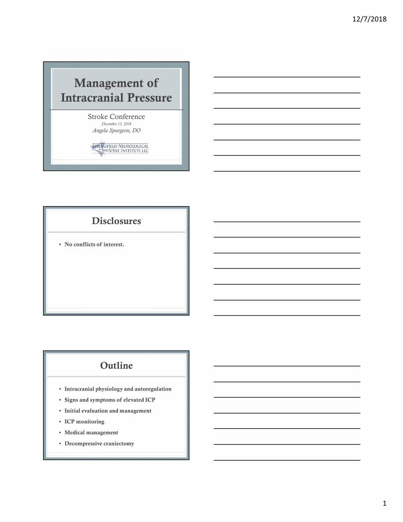
Management of
Intracranial Pressure
Stroke ConferenceDecember 13, 2018
Angela Spurgeon, DO
Disclosures
• No conflicts of interest.
Outline
• Intracranial physiology and autoregulation
• Signs and symptoms of elevated ICP
• Initial evaluation and management
• ICP monitoring
• Medical management
• Decompressive craniectomy
12/7/2018
2
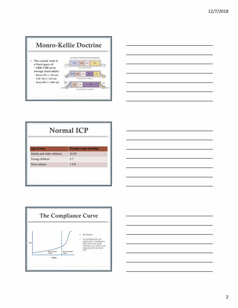
Monro-Kellie Doctrine
• The cranial vault is
a fixed space of
~1400-1700 ml in
average sized adults.
• Blood 10% (~150 ml)
• CSF 10% (~150 ml)
• Brain 80% (~1400 ml)
Normal ICP
Age Group Normal range (mmHg)
Adults and older children 10-20
Young children 3-7
Term infants 1.5-6
The Compliance Curve
• Nonlinear
• As mechanisms are exhausted, compliance falls and even small increases in volume can dramatically increase ICP.
12/7/2018
3
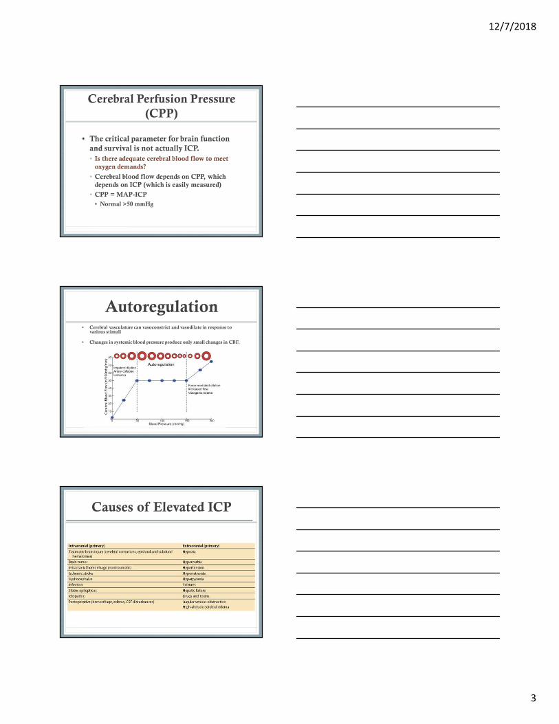
Cerebral Perfusion Pressure
(CPP)
• The critical parameter for brain function
and survival is not actually ICP.
• Is there adequate cerebral blood flow to meet
oxygen demands?
• Cerebral blood flow depends on CPP, which
depends on ICP (which is easily measured)
• CPP = MAP-ICP
• Normal >50 mmHg
Autoregulation• Cerebral vasculature can vasoconstrict and vasodilate in response to
various stimuli
• Changes in systemic blood pressure produce only small changes in CBF.
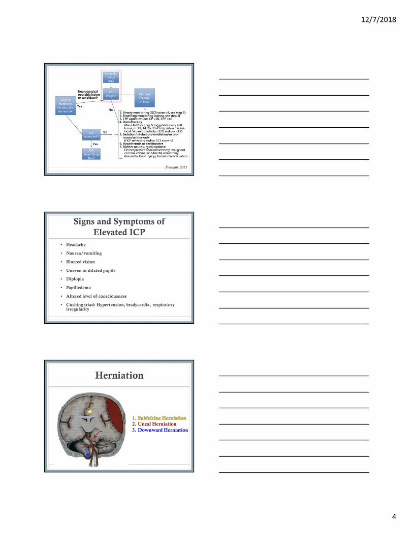
Causes of Elevated ICP
12/7/2018
4
Freeman, 2015
Signs and Symptoms of
Elevated ICP
• Headache
• Nausea/vomiting
• Blurred vision
• Uneven or dilated pupils
• Diplopia
• Papilledema
• Altered level of consciousness
• Cushing triad: Hypertension, bradycardia, respiratory irregularity
Herniation
2. Uncal Herniation
3. Downward Herniation
12/7/2018
5
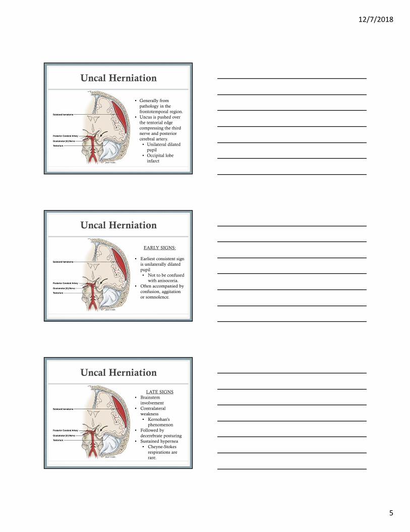
Uncal Herniation
• Generally from
pathology in the
frontotemporal region.
• Uncus is pushed over
the tentorial edge
compressing the third
nerve and posterior
cerebral artery.
• Unilateral dilated
pupil
• Occipital lobe
infarct
Uncal Herniation
EARLY SIGNS:
• Earliest consistent sign
is unilaterally dilated
pupil
• Not to be confused
with anisocoria.
• Often accompanied by
confusion, aggitation
or somnolence.
Uncal Herniation
LATE SIGNS
• Brainstem
involvement
• Contralateral
weakness
• Kernohan’s
phenomenon
• Followed by
decerebrate posturing
• Sustained hypernea
• Cheyne-Stokes
respirations are
rare.
12/7/2018
6
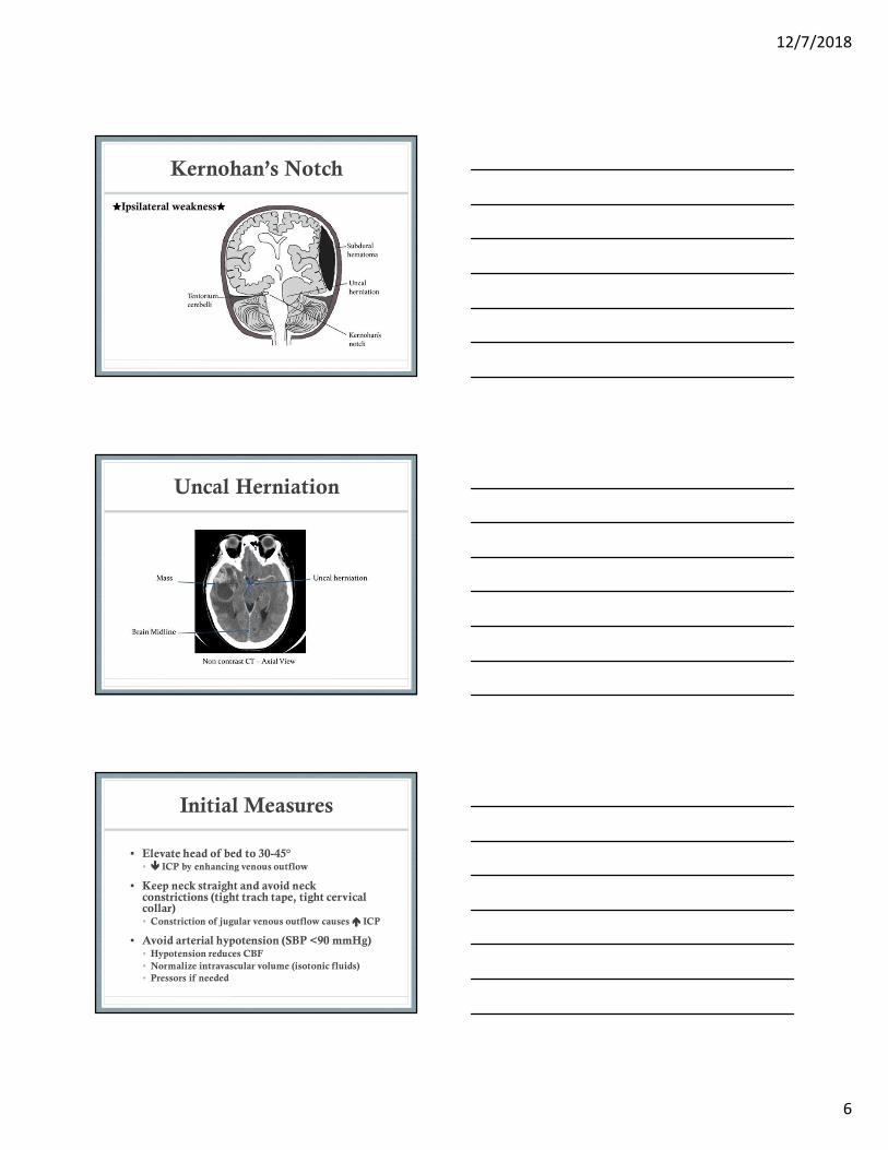
Kernohan’s Notch
★★★★Ipsilateral weakness★★★★
Uncal Herniation
Initial Measures
• Elevate head of bed to 30-45°• ���� ICP by enhancing venous outflow
• Keep neck straight and avoid neck constrictions (tight trach tape, tight cervical collar)• Constriction of jugular venous outflow causes ���� ICP
• Avoid arterial hypotension (SBP <90 mmHg)• Hypotension reduces CBF
• Normalize intravascular volume (isotonic fluids)
• Pressors if needed
12/7/2018
7
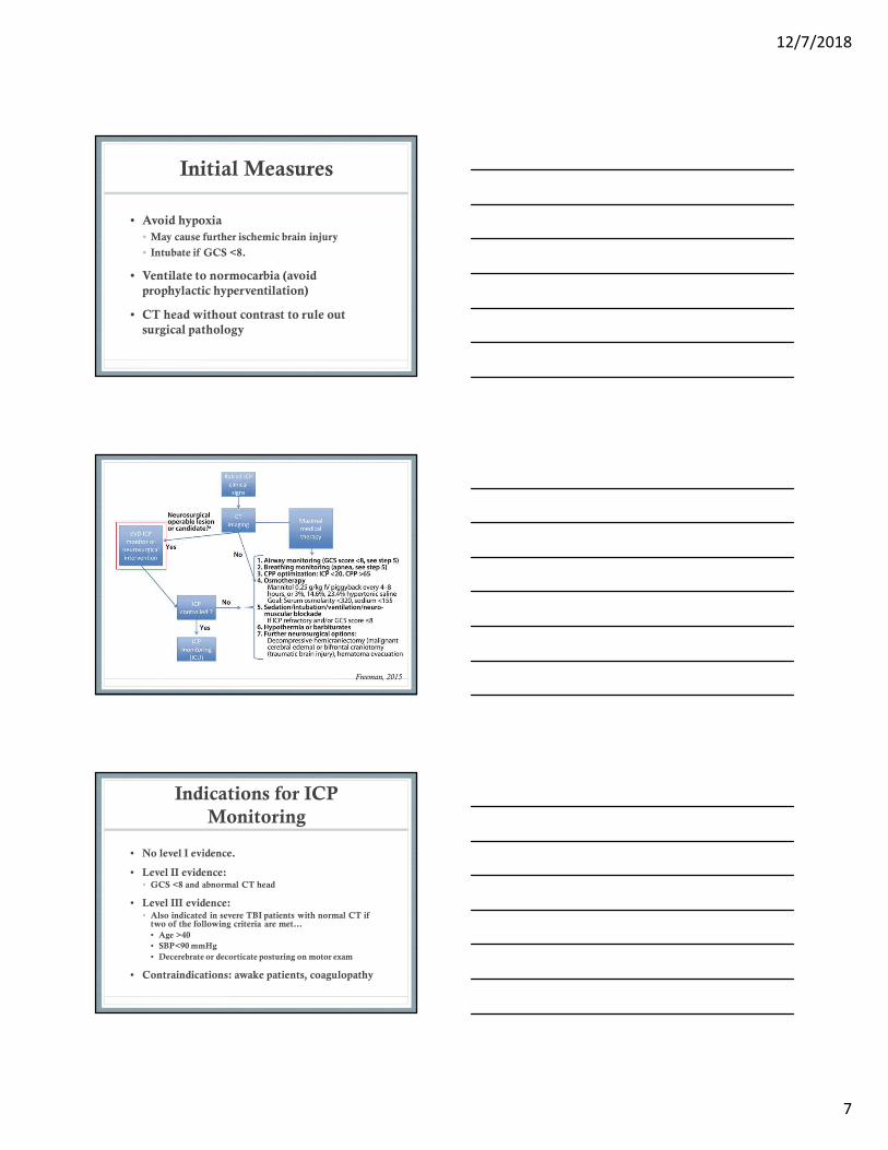
Initial Measures
• Avoid hypoxia
• May cause further ischemic brain injury
• Intubate if GCS <8.
• Ventilate to normocarbia (avoid
prophylactic hyperventilation)
• CT head without contrast to rule out
surgical pathology
Freeman, 2015
Indications for ICP
Monitoring
• No level I evidence.
• Level II evidence: • GCS <8 and abnormal CT head
• Level III evidence:• Also indicated in severe TBI patients with normal CT if
two of the following criteria are met…
• Age >40
• SBP<90 mmHg
• Decerebrate or decorticate posturing on motor exam
• Contraindications: awake patients, coagulopathy
12/7/2018
8
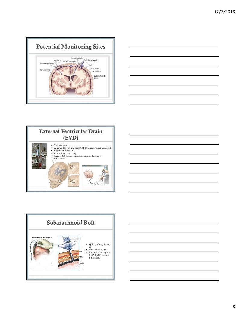
Potential Monitoring Sites
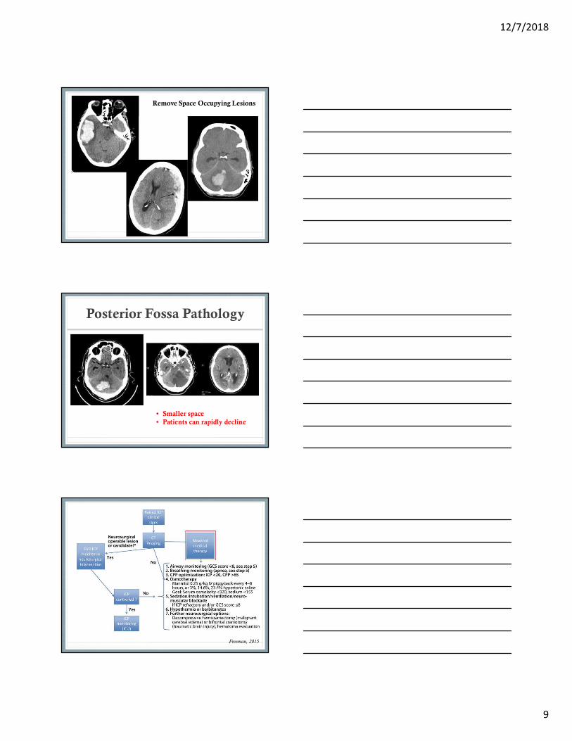
External Ventricular Drain
(EVD)
• Gold standard
• Can monitor ICP and drain CSF to lower pressure as needed.
• 10% risk of infection
• 1-2% risk of hemorrhage
• Frequently become clogged and require flushing or
replacement.
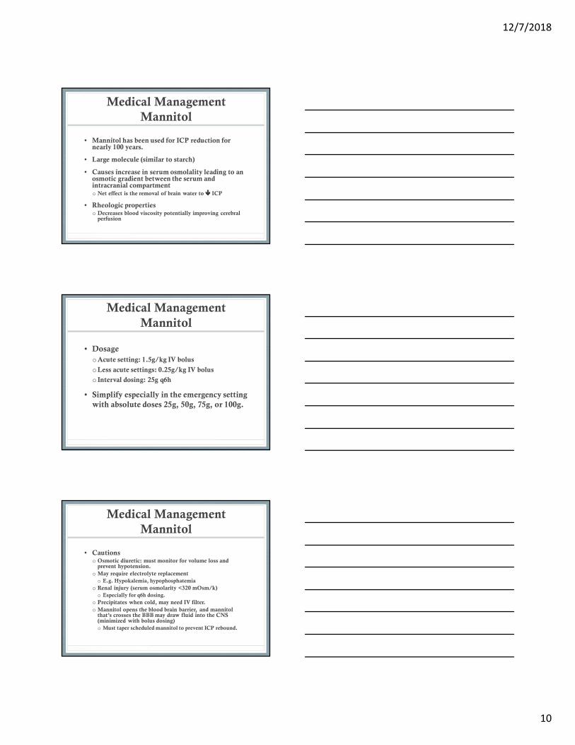
Subarachnoid Bolt
• Quick and easy to put
in.
• Low infection risk.
• May still need to place
EVD if CSF drainage
is necessary.
12/7/2018
9
Remove Space Occupying Lesions
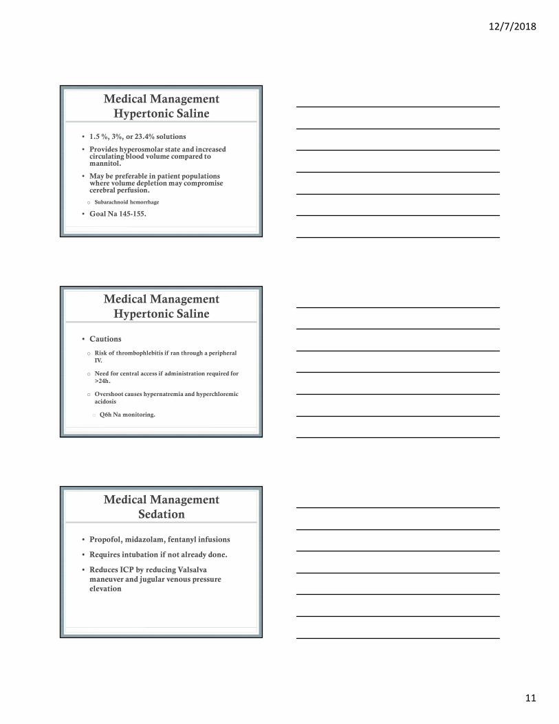
Posterior Fossa Pathology
• Smaller space
• Patients can rapidly decline
Freeman, 2015
12/7/2018
10
Medical Management
Mannitol
• Mannitol has been used for ICP reduction for nearly 100 years.
• Large molecule (similar to starch)
• Causes increase in serum osmolality leading to an osmotic gradient between the serum and intracranial compartmento Net effect is the removal of brain water to ���� ICP
• Rheologic propertieso Decreases blood viscosity potentially improving cerebral
perfusion
Medical Management
Mannitol
• Dosage
oAcute setting: 1.5g/kg IV bolus
oLess acute settings: 0.25g/kg IV bolus
o Interval dosing: 25g q6h
• Simplify especially in the emergency setting
with absolute doses 25g, 50g, 75g, or 100g.
Medical Management
Mannitol
• Cautionso Osmotic diuretic: must monitor for volume loss and
prevent hypotension.
o May require electrolyte replacement
o E.g. Hypokalemia, hypophosphatemia
o Renal injury (serum osmolarity <320 mOsm/k)
o Especially for q6h dosing.
o Precipitates when cold, may need IV filter.
o Mannitol opens the blood brain barrier, and mannitolthat’s crosses the BBB may draw fluid into the CNS (minimized with bolus dosing)
o Must taper scheduled mannitol to prevent ICP rebound.
12/7/2018
11
Medical Management
Hypertonic Saline
• 1.5 %, 3%, or 23.4% solutions
• Provides hyperosmolar state and increased circulating blood volume compared to mannitol.
• May be preferable in patient populations where volume depletion may compromise cerebral perfusion.
o Subarachnoid hemorrhage
• Goal Na 145-155.
Medical Management
Hypertonic Saline
• Cautions
o Risk of thrombophlebitis if ran through a peripheral
IV.
o Need for central access if administration required for
>24h.
o Overshoot causes hypernatremia and hyperchloremic
acidosis
o Q6h Na monitoring.
Medical Management
Sedation
• Propofol, midazolam, fentanyl infusions
• Requires intubation if not already done.
• Reduces ICP by reducing Valsalva
maneuver and jugular venous pressure
elevation
12/7/2018
12
Medical Management
Neuromuscular Paralysis
• Prevents coughing, which may cause ICP
spikes.
• Requires intubation if not already done.
• Disadvantage:
o Loss of neurologic exam aside from pupillary
reflexes.
Medical Management
Barbituates
• Reduces brain metabolism and therefore oxygen demands leading to ���� CBF.
• Anti-epileptic benefit.
• Cautions• Requires intubation and EEG monitoring to titrating
to burst-suppression pattern.
• Reduces cardiac output and may require vasopressor support
• Contraindicated in patients with heart history.
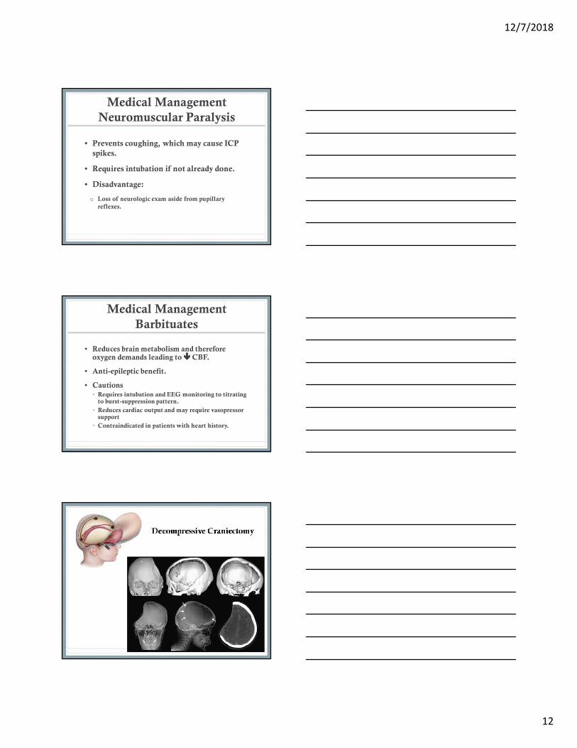
Decompressive Craniectomy
12/7/2018
13
Decompressive Craniectomy
• Most commonly used in large MCA or ICA
infarcts.
• Mortality rate in large MCA infarcts
approaches 80%
• Decompressive craniectomy may reduce
mortality to as low as 32% in nondominant
hemisphere strokes (37% in all comers)
Decompressive Craniectomy
• Meta-analysis of 3 randomized controlled
trials found that hemicraniectomy within
48 hrs after stroke onset:
• Decreased mortality
• Increased the number of patients with favorable
functional outcomes.
Vahedi et al. 2007
Decompressive Craniectomy
• Indications
• Age <70
• Usually more strongly considered in
nondominant hemisphere infarcts
• Clinical and CT evidence of acute, complete ICA
or MCA infarcts and direct signs of impending
swelling or herniation.
12/7/2018
14
Decompressive Craniectomy
• Technical Notes
• GO BIG! Bone flap
should be >12cm
• Dura should also be
opened, ± duraplasty
• Bone flap can be stored in
the abdomen or bone
freezer.
Decompressive Craniectomy
• Potential Complications
• Bleeding
• Herniation of the brain through the bone
opening (can cause local ischemia, minimized by
making a big craniectomy)
• Post-op fluid collections (subdural hygromas)
• Hydrocephalus
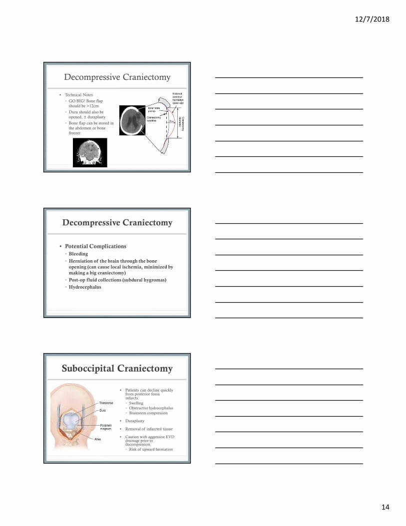
Suboccipital Craniectomy
• Patients can decline quickly from posterior fossa infarcts.
• Swelling
• Obstructive hydrocephalus
• Brainstem compression
• Duraplasty
• Removal of infarcted tissue
• Caution with aggressive EVD drainage prior to decompression.
• Risk of upward herniation
12/7/2018
15
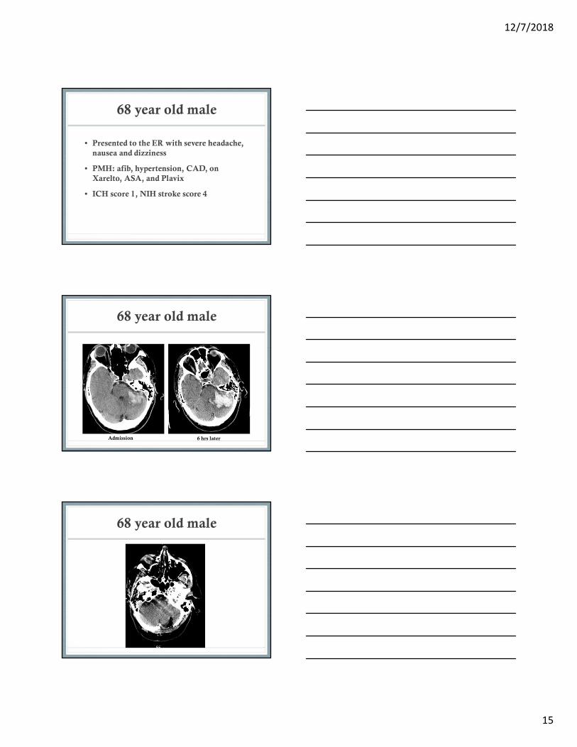
68 year old male
• Presented to the ER with severe headache,
nausea and dizziness
• PMH: afib, hypertension, CAD, on
Xarelto, ASA, and Plavix
• ICH score 1, NIH stroke score 4
68 year old male
Admission 6 hrs later
68 year old male
12/7/2018
16
68 year old male
• 9 months post-op
• Ambulating with a cane
• No headaches
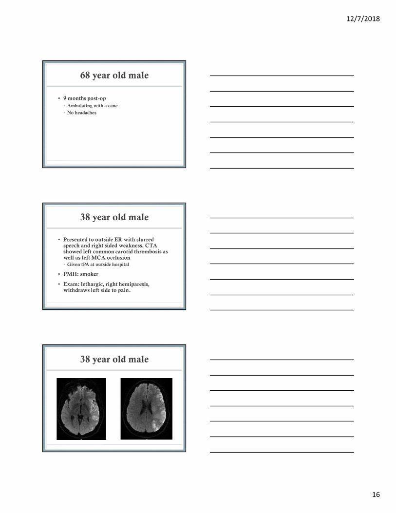
38 year old male
• Presented to outside ER with slurred speech and right sided weakness. CTA showed left common carotid thrombosis as well as left MCA occlusion• Given tPA at outside hospital
• PMH: smoker
• Exam: lethargic, right hemiparesis, withdraws left side to pain.
38 year old male

12/7/2018
17
38 year old male
38 year old male
• Taken to OR for decompressive
craniectomy and ICP monitor placement on
stroke day 2.
38 year old male
• Outcome (6 weeks out)
• Bone flap replaced
• Required shunt placement
for extra-axial hygroma
• Able to speak but still has
considerable word finding
difficulties, able to follow
commands on the right.
12/7/2018
18
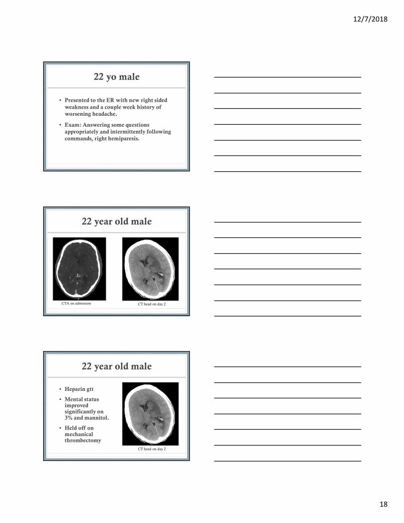
22 yo male
• Presented to the ER with new right sided
weakness and a couple week history of
worsening headache.
• Exam: Answering some questions
appropriately and intermittently following
commands, right hemiparesis.
22 year old male
CTA on admission CT head on day 2
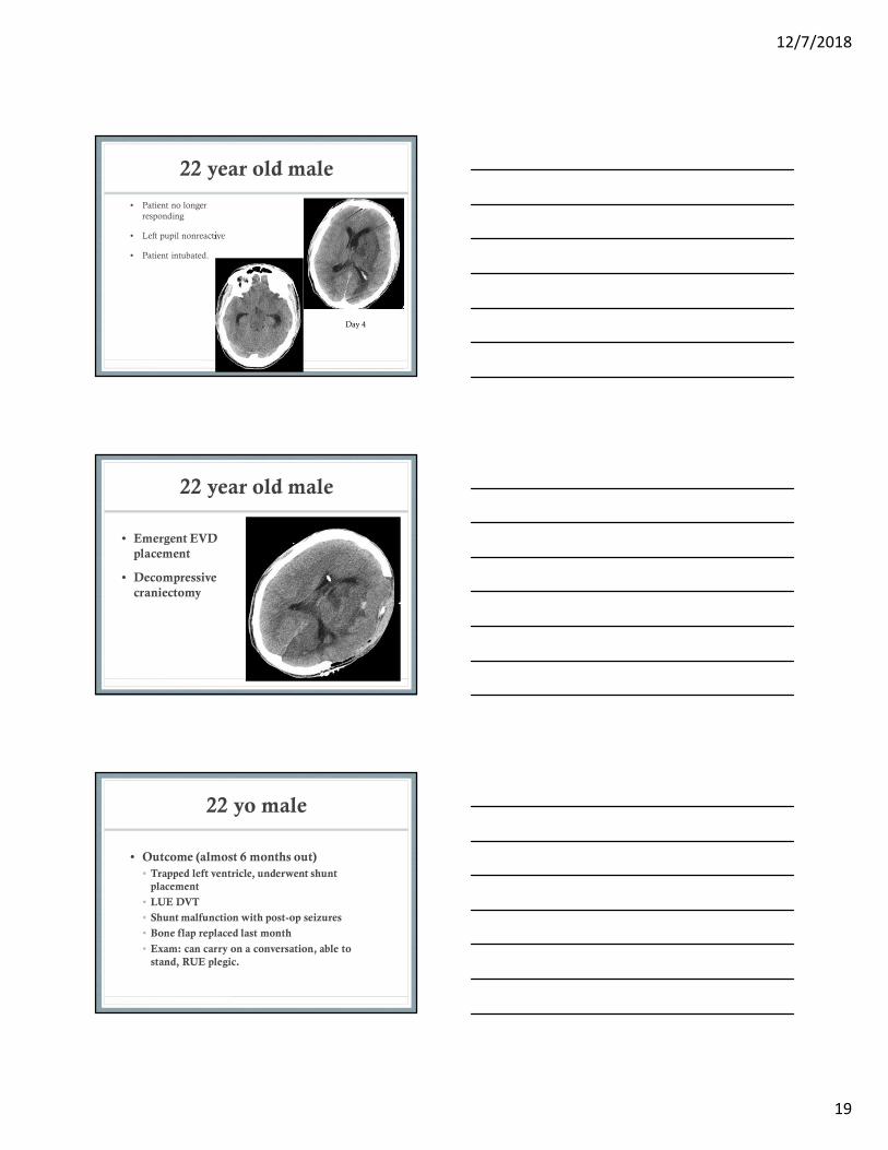
22 year old male
CT head on day 2
• Heparin gtt
• Mental status improved significantly on 3% and mannitol.
• Held off on mechanical thrombectomy
12/7/2018
19
22 year old male
• Patient no longer
responding
• Left pupil nonreactive
• Patient intubated.
Day 4
22 year old male
• Emergent EVD
placement
• Decompressive
craniectomy
22 yo male
• Outcome (almost 6 months out)
• Trapped left ventricle, underwent shunt
placement
• LUE DVT
• Shunt malfunction with post-op seizures
• Bone flap replaced last month
• Exam: can carry on a conversation, able to
stand, RUE plegic.

12/7/2018
20
References
• Dasenbrock HH et al. Timing of decompressivehemicraniectomy for stroke: A nationwide inpatient sample analysis. Stroke 2017; 48:704-711
• Freeman WD, Management of Intracranial Pressure. Continuum 2015;21(5):1299-1323.
• Greenberg MS, Handbood of Neurosurgery. 2016; 8th
Ed. Thieme Medical Publishers, New York.
• Vahedi et. al. Early decompressive surgery in malignant infarction of the middle cerebral artery: a pooled analysis of three randomised controlled trials. Lancet2007;6:215-222.


![MR Characteristics of Subdural Hematomas and Hygromas at 1.5 T · MR images is well established [1], subdural hemorrhage is less well characterized. ... hematoma, the entire volume](https://img.pdfslide.us/doc/110x75/5d2d74f488c9937b188c25a1/mr-characteristics-of-subdural-hematomas-and-hygromas-at-15-t-mr-images-is.jpg)