Embed Size (px)
Citation preview
ORIGINAL ARTICLE CLINICAL PRACTICE MANAGEMENT
Management of Incidental PancreaticCysts: A White Paper of the ACR IncidentalFindings Committee
Alec J. Megibow, MD, MPHa, Mark E. Baker, MDb, Desiree E. Morgan, MDc, Ihab R. Kamel, MD, PhDd,Dushyant V. Sahani, MDe, Elliot Newman, MD f, William R. Brugge, MDg, Lincoln L. Berland, MDc,Pari V. Pandharipande, MD, MPHe,hAbstract
The ACR Incidental Findings Committee (IFC) presents recommendations for managing pancreatic cysts that are incidentally detected onCT or MRI. These recommendations represent an update from the pancreatic component of the JACR 2010 white paper on managingincidental findings in the adrenal glands, kidneys, liver, and pancreas. The Pancreas Subcommittee—which included abdominalradiologists, a gastroenterologist, and a pancreatic surgeon—developed this algorithm. The recommendations draw from publishedevidence and expert opinion, and were finalized by informal iterative consensus. Algorithm branches successively categorize pancreaticcysts based on patient characteristics and imaging features. They terminate with an ascertainment of benignity and/or indolence (sufficientto discontinue follow-up), or a management recommendation. The algorithm addresses most, but not all, pathologies and clinicalscenarios. Our goal is to improve quality of care by providing guidance on how to manage incidentally detected pancreatic cysts.
Key Words: Pancreas, cyst, intraductal papillary mucinous neoplasm (IPMN), incidental finding
J Am Coll Radiol 2017;14:911-923. Copyright � 2017 American College of Radiology
OVERVIEW OF THE ACR INCIDENTALFINDINGS PROJECTThe core objectives of the Incidental Findings Project areto (1) develop consensus on patient characteristics andimaging features that are required to characterize anincidental finding; (2) provide guidance to manage suchfindings in ways that balance the risks and benefits topatients; (3) recommend reporting terms that reflect thelevel of confidence regarding a finding; and (4) focusfuture research by proposing a generalizable management
aDepartment of Radiology, NYU-Langone Medical Center, New York,New York.bDepartment of Radiology, Cleveland Clinic, Cleveland, Ohio.cDepartment of Radiology, University of Alabama at Birmingham, Bir-mingham, Alabama.dDepartment of Radiology, Johns Hopkins Hospital, Baltimore, Maryland.eDepartment of Radiology, Massachusetts General Hospital, Boston,Massachusetts.fDepartment of Surgery, NYU-Langone Medical Center, New York, NewYork.gGastrointestinal Unit, Department of Medicine, Massachusetts GeneralHospital, Boston, Massachusetts.hInstitute for Technology Assessment, Massachusetts General Hospital,Boston, Massachusetts.
ª 2017 American College of Radiology1546-1440/17/$36.00 n http://dx.doi.org/10.1016/j.jacr.2017.03.010
framework across practice settings. The Incidental Find-ings Committee (IFC) generated its first white paper in2010, addressing four algorithms for managing incidentalpancreatic, adrenal, kidney, and liver findings [1].
THE CONSENSUS PROCESS: THE PANCREATICCYST ALGORITHMThe current paper represents the first revision of the IFC’srecommendations regarding incidental pancreatic cysts.The process of developing this algorithm included naming
Corresponding author and reprints: Alec J. Megibow, MD, MPH,Department of Radiology, NYU-Langone Medical Center, NYU RadiologyAssociates, 530 1st Avenue, New York, NY 10016; e-mail: [email protected].
Dr. Megibow has nothing to disclose. Dr. Baker has nothing to disclose.Dr. Morgan reports grants from GE Healthcare, personal fees from GEHealthcare, outside the submitted work. Dr. Kamel has nothing to disclose.Dr. Sahani reports grants from GE Healthcare, textbook royalties fromElsevier, outside the submitted work. Dr. Newman has nothing to disclose.Dr. Brugge has nothing to disclose. Dr. Berland reports personal feesfrom Nuance Communications, Inc., outside the submitted work.Dr. Pandharipande reports a research grant from the Medical Imagingand Technology Alliance, outside the submitted work.
911
a Subcommittee Chair, who appointed four additionalabdominal radiologists, a gastroenterologist, and a pancreaticsurgeon. The Subcommittee then developed and gainedconsensus on a preliminary version of the algorithm. TheSubcommittee used published evidence as their primarysource. Where evidence was not available, they invoked thecollective expertise of their team. The preliminary algorithmunderwent review by additional members within the IFC,including the Body Commission Chair, the IFC Chair, andadditional IFCSubcommitteeChairs.The revised algorithmand corresponding white paper draft were submitted toadditional ACR stakeholders to gain input and feedback.Consensus was obtained iteratively after successive reviewsand revisions. After completion of this process, thealgorithm and white paper were finalized. The IFC’sconsensus processes meet policy standards of the ACR.However, they do not meet any specific, formal nationalstandards. This algorithm and set of recommendations doesnot represent policy of the ACR Practice Guidelines or the
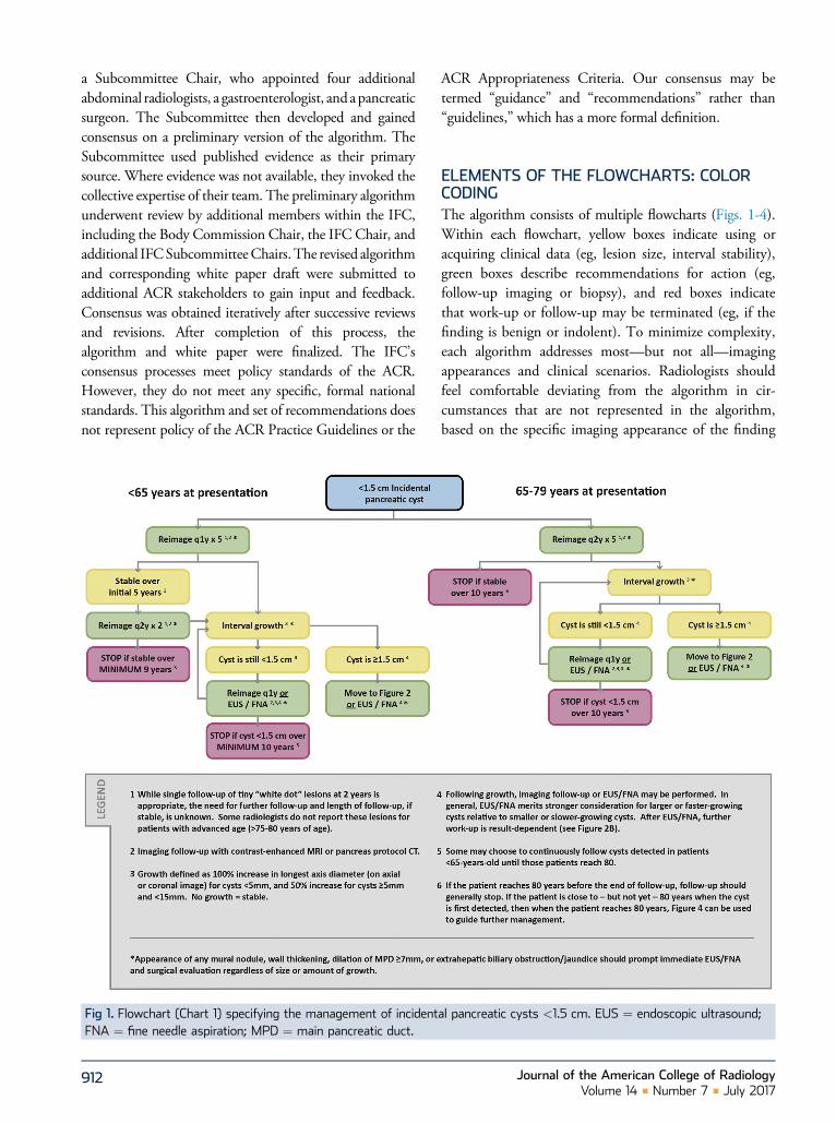
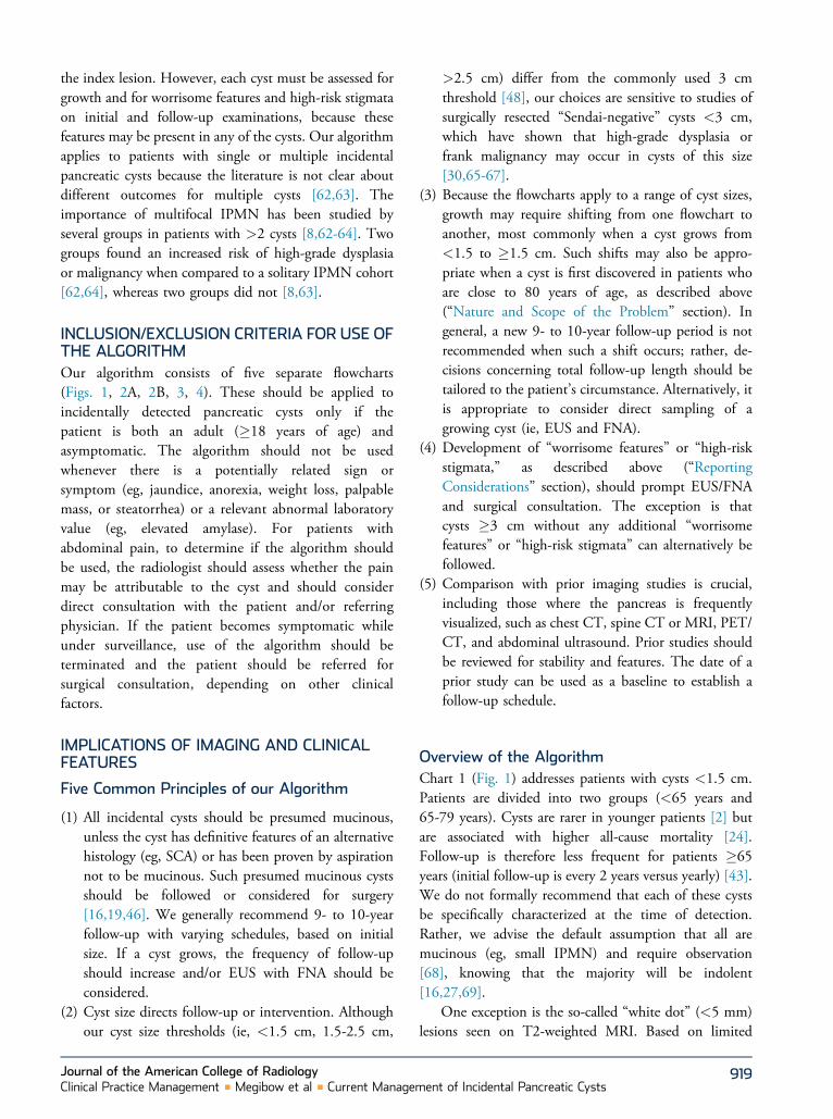
Fig 1. Flowchart (Chart 1) specifying the management of incidentFNA ¼ fine needle aspiration; MPD ¼ main pancreatic duct.
912
ACR Appropriateness Criteria. Our consensus may betermed “guidance” and “recommendations” rather than“guidelines,” which has a more formal definition.
ELEMENTS OF THE FLOWCHARTS: COLORCODINGThe algorithm consists of multiple flowcharts (Figs. 1-4).Within each flowchart, yellow boxes indicate using oracquiring clinical data (eg, lesion size, interval stability),green boxes describe recommendations for action (eg,follow-up imaging or biopsy), and red boxes indicatethat work-up or follow-up may be terminated (eg, if thefinding is benign or indolent). To minimize complexity,each algorithm addresses most—but not all—imagingappearances and clinical scenarios. Radiologists shouldfeel comfortable deviating from the algorithm in cir-cumstances that are not represented in the algorithm,based on the specific imaging appearance of the finding
al pancreatic cysts <1.5 cm. EUS ¼ endoscopic ultrasound;
Journal of the American College of RadiologyVolume 14 n Number 7 n July 2017
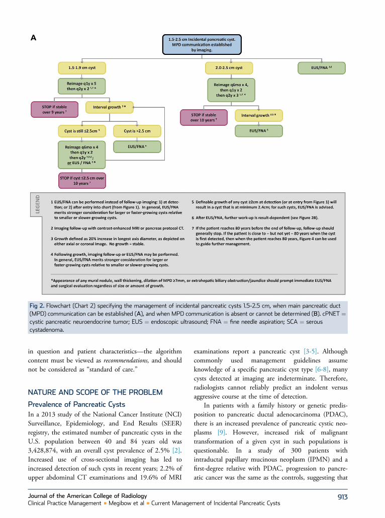
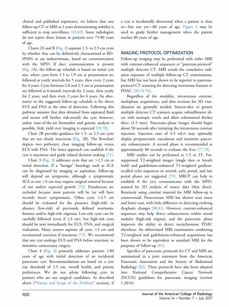
Fig 2. Flowchart (Chart 2) specifying the management of incidental pancreatic cysts 1.5-2.5 cm, when main pancreatic duct(MPD) communication can be established (A), and when MPD communication is absent or cannot be determined (B). cPNET ¼cystic pancreatic neuroendocrine tumor; EUS ¼ endoscopic ultrasound; FNA ¼ fine needle aspiration; SCA ¼ serouscystadenoma.
in question and patient characteristics—the algorithmcontent must be viewed as recommendations, and shouldnot be considered as “standard of care.”
NATURE AND SCOPE OF THE PROBLEM
Prevalence of Pancreatic CystsIn a 2013 study of the National Cancer Institute (NCI)Surveillance, Epidemiology, and End Results (SEER)registry, the estimated number of pancreatic cysts in theU.S. population between 40 and 84 years old was3,428,874, with an overall cyst prevalence of 2.5% [2].Increased use of cross-sectional imaging has led toincreased detection of such cysts in recent years; 2.2% ofupper abdominal CT examinations and 19.6% of MRI
Journal of the American College of RadiologyClinical Practice Management n Megibow et al n Current Managem
examinations report a pancreatic cyst [3-5]. Althoughcommonly used management guidelines assumeknowledge of a specific pancreatic cyst type [6-8], manycysts detected at imaging are indeterminate. Therefore,radiologists cannot reliably predict an indolent versusaggressive course at the time of detection.
In patients with a family history or genetic predis-position to pancreatic ductal adenocarcinoma (PDAC),there is an increased prevalence of pancreatic cystic neo-plasms [9]. However, increased risk of malignanttransformation of a given cyst in such populations isquestionable. In a study of 300 patients withintraductal papillary mucinous neoplasm (IPMN) and afirst-degree relative with PDAC, progression to pancre-atic cancer was the same as the controls, suggesting that
913ent of Incidental Pancreatic Cysts
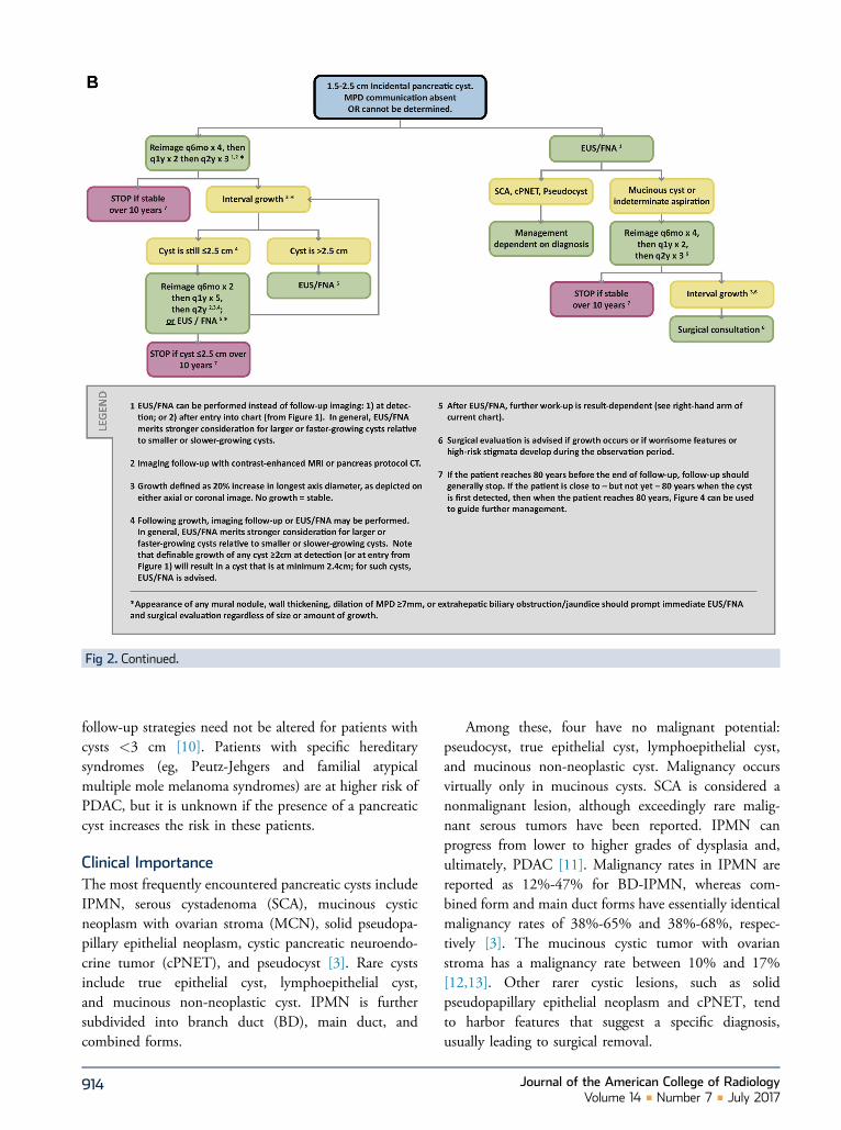
Fig 2. Continued.
follow-up strategies need not be altered for patients withcysts <3 cm [10]. Patients with specific hereditarysyndromes (eg, Peutz-Jehgers and familial atypicalmultiple mole melanoma syndromes) are at higher risk ofPDAC, but it is unknown if the presence of a pancreaticcyst increases the risk in these patients.
Clinical ImportanceThe most frequently encountered pancreatic cysts includeIPMN, serous cystadenoma (SCA), mucinous cysticneoplasm with ovarian stroma (MCN), solid pseudopa-pillary epithelial neoplasm, cystic pancreatic neuroendo-crine tumor (cPNET), and pseudocyst [3]. Rare cystsinclude true epithelial cyst, lymphoepithelial cyst,and mucinous non-neoplastic cyst. IPMN is furthersubdivided into branch duct (BD), main duct, andcombined forms.
914
Among these, four have no malignant potential:pseudocyst, true epithelial cyst, lymphoepithelial cyst,and mucinous non-neoplastic cyst. Malignancy occursvirtually only in mucinous cysts. SCA is considered anonmalignant lesion, although exceedingly rare malig-nant serous tumors have been reported. IPMN canprogress from lower to higher grades of dysplasia and,ultimately, PDAC [11]. Malignancy rates in IPMN arereported as 12%-47% for BD-IPMN, whereas com-bined form and main duct forms have essentially identicalmalignancy rates of 38%-65% and 38%-68%, respec-tively [3]. The mucinous cystic tumor with ovarianstroma has a malignancy rate between 10% and 17%[12,13]. Other rarer cystic lesions, such as solidpseudopapillary epithelial neoplasm and cPNET, tendto harbor features that suggest a specific diagnosis,usually leading to surgical removal.
Journal of the American College of RadiologyVolume 14 n Number 7 n July 2017

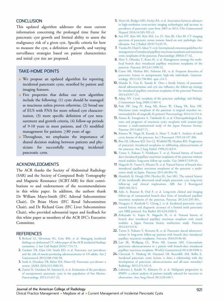
Fig 3. Flowchart (Chart 3) specifying the management of incidental pancreatic cysts >2.5 cm. EUS ¼ endoscopic ultrasound;FNA ¼ fine needle aspiration; MPD ¼ main pancreatic duct; SCA ¼ serous cystadenoma.
In a classic paper, small (<4 mm) pancreatic cystswere found in 24.3% of 300 consecutive all-cause au-topsies [14]. Coupled with the now-accepted concept ofan adenoma-carcinoma sequence [15], an incidentallydetected cyst may be a precursor to PDAC. However,observational data on BD-IPMN suggest that lesions�2 cm are indolent with only a small fraction progressingto malignancy [16-20] even when mural nodules arepresent [21]. Accurate rates of transition to malignancyfor small, incidental pancreatic cysts remain unknown.
Pancreatic cysts may reflect an elevated whole-glandrisk for developing PDAC at a location within thepancreas other than within the cyst; multiple authorshave observed PDAC separate from a cyst [22-25]. In alarge Veterans Affairs study, the incidence of pancreaticcancer in patients with previously diagnosed cysts was5.08 per 1,000 patient-years compared with 0.32 inpatients without cysts; however, the location of suchcancers relative to cysts was not reported [26]. Patients
Journal of the American College of RadiologyClinical Practice Management n Megibow et al n Current Managem
with cysts who are less than 65 years old also havebeen reported to have increased all-cause mortalityrelative to those without cysts; the same is not true forpatients who are aged �65 years [24].
Significance of Small Pancreatic CystsMost diagnostic uncertainty is centered on pancreaticcysts <2.5 cm. Helpful queries include the following: (1)Is the cyst mucinous? (2) If mucinous, what is its relationto the main pancreatic duct (MPD)? and (3) If mucinous,are mural nodules present? Several studies suggest thatreferring physicians are comfortable with imaging sur-veillance for small BD-IPMN without mural nodules[16,17,21,27], which is supported by pathology studiesconfirming a low rate of malignant transformation [28].However, even small so-called “Sendai-negative” cystsmay have microscopic invasion into the adjacent MPD,underscoring the limitations of imaging for identifyingaggressive lesions [29,30]. Presence or development of a
915ent of Incidental Pancreatic Cysts
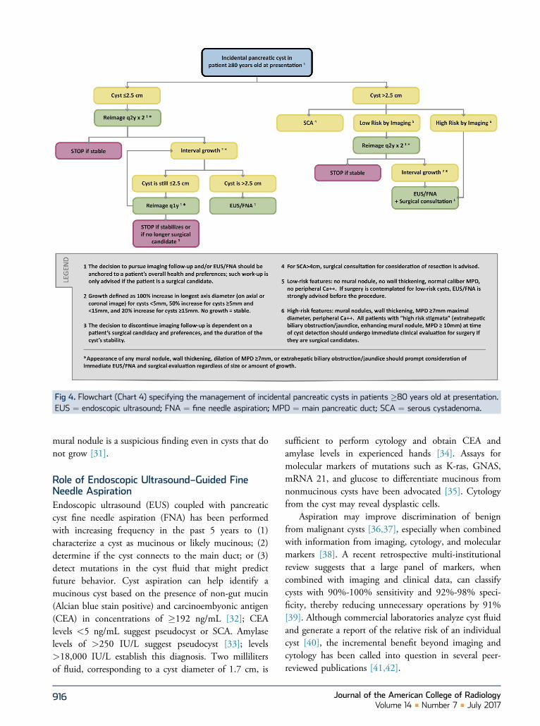
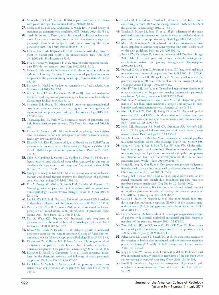
Fig 4. Flowchart (Chart 4) specifying the management of incidental pancreatic cysts in patients �80 years old at presentation.EUS ¼ endoscopic ultrasound; FNA ¼ fine needle aspiration; MPD ¼ main pancreatic duct; SCA ¼ serous cystadenoma.
mural nodule is a suspicious finding even in cysts that donot grow [31].
Role of Endoscopic Ultrasound–Guided FineNeedle AspirationEndoscopic ultrasound (EUS) coupled with pancreaticcyst fine needle aspiration (FNA) has been performedwith increasing frequency in the past 5 years to (1)characterize a cyst as mucinous or likely mucinous; (2)determine if the cyst connects to the main duct; or (3)detect mutations in the cyst fluid that might predictfuture behavior. Cyst aspiration can help identify amucinous cyst based on the presence of non-gut mucin(Alcian blue stain positive) and carcinoembyonic antigen(CEA) in concentrations of �192 ng/mL [32]; CEAlevels <5 ng/mL suggest pseudocyst or SCA. Amylaselevels of >250 IU/L suggest pseudocyst [33]; levels>18,000 IU/L establish this diagnosis. Two millilitersof fluid, corresponding to a cyst diameter of 1.7 cm, is
916
sufficient to perform cytology and obtain CEA andamylase levels in experienced hands [34]. Assays formolecular markers of mutations such as K-ras, GNAS,mRNA 21, and glucose to differentiate mucinous fromnonmucinous cysts have been advocated [35]. Cytologyfrom the cyst may reveal dysplastic cells.
Aspiration may improve discrimination of benignfrom malignant cysts [36,37], especially when combinedwith information from imaging, cytology, and molecularmarkers [38]. A recent retrospective multi-institutionalreview suggests that a large panel of markers, whencombined with imaging and clinical data, can classifycysts with 90%-100% sensitivity and 92%-98% speci-ficity, thereby reducing unnecessary operations by 91%[39]. Although commercial laboratories analyze cyst fluidand generate a report of the relative risk of an individualcyst [40], the incremental benefit beyond imaging andcytology has been called into question in several peer-reviewed publications [41,42].
Journal of the American College of RadiologyVolume 14 n Number 7 n July 2017
Length of Follow-upWe previously recommended 2-year follow-up to estab-lish cyst stability, concluding that stable cysts were benignor indolent [1]. Although this approach remains valid formost cysts [43], authors have documented delayedgrowth in cysts that were unchanged for several years[44,45]. We considered this observation, as well as newknowledge concerning age-related outcomes [24] andcysts as a marker for elevated whole-gland PDAC risk[24,25], when updating our recommendations.
For most patients, we advocate 9- to 10-year follow-up, terminating at the age of 80 years (Figs. 1-3). Forpatients who are <65 years old at the time of initialcyst detection, a follow-up terminating at age 80 willexceed the 9- to 10-year length, but may be prudent [24];such decisions regarding additional follow-up should bedetermined at the individual patient level. For patients�80 years old at the time of initial cyst detection, aseparate follow-up schedule is provided (Fig. 4). With ourapproach, many older patients will not undergosurveillance for the full follow-up period, whereasyounger patients may be subject to lengthier monitoringin comparison. The follow-up intervals are based onexperiential observations, and are not from randomizedcontrolled studies.
Follow-up beyond 80 years of age, for a cyst that wasfirst identified at <80 years, is generally not advised, asindicated above. The exception is when a cyst is discov-ered in a patient who is close to—but not yet—80 yearsof age. When this occurs, case-by-case decisions forongoing surveillance should be based on individual pa-tient characteristics (ie, overall patient health, willingnessto undergo treatment if needed) and the accumulatedknowledge about the cyst. In such circumstances, man-agement can shift to the same flowchart that addressescysts initially detected at �80 years of age (Fig. 4).
Challenges to a Perfect AlgorithmThe natural history of incidental pancreatic cysts remainsuncertain, and our recommendations cannot be simple orentirely definitive. Since 2010, several multi-institutionaland specialty society consensus papers, meta-analyses, andlarge-scale observational studies have appeared[1,21,34,46-50], but the quality of evidence has beencharacterized as poor or inconclusive, and conclusionsremain controversial [34]. Physicians must discuss suchuncertainty with their patients, integrating patients’ risktolerance, physicians’ clinical judgment, and localexpertise into management decisions. When local
Journal of the American College of RadiologyClinical Practice Management n Megibow et al n Current Managem
expertise is limited, referrals to sites of clinicalexcellence are strongly encouraged.
REPORTING CONSIDERATIONSThe following six elements must be reported when anincidental pancreatic cyst is detected on a CT orMRI study:
1. Cyst morphology, location2. Cyst size3. Possible communication with MPD4. Presence of “worrisome features” and/or “high-risk
stigmata”5. Growth on follow-up examination6. Multiplicity
1. Cyst Morphology, LocationAs mentioned, the most frequently encountered pancre-atic cysts include IPMN, SCA, MCN, solid pseudopa-pillary epithelial neoplasm, cPNET, and pseudocyst. Rarecysts include simple epithelial cyst, lymphoepithelial cyst,and mucinous non-neoplastic cyst. IPMN is furthersubdivided into BD, main duct, and combined forms.Cysts that are less than 10 mm are difficult or impossibleto specifically characterize. Cysts measuring 1-3 cm areoften “indeterminate” unless communication with theMPD can be established. If duct communication isestablished, the cyst is classified as either BD orcombined-type IPMN. Cysts �3 cm can be classified asoligocystic, microcystic, macrocystic, unilocular, or mul-tilocular [51]. If calcification is present within a cyst, itslocation should be reported. A cystic lesion with centralcalcification is most likely an SCA, whereas a cyst withperipheral calcification is likely an MCN. Peripheralcalcification in MCNs is more strongly associated withfrank malignancy [52].
Every attempt should be made to establish the diag-nosis of SCA or pseudocyst. SCA displays characteristicfeatures in >60% of cases [53], although “atypical”morphology can also be seen in a large proportion ofcases [6,54]. Clinical history and amylase levels in thecyst fluid of about 18,000 IU/L may help diagnose apseudocyst; however, elevated amylase is common inmucinous cysts [55]. We assume that incidental cyststhat cannot be characterized when detected are likely tobe mucinous (eg, IPMN). Follow-up imaging and/orEUS with FNA is typically needed.
Knowledge of a cyst’s location (uncinate process,head, neck, body, or tail) is important when evaluatingcomparison studies and can also aid in differential diag-nosis. For example, MCNs are common in the pancreatic
917ent of Incidental Pancreatic Cysts
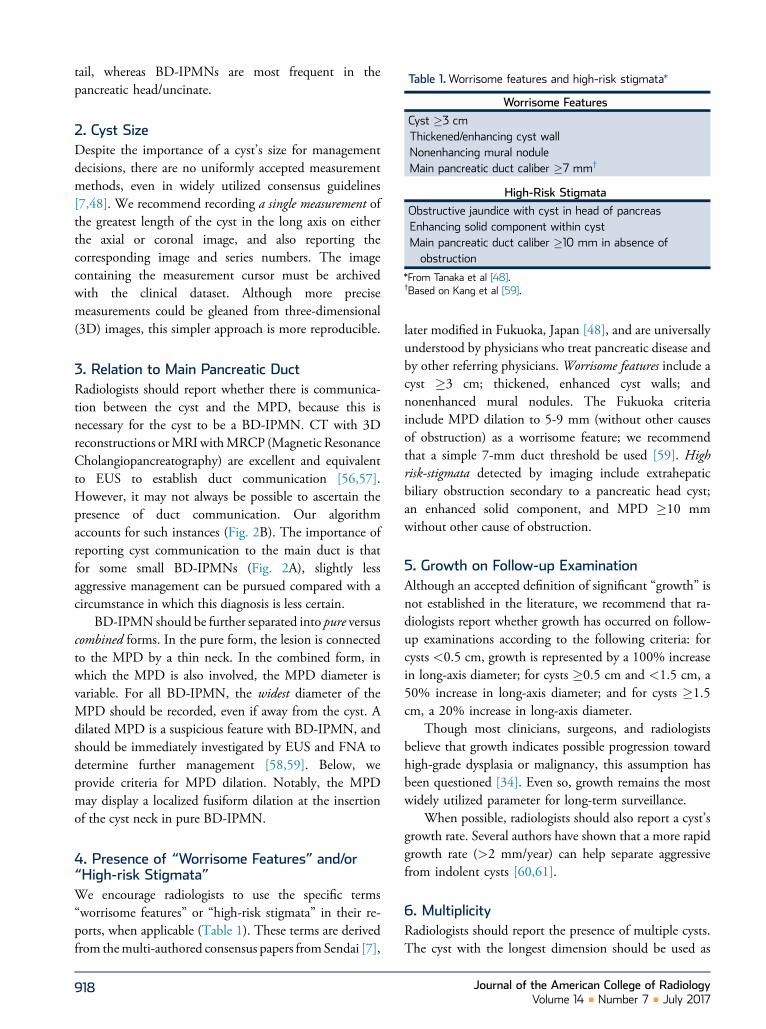
Table 1.Worrisome features and high-risk stigmata*
tail, whereas BD-IPMNs are most frequent in thepancreatic head/uncinate.Worrisome Features
Cyst �3 cmThickened/enhancing cyst wallNonenhancing mural noduleMain pancreatic duct caliber �7 mm†
High-Risk Stigmata
Obstructive jaundice with cyst in head of pancreasEnhancing solid component within cystMain pancreatic duct caliber �10 mm in absence ofobstruction
*From Tanaka et al [48].†Based on Kang et al [59].
2. Cyst SizeDespite the importance of a cyst’s size for managementdecisions, there are no uniformly accepted measurementmethods, even in widely utilized consensus guidelines[7,48]. We recommend recording a single measurement ofthe greatest length of the cyst in the long axis on eitherthe axial or coronal image, and also reporting thecorresponding image and series numbers. The imagecontaining the measurement cursor must be archivedwith the clinical dataset. Although more precisemeasurements could be gleaned from three-dimensional(3D) images, this simpler approach is more reproducible.
3. Relation to Main Pancreatic DuctRadiologists should report whether there is communica-tion between the cyst and the MPD, because this isnecessary for the cyst to be a BD-IPMN. CT with 3Dreconstructions orMRI withMRCP (Magnetic ResonanceCholangiopancreatography) are excellent and equivalentto EUS to establish duct communication [56,57].However, it may not always be possible to ascertain thepresence of duct communication. Our algorithmaccounts for such instances (Fig. 2B). The importance ofreporting cyst communication to the main duct is thatfor some small BD-IPMNs (Fig. 2A), slightly lessaggressive management can be pursued compared with acircumstance in which this diagnosis is less certain.
BD-IPMN should be further separated into pure versuscombined forms. In the pure form, the lesion is connectedto the MPD by a thin neck. In the combined form, inwhich the MPD is also involved, the MPD diameter isvariable. For all BD-IPMN, the widest diameter of theMPD should be recorded, even if away from the cyst. Adilated MPD is a suspicious feature with BD-IPMN, andshould be immediately investigated by EUS and FNA todetermine further management [58,59]. Below, weprovide criteria for MPD dilation. Notably, the MPDmay display a localized fusiform dilation at the insertionof the cyst neck in pure BD-IPMN.
4. Presence of “Worrisome Features” and/or“High-risk Stigmata”We encourage radiologists to use the specific terms“worrisome features” or “high-risk stigmata” in their re-ports, when applicable (Table 1). These terms are derivedfrom themulti-authored consensus papers fromSendai [7],
918
later modified in Fukuoka, Japan [48], and are universallyunderstood by physicians who treat pancreatic disease andby other referring physicians. Worrisome features include acyst �3 cm; thickened, enhanced cyst walls; andnonenhanced mural nodules. The Fukuoka criteriainclude MPD dilation to 5-9 mm (without other causesof obstruction) as a worrisome feature; we recommendthat a simple 7-mm duct threshold be used [59]. Highrisk-stigmata detected by imaging include extrahepaticbiliary obstruction secondary to a pancreatic head cyst;an enhanced solid component, and MPD �10 mmwithout other cause of obstruction.
5. Growth on Follow-up ExaminationAlthough an accepted definition of significant “growth” isnot established in the literature, we recommend that ra-diologists report whether growth has occurred on follow-up examinations according to the following criteria: forcysts <0.5 cm, growth is represented by a 100% increasein long-axis diameter; for cysts �0.5 cm and <1.5 cm, a50% increase in long-axis diameter; and for cysts �1.5cm, a 20% increase in long-axis diameter.
Though most clinicians, surgeons, and radiologistsbelieve that growth indicates possible progression towardhigh-grade dysplasia or malignancy, this assumption hasbeen questioned [34]. Even so, growth remains the mostwidely utilized parameter for long-term surveillance.
When possible, radiologists should also report a cyst’sgrowth rate. Several authors have shown that a more rapidgrowth rate (>2 mm/year) can help separate aggressivefrom indolent cysts [60,61].
6. MultiplicityRadiologists should report the presence of multiple cysts.The cyst with the longest dimension should be used as
Journal of the American College of RadiologyVolume 14 n Number 7 n July 2017
the index lesion. However, each cyst must be assessed forgrowth and for worrisome features and high-risk stigmataon initial and follow-up examinations, because thesefeatures may be present in any of the cysts. Our algorithmapplies to patients with single or multiple incidentalpancreatic cysts because the literature is not clear aboutdifferent outcomes for multiple cysts [62,63]. Theimportance of multifocal IPMN has been studied byseveral groups in patients with >2 cysts [8,62-64]. Twogroups found an increased risk of high-grade dysplasiaor malignancy when compared to a solitary IPMN cohort[62,64], whereas two groups did not [8,63].
INCLUSION/EXCLUSION CRITERIA FOR USE OFTHE ALGORITHMOur algorithm consists of five separate flowcharts(Figs. 1, 2A, 2B, 3, 4). These should be applied toincidentally detected pancreatic cysts only if thepatient is both an adult (�18 years of age) andasymptomatic. The algorithm should not be usedwhenever there is a potentially related sign orsymptom (eg, jaundice, anorexia, weight loss, palpablemass, or steatorrhea) or a relevant abnormal laboratoryvalue (eg, elevated amylase). For patients withabdominal pain, to determine if the algorithm shouldbe used, the radiologist should assess whether the painmay be attributable to the cyst and should considerdirect consultation with the patient and/or referringphysician. If the patient becomes symptomatic whileunder surveillance, use of the algorithm should beterminated and the patient should be referred forsurgical consultation, depending on other clinicalfactors.
IMPLICATIONS OF IMAGING AND CLINICALFEATURES
Five Common Principles of our Algorithm
(1) All incidental cysts should be presumed mucinous,unless the cyst has definitive features of an alternativehistology (eg, SCA) or has been proven by aspirationnot to be mucinous. Such presumed mucinous cystsshould be followed or considered for surgery[16,19,46]. We generally recommend 9- to 10-yearfollow-up with varying schedules, based on initialsize. If a cyst grows, the frequency of follow-upshould increase and/or EUS with FNA should beconsidered.
(2) Cyst size directs follow-up or intervention. Althoughour cyst size thresholds (ie, <1.5 cm, 1.5-2.5 cm,
Journal of the American College of RadiologyClinical Practice Management n Megibow et al n Current Managem
>2.5 cm) differ from the commonly used 3 cmthreshold [48], our choices are sensitive to studies ofsurgically resected “Sendai-negative” cysts <3 cm,which have shown that high-grade dysplasia orfrank malignancy may occur in cysts of this size[30,65-67].
(3) Because the flowcharts apply to a range of cyst sizes,growth may require shifting from one flowchart toanother, most commonly when a cyst grows from<1.5 to �1.5 cm. Such shifts may also be appro-priate when a cyst is first discovered in patients whoare close to 80 years of age, as described above(“Nature and Scope of the Problem” section). Ingeneral, a new 9- to 10-year follow-up period is notrecommended when such a shift occurs; rather, de-cisions concerning total follow-up length should betailored to the patient’s circumstance. Alternatively, itis appropriate to consider direct sampling of agrowing cyst (ie, EUS and FNA).
(4) Development of “worrisome features” or “high-riskstigmata,” as described above (“ReportingConsiderations” section), should prompt EUS/FNAand surgical consultation. The exception is thatcysts �3 cm without any additional “worrisomefeatures” or “high-risk stigmata” can alternatively befollowed.
(5) Comparison with prior imaging studies is crucial,including those where the pancreas is frequentlyvisualized, such as chest CT, spine CT or MRI, PET/CT, and abdominal ultrasound. Prior studies shouldbe reviewed for stability and features. The date of aprior study can be used as a baseline to establish afollow-up schedule.
Overview of the AlgorithmChart 1 (Fig. 1) addresses patients with cysts <1.5 cm.Patients are divided into two groups (<65 years and65-79 years). Cysts are rarer in younger patients [2] butare associated with higher all-cause mortality [24].Follow-up is therefore less frequent for patients �65years (initial follow-up is every 2 years versus yearly) [43].We do not formally recommend that each of these cystsbe specifically characterized at the time of detection.Rather, we advise the default assumption that all aremucinous (eg, small IPMN) and require observation[68], knowing that the majority will be indolent[16,27,69].
One exception is the so-called “white dot” (<5 mm)lesions seen on T2-weighted MRI. Based on limited
919ent of Incidental Pancreatic Cysts
clinical and published experience, we believe that onefollow-up CT or MRI at 2 years demonstrating stability issufficient to stop surveillance [43,69]. Some radiologistsdo not report these lesions in patients over 75-80 yearsof age.
Charts 2A and B (Fig. 2) separate 1.5- to 2.5-cm cystsby whether they can be definitively characterized as BD-IPMN or are indeterminate, based on communicationwith the MPD. If duct communication is present(Fig. 2A), the follow-up schedule is based on initial cystsize, where cysts from 1.5 to 1.9 cm at presentation arefollowed at yearly intervals for 5 years, then every 2 yearsfor 4 years. Cysts between 2.0 and 2.5 cm at presentationare followed at 6-month intervals for 2 years, then yearlyfor 2 years, and then every 2 years for 6 years. An alter-native to the suggested follow-up schedule is for directEUS and FNA at the time of detection. Following thispathway assumes that data obtained from aspirated fluidand serum will further risk-stratify the cyst; however,unless state-of-the-art biomarker and genetic analysis ispossible, little yield over imaging is expected [39,70].
Chart 2B provides guidance for 1.5- to 2.5-cm cyststhat are not clearly mucinous (Fig. 2B). The flowchartdepicts two pathways: close imaging follow-up versusEUS with FNA. The latter approach can establish if thecyst is mucinous and guide related decision making [71].
Chart 3 (Fig. 3) addresses cysts that are >2.5 cm atinitial detection. If a “benign” histology such as SCAcan be diagnosed by imaging or aspiration, follow-upwill depend on symptoms, although a symptomaticSCA or one >4 cm may require surgical removal becauseof size and/or expected growth [72]. Pseudocysts areexcluded because most patients will be (or will haverecently been) symptomatic. Other cysts >2.5 cmshould be evaluated for the presence (high-risk) orabsence (low-risk) of previously defined worrisomefeatures and/or high-risk stigmata. Low-risk cysts can becarefully followed (even if �3 cm), but high-risk cystsshould be sent immediately for EUS, FNA, and surgicalevaluation. Many centers aspirate all cysts >3 cm andrecommend resection if mucinous [73]. We recommendthat any cyst undergo EUS and FNA before resection, tominimize unnecessary surgery.
Chart 4 (Fig. 4) primarily addresses patients �80years of age with initial detection of an incidentalpancreatic cyst. Recommendations are based on a cystsize threshold of 2.5 cm, overall health, and patientpreferences. We do not advise following cysts inpatients who are not surgical candidates. As describedabove (“Nature and Scope of the Problem” section), if
920
a cyst is incidentally discovered when a patient is closeto—but not yet—80 years of age, Figure 4 may beused to guide further management when the patientreaches 80 years of age.
IMAGING PROTOCOL OPTIMIZATIONFollow-up imaging may be performed with either MRIwith contrast-enhanced sequences or “pancreas-protocol”multiple detector CT. MRI avoids the cumulative radi-ation exposure of multiple follow-up CT examinations,but MRI has not been shown to be superior to pancreas-protocol CT scanning for detecting worrisome features orPDAC [20,74-76].
Regardless of the modality, intravenous contrast,multiphase acquisitions, and thin sections for 3D visu-alization are generally needed. Sixteen-slice or greatermultiple detector CT scanners acquire submillimeter sli-ces with isotropic voxels and allow reformatted thickerslices (3-5 mm). Pancreatic-phase images should beginabout 50 seconds after initiating the intravenous contrastinjection. Injection rates of 4-5 mL/s may optimallydisplay peripancreatic vasculature and maximize pancre-atic enhancement. A second phase is recommended atapproximately 80 seconds to evaluate the liver [77,78].
MRI studies can be performed at 1.5 or 3T. Fat-suppressed T2-weighted images (single shot or breathhold) and gadolinium-enhanced T1-weighted gradient-recalled echo sequences in arterial, early portal, and lateportal phases are suggested [79]. MRCP can help toestablish if the cyst communicates with the MPD,assisted by 3D analysis of source data (thin slices).Routinely using contrast material for MRI follow-up iscontroversial. Noncontrast MRI has shorter scan timesand lower cost, with little difference in detecting evolvingdysplastic changes [80,81]. However, contrast-enhancedsequences may help detect enhancement within muralnodules (high-risk stigma), and the pancreatic phaseimproves the ability to detect metachronous PDACelsewhere. An abbreviated MRI examination combiningT2-weighted and gadolinium-enhanced acquisitions hasbeen shown to be equivalent to standard MRI for thepurposes of follow-up [81].
Specifics of pancreatic protocols for CT and MRI aresummarized in a joint statement from the AmericanPancreatic Association and the Society of AbdominalRadiology [82]. These protocols have also been adoptedinto National Comprehensive Cancer Network(NCCN) guidelines for pancreatic imaging (version1.2016).
Journal of the American College of RadiologyVolume 14 n Number 7 n July 2017
CONCLUSIONThis updated algorithm addresses the most currentinformation concerning the prolonged time frame forpancreatic cyst growth and limited ability to assess themalignancy risk of a given cyst. Specific criteria for howto measure the cyst, a definition of growth, and varyingsurveillance strategies based on patient characteristicsand initial cyst size are proposed.
JC
TAKE-HOME POINTS
- We propose an updated algorithm for reportingincidental pancreatic cysts, stratified by patient andimaging features.
- Five properties that define our new algorithminclude the following: (1) cysts should be managedas mucinous unless proven otherwise, (2) broad useof EUS with FNA for more refined cyst character-ization, (3) more specific definition of cyst mea-surement and growth criteria, (4) follow-up periodsof 9-10 years in most patients, and (5) modifiedmanagement for patients �80 years of age.
- Throughout, we emphasize the importance ofshared decision making between patients and phy-sicians for successfully managing incidentalpancreatic cysts.
ACKNOWLEDGMENTSThe ACR thanks the Society of Abdominal Radiology(SAR) and the Society of Computed Body Tomographyand Magnetic Resonance (SCBT-MR) for their contri-butions to and endorsement of the recommendationsin this white paper. In addition, the authors thankDr William Mayo-Smith (IFC Adrenal SubcommitteeChair), Dr Brian Herts (IFC Renal SubcommitteeChair), and Dr Richard Gore (IFC Liver SubcommitteeChair), who provided substantial input and feedback forthis white paper as members of the ACR IFC’s ExecutiveCommittee.
REFERENCES1. Berland LL, Silverman SG, Gore RM, et al. Managing incidentalfindings on abdominal CT: white paper of the ACR incidental findingscommittee. J Am Coll Radiol 2010;7:754-73.
2. Gardner TB, Glass LM, Smith KD, et al. Pancreatic cyst prevalenceand the risk of mucin-producing adenocarcinoma in US adults. Am JGastroenterol 2013;108:1546-50.
3. Stark A, Donahue TR, Reber HA, Hines OJ. Pancreatic cyst disease: areview. JAMA 2016;315:1882-93.
4. Zanini N, Giordano M, Smerieri E, et al. Estimation of the prevalenceof asymptomatic pancreatic cysts in the population of San Marino.Pancreatology 2015;15:417-22.
ournal of the American College of Radiologylinical Practice Management n Megibow et al n Current Managem
5. Moris M, Bridges MD, Pooley RA, et al. Association between advancesin high-resolution cross-section imaging technologies and increase inprevalence of pancreatic cysts from 2005 to 2014. Clin GastroenterolHepatol 2016;14:585-593 e3.
6. Sun HY, Kim SH, Kim MA, Lee JY, Han JK, Choi BI. CT imagingspectrum of pancreatic serous tumors: based on new pathologic clas-sification. Eur J Radiol 2010;75:e45-55.
7. TanakaM,Chari S, AdsayV, et al. International consensus guidelines formanagement of intraductal papillarymucinous neoplasms andmucinouscystic neoplasms of the pancreas. Pancreatology 2006;6:17-32.
8. Mori Y, Ohtsuka T, Kono H, et al. Management strategy for multi-focal branch duct intraductal papillary mucinous neoplasms of thepancreas. Pancreas 2012;41:1008-12.
9. Canto MI, Hruban RH, Fishman EK, et al. Frequent detection ofpancreatic lesions in asymptomatic high-risk individuals. Gastroen-terology 2012;142:796-804. quiz e14-5.
10. Mandai K, Uno K, Yasuda K. Does a family history of pancreaticductal adenocarcinoma and cyst size influence the follow-up strategyfor intraductal papillary mucinous neoplasms of the pancreas? Pancreas2014;43:917-21.
11. Adsay NV. Cystic neoplasia of the pancreas: pathology and biology.J Gastrointest Surg 2008;12:401-4.
12. Park JW, Jang JY, Kang MJ, Kwon W, Chang YR, Kim SW.Mucinous cystic neoplasm of the pancreas: is surgical resection rec-ommended for all surgically fit patients? Pancreatology 2014;14:131-6.
13. Yamao K, Yanagisawa A, Takahashi K, et al. Clinicopathological fea-tures and prognosis of mucinous cystic neoplasm with ovarian-typestroma: a multi-institutional study of the Japan Pancreas Society.Pancreas 2011;40:67-71.
14. Kimura W, Nagai H, Kuroda A, Muto T, Esaki Y. Analysis of smallcystic lesions of the pancreas. Int J Pancreatol 1995;18:197-206.
15. Brat DJ, Lillemoe KD, Yeo CJ, Warfield PB, Hruban RH. Progressionof pancreatic intraductal neoplasias to infiltrating adenocarcinoma ofthe pancreas. Am J Surg Pathol 1998;22:163-9.
16. Tanno S, Nakano Y, Nishikawa T, et al. Natural history of branchduct intraductal papillary-mucinous neoplasms of the pancreas withoutmural nodules: long-term follow-up results. Gut 2008;57:339-43.
17. Maguchi H, Tanno S, Mizuno N, et al. Natural history of branch ductintraductal papillary mucinous neoplasms of the pancreas: a multi-center study in Japan. Pancreas 2011;40:364-70.
18. Handrich SJ, Hough DM, Fletcher JG, Sarr MG. The natural historyof the incidentally discovered small simple pancreatic cyst: long-termfollow-up and clinical implications. AJR Am J Roentgenol2005;184:20-3.
19. Arlix A, Bournet B, Otal P, et al. Long-term clinical and imagingfollow-up of nonoperated branch duct form of intraductal papillarymucinous neoplasms of the pancreas. Pancreas 2012;41:295-301.
20. Nougaret S, Reinhold C, Chong J, et al. Incidental pancreatic cysts:natural history and diagnostic accuracy of a limited serial pancreaticcyst MRI protocol. Eur Radiol 2014;24:1020-9.
21. Kobayashi G, Fujita N, Maguchi H, et al. Natural history ofbranch duct intraductal papillary mucinous neoplasm with muralnodules: a Japan Pancreas Society multicenter study. Pancreas2014;43:532-8.
22. Tanno S, Nakano Y, Koizumi K, et al. Pancreatic ductal adenocarci-nomas in long-term follow-up patients with branch duct intraductalpapillary mucinous neoplasms. Pancreas 2010;39:36-40.
23. Law JK, Wolfgang CL, Weiss MJ, Lennon AM. Concomitantpancreatic adenocarcinoma in a patient with branch-duct intraductalpapillary mucinous neoplasm. World J Gastroenterol 2014;20:9200-4.
24. Chernyak V, Flusberg M, Haramati LB, Rozenblit AM, Bellin E.Incidental pancreatic cystic lesions: is there a relationship with thedevelopment of pancreatic adenocarcinoma and all-cause mortality?Radiology 2015;274:161-9.
25. Lafemina J, Katabi N, Klimstra D, et al. Malignant progression inIPMN: a cohort analysis of patients initially selected for resection orobservation. Ann Surg Oncol 2013;20:440-7.
921ent of Incidental Pancreatic Cysts
26. Munigala S, Gelrud A, Agarwal B. Risk of pancreatic cancer in patientswith pancreatic cyst. Gastrointest Endosc 2016;84:81-6.
27. Morris-Stiff G, Falk GA, Chalikonda S, Walsh RM. Natural history ofasymptomatic pancreatic cystic neoplasms.HPB (Oxford) 2013;15:175-81.
28. Terris B, Ponsot P, Paye F, et al. Intraductal papillary mucinous tu-mors of the pancreas confined to secondary ducts show less aggressivepathologic features as compared with those involving the mainpancreatic duct. Am J Surg Pathol 2000;24:1372-7.
29. Fritz S, Klauss M, Bergmann F, et al. Pancreatic main-duct involve-ment in branch-duct IPMNs: an underestimated risk. Ann Surg2014;260:848-55; discussion 855-6.
30. Fritz S, Klauss M, Bergmann F, et al. Small (Sendai negative) branch-duct IPMNs: not harmless. Ann Surg 2012;256:313-20.
31. Uehara H, Ishikawa O, Katayama K, et al. Size of mural nodule as anindicator of surgery for branch duct intraductal papillary mucinousneoplasm of the pancreas during follow-up. J Gastroenterol 2011;46:657-63.
32. Rockacy M, Khalid A. Update on pancreatic cyst fluid analysis. AnnGastroenterol 2013;26:122-7.
33. van der Waaij LA, van Dullemen HM, Porte RJ. Cyst fluid analysis inthe differential diagnosis of pancreatic cystic lesions: a pooled analysis.Gastrointest Endosc 2005;62:383-9.
34. Scheiman JM, Hwang JH, Moayyedi P. American gastroenterologicalassociation technical review on the diagnosis and management ofasymptomatic neoplastic pancreatic cysts. Gastroenterology 2015;148:824-848 e22.
35. Thiruvengadam N, Park WG. Systematic review of pancreatic cystfluid biomarkers: the path forward. Clin Transl Gastroenterol 2015;6:e88.
36. Freeny PC, Saunders MD. Moving beyond morphology: new insightsinto the characterization and management of cystic pancreatic lesions.Radiology 2014;272:345-63.
37. Khashab MA, Kim K, Lennon AM, et al. Should we do EUS/FNA onpatients with pancreatic cysts? The incremental diagnostic yield of EUSover CT/MRI for prediction of cystic neoplasms. Pancreas 2013;42:717-21.
38. Gillis A, Cipollone I, Cousins G, Conlon K. Does EUS-FNA mo-lecular analysis carry additional value when compared to cytology inthe diagnosis of pancreatic cystic neoplasm? A systematic review. HPB(Oxford) 2015;17:377-86.
39. Springer S, Wang Y, Dal Molin M, et al. A combination of molecularmarkers and clinical features improve the classification of pancreaticcysts. Gastroenterology 2015;149:1501-10.
40. Das A, Brugge W, Mishra G, Smith DM, Sachdev M, Ellsworth E.Managing incidental pancreatic cystic neoplasms with integrated mo-lecular pathology is a cost-effective strategy. Endosc Int Open 2015;3:E479-86.
41. Lee LS, Wu BU, Banks PA, et al. Utility of commercial DNA analysisin detecting malignancy within pancreatic cysts. JOP 2014;15:182-8.
42. Panarelli NC, Sela R, Schreiner AM, et al. Commercial molecularpanels are of limited utility in the classification of pancreatic cysticlesions. Am J Surg Pathol 2012;36:1434-43.
43. Das A, Wells CD, Nguyen CC. Incidental cystic neoplasms ofpancreas: what is the optimal interval of imaging surveillance? Am JGastroenterol 2008;103:1657-62.
44. Brook OR, Beddy P, Pahade J, et al. Delayed growth in incidentalpancreatic cysts: are the current American College of Radiology rec-ommendations for follow-up appropriate? Radiology 2016;278:752-61.
45. Khannoussi W, Vullierme MP, Rebours V, et al. The long term risk ofmalignancy in patients with branch duct intraductal papillarymucinous neoplasms of the pancreas. Pancreatology 2012;12:198-202.
46. Buscarini E, Pezzilli R, Cannizzaro R, et al. Italian consensus guide-lines for the diagnostic work-up and follow-up of cystic pancreaticneoplasms. Dig Liver Dis 2014;46:479-93.
47. Del Chiaro M, Verbeke C, Salvia R, et al. European experts consensusstatement on cystic tumours of the pancreas. Dig Liver Dis 2013;45:703-11.
922
48. Tanaka M, Fernandez-del Castillo C, Adsay V, et al. Internationalconsensus guidelines 2012 for the management of IPMN and MCN ofthe pancreas. Pancreatology 2012;12:183-97.
49. Tanaka S, Nakao M, Ioka T, et al. Slight dilatation of the mainpancreatic duct and presence of pancreatic cysts as predictive signs ofpancreatic cancer: a prospective study. Radiology 2010;254:965-72.
50. Moris M, Raimondo M, Woodward TA, et al. International intra-ductal papillary mucinous neoplasms registry: long-term results basedon the new guidelines. Pancreas 2017;46:306-10.
51. Sahani DV, Kadavigere R, Saokar A. Fernandez-del Castillo C, BruggeWR, Hahn PF. Cystic pancreatic lesions: a simple imaging-basedclassification system for guiding management. Radiographics2005;25:1471-84.
52. Procacci C, Carbognin G, Accordini S, et al. CT features of malignantmucinous cystic tumors of the pancreas. Eur Radiol 2001;11:1626-30.
53. Procacci C, Graziani R, Bicego E, et al. Serous cystadenoma of thepancreas: report of 30 cases with emphasis on the imaging findings.J Comput Assist Tomogr 1997;21:373-82.
54. Choi JY, Kim MJ, Lee JY, et al. Typical and atypical manifestations ofserous cystadenoma of the pancreas: imaging findings with pathologiccorrelation. AJR Am J Roentgenol 2009;193:136-42.
55. Park WG, Mascarenhas R, Palaez-Luna M, et al. Diagnostic perfor-mance of cyst fluid carcinoembryonic antigen and amylase in histo-logically confirmed pancreatic cysts. Pancreas 2011;40:42-5.
56. Kim JH, Eun HW, Park HJ, Hong SS, Kim YJ. Diagnostic perfor-mance of MRI and EUS in the differentiation of benign from ma-lignant pancreatic cyst and cyst communication with the main duct.Eur J Radiol 2012;81:2927-35.
57. Jones MJ, Buchanan AS, Neal CP, Dennison AR, Metcalfe MS,Garcea G. Imaging of indeterminate pancreatic cystic lesions: a sys-tematic review. Pancreatology 2013;13:436-42.
58. Fritz S, Hackert T, Buchler MW. Pancreatic intraductal papillarymucinous neoplasm–where is the challenge? Dig Dis 2015;33:99-105.
59. Kang MJ, Jang JY, Lee S, Park T, Lee SY, Kim SW. Clinicopatho-logical meaning of size of main-duct dilatation in intraductal papillarymucinous neoplasm of pancreas: proposal of a simplified morpholog-ical classification based on the investigation on the size of mainpancreatic duct. World J Surg 2015;39:2006-13.
60. Kang MJ, Jang JY, Kim SJ, et al. Cyst growth rate predicts malignancyin patients with branch duct intraductal papillary mucinous neoplasms.Clin Gastroenterol Hepatol 2011;9:87-93.
61. Kwong WT, Lawson RD, Hunt G, et al. Rapid growth rates of sus-pected pancreatic cyst branch duct intraductal papillary mucinousneoplasms predict malignancy. Dig Dis Sci 2015;60:2800-6.
62. Raman SP, Kawamoto S, Blackford A, et al. Histopathologic findingsof multifocal pancreatic intraductal papillary mucinous neoplasms onCT. AJR Am J Roentgenol 2013;200:563-9.
63. Castelli F, Bosetti D, Negrelli R, et al. Multifocal branch-duct intra-ductal papillary mucinous neoplasms (IPMNs) of the pancreas: mag-netic resonance (MR) imaging pattern and evolution over time. RadiolMed 2013;118:917-29.
64. Fritz S, Schirren M, Klauss M, et al. Clinicopathologic characteristicsof patients with resected multifocal intraductal papillary mucinousneoplasm of the pancreas. Surgery 2012;152:S74-80.
65. Woo SM, Ryu JK, Lee SH, Yoon WJ, Kim YT, Yoon YB. Branch ductintraductal papillary mucinous neoplasms in a retrospective series of190 patients. Br J Surg 2009;96:405-11.
66. Pelaez-Luna M, Chari ST, Smyrk TC, et al. Do consensus indicationsfor resection in branch duct intraductal papillary mucinous neoplasmpredict malignancy? A study of 147 patients. Am J Gastroenterol2007;102:1759-64.
67. Jang JY, Kim SW, Lee SE, et al. Treatment guidelines for branch ducttype intraductal papillary mucinous neoplasms of the pancreas: whencan we operate or observe? Ann Surg Oncol 2008;15:199-205.
68. Farrell JJ. Prevalence, diagnosis and management of pancreatic cysticneoplasms: current status and future directions. Gut Liver 2015;9:571-89.
Journal of the American College of RadiologyVolume 14 n Number 7 n July 2017

69. Allen PJ, D’Angelica M, Gonen M, et al. A selective approach to theresection of cystic lesions of the pancreas: results from 539 consecutivepatients. Ann Surg 2006;244:572-82.
70. Hoffman RL, Gates JL, Kochman ML, et al. Analysis of cyst size andtumor markers in the management of pancreatic cysts: support for theoriginal Sendai criteria. J Am Coll Surg 2015;220:1087-95.
71. Walsh RM, Henderson JM, Vogt DP, et al. Prospective preoperativedetermination of mucinous pancreatic cystic neoplasms. Surgery2002;132:628-33; discussion 633-4.
72. Jais B,ReboursV,MalleoG, et al. Serous cystic neoplasmof the pancreas: amultinational study of 2622patientsunder the auspices of the InternationalAssociation of Pancreatology and European Pancreatic Club (EuropeanStudy Group on Cystic Tumors of the Pancreas). Gut 2016;65:305-12.
73. Walsh RM, Vogt DP, Henderson JM, et al. Management of suspectedpancreatic cystic neoplasms based on cyst size. Surgery 2008;144:677-84; discussion 684-5.
74. Chen FM, Ni JM, Zhang ZY, Zhang L, Li B, Jiang CJ. Presurgicalevaluation of pancreatic cancer: a comprehensive imaging comparisonof CT versus MRI. AJR Am J Roentgenol 2016;206:526-35.
75. Lee HJ, Kim MJ, Choi JY, Hong HS, Kim KA. Relative accuracy ofCT and MRI in the differentiation of benign from malignantpancreatic cystic lesions. Clin Radiol 2011;66:315-21.
76. Sainani NI, Saokar A, Deshpande V, Fernandez-del Castillo C,Hahn P, Sahani DV. Comparative performance of MDCT and MRI
Journal of the American College of RadiologyClinical Practice Management n Megibow et al n Current Managem
with MR cholangiopancreatography in characterizing small pancreaticcysts. AJR Am J Roentgenol 2009;193:722-31.
77. Fletcher JG, Wiersema MJ, Farrell MA, et al. Pancreatic malignancy:value of arterial, pancreatic, and hepatic phase imaging with multi-detector row CT. Radiology 2003;229:81-90.
78. McNulty NJ, Francis IR, Platt JF, Cohan RH, Korobkin M,Gebremariam A. Multi–detector row helical CT of the pancreas: effectof contrast-enhanced multiphasic imaging on enhancement of thepancreas, peripancreatic vasculature, and pancreatic adenocarcinoma.Radiology 2001;220:97-102.
79. Matos C, Bali MA, Delhaye M, Deviere J. Magnetic resonance im-aging in the detection of pancreatitis and pancreatic neoplasms. BestPract Res Clin Gastroenterol 2006;20:157-78.
80. Macari M, Lee T, Kim S, et al. Is gadolinium necessary for MRIfollow-up evaluation of cystic lesions in the pancreas? Preliminary re-sults. AJR Am J Roentgenol 2009;192:159-64.
81. Pozzi-Mucelli RM, Rinta-Kiikka I, Wunsche K, et al. PancreaticMRI for the surveillance of cystic neoplasms: comparison of ashort with a comprehensive imaging protocol. Eur Radiol 2017;27:41-50.
82. Al-Hawary MM, Francis IR, Chari ST, et al. Pancreatic ductaladenocarcinoma radiology reporting template: consensus statement ofthe Society of Abdominal Radiology and the American PancreaticAssociation. Gastroenterology 2014;146:291-304 e1.
Credits awarded for this enduring activity are designated “SA-CME” by the AmericanBoard of Radiology (ABR) and qualify toward fulfilling requirements for Maintenance ofCertification (MOC) Part II: Lifelong Learning and Self-assessment. Scan the QR codeto access the SA-CME activity or visit http://bit.ly/ACRSACME.
923ent of Incidental Pancreatic Cysts