Embed Size (px)
Citation preview
Management of
Acute Lower Extremity Ischemia
Deepak G. Nair MD, MS, MHA, RVT FACS Sarasota, FL

• No Financial Support from Industry
• No Stocks in Medical Devices/Pharma
• No Speakers Bureau
Financial Disclosures

The ‘Cold Leg Call’
• Not often well
received
• One of the most
common and
potentially
devastating
problems in
Vascular

The ‘Cold Leg Call’
• Don’t get
angry…after
all…you are the one
on-call
• Listen carefully
– Timely recognition of
acute limb ischemia
is difficult
– Presentation can
range from subtle to
dramatic

The ‘Cold Leg Call’
• Do not delay
• Consequences are
dependent on the
speed and
accuracy of
diagnosis and
treatment

The ‘Cold Leg Call’
• Questions: – What?...chief
complaint
– When?...did it happen
– Which?...leg
– Who?...medical history
– Where?...do you feel pulses…hear doppler signals
– Why?...haven’t you called the fellow
Pathophysiology
• Lack of oxygen
delivery to tissues
leads to progressive
depletion of high-
energy substrate
• Result is anaerobic
metabolism

Pathophysiology
• Tissues differ in ability to tolerate ischemia
– Skin and subcutaneous tissue are relatively
resistant
– Peripheral nerves are sensitive
• Prolonged functional deficits are seen after 3
hours
– Skeletal muscle is relatively tolerant
• Slow resting metabolic rate
• Stores of glycogen
• Ability to function anaerobically

Pathophysiology
• ‘Safe Period of Ischemia’ beyond which the
viability of the tissue is unlikely, cannot be
substantiated
– TIME is not a reliable predictor of ischemic injury
• Depends on
– Location of vascular occlusion
– Rapidity with which it developed
– Presence of collateral circulation before the occlusion

Pathophysiology
• Ischemia leads to anoxia
– Cells are unable to sustain cellular functions
– Transmembrane gradients cannot be
maintained
– Cell membrane becomes compromised
– A net cellular calcium influx

Pathophysiology
• Two Distinct Phases of Injury – Ischemia
– Reperfusion • Ischemia leads to massive catabolism of
nucleotides
• Adenosine Inosine Hypoxanthine
• An abundance of Xanthine oxidase and its substrate hypoxanthine await the introduction of its other substrate: oxygen
• During reperfusion a burst of superoxide is produced
• Xanthine oxidase independent pathways – Also involved in oxygen radical injury
– Likely from ischemic changes to mitochondria

Pathophysiology
• Reperfusion Injury
– “No-reflow” phenomenon
• Etiology
– Leukocyte-capillary plugging
– Leukocyte adhesion to venules
– Endothelial swelling
• In spite of flow restoration, ischemia continues
• Muscles subjected to brief ischemia (in vitro) do
not exhibit this phenomenon

Etiology
• Embolism
• Thrombosis (native artery or graft)
• Trauma
• Dissection
• Outflow Venous Occlusion
• Popliteal entrapment or cyst

Etiology
• Embolism – Heart
• CAD – Acute MI – Arrhythmia
• Valvular Heart Disease – Rheumatic – Degenerative – Congenital – Bacterial – Prosthetic
– Artery-to-artery • Aneurysm
• Atherosclerotic plaque
– Idiopathic
– Paradoxical Embolus

Etiology
• Thrombosis
– Atherosclerosis
– Low Flow States
• CHF
• Hypovolemia
• Hypotension
– Hypercoagulable States
– Vascular grafts
• Progression of disease
• Intimal hyperplasia
• Mechanical
Etiology
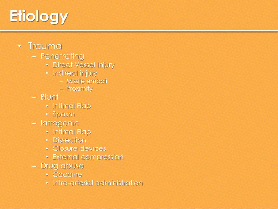
• Trauma – Penetrating
• Direct Vessel Injury
• Indirect injury – Missile emboli
– Proximity
– Blunt • Intimal Flap
• Spasm
– Iatrogenic • Intimal Flap
• Dissection
• Closure devices
• External compression
– Drug abuse • Cocaine
• Intra-arterial administration

Etiology
• Outflow Venous
Occlusion
– Compartment
syndrome
– Phlegmasia

Differential Diagnosis
• Mimics
– Low Flow States
• In the presence of chronic occlusive disease
– Venous Thrombosis
• Especially in early stages
– Acute Compressive Neuropathy
• Peroneal Nerve
• Tibial Nerve
• Saphenous Nerve

Initial Evaluation
• Symptoms
– Assess severity of limb ischemia
• Suddenness
• Time of onset of pain
• Weakness
• Numbness
– Location, intensity, and change over time
– Determine functional status of extremity

Initial Evaluation
• Past Medical History
– Claudication
– Coronary artery disease
– Arrhythmias
– Atherosclerotic risk factors
– Clotting problems
– Recent percutaneous interventions
– AGE
– LONGETIVITY OUTLOOK
– ANESTHETIC RISK

Initial Evaluation
• Physical
Examination
– Pulses
– Skin color and
temperature
– Focus on sensory
and motor defecits
– Compare with
normal opposite
extremity

Initial Evaluation
• Doppler Interrogation
– Check pedal vessels for signals
• If doppler signals are
clearly audible:
– Can allow delay for
transfer or referral,
arteriography, or
identification / treatment
of causative factors and
co-morbidities.
• Ankle/Brachial Index
– Normal >0.95
– Claudication 0.40-0.80
– Rest Pain 0.20-0.40
– Ulceration/Gangrene
<0.10

Staging
• Rutherford Criteria – SVS standardized criteria
– Class I
• Limb is viable and will remain so without intervention – Life style limiting claudication
– Class IIa
• Limbs are threatened and require revascularization for salvage, albeit not always on an emergency basis
– Parasthesias and numbness w/o motor deficit
– Class IIb
• Limbs require very urgent revascularization to prevent limb loss – Sensory and motor deficits
– Class III
• Irreversible ischemia – Permanent paralysis and sensory loss

Treatment
• Heparin – Prevent clot propagation
– Obviate further embolism
– NO studies have established a role for any antithrombotic agent in ALI
– Increased wound complications and hematomas peri-opeartively
– Patients with ALI should be treated with unfractionated heparin to prevent further clot propagation • Class 1
– Conditions for which there is evidence for and/or general agreement that a given procedure or treatment is beneficial, useful, and effective
• Level of Evidence C – Only consensus opinion of experts, case studies, or standard-of-care
Treatment
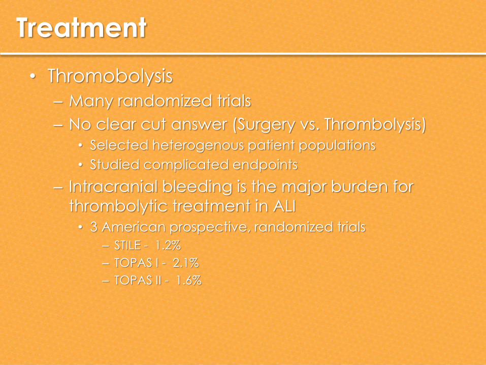
• Thromobolysis
– Many randomized trials
– No clear cut answer (Surgery vs. Thrombolysis)
• Selected heterogenous patient populations
• Studied complicated endpoints
– Intracranial bleeding is the major burden for
thrombolytic treatment in ALI
• 3 American prospective, randomized trials
– STILE - 1.2%
– TOPAS I - 2.1%
– TOPAS II - 1.6%

Treatment
• Thromobolysis
– Consensus
• Immediate surgical revascularization is
preferred if thrombolysis would lead to an
unacceptable delay in effective reperfusion.
• In patients with irreversible ischemia, primary
amputation is indicated.
• In native artery occlusion, thrombolysis
followed by correction of the causative lesion
in patients with ischemia of < 14 days in
duration.

Treatment
• Thromobolysis – Consensus
• For occluded bypass grafts – surgical revision and thrombectomy
– catheter-directed thrombolysis
– insertion of a new graft
• Factors to consider in therapeutic decision making – age and nature of the graft
– the duration and degree of ischemia
– availability of vein for a new distal bypass
• Recent occlusion of a well-established graft – thrombolytic therapy as a primary treatment
modality
– May clear the thrombosed outflow vessels as well

Embolectomy


Compartment Syndrome
• Definition: – > 40mm Hg
– > 30 mm Hg for 4 hours
– Pressure within 30 mm Hg of MAP
– Pressure within 20 mm Hg of the diastolic pressure
– > 25 mm Hg consistent with diagnosis
• Incidence: 8% in Acute Leg Ischemia – 30% if associated with fracture
• Predictors of need for fasciotomy – ‘tight swelling’ pre-op or intra-op
– Combination of arterial and venous injury
– Soft tissue crush injury
• May be seen after thrombolysis as well

Compartment Syndrome
• Lower extremity
– Anterior
compartment is the
most sensitive
– Lateral > Deep
Posterior >
Superficial Posterior

Spasm
• Montefiore Cocktail (Dr. Frank Veith)

Success is not final, failure is not fatal: it is the courage to continue that counts.
Winston Churchill