Embed Size (px)
Citation preview
Managed by UT-Battellefor the Department of Energy
Development of Computational Methods for Neurobiological Imaging Research
Shaun Gleason, PhD
Group Leader
Image Science and Machine Vision
Measurement Science and Engineering Division
Oak Ridge National Laboratory
Biomedical Science and Engineering Conference
Measurement Science and Imaging Session
March 18-19, 2009
2 Managed by UT-Battellefor the Department of Energy
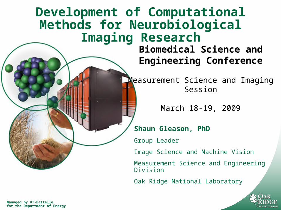
Outline
Background– Neuron Morphology
– Neuron Migration
Research Areas– Develop algorithms to compare neuron morphology
– Develop algorithms to study mechanism of neuron migration
Target applications of research– Neurological disease characterization
– Neuronal interfacing
3 Managed by UT-Battellefor the Department of Energy
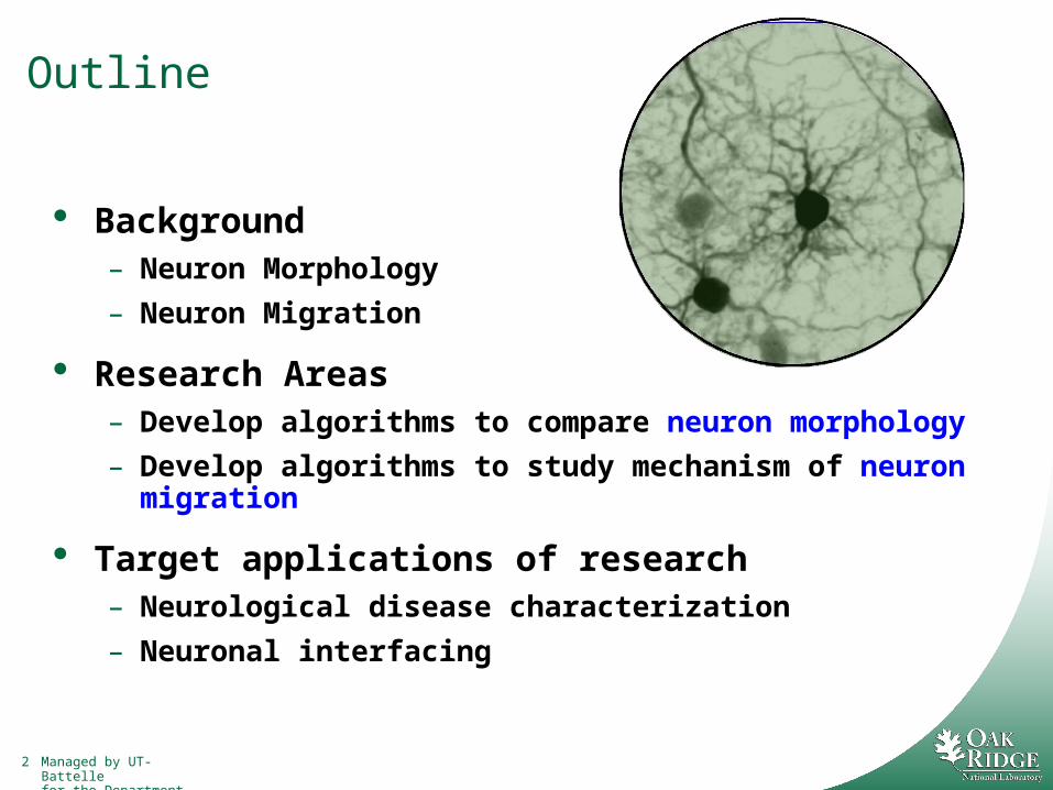
Neuronal Morphology: Form Equals Function
Guiding principle of neurobiology
Structures are generated during development
Structures are extremely heterogeneous
Changes in structure can alter function and vice versa
Santiago Ramon y Cajal, 1900
4 Managed by UT-Battellefor the Department of Energy
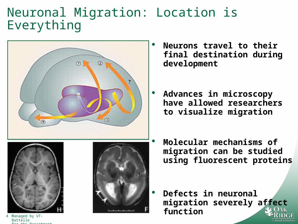
Neuronal Migration: Location is Everything
Neurons travel to their final destination during development
Advances in microscopy have allowed researchers to visualize migration
Molecular mechanisms of migration can be studied using fluorescent proteins
Defects in neuronal migration severely affect function
5 Managed by UT-Battellefor the Department of Energy
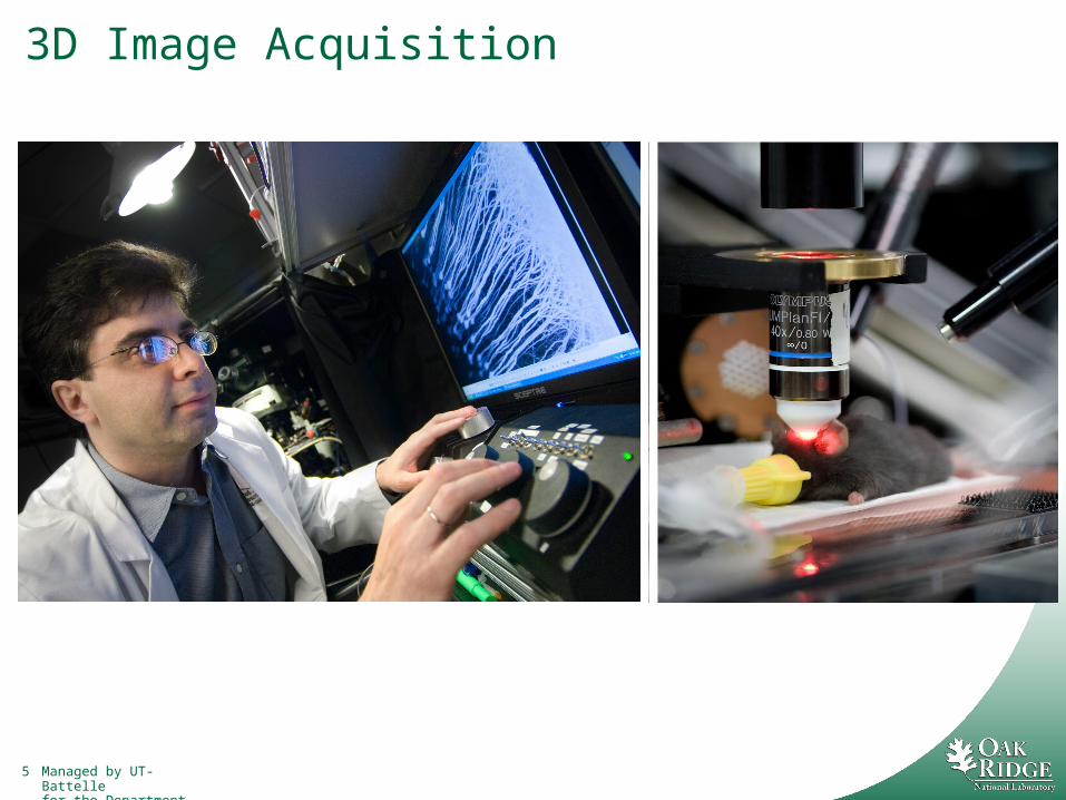
3D Image Acquisition
6 Managed by UT-Battellefor the Department of Energy
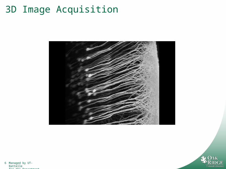
3D Image Acquisition
7 Managed by UT-Battellefor the Department of Energy
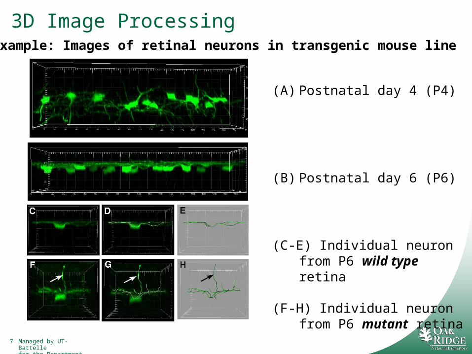
3D Image Processing
(A) Postnatal day 4 (P4)
(B) Postnatal day 6 (P6)
(C-E) Individual neuron from P6 wild type retina
(F-H) Individual neuron from P6 mutant retina
Example: Images of retinal neurons in transgenic mouse line
8 Managed by UT-Battellefor the Department of Energy
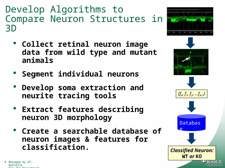
Develop Algorithms to Compare Neuron Structures in 3D
Collect retinal neuron image data from wild type and mutant animals
Segment individual neurons
Develop soma extraction and neurite tracing tools
Extract features describing neuron 3D morphology
Create a searchable database of neuron images & features for classification.
{f0, f1, f2, …fN-1}
Database
Classified Neuron:WT or KO
9 Managed by UT-Battellefor the Department of Energy
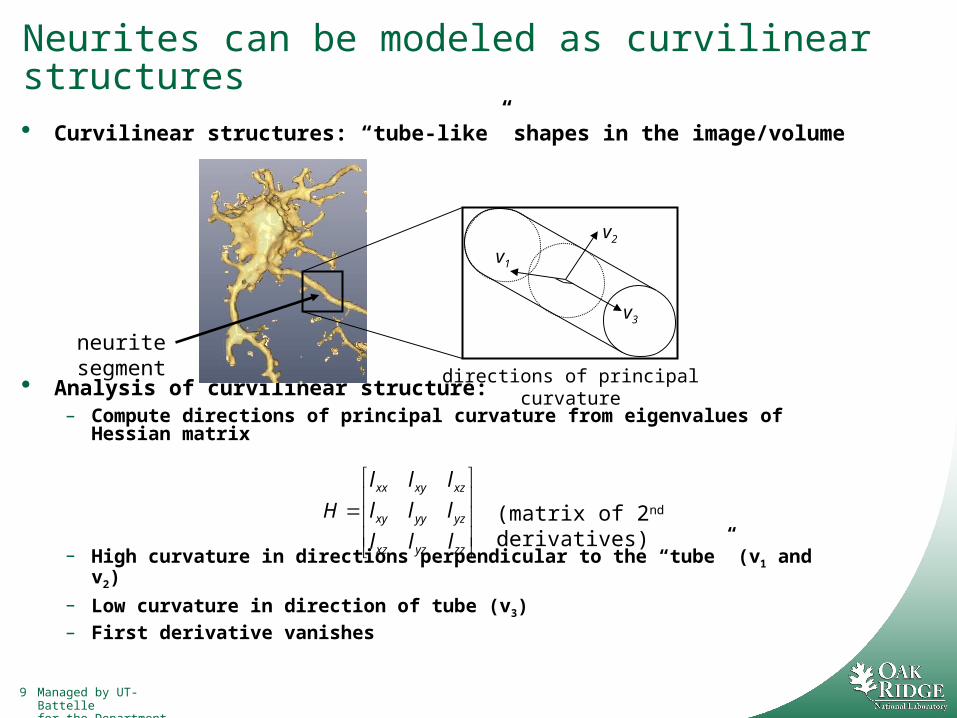
Neurites can be modeled as curvilinear structures Curvilinear structures: “tube-like” shapes in the image/volume
Analysis of curvilinear structure:– Compute directions of principal curvature from eigenvalues of Hessian
matrix
– High curvature in directions perpendicular to the “tube” (v1 and v2)
– Low curvature in direction of tube (v3)– First derivative vanishes
v1
v2
v3
neurite segment directions of principal curvature
zzyzxz
yzyyxy
xzxyxx
lll
lll
lll
H (matrix of 2nd derivatives)
10 Managed by UT-Battellefor the Department of Energy
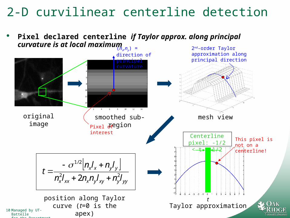
2-D curvilinear centerline detection
Pixel declared centerline if Taylor approx. along principal curvature is at local maximum
yyyxyyxxxx
yyxx
lnlnnln
lnlnt
22
2/1
2
2 4 6 8 10 12 14
2
4
6
8
10
12
14
(nx,ny) = direction of principal curvature
smoothed sub-region
Pixel of interest
2nd-order Taylor approximation along principal direction
mesh view
Taylor approximationt
Centerline pixel: -1/2 < t < 1/2
This pixel is not on a centerline!
position along Taylor curve (t=0 is the apex)
original image

11 Managed by UT-Battellefor the Department of Energy
3-D neurite centerline detection by curvilinear analysis We extend this method from 2-D to 3-D
At every voxel:– Analyze Hessian matrix
– Classify the voxel as “centerline” or “not centerline”
– 3D extension of 2D approach [Xiong, Zhou, Degterev, Ji, Wong, Automated Neurite Labeling and Analysis in Fluorescence Microscopy Images, Cytometry Part A 69A:494–505 (2006)]
Original volume (isosurface rendering)
Detected centerlines
EXAMPLE: retinal neurons
somas
12 Managed by UT-Battellefor the Department of Energy
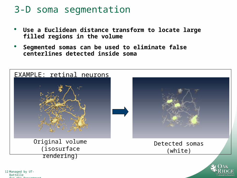
3-D soma segmentation
Use a Euclidean distance transform to locate large filled regions in the volume
Segmented somas can be used to eliminate false centerlines detected inside soma
Original volume (isosurface rendering)
Detected somas (white)
EXAMPLE: retinal neurons
13 Managed by UT-Battellefor the Department of Energy
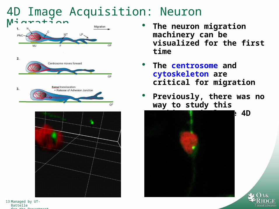
4D Image Acquisition: Neuron Migration The neuron migration
machinery can be visualized for the first time
The centrosome and cytoskeleton are critical for migration
Previously, there was no way to study this machinery in large 4D datasets

14 Managed by UT-Battellefor the Department of Energy
Develop Algorithms to Study Mechanism of Neuron Migration
Collect time-series images of migrating cerebellum neurons
Enhance existing centrosome motion tracking algorithms
Add cytoskeletal characterization methods
Investigate mechanistic model of migration
iii
iii
vHxy
GuFxx 1
F ix
iy
G
1ix ivmeasurement error
transition function
state uncertainty
MOTION MODEL
15 Managed by UT-Battellefor the Department of Energy
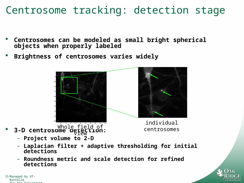
Centrosome tracking: detection stage
Centrosomes can be modeled as small bright spherical objects when properly labeled
Brightness of centrosomes varies widely
3-D centrosome detection:– Project volume to 2-D– Laplacian filter + adaptive thresholding for initial detections– Roundness metric and scale detection for refined detections
individual centrosomesWhole field of view

16 Managed by UT-Battellefor the Department of Energy
Apply joint probabilistic data association filter (JPDAF) tracking algorithm
[Bar-Shalom and Fortmann, Tracking and Data Association. New York, NY: Academic, 1988]
– Tracks multiple objects simultaneously using multi-hypothesis analysis
Use a Newtonian state-space motion model allowing for random acceleration of centrosomes in x, y, and z directions
Centrosome tracking: linking stage
?
detections and tracks from previous frames
detections in current frame
1-D ILLUSTRATION 3-D RESULTANT TRACKS
17 Managed by UT-Battellefor the Department of Energy
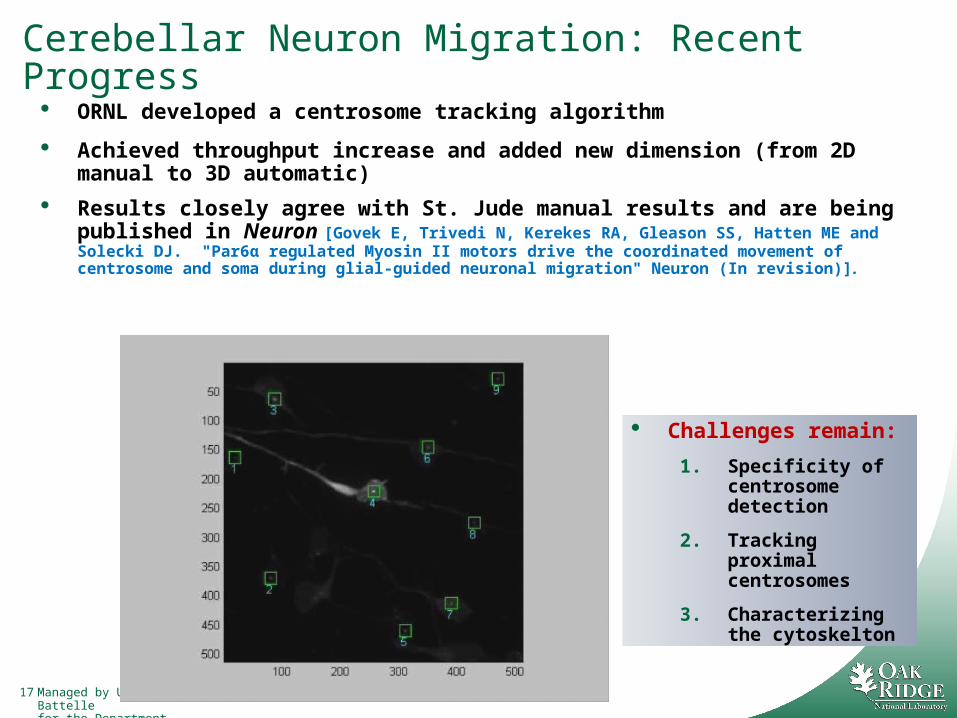
Cerebellar Neuron Migration: Recent Progress
ORNL developed a centrosome tracking algorithm
Achieved throughput increase and added new dimension (from 2D manual to 3D automatic)
Results closely agree with St. Jude manual results and are being published in Neuron [Govek E, Trivedi N, Kerekes RA, Gleason SS, Hatten ME and Solecki DJ. "Par6α regulated Myosin II motors drive the coordinated movement of centrosome and soma during glial-guided neuronal migration" Neuron (In revision)].
Challenges remain:
1. Specificity of centrosome detection
2. Tracking proximal centrosomes
3. Characterizing the cytoskelton
18 Managed by UT-Battellefor the Department of Energy
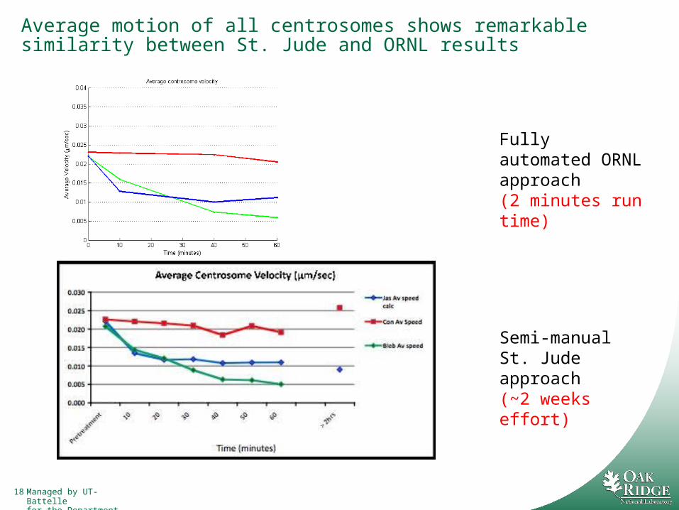
Average motion of all centrosomes shows remarkable similarity between St. Jude and ORNL results
Fully automated ORNL approach (2 minutes run time)
Semi-manual St. Jude approach (~2 weeks effort)
19 Managed by UT-Battellefor the Department of Energy
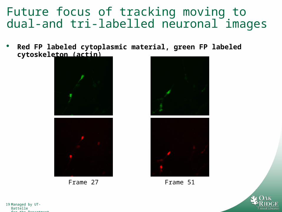
Future focus of tracking moving to dual-and tri-labelled neuronal images
Red FP labeled cytoplasmic material, green FP labeled cytoskeleton (actin)
Frame 27 Frame 51

20 Managed by UT-Battellefor the Department of Energy
Applications of morphological and migration research are numerous
Neurological disease characterization and treatment:– Alzheimer’s, Parkinson’s, schizophrenia, epilepsy,
cancer of the nervous system, retinal disorders, autism, etc.
Neurotechnology, the application of electronics and engineering to the human nervous system (neuronal interfacing)– We need to understand how neurons respond at
the cellular level to probes used for Neuronal prostheses Neuronal stimulation (e.g. deep brain)
B. Beckerman_ LDRD08

21 Managed by UT-Battellefor the Department of Energy
We are investigating the application ofnanostructured materials as a multimodaltissue interface for neural prostheses.
Physical:Quasi 3-Dimensionalcell and tissue scaffolding
10 m
Electrical:Electroanalytical Probes/Actuators
Genetic Level:Localized modulation of tissue response via geneticlevel manipulation
Fluidic:Localized modulation of tissue viareagent delivery.

22 Managed by UT-Battellefor the Department of Energy
Collaboration with the Morrison Lab at Columbiahas demonstrated these arrays may be repeatedly used for
whole tissue electrophysiological recording....
1 M TTX
1 M TTX
E38
E38
0
2
4
6
8
10
12
14
0 50 100 150 200 250 300 350 400Time (sec)
Fir
ing
Ra
te (
Hz
)
E01 E40
DG
CA3
CA1
100 ms
100 V
E01 E40
DG
CA3
CA1
E03 E04
E39 E40
200 ms
200 V
50 M BIC
50 M BIC
50 M BIC
50 M BIC
Yu Z, et al, J Neurotrauma 24 (7), 2007, Yu Z, et al. Nanoletters, 7 (8), 2007.
23 Managed by UT-Battellefor the Department of Energy
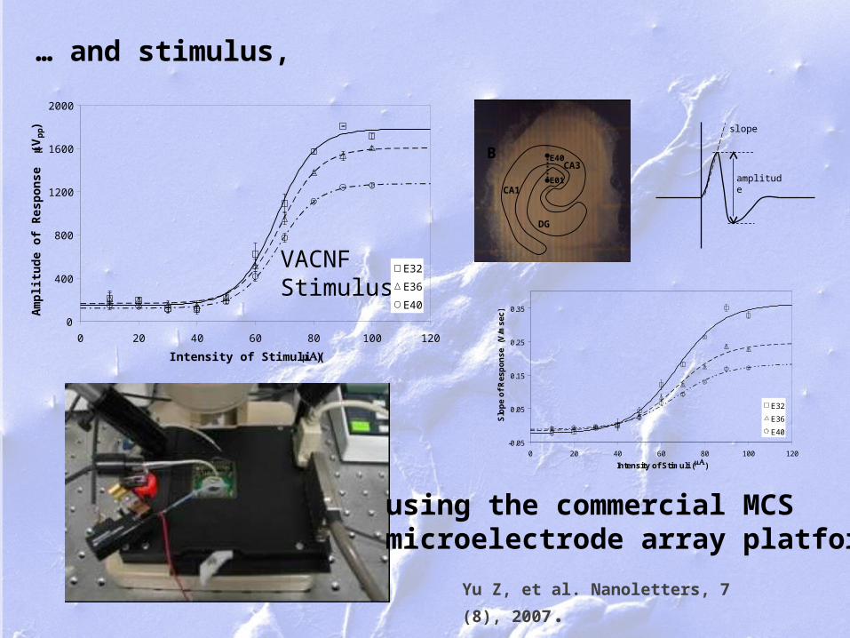
… and stimulus,
slope
amplitude
-0.05
0.05
0.15
0.25
0.35
0 20 40 60 80 100 120
Intensity of Stimuli (A)
Slo
pe
of
Re
sp
on
se
(V
/ms
ec
)
E32
E36
E40
VACNFStimulus
0
400
800
1200
1600
2000
0 20 40 60 80 100 120
Intensity of Stimuli ( A)
Am
plit
ude
of R
espo
nse
( V
pp)
E32
E36
E40
Yu Z, et al. Nanoletters, 7 (8), 2007.
using the commercial MCSmicroelectrode array platform.
E40
E01
DG
CA3
CA1
B
Managed by UT-Battellefor the Department of Energy
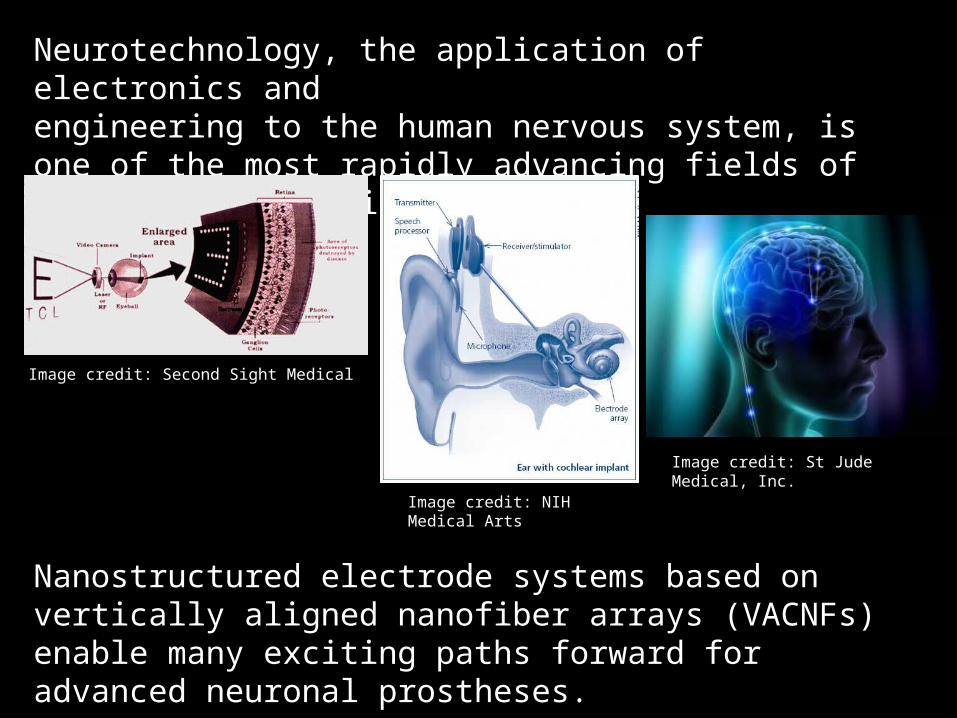
Neurotechnology, the application of electronics andengineering to the human nervous system, is one of the most rapidly advancing fields of translational medicine.
Image credit: NIH Medical Arts
Image credit: Second Sight Medical
Image credit: St Jude Medical, Inc.
Nanostructured electrode systems based on vertically aligned nanofiber arrays (VACNFs) enable many exciting paths forward for advanced neuronal prostheses.
25 Managed by UT-Battellefor the Department of Energy
Summary & Conclusions
Neurobiologists need tools to help them analyze complex neurobiological processes systematically and efficiently.
Methods are being developed to address:– Morphological-based neuron classification
– Characterization of neuron migration
Result will be a foundation upon which more sophisticated and powerful tools can be built.
Methods will enable new ways to conduct research in various neurobiological fields, e.g.: Alzheimer’s, Parkinson’s, schizophrenia, epilepsy, cancer of the nervous
system, retinal disorders, autism
Neurotechnology

26 Managed by UT-Battellefor the Department of Energy
Acknowledgements
ORNLRyan Kerekes, PhD, MSSERichard Ward, PhD, CSEDBarbara Beckerman, MBA, CSEDM. Nance Ericson, PhD, MSSETim McKnight, MSSE
St. Jude Children’s Research HospitalMichael Dyer, PhD, SJCRHDavid Solecki, PhD, SJCRHStanislav Zakharenko, MD, PhD, SJCRH
Columbia UniversityBarclay Morrison, PhD