Embed Size (px)
Citation preview

Male Osteoporosis
Matthew T. Drake, MD, PhD*, Sundeep Khosla, MD
KEYWORDS
� Osteoporosis � Men � Fracture � Epidemiology � DXA � QCT � Sex steroids
KEY POINTS
� Osteoporosis is a major health threat in aging men, with hip fractures associated with thegreatest morbidity and mortality.
� Trabecular bone loss in men begins in early adulthood, while cortical bone loss occurs frommidlife onwards.
� Declining bioavailable estradiol levels are closely associated with age-associated bone lossin men.
INTRODUCTION
Osteoporosis is defined as an asymptomatic bone disease marked by low bone massand skeletal microarchitectural deterioration resulting in increased fracture risk.1
Although osteoporosis has been widely recognized by medical professionals andthe public as a significant health issue in aging women, it is now also increasinglyviewed as a major health threat to the aging male population in terms of morbidity,mortality, and health care resource expenditure.
MALE FRACTURE EPIDEMIOLOGY
Despite not undergoing a menopausal transition as women do, men sustain bone lossof approximately 0.5% to 1.0% per year beginning by the sixth decade.2 Current esti-mates are that approximately 20% of Americans with osteopenia or osteoporosis aremen and that 1 in 8 men aged older than 50 years will incur an osteoporotic-type frac-ture during his lifetime, with roughly 30% of hip fractures occurring in men.3 Fracturesat both the hip and vertebrae have been shown to increase morbidity and mortality inmen,4,5 with hip fractures in men associated with an approximately 2- to 3-foldincreased mortality risk relative to women.6
Department of Medicine, Division of Endocrinology, College of Medicine, Mayo Clinic, 200 FirstStreet Southwest, Rochester, MN 55905, USA* Corresponding author. Endocrine Research Unit, Guggenheim 7-11, Mayo Clinic, 200 FirstStreet Southwest, Rochester, MN 55905.E-mail address: [email protected]
Endocrinol Metab Clin N Am 41 (2012) 629–641doi:10.1016/j.ecl.2012.05.001 endo.theclinics.com0889-8529/12/$ – see front matter � 2012 Elsevier Inc. All rights reserved.
Drake & Khosla630
Fracture incidence in men has 2 peaks: one around adolescence and a second sus-tained peak that occurs at advanced age. Before 50 years of age, men experiencemore fractures than women, which is likely primarily caused by increased rates ofhigh-energy trauma, such as athletic and work-place related injuries, in youngermen relative to women.7 Men subsequently experience an exponential increase infractures beginning after about 75 years of age, although the absolute fracture inci-dence in older men remains less than in age-matched women.Using data from the year 2000, it has been estimated that approximately 9.0 million
new osteoporotic fractures occur yearly, and that roughly 39% of these occur inmen.5 In the General Practice Research Database in the United Kingdom followedfrom 1988 to 1998, 103,052 men among the 5 million adults followed sustained a frac-ture over 10.4 million person-years of follow-up.7 In men, aging was associated with anincreased risk for fracture of the vertebrae, femur, forearm, humerus, clavicle, scapula,pelvis, and ribs; thus, these fracture typeswere consideredmore likely to be associatedwith osteoporosis. Similar to these findings, other studies of agedmenhave also shownincreased rates of nonvertebral fractures of the proximal femur, humerus, and fore-arm.8,9 Taken together, these findings suggest that osteoporotic fractures in menprimarily involve the hip, vertebrae, humerus, and forearm, although fractures at othersites, including the ribs, pelvis, and clavicle, are also associated with male aging.
Hip Fractures
In men, hip fractures are associated with the greatest morbidity and mortality.Although current evidence suggests that hip fracture rates in Western populationsseem to have stabilized over the past 2 decades, hip fracture rates may be increasingin other parts of the world, including Asia.10 Indeed, current estimates are that thenumber of men worldwide with hip fractures will be between 1.8 and 6.8 million by2050.3,11 Although absolute incidence rates vary geographically, overallincidence rates worldwide have been found to increase exponentially beginning atroughly 75 years of age.12 Mortality is substantially higher in men compared withwomen following hip fracture. Men are nearly twice as likely to die during their imme-diate postfracture hospitalization,13 and roughly one-third of men die within the firstyear following fracture.14,15 Morbidity is also increased in men compared with womenfollowing hip fracture; nearly half of men require skilled institutionalized care followinghip fracture,16 and men are far less likely when compared with women to return to fullindependence at 1 year following hip fracture.17 Further, although it is well docu-mented that both men and women continue to be undertreated for their osteoporosisfollowing hip fracture,18 men seem to be disproportionally more likely to beundertreated.19
Vertebral Fractures
Like hip fractures, vertebral fracture incidence in men increases with age. As shown inthe European Prospective Osteoporosis Study of 3174 men (mean age 63.1 years)who had spinal radiographs performed at a mean of 3.8 years following a baselinefilm, the age-standardized incidence of morphometric fractures was 5.7 per 1000person-years, a rate slightly lower than the 10.7 per 1000 person-years determinedin women.20 As expected, the incidence in both sexes increased markedly withprogressive age. Similar trends have been demonstrated in numerous other studiesperformed both in the United States and Europe,21–24 with small differences in inci-dence/prevalence rates between the studies likely reflecting subject inclusion criteria,study methodologies, and geographic variation. Importantly, vertebral fracturesincrease the likelihood of subsequent fractures in both sexes,25,26 have been shown

Male Osteoporosis 631
to be a positive predictor of age-adjusted mortality in men,26 and are associated withmultiple indices of poorer life quality.27
Although fracture data in men at sites other than the spine and hip are more limited,there is now good evidence that low-trauma fractures of the upper arm (forearm andhumerus) and ankle are associated with an increased risk for future fractures in men.28
PREVALENCE OF LOW BONE MASS IN MEN
As defined by the World Health Organization, osteopenia is a bone mineral density(BMD) measured by dual-energy x-ray absorptiometry (DXA) between 1.0 and 2.5standard deviations (SD) less than the young adult (aged 20–29 years) normative refer-encemean, with osteoporosis defined as a BMD value of less than 2.5 SD less than themean value. Using femoral BMD data from the Third National Health and NutritionSurvey and a BMD normative reference range derived from non-Hispanic whites,Looker and colleagues29 found that substantial numbers of men had low bonemass. Using a male reference range, 3% to 6% of men had osteoporosis and 28%to 47% had osteopenia. Using a female reference mean, 1% to 4% of men had oste-oporosis, whereas 15% to 33% were osteopenic. By comparison, Melton andcolleagues30 used a population-based sample of Rochester, Minnesota men to deter-mine a prevalence estimate of 19% for osteoporosis in men aged 50 years and olderwhen using a male reference range but only 3% when using a female reference range.There remains some debate as to whether sex-specific reference ranges may be moreappropriate than a single female-based reference range for describing low bone massin men,31,32 although it is clear that the use of a male-specific database identifies moremen as having diminished bone mass (osteopenia and osteoporosis). Estimates of lowbone mass from DXA data in men other than non-Hispanic whites are more limited butsuggest that non-Hispanic white men have the highest prevalence of osteopenia/oste-oporosis, followed by Mexican-American men and non-Hispanic black men,29 whichare results that are consistent with the hip fracture incidence among men of differentethnic backgrounds.33
RISK FACTORS FOR BONE LOSS IN MEN
Because of the combination of an improved male longevity, the significant morbidityassociated with fractures in aged men, and the broad availability of pharmacologicagents that documented efficacy for reducing fracture risk, it is increasingly importantto identify men at an increased fracture risk for low BMD-associated fractures. Thereare many reasons why male bone loss occurs, which can broadly be grouped intoprimary (age-associated and idiopathic) and secondary (all other) causes (Box 1). Inindividual men, bone loss may result from a single cause or be caused by a combina-tion of multiple factors. A more thorough review of risk factors associated with boneloss is men is beyond the scope of this review but has been reviewed recently.34
CAUSE OF MALE BONE LOSS
Although DXA is the best-established clinical method for assessing bone loss, DXAonly allows for assessment of areal BMD and, therefore, lacks the ability to distinguishbetween cortical and trabecular bone components. Recent advances in boneimaging, however, have allowed for significant enhancement in our understandingof age-associated bone loss. Chief among these advances has been the applicationof quantitative computed tomography (QCT) to both central and peripheral skeletalsites, which is technology that allows for true assessments of volumetric BMD
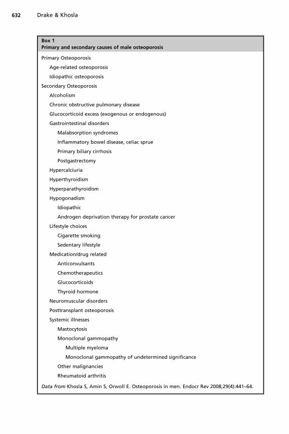
Box 1
Primary and secondary causes of male osteoporosis
Primary Osteoporosis
Age-related osteoporosis
Idiopathic osteoporosis
Secondary Osteoporosis
Alcoholism
Chronic obstructive pulmonary disease
Glucocorticoid excess (exogenous or endogenous)
Gastrointestinal disorders
Malabsorption syndromes
Inflammatory bowel disease, celiac sprue
Primary biliary cirrhosis
Postgastrectomy
Hypercalciuria
Hyperthyroidism
Hyperparathyroidism
Hypogonadism
Idiopathic
Androgen deprivation therapy for prostate cancer
Lifestyle choices
Cigarette smoking
Sedentary lifestyle
Medication/drug related
Anticonvulsants
Chemotherapeutics
Glucocorticoids
Thyroid hormone
Neuromuscular disorders
Posttransplant osteoporosis
Systemic illnesses
Mastocytosis
Monoclonal gammopathy
Multiple myeloma
Monoclonal gammopathy of undetermined significance
Other malignancies
Rheumatoid arthritis
Data from Khosla S, Amin S, Orwoll E. Osteoporosis in men. Endocr Rev 2008;29(4):441–64.
Drake & Khosla632

Male Osteoporosis 633
(vBMD) and bone microstructural changes either cross-sectional or longitudinal.Accordingly, Riggs and colleagues35 recently assessed bone geometry and vBMDby QCT at the lumbar spine and femoral neck and peripheral QCT at the distal radiusand tibia in a population-based sample of 323 men aged 20 to 97 years from Roches-ter, Minnesota. As seen in Fig. 1, a substantial (approximately 46%) nearly linear lossof lumbar spine (comprised primarily of trabecular bone) vBMD occurred across theadult male lifespan. In contrast, cortical vBMD as assessed at the radius remainedessentially stable until approximately the sixth decade in men, thereafter decreasingnearly linearly by approximately 18%.In addition to changes in vBMD, changes in bone geometry also occur with aging.
As again demonstrated by Riggs and colleagues,35 aging in men was associated withincreases in bone cross-sectional area at the femoral neck caused by progressiveperiosteal apposition with concomitant increases in endocortical resorption, ultimatelyresulting in a slight decline in cortical area and thickness. Importantly, however, thisnet outward cortical displacement increases resistance to bending stresses, therebyproviding a partial biomechanical adaptation to limit the overall loss of bone strengthcaused by the decreases in cortical area and thickness.36
Additional studies from the Osteoporotic Fractures in Men cohort examiningchanges in vBMD and bone dimensions at the femoral neck and shaft by QCT ina cohort of 3358 men aged 65 to 100 years demonstrated that at the femoral neck,trabecular vBMD was 22.1% lower in men aged more than 85 years compared withmen aged 65 to 69 years but that cortical vBMDwas similar between the two groups.37
Confirming the results described by Riggs and colleagues,36 increased endocorticalresorption and periosteal apposition with resultant cortical thinning but outwardcortical displacement was also observed at the femoral neck in the older versusyounger men. At the femoral shaft by comparison, both the cross-sectional andmedullary areas were increased (9% and 22% respectively) in the older versusyounger men, with percent shaft cortical bone and shaft cortical vBMD both 4% lowerin the older men.Together, these studies demonstrate that although trabecular bone loss in men
(and, indeed, in women) begins in early adulthood, cortical bone loss only seems tobegin with midlife. Further, although an increase in periosteal apposition leads to an
Fig. 1. (A) Total vertebral body vBMD values (milligrams per cubic centimeter) from a popula-tion sample (n5 323) of men in Rochester, Minnesota aged 20 to 97 years. (B) Cortical vBMDvalues at the distal radius in the same male cohort. (Adapted from Riggs BL, Melton Iii LJ 3rd,Robb RA, et al. Population-based study of age and sex differences in bone volumetric density,size, geometry, and structure at different skeletal sites. J BoneMiner Res 2004;19(12):1945–54;with permission from the American Society of Bone and Mineral Research.)

Drake & Khosla634
increase in bone cross-sectional area with aging, a simultaneous increase in endocort-ical resorption ultimately results in the overall decline in cortical area.
MALE BONE MICROSTRUCTURAL CHANGES WITH AGING
Themore recent development of high-resolution peripheral QCT (HRpQCT), which canbe used to image peripheral skeletal sites (wrist and tibia) at much higher resolutionsthan standard QCT, has allowed for an even more accurate noninvasive assessmentof bone microstructure. In a population-based cross-sectional study of 278 men inRochester, Minnesota aged 21 to 97 years highly characteristic of the United Stateswhite population, Khosla and colleagues38 used HRpQCT to examine bonemicrostructure at the ultradistal radius. Relative to young aged-matched womenaged 20 to 29 years, men had markedly greater bone volume (BV)/tissue volume (TV)and trabecular thickness (26% and 28% respectively) but little difference in trabecularnumber or separation. Although cross-sectional decreases in trabecular BV/TV wereapproximately 26% in both sexes between 20 to 90 years of age, the structural basisfor these declines was fundamentally different. Women lost trabeculae and increasedtrabecular separation over the lifespan, whereas men primarily sustained trabecularthinning without trabecular loss. Importantly, these data are in accordance withprevious histomorphometric studies of cadaveric transiliac bone biopsies obtainedover approximately the same age range of 20 to 90 years, suggesting that the changesdetermined by HRpQCT may be similar at other skeletal sites. If so, this may partiallyexplain the reduced fracture incidence seen in men compared with women with agingbecause decreases in trabecular number are anticipated to have a greater impact onbone strength relative to decreases in trabecular thickness.39
THE ROLE OF SEX STEROIDS IN MALE OSTEOPOROSIS
Although the role of declining sex steroid levels in bone loss is best recognized inwomen, marked changes in sex steroid levels also occur over the male lifespan. Unlikewomen in whom menopausal-associated ovarian failure is the primary cause,declining sex-steroid levels in men are principally caused by a greater than twofoldage-associated increase in sex hormone binding globulin (SHBG) levels.40 ThisSHBG increase limits the biologically available (free [1%–3%] and albumin-associated [35%–55%]) sex steroids, leading to declines in bioavailable testosterone(T) and estrogen (E) levels of 64% and 47%, respectively, over the male lifespan.40
Although T is the predominant male sex steroid, both cross-sectional41–46 and longi-tudinal47 evidence indicates that levels of bioavailable estradiol (E2) rather than T arebetter correlated with male BMD. In support of this, a longitudinal study that evaluatedsex steroid levels in a younger male cohort (aged 22–39 years, an age range in the finalstages of skeletal maturation) versus an older male cohort (aged 60–90 years, an agerange in which age-related bone loss occurs) over a 4-year interval found that althoughdistal forearm BMD (primarily reflecting cortical bone compartment changes) in theyounger cohort increased approximately 0.42% to 0.43% per year, distal forearmBMD in the older men declined by 0.49% to 0.66% per year.47 Importantly, theincreasedBMD in youngermenand thedecreasedBMD inoldermenweremore closelyassociated with bioavailable E2 levels than with T levels. Further, in older men, thereseemed to be a threshold bioavailable E2 level of approximately 40 pM (11 pg/mL),which was less than the bone loss rates seemed to increase. Further vBMD analysesshowed that this bioavailable E2 threshold correlated better with cortical than trabec-ular sites.48 Above this threshold, however, therewas no firm relationship seenbetweenbone loss rates andbioavailable E2 levels. Additionalwork has supported this threshold
Male Osteoporosis 635
effect for bioavailable and total E2,49 although the absolute threshold determined byother investigators has varied slightly depending onwhether E2 levelswere determinedby immunoassay (approximately 20–25 pg/mL) or mass spectroscopy (16 pg/mL).50,51
Although correlative, the previous findings provide no direct evidence of a causal rolefor E in male bone health with aging. To directly assess the role of sex steroids (E and T)on skeletal health in agingmen, Falahati-Nini and colleagues52 undertook a direct inter-ventional study inwhich they suppressedendogenousTandEproduction (by treatmentwith both a gonadotropin-releasing hormone agonist and an aromatase inhibitor) whilesimultaneously providing physiologic replacement levels of E and T by topical patchplacement. Following the determination of the baseline bone turnover markers whileon the previous endogenous suppression/physiologic replacement regime, patientswere randomized into 1 of 4 groups: group A (-T, -E) discontinued both patches, groupB (-T,1E) continued only the E patch, group C (1T, -E) continued only the T patch, andgroup D (1T,1E) continued both patches. Accordingly, the study design permitted thedetermination of bone metabolism changes caused by either E or T because endoge-nous sex steroid production was suppressed throughout the study.As seen in Fig. 2, the complete absence of both T and E (group A) resulted in signif-
icant increases in bone resorption, which was completely prevented by treatment withboth T and E (group D). Treatment with E alone was almost completely able to preventthe increase in bone resorption (group B), whereas T alone had only modest effects(group C). Comparatively, the significant decline in serum bone formation markers(osteocalcin and amino-terminal propeptide of type I collagen [P1NP]) that occurredwith a deficiency of both T and E (group A) was completely prevented by the supple-mentation of both T and E (group D); treatment with either E or T alone lead to onlyslight decreases in osteocalcin levels, whereas P1NP levels were maintained with E(group B) but not T (group C) treatment. Together, these results are consistent witha dominant role for E as the major sex steroid regulating skeletal health in aged men.With male aging, levels of bioavailable T decline even more precipitously than those
of bioavailable E. Despite this decrease, however, the extent to which the diminution ofbioavailable T levels impacts bone loss associated with male aging is less clear whencompared with the role of declining bioavailable E levels. As shown, T imparts modesteffects on bone resorption and also affects bone formation (see Fig. 2). As recentlydemonstrated, increased femoral neck BMD occurred in aged men who receivedlow-dose T replacement for 2 years53 despite the absence of BMD increases at anyother site examined (lumbar spine, total hip, or distal radius). Whether the effect ofT on femoral neck BMD in this study was direct or rather the result of T aromatizationto E, however, is unclear. Current evidence suggests that T also likely plays a role incortical appositional bone growth, although this has been most convincingly demon-strated in rodent studies54 and the extent to which T has this function with aging inmen is less clear.Finally, it hasbeensuggested thatTmayplayamore indirect role inmaleskeletal health
with aging by allowing for relative maintenance of balance and muscle strength in mencomparedwithwomen.51 As such, the slightly lower fracture incidence in agedmen rela-tive to age-matched women may be partially reflective of the increased frequency offalling that occurswith aging inwomen relative tomen,55,56with fracturesafter fallsoccur-ring 2.2-fold higher in women than men.57
OTHER POTENTIAL CAUSES OF MALE BONE LOSS
A variety of other factors have been postulated to play a role in age-associated malebone loss. These factors include an age-associated increase in parathyroid hormone

Fig. 2. Percent changes in markers of (A) bone resorption (urinary deoxypyridinoline [Dpd]and N-telopeptide of type I collagen [NTX]) and (B) bone formation (serum osteocalcin andN-terminal extension peptide of type I collagen [P1NP]) in elderly men (mean age, 68 years)pharmacologically made acutely hypogonadal and subsequently treated with either an ar-omatase inhibitor (group A), E alone (group B), T alone (group C), or both E and T (group D).Significance for change from baseline: asterisk, P<.05; double asterisk, P<.01; triple asterisk,P<.001. (Adapted from Falahati-Nini A, Riggs BL, Atkinson EJ, et al. Relative contributions oftestosterone and estrogen in regulating bone resorption and formation in normal elderlymen. J Clin Invest 2000;106(12):1553–60; with permission from the American Society for Clin-ical Investigation.)
Drake & Khosla636
levels,58 decreases in growth hormone and circulating bioavailable insulinlike growthfactor levels,59 and inherent changes or loss of stem or osteoprogenitor cells.60 Addi-tionally, nutrition also seems to be critical for optimal skeletal health because thefailure to maintain an adequate intake of vitamin D, calcium, or protein has shownto lead to worsened bone health in men.12
CARE FOR MALE PATIENTS WITH BONE LOSS
Reducing the substantial fracture risk that occurs with bone loss in men is now morewidely recognized as an important goal of optimal patient care. Despite this recogni-tion, however, most studies using pharmacologic interventions have been performedin women; further, the comparatively fewer studies in men have typically assessedBMD changes as a surrogate for fracture risk. Nonetheless, current evidence suggeststhat the most commonly prescribed therapies (nitrogen-containing bisphosphonatesand teriparatide) are as efficacious in men as they are in women.

Male Osteoporosis 637
Bisphosphonates
Alendronate,61 risedronate,62 ibandronate,63 and zoledronate64 have all been shownto increase BMD and reduce biochemical markers of bone turnover in men with lowbone mass, with alendronate, risedronate, and zoledronate having received Foodand Drug Administration (FDA) approval for the treatment of osteoporosis in men.Importantly, BMD changes with bisphosphonate therapy seem to be equivalent inmen with low free T levels as in men with normal levels,61 perhaps indicating whybisphosphonate therapy is effective in limiting bone loss in men receiving androgendeprivation therapy for prostate cancer. Bisphosphonates have also been shown tobe effective for limiting bone loss in men receiving corticosteroid therapy65 or in thesetting of acute immobilization.66
Teriparatide
Once daily recombinant parathyroid hormone 1–34 increases BMD in men with lowbone mass and seems to reduce the risk for vertebral fractures.67 As with bisphosph-onates, results seem to be similar in magnitude to those seen in women, and teripara-tide has received FDA approval for the treatment of osteoporosis in men.
Denosumab
The receptor activator of nuclear factor kappa-B (RANK) ligand inhibitor was initiallyapproved for the treatment of osteoporosis in women and was more recently FDA-approved for the treatment ofmenwith nonmetastatic prostate cancer with osteopeniaand a high risk of fracture receiving androgen-deprivation therapy.A more complete review of additional potential pharmacologic agents for the
management of male bone loss is beyond the scope of this review. Accordingly,the reader is referred to a recently published meta-analysis of the comparativeeffectiveness of currently available pharmacologic agents for reducing fracture riskin men.68
ADDITIONAL SUPPORTIVE CARE
In addition to pharmacologic interventions, ensuring that men with low bone massreceive appropriate supportive care is essential for limiting fracture risk. Suchcare includes confirming that patients have adequate vitamin D and calcium intakeboth before and while maintained on therapy. In older community-dwelling menaged 65 years and older, moderate reductions in bone loss at the hip and spineand reduced nonvertebral fracture incidence were seen in patients receiving dietaryvitamin D and calcium supplementation for 3 years compared with patients whoreceived placebo.69 Notably, recommendations from the Institute of Medicine ondietary intakes of calcium and vitamin D have been recently published, withskeletal-related outcomes as the primary end point for most included studies.70,71
Finally, defining appropriate generalized exercise prescriptions in patients atincreased fracture risk remains an important part of the care plan for most patientsbecause increases in physical performance are expected to increase muscle massand tone, thereby reduce frailty and decreasing fall risk. To date, however, studiesthat have attempted to examine this approach to exercise in at-risk patients havenot used standardized methods. Because of the significant heterogeneity amongstudies that examined the role of exercise in fracture reduction, a recent reviewconcluded that currently available data do not allow for proper quantitative assess-ment of the risk associated with exercise in at-risk patients.34

Drake & Khosla638
SUMMARY
Althoughmuch work over the preceding 2 decades has enlightened our understandingof male bone loss, significant challenges to providing optimal care to our male patientspersist. Osteoporosis in men remains an underrecognized clinical problem that willcontinue to grow in importance with the aging of the population. Despite significantadvances in our understanding of the epidemiology, risk factors, causes, and thera-peutic approaches to male osteoporosis, much work awaits if we are to limit futuremorbidity and mortality associated with bone loss in men.
REFERENCES
1. Consensus development conference: diagnosis, prophylaxis, and treatment ofosteoporosis. Am J Med 1993;94(6):646–50.
2. Melton LJ 3rd, Khosla S, Achenbach SJ, et al. Effects of body size and skeletalsite on the estimated prevalence of osteoporosis in women and men. OsteoporosInt 2000;11(11):977–83.
3. Cooper C, Campion G, Melton LJ 3rd. Hip fractures in the elderly: a world-wideprojection. Osteoporos Int 1992;2(6):285–9.
4. Johnell O, Kanis JA. An estimate of the worldwide prevalence, mortality anddisability associated with hip fracture. Osteoporos Int 2004;15(11):897–902.
5. Johnell O, Kanis JA. An estimate of the worldwide prevalence and disability asso-ciated with osteoporotic fractures. Osteoporos Int 2006;17(12):1726–33.
6. Center JR, Nguyen TV, Schneider D, et al. Mortality after all major types of oste-oporotic fracture in men and women: an observational study. Lancet 1999;353(9156):878–82.
7. van Staa TP, Dennison EM, Leufkens HG, et al. Epidemiology of fractures inEngland and Wales. Bone 2001;29(6):517–22.
8. Schuit SC, van der Klift M, Weel AE, et al. Fracture incidence and association withbone mineral density in elderly men and women: the Rotterdam study. Bone2004;34(1):195–202.
9. Jonsson BY, Siggeirsdottir K, Mogensen B, et al. Fracture rate in a population-based sample of men in Reykjavik. Acta Orthop Scand 2004;75(2):195–200.
10. Cooper C, Cole ZA, Holroyd CR, et al. Secular trends in the incidence of hip andother osteoporotic fractures. Osteoporos Int 2011;22(5):1277–88.
11. Gullberg B, Johnell O, Kanis JA. World-wide projections for hip fracture. Osteo-poros Int 1997;7(5):407–13.
12. Khosla S, Amin S, Orwoll E. Osteoporosis in men. Endocr Rev 2008;29(4):441–64.13. Diamond TH, Thornley SW, Sekel R, et al. Hip fracture in elderly men: prognostic
factors and outcomes. Med J Aust 1997;167(8):412–5.14. Kiebzak GM, Beinart GA, Perser K, et al. Undertreatment of osteoporosis in men
with hip fracture. Arch Intern Med 2002;162(19):2217–22.15. Bass E, French DD, Bradham DD, et al. Risk-adjusted mortality rates of elderly
veterans with hip fractures. Ann Epidemiol 2007;17(7):514–9.16. Poor G, Atkinson EJ, O’Fallon WM, et al. Determinants of reduced survival
following hip fractures in men. Clin Orthop Relat Res 1995;(319):260–5.17. Schurch MA, Rizzoli R, Mermillod B, et al. A prospective study on socioeconomic
aspects of fracture of the proximal femur. J Bone Miner Res 1996;11(12):1935–42.
18. Curtis JR, McClure LA, Delzell E, et al. Population-based fracture risk assessmentand osteoporosis treatment disparities by race and gender. J Gen Intern Med2009;24(8):956–62.

Male Osteoporosis 639
19. Shibli-Rahhal A, Vaughan-Sarrazin MS, Richardson K, et al. Testing and treatmentfor osteoporosis following hip fracture in an integrated U.S. healthcare deliverysystem. Osteoporos Int 2011;22(12):2973–80.
20. Incidence of vertebral fracture in Europe: results from the European ProspectiveOsteoporosis Study (EPOS). J Bone Miner Res 2002;17(4):716–24.
21. O’Neill TW, Felsenberg D, Varlow J, et al. The prevalence of vertebral deformity inEuropean men and women: the European Vertebral Osteoporosis Study. J BoneMiner Res 1996;11(7):1010–8.
22. Davies KM,StegmanMR,HeaneyRP, et al. Prevalence and severity of vertebral frac-ture: the Saunders County Bone Quality Study. Osteoporos Int 1996;6(2):160–5.
23. Santavirta S, Konttinen YT, Heliovaara M, et al. Determinants of osteoporoticthoracic vertebral fracture. Screening of 57,000 Finnish women and men. ActaOrthop Scand 1992;63(2):198–202.
24. Szulc P, Munoz F, Marchand F, et al. Semiquantitative evaluation of prevalentvertebral deformities in men and their relationship with osteoporosis: the MINOSstudy. Osteoporos Int 2001;12(4):302–10.
25. Melton LJ 3rd, Atkinson EJ, Cooper C, et al. Vertebral fractures predict subse-quent fractures. Osteoporos Int 1999;10(3):214–21.
26. Hasserius R, Karlsson MK, Nilsson BE, et al. Prevalent vertebral deformitiespredict increased mortality and increased fracture rate in both men and women:a 10-year population-based study of 598 individuals from the Swedish cohort inthe European Vertebral Osteoporosis Study. Osteoporos Int 2003;14(1):61–8.
27. Burger H, Van Daele PL, Grashuis K, et al. Vertebral deformities and functionalimpairment in men and women. J Bone Miner Res 1997;12(1):152–7.
28. Center JR, Bliuc D, Nguyen TV, et al. Risk of subsequent fracture after low-traumafracture in men and women. JAMA 2007;297(4):387–94.
29. Looker AC, Orwoll ES, Johnston CC Jr, et al. Prevalence of low femoral bonedensity in older U.S. adults from NHANES III. J Bone Miner Res 1997;12(11):1761–8.
30. Melton LJ 3rd, Atkinson EJ, O’Connor MK, et al. Bone density and fracture risk inmen. J Bone Miner Res 1998;13(12):1915–23.
31. Khosla S. Update in male osteoporosis. J Clin Endocrinol Metab 2010;95(1):3–10.32. Kanis JA, Bianchi G, Bilezikian JP, et al. Towards a diagnostic and therapeutic
consensus in male osteoporosis. Osteoporos Int 2011;22(11):2789–98.33. Silverman SL, Madison RE. Decreased incidence of hip fracture in Hispanics,
Asians, and blacks: California hospital discharge data. Am J Public Health1988;78(11):1482–3.
34. Drake MT, Murad MH, Mauck KF, et al. Risk factors for low bone mass-relatedfractures in men: a systematic review and meta-analysis. J Clin Endocrinol Metab2012;97(6):1861–70.
35. Riggs BL, Melton Iii LJ 3rd, Robb RA, et al. Population-based study of age andsex differences in bone volumetric density, size, geometry, and structure atdifferent skeletal sites. J Bone Miner Res 2004;19(12):1945–54.
36. Turner CH. Bone strength: current concepts. Ann N Y Acad Sci 2006;1068:429–46.
37. Marshall LM, Lang TF, Lambert LC, et al. Dimensions and volumetric BMD of theproximal femur and their relation to age among older U.S. men. J Bone Miner Res2006;21(8):1197–206.
38. Khosla S, Riggs BL, Atkinson EJ, et al. Effects of sex and age on bone micro-structure at the ultradistal radius: a population-based noninvasive in vivo assess-ment. J Bone Miner Res 2006;21(1):124–31.

Drake & Khosla640
39. Silva MJ, Gibson LJ. Modeling the mechanical behavior of vertebral trabecularbone: effects of age-related changes in microstructure. Bone 1997;21(2):191–9.
40. Khosla S, Melton LJ 3rd, Atkinson EJ, et al. Relationship of serum sex steroidlevels and bone turnover markers with bone mineral density in men and women:a key role for bioavailable estrogen. J Clin Endocrinol Metab 1998;83(7):2266–74.
41. Slemenda CW, Longcope C, Zhou L, et al. Sex steroids and bone mass in oldermen. Positive associations with serum estrogens and negative associations withandrogens. J Clin Invest 1997;100(7):1755–9.
42. Greendale GA, Edelstein S, Barrett-Connor E. Endogenous sex steroids andbone mineral density in older women and men: the Rancho Bernardo Study.J Bone Miner Res 1997;12(11):1833–43.
43. Center JR, Nguyen TV, Sambrook PN, et al. Hormonal and biochemical parame-ters in the determination of osteoporosis in elderly men. J Clin Endocrinol Metab1999;84(10):3626–35.
44. van den Beld AW, de Jong FH, Grobbee DE, et al. Measures of bioavailableserum testosterone and estradiol and their relationships with muscle strength,bone density, and body composition in elderly men. J Clin Endocrinol Metab2000;85(9):3276–82.
45. Amin S, Zhang Y, Sawin CT, et al. Association of hypogonadism and estradiollevels with bone mineral density in elderly men from the Framingham study.Ann Intern Med 2000;133(12):951–63.
46. Szulc P, Munoz F, Claustrat B, et al. Bioavailable estradiol may be an importantdeterminant of osteoporosis in men: the MINOS study. J Clin Endocrinol Metab2001;86(1):192–9.
47. Khosla S, Melton LJ 3rd, Atkinson EJ, et al. Relationship of serum sex steroidlevels to longitudinal changes in bone density in young versus elderly men.J Clin Endocrinol Metab 2001;86(8):3555–61.
48. Khosla S, Riggs BL, Robb RA, et al. Relationship of volumetric bone density andstructural parameters at different skeletal sites to sex steroid levels in women.J Clin Endocrinol Metab 2005;90(9):5096–103.
49. Gennari L, Merlotti D, Martini G, et al. Longitudinal association between sexhormone levels, bone loss, and bone turnover in elderly men. J Clin EndocrinolMetab 2003;88(11):5327–33.
50. Mellstrom D, Vandenput L, Mallmin H, et al. Older men with low serum estradioland high serum SHBG have an increased risk of fractures. J Bone Miner Res2008;23(10):1552–60.
51. Khosla S, Melton LJ 3rd, Riggs BL. The unitary model for estrogen deficiency andthe pathogenesis of osteoporosis: is a revision needed? J Bone Miner Res 2011;26(3):441–51.
52. Falahati-Nini A, Riggs BL, Atkinson EJ, et al. Relative contributions of testosteroneand estrogen in regulating bone resorption and formation in normal elderly men.J Clin Invest 2000;106(12):1553–60.
53. Nair KS, Rizza RA, O’Brien P, et al. DHEA in elderly women and DHEA or testos-terone in elderly men. N Engl J Med 2006;355(16):1647–59.
54. Wakley GK, Schutte HD Jr, Hannon KS, et al. Androgen treatment prevents lossof cancellous bone in the orchidectomized rat. J Bone Miner Res 1991;6(4):325–30.
55. Sattin RW, Lambert Huber DA, DeVito CA, et al. The incidence of fall injury eventsamong the elderly in a defined population. Am J Epidemiol 1990;131(6):1028–37.
56. Paspati I, Galanos A, Lyritis GP. Hip fracture epidemiology in Greece during1977-1992. Calcif Tissue Int 1998;62(6):542–7.
Male Osteoporosis 641
57. Stevens JA, Sogolow ED. Gender differences for non-fatal unintentional fallrelated injuries among older adults. Inj Prev 2005;11(2):115–9.
58. Kennel KA, Riggs BL, Achenbach SJ, et al. Role of parathyroid hormone in medi-ating age-related changes in bone resorption in men. Osteoporos Int 2003;14(8):631–6.
59. Boonen S, Mohan S, Dequeker J, et al. Down-regulation of the serum stimulatorycomponents of the insulin-like growth factor (IGF) system (IGF-I, IGF-II, IGFbinding protein [BP]-3, and IGFBP-5) in age-related (type II) femoral neck oste-oporosis. J Bone Miner Res 1999;14(12):2150–8.
60. Undale AH, Westendorf JJ, Yaszemski MJ, et al. Mesenchymal stem cells forbone repair and metabolic bone diseases. Mayo Clin Proc 2009;84(10):893–902.
61. Orwoll E, Ettinger M, Weiss S, et al. Alendronate for the treatment of osteoporosisin men. N Engl J Med 2000;343(9):604–10.
62. Ringe JD, Faber H, Farahmand P, et al. Efficacy of risedronate in men withprimary and secondary osteoporosis: results of a 1-year study. Rheumatol Int2006;26(5):427–31.
63. Orwoll ES, Binkley NC, Lewiecki EM, et al. Efficacy and safety of monthly ibandr-onate in men with low bone density. Bone 2010;46(4):970–6.
64. Orwoll ES, Miller PD, Adachi JD, et al. Efficacy and safety of a once-yearly i.v.infusion of zoledronic acid 5 mg versus a once-weekly 70-mg oral alendronatein the treatment of male osteoporosis: a randomized, multicenter, double-blind,active-controlled study. J Bone Miner Res 2010;25(10):2239–50.
65. Sambrook PN, Roux C, Devogelaer JP, et al. Bisphosphonates and glucocorti-coid osteoporosis in men: results of a randomized controlled trial comparing zo-ledronic acid with risedronate. Bone 2012;50(1):289–95.
66. Bauman WA, Wecht JM, Kirshblum S, et al. Effect of pamidronate administrationon bone in patients with acute spinal cord injury. J Rehabil Res Dev 2005;42(3):305–13.
67. Girotra M, Rubin MR, Bilezikian JP. The use of parathyroid hormone in the treat-ment of osteoporosis. Rev Endocr Metab Disord 2006;7(1–2):113–21.
68. Murad MH, Drake MT, Mullan RJ, et al. Comparative effectiveness of drug treat-ments to prevent fragility fractures: a systematic review and network meta-anal-ysis. J Clin Endocrinol Metab 2012, Mar 30. [Epub ahead of print].
69. Dawson-Hughes B, Harris SS, Krall EA, et al. Effect of calcium and vitamin Dsupplementation on bone density in men and women 65 years of age or older.N Engl J Med 1997;337(10):670–6.
70. Ross AC, Manson JE, Abrams SA, et al. The 2011 report on dietary referenceintakes for calcium and vitamin D from the Institute of Medicine: what cliniciansneed to know. J Clin Endocrinol Metab 2011;96(1):53–8.
71. Rosen CJ, Gallagher JC. The 2011 IOM report on vitamin D and calcium require-ments for North America: clinical implications for providers treating patients withlow bone mineral density. J Clin Densitom 2011;14(2):79–84.