Embed Size (px)
Citation preview
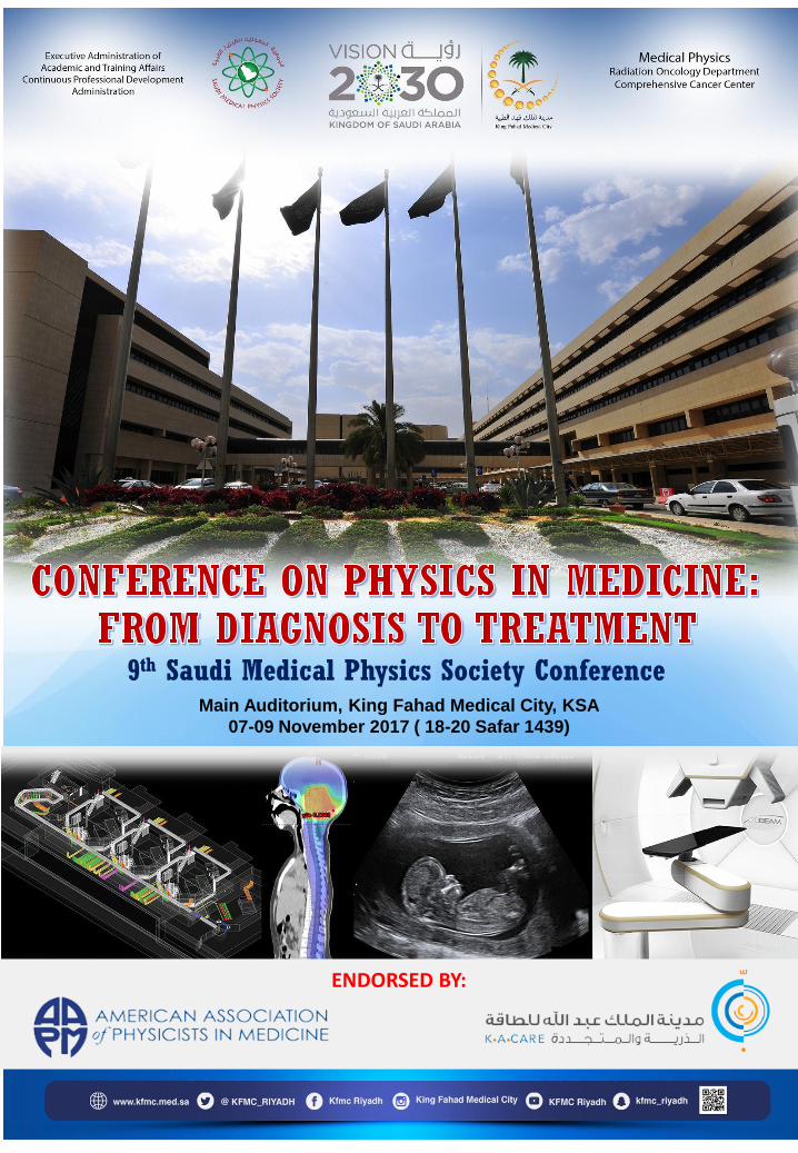
9th Saudi Medical Physics Society ConferenceMain Auditorium, King Fahad Medical City, KSA
07-09 November 2017 ( 18-20 Safar 1439)
ENDORSED BY:
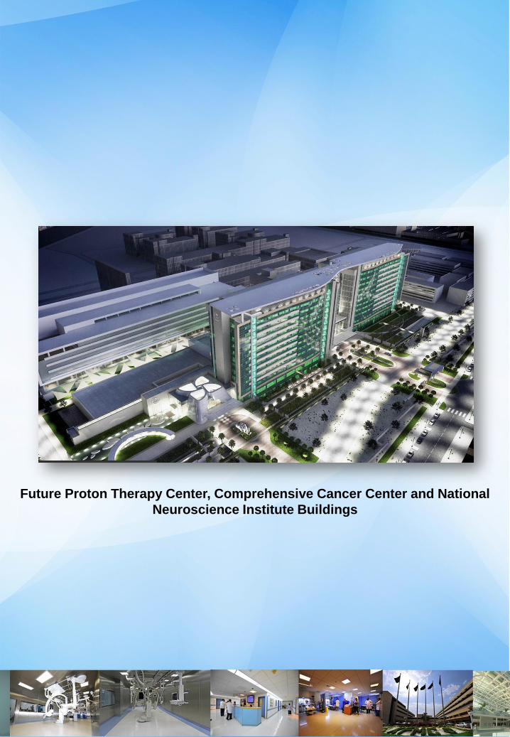
Future Proton Therapy Center, Comprehensive Cancer Center and National
Neuroscience Institute Buildings
MESSAGE
On behalf of the Conference Committee, I am honored and delighted to welcome you to the
KFMC International Conference on Physics in Medicine. This meeting is an opportunity to meet
some of the leading experts in healthcare for scientific discussion and to share experience on
the applications of physics and engineering in the medical field.
This conference is a continuation to the successful annual meetings organized in collaboration
with the Saudi Medical Physics Society (SMPS), which attracts over 500 scientists, engineers,
professors, students, and healthcare professionals. It has been successfully held in different
cities around the kingdom after the first conference that was held at King Fahad Medical City
(KFMC) back in 2006. Now in 2017, it is back where it all started.
KFMC conference on physics in medicine provides a unique opportunity for all participants to
exchange ideas and share their knowledge and experience. In addition to the scientific
program, I hope you will get to now KFMC and enjoy your stay. November is an excellent time
to come to Riyadh, you will be able to enjoy the landmarks and attractions of the city in a very
nice weather.
I would like to take this opportunity to thank the organizing and scientific program committees
for their time and effort to bring the conference a success. I would also express my heartfelt
gratitude to all the sponsors for their support that made this meeting possible.
Dr. Mukhtar AlShanqity, MSc, PhD, DABR
Chairman of organizing Committee
Head of Medical Physicists
King Fahad Medical City
Riyadh, Saudi Arabia
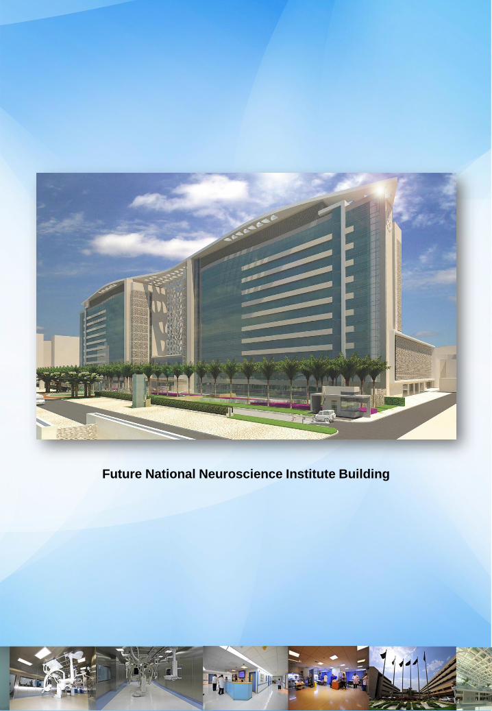
Future National Neuroscience Institute Building
MESSAGE
I take this opportunity to thank King Fahad Medical City (KFMC) for being a partner of Saudi
Medical Physics Society (SMPS) in promoting the medical physics profession. Today's
conference is another collaborative effort between KFMC and SMPS for its 9th Saudi Medical
Physics Conference in developing awareness of the medical physicists' endeavor to provide
services that ensure patient safety and quality health care in using radiation in medicine.
The theme of this conference is bridging the relationship between diagnosis and treatment.
With the development of new and hybrid technologies in both areas medical physicists are
challenged in improving services from conservative to ultra modern. This conference provides
an excellent venue for introducing new frontiers to young medical physicists and sharing of
experiences for consultant medical physicists.
As this very important event coincides with the celebration of the International Day of Medical
Physics with the theme " Medical Physics: Providing a Holistic Approach to Women Patients
and Women Staff Safety in Radiation Medicine" we join all the medical physicists around the
world for honoring the contributions of women medical physicists in providing quality medical
physics services from diagnosis to treatment.
I wish each one of you a fruitful three days of participation in the conference and bring home to
your institution the lessons and experiences learned from this conference. Saudi Arabia, being
the country with the most number of medical physicists in the Middle East region, let us
continue to strive to be more competitive internationally.
Abdalla N. Al-Haj, PhD, FIPEM, CSci, MSRP
President, Saudi Medical Physics Society
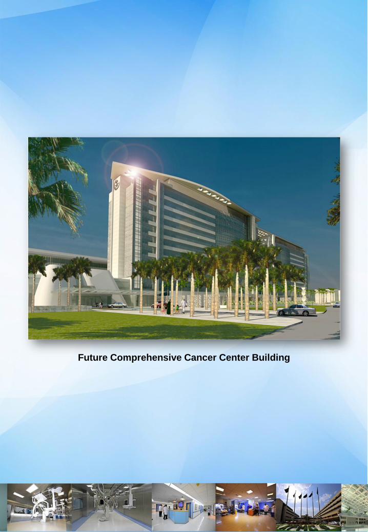
Future Comprehensive Cancer Center Building
COURSE OBJECTIVE
1. Provide education on the different applications of physics in medicine.
2. Discuss the emerging technologies and applications of radiation in diagnostic X-ray
imaging, nuclear medicine and radiation therapy.
3. Apply radiation safety practices in the use of radiation in medicine for quality medical
physics services in healthcare.
4. Identify gaps on safety and quality in the use of radiation in medicine.
5. Discuss current trends , issues and practices on uses of radiation from diagnosis to
treatment
DESIRED RESULTS
1. Increased awareness of the applications of physics in medicine.
2. Provision of interactive forum for discussion of technical advances and research on
application of physics in medicine.
3. Increased capability on safe uses of radiation in diagnosis and treatment to improve health
care services.
4. Bridge gaps on the uses of radiation from diagnose to treatment.
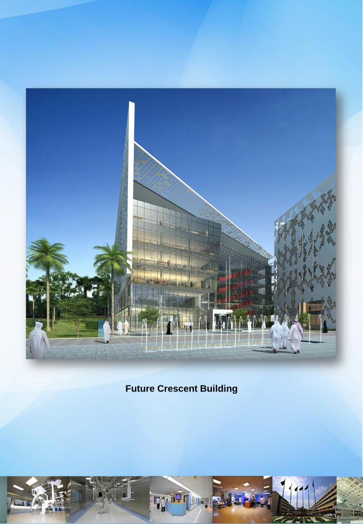
Future Crescent Building
SCIENTIFIC COMMITTEE ORGANIZING COMMITTEE
Dr. Ahmed Outif
Consultant Medical Physicist
Dr. Abdullah Abuhaimed
Assistant professor
Dr. Awad AlZahrani
Consultant
Dr. Faris Al Zahrani
Consultant
Ms. Joelle Amiouni
Medical Physicist
Mr. Rami Al Harbi
Medical Physicist
Mr. Abdullah Al Qarni
Medical Physicist
Dr. Mukhtar AlShanqity
Consultant Medical Physicist
Dr. Abousleh El Awadi
Consultant Medical Physicist
Ms. Laura Stanciu
Medical Physicist
Mr. Hosam Allazkani
Medical Physicist
Mr. Tariq Al Kwaitim
CME Coordinator
Mr. Melchor Malavi
Committee Secretary
Ms. Jaina A. Abdulkadil
Medical Conference Registrar
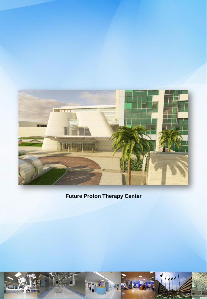
Future Proton Therapy Center
CONFERENCE SPEAKERS
Dr. Mukhtar AlShanqity
Consultant Medical Physicist
Radiation Oncology Department
King Fahad Medical City
Riyadh, KSA
Dr. Renato Padovani
Director & Consultant Medical Physicist
International Centre for Theoretical Physics
Trieste, Italy
Dr. Hussain AlHussain
Consultant Radiation Oncologist
Radiation Oncology Department
King Fahad Medical City
Riyadh, KSA
Dr. Awad Al Zahrani
Consultant
National Center for Radiation Protection
King Abdullah City for Atomic & Renewable Energy
(KACARE)
Dr. Faris Mayia
Consultant Medical Physicist
Radiation Oncology Department
King Fahad Medical City
Riyadh, KSA
Dr. Ahmad Outif
Consultant Medical Physicist
Security Forces Hospital
Riyadh, KSA
Dr. Wamied Abdulrahman
Chief/Consultant Radiation Oncology Physicist
Department of Radiation Oncology
King Fahad Specialist Hospital
Dammam, KSA
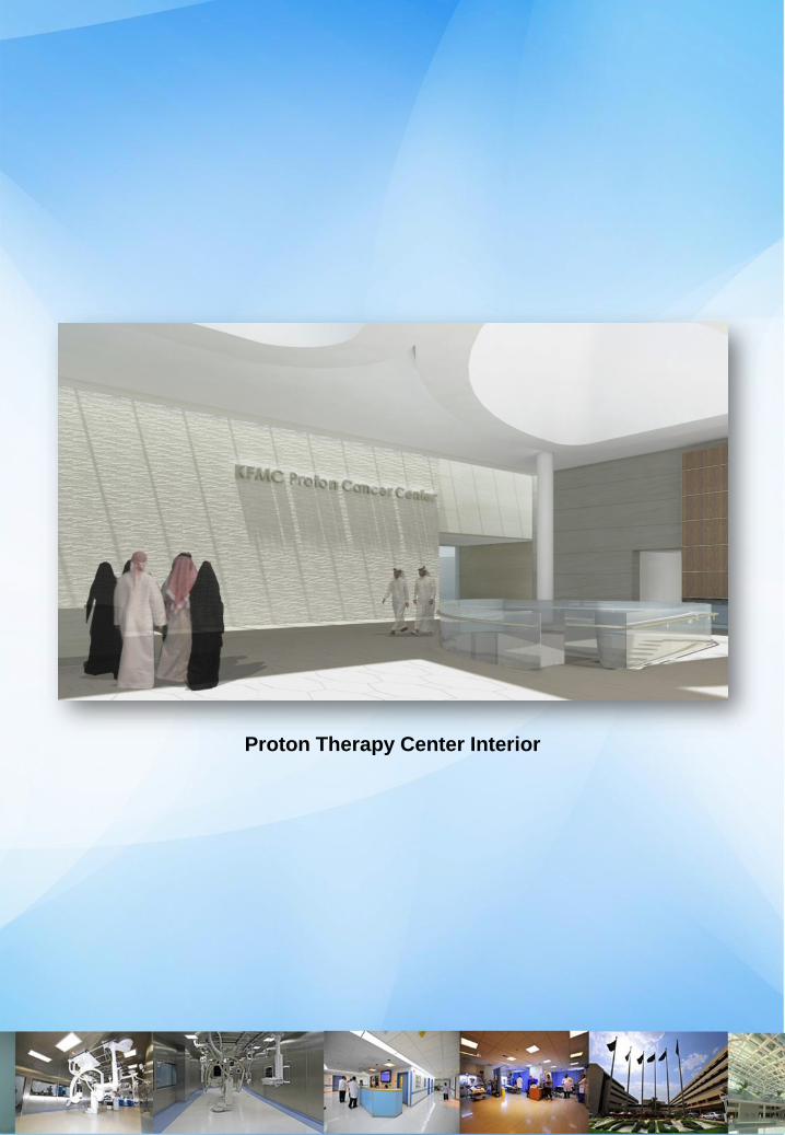
Proton Therapy Center Interior
CONFERENCE SPEAKERS
Dr. Abdullah H. Jamea
Consultant Medical Physicist
Assistant Professor
Radiology Department College of Medicine
King Khalid University Hospital
Riyadh, KSA
Dr. Abdullah A. Abuhaimed
Assistant Professor
National Center for Applied Physics
King Abdulaziz City for Science and
Technology (KACST)
Dr. Yasser Bayoumi
Consultant Radiation Oncologist
Director of Radiation Oncology Department
Residency Program
King Fahad Medical City, Riyadh, KSA
Dr. Moamen Aly
Consultant Medical Physicist
Radiation Oncology Department
King Fahad Medical City
Riyadh, KSA
Dr. Mohamed Zaghloul
Chairman
Radiation Oncology Department
Children's Cancer Hospital
Cairo, Egypt
Dr. Shanker Raja
Consultant
Nuclear Medicine
Medical Imaging Administration
King Fahad Medical City
Dr. Mamdoh AlGathami
Consultant Medical Physicist
Radiation Oncology Department
National Guard Hospital
Riyadh, KSA
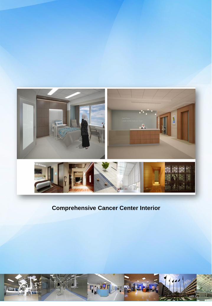
Comprehensive Cancer Center Interior
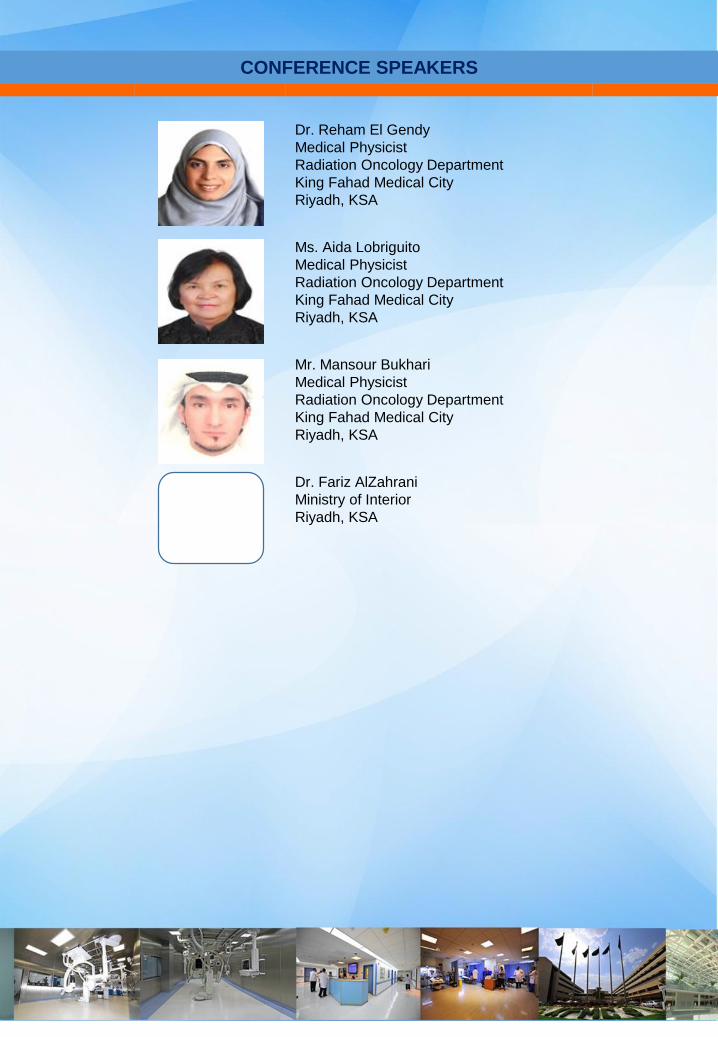
CONFERENCE SPEAKERS
Dr. Reham El Gendy
Medical Physicist
Radiation Oncology Department
King Fahad Medical City
Riyadh, KSA
Ms. Aida Lobriguito
Medical Physicist
Radiation Oncology Department
King Fahad Medical City
Riyadh, KSA
Mr. Mansour Bukhari
Medical Physicist
Radiation Oncology Department
King Fahad Medical City
Riyadh, KSA
Dr. Fariz AlZahrani
Ministry of Interior
Riyadh, KSA
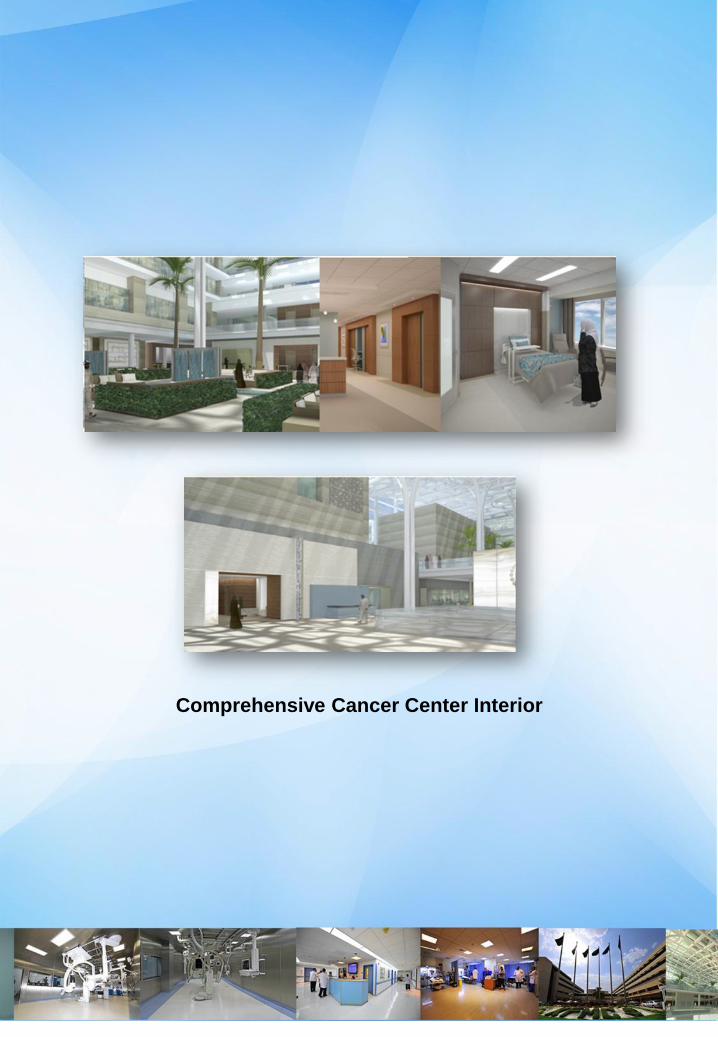
Comprehensive Cancer Center Interior
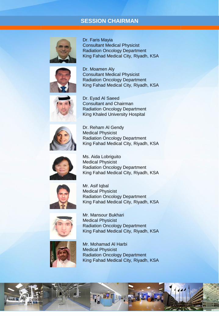
SESSION CHAIRMAN
Dr. Faris Mayia
Consultant Medical Physicist
Radiation Oncology Department
King Fahad Medical City, Riyadh, KSA
Dr. Moamen Aly
Consultant Medical Physicist
Radiation Oncology Department
King Fahad Medical City, Riyadh, KSA
Dr. Eyad Al Saeed
Consultant and Chairman
Radiation Oncology Department
King Khaled University Hospital
Dr. Reham Al Gendy
Medical Physicist
Radiation Oncology Department
King Fahad Medical City, Riyadh, KSA
Ms. Aida Lobriguito
Medical Physicist
Radiation Oncology Department
King Fahad Medical City, Riyadh, KSA
Mr. Asif Iqbal
Medical Physicist
Radiation Oncology Department
King Fahad Medical City, Riyadh, KSA
Mr. Mansour Bukhari
Medical Physicist
Radiation Oncology Department
King Fahad Medical City, Riyadh, KSA
Mr. Mohamad Al Harbi
Medical Physicist
Radiation Oncology Department
King Fahad Medical City, Riyadh, KSA
Future Cochlear Implant Building
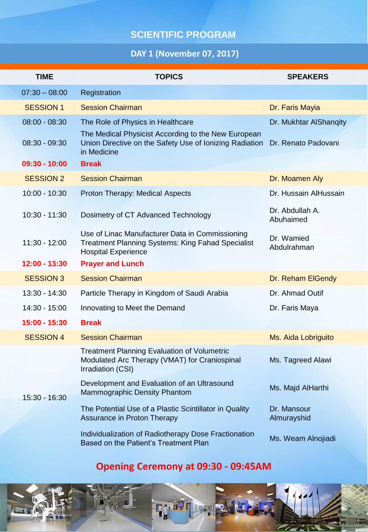
DAY 1 (November 07, 2017)
TIME TOPICS SPEAKERS
07:30 – 08:00 Registration
SESSION 1 Session Chairman Dr. Faris Mayia
08:00 - 08:30 The Role of Physics in Healthcare Dr. Mukhtar AlShanqity
08:30 - 09:30
The Medical Physicist According to the New European
Union Directive on the Safety Use of Ionizing Radiation
in Medicine
Dr. Renato Padovani
09:30 - 10:00 Break
SESSION 2 Session Chairman Dr. Moamen Aly
10:00 - 10:30 Proton Therapy: Medical Aspects Dr. Hussain AlHussain
10:30 - 11:30 Dosimetry of CT Advanced TechnologyDr. Abdullah A.
Abuhaimed
11:30 - 12:00
Use of Linac Manufacturer Data in Commissioning
Treatment Planning Systems: King Fahad Specialist
Hospital Experience
Dr. Wamied
Abdulrahman
12:00 - 13:30 Prayer and Lunch
SESSION 3 Session Chairman Dr. Reham ElGendy
13:30 - 14:30 Particle Therapy in Kingdom of Saudi Arabia Dr. Ahmad Outif
14:30 - 15:00 Innovating to Meet the Demand Dr. Faris Maya
15:00 - 15:30 Break
SESSION 4 Session Chairman Ms. Aida Lobriguito
15:30 - 16:30
Treatment Planning Evaluation of Volumetric
Modulated Arc Therapy (VMAT) for Craniospinal
Irradiation (CSI)
Ms. Tagreed Alawi
Development and Evaluation of an Ultrasound
Mammographic Density PhantomMs. Majd AlHarthi
The Potential Use of a Plastic Scintillator in Quality
Assurance in Proton Therapy
Dr. Mansour
Almurayshid
Individualization of Radiotherapy Dose Fractionation
Based on the Patient’s Treatment Plan Ms. Weam Alnojiadi
SCIENTIFIC PROGRAM
Opening Ceremony at 09:30 - 09:45AM

NOTES

DAY 2 (November 08, 2017)
TIME TOPICS SPEAKERS
SESSION 5 Session Chairman Mr. Asif Iqbal
08:00 - 08:30
Impact of AI (Artificial Intelligence) and
Deeplearning in Medicine, Medical Imaging and
Radiotherapy
Dr. Shanker Raja
08:30 - 09:30 MRI Diffusion Imaging and Tractography Dr. Abdullah Jamea
09:30 - 10:00 Break
SESSION 6 Session Chairman Dr. Eyad Al Saeed
10:00 - 10:30 Intensity Modulated Therapy for Breast Cancer Dr. Mohamed Zaghloul
10:30 - 11:30New ICRP Recommendations on Assessment
and Use of Diagnostic Reference LevelsDr. Renato Padovani
11:30 - 12:00Education, Training, Certification and
Credentialing of Medical PhysicistsMs. Aida Lobriguito
12:00 - 13:30 Lunch
SESSION 7 Session Chairman Mr. Mohamad Al Harbi
13:30 - 14:30 Shielding Design in Proton Facilities Dr. Ahmad Outif
14:30 - 15:00 Break
15:00 - 16:30 Eye Lens Dosimetry Workshop Dr. Renato Padovani
SCIENTIFIC PROGRAM

NOTES
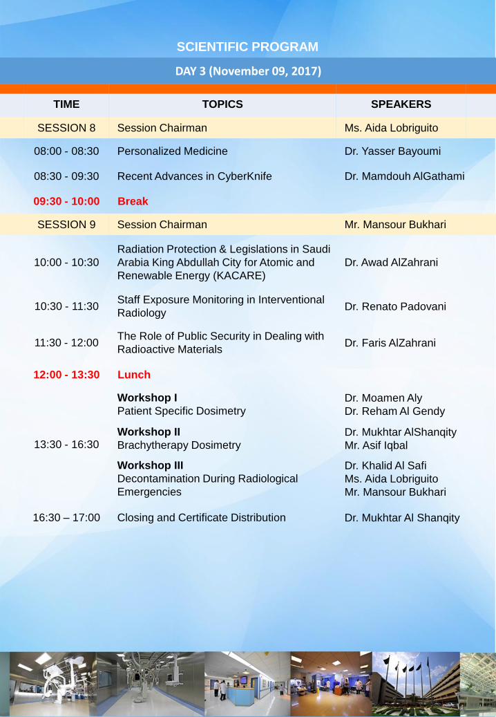
DAY 3 (November 09, 2017)
TIME TOPICS SPEAKERS
SESSION 8 Session Chairman Ms. Aida Lobriguito
08:00 - 08:30 Personalized Medicine Dr. Yasser Bayoumi
08:30 - 09:30 Recent Advances in CyberKnife Dr. Mamdouh AlGathami
09:30 - 10:00 Break
SESSION 9 Session Chairman Mr. Mansour Bukhari
10:00 - 10:30
Radiation Protection & Legislations in Saudi
Arabia King Abdullah City for Atomic and
Renewable Energy (KACARE)
Dr. Awad AlZahrani
10:30 - 11:30Staff Exposure Monitoring in Interventional
Radiology Dr. Renato Padovani
11:30 - 12:00The Role of Public Security in Dealing with
Radioactive MaterialsDr. Faris AlZahrani
12:00 - 13:30 Lunch
13:30 - 16:30
Workshop I
Patient Specific Dosimetry
Dr. Moamen Aly
Dr. Reham Al Gendy
Workshop II
Brachytherapy Dosimetry
Dr. Mukhtar AlShanqity
Mr. Asif Iqbal
Workshop III
Decontamination During Radiological
Emergencies
Dr. Khalid Al Safi
Ms. Aida Lobriguito
Mr. Mansour Bukhari
16:30 – 17:00 Closing and Certificate Distribution Dr. Mukhtar Al Shanqity
SCIENTIFIC PROGRAM

NOTES

NOTES
ABSTRACT
The potential use of a plastic scintillator in quality assurance in proton therapy
Mansour Almurayshid 1,2, Yusuf Helo2, Andrzej Kacperek3, Jennifer Griffiths2, Jem Hebden2
and Adam Gibson2
1. National Center for Nuclear Technology, King Abdulaziz City for Science and Technology
– KACST, Saudi Arabia
2. Medical Physics and Biomedical Engineering, University College London, UK
3. National Eye Proton Therapy Centre, Clatterbridge Cancer Centre, UK
METHOD: Plastic scintillators emit visible light when irradiated. They are inexpensive,
robust, have tissue-equivalent density and can easily be shaped and fabricated. RESULTS:
We evaluate a BC-408 plastic scintillator (Saint-Gobain Crystal Corporation, USA), chosen
due to its high scintillation efficiency, optical transparency and large volume
(20 cm×20cm×10 cm), imaged with a commercial Nikon D7100 camera as a quality
assurance tool in proton therapy. The whole 2D dose distribution was then obtained in a
single image (Fig 1a). Possible optical artefacts, which could affect the signal, were
investigated and taken in conisation when needed. The scintillation responses were
reproducible to within 0.55%, linear with dose, and independent of dose rate. However, there
was a reduction in the light intensity in the Bragg peak region because the protons’ high
linear energy transfer (LET) leads to quenching where less light is produced than expected.
We corrected the measured scintillation light distribution for quenching using Birks’ equation.
LET was obtained produced by Geant4 as a function of depth and was combined with the
measured scintillation light to calculate Birks’ constant as 0.15 ± 0.02 mm MeV-1. Ideal and
quenched distributions were then simulated by Geant4 and used to generate a correction
which was applied to the measured distribution to produce the corrected scintillation output
(Fig 1b).
ABSTRACT
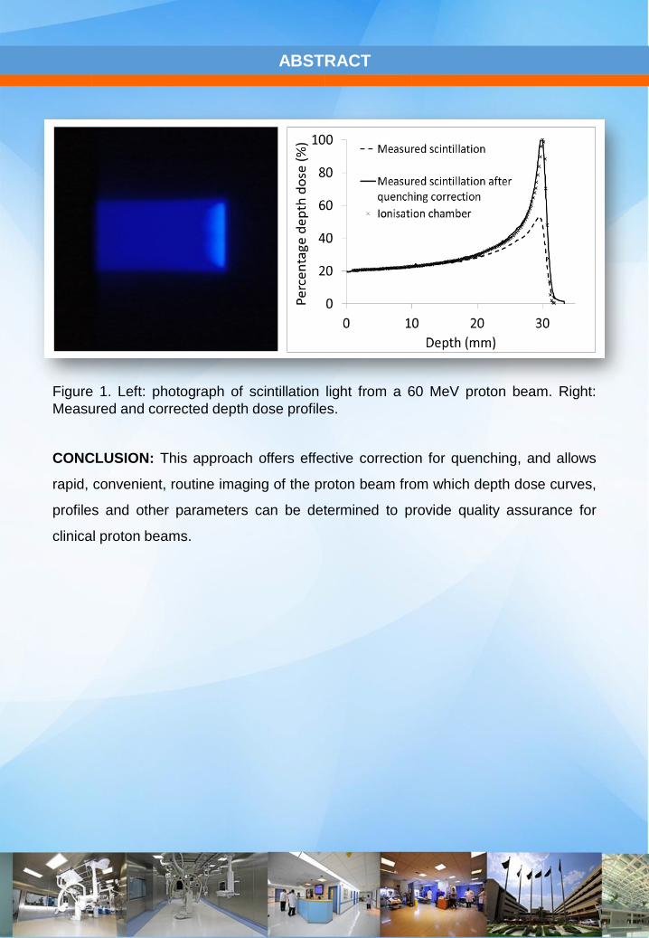
CONCLUSION: This approach offers effective correction for quenching, and allows
rapid, convenient, routine imaging of the proton beam from which depth dose curves,
profiles and other parameters can be determined to provide quality assurance for
clinical proton beams.
Figure 1. Left: photograph of scintillation light from a 60 MeV proton beam. Right:
Measured and corrected depth dose profiles.
ABSTRACT
Treatment Planning Evaluation of Volumetric Modulated Arc Therapy (VMAT) for
Craniospinal Irradiation (CSI)
Salwa Fathy, Tagreed ALawi, Nasser Al-Dhaibani
PURPOSE: The aim of this study is to assess the feasibility and accuracy of Volumetric
Modulated Arc Therapy (VMAT) technique for CSI regarding to improving the PTV dose
conformity and homogeneity with reducing the dose to organs at risk (OAR) with reporting the
integral dose (ID) received by non-target tissue (NTT).
METHODS: Four male patients (n=4) had received crainiospinal irradiation by volumetric
modulated arc therapy [VMAT] treatment planning technique [normalized such that 95% of PTV
(planning target volume) received at least 95% of the prescribed dose which is 36Gy in 20
fractions]. Plans were assessed using PTV dose coverage as D95% (the dose received by 95% of
PTV); D2% was taken as a reference for the maximum dose and D98% as a reference for the
minimum dose, the mean doses of OARs, conformity index 95% (CI95%), and homogeneity index
(HI), and non-target tissue integral doses (NTTID).
RESULTS: VMAT CSI is able to produce satisfied dose distributions with high PTV conformity
and homogeneity. The average D95% coverage of the PTV was 34Gy, with an average
conformity index 95% (CI95%) of 0.99 and an average homogeneity index (HI) of 1.13. In terms of
OAR sparing, VMAT technique allowed average mean doses acceptably low for all organs
[lenses, eyes, liver, kidneys, lungs, and heart were 7.73Gy, 14.39Gy, 8.16Gy, 6.97Gy, 8.06Gy,
and 8.18Gy respectively]. As regard the average integral dose for the non target tissue NTTID
was 307.21Gy.
CONCLUSION: CSI is considered one of the most challenging processes in radiotherapy. This
study provides a practically useful planning method for VMAT treatment of CSI that shows its
ability to achieve highly conformal and homogeneous treatment plans, while limiting the dose to
the surrounding OARs. However, this technical advantage of this VMAT technique was
associated with an increase in non target tissue integral dose that may be associated with
increase in risk of secondary malignancies. This VMAT approach should therefore be tested with
respect to late toxicity with further studies.

NOTES

ABSTRACT
Development and Evaluation of an Ultrasound Mammographic Density
Phantom
Majd Alharthi, Christian Langton
Science and Engineering Faculty, Queensland University of Technology, Brisbane,
Queensland, Australia
PURPOSE:This paper reports on the building of five breast phantoms, two of which simulated
different types of normal breast tissues: fatty tissues (subcutaneous fat and retromammary fat)
and glandular tissues. The other three phantoms simulated breast tissues abnormal with
respect to attenuation coefficients, velocity of sound, densities and broadband ultrasound
attenuation (BUA).
METHODS: Phantom composition and production are described in detail. Ultrasonic
properties were tested at 22ºC and 34ºC using a transmission technique with a 1-MHz
transducer, and a 5-MHz transducer was used for BUA.
RESULTS: Values for attenuation coefficients and the velocity of sound agree with published
values, and increased temperature was found to reduce the attenuation coefficients of all
phantoms. However, increased temperature was also found to increase the speed of sound in
non-fatty phantoms, while it reduced the speed in fatty phantoms. BUA measurements of non-
fatty phantoms had attenuations are linear proportional to frequencies between 3.3-3.7MHz
and higher values than those of fatty phantoms, while the signals for fatty phantoms fluctuated
due to lesser thickness.
CONCLUSION: Phantoms exhibited ultrasound properties similar to those found in clinical
scans of breast tissues, and attenuation coefficients and velocity of sound were also shown to
be affected by changes in temperature. A potential use for these phantoms is to build a
phantom that incorporates all five, which can be used to help develop ultrasound machines.

NOTES
ABSTRACT
Individualisation of radiotherapy dose fractionation based on the patient’s treatment
plan
Weam Alnojiadi, Hussein Albarakaty, Mukhtar Alshanqity, A Nahum
Hoffman and Nahum introduce modifications to the Withers iso-effective dose formula, which
is widely used widely used in external-beam radiotherapy to derive a new tumour dose
prescription such that there is normal-tissue (NT) iso-effect when changing the fraction size
and/or number. The modified formula takes account of the heterogeneity of normal tissue dose
and architecture ( i.e whether the organ is serial or parallel), which is not considered in the
basic Withers formula. In this study, we work on using the modified Withers formula in highly
conformal radiotherapy modalities like Intensity modulated radiotherapy (IMRT), stereotactic
ablative radiotherapy (SABR), rapid arc, and proton therapy. We work on the development of a
procedure for Individualization of the dose fractionation for each patient based on his/her own
treatment plan. The producer compromises of three stages 1) identifying the normal tissue,
which the most sensitive to hypofraciation using effective a/b ratio 2) calculating the iso toxic
dose 3) comparing the iso-dose with the tolerance and investigate the possibility of dose
escalation. Because the producer involves complex calculations, a software was developed to
carry out the calculations .

NOTES
ABSTRACT
Lung tumour imaging and volume delineation using PET scan
Bayan Baatiyah , Asma'a Basahel , Bashaier Al Mould.
Lung cancer is by far the most common cause of cancer death . The Reasons for this
High mortality rate: symptoms of lung cancer do not appear until the disease is already
in an advanced (non curable stage), many people may mistake symptoms of lung
cancer for other problems. Positron emission tomography (PET) play an essential role
in the diagnosis, staging, and follow-up of patients with lung cancer. PET scans are
often used to diagnose a condition or how a condition is developing PET is now an
important cancer imaging tool. FDG is not cancer specific and will accumulate in areas
with high levels of metabolism and glycolysis.
Accurate staging and target volume delineation (TVD) are crucial for lung cancer
patients because the treatment strategy and prognosis drastically differ according to
them . By reviewing the literature, accurate target volume delineation using the PET
has proved challenging due to the intrinsic properties PET data. Wide variety of image
segmentation techniques have been proposed (Zaidi, 2012). Because of the limitation
of using PET in TVD, the choice of method for tumour delineation via PET may
influence the TVD, with consequences for the outcome of the radiation therapy (Nestle
et al,2006).
As a result, lung tumour is fatal if not diagnostic early and the use of PET help in
diagnostic early. PET is more helpful in staging but not in TVD.

NOTES

NOTES
COMPANY SPONSORS
Varian Medical Systems’ vision is a world without fear of cancer. To meet this challenge, we
equip the world with new tools for fighting cancer.
Since the 1950s we have been producing tools that harness the power of X-ray energy to
benefit humankind. Our history is one of pioneering developments in the fields of
radiotherapy, radiosurgery, X-ray tube technology, digital image detectors, cargo screening,
and non-destructive testing. Today, we have a robust product portfolio and long-standing
relationships with many of the world’s leading clinicians. As Varian continues to grow, our
staff of over 6,600 people in 70 sales and support offices around the globe is developing
innovative, cost-effective solutions that help make the world a healthier place.
Treatment
Delivery
Varian offers the
widest range of
advanced
radiation
treatment
systems that
provide precise
treatment,
personalized
patient care, and
clinical
efficiencies.
Software
Varian is
committed to
cultivating an
environment that
connects the
entire oncology
community –
from diagnosis
to survivorship.
Real-time
Tracking &
Motion
Management
Intracranial and
extracranial
tracking
solutions from
Varian provide
continuous and
real-time
tracking to
enable radiation
oncology
clinicians to
keep the target
in the path of the
radiation beam
at all times.
Cloud-Based
Apps
Varian offers
innovative, cloud
applications for
users to help
with their clinical
workflow.

NOTES
COMPANY SPONSORS
GNMED is fast growing company, specializing in providing the best
Customer Support and the State-of-the-art technology in the field of
Diagnostic and Therapeutic medical Equipment as well as in Radiation
protection and health physics fields. We collaborate with our clients in
Healthcare Industry for successful rendering of quality business solutions.
We are known as a professional in promoting medical equipment’s and systems that
contribute to quality of life enhancement with cost effective services for the health care
system and life sciences.
Radiochemistry
Global Expertise Provider of PET and
SPECT Tracer Implementation, GMP
site Consultation, planning,
Installation and site readiness, Start
up and Operation, Supply Chain
Management, Quality Management
System
Personal Dosimetry
Full solution provider for a personnel
dosimetry laboratory covering state of
the art OSL technology, equipment
and management software to help
establish an optimized workflow.

NOTES

NOTES
MEDICAL PHYSICS SERVICES
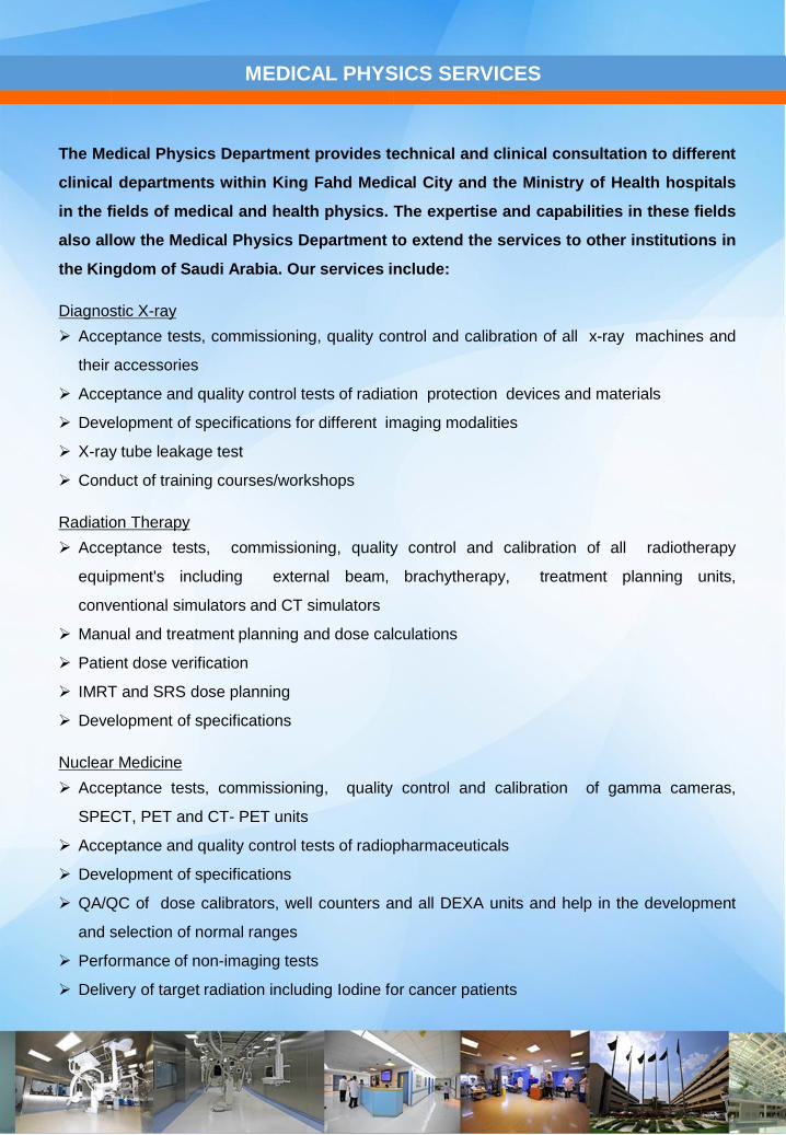
The Medical Physics Department provides technical and clinical consultation to different
clinical departments within King Fahd Medical City and the Ministry of Health hospitals
in the fields of medical and health physics. The expertise and capabilities in these fields
also allow the Medical Physics Department to extend the services to other institutions in
the Kingdom of Saudi Arabia. Our services include:
Diagnostic X-ray
Acceptance tests, commissioning, quality control and calibration of all x-ray machines and
their accessories
Acceptance and quality control tests of radiation protection devices and materials
Development of specifications for different imaging modalities
X-ray tube leakage test
Conduct of training courses/workshops
Radiation Therapy
Acceptance tests, commissioning, quality control and calibration of all radiotherapy
equipment's including external beam, brachytherapy, treatment planning units,
conventional simulators and CT simulators
Manual and treatment planning and dose calculations
Patient dose verification
IMRT and SRS dose planning
Development of specifications
Nuclear Medicine
Acceptance tests, commissioning, quality control and calibration of gamma cameras,
SPECT, PET and CT- PET units
Acceptance and quality control tests of radiopharmaceuticals
Development of specifications
QA/QC of dose calibrators, well counters and all DEXA units and help in the development
and selection of normal ranges
Performance of non-imaging tests
Delivery of target radiation including Iodine for cancer patients

NOTES
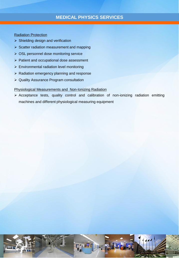
MEDICAL PHYSICS SERVICES
Radiation Protection
Shielding design and verification
Scatter radiation measurement and mapping
OSL personnel dose monitoring service
Patient and occupational dose assessment
Environmental radiation level monitoring
Radiation emergency planning and response
Quality Assurance Program consultation
Physiological Measurements and Non-Ionizing Radiation
Acceptance tests, quality control and calibration of non-ionizing radiation emitting
machines and different physiological measuring equipment

NOTES
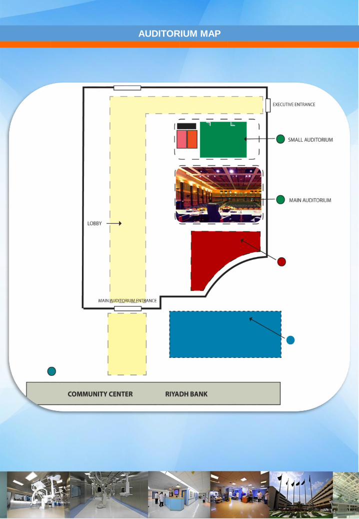
AUDITORIUM MAP
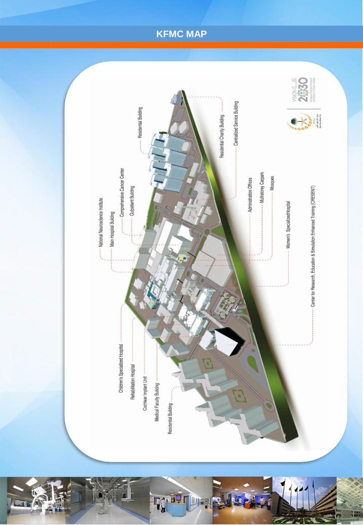
KFMC MAP
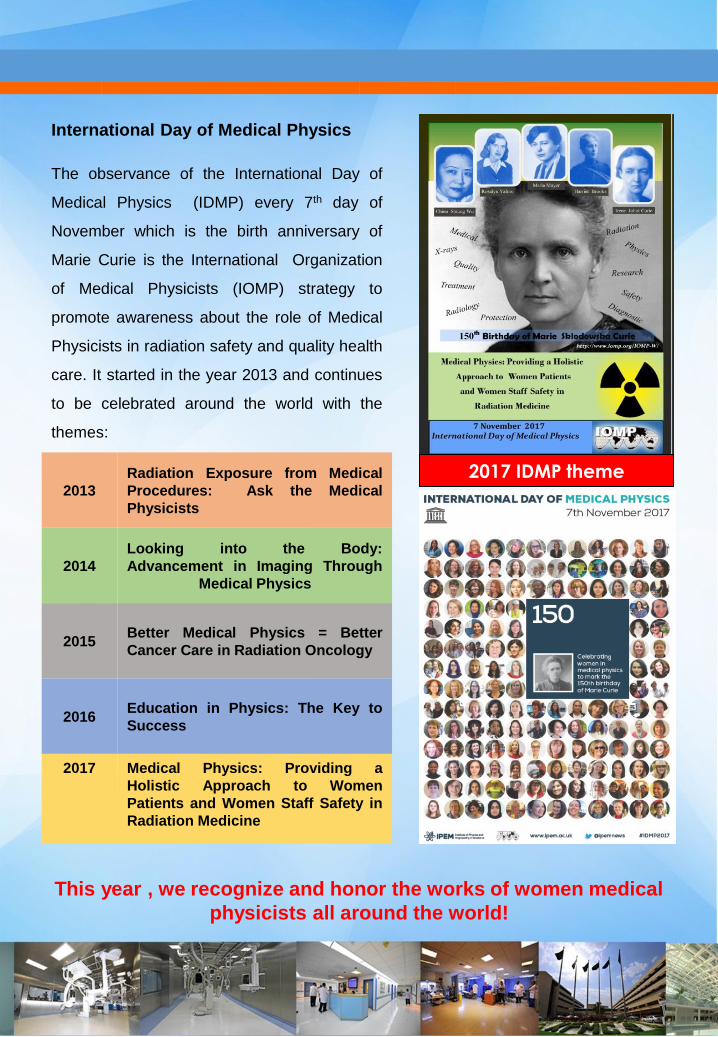
The observance of the International Day of
Medical Physics (IDMP) every 7th day of
November which is the birth anniversary of
Marie Curie is the International Organization
of Medical Physicists (IOMP) strategy to
promote awareness about the role of Medical
Physicists in radiation safety and quality health
care. It started in the year 2013 and continues
to be celebrated around the world with the
themes:
2017 IDMP theme
This year , we recognize and honor the works of women medical
physicists all around the world!
2013
Radiation Exposure from Medical
Procedures: Ask the Medical
Physicists
2014
Looking into the Body:
Advancement in Imaging Through
Medical Physics
2015Better Medical Physics = Better
Cancer Care in Radiation Oncology
2016Education in Physics: The Key to
Success
2017 Medical Physics: Providing a
Holistic Approach to Women
Patients and Women Staff Safety in
Radiation Medicine
International Day of Medical Physics