Embed Size (px)
Citation preview
The Egyptian Journal of Radiology and Nuclear Medicine (2016) 47, 297–304
Egyptian Society of Radiology and Nuclear Medicine
The Egyptian Journal of Radiology andNuclearMedicine
www.elsevier.com/locate/ejrnmwww.sciencedirect.com
ORIGINAL ARTICLE
Magnetic resonance imaging versus transcranial
ultrasound in early identification of cerebral injuries
in neonatal encephalopathy
* Corresponding author at: 81 Mohy Eldeen Abdel Hameed, 8th
District, Nasr City, Cairo, Egypt. Mobile: +20 1001798342.
E-mail addresses: [email protected] (E.A.Sh. Genedi),
[email protected] (N.M. Osman), drmarwa_eldeeb@yahoo.
com (M.T. El-deeb).
Peer review under responsibility of Egyptian Society of Radiology and
Nuclear Medicine.
http://dx.doi.org/10.1016/j.ejrnm.2016.01.0010378-603X � 2016 The Egyptian Society of Radiology and Nuclear Medicine. Production and hosting by Elsevier B.V.This is an open access article under the CC BY-NC-ND license (http://creativecommons.org/licenses/by-nc-nd/4.0/).
Eman A.Sh. Genedia, Noha Mohamed Osman
a,*, Marwa Talaat El-deebb
aDepartment of Radio-Diagnosis, Faculty of Medicine, Ain Shams University, Cairo, EgyptbDepartment of Pediatrics, Faculty of Medicine, Ain Shams University, Cairo, Egypt
Received 12 September 2015; accepted 3 January 2016
Available online 14 January 2016
KEYWORDS
Neonatal encephalopathy;
Hypoxic–ischemic
encephalopathy;
HIE;
MRI;
Transcranial ultrasound
Abstract Objective: Neonatal encephalopathy (NE) is a condition that causes significant morbid-
ity and mortality to the infant. The diagnosis and severity of NE rely heavily on clinical presenta-
tion and imaging findings.
The present study was planned to assess the role of MRI and Transcranial ultrasound (TCUS) in
the early identification of cerebral injuries in NE.
Patients and methods: Our study enrolled 38 newborns presented with NE. Brain MRI and TCUS
were carried out for each case and their results were compared.
Results: MRI was positive in 33 cases. Findings at MRI supported hypoxic-ischemic encephalopa-
thy as an etiology in 25 neonates, and other etiologies included metabolic disorders in 2, congenital
neonatal infection in 1, 2 cases of neonatal stroke, congenital brain anomalies in 2 neonates and
cerebral venous sinus thrombosis in 1. The overall diagnostic accuracy of TCUS compared to
MRI was 78.9%, while the overall sensitivity and specificity were 81.8% and 60% respectively.
Conclusion: TCUS is an effective screening tool in detecting the etiology of NE in suspected cases;
it is sometimes crucial in critically sick neonates; however, early MRI is mandatory as it can detect
precisely the extent of brain injury compared with TCUS alone.� 2016 The Egyptian Society of Radiology and Nuclear Medicine. Production and hosting by Elsevier
B.V. This is an open access article under the CC BY-NC-ND license (http://creativecommons.org/
licenses/by-nc-nd/4.0/).
1. Introduction
Neonatal encephalopathy (NE) is a heterogeneous, clinicallydefined syndrome characterized by disturbed neurological
function in the earliest days of life, manifested by feeding dif-ficulties, irritability, abnormality of tone, seizures, and reducedlevel of consciousness, and often accompanied by difficulty
with initiating and maintaining respiration (1). The terminol-ogy NE is preferred to Hypoxic Ischemic Encephalopathy
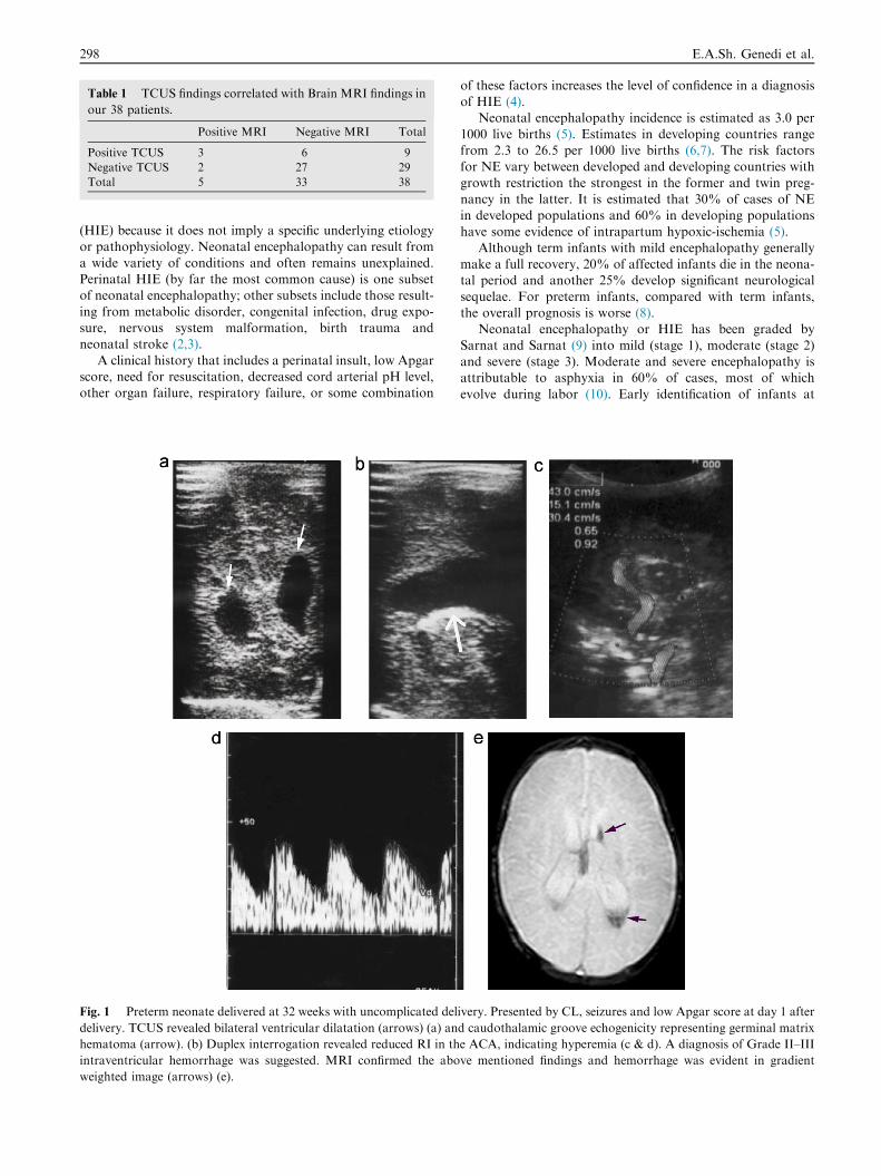
Table 1 TCUS findings correlated with Brain MRI findings in
our 38 patients.
Positive MRI Negative MRI Total
Positive TCUS 3 6 9
Negative TCUS 2 27 29
Total 5 33 38
298 E.A.Sh. Genedi et al.
(HIE) because it does not imply a specific underlying etiologyor pathophysiology. Neonatal encephalopathy can result from
a wide variety of conditions and often remains unexplained.Perinatal HIE (by far the most common cause) is one subsetof neonatal encephalopathy; other subsets include those result-
ing from metabolic disorder, congenital infection, drug expo-sure, nervous system malformation, birth trauma andneonatal stroke (2,3).
A clinical history that includes a perinatal insult, low Apgarscore, need for resuscitation, decreased cord arterial pH level,other organ failure, respiratory failure, or some combination
Fig. 1 Preterm neonate delivered at 32 weeks with uncomplicated del
delivery. TCUS revealed bilateral ventricular dilatation (arrows) (a) an
hematoma (arrow). (b) Duplex interrogation revealed reduced RI in th
intraventricular hemorrhage was suggested. MRI confirmed the abo
weighted image (arrows) (e).
of these factors increases the level of confidence in a diagnosisof HIE (4).
Neonatal encephalopathy incidence is estimated as 3.0 per
1000 live births (5). Estimates in developing countries rangefrom 2.3 to 26.5 per 1000 live births (6,7). The risk factorsfor NE vary between developed and developing countries with
growth restriction the strongest in the former and twin preg-nancy in the latter. It is estimated that 30% of cases of NEin developed populations and 60% in developing populations
have some evidence of intrapartum hypoxic-ischemia (5).Although term infants with mild encephalopathy generally
make a full recovery, 20% of affected infants die in the neona-tal period and another 25% develop significant neurological
sequelae. For preterm infants, compared with term infants,the overall prognosis is worse (8).
Neonatal encephalopathy or HIE has been graded by
Sarnat and Sarnat (9) into mild (stage 1), moderate (stage 2)and severe (stage 3). Moderate and severe encephalopathy isattributable to asphyxia in 60% of cases, most of which
evolve during labor (10). Early identification of infants at
ivery. Presented by CL, seizures and low Apgar score at day 1 after
d caudothalamic groove echogenicity representing germinal matrix
e ACA, indicating hyperemia (c & d). A diagnosis of Grade II–III
ve mentioned findings and hemorrhage was evident in gradient
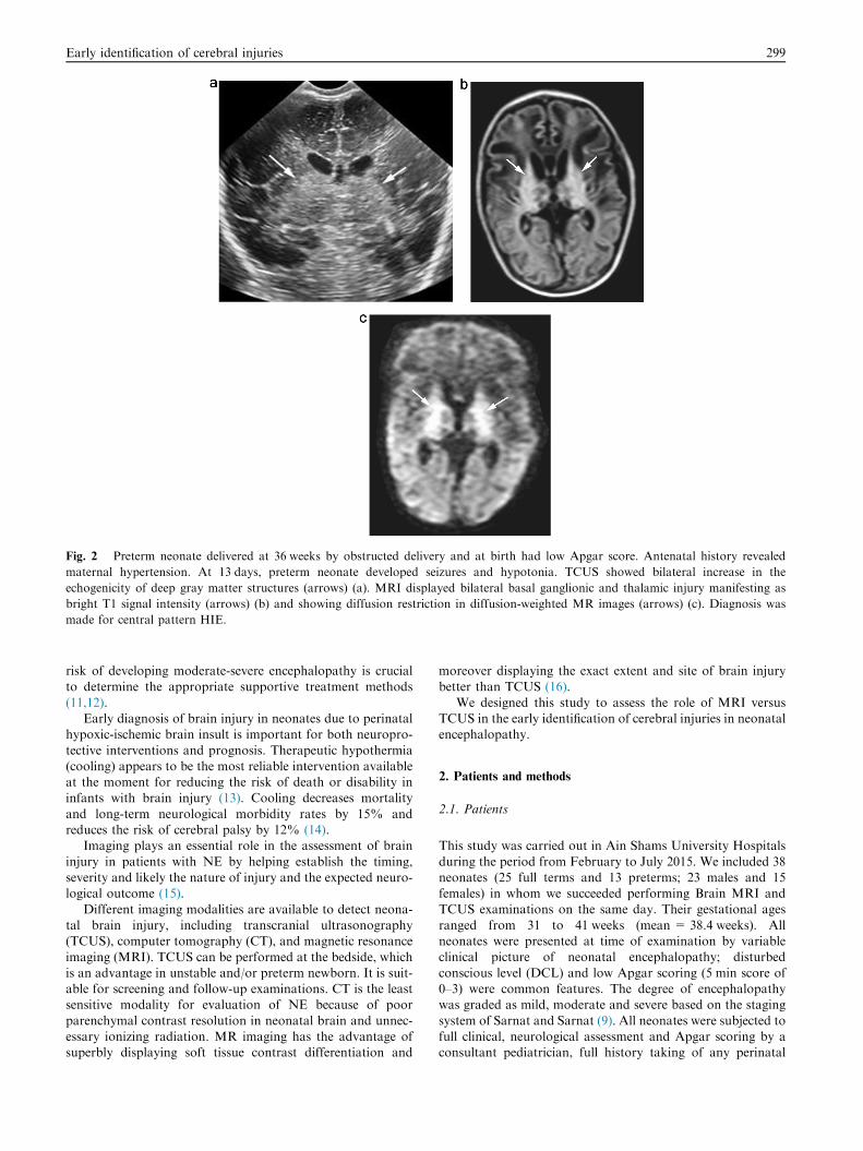
Fig. 2 Preterm neonate delivered at 36 weeks by obstructed delivery and at birth had low Apgar score. Antenatal history revealed
maternal hypertension. At 13 days, preterm neonate developed seizures and hypotonia. TCUS showed bilateral increase in the
echogenicity of deep gray matter structures (arrows) (a). MRI displayed bilateral basal ganglionic and thalamic injury manifesting as
bright T1 signal intensity (arrows) (b) and showing diffusion restriction in diffusion-weighted MR images (arrows) (c). Diagnosis was
made for central pattern HIE.
Early identification of cerebral injuries 299
risk of developing moderate-severe encephalopathy is crucialto determine the appropriate supportive treatment methods
(11,12).Early diagnosis of brain injury in neonates due to perinatal
hypoxic-ischemic brain insult is important for both neuropro-
tective interventions and prognosis. Therapeutic hypothermia(cooling) appears to be the most reliable intervention availableat the moment for reducing the risk of death or disability ininfants with brain injury (13). Cooling decreases mortality
and long-term neurological morbidity rates by 15% andreduces the risk of cerebral palsy by 12% (14).
Imaging plays an essential role in the assessment of brain
injury in patients with NE by helping establish the timing,severity and likely the nature of injury and the expected neuro-logical outcome (15).
Different imaging modalities are available to detect neona-tal brain injury, including transcranial ultrasonography(TCUS), computer tomography (CT), and magnetic resonance
imaging (MRI). TCUS can be performed at the bedside, whichis an advantage in unstable and/or preterm newborn. It is suit-able for screening and follow-up examinations. CT is the leastsensitive modality for evaluation of NE because of poor
parenchymal contrast resolution in neonatal brain and unnec-essary ionizing radiation. MR imaging has the advantage ofsuperbly displaying soft tissue contrast differentiation and
moreover displaying the exact extent and site of brain injurybetter than TCUS (16).
We designed this study to assess the role of MRI versusTCUS in the early identification of cerebral injuries in neonatalencephalopathy.
2. Patients and methods
2.1. Patients
This study was carried out in Ain Shams University Hospitals
during the period from February to July 2015. We included 38neonates (25 full terms and 13 preterms; 23 males and 15females) in whom we succeeded performing Brain MRI andTCUS examinations on the same day. Their gestational ages
ranged from 31 to 41 weeks (mean = 38.4 weeks). Allneonates were presented at time of examination by variableclinical picture of neonatal encephalopathy; disturbed
conscious level (DCL) and low Apgar scoring (5 min score of0–3) were common features. The degree of encephalopathywas graded as mild, moderate and severe based on the staging
system of Sarnat and Sarnat (9). All neonates were subjected tofull clinical, neurological assessment and Apgar scoring by aconsultant pediatrician, full history taking of any perinatal
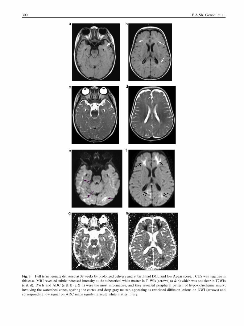
Fig. 3 Full term neonate delivered at 38 weeks by prolonged delivery and at birth had DCL and low Apgar score. TCUS was negative in
this case. MRI revealed subtle increased intensity at the subcortical white matter in T1WIs (arrows) (a & b) which was not clear in T2WIs
(c & d). DWIs and ADC (e & f) (g & h) were the most informative, and they revealed peripheral pattern of hypoxic/ischemic injury,
involving the watershed zones, sparing the cortex and deep gray matter, appearing as restricted diffusion lesions on DWI (arrows) and
corresponding low signal on ADC maps signifying acute white matter injury.
300 E.A.Sh. Genedi et al.
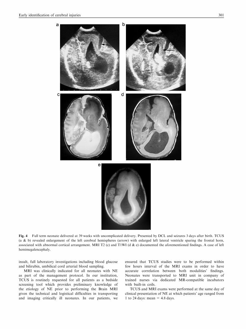
Fig. 4 Full term neonate delivered at 39 weeks with uncomplicated delivery. Presented by DCL and seizures 3 days after birth. TCUS
(a & b) revealed enlargement of the left cerebral hemispheres (arrow) with enlarged left lateral ventricle sparing the frontal horn,
associated with abnormal cortical arrangement. MRI T2 (c) and T1WI (d & e) documented the aforementioned findings. A case of left
hemimegalencephaly.
Early identification of cerebral injuries 301
insult, full laboratory investigations including blood glucoseand bilirubin, umbilical cord arterial blood sampling.
MRI was clinically indicated for all neonates with NE
as part of the management protocol. In our institution,TCUS is routinely requested for all patients as a bedsidescreening tool which provides preliminary knowledge ofthe etiology of NE prior to performing the Brain MRI
given the technical and logistical difficulties in transportingand imaging critically ill neonates. In our patients, we
ensured that TCUS studies were to be performed withinfew hours interval of the MRI exams in order to haveaccurate correlation between both modalities’ findings.
Neonates were transported to MRI unit in company oftrained nurses via dedicated MR-compatible incubatorswith built-in coils.
TCUS and MRI exams were performed at the same day of
clinical presentation of NE at which patients’ age ranged from1 to 24 days: mean = 4.8 days.
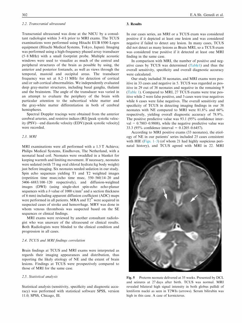
Fig. 5 Preterm neonate delivered at 35 weeks. Presented by DCL
and seizures at 27 days after birth. TCUS was normal. MRI
revealed bilateral high signal intensity in both globus pallidi of
lentiform nuclei as seen in T2WIs (arrows). Serum bilirubin was
high in this case. A case of kernicterus.
302 E.A.Sh. Genedi et al.
2.2. Transcranial ultrasound
Transcranial ultrasound was done at the NICU by a consul-tant radiologist within 3–4 h prior to MRI exams. The TCUSexaminations were performed using Hitachi EUB 8500 Logos
equipment (Hitachi Medical Systems, Tokyo, Japan). Imagingwas performed using a high-frequency phased array transducer(5–8 MHz) with a small footprint probe. Multiple acousticwindows were used to visualize as much of the central and
peripheral structures of the brain as possible by using, theanterior and posterior fontanels, as well as views through thetemporal, mastoid and occipital areas. The transducer
frequency was set at 8.2–11 MHz for detection of corticaland/or sub cortical abnormalities. We independently evaluateddeep gray-matter structures, including basal ganglia, thalami
and the brainstem. The angle of the transducer was varied inan attempt to evaluate the periphery of the brain withparticular attention to the subcortical white matter and
the gray-white matter differentiation in both of cerebralhemispheres.
Spectral Doppler tracings were obtained from the anteriorcerebral arteries, and resistive indices (RI) [peak systolic veloc-
ity (PSV)—end diastolic velocity (EDV)/peak systolic velocity]were recorded.
2.3. MRI
MRI examinations were all performed with a 1.5 T Achieva;Philips Medical Systems, Eindhoven, The Netherland, with a
neonatal head coil. Neonates were swaddled in a blanket forkeeping warmth and limiting movement. If necessary, neonateswere sedated (with 75 mg oral chloral hydrate/kg body weight)just before imaging. Six neonates needed sedation in our study.
Spin echo sequences yielding T1 and T2 weighted images(repetition time msec/echo time msec, 550–560/14–20 and5406–6883/100–120 respectively), and diffusion-weighted
images (DWI) (using single-shot spin-echo echo-planarsequences with a b value of 1000 s/mm2 and a section thicknessof 6 mm) including apparent diffusion coefficient (ADC) maps
were performed in all patients. MRA and T2* were acquired insuspected cases of stroke and hemorrhage. MRV was done inwhom venous thrombosis was suspected based on the SE
sequences or clinical findings.MRI exams were reviewed by another consultant radiolo-
gist who was unaware of the ultrasound or clinical results.Both Radiologists were blinded to the clinical condition and
progression in all cases.
2.4. TCUS and MRI findings correlation
Brain findings at TCUS and MRI exams were interpreted asregards their imaging appearances and distribution, thusreporting the likely etiology of NE and the extent of brain
lesions. Findings at TCUS were prospectively compared tothose of MRI for the same case.
2.5. Statistical analysis
Statistical analysis (sensitivity, specificity and diagnostic accu-
racy) was performed with statistical software SPSS, version11.0; SPSS, Chicago, Ill.
3. Results
In our cases series, an MRI or a TCUS exam was consideredpositive if it depicted at least one lesion and was considered
negative if failed to detect any lesion. In many cases, TCUSdid not detect as many lesions as Brain MRI, so a TCUS examwas considered true positive if it detected at least one MRI
finding in the same case.In comparison with MRI, the number of positive and neg-
ative cases by TCUS was determined (Table1) and thus theoverall sensitivity, specificity and overall diagnostic accuracy
were calculated.Our study included 38 neonates, and MRI exams were pos-
itive in 33 cases and negative in 5. TCUS was regarded as pos-
itive in 29 out of 38 neonates and negative in the remaining 9(Table 1). Compared to MRI, 27 TCUS exams were true pos-itive while 2 were false positive, and 3 cases were true negatives
while 6 cases were false negatives. The overall sensitivity andspecificity of TCUS in detecting imaging findings in our 38neonates with NE compared to MRI were 81.8% and 60%
respectively, yielding overall diagnostic accuracy of 78.9%.The positive predictive value was 93.1 (95% confidence inter-val = 0.7803–0.9808), while the negative predictive value was33.3 (95% confidence interval = 0.1205–0.6457).
According to MRI positive exams (33 neonates), the etiol-ogy of NE in our patients’ series included 25 cases consistentwith HIE (Figs. 1–3) (of whom 21 had highly suspicious peri-
natal history), and TCUS agreed with MRI in 22. MRI
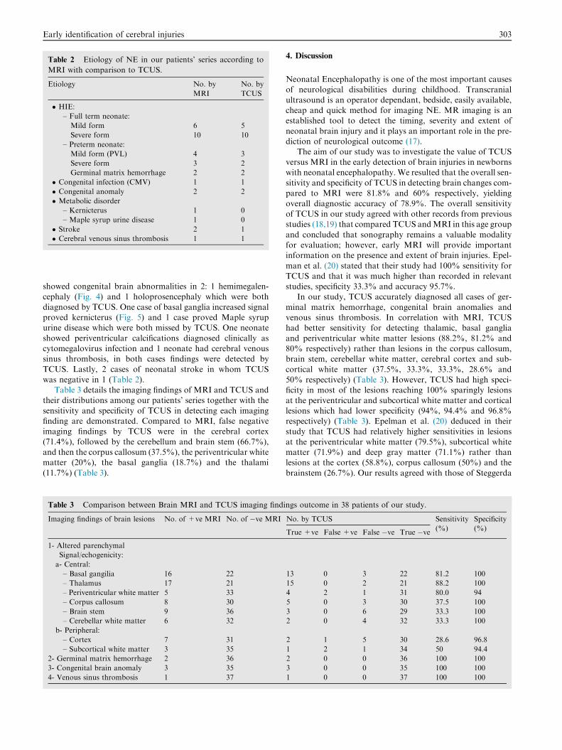
Table 2 Etiology of NE in our patients’ series according to
MRI with comparison to TCUS.
Etiology No. by
MRI
No. by
TCUS
� HIE:
– Full term neonate:
Mild form 6 5
Severe form 10 10
– Preterm neonate:
Mild form (PVL) 4 3
Severe form 3 2
Germinal matrix hemorrhage 2 2
� Congenital infection (CMV) 1 1
� Congenital anomaly 2 2
� Metabolic disorder
– Kernicterus 1 0
– Maple syrup urine disease 1 0
� Stroke 2 1
� Cerebral venous sinus thrombosis 1 1
Early identification of cerebral injuries 303
showed congenital brain abnormalities in 2: 1 hemimegalen-cephaly (Fig. 4) and 1 holoprosencephaly which were both
diagnosed by TCUS. One case of basal ganglia increased signalproved kernicterus (Fig. 5) and 1 case proved Maple syrupurine disease which were both missed by TCUS. One neonate
showed periventricular calcifications diagnosed clinically ascytomegalovirus infection and 1 neonate had cerebral venoussinus thrombosis, in both cases findings were detected by
TCUS. Lastly, 2 cases of neonatal stroke in whom TCUSwas negative in 1 (Table 2).
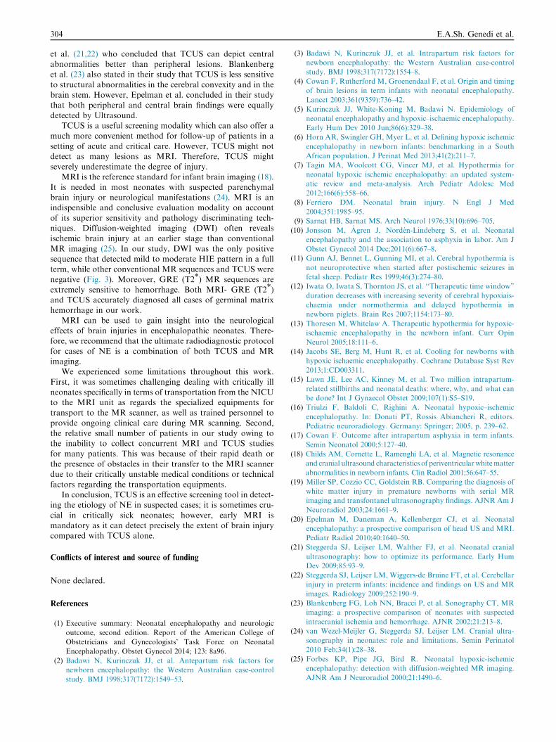
Table 3 details the imaging findings of MRI and TCUS andtheir distributions among our patients’ series together with the
sensitivity and specificity of TCUS in detecting each imagingfinding are demonstrated. Compared to MRI, false negativeimaging findings by TCUS were in the cerebral cortex
(71.4%), followed by the cerebellum and brain stem (66.7%),and then the corpus callosum (37.5%), the periventricular whitematter (20%), the basal ganglia (18.7%) and the thalami
(11.7%) (Table 3).
Table 3 Comparison between Brain MRI and TCUS imaging findi
Imaging findings of brain lesions No. of +ve MRI No. of �ve MRI
1- Altered parenchymal
Signal/echogenicity:
a- Central:
– Basal gangilia 16 22
– Thalamus 17 21
– Periventricular white matter 5 33
– Corpus callosum 8 30
– Brain stem 9 36
– Cerebellar white matter 6 32
b- Peripheral:
– Cortex 7 31
– Subcortical white matter 3 35
2- Germinal matrix hemorrhage 2 36
3- Congenital brain anomaly 3 35
4- Venous sinus thrombosis 1 37
4. Discussion
Neonatal Encephalopathy is one of the most important causesof neurological disabilities during childhood. Transcranial
ultrasound is an operator dependant, bedside, easily available,cheap and quick method for imaging NE. MR imaging is anestablished tool to detect the timing, severity and extent of
neonatal brain injury and it plays an important role in the pre-diction of neurological outcome (17).
The aim of our study was to investigate the value of TCUSversus MRI in the early detection of brain injuries in newborns
with neonatal encephalopathy. We resulted that the overall sen-sitivity and specificity of TCUS in detecting brain changes com-pared to MRI were 81.8% and 60% respectively, yielding
overall diagnostic accuracy of 78.9%. The overall sensitivityof TCUS in our study agreed with other records from previousstudies (18,19) that compared TCUS andMRI in this age group
and concluded that sonography remains a valuable modalityfor evaluation; however, early MRI will provide importantinformation on the presence and extent of brain injuries. Epel-
man et al. (20) stated that their study had 100% sensitivity forTCUS and that it was much higher than recorded in relevantstudies, specificity 33.3% and accuracy 95.7%.
In our study, TCUS accurately diagnosed all cases of ger-
minal matrix hemorrhage, congenital brain anomalies andvenous sinus thrombosis. In correlation with MRI, TCUShad better sensitivity for detecting thalamic, basal ganglia
and periventricular white matter lesions (88.2%, 81.2% and80% respectively) rather than lesions in the corpus callosum,brain stem, cerebellar white matter, cerebral cortex and sub-
cortical white matter (37.5%, 33.3%, 33.3%, 28.6% and50% respectively) (Table 3). However, TCUS had high speci-ficity in most of the lesions reaching 100% sparingly lesions
at the periventricular and subcortical white matter and corticallesions which had lower specificity (94%, 94.4% and 96.8%respectively) (Table 3). Epelman et al. (20) deduced in theirstudy that TCUS had relatively higher sensitivities in lesions
at the periventricular white matter (79.5%), subcortical whitematter (71.9%) and deep gray matter (71.1%) rather thanlesions at the cortex (58.8%), corpus callosum (50%) and the
brainstem (26.7%). Our results agreed with those of Steggerda
ngs outcome in 38 patients of our study.
No. by TCUS Sensitivity
(%)
Specificity
(%)True +ve False +ve False �ve True �ve
13 0 3 22 81.2 100
15 0 2 21 88.2 100
4 2 1 31 80.0 94
5 0 3 30 37.5 100
3 0 6 29 33.3 100
2 0 4 32 33.3 100
2 1 5 30 28.6 96.8
1 2 1 34 50 94.4
2 0 0 36 100 100
3 0 0 35 100 100
1 0 0 37 100 100
304 E.A.Sh. Genedi et al.
et al. (21,22) who concluded that TCUS can depict centralabnormalities better than peripheral lesions. Blankenberget al. (23) also stated in their study that TCUS is less sensitive
to structural abnormalities in the cerebral convexity and in thebrain stem. However, Epelman et al. concluded in their studythat both peripheral and central brain findings were equally
detected by Ultrasound.TCUS is a useful screening modality which can also offer a
much more convenient method for follow-up of patients in a
setting of acute and critical care. However, TCUS might notdetect as many lesions as MRI. Therefore, TCUS mightseverely underestimate the degree of injury.
MRI is the reference standard for infant brain imaging (18).
It is needed in most neonates with suspected parenchymalbrain injury or neurological manifestations (24). MRI is anindispensible and conclusive evaluation modality on account
of its superior sensitivity and pathology discriminating tech-niques. Diffusion-weighted imaging (DWI) often revealsischemic brain injury at an earlier stage than conventional
MR imaging (25). In our study, DWI was the only positivesequence that detected mild to moderate HIE pattern in a fullterm, while other conventional MR sequences and TCUS were
negative (Fig. 3). Moreover, GRE (T2*) MR sequences areextremely sensitive to hemorrhage. Both MRI- GRE (T2*)and TCUS accurately diagnosed all cases of germinal matrixhemorrhage in our work.
MRI can be used to gain insight into the neurologicaleffects of brain injuries in encephalopathic neonates. There-fore, we recommend that the ultimate radiodiagnostic protocol
for cases of NE is a combination of both TCUS and MRimaging.
We experienced some limitations throughout this work.
First, it was sometimes challenging dealing with critically illneonates specifically in terms of transportation from the NICUto the MRI unit as regards the specialized equipments for
transport to the MR scanner, as well as trained personnel toprovide ongoing clinical care during MR scanning. Second,the relative small number of patients in our study owing tothe inability to collect concurrent MRI and TCUS studies
for many patients. This was because of their rapid death orthe presence of obstacles in their transfer to the MRI scannerdue to their critically unstable medical conditions or technical
factors regarding the transportation equipments.In conclusion, TCUS is an effective screening tool in detect-
ing the etiology of NE in suspected cases; it is sometimes cru-
cial in critically sick neonates; however, early MRI ismandatory as it can detect precisely the extent of brain injurycompared with TCUS alone.
Conflicts of interest and source of funding
None declared.
References
(1) Executive summary: Neonatal encephalopathy and neurologic
outcome, second edition. Report of the American College of
Obstetricians and Gynecologists’ Task Force on Neonatal
Encephalopathy. Obstet Gynecol 2014; 123: 8a96.
(2) Badawi N, Kurinczuk JJ, et al. Antepartum risk factors for
newborn encephalopathy: the Western Australian case-control
study. BMJ 1998;317(7172):1549–53.
(3) Badawi N, Kurinczuk JJ, et al. Intrapartum risk factors for
newborn encephalopathy: the Western Australian case-control
study. BMJ 1998;317(7172):1554–8.
(4) Cowan F, Rutherford M, Groenendaal F, et al. Origin and timing
of brain lesions in term infants with neonatal encephalopathy.
Lancet 2003;361(9359):736–42.
(5) Kurinczuk JJ, White-Koning M, Badawi N. Epidemiology of
neonatal encephalopathy and hypoxic–ischaemic encephalopathy.
Early Hum Dev 2010 Jun;86(6):329–38.
(6) Horn AR, Swingler GH, Myer L, et al. Defining hypoxic ischemic
encephalopathy in newborn infants: benchmarking in a South
African population. J Perinat Med 2013;41(2):211–7.
(7) Tagin MA, Woolcott CG, Vincer MJ, et al. Hypothermia for
neonatal hypoxic ischemic encephalopathy: an updated system-
atic review and meta-analysis. Arch Pediatr Adolesc Med
2012;166(6):558–66.
(8) Ferriero DM. Neonatal brain injury. N Engl J Med
2004;351:1985–95.
(9) Sarnat HB, Sarnat MS. Arch Neurol 1976;33(10):696–705.
(10) Jonsson M, �Agren J, Norden-Lindeberg S, et al. Neonatal
encephalopathy and the association to asphyxia in labor. Am J
Obstet Gynecol 2014 Dec;2011(6):667–8.
(11) Gunn AJ, Bennet L, Gunning MI, et al. Cerebral hypothermia is
not neuroprotective when started after postischemic seizures in
fetal sheep. Pediatr Res 1999;46(3):274–80.
(12) Iwata O, Iwata S, Thornton JS, et al. ‘‘Therapeutic time window”
duration decreases with increasing severity of cerebral hypoxiais-
chaemia under normothermia and delayed hypothermia in
newborn piglets. Brain Res 2007;1154:173–80.
(13) Thoresen M, Whitelaw A. Therapeutic hypothermia for hypoxic-
ischaemic encephalopathy in the newborn infant. Curr Opin
Neurol 2005;18:111–6.
(14) Jacobs SE, Berg M, Hunt R, et al. Cooling for newborns with
hypoxic ischaemic encephalopathy. Cochrane Database Syst Rev
2013;1:CD003311.
(15) Lawn JE, Lee AC, Kinney M, et al. Two million intrapartum-
related stillbirths and neonatal deaths: where, why, and what can
be done? Int J Gynaecol Obstet 2009;107(1):S5–S19.
(16) Triulzi F, Baldoli C, Righini A. Neonatal hypoxic–ischemic
encephalopathy. In: Donati PT, Rossis Abiancheri R, editors.
Pediatric neuroradiology. Germany: Springer; 2005, p. 239–62.
(17) Cowan F. Outcome after intrapartum asphyxia in term infants.
Semin Neonatol 2000;5:127–40.
(18) Childs AM, Cornette L, Ramenghi LA, et al. Magnetic resonance
and cranial ultrasound characteristics of periventricular whitematter
abnormalities in newborn infants. Clin Radiol 2001;56:647–55.
(19) Miller SP, Cozzio CC, Goldstein RB. Comparing the diagnosis of
white matter injury in premature newborns with serial MR
imaging and transfontanel ultrasonography findings. AJNR Am J
Neuroradiol 2003;24:1661–9.
(20) Epelman M, Daneman A, Kellenberger CJ, et al. Neonatal
encephalopathy: a prospective comparison of head US and MRI.
Pediatr Radiol 2010;40:1640–50.
(21) Steggerda SJ, Leijser LM, Walther FJ, et al. Neonatal cranial
ultrasonography: how to optimize its performance. Early Hum
Dev 2009;85:93–9.
(22) Steggerda SJ, Leijser LM, Wiggers-de Bruine FT, et al. Cerebellar
injury in preterm infants: incidence and findings on US and MR
images. Radiology 2009;252:190–9.
(23) Blankenberg FG, Loh NN, Bracci P, et al. Sonography CT, MR
imaging: a prospective comparison of neonates with suspected
intracranial ischemia and hemorrhage. AJNR 2002;21:213–8.
(24) van Wezel-Meijler G, Steggerda SJ, Leijser LM. Cranial ultra-
sonography in neonates: role and limitations. Semin Perinatol
2010 Feb;34(1):28–38.
(25) Forbes KP, Pipe JG, Bird R. Neonatal hypoxic-ischemic
encephalopathy: detection with diffusion-weighted MR imaging.
AJNR Am J Neuroradiol 2000;21:1490–6.