Embed Size (px)
Citation preview
1
MACROPHAGE SKEWING BY PHD2 HAPLODEFICIENCY PREVENTS ISCHEMIA BY INDUCING ARTERIOGENESIS
Yukiji Takeda1,2,3,4, Sandra Costa1,2,5#, Estelle Delamarre1,2#, Carmen Roncal1,2,3,4,6#, Rodrigo
Leite De Oliveira1,2,3,4, Mario Leonardo Squadrito7, Veronica Finisguerra1,2, Françoise Bruyère3,4, Sofie Deschoemaeker1,2, Mathias Wenes1,2, Alexander Hamm1,2, Jens
Serneels1,2, Julie Magat8, Tapan Bhattacharrya9, Andrey Anisimov10, Benedicte F. Jordan8, Kari Alitalo10, Patrick Maxwell9, Bernard Gallez8, Zhenwu Zhuang11, Yoshihiko Saito12,
Michael Simons11, Michele De Palma5 & Massimiliano Mazzone1,2
1Lab of Molecular Oncology and Angiogenesis, Vesalius Research Center, VIB, Leuven, Belgium; 2Lab of Molecular Oncology and Angiogenesis, Vesalius Research Center, K.U.Leuven, Leuven, Belgium; 3Lab of
Angiogenesis and Neurovascular link, Vesalius Research Center, VIB, Leuven, Belgium; 4Lab of Angiogenesis and Neurovascular link, Vesalius Research Center, K.U.Leuven, Belgium; 5Life and Health Sciences Research Institute, Minho University, Braga, Portugal; 6Atherosclerosis Research Laboratory, CIMA-University of Navarra,
Pamplona, Spain; 7Angiogenesis and Tumor Targeting Unit, HSR-TIGET and Vita-Salute University, San Raffaele Institute, Milan, Italy; 8Biomedical Magnetic Resonance Unit, Medicinal Chemistry and Radiopharmacy
U.C.Louvain, Brussels, Belgium; 9Rayne Institute, University College London, London, UK; 10Molecular/Cancer Biology Laboratory, Research Programs Unit, Institute for Molecular Medicine, Helsinki, Finland; 11Cardiovascular
Medicine, Yale University, New Haven, CT, USA; 12The First Department of Internal Medicine, Nara Medical University, Nara, Japan; #These authors contributed equally to this work.
Editorial correspondence: Massimiliano Mazzone, Ph.D. Vesalius Research Center, VIB, KULeuven, Campus Gasthuisberg, Herestraat 49,
B-3000, Leuven, Belgium; Tel: +32-16-34.61.76; fax: +32-16-34.59.90
e-mail: [email protected]

2
SUMMARY
PHD2 serves as oxygen sensor that rescues blood supply by regulating vessel
formation and shape in case of oxygen shortage. However, it is unknown whether PHD2
can influence arteriogenesis. By using hindlimb ischemia as a model, we studied the role
of PHD2 in collateral artery growth, a process that compensates the lack of blood flow in
case of major arterial occlusion. Here, we show that PHD2+/- mice displayed preformed
collateral arteries that ameliorated limb perfusion and prevented ischemic necrosis. The
improved arteriogenesis in PHD2+/- mice was due to the expansion of circulating and
tissue-resident Tie2-expressing monocytes/macrophages (TEMs) and their enhanced
release of PDGFB and SDF1, resulting in increased smooth muscle cell recruitment and
growth. This program relied on activation of the canonical NF-κB pathway in PHD2+/-
mononuclear phagocytes. These results show how PHD2 controls blood flow and thus
tissue oxygenation by skewing the macrophage towards an arteriogenic phenotype in
ischemia.

3
INTRODUCTION
Vascular stenosis reduces blood supply resulting in ischemia, which causes tissue
dysfunction and demise. This condition is however associated with the formation of new
blood vessels (angiogenesis) and remodeling of preexisting collateral arterioles
(arteriogenesis) that reestablish blood flow to the downstream tissue1-3. Spontaneous
angiogenesis and arteriogenesis thus attenuate local tissue ischemia and improve the
clinical outcome of the disease2,4,5. Upon occlusion of an artery, the blood flow is
redirected into preexisting arteriolar anastomoses, causing enhanced shear stress on the
endothelium of the collateral circulation6-8. As a consequence, endothelial cells (ECs)
secrete VEGF, which induces the production of monocyte chemotactic protein-1
(CCL2/MCP1) from the endothelium itself and from adjacent smooth muscle cells (SMCs),
leading to monocyte recruitment2,7,9,10. Once in the periarteriolar region, monocyte-derived
macrophages produce growth factors that enhance the motility and proliferation of SMCs,
as well as proteases that digest the extracellular matrix to provide space for new SMCs2,8.
Recent studies have analyzed the functional plasticity of mononuclear phagocytes in
response to different environmental cues. For instance, in cancer and atherosclerosis,
macrophages generally display an “alternatively activated” (M2) phenotype11,12, which
enhances debris scavenging, angiogenesis, tissue remodeling, wound healing, and the
promotion of type II immunity. On the other hand, in inflamed tissues, macrophages
display a “classically activated” (M1) phenotype, which facilitates eradication of invading
microorganisms and the promotion of type I immune responses11,12. However,
macrophage heterogeneity during ischemia-induced arteriogenesis has not been
elucidated yet. Although initiation of arteriogenesis by macrophages takes place in a non-
hypoxic environment distant from the ischemic area13,14, some of the cytokines that
stimulate arteriogenesis are under the control of the prolyl hydroxylase domain protein
PHD215,16. PHD2 belongs to a larger family of proteins that utilize oxygen to hydroxylate
the hypoxia-inducible transcription factors (HIF)-1α and HIF-2α and, thereby, target the
latter for proteasomal degradation and hence inactivation17. In hypoxic conditions, PHDs
are inactive, which allows HIFs to become stabilized and mount an adaptive response to
hypoxia. Besides negatively regulating HIF accumulation, PHDs display a repressive role

4
in controlling the activity of NF-κB, a key signaling molecule for inflammation18. The control
of NF-κB by PHDs can be both dependent and independent of the hydroxylase activity
and therefore from the presence of oxygen used as a substrate15,19-21.
Pharmacological inhibition, silencing, or genetic inactivation of PHD2 after birth
stimulates angiogenesis and therapeutic revascularization through upregulation of
angiogenic factors from the parenchymal tissue16,22-24. Recently, we have shown that
heterozygous deficiency of PHD2 in ECs does not affect vessel density or lumen size, but
normalizes their endothelial lining, barrier, and stability, thus resulting in increased blood
vessel perfusion in tumors25. However, whether and how PHD2 plays a role in the
regulation of the arteriogenic process remains enigmatic. By using hindlimb ischemia as a
model of arteriogenesis, we found that reduced PHD2 levels in macrophages increases
the production of arteriogenic cytokines, including SDF1 and PDGFB, in an NF-κB
dependent manner. An increase of Tie2-expressing monocytes/macrophages (TEMs) in
the blood and tissues accounts for the superior arteriogenesis in PHD2 haplodeficient
mice. As a consequence of TEMs’ production of SDF1 and PDGFB, the remodeling of
collateral anastomoses is enhanced, thus conferring protection against ischemic damage.
Altogether, these data indicate that a reduction of PHD2 levels in monocytes/macrophages
unleashes NF-κB signals that skew their polarization towards an arteriogenic phenotype.
5
RESULTS
PHD2 HAPLODEFICIENCY PRESERVES TISSUE PERFUSION AND VIABILITY IN ISCHEMIA
We recently showed that stromal haplodeficiency of PHD2 increases tumor
perfusion25. Prompted by these results, we examined whether partial loss of PHD2 also
enhances perfusion of ischemic tissues. We therefore subjected mice to femoral artery
ligation, an established procedure that reduces perfusion of the lower limb and causes
ischemia in the calf (i.e. crural muscle). Laser-doppler measurements revealed that
perfusion of the lower hindlimb was higher in PHD2+/- than wild-type (WT) mice 12, 24 and
48 hours after femoral artery ligation, during the critical period when myofibers die if they
do not receive sufficient oxygen (Figure 1A). The increased perfusion in PHD2+/- mice
translated into enhanced physical endurance in ischemic conditions (12 hours post-
ligation), whereas both genotypes exhibited similar running capacity at baseline (Figure
1B). Quantification of oxygen levels in the calf by MRI-based oxymetry 12 hours after
ligation revealed that femoral occlusion induced a drop of oxygen tension by 66% in WT
and 46% in PHD2+/- mice (Figure 1C-E). Such differences in oxygen tension have been
shown to influence the outcome of the ischemic disease26. Staining for the hypoxia-marker
pimonidazole showed that the hypoxic area in the crural muscle of ligated limbs was 37.1
± 3.0% in WT mice but only 16.0 ± 7.0% in PHD2+/- mice (Figure 1H-J). Pimonidazole
staining of baseline WT and PHD2+/- crural muscles was negative (Figure 1F,G). In
accordance with findings that oxygen consumption in conditions of low oxygen availability
is associated with formation of reactive oxygen species (ROS), WT but not PHD2+/- crural
muscles at 12 hours post-ligation stained strongly for 8-hydroxy-2-deoxyguanosine (8-
OHdG), a marker of deoxyguanosine oxidation (Supplementary Figure 1C-E). At baseline,
oxidative stress in the crural muscle was comparable in both genotypes (Supplementary
Figure 1A,B,E). We next determined whether the decreased drop in perfusion and thus
oxygen tension in PHD2+/- ligated limbs prevented ischemic necrosis. Histological analysis
of the crural muscle (i.e. soleus) showed extensive ischemic damage in WT mice 72 hours
after ischemia (Figure 1K). In PHD2+/- mice, ischemic necrosis of the soleus was reduced
by more than 50% (Figure 1K-M). In accordance, crural muscle viability after ischemia was
almost double in PHD2+/- than in WT mice (Figure 1N-P). Compared to WT mice, muscle

6
fibers in PHD2+/- mice also showed fewer signs of regeneration as assessed by BrdU
staining, confirming that they were less damaged (Supplementary Figure 1F-H).
Upon femoral artery ligation, growth factors released by the ischemic crural muscle
promote angiogenesis1,27. Indeed, in WT mice, 14 days after femoral artery occlusion,
vessel density and total vessel area in near-completely regenerated regions of the soleus
(an oxidative unit of the crural muscle) were increased respectively by 33% and 70%
(Supplementary Figure 1I,J). In contrast, in PHD2+/- mice, these parameters remained
unchanged compared to the baseline, likely because these muscles never experienced
sufficient ischemia to stimulate angiogenesis (Supplementary Figure 1I,J).
We also wanted to assess whether PHD2+/- mice were protected against myocardial
ischemia and therefore performed ligation of the left anterior descending coronary artery of
WT and PHD2+/- hearts. The infarcted area was measured in desmin stained cross-
sections 24 hours after coronary ligation. Desmin-negative area (a readout of
cardiomyocyte death) was about 60% of the left ventricle in WT hearts and only 40% in
PHD2+/- hearts (Figure 1Q-S). Compared to WT, PHD2+/- hearts displayed higher perfusion
in both the infarcted and remote myocardium (Figure 1T-W).
Thus, PHD2 haplodeficiency greatly preserves perfusion and reduces tissue
damage in ischemia.
PHD2 HAPLODEFICIENCY ELICITS “COLLATERAL VESSEL PRECONDITIONING”
Since PHD2+/- muscles were protected against ischemic damage already 12 hours
after femoral artery ligation, we hypothesized that PHD2 haplodeficient mice were
preadapted to and therefore capable of better tolerating the ischemic insult. The number
and caliber of preexisting collaterals (primary, secondary, and tertiary branches) are major
determinants of the severity of tissue injury in occlusive diseases since these conduits
allow blood flow to bypass the obstruction2,4,5. We therefore investigated whether PHD2+/-
mice showed increased collaterals at baseline, independently of ischemia. Macroscopic
counting of collateral arteries on gelatin-bismuth angiographies in the thigh of non-
occluded limbs revealed a similar number of primary branches in both genotypes (Figure
2A,B). PHD2+/- mice however, had 1.7 and 2 fold more secondary and tertiary collateral
arteries, respectively (Figure 2A-D). Histological analysis of the adductor muscles (located

7
in the inner thigh, where collaterals form) showed that the total area and density of
bismuth-positive collaterals at baseline were respectively 2.0 and 2.3 fold higher in
PHD2+/- than WT mice (Figure 2E-H). In contrast, capillary density and area in the
adductor were comparable in both genotypes (Supplementary Figure 2A,B). Consistent
with these results, X-ray radiography (Figure 2I,J) and micro-CT scans showed a higher
number of large vessels (>200 µm in diameter) in PHD2+/- than WT thighs at baseline
(Figure 2K-M), whereas smaller vessels (<200 µm in diameter) were not changed
(Supplementary Figure 2C). Similar results were obtained in PHD2+/- hearts at baseline,
displaying a higher density of large vessels (Figure 2N-P), and a similar number of small
vessels and capillaries, compared to WT (Supplementary Figure 2D,E).
After femoral artery ligation, evaluation of gelatin-bismuth angiographies in WT
limbs showed a 30% induction of the collateral vascularization 12 and 72 hours post-
ischemia (Figure 2C,D). Conversely, in PHD2+/- mice the number of collaterals in the
adductor did not significantly change after occlusion, likely because they were already
expanded at baseline (Figure 2C,D). Nevertheless, there were still more secondary and
tertiary collateral branch arteries after ischemia in PHD2+/- than WT mice (Figure 2C,D).
Histological analysis confirmed that 12 and 72 hours post-ligation, the bismuth-positive
collateral area and density in adductor muscles were still higher in PHD2+/- than in WT
controls (Figure 2G,H).
To increase blood flow, collateral vessels undergo extensive remodeling
(arteriogenesis) and thus the tunica media, consisting of α-smooth muscle actin (αSMA)-
positive contractile SMCs, becomes thicker and the diameter of the conduit enlarges.
Staining of adductor tissue sections for αSMA revealed that number and total area of
αSMA+ collateral vessels were respectively 1.5 and 2 times increased in PHD2+/- muscles
at both baseline and after ischemia (Figure 2Q,R). However, the mean area of αSMA+
collaterals was higher in PHD2+/- than WT mice only at baseline, since, 72 hours post-
ligation, WT collaterals enlarged to the same size as PHD2+/- collaterals (Figure 2S,U-Y).
A similar trend was observed by measuring the thickness of the tunica media (Figure 2T).
These data show that, at baseline, the collateral vessels of PHD2+/- mice were similar to
those of WT mice after femoral artery ligation. This “collateral vessel preconditioning”
offered PHD2+/- mice a remarkable protection against lethal muscle ischemia.

8
PHD2+/- MACROPHAGES DISPLAY A SPECIFIC PHENOTYPE
Since inflammatory cells and in particular macrophages are known to produce
SMC/EC-mitogens, cytokines and proteases during collateral growth, we expected that the
increased collateralization in PHD2+/- muscles was due to enhanced infiltration of
leukocytes in response to HIF-mediated release of chemoattractant proteins28.
Surprisingly, when we measured the density of leukocytes and macrophages by staining
adductor tissue sections for CD45 and F4/80, respectively, there was no difference
between both genotypes at baseline (Figure 3A,B). Ligation of the femoral artery induced
a significant, but comparable increase in inflammatory cell accumulation in WT and
PHD2+/- adductors. Consistently, RNA levels of MCP1, one of the most important
proinflammatory cytokines in limb ischemia2,7,9,10, did not differ in the two genotypes either
at baseline or after femoral artery occlusion (Supplementary Figure 3A).
We therefore explored whether the phenotype, not the quantity, of the infiltrating
macrophages was different in PHD2+/- and WT mice. We measured the density of wound-
healing/proangiogenic macrophages, which can be identified by their enhanced
expression of the mannose receptor, MRC1/CD20629-31, and correspond to M2-polarized
macrophages11,12. Notably, costaining adductor sections for MRC1 and the macrophage
specific marker F4/80 revealed that the number of F4/80+MRC1+ cells was augmented by
75% at baseline conditions in PHD2+/- as compared to WT mice (Figure 3C-E). Seventy-
two hours after ligation, their numbers were increased by 95% in WT mice and by 54% in
PHD2+/- mice, but remained higher by 35% in ischemic PHD2+/- than WT mice (Figure
3C,F,G).
Prompted by these results, we gene-profiled WT and PHD2+/- macrophages
collected by peritoneal lavage (peritoneal macrophages, pMØ) and analyzed the
expression level of proangiogenic/proarteriogenic, proinflammatory, and antiangiogenic
genes. Remarkably, the genes that were upregulated in PHD2+/- macrophages were
markers of wound-healing/proangiogenic (i.e. M2-like) macrophages11,29 and comprised
Tie2, Arg1, CXCR4, CCR2, Nrp1, HGF, MMP2, FIZZ, CXCL12/SDF1, PDGFB, and TGFβ
(Figure 3H). Of note, most of these molecules have been reported to play an important
role during the arteriogenic process6,32-36. Conversely, several proinflammatory or
antiangiogenic (i.e. M1-type) molecules were downregulated in PHD2+/- macrophages;

9
these included IL1ß, IL6, NOS2, and IL12 (Figure 3H). The changes in the molecular
signature of macrophages were already detectable at baseline conditions, since F4/80+
cells freshly sorted from adductor muscles of PHD2+/- mice expressed higher levels of
PDGFB, SDF1, Tie2, MMP2, Nrp1 (Figure 3I). After 72 hours of ischemia, the expression
levels of these markers caught up in WT tissue macrophages (Figure 3I). Interestingly, in
ischemia, PHD2 levels were reduced by almost 50% in WT macrophages while they were
half in PHD2+/- macrophages both at baseline and after ischemia (versus WT
macrophages at baseline; Figure 3I). No differences were detected in WT and PHD2+/-
ECs, freshly isolated from adductor muscles at baseline and in ischemia (Supplementary
Table 1). Thus, PHD2+/- macrophages display a unique and cell specific gene signature,
which is reminiscent, at least in part, of M2-polarized macrophages11,29,30,37.
HETEROZYGOUS DEFICIENCY OF PHD2 IN MYELOID CELLS PREVENTS ISCHEMIC DAMAGE
To investigate whether reduced levels of macrophage-derived PHD2 displays
collateral vessel preconditioning and thus protection against ischemia, we generated
conditional PHD2 deficient mice lacking one or two PHD2 alleles specifically in myeloid
cells (PHD2LysCre;lox/wt and PHD2LysCre;lox/lox respectively) by intercrossing PHD2lox/wt and
PHD2lox/lox mice with LysM:Cre mice expressing the Cre-recombinase under the control of
the myeloid-specific lysozyme M promoter38. In contrast to PHD2 knockout mice, which die
between E12.5 and E14.5 due to placental defects25,39, mice with homozygous deficiency
of PHD2 in myeloid cells (PHD2LysCre;lox/lox) are viable and fertile. Gelatin-bismuth
angiographies revealed a higher number of secondary and tertiary collateral branch
arteries in heterozygous PHD2LysCre;lox/wt mice while arterialization was unchanged in
PHD2LysCre;lox/lox mice (Figure 4A,B). This was likely due to compensatory activity of PHD3
in PHD2LysCre;lox/lox macrophages (see below). Histological analysis of the same adductor
samples showed that the total area and density of bismuth positive collaterals were higher
in PHD2LysCre;lox/wt, but not in PHD2LysCre;lox/lox mice compared to control mice (Figure 4C,D).
Collateral vessel preconditioning conferred protection against ischemia since, 72 hours
after femoral artery occlusion, muscle necrosis was reduced by 67% in PHD2LysCre;lox/wt,
but not in PHD2LysCre;lox/lox mice (Figure 4E-H). Similarly, in ischemia, the running capacity
of PHD2LysCre;lox/wt, but not of PHD2LysCre;lox/lox mice was 1.6-fold higher compared to
PHD2LysCre;wt/wt mice while comparable at baseline (Figure 4I).

10
To further explore whether the increased arteriogenesis in PHD2 haplodeficient
mice could be attributed to the lack of one PHD2 allele in macrophages, we transplanted
WT or PHD2+/- (hereafter HE for “heterozygous”) bone marrow of syngenic mice,
ubiquitously expressing GFP, into lethally irradiated WT recipients (referred to as WTàWT
and HEàWT mice, respectively) or into lethally irradiated PHD2+/- recipients (referred to as
WTàHE and HEàHE mice, respectively). Collateral arteries were quantified 5 weeks
after bone marrow transplantation, when hematopoietic reconstitution by GFP+ blood cells
was, on average, 82 ± 8% and differential white blood counts were comparable in all the
groups (not shown). Histological analysis of gelatin-bismuth-based angiographies revealed
greater numbers and area of collateral vessels in HEàWT than WTàWT mice, while not
differing from HEàHE mice, supporting the key role of bone marrow derived cells in
enhancing collateralization (Figure 4J,K). Interestingly, collateral vessel parameters in
WTàWT and WTàHE mice were comparable (Figure 4J,K), indicating that bone marrow
derived cells are also important to sustain preexisting arteries in PHD2 heterozygous mice.
In accordance, ischemic necrosis at 72 hours post-ligation was prevented in HEàHE and
HEàWT mice, while it did not reach statistical significance in WTàHE mice (Figure 4L).
We also assessed whether transplantation of HE bone marrow into lethally irradiated WT
recipients would suffice to improve the physical endurance in ischemia. In a treadmill test,
the running capacity of HEàWT mice was twice as good as in WTàWT mice 12 hours
after femoral artery ligation while no differences were detected at baseline (Figure 4M).
Finally, we generated another strain lacking one PHD2 allele in both the
hematopoietic and endothelial lineage (PHD2Tie2Cre;lox/wt) by using the Tie2:Cre deleter
mouse line25,40. Reciprocal bone marrow transplantation of PHD2Tie2Cre;lox/wt and
PHD2Tie2Cre;wt/wt mice revealed that increased arteriogenesis of PHD2 heterozygous mice
was specifically caused by loss of one PHD2 allele in bone marrow derived hematopoietic
cells, but not in endothelial cells (Supplementary Table 2). Reduction of collateral
branches in PHD2Tie2Cre;lox/wt recipient mice transplanted with a WT bone marrow
(PHD2Tie2Cre;wt/wt) further supported the concept that PHD2 heterozygous macrophages are
required not only to trigger arteriogenesis but also to preserve existing arteries
(Supplementary Table 2). Deletion of one PHD2 allele selectively in ECs or SMCs did not
affect arteriogenesis (Supplementary Table 3). Thus, lower levels of PHD2 in bone marrow
derived myeloid cells, but not in ECs and/or SMCs, increase collateral vessel formation

11
and prevent ischemic damage.
MACROPHAGE-DERIVED SDF1 AND PDGFB PROMOTE ARTERIOGENESIS
In order to unravel the biological mechanism underlying the arteriogenic phenotype,
we assessed how WT and PHD2+/- macrophages affect the behavior of ECs and SMCs,
the two main cellular components of arteries. First, we evaluated the chemotactic potential
of primary ECs and SMCs towards WT and PHD2+/- macrophages. EC migration towards
WT or PHD2+/- macrophages was comparable and 50-times higher than towards culture
medium alone (Figure 5A-D). SMCs migrated only 6.5-times more efficiently when WT
macrophages were seeded in the lower chamber of the transwell (compared to control
medium), whereas migration towards PHD2+/- macrophages was 44-times higher (Figure
5E-H). Given the established role of SDF1 and PDGFB in the recruitment of SMCs and/or
SMC progenitors41-43 and the aforementioned finding that these two cytokines were
upregulated the highest in PHD2+/- macrophages (see Figure 3H), we tested whether
inhibiting these pathways, alone or in combination, would abrogate chemoattraction of
SMCs towards PHD2+/- macrophages. Combined inhibition of SDF1 and PDGFB signaling
by AMD3100 and imatinib, respectively, abrogated the increased migration of SMCs
towards PHD2+/- macrophages, while either treatment alone was not effective (Figure 5I).
Similarly, when silencing both SDF1 and PDGFB in PHD2+/- macrophages, SMC migration
was almost completely prevented, though each shRNA alone was already partly effective
(Supplementary Figure 4A and Supplementary Note 1).
To assess the influence of soluble factors released by WT and PHD2+/-
macrophages on EC and SMC growth, we performed a cell viability assay. We seeded
ECs or SMCs on the upper side of a 0.4 µm-pore filter (that does not allow cell migration
but only protein diffusion), and WT or PHD2+/- macrophages in the lower chamber.
Notably, growth of SMCs was enhanced by soluble factors released from PHD2+/- (versus
WT) macrophages (Figure 5J). EC growth was not differently affected by WT and PHD2+/-
macrophages (Figure 5K). SMCs display a proliferative (or synthetic) phenotype during the
phase of active growth in contrast to the contractile phenotype in mature vessels44. The
proliferative or synthetic phenotype is characterized by the reduction of contractile proteins
including smoothelin, NmMHC, αSMA, and of calponin family proteins, i.e. calponin-1 and

12
Sm22α44, 45. The down-modulation of these genes in SMCs indicates that these cells are
under the influence of growth factors and are able to migrate and to proliferate. Consistent
with the enhanced growth of SMCs seeded in the presence of PHD2+/- macrophages,
conditioned medium from PHD2+/- macrophages reduced the expression level of calponin-
1, SM22α, smoothelin, NmMHC and αSMA, therefore supporting a proliferative phenotype
(Figure 5L-P). Unlike what we observed in the migration assays, AMD3100 or imatinib
alone abrogated the increased SMC growth by PHD2+/- macrophages. The combination of
both AMD3100 and imatinib did not elicit an additive effect (Figure 5Q). Similarly, both
single and combined knockdown of SDF1 and PDGFB in PHD2+/- macrophages hindered
SMC growth (Supplementary Figure 4B).
Prompted by the in vitro results, we treated WTàWT and HEàWT mice with daily
administration of AMD3100 (5 mg/kg) or imatinib (50 mg/kg), alone or in combination. In
vivo, each drug alone only partially prevented the increased formation of second
generation collateral branches in HEàWT mice (Figure 5R), while third generation
collaterals were affected by either treatment alone (Figure 5S). However, the combination
of AMD3100 and imatinib more potently prevented collateralization in the adductor of
these mice. In WTàWT mice, the number of collateral branch arteries was not affected in
all conditions tested (Figure 5R,S). Thus, in mice with reduced level of myeloid PHD2,
combined PDGFB and SDF1 pathway activation is necessary to complete the arteriogenic
process.
MACROPHAGES PROMOTING ARTERIOGENESIS IN PHD2+/- MICE ARE TEMS
Tie2 is a gene recently found to be significantly upregulated in a subpopulation of
macrophages, known as TEMs, which express an M2-like, wound healing / proangiogenic
phenotype29-31,46. Although TEMs express genes that are commonly expressed by other
macrophage subsets (including PDGFB), they display greatly enhanced expression of
SDF129. Since Tie2 was strongly induced in PHD2+/- macrophages, we explored if this
increase was due to an enhanced fraction of TEMs in the total macrophage population. As
tumor TEMs express MRC1 to higher levels than classically activated macrophages /
inflammatory macrophages29,31 and because we found that PHD2+/- adductors display
enhanced infiltration of F4/80+MRC1+ macrophages (Figure 3C), we stained adductor

13
sections from WT and PHD2+/- mice for F4/80, MRC1 and Tie2 in order to rigorously
identify TEMs. At baseline, F4/80+MRC1+Tie2+ TEMs were scarce in WT mice but were 4-
times more abundant in PHD2+/- mice (Figure 6A). Seventy-two hours after femoral artery
occlusion, the density of TEMs was 3.2-times higher in WT but 1.3-fold increased in
PHD2+/- mice towards the baseline (Figure 6A). Thus, TEM density was still 1.6-fold higher
in ischemic PHD2+/- than WT mice (Figure 6A). The increased presence of tissue-resident
TEMs in PHD2+/- than WT mice was not due to a differential expression of the Tie2
ligands, angiopoietin-1 and angiopoietin-2, since transcript levels of these two cytokines
were similar in WT and PHD2+/- adductors at baseline and ischemic conditions
(Supplementary Figure 3B,C). When we measured Tie2-expressing monocytes (gated as
CD115+Tie2+ leukocytes) in the blood, we found a 3.4-fold higher TEM frequency in
PHD2+/- than WT mice at baseline (Figure 6B). Interestingly, 72 hours after femoral artery
ligation, the frequency of circulating TEMs was reduced by 3.4-fold in WT and 2.2-fold in
PHD2+/- mice, although this decrease reached statistical significance in WT mice only
(Figure 6B). Similar results were observed when quantifying the transcript levels of Tie2 in
WT and PHD2+/- CD115+ circulating monocytes, though the overall expression of Tie2 was
low (Figure 6C). In F4/80+ tissue macrophages, Tie2 transcripts were almost 100-times
higher than in monocytes. After ligation, Tie2 transcript levels were further augmented, but
only in WT macrophages, likely because PHD2+/- macrophages presented increased Tie2
expression already at baseline (Figure 6C). In mice, expression of Gr1 distinguishes
“inflammatory” monocytes (CD115+Gr1high) from “resident” monocytes
(CD115+Gr1low/neg)47,48. As previously reported29, circulating TEMs in PHD2+/- mice are
mostly CD115+Gr1low/neg (Supplementary Figure 5). Altogether, these data suggest that, in
ischemia, TEMs are recruited from the blood to the adductor where they trigger
arteriogenesis.
To address if TEMs are functionally involved in the maturation of collateral arteries
and thus preadaptation to ischemia in PHD2+/- mice, we used a 'suicide' gene strategy
based on the Herpes simplex virus (HSV) thymidine kinase (tk)-ganciclovir (GCV)
system49. We transplanted mice with WT or PHD2+/- bone marrow-derived lineage-
negative cells transduced with a lentiviral vector (LV) expressing the HSV-tk cDNA under
the control of the Tie2 promoter/enhancer (Tie2:tk-BMT mice; Supplementary Note 2). By
this approach, bone marrow-derived TEMs can be specifically eliminated upon GCV

14
administration in the transplanted mice (Supplementary Note 3). GCV-treated and
untreated mice were monitored for arterial growth at baseline as well as 3 and 7 days after
ligation by staining adductor sections for αSMA. GCV-untreated PHD2+/- Tie2:tk-BMT mice
displayed increased number of αSMA+ arteries and this density only slightly augmented at
3 and 7 days after ischemia. Remarkably, the arterial vessel preconditioning was
completely abolished in GCV-treated PHD2+/- Tie2:tk-BMT mice and thus protection
against ischemia was lost at both 3 and 7 days post-ligation (Figure 6D,E). In WT mice,
TEM depletion by GCV administration prevented ischemia-induced arteriogenesis and
consequent tissue healing 7 days post-ligation (Figure 6D,E). Thus, TEMs fuel
arteriogenesis in PHD2+/- mice at baseline and in WT mice during ischemia.
ACUTE DELETION OF PHD2 FAVORS TEMS, ARTERIOGENESIS AND ISCHEMIA
PROTECTION In order to strengthen the possible therapeutic value of our findings, we assessed
whether acute deletion of PHD2 in macrophages induced arteriogenesis and protection
against ischemia as observed in PHD2+/- mice. To this end, we generated tamoxifen-
inducible PHD2 haplodeficient mice (PHD2Rosa26CreERT;lox/wt) where the Rosa26 promoter
directs the ubiquitous expression of the fusion protein Cre-ERT2. Administration of 4-
hydroxytamoxifen to PHD2Rosa26CreERT;lox/wt peritoneal macrophages induced a 50%
reduction of PHD2 levels and increased the expression of PDGFB, SDF1, and Tie2,
therefore resembling the phenotype of PHD2+/- macrophages (Supplementary Figure 6A).
To address whether acute deletion of PHD2 in macrophage fuels arteriogenesis, bone
marrows from PHD2Rosa26CreERT;lox/wt mice were transplanted into lethally irradiated WT
recipient mice (HERosa26CreERTàWT). After five weeks, transplanted mice were treated with
vehicle or tamoxifen (1 mg/mouse for 5 days). Fourteen days after tamoxifen treatment,
circulating TEMs were almost 3-fold higher (Supplementary Figure 6B) and both
secondary and tertiary collateral branches were respectively 1.6 and 2.3 times more
abundant than in vehicle-treated mice (Supplementary Figure 6C). Consistent with an
increased arteriogenesis, ischemic damage in tamoxifen-treated HERosa26CreERTàWT mice
was greatly reduced (Supplementary Figure 6D). These data suggest that acute
inactivation of PHD2 might represent a preventive medicine for ischemic diseases.

15
HETEROZYGOUS DEFICIENCY OF PHD2 IN MACROPHAGES ENHANCES NF-κB ACTIVITY
PHD2 oxygen sensor negatively regulates HIF accumulation and NF-κB activity15,25.
When analyzing the accumulation of HIF-1α and HIF-2α by Western blot analysis, we
observed that the levels of HIF-1α and HIF-2α in PHD2 haplodeficient macrophages
(PHD2LysCre;lox/wt) were comparable to the control (PHD2LysCre;wt/wt). In contrast, HIF-1α and
HIF-2α levels in PHD2 null macrophages (PHD2LysCre;lox/lox) were respectively 4 and 2
times higher than in control macrophages (PHD2LysCre;wt/wt; Figure 6F). We therefore
quantified NF-κB activity by transducing PHD2LysCre;lox/wt, PHD2LysCre;lox/lox, PHD2LysCre;wt/wt
macrophages with a lentiviral vector carrying an NF-κB-responsive firefly luciferase
reporter (Figure 6G). Interestingly, NF-κB activity was increased by 65% in PHD2
haplodeficient macrophages but unaffected in PHD2 null macrophages.
We hypothesized that other PHD oxygen sensors might compensate for the
complete loss of PHD2. We therefore measured RNA levels of PHD1, PHD2, and PHD3 in
PHD2LysCre;wt/wt, PHD2LysCre;lox/wt and PHD2LysCre;lox/lox macrophages. While PHD2 levels
were decreased by 40% and 93% in PHD2LysCre;lox/wt and PHD2LysCre;lox/lox macrophages,
respectively, PHD1 and PHD3 transcript levels were 1.2 and 2.5 fold higher in PHD2
haplodeficient macrophages, and 1.3 and 12.2 fold higher in PHD2 null macrophages
(Supplementary Figure 7). PHD3 silencing induced NF-κB activity by 22% and 14% in
PHD2LysCre;wt/wt and PHD2LysCre;lox/wt macrophages but by 70% in PHD2LysCre;lox/lox
macrophages compared to their scramble controls (Figure 6G and Supplementary Note 4).
These data indicate that PHD3 induction in PHD2 null macrophages is responsible for the
repression of NF-κB activity. This may explain, at least in part, the absence of enhanced
collateral growth and ischemic protection in mice lacking two PHD2 alleles in myeloid
cells. To understand if hydroxylase function was necessary for PHD2 mediated NF-kB
regulation, PHD2+/- macrophages were electroporated with a plasmid carrying a wild type
PHD2 (PHD2wt) or a hydroxylase-deficient PHD2 containing a mutation in a critical residue
of the catalytic site (PHD2H313A)50. Ectopic expression of PHD2wt greatly blunted the
activity of NF-κB luciferase induced by PHD2 haplodeficiency, whereas PHD2H313A had no
effect (Figure 6H), suggesting a functional role of PHD2 hydroxylase activity in the
downregulation of NF-κB pathway. We also assessed the effect of TNF-α, archetypal

16
cytokine activating the canonical NF-κB pathway18, in WT and PHD2+/- macrophages and
found that TNF-α-induced NF-κB activation was significantly stronger in PHD2
haplodeficient macrophages (Figure 6I). In contrast, basal and TNF-α-induced NF-κB
activity were comparable in WT and PHD2+/- ECs (Supplementary Figure 8A). When
measuring the nuclear accumulation of NF-κB subunits, we found that the members of the
canonical pathway p65 (RelA) and p50 (NF-κB1) were more abundant in PHD2+/- than WT
macrophages (Figure 6J). Silencing of p65 or p50 blocked NF-κB hyperactivation in
PHD2+/- macrophages and the combined knockdown of both subunits restored NF-κB
function back to the WT levels (Figure 6K and Supplementary Note 5), thus highlighting
the prominent role of NF-κB p65/p50 heterodimers in macrophages.
To evaluate the involvement of the canonical NF-κB signaling in macrophage
skewing by PHD2 haplodeficiency, we generated a myeloid specific double transgenic
strain, heterozygous deficient for PHD2 and null for IKKβ, a positive regulator of NF-κB
canonical pathway. Disruption of NF-κB canonical pathway via genetic deletion of IKKβ
prevented the upregulation of Tie2, PDGFB and SDF1 in cultured PHD2 haplodeficient
macrophages (Figure 6L). Similar results were obtained by treating macrophages with the
NF-κB inhibitor 6-amino-4-(4-phenoxyphenylethylamino)quinazoline (Supplementary
Figure 8B). In vivo, genetic inactivation of IKKβ in myeloid cells abolished the enhanced
production of circulating TEMs (Figure 6M) and collateral vessel preconditioning induced
by myeloid PHD2 haplodeficiency (Figure 6N). Remarkably, myeloid specific IKKβ gene
targeting in WT mice greatly prevented ischemia-induced arteriogenesis, occuring 7 days
post-ligation (Figure 6N).
Thus, skewing of PHD2 haplodeficient macrophages towards an arteriogenic
phenotype relies on the activation of the NF-κB canonical pathway.
17
DISCUSSION
Specific macrophage subsets / differentiation states have been implicated in the
promotion of angiogenesis during cancer and atherosclerosis progression11,12. However,
little is known of the significance of macrophage heterogeneity in arteriogenesis and its
implications on ischemic diseases. This study identifies a role of myeloid PHD2 in oxygen
delivery by regulating arteriogenesis. Reduced PHD2 levels in macrophages determine a
specific gene signature that fosters the arteriogenic program by inducing recruitment and
growth of SMCs. This program relies on NF-κB-dependent upregulation of macrophage-
derived SDF1, PDGFB, and the angiopoietin receptor, Tie2. We show that the phenotype
of macrophages induced by reduced levels of myeloid PHD2 not only favors the formation
of new collateral branches, but is also important for collateral vessel homeostasis. Under
steady-state conditions, blood monocytes act as circulating precursors that migrate into
non-inflamed tissue to replace certain subsets of tissue macrophages51. PHD2
haplodeficient bone marrows in WT recipient mice enhanced collateral formation already
at baseline (“collateral vessel preconditioning”). However, when PHD2+/- mice were
transplanted with a WT bone marrow, preexisting collaterals regressed to the same level
as in WT mice, suggesting a role of tissue macrophages in sustaining artery maintenance.
The proarteriogenic tissue macrophages identified in the present study are reminiscent of
the M2-like, proangiogenic macrophage subset, known as TEMs, which are found in
tumors and developing or regenerating tissues29-31,49. The macrophages here described do
not upregulate either VEGF or inflammatory genes, but express increased levels of Tie2,
Nrp1, CXCR4, PDGFB and SDF1. Unlike tumor-associated TEMs, the cells described
here express high levels of the MCP1 receptor, CCR2. Tissue- and tumor-infiltrating TEMs
appear to originate from a distinct subset of circulating Tie2-expressing monocytes (our
data and 29). In agreement with previous studies49, Tie2-expressing monocytes as well as
Tie2-expressing macrophages were scarce in the peripheral blood and adductor of WT
mice, but were abundant in PHD2 haplodeficient mice in resting conditions or in the
pericollaterel region of WT mice after occlusion of the major arterial route. Their depletion
abrogated the arterial vessel preconditioning in PHD2+/- mice and prevented ischemia-
induced arteriogenesis in WT mice, supporting the role of TEMs in blood flow recovery
during occlusive diseases. The model we propose is described in Supplementary Figure 9.

18
After femoral artery ligation, a surge of chemoattractants recruits TEMs from the blood to
the pericollateral region. Here, TEMs fuel the tissue with SDF1 and PDGFB. The
combined activity of these two cytokines will induce SMC migration, positioning,
dedifferentiation, and growth, altogether resulting in artery maturation. PHD2
haplodeficiency unleashes constitutive NF-κB signals that result in a larger reservoir of
circulating and tissue-resident TEMs. Production of SDF1 and PDGFB by TEMs accounts
for the enhanced arteriogenesis at baseline and thus protection against ischemic tissue
demise. It remains an open question by which mechanism PHD2 activity / levels are
reduced in WT mononuclear phagocytes. In general, PHD2 needs oxygen as cosubstrate
and partly loses its activity under hypoxia. Furthermore, cytokine-driven downregulation of
PHD2 expression will also result in reduced enzymatic activity independent of oxygen
availability52,53. Here, we show that PHD2 hydroxylase function (and thus oxygen) is
required to repress NF-κB and that PHD2 levels are decreased in tissue macrophages
after ischemia. Thus, there might be two potentially different levels of regulation of
monocyte/macrophage polarization by PHD2. The first might occur in the bone marrow
where mean oxygen tension is normally 50 mmHg (ca. 7%)54. At this oxygen tension,
PHD2 activity will be partly inhibited in WT macrophages, to a similar extent as it occurs in
PHD2+/- macrophages25,55-57. After femoral artery occlusion, the bulk of blood flow is
redirected into collateral conduits, generating shear stress that causes the release of
different cytokines by the endothelium. Collateral formation is recognized as a hypoxia-
independent process13, 14. Nevertheless, stimulation by these cytokines might explain the
transcriptional repression of PHD2 measured in infiltrating WT macrophages (but not in
PHD2+/- macrophages that constitutively express half levels of PHD2) and thus strengthen
their arteriogenic response. Noteworthy, separate studies have reported that
angiopoietin(s) as well as TGFß reduce PHD2 expression (52,53 and M.M., unpublished),
and enhance collateral vascularization, in part through a direct effect on
monocytes/macrophages58-61. Overall, genetic deletion of one PHD2 allele preadapts
macrophages to ischemia and causes polarization towards an arteriogenic phenotype.
Genetic studies in mice on macrophage-associated cytokine receptors, or on
cytokines, have elucidated the importance of specific biological axes such as
SDF1/CXCR4, MCP1/CCR2, VEGF/Nrp1 and others, in arteriogenesis and post-ischemic
revascularization6, 32-36, 62-66. The population of macrophages described in the current

19
manuscript is enriched in Tie2, CXCR4, CCR2, and Nrp1 expression. By using Tie2 as a
marker to identify (and to deplete) a subset of monocytes/macrophages, we define for the
first time the role of PHD2 in macrophage skewing and unravel a new mechanism
whereby an oxygen sensor leads to blood recovery through collateral arteriogenesis,
fostered by M2-like macrophages. Further investigations will be needed to understand if
this model plays a role during developmental arteriogenesis, if the Tie2-expressing
population here described is similar to the one found in cancer, and if Tie2 expressed by
these cells has a functional role as in tumor-associated TEMs29-31. Recently, TEM-like
embryonic macrophages have been found to be involved in anastomosis of vessels during
development46. Given the fact that collateral vessels in the limb grow and mature from
preexisting anastomoses6, we cannot exclude that enhanced collateralization in PHD2+/-
mice is, at least in part, due to this process as well. Nevertheless, acute deletion of one
PHD2 allele in macrophages leads to almost complete protection against ischemic
necrosis by induction of arterial collateral branches, thus supporting the idea that
enhanced arteriogenesis by PHD2 haplodeficiency can promptly occur in adults.
Although different proarteriogenic molecules such as MMP2 are upregulated in
PHD2+/- macrophages, SDF1 and PDGFB were expressed more abundantly. Both
cytokines are potent chemoattractants for SMCs and/or SMC progenitors41-43. SDF1 more
specifically plays a key-role in recruiting, retaining and positioning CXCR4+ cells67,68. This
might be the case for SMCs and SMC progenitors, both positive for the SDF1 receptor
CXCR441,69, which can find their way towards collaterals by following a gradient of SDF1
released by pericollateral TEMs. PDGFB sustains recruitment and proliferation of SMCs
and SMC progenitors at the site of expression42. In our experiments, combined activation
of SDF1 and PDGFB results in the complete formation of collateral branches, suggesting
that in SMCs, these two pathways can converge to, at least in part, overlapping
downstream effectors. An open question is to understand whether, in PHD2+/-
monocytes/macrophages, the release of SDF1 and PDGFB are directly downstream of the
NF-κB pathway or are consequent to Tie2 signaling activation29.
TEMs are a subpopulation of alternatively activated (M2) macrophages. We show
here that macrophage skewing by PHD2 haplodeficiency is driven by the canonical NF-κB
pathway. The NF-κB family consists of 5 members: NF-κB1 (p105/p50), NF-κB2
(p100/p52), RelA (p65), RelB, and c-Rel18,70,71, which may form different homo- and

20
heterodimers associated with differential regulation of target genes. Activation of NF-κB
typically involves phosphorylation-dependent degradation of IκB inhibitors by the IκB
kinase (IKK) complex. This releases NF-κB and allows it to translocate freely to the
nucleus. HIF-prolyl hydroxylases are repressors of NF-κB activity, likely via their potential
to directly hydroxylate IKKβ20,21. Alternatively, PHD3 has been also shown to associate
with IKKβ independently of its hydroxylase function, thereby blocking further interaction
between IKKβ and the chaperone Hsp90, which is required for IKKβ phosphorylation and
release of NF-κB19. Thus, the hydroxylase function of PHD oxygen sensors can be either
necessary or dispensable for the downregulation of NF-κB depending on the cellular
context (our data and 15). By different means, we prove that PHD2 haplodeficiency results
in the hyperactivation of the canonical NF-κB pathway in macrophages via accumulation of
p65 and p50 subunits. Genetic or pharmacological inhibition of the NF-κB pathway
prevents the upregulation of Tie2, PDGFB, and SDF1 in cultured PHD2 haplodeficient
macrophages. In vivo, genetic inactivation of myeloid IKKβ inhibits TEM production and
collateral vessel preconditioning induced by PHD2 haplodeficiency. Previous evidence has
shown that IKKβ suppresses the M1 phenotype and promotes M2 macrophage skewing
through positive regulation of the canonical NF-κB pathway72-74. From a molecular point of
view, the main downstream effectors of the canonical NF-κB pathway are p65 and p50,
existing mostly as heterodimers. Homodimers of the p50 subunit of NF-κB, which lack
transactivation domains, can repress expression of NF-κB target genes and inhibit
inflammation, whereas the homodimers of p65 as well as the p65/p50 heterodimers are
responsible for NF-κB-mediated gene transcription18,70,71. Consistent with a role of PHD
oxygen sensors in the negative regulation of IKKβ, we show that PHD2 haplodeficiency
triggers p65 and p50 accumulation and thus leads to the repression of M1 genes and
reinforcement of M2 genes. This is in line with the above-cited literature showing
repression of M2-markers and increased expression of M1-markers in IKKβ deficient
macrophages72-74. Another study shows that deficiency, not accumulation, of p50 in bone
marrow cells prompts macrophage infiltration and elicits arteriogenesis75. At first glance,
this might appear in conflict with our findings. Nevertheless, the same study reports that
p50 ablation favors a (compensatory) p65 accumulation. Thus, both the elimination of
negative (p50-mediated) NF-κB breaking cues and endorsement of the (p65-mediated)

21
NF-κB transactivating potential will tilt the balance towards a positive regulation of the NF-
κB canonical pathway and enhanced macrophage-initiated arteriogenesis. Interestingly,
complete deletion of PHD2 in macrophages elicits a potent induction of PHD3 levels that
compensate for the absence of PHD2 and thus reestablish the activity of NF-κB to the WT
levels. In contrast, a mild induction of PHD3 in PHD2 haplodeficient macrophages does
not counterbalance the positive effect of PHD2 downmodulation on NF-κB pathway.
Therefore, consistent with previous findings76, feedback loops involving PHD3 oxygen
sensor tune the genetic program triggered by PHD2 inactivation. As a consequence,
heterozygous but not homozygous inactivation of PHD2 is able to mount a safety program
in myeloid cells through NF-κB activation that enhances collateralization and thus prevents
ischemic necrosis.
In our previous work, we show that deficiency or inhibition of the oxygen sensor
PHD1, belonging to the same family as PHD2, induces hypoxia tolerance by
reprogramming basal metabolism towards glycolysis, that allows anaerobic ATP
production in ischemia77. This phenotype was specific for PHD1-/- mice but not for PHD2+/-
and PHD3-/- mice77. However, while the previous investigation was carried out in a mixed
background (Swiss/129), the current study was performed in a BalbC and 129S6
background. It is known that strain-related genetic differences profoundly affect the
formation of collateral vasculature and thus the outcome of ischemia after femoral artery
ligation78. Indeed, collateral formation in PHD2+/- mice and ischemic necrosis in a
Swiss/129 background were comparable to their littermate controls (data not shown). Also,
in any of the PHD2+/- strains analyzed, the levels of the peroxisome proliferator-activated
receptor α (PPARα) and the pyruvate dehydrogenase kinases PDK1 and PDK4 (induced
in PHD1-/- fibers and driving the metabolic reprogramming at baseline) were not affected77,
supporting a different mechanism of protection against ischemia in PHD2+/- mice.
Finally, our findings have potential medical implications. Previous studies have
shown that unspecific inhibitors of PHD2 or silencing of PHD2 promotes therapeutic
revascularization against ischemia16,22-24. However, this approach can have some
limitations. First, angiogenesis is a late response; therefore, organ function might be
compromised until new vessel formation is complete. In contrast, arteriogenesis takes
place on preexisting vascular shunts and this process is actually the first to be triggered in

22
case of ischemia6. Second, the generation of PHD2-specific inhibitors will be challenging
due to the high homology of the catalytic pocket of the three PHD family members (PHD1,
PHD2 and PHD3)17. Overall, a cell-based therapy with PHD2 hypomorphic macrophages
or Tie2-expressing macrophages might promote collateral vascularization in patients at
risk of ischemic damage, i.e. diabetic or hypercholesterolemic patients79; similar results
may be obtained by combined administration of SDF1 and PDGFB. At this stage, our
study provides insight into how PHD2 oxygen sensor regulates arteriogenesis via
controlling a specific monocyte / macrophage phenotype and thus guarantees oxygen
delivery in case of shortage, as it occurs during ischemia.

23
METHODS SUMMARY
129/S6 or Balb/C WT and PHD2+/- mice (8-12 weeks old) were obtained from our mouse
facility. PHD2+/- and PHD2 conditional knockout mice were obtained as previously
described25. To induce hind limb ischemia, unilateral or bilateral ligations of the femoral
artery and vein and the cutaneous vessels branching from the caudal femoral artery side
branch were performed without damaging the nervus femoralis80. Oxgen tension (pO2) in
the lower limb was measured 12 hours after femoral artery ligation by using 19F-MRI
oximetry. Adductor crural muscles were dissected, fixed in 2% PFA, dehydrated,
embedded in paraffin and sectioned at 7µm thickness for histology, immunostaining and
morphometry analysis. Macrophages were either harvested from the peritoneal cavity of
the mice (peritoneal macrophages (pMØ)) or derived from bone marrow precursors as
described before81. Balb/c WT and PHD2+/- recipient mice were irradiated with 7.5 Gy.
Subsequently, 5x10⁶ bone marrow cells from green fluorescent protein+ (GFP+) WT or
GFP+ PHD2+/- mice were injected intravenously via the tail vein. Femoral artery ligation,
treadmill running test and bismuth angiography were performed 6 weeks after bone
marrow reconstitution. Full Methods and any associated references are available in the
Supplementary Information.

24
REFERENCES
1. Carmeliet, P. Mechanisms of angiogenesis and arteriogenesis. Nat Med 6, 389-95 (2000).
2. Schirmer, S.H., van Nooijen, F.C., Piek, J.J. & van Royen, N. Stimulation of collateral artery growth: travelling further down the road to clinical application. Heart 95, 191-7 (2009).
3. Simons, M. & Ware, J.A. Therapeutic angiogenesis in cardiovascular disease. Nat Rev Drug Discov 2, 863-71 (2003).
4. Yu, J. et al. Endothelial nitric oxide synthase is critical for ischemic remodeling, mural cell recruitment, and blood flow reserve. Proc Natl Acad Sci U S A 102, 10999-1004 (2005).
5. Schultz, A. et al. Interindividual heterogeneity in the hypoxic regulation of VEGF: significance for the development of the coronary artery collateral circulation. Circulation 100, 547-52 (1999).
6. Schaper, W. Collateral circulation: past and present. Basic Res Cardiol 104, 5-21 (2009).
7. van Royen, N. et al. Effects of local MCP-1 protein therapy on the development of the collateral circulation and atherosclerosis in Watanabe hyperlipidemic rabbits. Cardiovasc Res 57, 178-85 (2003).
8. Heil, M. & Schaper, W. Influence of mechanical, cellular, and molecular factors on collateral artery growth (arteriogenesis). Circ Res 95, 449-58 (2004).
9. Heil, M. et al. Blood monocyte concentration is critical for enhancement of collateral artery growth. Am J Physiol Heart Circ Physiol 283, H2411-9 (2002).
10. Ito, W.D. et al. Monocyte chemotactic protein-1 increases collateral and peripheral conductance after femoral artery occlusion. Circ Res 80, 829-37 (1997).
11. Mantovani, A. & Sica, A. Macrophages, innate immunity and cancer: balance, tolerance, and diversity. Curr Opin Immunol 22, 231-7.
12. Mantovani, A., Garlanda, C. & Locati, M. Macrophage diversity and polarization in atherosclerosis: a question of balance. Arterioscler Thromb Vasc Biol 29, 1419-23 (2009).
13. Ito, W.D. et al. Angiogenesis but not collateral growth is associated with ischemia after femoral artery occlusion. Am J Physiol 273, H1255-65 (1997).
14. Gray, C. et al. Ischemia is not required for arteriogenesis in zebrafish embryos. Arterioscler Thromb Vasc Biol 27, 2135-41 (2007).
15. Chan, D.A. et al. Tumor vasculature is regulated by PHD2-mediated angiogenesis and bone marrow-derived cell recruitment. Cancer Cell 15, 527-38 (2009).
16. Loinard, C. et al. Inhibition of prolyl hydroxylase domain proteins promotes therapeutic revascularization. Circulation 120, 50-9 (2009).

25
17. Fraisl, P., Aragones, J. & Carmeliet, P. Inhibition of oxygen sensors as a therapeutic strategy for ischaemic and inflammatory disease. Nat Rev Drug Discov 8, 139-52 (2009).
18. Lawrence, T. The nuclear factor NF-kappaB pathway in inflammation. Cold Spring Harb Perspect Biol 1, a001651 (2009).
19. Xue, J. et al. Prolyl hydroxylase-3 is down-regulated in colorectal cancer cells and inhibits IKKbeta independent of hydroxylase activity. Gastroenterology 138, 606-15.
20. Cummins, E.P. et al. Prolyl hydroxylase-1 negatively regulates IkappaB kinase-beta, giving insight into hypoxia-induced NFkappaB activity. Proc Natl Acad Sci U S A 103, 18154-9 (2006).
21. Fu, J. & Taubman, M.B. Prolyl hydroxylase EGLN3 regulates skeletal myoblast differentiation through an NF-kappaB-dependent pathway. J Biol Chem 285, 8927-35.
22. Milkiewicz, M., Pugh, C.W. & Egginton, S. Inhibition of endogenous HIF inactivation induces angiogenesis in ischaemic skeletal muscles of mice. J Physiol 560, 21-6 (2004).
23. Nangaku, M. et al. A novel class of prolyl hydroxylase inhibitors induces angiogenesis and exerts organ protection against ischemia. Arterioscler Thromb Vasc Biol 27, 2548-54 (2007).
24. Huang, M. et al. Short hairpin RNA interference therapy for ischemic heart disease. Circulation 118, S226-33 (2008).
25. Mazzone, M. et al. Heterozygous deficiency of PHD2 restores tumor oxygenation and inhibits metastasis via endothelial normalization. Cell 136, 839-51 (2009).
26. Aranguren, X.L. et al. MAPC transplantation confers a more durable benefit than AC133+ cell transplantation. Cell Transplant.
27. Pugh, C.W. & Ratcliffe, P.J. Regulation of angiogenesis by hypoxia: role of the HIF system. Nat Med 9, 677-84 (2003).
28. Patel, T.H., Kimura, H., Weiss, C.R., Semenza, G.L. & Hofmann, L.V. Constitutively active HIF-1alpha improves perfusion and arterial remodeling in an endovascular model of limb ischemia. Cardiovasc Res 68, 144-54 (2005).
29. Pucci, F. et al. A distinguishing gene signature shared by tumor-infiltrating Tie2-expressing monocytes, blood "resident" monocytes, and embryonic macrophages suggests common functions and developmental relationships. Blood 114, 901-14 (2009).
30. De Palma, M. et al. Tie2 identifies a hematopoietic lineage of proangiogenic monocytes required for tumor vessel formation and a mesenchymal population of pericyte progenitors. Cancer Cell 8, 211-26 (2005).
31. Mazzieri, R. et al. Targeting the ANG2/TIE2 Axis Inhibits Tumor Growth and Metastasis by Impairing Angiogenesis and Disabling Rebounds of Proangiogenic Myeloid Cells. Cancer Cell 19, 512-26.

26
32. Nickerson, M.M. et al. Bone marrow-derived cell-specific chemokine (C-C motif) receptor-2 expression is required for arteriolar remodeling. Arterioscler Thromb Vasc Biol 29, 1794-801 (2009).
33. Cochain, C. et al. Regulation of monocyte subset systemic levels by distinct chemokine receptors controls post-ischaemic neovascularization. Cardiovasc Res 88, 186-95.
34. Heil, M. et al. Collateral artery growth (arteriogenesis) after experimental arterial occlusion is impaired in mice lacking CC-chemokine receptor-2. Circ Res 94, 671-7 (2004).
35. Fujii, T. et al. Nonendothelial mesenchymal cell-derived MCP-1 is required for FGF-2-mediated therapeutic neovascularization: critical role of the inflammatory/arteriogenic pathway. Arterioscler Thromb Vasc Biol 26, 2483-9 (2006).
36. Zacchigna, S. et al. Bone marrow cells recruited through the neuropilin-1 receptor promote arterial formation at the sites of adult neoangiogenesis in mice. J Clin Invest 118, 2062-75 (2008).
37. Qian, B.Z. & Pollard, J.W. Macrophage diversity enhances tumor progression and metastasis. Cell 141, 39-51.
38. Clausen, B.E., Burkhardt, C., Reith, W., Renkawitz, R. & Forster, I. Conditional gene targeting in macrophages and granulocytes using LysMcre mice. Transgenic Res 8, 265-77 (1999).
39. Takeda, K. et al. Placental but not heart defects are associated with elevated hypoxia-inducible factor alpha levels in mice lacking prolyl hydroxylase domain protein 2. Mol Cell Biol 26, 8336-46 (2006).
40. El Kasmi, K.C. et al. Toll-like receptor-induced arginase 1 in macrophages thwarts effective immunity against intracellular pathogens. Nat Immunol 9, 1399-406 (2008).
41. Karshovska, E., Zagorac, D., Zernecke, A., Weber, C. & Schober, A. A small molecule CXCR4 antagonist inhibits neointima formation and smooth muscle progenitor cell mobilization after arterial injury. J Thromb Haemost 6, 1812-5 (2008).
42. Hellstrom, M., Kalen, M., Lindahl, P., Abramsson, A. & Betsholtz, C. Role of PDGF-B and PDGFR-beta in recruitment of vascular smooth muscle cells and pericytes during embryonic blood vessel formation in the mouse. Development 126, 3047-55 (1999).
43. Cao, R. et al. Angiogenic synergism, vascular stability and improvement of hind-limb ischemia by a combination of PDGF-BB and FGF-2. Nat Med 9, 604-13 (2003).
44. Wolf, C. et al. Vascular remodeling and altered protein expression during growth of coronary collateral arteries. J Mol Cell Cardiol 30, 2291-305 (1998).
45. Kumar, M.S. & Owens, G.K. Combinatorial control of smooth muscle-specific gene expression. Arterioscler Thromb Vasc Biol 23, 737-47 (2003).
46. Fantin, A. et al. Tissue macrophages act as cellular chaperones for vascular anastomosis downstream of VEGF-mediated endothelial tip cell induction. Blood.

27
47. Sunderkotter, C. et al. Subpopulations of mouse blood monocytes differ in maturation stage and inflammatory response. J Immunol 172, 4410-7 (2004).
48. Geissmann, F. et al. Blood monocytes: distinct subsets, how they relate to dendritic cells, and their possible roles in the regulation of T-cell responses. Immunol Cell Biol 86, 398-408 (2008).
49. De Palma, M., Venneri, M.A., Roca, C. & Naldini, L. Targeting exogenous genes to tumor angiogenesis by transplantation of genetically modified hematopoietic stem cells. Nat Med 9, 789-95 (2003).
50. Jokilehto, T. et al. Retention of prolyl hydroxylase PHD2 in the cytoplasm prevents PHD2-induced anchorage-independent carcinoma cell growth. Exp Cell Res 316, 1169-78.
51. Geissmann, F., Jung, S. & Littman, D.R. Blood monocytes consist of two principal subsets with distinct migratory properties. Immunity 19, 71-82 (2003).
52. Chen, J.X. & Stinnett, A. Ang-1 gene therapy inhibits hypoxia-inducible factor-1alpha (HIF-1alpha)-prolyl-4-hydroxylase-2, stabilizes HIF-1alpha expression, and normalizes immature vasculature in db/db mice. Diabetes 57, 3335-43 (2008).
53. McMahon, S., Charbonneau, M., Grandmont, S., Richard, D.E. & Dubois, C.M. Transforming growth factor beta1 induces hypoxia-inducible factor-1 stabilization through selective inhibition of PHD2 expression. J Biol Chem 281, 24171-81 (2006).
54. Harrison, J.S., Rameshwar, P., Chang, V. & Bandari, P. Oxygen saturation in the bone marrow of healthy volunteers. Blood 99, 394 (2002).
55. Chan, D.A., Sutphin, P.D., Yen, S.E. & Giaccia, A.J. Coordinate regulation of the oxygen-dependent degradation domains of hypoxia-inducible factor 1 alpha. Mol Cell Biol 25, 6415-26 (2005).
56. Pan, Y. et al. Multiple factors affecting cellular redox status and energy metabolism modulate hypoxia-inducible factor prolyl hydroxylase activity in vivo and in vitro. Mol Cell Biol 27, 912-25 (2007).
57. Epstein, A.C. et al. C. elegans EGL-9 and mammalian homologs define a family of dioxygenases that regulate HIF by prolyl hydroxylation. Cell 107, 43-54 (2001).
58. van Royen, N. et al. Exogenous application of transforming growth factor beta 1 stimulates arteriogenesis in the peripheral circulation. FASEB J 16, 432-4 (2002).
59. Kobayashi, K. et al. Combination of in vivo angiopoietin-1 gene transfer and autologous bone marrow cell implantation for functional therapeutic angiogenesis. Arterioscler Thromb Vasc Biol 26, 1465-72 (2006).
60. Shyu, K.G., Manor, O., Magner, M., Yancopoulos, G.D. & Isner, J.M. Direct intramuscular injection of plasmid DNA encoding angiopoietin-1 but not angiopoietin-2 augments revascularization in the rabbit ischemic hindlimb. Circulation 98, 2081-7 (1998).
61. Tressel, S.L. et al. Angiopoietin-2 stimulates blood flow recovery after femoral artery occlusion by inducing inflammation and arteriogenesis. Arterioscler Thromb Vasc Biol 28, 1989-95 (2008).

28
62. Zhou, J. et al. CXCR3-dependent accumulation and activation of perivascular macrophages is necessary for homeostatic arterial remodeling to hemodynamic stresses. J Exp Med 207, 1951-66.
63. Jin, D.K. et al. Cytokine-mediated deployment of SDF-1 induces revascularization through recruitment of CXCR4+ hemangiocytes. Nat Med 12, 557-67 (2006).
64. Seeger, F.H. et al. CXCR4 expression determines functional activity of bone marrow-derived mononuclear cells for therapeutic neovascularization in acute ischemia. Arterioscler Thromb Vasc Biol 29, 1802-9 (2009).
65. Yla-Herttuala, S. & Alitalo, K. Gene transfer as a tool to induce therapeutic vascular growth. Nat Med 9, 694-701 (2003).
66. Seiler, C. et al. Promotion of collateral growth by granulocyte-macrophage colony-stimulating factor in patients with coronary artery disease: a randomized, double-blind, placebo-controlled study. Circulation 104, 2012-7 (2001).
67. Grunewald, M. et al. VEGF-induced adult neovascularization: recruitment, retention, and role of accessory cells. Cell 124, 175-89 (2006).
68. Bagri, A. et al. The chemokine SDF1 regulates migration of dentate granule cells. Development 129, 4249-60 (2002).
69. Zernecke, A. et al. SDF-1alpha/CXCR4 axis is instrumental in neointimal hyperplasia and recruitment of smooth muscle progenitor cells. Circ Res 96, 784-91 (2005).
70. Li, Q. & Verma, I.M. NF-kappaB regulation in the immune system. Nat Rev Immunol 2, 725-34 (2002).
71. Perkins, N.D. Integrating cell-signalling pathways with NF-kappaB and IKK function. Nat Rev Mol Cell Biol 8, 49-62 (2007).
72. Fong, C.H. et al. An antiinflammatory role for IKKbeta through the inhibition of "classical" macrophage activation. J Exp Med 205, 1269-76 (2008).
73. Hagemann, T. et al. "Re-educating" tumor-associated macrophages by targeting NF-kappaB. J Exp Med 205, 1261-8 (2008).
74. Greten, F.R. et al. NF-kappaB is a negative regulator of IL-1beta secretion as revealed by genetic and pharmacological inhibition of IKKbeta. Cell 130, 918-31 (2007).
75. de Groot, D., Haverslag, R.T., Pasterkamp, G., de Kleijn, D.P. & Hoefer, I.E. Targeted deletion of the inhibitory NF-kappaB p50 subunit in bone marrow-derived cells improves collateral growth after arterial occlusion. Cardiovasc Res 88, 179-85.
76. Moslehi, J. et al. Loss of Hypoxia-Inducible Factor Prolyl Hydroxylase Activity in Cardiomyocytes Phenocopies Ischemic Cardiomyopathy. Circulation (2010).
77. Aragones, J. et al. Deficiency or inhibition of oxygen sensor Phd1 induces hypoxia tolerance by reprogramming basal metabolism. Nat Genet 40, 170-80 (2008).
78. Helisch, A. et al. Impact of mouse strain differences in innate hindlimb collateral vasculature. Arterioscler Thromb Vasc Biol 26, 520-6 (2006).

29
79. Sacco, R.L. Risk factors and outcomes for ischemic stroke. Neurology 45, S10-4 (1995).
80. Luttun A, T.M., Moons L, Wu Y, Angelillo-Scherrer A, Liao F, Nagy JA, Hooper A, Priller J, De Klerck B, Compernolle V, Daci E, Bohlen P, Dewerchin M, Herbert JM, Fava R, Matthys P, Carmeliet G, Collen D, Dvorak HF, Hicklin DJ, Carmeliet P. Revascularization of ischemic tissues by PlGF treatment, and inhibition of tumor angiogenesis, arthritis and atherosclerosis by anti-Flt1. Nat Med 8, 831-840 (2002).
81. Meerpohl, H.G., Lohmann-Matthes, M.L. & Fischer, H. Studies on the activation of mouse bone marrow-derived macrophages by the macrophage cytotoxicity factor (MCF). Eur J Immunol 6, 213-7 (1976).

30
SUPPLEMENTARY INFORMATION (SI)
Supplementary Information is linked to the main manuscript.
A figure (Supplementary Figure 9) summarizing the main result of this paper is also
included as SI.
31
ACKNOWLEDGEMENTS
The authors thank Y. Jonsson, T. Janssens, A. Bouché, A. Carton, A. Manderveld, B.
Vanwetswinkel and N. Dai for technical assistance. This work was supported by grants
from FWO (G.0726.10), Belgium, and from VIB. The authors are thankful to Dr. P.
Carmeliet for scientific discussion and support. VE-Cadherin:CreERT and PDGFRB:Cre
transgenic mice were generated at the Cancer Research UK (London, UK) and kindly
donated by Dr. R. Adams. The IKKβ floxed mice are a generous gift of Dr. M. Karin
(UCSD, La Jolla, CA). The hydroxylase-deficient PHD2 construct was given by Dr. P.
Ratcliffe (Oxford, UK). ED was granted by ARC, SC by FCT, RLO and VF by FWO, AH by
DFG. CR was supported by COST action TD0901. MDP was supported by an ERC
starting grant.

32
AUTHOR INFORMATION
AFFILIATIONS
Vesalius Research Center, VIB, KUL, Leuven, Belgium
Y. Takeda, S. Costa, E. Delamarre, C. Roncal, R. Leite De Oliveira, V. Finisguerra, P. F.
Bruyère, S. Deschoemaeker, M. Wenes, A. Hamm, J. Serneels & M. Mazzone
Life and Health Sciences Research Institute, Minho University, Braga, Portugal
S. Costa
Atherosclerosis Research Laboratory, CIMA-University of Navarra, Pamplona, Spain
C. Roncal
Angiogenesis and Tumor Targeting Unit, HSR-TIGET and Vita-Salute University, San
Raffaele Institute, Milan, Italy
M.L. Squadrito & M. De Palma
Biomedical Magnetic Resonance Unit, Medicinal Chemistry and Radiopharmacy
U.C.Louvain, Brussels, Belgium
J. Magat, B.F. Jordan & B. Gallez
Rayne Institute, University College London, London, UK
T. Bhattacharrya & Patrick Maxwell
Cardiovascular Medicine, Yale University, New Haven, CT, USA
Z. Zhuang & M. Simons
Molecular/Cancer Biology Laboratory, Research Programs Unit, Institute for Molecular
Medicine, Helsinki, Finland
A. Anisimov & K. Alitalo
The First Department of Internal Medicine, Nara Medical University, Nara, Japan
Y. Saito

33
CONTRIBUTIONS
Y.T., E.D. and S.C. performed experimental design, all experiments, acquisition of data
and analysis and interpretation of all data. C.R. performed analysis of histological
stainings, angiographies. R.L.O., C.R and S.C. performed the western blots. R.L.O and
V.F performed treadmill-running tests, RT-PCR experiments and drug administrations.
M.L.S. performed bone marrow derived, lineage negative hematopoietic cells isolation and
transduction and vector copy number analysis. F.B. performed EC isolation and
angiography measurements. J.M., B.F.J. and B.G. performed oxymetry experiments. S.D.
performed luciferase assays and management of the colonies. M.W. and A.H performed
transplantation experiments and electroporations. Y.T. and J.S. performed the ligations of
the femoral artery. Z.Z and M.S. performed micro-CT angiograms. A.A. and K.A. have
contributed with viral vector productions. T.B. and P.M. contributed to the generation of the
PHD2 targeting vector. Y.T., E.D., S.C., C.R., Y.S. and M.D.P. participated in scientific
discussion and drafting of the manuscript. M.M. performed experimental design, analysis
of data, conducted scientific direction, wrote manuscript.
COMPETING FINANCIAL INTERESTS
No competing financial interests to declare.
CORRESPONDING AUTHOR
Correspondence to: M. Mazzone ([email protected])
34
FIGURE LEGENDS
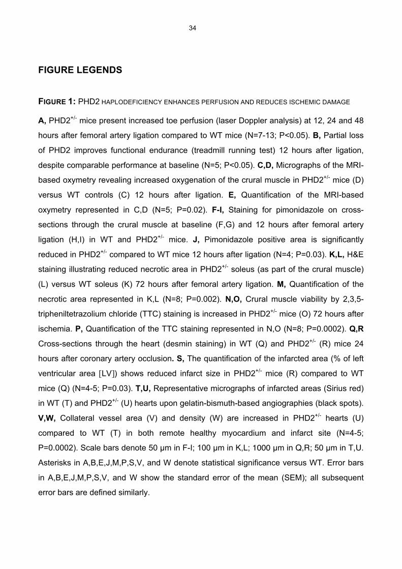
FIGURE 1: PHD2 HAPLODEFICIENCY ENHANCES PERFUSION AND REDUCES ISCHEMIC DAMAGE
A, PHD2+/- mice present increased toe perfusion (laser Doppler analysis) at 12, 24 and 48
hours after femoral artery ligation compared to WT mice (N=7-13; P<0.05). B, Partial loss
of PHD2 improves functional endurance (treadmill running test) 12 hours after ligation,
despite comparable performance at baseline (N=5; P<0.05). C,D, Micrographs of the MRI-
based oxymetry revealing increased oxygenation of the crural muscle in PHD2+/- mice (D)
versus WT controls (C) 12 hours after ligation. E, Quantification of the MRI-based
oxymetry represented in C,D (N=5; P=0.02). F-I, Staining for pimonidazole on cross-
sections through the crural muscle at baseline (F,G) and 12 hours after femoral artery
ligation (H,I) in WT and PHD2+/- mice. J, Pimonidazole positive area is significantly
reduced in PHD2+/- compared to WT mice 12 hours after ligation (N=4; P=0.03). K,L, H&E
staining illustrating reduced necrotic area in PHD2+/- soleus (as part of the crural muscle)
(L) versus WT soleus (K) 72 hours after femoral artery ligation. M, Quantification of the
necrotic area represented in K,L (N=8; P=0.002). N,O, Crural muscle viability by 2,3,5-
tripheniltetrazolium chloride (TTC) staining is increased in PHD2+/- mice (O) 72 hours after
ischemia. P, Quantification of the TTC staining represented in N,O (N=8; P=0.0002). Q,R
Cross-sections through the heart (desmin staining) in WT (Q) and PHD2+/- (R) mice 24
hours after coronary artery occlusion. S, The quantification of the infarcted area (% of left
ventricular area [LV]) shows reduced infarct size in PHD2+/- mice (R) compared to WT
mice (Q) (N=4-5; P=0.03). T,U, Representative micrographs of infarcted areas (Sirius red)
in WT (T) and PHD2+/- (U) hearts upon gelatin-bismuth-based angiographies (black spots).
V,W, Collateral vessel area (V) and density (W) are increased in PHD2+/- hearts (U)
compared to WT (T) in both remote healthy myocardium and infarct site (N=4-5;
P=0.0002). Scale bars denote 50 µm in F-I; 100 µm in K,L; 1000 µm in Q,R; 50 µm in T,U.
Asterisks in A,B,E,J,M,P,S,V, and W denote statistical significance versus WT. Error bars
in A,B,E,J,M,P,S,V, and W show the standard error of the mean (SEM); all subsequent
error bars are defined similarly.

35
FIGURE 2: ENHANCED COLLATERALIZATION IN PHD2+/- MUSCLES
A,B, Macroscopic view of gelatin-bismuth-based angiographies at baseline. PHD2+/-
adductors (B) show comparable primary (blue arrow) and enhanced secondary (red arrow)
and tertiary (black arrow) collateral vessels compared to WT (A) mice. The green arrow
points to the femoral artery. C,D, PHD2+/- mice present increased number of secondary
(C) and tertiary (D) collateral vessels, both at baseline and after ischemia (12 and 72
hours post-ischemia) (N=6-11; P<0.05). E,F, H&E staining of adductor sections after
gelatin-bismuth-based angiography. Bismuth+ collaterals appear black. G,H, PHD2
haplodeficient mice present increased collateral vessel area (G) and density (H) compared
to WT mice (N=8-14; P<0.01). I,J, Increased number of collaterals in PHD2+/- hindlimbs
evaluated by X-ray radiography. K-M, Quantification of the micro-CT angiograms of hind
limbs at baseline (K) showing increased number of large vessels (>200 µm in diameter) in
the thigh of PHD2+/- (M) versus WT (L) mice (N=6; P=0.04). N-P, Quantification of micro-
CT angiograms at baseline (N) showing increased number of large vessels (>200 µm in
diameter) in PHD2+/- hearts (P) versus WT (O) hearts (N=6; P=0.04). Q-T, Morphometric
analysis of α-smooth muscle actin (αSMA) collateral vessels in non-occluded and
occluded adductor muscles of WT and PHD2+/- mice: Q, Density of αSMA+ collateral
vessels (N=12; P<0.04); R, Total αSMA+ collateral area (N=12; P<0.05); S, Mean αSMA+
collateral vessel area (N=12; P<0.05). T, Thickness of the tunica media (N=8; P=0.04). U-
Y, Staining of adductor sections for αSMA showing increased caliber and tunica media
thickness of PHD2+/- collateral arteries at baseline; 72 hours after ligation, WT collaterals
enlarge to the same size and thickness of PHD2+/- collaterals. Scale bars denote 50 µm in
E,F and 10 µm in U,V,W,Y. Asterisks in C,D,G,H,K,N,Q,R,S, and T denote statistical
significance versus WT. Hash signs in C,D,R,S, and T denote statistical significance
compared to the baseline.
FIGURE 3: PHD2+/- MACROPHAGES DISPLAY A SPECIFIC PHENOTYPE
A,B, Quantification of leukocytes by CD45 immunostaining (A) and macrophages by F4/80
immunostaining (B) in adductor sections of WT and PHD2+/- mice at baseline and after

36
femoral artery ligation (N=8-20; P=NS). C, Histogram showing increased percentage of
mannose receptor C, type 1+ (MRC1+) cells out of the F4/80+ population in PHD2+/-
adductors at baseline and 72 hours post-ligation (N=8; P=0.04 in baseline and N=8;
P=0.03 in ischemia); MRC1+F4/80+ cells are significantly augmented in occluded WT and
PHD2+/- limbs compared to the baseline (N=8; P<0.001 in WT mice; N=8; P=0.03 in
PHD2+/- mice). D-G, Costaining of adductor sections for F4/80 (green), and MRC1 (red)
quantified in C. Arrowheads in panels F and G point to F4/80 and MRC1 double positive
cells. Collateral vessels are stained for αSMA (blue). H, Gene expression analysis (RT-
PCR) in WT and PHD2+/- peritoneal macrophages (pMØ). PHD2 haplodeficiency
upregulates some M2-like-markers, whereas some other M2-like-markers and all the M1-
like genes tested are downmodulated (N=8-23, P<0.05). Data are expressed in fold
change relative to WT, where the RNA levels in WT macrophages are represented by
1; all subsequent gene expression data are defined similarly, unless otherwise specified. I,
Gene expression analysis (RT-PCR) in F4/80+ tissue macrophages sorted from adductor
muscles confirms increased levels of M2-markers (PDGFB, SDF1, Tie2, MMP2, Nrp1) in
PHD2+/- mice at baseline (N=6; P<0.03). Seventy-two hours after femoral artery occlusion,
RNA expression levels of all the genes tested, except SDF1 (N=6; P=0.01), caught up in
WT macrophages (N=6; P=NS). Conversely, at baseline, PHD2 expression is about half in
PHD2+/- versus WT macrophages; in ischemia (72 hours post-ligation), PHD2 expression
in WT macrophages goes down to the levels in PHD2+/- macrophages, while it remains
unchanged in PHD2+/- macrophages. Scale bars denote 20 µm in D,E,F and G. Asterisks
in C,H and I denote statistical significance versus WT. Hash signs in A,B,C and I denote
statistical significance compared to baseline.
FIGURE 4: MYELOID SPECIFIC DELETION OF ONE PHD2 ALLELE PREVENTS ISCHEMIC DAMAGE
A,B, Heterozygous deficiency of PHD2 in myeloid cells (PHD2LysCre;lox/wt; labeled as lox/wt)
increases the basal number of secondary (A) and tertiary (B) collateral branches
(assessed by gelatin bismuth-based angiography) compared to both WT (PHD2LysCre;wt/wt;
labeled as wt/wt) and PHD2 homozygous deficiency (PHD2LysCre;lox/lox; labeled as lox/lox)
(N=18; P=0.01 and P=0.02, respectively). C,D, Histological quantification on adductor

37
sections of bismuth+ collateral vessel area (C) and density (D) at baseline (N=20; P=0.01
and P=0.003 respectively). E, Quantification of necrotic area (%) represented in F,G, and
H (N=5-12; P=0.03). F-H, H&E staining of crural muscle sections showing that
heterozygous (G), but not homozygous (H) deficiency of PHD2 in myeloid cells preserves
perfusion (black spots of bismuth-gelatin) and thus protects fibers against ischemic
necrosis when compared to wt/wt (F) mice. I, Heterozygous, but not homozygous loss of
PHD2 in myeloid cells improves functional endurance (treadmill running test) 12 hours
after ligation, despite comparable performance at baseline (N=5; P<0.05). J,K, Histograms
showing collateral vessel density (J) and area (K) of non-occluded limbs 5 weeks after
bone marrow transplantation. PHD2+/- bone marrow in WT and PHD2+/- recipient mice
(HEàWT and HEàHE respectively) increases the number of bismuth+ collateral vessels
at baseline; WT bone marrow transplants result in a lower number of collateral branches
regardless of the genotype of the recipient mice (WTàWT and WTàHE). L, Quantification
of ischemic necrosis 72 hours post-ischemia. M, The running capacity at 12 hours after
femoral artery occlusion is increased in HEàWT mice compared to controls (WTàWT).
Scale bars denote 100 µm in F,G, and H. Asterisks in A,B,C,D,E,I,J,K,L, and M denote
statistical significance towards wt/wt and lox/lox (or WTàWT in J,K,L, and M).
FIGURE 5: PHD2+/- MACROPHAGE DERIVED SDF1 AND PDGFB PROMOTE ARTERIOGENESIS
A-D, Migration of primary endothelial cells (ECs) towards control medium (A), WT (B) and
PHD2+/- (C) macrophages. Quantification of transmigrating ECs is represented in D (N=8;
P=NS). E-H, Migration of primary smooth muscle cells (SMCs) towards control medium
(E), WT (F) and PHD2+/- (G) macrophages. Quantification of transmigrating SMCs is
represented in H (N=16; P<0.0001). I, Combined pharmacological inhibition of SDF1
pathway by AMD3100 and PDGFB pathway by imatinib reduces SMC migration towards
PHD2+/- macrophages (N=8; P<0.02). J,K, SMC growth (J) is enhanced in presence of
medium conditioned by PHD2+/- macrophages (N=4; P<0.001). Conversely, EC growth (K)
is comparable (N=4; P=NS). L-P, The stimulation of SMCs with PHD2+/- macrophage-
conditioned medium promotes a synthetic (proliferative) phenotype characterized by
reduced RNA expression of calponin-1 (L), SM22α (M), smoothelin (N), NmMHC (O), and
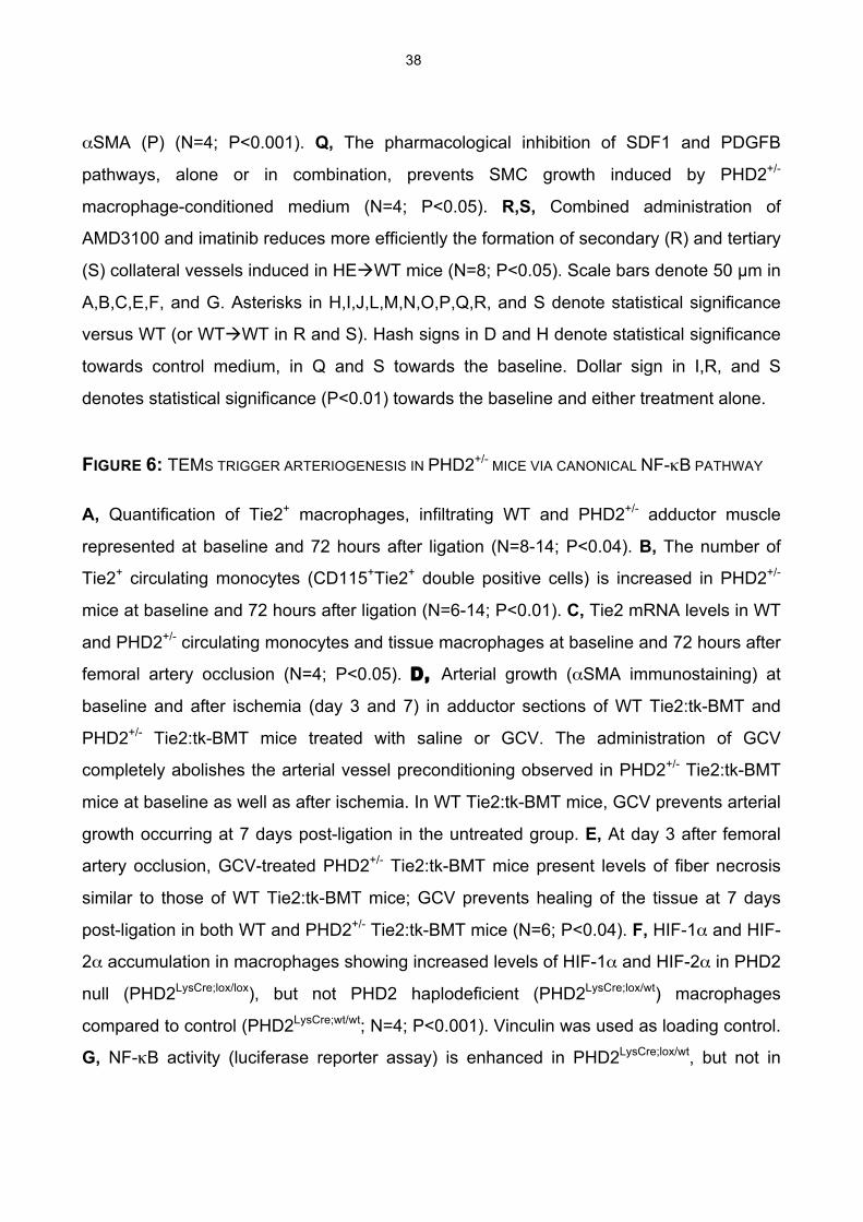
38
αSMA (P) (N=4; P<0.001). Q, The pharmacological inhibition of SDF1 and PDGFB
pathways, alone or in combination, prevents SMC growth induced by PHD2+/-
macrophage-conditioned medium (N=4; P<0.05). R,S, Combined administration of
AMD3100 and imatinib reduces more efficiently the formation of secondary (R) and tertiary
(S) collateral vessels induced in HEàWT mice (N=8; P<0.05). Scale bars denote 50 µm in
A,B,C,E,F, and G. Asterisks in H,I,J,L,M,N,O,P,Q,R, and S denote statistical significance
versus WT (or WTàWT in R and S). Hash signs in D and H denote statistical significance
towards control medium, in Q and S towards the baseline. Dollar sign in I,R, and S
denotes statistical significance (P<0.01) towards the baseline and either treatment alone.
FIGURE 6: TEMS TRIGGER ARTERIOGENESIS IN PHD2+/- MICE VIA CANONICAL NF-κB PATHWAY
A, Quantification of Tie2+ macrophages, infiltrating WT and PHD2+/- adductor muscle
represented at baseline and 72 hours after ligation (N=8-14; P<0.04). B, The number of
Tie2+ circulating monocytes (CD115+Tie2+ double positive cells) is increased in PHD2+/-
mice at baseline and 72 hours after ligation (N=6-14; P<0.01). C, Tie2 mRNA levels in WT
and PHD2+/- circulating monocytes and tissue macrophages at baseline and 72 hours after
femoral artery occlusion (N=4; P<0.05). D, Arterial growth (αSMA immunostaining) at
baseline and after ischemia (day 3 and 7) in adductor sections of WT Tie2:tk-BMT and
PHD2+/- Tie2:tk-BMT mice treated with saline or GCV. The administration of GCV
completely abolishes the arterial vessel preconditioning observed in PHD2+/- Tie2:tk-BMT
mice at baseline as well as after ischemia. In WT Tie2:tk-BMT mice, GCV prevents arterial
growth occurring at 7 days post-ligation in the untreated group. E, At day 3 after femoral
artery occlusion, GCV-treated PHD2+/- Tie2:tk-BMT mice present levels of fiber necrosis
similar to those of WT Tie2:tk-BMT mice; GCV prevents healing of the tissue at 7 days
post-ligation in both WT and PHD2+/- Tie2:tk-BMT mice (N=6; P<0.04). F, HIF-1α and HIF-
2α accumulation in macrophages showing increased levels of HIF-1α and HIF-2α in PHD2
null (PHD2LysCre;lox/lox), but not PHD2 haplodeficient (PHD2LysCre;lox/wt) macrophages
compared to control (PHD2LysCre;wt/wt; N=4; P<0.001). Vinculin was used as loading control.
G, NF-κB activity (luciferase reporter assay) is enhanced in PHD2LysCre;lox/wt, but not in

39
PHD2LysCre;lox/lox macrophages. Silencing of PHD3 unleashes NFκB in PHD2LysCre;lox/lox
macrophages. H, NF-κB is modulated by the hydroxylase activity of PHD2 in
macrophages. The electroporation of PHD2+/- macrophages with a wild type PHD2
(PHD2wt) blunts NF-κB activation, whereas a PHD2 construct containing a mutation at the
catalytic site (PHD2H313A) is not effective (N=4; P<0.05). I, PHD2+/- macrophages present
enhanced NF-κB activity at baseline and upon TNF-α stimulation compared to WT
macrophages (N=4; P<0.05). J, Western blot analysis of nuclear extracts from WT and
PHD2+/- macrophages showing increased accumulation of both p65 (RelA) and p50 (NF-
κB1) in PHD2+/- macrophages at baseline (N=3; P<0.05). K, Silencing of p65 or p50
inhibits NF-κB hyperactivation in PHD2+/- macrophages and combined knockdown of both
subunits restores NF-κB function back to the WT levels (N=4; P<0.05). L, Genetic
inactivation of IKKβ in PHD2 haplodeficient pMØ abrogates the upregulation of PDGFB,
SDF1, and Tie2, while it did not have any effect on PHD2wt/wt macrophages (N=4; P<0.05).
M, Histogram showing reduction of circulating CD115+Tie2+ double positive cells at
baseline upon myeloid specific IKKβ gene targeting in myeloid specific PHD2
haplodeficient mice (N=6-12; P<0.05). N, αSMA immunostainings of adductor sections,
revealing abrogation of collateral vessel preconditioning induced by PHD2 haplodeficiency
upon myeloid specific IKKβ knockout; in WT mice, IKKβ gene deletion greatly prevented
ischemia-induced arteriogenesis 7 days post-ligation (N=4-12, P<0.05). Asterisks in
A,B,C,D,E,G,H,I,K,L,M, and N denote statistical significance (P<0.05). Hash signs in
A,B,C,D,E,I, and N denote statistical significance compared to baseline, in G and K
towards their scramble controls. Dollar signs in E denote statistical significance towards
the baseline and GCV treatment, in K towards the baseline and either treatment alone.

A
60
20
E80
40
0WT PHD2+/-
*
Figure 1
Toe perfusion (laser Doppler analysis)
0 12 24 48 72
100
50
0
Per
fusi
on (%
)
* * *75
5
Hours
MRI-based oxymetry
pO2 (
% o
f bas
elin
e)WT PHD2+/-
M PON
10
15
0Nec
rotic
are
a (%
)
WT PHD2+/-
*5
80
20
*
40
602025
Viab
le a
rea
(%)
LK WT PHD2+/- WT PHD2+/-Ischemic necrosis Muscle viability (TTC)
B
50
End
uran
ce (m
in)
Baseline 12h
*10
25 WT PHD2+/-
Treadmill running test
40
10
J50
20
0Pim
onid
azol
e+ are
a (%
)
WT PHD2+/-
*30
Hypoxia
125
GF WT PHD2+/-
C DWT PHD2+/-
Oxygen tensionHigh pO2
Low pO2
Oxygen tensionHigh pO2
Low pO2
S
80
20
0WT PHD2+/-
*40
60
Infa
rcte
d ar
ea (%
of L
V)
Myocardial infarction
WT PHD2+/-0
1520
UT WT PHD2+/-
Bis
mut
h+ ves
sel
area
(%)
Bis
mut
h+
vess
els/
mm
2
V
0Remote
myocardium
1
WT PHD2+/-
Infarct
2
3
Collateral vessel densityCollateral vessel area
**
W
0
20
WT PHD2+/-
Infarct
40
60
**
Remote myocardium
R
H
Q
WT I PHD2+/-
WT PHD2+/-

K
800
200
0 Num
ber o
f ves
sels
/ cr
oss-
sect
ion
WT PHD2+/-
400600
B
A
WT
PHD2+/-
C
50N
umbe
r of c
olla
tera
ls
Baseline 72h
10
15
WT PHD2+/-B
ism
uth+ v
esse
l ar
ea (%
)
*
FE WT PHD2+/-
Bis
mut
h+
vess
els/
mm
2
12h
2025
D
50N
umbe
r of c
olla
tera
ls
Baseline 72h
10
15
WT PHD2+/-
12h
2025
G
0.5
0 Baseline 72h
1.0
1.5
WT PHD2+/-
12h
2.02.5
H
20
0 Baseline 72h
40
60 WT PHD2+/-
12h
Figure 2
2nd generation collateral branches 3rd generation collateral branches
I JWT PHD2+/-Collateral vessel densityCollateral vessel area
Large vessels in the thigh (diameter >200 m)
U V
W
WT PHD2+/-
WT PHD2+/-
Bas
elin
e
SMA SMA
SMA SMA
Liga
ted
Q
10
0Num
ber o
f ves
sels
/ cr
oss-
sect
ion
Baseline 72h
20
30WT PHD2+/-
S
0Baseline 72h
200
400
600WT PHD2+/-
R
0Baseline 72h
5000
10000
15000WT PHD2+/-
Tota
l are
a (
m2 )
Density of SMA+ collaterals Total lumen area of SMA+ collaterals
Mean lumen area of SMA+ collaterals
Mea
n ar
ea (
m2 )
#
#
# #* *
**
* ** * *
*
* * * * *#
T
0Baseline 72h
*200
400
600
WT PHD2+/-
Mean thickness of the tunica media
SM
A+ a
rea/
vess
el
(m
2 )
800 ##
N O PLarge vessels in the heart (diameter >200 m)
0
4000
6000
WT PHD2+/- Num
ber o
f ves
sels
/ cr
oss-
sect
ion
2000
*
L MWT PHD2+/- WT PHD2+/-
Y
1000*

BA C
E
H
20
0F4/8
0+ MR
C1+ c
ells
(%
of F
4/80
+ cel
ls)
40
60
400
100
0CD
45+ c
ells
/mm
2
Baseline 72h
200
300
100
0F4/8
0+ cel
ls /m
m2
Baseline 72h
150
200
50
Baseline 72h
Fold
cha
nge
rela
tive
to W
T
WT PHD2+/- WT PHD2+/- WT PHD2+/-
3.0
1.0
0.0
2.0
2.5
0.5
1.5
3.5
Tie2
Ang
1A
ng2
Arg
1C
XC
L1C
XC
L2C
XC
R4
HG
FP
DG
FBP
lGF
Ran
tes
CC
L17
Ym
1FI
ZZN
rp1
VE
GF
MM
P2
MM
P9
SD
F1TG
FIL
1 IL6
NO
S2
MC
P1
TNF
CX
CL1
0IL
12IF
NC
ox2
ICA
M1
CC
L22
* *
*
**
**
* *
*
*** * ** *
** **
M2-markers M1-markers
*
D PHD2+/-
Figure 3
I
Fold
cha
nge
rela
tive
to W
T at
bas
elin
e
2
0
1
3
Tie2PDGFB Nrp1MMP2SDF1
*HQH�SUR¿OH�RI�3+'�+/- adductor macrophagesWT Baseline PHD2+/- Baseline
4
CD45+ cells in adductor F4/80+ cells in adductor F4/80+MRC1+macrophages in adductor
*
* *
** *
#
*HQH�SUR¿OH�RI�3+'�+/- pM
GF WT PHD2+/-
80
SMA F4/80 MRC1 SMA F4/80 MRC1 SMA F4/80 MRC1 SMA F4/80 MRC1
#
Baseline Ligated
**# ## #
#
#
# #*
WT 72h PHD2+/-72h
CC
R2
*
CX
3CR
1
Flt1
Flk1 Flt4
WT
#
PHD2
*

A
20
5
0Num
ber o
f col
late
rals
10
15
B
10
0
20
30
C
1.0
1.5
0
Bis
mut
h+ ves
sel
area
(%)
0.5
D
60
80
0
20
40
Figure 4
2nd generation collateral branches (LysCre+)
3rd generation collateral branches (LysCre+)
Num
ber o
f col
late
rals
Collateral vessel density(LysCre+)
Collateral vessel area (LysCre+)
Bis
mut
h+ ves
sels
/mm
2
L
1015
05
* *
Nec
rotic
are
a (%
)
M
1015
05
Baseline 12h
End
uran
ce (m
in)20
25
Ischemic necrosis Treadmill running testK
*
Collateral vessel areaCollateral vessel density
Bis
mut
h+ ve
ssel
ar
ea (%
)
Bis
mut
h+
vess
els/
mm
2
0.5
0
1.0
1.5
2.0
J
20
0
40
60
*WT WT HE WT
* * * *
*
**
lox/wt lox/loxwt/wt lox/wt lox/loxwt/wt lox/wt lox/loxwt/wt lox/wt lox/loxwt/wt
WT
WT
HE W
T
WT
HE
HE H
E
WT
WT
HE W
T
WT
HE
HE H
E
WT
WT
HE W
T
WT
HE
HE H
E
I
10
25
05
Baseline 12h
End
uran
ce (m
in)
Treadmill running test (LysCre+)
* *1520
E
20
30
0
Nec
rotic
are
a (%
)
lox/wt lox/lox
*10
40
G HF
wt/wt
lox/wt lox/loxwt/wt
Ischemic necrosis (LysCre+)
wt/wt lox/wt lox/lox
2025
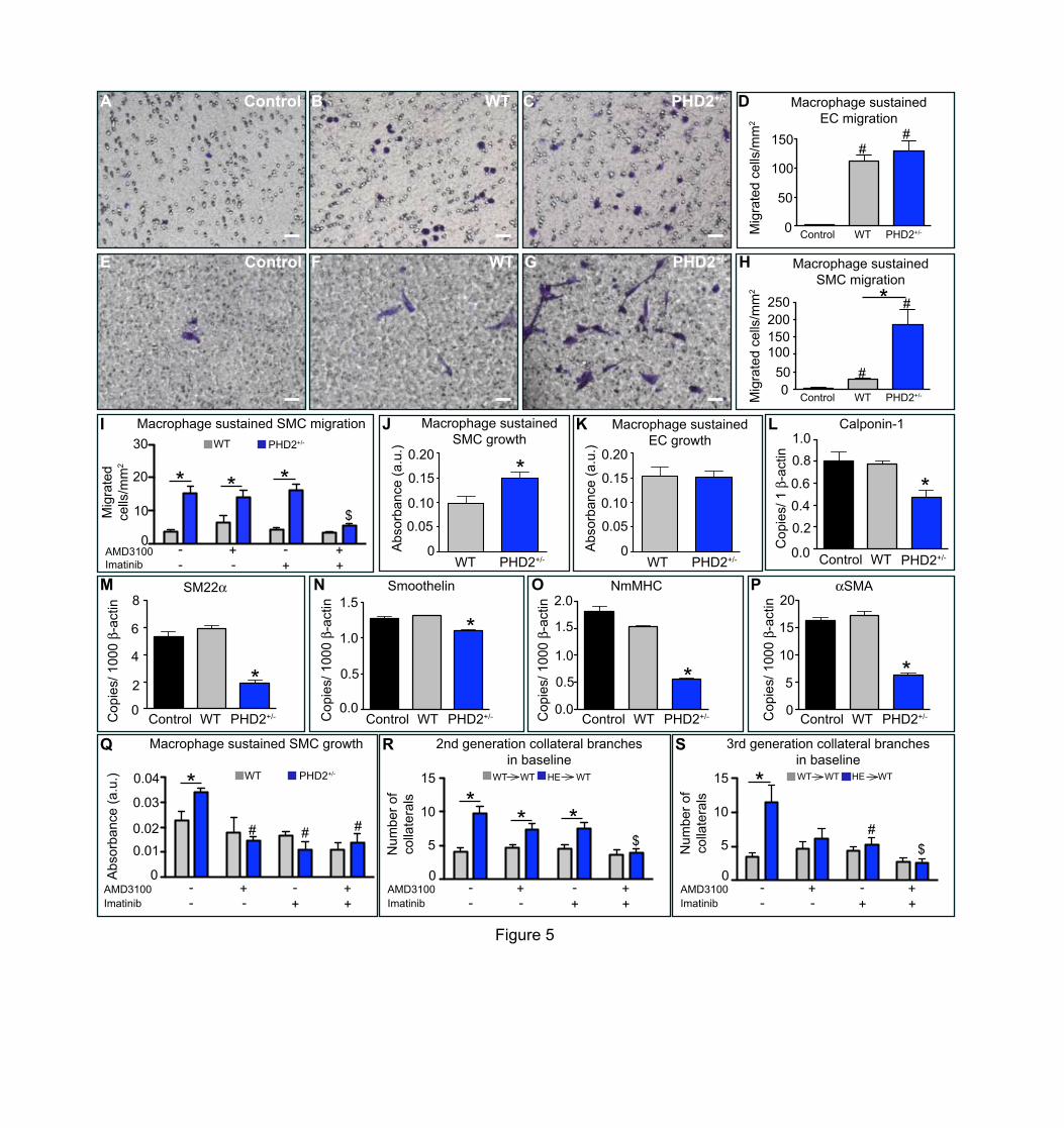
HF G
B C
I
10
0AMD3100
20
30
Imatinib- -- -
++
++
*
Figure 5
0.15
0.05
K
0.10
0Abs
orba
nce
(a.u
.)
WT PHD2+/-
0.20
Macrophage sustained EC growth
0.15
0.05
J
0.10
0Abs
orba
nce
(a.u
.)
WT PHD2+/-
*0.20
Macrophage sustained SMC growth
Q
0.01
0AMD3100
0.02
0.03
Imatinib- -- -
++
++
#
Abs
orba
nce
(a.u
.) 0.04
# #
Macrophage sustained SMC growth
WT PHD2+/-
WT PHD2+/-
Macrophage sustained SMC migration
0.4
0.0
L
0.2
Cop
ies/
1
-act
in
0.6
Control WT PHD2+/-
0.81.0
Calponin-1
4
0
M
2
Cop
ies/
100
0 -a
ctin
6
8SM22
1.0
0.0
N
0.5
Cop
ies/
100
0 -a
ctin
1.5Smoothelin
1.0
0.0
O
0.5
Cop
ies/
100
0 -a
ctin
1.5
NmMHC2.0
10
0
P
5
Cop
ies/
100
0 -a
ctin
15
SMA20
*
*
*
* *
WT PHD2+/-
WT PHD2+/- Macrophage sustained SMC migration
Control WT PHD2+/- Control WT PHD2+/- Control WT PHD2+/- Control WT PHD2+/-
R
5
0AMD3100
10
15
Imatinib- -- -
++
++
*
Num
ber o
f co
llate
rals
2nd generation collateral branches in baseline
S
5
0AMD3100
10
15
Imatinib- -- -
++
++
Num
ber o
f co
llate
rals
3rd generation collateral branches in baseline
* *
** * #
*
$
WT WT HE WT WT WT HE WT
$$
50
D
100
0Mig
rate
d ce
lls/m
m2
150
Macrophage sustained EC migration
WT PHD2+/-Control
A Control
E Control
WT PHD2+/-Control
50100
0
150200250 * #
#
##
Mig
rate
d ce
lls/m
m2
Mig
rate
d ce
lls/m
m2

J
Figure 6
F
Vinculin
HIF-1
wt/wt lox/wt lox/lox
HIF-2
G
Lum
inis
cenc
e (a
.u)
siPHD3Scramble
6
2
4
0
wt/wt lox/wt lox/lox
NF- B activity in macrophages(LysCre+)
* n.s.
##
HIF accumulation in macrophages (LysCre+)
* *
B
#
CD
115+ T
ie2+ c
ells
(%
of C
D11
5+ cel
ls)
Tie2-expressing monocytes in blood
6
0
4
8
2
AF4
/80+
MR
C1+
Tie2
+ ce
lls/m
m2
Tie2-expressing macrophages in adductor
120
0
60
30
Baseline
*
72h
WT PHD2+/-
Baseline 72h
WT PHD2+/-
#*
*90
Nec
rotic
are
a
(%)
Ischemic necrosisE
GCV
*30
0
10
20
- -+ +
WT Tie2:tk BMT PHD2+/- Tie2:tk BMT
C
#
Tie2 transcripts in monocytes and macrophages
50
0.4
25
0.8
Monocytes Macrophages
WT baseline PHD2+/- baseline
*
*
Cop
ies/
106 c
opie
s -a
ctin
I NF- B activity in macrophages after TNF- stimulation
Lum
inis
cenc
e (a
.u)
24
0
100
*
*#
#150
H
Lum
inis
cenc
e (fo
ld
chan
ge re
lativ
e to
WT)
NF- B regulation by prolyl hydroxylase function
3
0
2
4
1
PHD2wt PHD2H313A
WT PHD2+/-
* *
Control
WT 72h PHD2+/- 72h
0.0
*
Tie2-expressing monocytes in blood (LysCre+)
M N
#
WT PHD2+/-
200
p50
p65
WT PHD2+/-
Lamin
Nuclear accumulation of p65 and p50 in macrophages
K
siRNAp50
NF- B activity in macrophagesafter p65/p50 knockdown
Lum
inis
cenc
e (a
.u)
0
10
*
#
15WT PHD2+/-
- -+ +
5
siRNAp65
##
## $
* *
- - + +
- -+ +3d 7d
D
GCV
*30
0
10
20
WT Tie2:tk BMT PHD2+/- Tie2:tk BMT
7d
Num
ber o
f ves
sels
/ cr
oss-
sect
ion
Density of SMA+ collaterals
- -+ +Baseline
##
$#
#
$ $
# ##
*
TNF-Control
CD
115+ T
ie2+ c
ells
(%
of C
D11
5+ cel
ls)
6
2
4
0
8 *
L TEM signature after genetic inactivation of IKK (LysCre+)
Fold
cha
nge
rela
tive
to W
T
3
0
2
4
1
Tie2SDF1PDGFB
PHD2wt/wt;IKK wt/wt PHD2wt/wt;IKK lox/lox
PHD2lox/wt;IKK wt/wt PHD2lox/wt;IKK lox/lox
5
3d- -+ +- -+ +
PHD2wt/wt;IKK wt/wt PHD2wt/wt;IKK lox/lox
PHD2lox/wt;IKK wt/wt PHD2lox/wt;IKK lox/loxPHD2wt/wt;IKK wt/wt PHD2wt/wt;IKK lox/lox
PHD2lox/wt;IKK wt/wt PHD2lox/wt;IKK lox/lox
Num
ber o
f ves
sels
/ cr
oss-
sect
ion
Density of SMA+ collaterals
0
10
15
5
Baseline 7d
2025
* #****
* *
* **
*

SUPPLEMENTARY INFORMATION
MACROPHAGE SKEWING BY PHD2 HAPLODEFICIENCY PREVENTS
ISCHEMIA BY INDUCING ARTERIOGENESIS
Yukiji Takeda, Sandra Costa, Estelle Delamarre, Carmen Roncal, Rodrigo Leite De Oliveira, Mario Leonardo Squadrito, Veronica Finisguerra, Françoise Bruyère, Sofie
Deschoemaeker, Mathias Wenes, Alexander Hamm, Jens Serneels, Julie Magat, Tapan Bhattacharrya, Andrey Anisimov, Benedicte F. Jordan, Kari Alitalo, Patrick Maxwell,
Bernard Gallez, Zhenwu Zhuang, Yoshihiko Saito, Michael Simons, Michele De Palma & Massimiliano Mazzone
SUPPLEMENTARY METHODS
ANIMALS: 129/S6 or Balb/C WT and PHD2+/- mice (8-12 weeks old) were obtained from
our mouse facility. PHD2+/- and PHD2 conditional knock-out mice were obtained as
previously described1. VE-Cadherin:CreERT and PDGFRB:Cre transgenic mice were
generated by Dr. Adams at the Cancer Research UK (London, UK), currently at the
Max-Planck-Institute (Munster, Germany)2,3. IKKβ conditional knock-out mice were
obtained from Dr. Karin (UCSD, California)4. Tie2:Cre and Rosa26:CreERT transgenic
mice were purchased by the Jackson Laboratory. Housing and all experimental animal
procedures were approved by the Institutional Animal Care and Research Advisory
Committee of the K.U.Leuven.
MOUSE MODEL OF HINDLIMB ISCHEMIA: To induce hindlimb ischemia, unilateral or bilateral
ligations of the femoral artery and vein (proximal to the popliteal artery) and the
cutaneous vessels branching from the caudal femoral artery side branch were
performed without damaging the nervus femoralis5. By using this procedure, collateral
flow to adductor muscles is preserved via arterioles branching from the femoral artery,
therefore 50% up to 60% of perfusion is preserved by this method. Two superficial
preexisting collateral arterioles, connecting the femoral and saphenous artery, were
used for analysis. Functional perfusion measurements of the collateral region were
performed using a Lisca PIM II camera (Gambro). Gelatin-bismuth-based angiography
was performed as described6 and analyzed by photoangiographs (Nikon D1 digital
camera). Collateral side branches were categorized as follows: second-generation
collateral arterioles directly branched from the main collateral, whereas third-generation
collateral arterioles were orientated perpendicularly to the second-generation branches.
The number of secondary and tertiary collateral arterioles was counted. After perfusion-
fixation, the muscle tissue between the 2 superficial collateral arterioles (adductor) was
post-fixed in 2% paraformaldehyde (PFA), paraffin-embedded, and morphometrically
analyzed. An endurance treadmill-running test was performed at baseline and 12 hours
post-bilateral-ligation.
MOUSE MODEL OF MYOCARDIAL INFARCTION: Myocardial infarction (MI) was induced by
permanent ligation of the left anterior descending coronary artery as previously

described7. Briefly, animals were anesthetized with pentobarbital (100 mg/kg i.p.), fixed
in the supine position and the trachea was intubated with a 1.1-mm steel tube. Positive
pressure respiration (1.5-2 ml, 70 strokes/min) was started and the left thorax was
opened in the fourth intercostal space. All muscles overlying the intercostal space were
dissected and retracted with 5-0 silk threads; only the intercostal muscles were
transected. After opening the pericardium, the main left coronary artery, which was
clearly visible, was ligated just proximal to the main bifurcation by using 6-0 silk and an
atraumatic needle (Ethicon K801). Infarction was evident from discoloration of the
ventricle. The thorax was closed and the skin sutured with 5-0 silk. Animals recovered
at 30ºC. Gelatin-bismuth-based angiography was performed 24 hours after ligation and
hearts were then collected in 2% PFA.
OXYMETRY: Oxygen tension (pO2) in the lower limb was measured using 19F-MRI
oxymetry in non-ligated and ligated legs 12 hours after femoral artery ligation. The
oxygen reporter probe hexafluorobenzene (HFB) was injected directly into the crural
muscle. MRI was performed with a 4.7T (200 MHz, 1H), 40 cm inner diameter bore
system (Bruker Biospec). A tunable 1H/19F surface coil was used for radiofrequency
transmission and reception8.
HISTOLOGY, IMMUNOSTAINING, AND MORPHOMETRY: Adductor, crural muscles and hearts
were dissected, fixed in 2% PFA, dehydrated, embedded in paraffin, and sectioned at 7-
µm-thickness. Area of necrotic tissues in the crural muscle was analyzed by
Hematoxylin & Eosin (H&E) staining. Necrotic cells display a more glassy homogeneous
appearance in the cytoplasm with increased eosinophilia, while the nuclear changes are
reflected by karyolysis, pyknosis, and karyorrhexis. Necrotic area was defined as the
percentage of area which includes these necrotic myocytes, inflammatory cells, and
interstitial cells, compared to the total soleus area. Infarct size was measured in desmin
stained hearts 24 hours after ischemia as previously described9. After deparaffinization
and rehydration, sections were blocked and incubated overnight with primary
antibodies: rat anti-CD31, dilution 1/500 (BD-pharmingen), mouse anti-aSMA, dilution
1/500 (Dako), rat anti-F4/80, dilution 1/100 (Serotec), dilution 1/50 (BD-pharmingen), rat
anti-CD45, dilution 1/100 (BD-pharmingen), goat anti-MRC1, dilution 1/200 (R&D), rat
anti-Tie-2, dilution 1/100 (Reliatech), rabbit anti-desmin dilution 1/150 (Cappel). In order

to analyze capillary density and area, images of CD31 stained sections of the entire
soleus were taken at 40x. In order to measure bismuth-positive vessel density and area,
H&E stained paraffin sections were analyzed and vessels filled with bismuth-gelatin
(black spots) were taken in account. Images of the entire soleus were acquired at 20x
for this analysis. The values in the graph represent the averages of the mean vessel
density and area per soleus muscle. The same method was used to quantify vessel
capillaries and collateral branches in cardiac tissues. Density and area were measure
by using a KS300 (Leica) software analysis. Hypoxic cells were analyzed 2 hours after
injection of 60 mg/kg pimonidazole into operated mice. Mice were sacrificed and
muscles were harvested. Paraffin sections were stained with Hypoxiprobe-1-Mab-1
(Hypoxiprobe kit; Chemicon International) following the manufacturer’s instructions.
Oxidative stress and proliferation rate were assessed on 7µm-thick cryosections by
using the goat anti-8-OHdG antibody, dilution 1/200 (Serotec) and the rat anti-BrdU
antibody, dilution 1/300 (Serotec). Sections were subsequently incubated with
appropriate secondary antibodies, developed with fluorescent dies or 3,3’-
disminobenzidine (DAB, Sigma). Whole muscle viability was assessed on unfixed 2-
mm-thick tissue slices by staining with 2,3,5-triphenyltetrazolium chloride (TCC). Viable
area, stained in red, was traced and analyzed. Pictures for morphometric analysis were
taken using a Retiga EXi camera (Q Imaging) connected to a Nikon E800 microscope or
a Zeiss Axio Imager connected to an Axiocam MRc5 camera (Zeiss) and analysis was
performed using KS300 (Leica). Angiograms were obtained by X-Ray and CT
angiographies of hearts and legs at baseline.
MACROPHAGE PREPARATION: To harvest peritoneal macrophages (pMØ), the peritoneal
cavity was washed with 5 ml of RPMI 10% FBS. The pooled cells were then seeded in
RPMI 10% FBS in 6-well plates (2x10⁶ cells/well), 12-well plates (1x10⁶ cells/well), or
24-well plates (5x10⁵ cells/well). After 6 hours of incubation at 37°C in a moist
atmosphere of 5% CO2 and 95% air, non-adhering cells on each plate were removed by
rinsing with phosphate-buffered saline (PBS). The attached macrophages were cultured
in different media for 12 hours or 48 hours depending on the experiments performed, as
described below. When high amounts of cells were needed (analysis for HIF
accumulation and NF-κB activity), macrophages were derived from bone marrow

precursors as described before10. Briefly, bone marrow cells (2x10⁶ cells/ml) were
cultured in a volume of 5 ml in a 10 cm Petri dish (non tissue culture treated, bacterial
grade) for 7 to 10 days in DMEM supplemented with 20% FBS and 30% L929
conditioned medium as a source of M-CSF. The cells obtained in those cultures are
uniformly macrophages. Tamoxifen-inducible PHD2 haplodeficient pMØ
(PHD2Rosa26CreERT;lox/wt) were isolated as described above. After 8 hours in culture
(RPMI, 10% FBS), pMØ were washed twice with PBS and treated with or without 2 µM
4-hydroxytamoxifen (4-OHT, Sigma) in complete medium for 48 hours to allow Cre
recombinase activity. Cells were then washed and kept in culture for other 48 hours
before mRNA isolation and gene expression analysis.
QUANTITATIVE PCR ANALYSIS: In order to investigate gene expression in pMØ,
quantitative RT-PCR was performed. After preparing pMØ, the cells were cultured in
normoxic condition for 12 hours and RNA was extracted. To analyze the expression
levels of chemoattractants in the adductor, tissues were collected at baseline or 24
hours post-ischemia and RNA was extracted. Macrophages and ECs were freshly
sorted from dissected adductors as described below and RNA was extracted.
Quantitative RT-PCR was performed with commercially available or home-made
primers and probes for the studied genes. The assay ID (Applied Biosystems, Foster
City, CA) or the sequence of primers and probes (when home-made) are listed in
Supplementary Table 5. RNA levels of Tie-2, SDF1, and PDGFB after inhibition of NF-
kB pathway were measured by RT-PCR on pMØ exposed for 12 hours to 500 nM 6-
amino-4-(4-phenoxyphenylethylamino)quinazoline.
PROTEIN EXTRACTION AND IMMUNOBLOT: Protein extraction was performed using 8M urea
buffer (10% glycerol, 1% SDS, 5 mM DTT, 10mM Tris-HCl, pH 6.8) as previously
described1. Nuclear proteins were extracted in 1% SDS buffer upon cytoplasmic
separation by using a hypotonic lysis buffer (10 mM KCl, 10 mM EDTA, 0.5% NP40, 10
mM HEPES, pH=8, supplemented with phosphatase and protease inhibitors, from
Roche). Signal was detected using ECL system (Invitrogen) according to the
manufacture’s instructions. The following antibodies were used: rabbit anti-HIF-1α
(Novus), rabbit anti-p105/50, rabbit anti-HIF-2α (Abcam), PM9 rabbit anti-HIF-2α (from

Dr. Maxwell), mouse anti-vinculin (Sigma), rabbit anti-lamin A/C, rabbit anti-p65 (Cell
Signaling).
TRANSDUCTION AND TRANSFECTION OF BONE MARROW DERIVED MACROPHAGES AND LUNG
ENDOTHELIAL CELLS: To express an inducible NF-κB responsive firefly luciferase
reporter, commercially available lentiviral vector particles (LV) were used (Cignal Lenti
NF-κB Reporter; SABiosciences). 2.5x10⁵ bone marrow derived macrophages and 105
primary lung endothelial cells (isolated as previously described1) were seeded in a 24-
well plate in DMEM 10% FBS or M199 20% FBS for 8 hours. Cells were transduced
with 108 TU/ml. Eight hours after transduction, the medium was replaced. After 48
hours, cells were stimulated with TNFα (20 ng/mL) for 8 hours and the same amount of
protein extract was read in a luminometer. For PHD3 silencing, siRNA oligonucleotides
were designed using the Invitrogen online siRNA design tool
(http://rnaidesigner.invitrogen.com). The following sequences (sense strands) and target
positions were used: PHD3 siRNA: 5’-GCCGGCUGGGCAAAUACUAUGUCA-3’;
scramble siRNA:5’-CACCGCTTAACCCGTATTGCCTAT-3’. In brief, one day after the
transduction of macrophages with LV, cells were transfected using Lipofectamine 2000
(Invitrogen) according to the manufacturer’s instructions. Preparation of the
oligonucleotide-Lipofectamine 2000 complexes was done as followed: 25 pmol siRNA
oligonucleotide (stock: 20 µM) was diluted in 50 µl Opti-MEM I reduced serum medium.
Lipofectamine 2000 (1.5 µl) was diluted in 50 µl Opti-MEM I reduced serum medium and
incubated for 5 minutes at room temperature. siRNA oligonucleotides were gently mixed
with Lipofectamine 2000 and allowed to incubate at room temperature for 20 minutes to
form complexes. Just before transfection, the cell culture medium was removed and
cells were rinsed twice with serum-free Opti-MEM I medium. The Lipofectamine 2000-
siRNA oligonucleotide complexes were added to each well in 400 µl of serum-free Opti-
MEM medium for 5 hours. Afterwards, cells were incubated in complete medium for 48
hours at 37°C in a CO2 incubator and assayed for gene knockdown (RT-PCR) and
luciferase activity. To assess if the increased NF-κB activity observed in PHD2+/-
macrophages was dependent on the hydroxylase activity of PHD2, 48h before

transduction, 4x10⁶ bone marrow derived macrophages were resuspended in 240 µl of
Opti-MEM and were electroporated (250V, 950uF,∞ Ω) with 7 µg of plasmids
expressing a wild type PHD2 (PHD2wt) or a PHD2 containing a mutation at the catalytic
site (PHD2H313A). Silencing of the canonical pathway subunits p65 (RelA) and p50 (NF-
κB1) was achieved by electroporation with specific siRNAs. Briefly, 48h before
transduction, 2.4x10⁶ bone marrow derived macrophages were resuspended in 320 µl
of Opti-MEM and were electroporated (250V, 950µF, ∞Ω) with 60 pmol of siRNA for
either scramble, p65, p50, or combination of p50 and p65. For higher efficiency of
silencing, two different siRNA sequences for each respective gene were designed
(http://rnaidesigner.invitrogen.com). For p65: 5’-TGTCTGCACCTGTTCCAAATT-3’ and
5’-TGCTGATGGAGTACCCTGATT-3’; for p50: 5’-GAATACTTCATGTGACTAATT-3’
and 5’-CAAAGGTTATCGTTCAGTTTT-3’; for the scramble siRNA: 5’-
CACCGCTTAACCCGTATTGCCTAT-3’.
CELL MIGRATION AND VIABILITY ASSAYS: Migration and proliferation of SMCs and ECs
were assessed by using 8µm-pore Transwell permeable plate for migration assays and
0.4µm-pore Transwell permeable plate for proliferation assays (Corning Life Science).
To determine cell migration towards the factors secreted by pMØ, pMØ were cultured in
the lower chamber for 12 hours in RPMI 1% FBS or in M-199 1% FBS (migration
assay), or 48 hours in DMEM-F12 1% FBS or in M-199 1% FBS (proliferation assay).
For migration assays, hCASMC (Human coronary artery smooth muscle cells; from
Lonza) and HUVEC (Human umbilical vein endothelial cells; from Lonza) were starved
for 12 hours in their own medium at 1% FBS and then seeded in the upper chamber
(5x10³ cells in 200 µl of medium 1% FBS), with or without AMD3100 (1 µg/ml, Sigma-
Aldrich, Dorset, U.K) and/or imatinib (2.5 µg/ml, Novartis). SMCs and HUVECs were
incubated for 2 days or 24 hours respectively, and migrated cells were fixed with 4%
PFA, stained with 0.25% crystal violet/ 50% methanol and counted under the
microscope. VEGF (100 ng/mL, R&D), PDGFB (100 ng/mL, R&D), or SDF1 (100 ng/mL,
R&D) were used as positive controls. For cell growth assays, RAOSMC (Rat Aortic
Smooth Muscle Cells) and HUVEC were seeded on the upper chambers (5x10³ cells/
transwell) and cultivated with pMØ for 24 hours in DMEM-F12 1% FBS or M-199 1%

FBS for RAOSMC and HUVEC cells, respectively. The cell proliferative ability was then
analyzed using WST-1 Cell Proliferation Assay (Roche Applied Biosciences) according
to the manufacturer instructions. Alternatively, WT and PHD2+/- pMØ were seeded in the
lower chamber of a Transwell and transduced with lentiviral vectors (108 TU/ml; Sigma)
carrying an shRNA against SDF1, PDGFB, or a scramble control (purchased from
Sigma). Sixty hours after macrophage transduction, SMC migration or growth assays
were performed by seeding the SMCs in the upper side of the Transwell as described
above.
SMC DIFFERENTIATION ASSAY: pMØ were seeded in a 24-well plate with DMEM F-12 5%
FBS. Conditioned medium was harvested after 2 days and supplemented with 25 mM
HEPES. RAOSMC were seeded in a 24-well plate (80x103 cells/ well) and incubated for
5 hours at 37°C in a moist atmosphere of 5% CO2 and 95% air. After 2 hours of
starvation in DMEM-F12 1% FBS, SMC were stimulated with conditioned medium from
WT and PHD2+/- pMØ. After 24 hours, differentiation status of the SMCs was assessed
by RT-PCR.
FACS ANALYSIS AND MACROPHAGE AND ENDOTHELIAL CELL SORTING: FACS analysis of
circulating TEMs was performed in 200 µl of peripheral blood, harvested by eye
bleeding at baseline or 3 days after femoral artery ligation. Blood samples were
incubated for 20 minutes at 4°C with a rat APC-conjugated anti-CD115, a mouse PE-
conjugated anti-Tie2 (eBiosciences), or a rat FITC conjugated anti-Gr1 (BD-
pharmingen). For cell sorting of adductor macrophages and ECs, the adductors were
dissected, dissociated mechanically, and digested using collagenase I for 45 minutes at
37°C. For macrophage sorting, the digested cell suspension was incubated for 15
minutes with mouse anti-CD16/CD32 mAb (Fc Block™, BD-pharmingen) and stained
with rat FITC-conjugated anti-F4/80 antibody (Serotec) for 20 minutes at 4 °C.
CD31+CD45- endothelial cells were sorted from the digested adductor cell suspension
after incubation with rat APC-conjugated anti-CD31 and rat FITC-conjugated anti-CD45
(BD-pharmingen) for 20 minutes at 4 °C.
BONE MARROW (BM) TRANSPLANTATION AND HEMATOLOGICAL ANALYSIS: Balb/c WT and
PHD2+/- recipient mice were irradiated with 7.5 Gy. Subsequently, 5 x 10⁶ bone marrow

cells from green fluorescent protein+ (GFP+) WT or GFP+ PHD2+/- mice were injected
intravenously via the tail vein. After one week, saline or AMD3100 (5mg/kg) was
administered intravenously and saline or imatinib (50mg/ml) was administered by oral
gavage for 4 weeks. Femoral artery ligation, treadmill running test, and bismuth
angiography were performed at 6 weeks after bone marrow reconstitution. Red and
white blood cell count was determined using a hemocytometer (Cell-Dyn 3700, Abbott)
on peripheral blood collected in heparin by retro-orbital bleeding. To assess the effect of
acute deletion of macrophage borne PHD2 on arteriogenesis, PHD2Rosa26CreERT;lox/wt
bone marrows were transplanted into lethally irradiated WT recipient mice. After five
weeks, transplanted mice were injected i.p. with tamoxifen (1 mg/mouse; Sigma) or
vehicle for 5 days. TEM quantification and femoral artery ligation were performed 10
days after tamoxifen treatment as explained above.
BONE MARROW-DERIVED, LINEAGE NEGATIVE HEMATOPOIETIC CELL ISOLATION, TRANSDUCTION AND TRANSPLANTATION: Six to 12-week–old WT or PHD2+/- Balb/C mice
were killed and their BM was harvested by flushing the femurs and the tibias. Lineage-
negative cells (lin– cells) enriched in hematopoietic stem/progenitor cells (HS/PCs) were
isolated from BM cells using a cell purification kit (StemCell Technologies) and
transduced by concentrated lentiviral vectors. Briefly, 106 cells/ml were pre-stimulated
for 4-6 hours in serum-free StemSpan medium (StemCell Technologies) containing a
cocktail of IL-3 (20 ng/ml), SCF (100 ng/ml), TPO (100 ng/ml), and FLT-3L (100 ng/ml)
(all from Peprotech), and transduced with 108 TU/ml of two lentiviral vectors (LVs),
Tie2:tk (to deplete TEMs in transplanted mice) and PGK:GFP (to assess the efficiency
of BM reconstitution in transplanted mice). After 12 hours, 106 cells were washed and
infused into the tail vein of lethally irradiated 6-week-old female Balb/C mice (radiation
dose: 7.5 Gy).
VECTOR COPY NUMBER ANALYSIS: Transduced lin– cells were cultured and collected after
9 days while blood from the transplanted mice was collected 4 weeks after HS/PCs tail
vein injection to measure the number of integrated LV copies/cell genome (vector copy
number, VCN) by RT-PCR as previously described11. Briefly, for vector copy number
(VCN) analysis, we performed RT-PCR using custom TaqMan assays specific for β-
actin, HSV-tk, or HIV-gag sequences (Applied Biosystems). Standard curves for HSV-tk

(contained by Tie2:tk LV) or HIV-gag (contained by both Tie2:tk and PGK:GFP LVs)
were obtained from genomic DNA samples containing known amounts of integrated LV.
The VCN of genomic DNA standard curves was determined using custom TaqMan
assays specific for LVs (Applied Biosystems). The SDS 2.2.1 software was used to
extract raw data (CT) and to perform VCN analysis. To calculate VCN, we used the
following formula: VCN = VCN(standard curve) * ng of "LV of interest" / ng of β-actin, where
"LV of interest" is either HIV-gag or HSV-tk. The VCN of PGK:GFP LV was obtained by
subtracting the VCN of HSV-tk from the total HIV-gag VCN.
STATISTICS: The data were represented as mean ± SEM of the indicated number of
measurements. Statistical significance was calculated by t test where indicated (Prism
v4.0b), with p<0.05 considered statistically significant.

ADDITIONAL REFERENCES
1. Mazzone, M. et al. Heterozygous deficiency of PHD2 restores tumor oxygenation and inhibits metastasis via endothelial normalization. Cell 136, 839-51 (2009).
2. Foo, S.S. et al. Ephrin-B2 controls cell motility and adhesion during blood-vessel-wall assembly. Cell 124, 161-73 (2006).
3. Benedito, R. et al. The notch ligands Dll4 and Jagged1 have opposing effects on angiogenesis. Cell 137, 1124-35 (2009).
4. Chen, L.W. et al. The two faces of IKK and NF-kappaB inhibition: prevention of systemic inflammation but increased local injury following intestinal ischemia-reperfusion. Nat Med 9, 575-81 (2003).
5. Luttun A, T.M., Moons L, Wu Y, Angelillo-Scherrer A, Liao F, Nagy JA, Hooper A, Priller J, De Klerck B, Compernolle V, Daci E, Bohlen P, Dewerchin M, Herbert JM, Fava R, Matthys P, Carmeliet G, Collen D, Dvorak HF, Hicklin DJ, Carmeliet P. Revascularization of ischemic tissues by PlGF treatment, and inhibition of tumor angiogenesis, arthritis and atherosclerosis by anti-Flt1. Nat Med 8, 831-840 (2002).
6. Carmeliet P, M.L., Luttun A, Vincenti V, Compernolle V, De Mol M, Wu Y, Bono F, Devy L, Beck H, Scholz D, Acker T, DiPalma T, Dewerchin M, Noel A, Stalmans I, Barra A, Blacher S, Vandendriessche T, Ponten A, Eriksson U, Plate KH, Foidart JM, Schaper W, Charnock-Jones DS, Hicklin DJ, Herbert JM, Collen D, Persico MG. Synergism between vascular endothelial growth factor and placental growth factor contributes to angiogenesis and plasma extravasation in pathological conditions. Nat Med 7, 575-583 (2001).
7. Lutgens, E. et al. Biphasic pattern of cell turnover characterizes the progression from fatty streaks to ruptured human atherosclerotic plaques. Cardiovasc Res 41, 473-9 (1999).
8. Jordan, B.F., Cron, G.O. & Gallez, B. Rapid monitoring of oxygenation by 19F magnetic resonance imaging: Simultaneous comparison with fluorescence quenching. Magn Reson Med 61, 634-8 (2009).
9. Pfeffer, M.A. et al. Myocardial infarct size and ventricular function in rats. Circ Res 44, 503-12 (1979).
10. Meerpohl, H.G., Lohmann-Matthes, M.L. & Fischer, H. Studies on the activation of mouse bone marrow-derived macrophages by the macrophage cytotoxicity factor (MCF). Eur J Immunol 6, 213-7 (1976).
11. De Palma, M. et al. Tumor-targeted interferon-alpha delivery by Tie2-expressing monocytes inhibits tumor growth and metastasis. Cancer Cell 14, 299-311 (2008).

SUPPLEMENTARY TABLES
SUPPLEMENTARY TABLE 1: GENE EXPRESSION IN WT AND PHD2+/- ENDOTHELIAL CELLS.
WT PHD2+/-
GENE BASELINE LIGATED BASELINE LIGATED
Phd2 1 ± 0.16 1.2 ± 0.17 0.5 ± 0.03 * 0.5 ± 0.09 *
Tie2 1 ± 0.02 1.1 ± 0.39 1.1 ± 0.13 0.9 ± 0.40
Ang1 1 ± 0.25 1 ± 0.5 1.25 ± 0.5 1.25 ± 0.75
Ang2 1 ± 0.19 1.5 ± 0.05 # 1.04 ± 0.09 1.5 ± 0.16 #
Cxcl1 1 ± 0.38 2.5 ± 0.71 1.3 ± 0.29 2.8 ± 0.80
Cxcl2 n.d. n.d.
Cxcr4 1 ± 0.11 0.6 ± 0.11 # 0.9 ± 0.17 0.5 ± 0.08 #
Cx3cr1 1 ± 0.13 0.6 ± 0.13 0.9 ± 0.13 0.6 ± 0.11
hgf 1 ± 0.01 1.2 ± 0.36 1.2 ± 0.11 1.0 ± 0.25
Pdgfb 1 ± 0.06 0.9 ± 0.08 0.9 ± 0.15 0.9 ±0 .19
Plgf 1 ± 0.16 3.1 ± 0.84 1.5 ± 0.38 3.3 ± 0.56
Rantes n.d. n.d.
Ccl17 n.d. n.d.
Ccl22 n.d. n.d.
Flk1 1 ± 0.14 0.4 ± 0.09 # 0.86 ± 0.09 0.4 ± 0.04 #
Flt1 1 ± 0.26 0.7 ± 0.17 0.9 ± 0.47 0.7 ± 0.24
sFlt1 1 ± 0.10 0.7 ± 0.03 1.0 ± 0.10 0.7 ± 0.12
Nrp1 1 ± 0.04 0.4 ± 0.08 # 0.9 ± 0.38 0.5 ± 0.05 #
Cdh5 1 ± 0.06 0.8 ± 0.06 1.2 ± 0.17 0.8 ± 0.17
Vegfa 1 ± 0.5 1 ± 0.30 0.8 ± 0.20 1.2 ± 0.20
Mmp2 1 ± 0.30 1.5 ± 0.42 1.2 ±0.37 1.4 ± 0.36
Mmp9 1 ± 0.48 0.6 ± 0.26 1.2 ±0.68 0.5 ± 0.11
Sdf1 1 ± 0.12 1.2 ± 0.26 0.8 ±0.12 1,0 ± 0.22
Tgfβ 1 ± 0.04 0.8 ± 0.05 0.9 ±0.09 1.0 ± 0.38
IL1β n.d. n.d.

IL6 1 ± 0.23 1.6 ± 0.28 1.1 ± 0.14 1.72 ± 0.31
Nos3 1 ± 0.17 0.8 ± 0.11 0.8 ± 0.16 0.8 ± 0.14
Mcp1 1 ± 0.18 5.4 ± 0.13 # 0.9 ± 0.16 6.0 ±0.13 #
Tnfα 1 ± 0.21 0.8 ± 0.28 1.0 ± 0.18 0.7 ± 0.15
Cxcl10 1 ± 0.23 2.1 ± 0.19 # 1.1 ± 0.41 2.4 ± 0.27 #
IL12α n.d. n.d.
IFNβ n.d. n.d.
Cox2 1 ± 0.12 1.2 ± 0.04 1.1 ± 0.2 1.1 ± 0.2
Jagged1 1 ± 0.09 0.9 ± 0.07 1.1 ± 0.10 1.0 ± 0.15
Icam1 1 ± 0.11 0.8 ± 0.13 0.8 ± 0.09 0.6 ± 0.08
The data represent the expression analysis of endothelial cells, freshly sorted from WT
and PHD2+/- adductor muscles at baseline and 72 hours after femoral artery occlusion
(N=4-8, P<0.05). Data are normalized towards the expression levels of WT ECs at
baseline; n.d.= not determined. Asterisks denote statistical significance versus WT.
Hash signs denote statistical significance compared to the baseline.

SUPPLEMENTARY TABLE 2: COLLATERALIZATION IN MICE HAPLODEFICIENT FOR PHD2 IN
HEMATOPOIETIC CELLS AND/OR ENDOTHELIAL CELLS.
Donor PHD2Tie2Cre;wt/wt PHD2Tie2Cre;lox/wt PHD2Tie2Cre;wt/wt PHD2Tie2Cre;lox/wt
Recipient PHD2Tie2Cre;wt/wt PHD2Tie2Cre;wt/wt PHD2Tie2Cre;lox/wt PHD2Tie2Cre;lox/wt
2nd generation
collaterals 5.9 ± 1.0 13.3 ± 1.4 *# 8.2 ± 0.7 14.4 ± 1.6 *#
3rd generation
collaterals 3.8 ± 0.7 7.2 ± 1.3 *# 2.7 ± 1.1 7.1 ± 1.0 *#
Reciprocal bone marrow transplantation in lethally irradiated mice reveals that the
enhanced arteriogenesis of PHD2 heterozygous mice is specifically caused by loss of
one PHD2 allele in bone marrow derived hematopoietic cells (green column) but not in
endothelial cells (pink column) compared to WT controls (yellow column). Combined
deletion of one PHD2 allele in both the hematopoietic and EC lineage (light blue
column) does not modify the biological effect elicited on collateral arteries by PHD2
haplodeficient hematopoietic cells alone. Asterisks denote statistical significance versus
PHD2Tie2Cre;wt/wt àPHD2Tie2Cre;wt/wt. Hash signs denote statistical significance compared
to PHD2Tie2Cre;wt/wt àPHD2Tie2Cre;lox/wt .
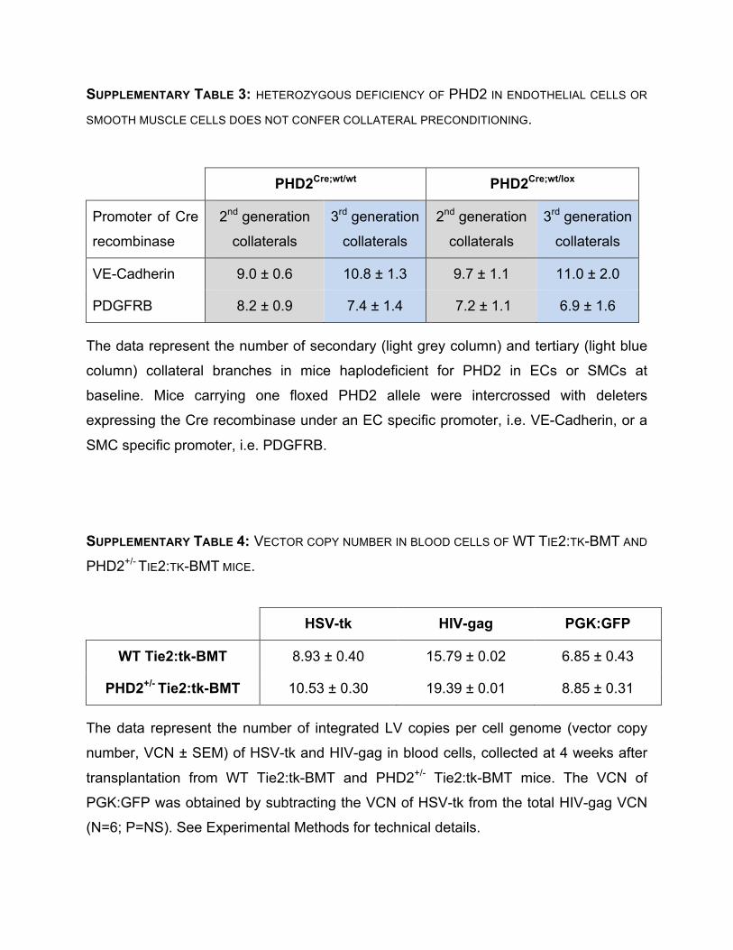
SUPPLEMENTARY TABLE 3: HETEROZYGOUS DEFICIENCY OF PHD2 IN ENDOTHELIAL CELLS OR
SMOOTH MUSCLE CELLS DOES NOT CONFER COLLATERAL PRECONDITIONING.
PHD2Cre;wt/wt PHD2Cre;wt/lox
Promoter of Cre
recombinase 2nd generation
collaterals
3rd generation
collaterals
2nd generation
collaterals
3rd generation
collaterals
VE-Cadherin 9.0 ± 0.6 10.8 ± 1.3 9.7 ± 1.1 11.0 ± 2.0
PDGFRB 8.2 ± 0.9 7.4 ± 1.4 7.2 ± 1.1 6.9 ± 1.6
The data represent the number of secondary (light grey column) and tertiary (light blue
column) collateral branches in mice haplodeficient for PHD2 in ECs or SMCs at
baseline. Mice carrying one floxed PHD2 allele were intercrossed with deleters
expressing the Cre recombinase under an EC specific promoter, i.e. VE-Cadherin, or a
SMC specific promoter, i.e. PDGFRB.
SUPPLEMENTARY TABLE 4: VECTOR COPY NUMBER IN BLOOD CELLS OF WT TIE2:TK-BMT AND
PHD2+/- TIE2:TK-BMT MICE.
HSV-tk HIV-gag PGK:GFP
WT Tie2:tk-BMT 8.93 ± 0.40 15.79 ± 0.02 6.85 ± 0.43
PHD2+/- Tie2:tk-BMT 10.53 ± 0.30 19.39 ± 0.01 8.85 ± 0.31
The data represent the number of integrated LV copies per cell genome (vector copy
number, VCN ± SEM) of HSV-tk and HIV-gag in blood cells, collected at 4 weeks after
transplantation from WT Tie2:tk-BMT and PHD2+/- Tie2:tk-BMT mice. The VCN of
PGK:GFP was obtained by subtracting the VCN of HSV-tk from the total HIV-gag VCN
(N=6; P=NS). See Experimental Methods for technical details.

SUPPLEMENTARY TABLE 5: LIST OF PRIMERS USED FOR RT-PCR.
GENE PROBE FORWARD REVERSE
Nos2
ACT-ATA-ACT-
CCA-TCA-AAA-
GGA-GTG-GCT-
CCC-AG
AGC-CCG-GGA-
CTT-CAT-CAA-TC
TGA-AGC-CGC-TGC-
TCA-TGA-G
Nos3
ACT-ATA-ACT-
CCA-TCA-
AAAGGA-GTG-
GCT-CCC-AG
AGC-CCG-GGA-
CTTCAT-CAA-TC
TGA-AGC-CGC-
TGCTCA-TGA-G
Icam1 CCT-CAT-GCA-
AGG-AGG-ACC-
TCA-GCC-T
GTC-CGT-GCA-
GGT-GAA-CTG-
TTC
CTT-TCA-GCC-ACT-
GAG-TCT-CCA-A
Pdgfb
CCC-ATC-TTC-
AAG-AAG-GCC-
ACA-GTG-ACC-T
CGG-TCC-AGG-
TGA-GAA-AGA-
TTG
CGT-CTT-GGC-TCG-
CTG-CTC
Egln1/Phd2 ACG-AAA-GCC-
ATG-GTT-GCT-
TGT-TAC-CCA
GCT-GGG-CAA-
CTA-CAG-GAT-
AAA-C
CAT-AGC-CTG-TTC-
GTT-GCC-T
Flt4 CGG-CGA-GCC-
CCA-CTT-GTC-CA
GGT-TCC-TGA-
TGG-GCA-AAG-G
TCA-GTG-GGC-TCA-
GCC-ATA-GG
sFlt1 TTT-GCC-GCA-
GTG-CTCACC-
TCT-AAC-G
GAA-GAC-ATC-
CTTCGG-AAG-
CAC-GAA
TTG-GAG-ATC-
CGAGAG-AAA-ATG-G
Cxcl12/Sdf1 CGG-TAA-ACC-
AGT-CAG-CCT-
GAG-CTA-CCG
CCG-CGC-TCT-
GCAT-CAG-T
GCG-ATG-TGG-CTC-
TCGAAG-A

Tgfβ TGC-TAA-AGA-
GGT-CAC-CCG-
CGT-GCT
GAG-CCC-GAA-
GCG-GAC-TAC-T
GCG-TTG-TTG-CGG-
TCC-AC
For the following genes with sequence ID (enclosed between brackets), commercially
available primers were ordered from Applied Biosystems
(https://products.appliedbiosystems.com): Ang1 (Mm00456498_m1), Ang2
(Mm00545822_m1), Arg1 (Mm00475991_m1), Calponin-1 (Mm00487032_m1), Ccl17
(Mm00516136_m1), Ccl22 (Mm00436439_m1), Cox2 (Mm00478374_m1), Cxcl1
(Mm00433859_m1), Cxcl10 (Mm99999072_m1), Cxcl2 (Mm00436450_m1), Cx3cr1 (Mm0262011_s1), Cxcr4 (Mm01292123_m1), Fizz (Mm00445109_m1), Flt1
(Mm01210866_m1), Hgf (Mm01135185_m1), Ifnβ (Mm00439552_s1),
IL12α (Mm00434169_m1), IL1β (Mm01336189_m1), IL6 (Mm01210733_m1), Mcp1
(Mm00441242_m1), Mmp2 (Mm00439506_m1), Mmp9 (Mm00442991_m1), Nrp1
(Mm01253210_m1), NmMHC (Mm00805131_m1), Egln2/Phd1 (Mm00519067_m1),
Egln3/Phd3 (Mm00472200_m1), Plgf (Mm00435613_m1), Rantes
(Mm01302428_m1), SM22α (Mm00441660_m1), Smoothelin (Mm00449973_m1),
Tie2 (Mm00443243_m1), Tnfα (Mm00443258_m1), Vegfa (Mm00437304_m1), Ym1
(Mm00657889_mH), αSMA (Mm01546133_m1), Jagged1 (Mm00496902_m1), Flk1
(Mm01222419_m1), Cdh5 (Mm00486938_M1).

SUPPLEMENTARY NOTES
SUPPLEMENTARY NOTE 1: To measure the silencing of SDF1 and PDGFB, we performed
RT-PCR on WT and PHD2+/- pMØ at 4 days after transduction with a lentiviral vector
carrying an shRNA against SDF1 or PDGFB. Compared to their scramble control, the
knockdown for SDF1 was 77 ± 5.0% and 71 ± 2.6%, and for PDGFB 81 ± 3.2% and 87
± 5.9% in WT and PHD2+/- pMØ, respectively (N=4; P<0.01).
SUPPLEMENTARY NOTE 2: We also cotransduced WT and PHD2+/- bone marrow cells
with a lentiviral vector ubiquitously expressing GFP under the PGK promoter, in order to
measure bone marrow engraftment in the transplanted mice by scoring GFP
expression. By using flow cytometry of GFP+ cells and RT-PCR analysis of integrated
vectors in blood cells, we found that PHD2 haplodeficiency in bone marrow
hematopoietic cells did not preclude their full engraftment upon transplantation in
irradiated mice (GFP+ cells, % of leukocyte population: 92.6 ± 2.5% in WT Tie2:tk-BMT
mice and 89.3 ± 5.5% in PHD2+/- Tie2:tk-BMT mice; N=6; P=NS; and Supplementary
Table 4).
SUPPLEMENTARY NOTE 3: Four weeks after transplantation, WT and PHD2+/- Tie2:tk-BMT
mice were treated with either saline or GCV (50 mg/kg daily) for ten days before and
three days after femoral artery ligation. The depletion of TEMs was assessed by F4/80
and Tie2 double staining of baseline and ligated adductor sections. We found that GCV
treatment reduced the density of F4/80+Tie2+ cells by 46 ± 10% in WT Tie2:tk-BMT
mice and 58 ± 11% in PHD2+/- Tie2:tk-BMT mice at baseline (N=6; P<0.001), and by 39
± 6% in WT Tie2:tk-BMT mice and 68 ± 5% in PHD2+/- Tie2:tk-BMT mice 3 days post-
ligation (N=6; P<0.001); similar levels of TEM depletion were obtained at 7 days post-
ligation carrying on the GCV treatment for the entire period of ischemia (not shown).
SUPPLEMENTARY NOTE 4: To address whether the induction of PHD3 rescued the
activation of NF-κB pathway by loss of PHD2, we silenced PHD3 in PHD2LysCre;wt/wt,
PHD2LysCre;lox/wt and PHD2LysCre;lox/lox macrophages carrying the NF-κB-responsive

luciferase reporter. The knockdown of PHD3 was 63 ± 0.03%, 60 ± 0.04%, and 37 ±
0.01% in PHD2LysCre;wt/wt, PHD2LysCre;lox/wt, and PHD2LysCre;lox/lox macrophages,
respectively, compared to their scramble controls (N=4; P<0.001).
SUPPLEMENTARY NOTE 5: To dissect the axis of NF-κB activation in PHD2+/-
macrophages, we silenced the main components of the canonical pathway, i.e. p65
(RelA) and p50 (NF-κB1) in macrophages carrying the NF-κB-responsive luciferase
reporter. The efficiency of knockdown for p65 was 52.7% ± 1.4% and 50.2% ± 0.4% in
WT and PHD2+/- macrophages compared to their scramble control. Respectively, the
efficiency of knockdown for p50 was 49.6% ± 0.9% and 59.1% ± 1.2% in WT and
PHD2+/- macrophages compared to their scramble control (N=4, p<0.001).

HGF500
200
0
Brd
U+ c
ells
/mm
2
WT PHD2+/-
*300
400
100
WT PHD2+/- Proliferation
40
20
08-O
HdG
+ are
a (%
) *
PHD2+/-A B DWTBaseline
WT PHD2+/-Ligated
E Oxidative stress
60
I
Baseline 3d 14d
400
1000
200300
Vess
els/
mm
2
500 #
WT PHD2+/-
Vessel density (soleus) J
Baseline
8
2
0
4
6
Lum
en a
rea
(%)
WT PHD2+/-
Total vessel area (soleus)
#**
3d 14d
#
Baseline 12h
SUPPLEMENTARY FIGURE 1: PHD2 HAPLODEFICIENCY PREVENTS ISCHEMIC DAMAGE
A-D, Staining for 8-hydroxy-2-deoxyguanosine (8-OHdG; red) and cell nuclei (DAPI, blue) in WT and PHD2+/- crural muscles before and after ischemia (12h). At baseline, 8-OHdG+ area is similar in both genotypes (A,B). After occlusion, WT (C), but not PHD2+/- crural muscles present enhanced oxidative stress. E, 4XDQWL¿FDWLRQ�RI�WKH���2+G*+ area represented in A-D (N=8; P=0.02). F,G, Cell proliferation assessed by BrdU immunostaining indicates reduced muscle regeneration in PHD2+/- mice (G) 72 hours post-ischemia. H,�4XDQWL¿FDWLRQ�RI�%UG8+ cells in the crural muscle represented in F,G (N=3; P=0.02). I, J, Ves-sel density (I) and area (J) at baseline and after femoral artery ligation in the soleus of WT and PHD2+/-�PLFH��1 �����3��������6FDOH�EDUV�GHQRWH����ȝP�LQ�
$�%�&�'�)�DQG�*��$VWHULVN�LQ�(�+�,��DQG�-�GHQRWHV�VWDWLVWLFDO�VLJQL¿FDQFH��3�������FRPSDUHG�WR�:7��+DVK�VLJQV�LQ�(�,��DQG�-�GHQRWH�VWDWLVWLFDO�VLJQL¿FDQFH�
(P<0.05) towards baseline.
CWT PHD2+/-

A
400
1000
200300
Cap
illar
ies/
mm
2
Capillary density (adductor)
B
0.5
0
1.0
1.5
Lum
en a
rea
(%)
Total capillary area (adductor)
SUPPLEMENTARY FIGURE 2: PHD2 HAPLODEFICIENCY DOES NOT AFFECT CAPILLARY VESSELS A,B, Capillary density (A) and total capillary area (B) in the adductor of WT and PHD2+/- mice at baseline (N=8; P=NS). C, Number of small vessels (<200 m in diameter) in the thigh of non-ligated WT and PHD2+/- mice by micro-CT angiography (N=6; P=NS). D, Capillary density in WT and PHD2+/- hearts at baseline (N=5; P=NS). E, Number of small vessels (<200 m in diameter) in WT and PHD2+/- hearts at baseline by micro-CT angiography (N=5; P=NS).
C
1500
0 Num
ber o
f ves
sels
/ cr
oss-
sect
ion
WT PHD2+/-
3000
4500
Small vessels in the thigh (diameter <200 m)
WT PHD2+/-WT PHD2+/-
E
5000
0 Num
ber o
f ves
sels
/ cr
oss-
sect
ion
WT PHD2+/-
10000
15000
Small vessels in the heart (diameter <200 m)
D
4000
10000
20003000
Cap
illar
ies/
mm
2
Capillary density (heart)
WT PHD2+/-

SUPPLEMENTARY FIGURE 3: PHD2 HAPLODEFICIENCY DOES NOT CHANGE THE EXPRESSION OF CHEMOATTRACTANTS
A-C, Histograms showing comparable expression (qRT-PCR) of MCP1 (A), angiopoietin-1 (B) and angiopoietin-2 (C) in adductor muscles of non-ligated and ligated WT and PHD2+/- mice (N=6-18; P=NS). MCP1, angiopoietin-1 and angiopoietin-2 levels increased after ligation and were comparable in both genotypes (N=6-18; P<0.005). +DVK�VLJQV�LQ�$��%�DQG�&�GHQRWH�VWDWLVWLFDO�VLJQL¿FDQFH��3�������YHUVXV�EDVHOLQH�
0.06
0.02
0.04
0.00Cop
ies/
100
0 co
pies
-a
ctin
Baseline 24h
2.00
MCP1
#
2.503.003.504.00 WT PHD2+/-
#
0.008
0.002
B
0.004
0.000Cop
ies/
100
0 co
pies
-a
ctin
Baseline 24h
0.400
Angiopoietin-1
#0.6
0.2
C
0.0Cop
ies/
100
0 co
pies
-a
ctin
Baseline 24h
0.4
Angiopoietin-2
0.006
0.600
0.800 WT PHD2+/- WT PHD2+/-
# # #
A
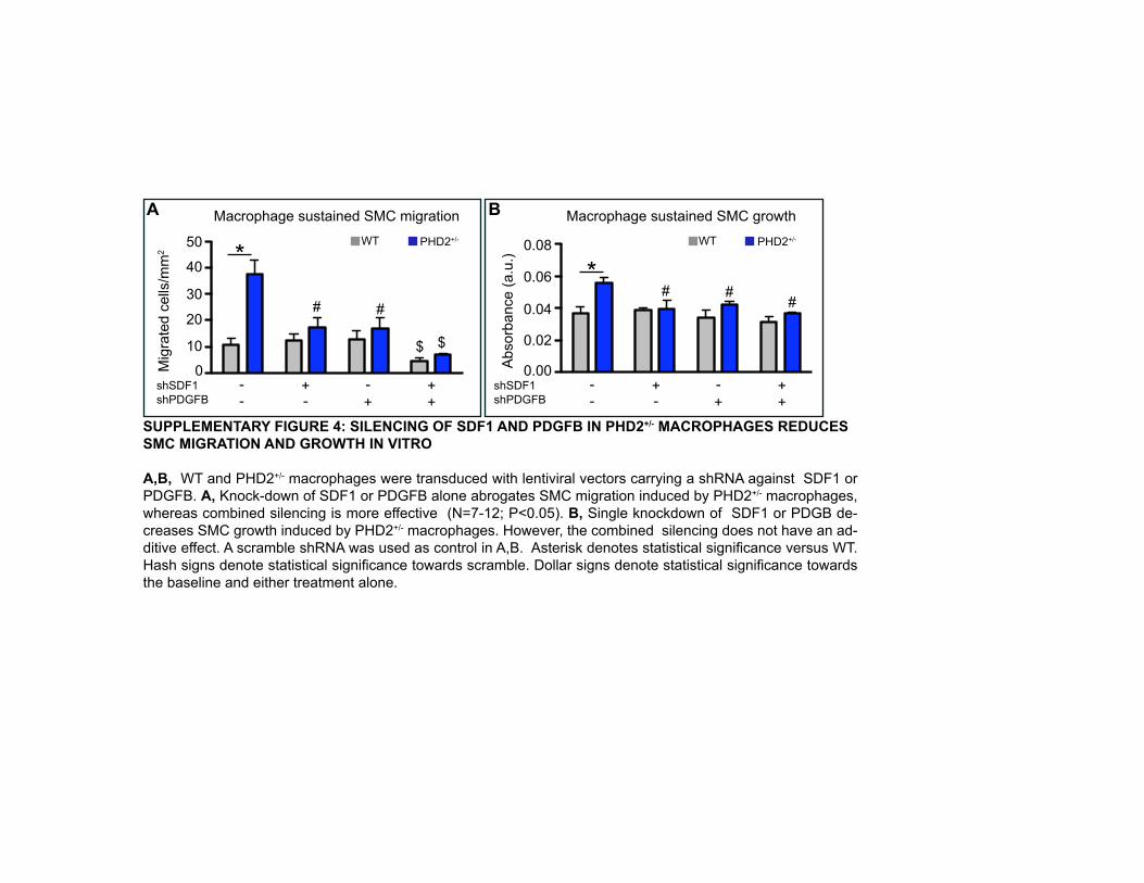
SUPPLEMENTARY FIGURE 4: SILENCING OF SDF1 AND PDGFB IN PHD2+/- MACROPHAGES REDUCES SMC MIGRATION AND GROWTH IN VITRO
A,B, WT and PHD2+/- macrophages were transduced with lentiviral vectors carrying a shRNA against SDF1 or PDGFB. A, Knock-down of SDF1 or PDGFB alone abrogates SMC migration induced by PHD2+/- macrophages, whereas combined silencing is more effective (N=7-12; P<0.05). B, Single knockdown of SDF1 or PDGB de-creases SMC growth induced by PHD2+/- macrophages. However, the combined silencing does not have an ad-GLWLYH�HIIHFW��$�VFUDPEOH�VK51$�ZDV�XVHG�DV�FRQWURO�LQ�$�%���$VWHULVN�GHQRWHV�VWDWLVWLFDO�VLJQL¿FDQFH�YHUVXV�:7��
+DVK�VLJQV�GHQRWH�VWDWLVWLFDO�VLJQL¿FDQFH�WRZDUGV�VFUDPEOH��'ROODU�VLJQV�GHQRWH�VWDWLVWLFDO�VLJQL¿FDQFH�WRZDUGV�
the baseline and either treatment alone.
A
10
0Mig
rate
d ce
lls/m
m2
shSDF1
20
30
shPDGFB- -- -
++
++
*WT PHD2+/-
Macrophage sustained SMC migration
$
40
50
# #
$
B
0.02
0.00shSDF1
0.04
0.06
shPDGFB- -- -
++
++
*WT PHD2+/-
Macrophage sustained SMC growth
0.08
# #
Abs
orba
nce
(a.u
.)
#

SS
C-A
SS
C-A
SS
C-A
SS
C-A
SS
C-A
SS
C-A
SS
C-A
SS
C-A
SS
C-A
SUPPLEMENTARY FIGURE 5: FLOW CYTOMETRIC ANALYSIS OF CIRCULATING MONOCYTES
A-C,�%ORRG�PRQRQXFOHDU�FHOOV�IURP�:7�PLFH�ZHUH�JDWHG�DV�LQ�WKH�LQVHW�LQ�$��DQG�DXWRÀXRUHVFHQFH�RI�WKH�JDWHG�FHOOV�ZDV�DQDO\]HG�XVLQJ�WKH�$3&��3(�
and FITC channels. D-F,�%ORRG�PRQRQXFOHDU�FHOOV�IURP�:7�PLFH�ZHUH�JDWHG�DV�LQ�WKH�LQVHW�LQ�'�DQG�DQDO\]HG�IRU�WKH�H[SUHVVLRQ�RI�WKH�PRQRF\WH�PDUNHU�
CD115 (D). CD115+�PRQRF\WHV��JDWH�LQ�'��ZHUH�WKHQ�DQDO\]HG�IRU�WKH�H[SUHVVLRQ�RI�7LH���(���&'���+7LH�+�7(0V��JDWH�LQ�(��ZHUH�IXUWKHU�DQDO\]HG�IRU�WKH�
H[SUHVVLRQ�RI�*U���)���G-I,�%ORRG�PRQRQXFOHDU�FHOOV�IURP�3+'�+/-�PLFH�ZHUH�JDWHG�DV�LQ�WKH�LQVHW�LQ�*�DQG�DQDO\]HG�DV�LQ�'�)�DERYH��J-L,�1HXWURSKLOV�ZHUH�LGHQWL¿HG�E\�WKHLU�OLJKW�VFDWWHULQJ�IHDWXUHV�DQG�JDWHG�DV�LQ�WKH�LQVHWV��([SUHVVLRQ�RI�*U��ZDV�WKHQ�DQDO\]HG�LQ�WKH�JDWHG�FHOOV�IURP�HLWKHU�:7��.��RU�
3+'�+/-��/��PLFH��DQG�XVHG�DV�*U�high�UHIHUHQFH�LQ�)�,��WKH�XQVWDLQHG�FRQWURO�LV�UHSUHVHQWHG�LQ�-��$VWHULVN�LQ�+�GHQRWHV�VWDWLWVWLFDO�VLJQL¿FDQFH�WRZDUGV�:7�
�1 ����3 ������
A
D
B C
E F
G H I
1R�DQWLERG\��$3&�
CD115+ ���������
7LH���3(� *U���),7&�&'�����$3&�
7LH���3(� *U�
CD115+ ���������
7LH�+ ���������
7LH�+ ���������� *U�Low/neg ����������
*U�Low/neg ����������
&'�����$3&�
unst
aine
d W
T 3+'�
+/-
1R�DQWLERG\��3(� 1R�DQWLERG\��),7&�
SS
C-A
SS
C-A
J L
*U���),7&�1R�DQWLERG\��),7&�
*U��FRQWUROV
unstained 3+'�+/-
*U���),7&�
K
SS
C-A
WT

SUPPLEMENTARY FIGURE 6: ACUTE DELETION OF ONE PHD2 ALLELE PROMOTES ARTERIOGENIC MACROPHAGES
A, PHD2Rosa26CreERT;lox/wt�SHULWRQHDO�PDFURSKDJHV�WUHDWHG�ZLWK���ȝ0���K\GUR[\WDPR[LIHQ�IRU����KRXUV�SUHVHQW�LQFUHDVHG�H[SUHVVLRQ�RI�
3'*)%��6')���DQG�7LH��UHVHPEOLQJ�WKH�SKHQRW\SH�RI�3+'�����PDFURSKDJHV��1 ���3��������B, WT mice transplanted with the bone PDUURZ�RI�3+'�Rosa26CreERT;lox/wt mice (HERosa26CreERT����:7��SUHVHQW�LQFUHDVHG�QXPEHU�RI�7LH�+�FLUFXODWLQJ�PRQRF\WHV��&'���+Tie2+ GRXEOH�SRVLWLYH�FHOOV��DIWHU�WDPR[LIHQ�WUHDWPHQW��1 ���3��������C,�7DPR[LIHQ�WUHDWHG�+(Rosa26CreERT WT mice present increased QXPEHU�RI��QG�DQG��UG�JHQHUDWLRQ�FROODWHUDO�YHVVHOV�FRPSDUHG�WR�XQWUHDWHG�PLFH�DW�EDVHOLQH��1 �������3��������D,����KRXUV�DIWHU�IHPRUDO�DUWHU\�OLJDWLRQ��WDPR[LIHQ�WUHDWHG�+(Rosa26CreERT����:7�PLFH�SUHVHQW�UHGXFHG�QHFURWLF�DUHD�FRPSDUHG�WR�XQWUHDWHG�DQLPDOV��
$VWHULVNV�LQ�$�%�&��DQG�'�GHQRWH�VWDWLVWLFDO�VLJQL¿FDQFH��3�������FRPSDUHG�WR�XQWUHDWHG�+(Rosa26CreERT����:7�PLFH��
�
�
2
6
�
&'���
+ Tie
2+ �FHOOV�����
7LH��H[SUHVVLQJ��PRQRF\WHV�LQ�
blood (HERosa26CreERT�����:7�
B
Vehicle 7DPR[LIHQTie2PDGFB SDF1PHD2
�
�
2
6
*HQH�SUR¿OH�RI�3+'�Rosa26CreERT;lox/wt�S02
Vehicle 7DPR[LIHQ
)ROG�FKDQJH�UHODWLYH�WR�FRQWURO
*
A
*
**
*
1XPEHU�RI�FROODWHUDOV
Vehicle 7DPR[LIHQ
�QG�DQG��UG�JHQHUDWLRQ�FROODWHUDO�EUDQFKHV�
(HERosa26CreERT����:7�
��
�
�
��
C
* *
2nd generation �UG�JHQHUDWLRQ
��
�
�
��
��
D
Vehicle 7DPR[LIHQ
1HFURWLF�DUHD����
��
Ischemic necrosis (HERosa26CreERT����:7�
��
*

Fold
cha
nge
rela
tive
to w
t/wt
5
0
1
10
PHD3PHD1 PHD2
Expression of PHDs in PHD2 heterozygous and null macrophages (LysCre+)
15
*
lox/wt lox/lox
*
SUPPLEMENTARY FIGURE 7: PHD EXPRESSION IN PHD2 HETEROZYGOUS AND PHD2 NULL MACROPHAGES RNA levels of PHD1, PHD2, and PHD3 in macrophages from PHD2LysCre;wt/wt, PHD2LysCre;lox/wt, and PHD2LysCre;lox/lox (labeled as wt/wt, lox/wt, and lox/lox respectively) mice. As expected, PHD2 OHYHOV�ZHUH�VLJQL¿FDQWO\�GHFUHDVHG�LQ�3+'�LysCre;lox/wt and PHD2LysCre;lox/lox macrophages. PHD1 and PHD3 transcript levels were higher in PHD2 heterozygous and null macrophages (N=4; 3��������$VWHULVNV�GHQRWH�VWDWLVWLFDO�VLJQL¿FDQFH��3�������FRPSDUHG�WR�ZW�ZW�PDFURSKDJHV�
*
* *
*2 3

A NF- B activity in ECs after TNF- stimulation
Lum
inis
cenc
e (a
.u) 600
10
20
0
400200
SUPPLEMENTARY FIGURE 8: NF-KB IS SELECTIVELY ACTIVATED IN PHD2+/- MACROPHAGES
A, Histogram showing comparable NF- B activity in WT and PHD2+/- ECs at baseline; stimulation with TNF- (20 ng/mL, 8 hours) unleashes NF- B activity to the same level in both genotypes (N=4, P< 0.05). B,� 3KDUPDFRORJLFDO� LQKLELWLRQ� RI�1)�ț%�DFWLYLW\� UHGXFHV� WKH� H[SUHVVLRQ� RI�3'*)%��6')��DQG�7LH�� LQ�3+'�� KDSORGH¿FLHQW�PDFURSKDJHV� �1 ����� 3��������$VWHULVNV� GHQRWH� VWDWLVWLFDO� VLJQL¿FDQFH� �3�������
FRPSDUHG�WR�:7�YHKLFOH� WUHDWHG�PDFURSKDJHV��+DVK�VLJQV�GHQRWH�VWDWLVWLFDO�VLJQL¿FDQFH�FRPSDUHG�WR�
FRQWURO�LQ�$�RU�WR�:7�YHKLFOH�LQ�%��
TNF-
WT PHD2+/-
Control
# #
�
1
B
2
0Fold
cha
nge
rela
tive
to
WT
vehi
cle
Tie26')�
4
TEM signature after NF- B inhibition
*
3'*)%
##
5
#
**
#
VehicleVehicle
NF- B inhibitorNF- B inhibitor
WT PHD2+/- WT PHD2+/- WT PHD2+/-

SUPPLEMENTARY FIGURE 9: SCHEME OF COLLATERAL ARTERIAL GROWTH IN WT AND PHD2+/- MICE
Collateral vessels are preexisting small conduits (shown in panel a) that undergo growth and maturation in case of occlusion of the major arterial flow, a process named arteriogenesis. PHD2+/- mice are preadapted to ischemia and display enhanced collateralization at baseline (panel b). The absence of one PHD2 allele in monocytes unleashes NF-κB signals that expand the reservoir of Tie2-expressing monocytes (TEMs) in the blood. Once extravasated into the pericollateral region, they differentiate into Tie2-expressing macrophages and promote collateral vessel maturation and homeostasis. SDF1 and PDGFB production by TEMs induces smooth muscle cell/progenitor recruitment and proliferation, ultimately leading to more abundant, larger, thicker, mature, and functional collateral arteries. In ischemia, shear stress-induced cytokines recruit circulating TEMs in WT mice (panel c). Tissue infiltrating TEMs foster arteriogenesis in a NF-κB dependent fashion and newly formed collateral arteries will partly reestablish blood flow. In PHD2+/- mice, femoral artery occlusion also induces extravasation and recruitment of TEMs, but to a lower extent (panel d), likely because of their collateral vessel preconditioning at baseline.
Tie2-expressing macrophage
Tie2-expressing monocyte Endothelial cell
Smooth muscle cell
Macrophage-borne SDF1
Macrophage-borne PDGFB
ISCHEMIA BASELINE
• Collateral growth and maintenance • SMC/SMP recruitment • SMC proliferation
PHD2+/-
release of shear-stress induced cytokines
WT PHD2+/- WT a) b) c) d)
NF-κB off! NF-κB on! NF-κB on! NF-κB on!
![Inducing Patent Infringement - Law Review...2005] Inducing Patent Infringement 229 understanding, inducing infringement is a natural outgrowth of the common law principle of respondeat](https://img.pdfslide.us/doc/110x75/5f9608d795a783197246401f/inducing-patent-infringement-law-review-2005-inducing-patent-infringement.jpg)