Embed Size (px)
Citation preview

Morphological Recovery in the Reafrached RetinaDon H. Anderson, Christopher J. Guerin, Page A. Erickson, Walter H. Stern, and Steven K. Fisher
After experimental retinal detachment in the cat, a number of morphological changes take place inretinal and RPE cells. Following reattachment, the ultrastructural relationship between the photoreceptorsand the RPE is re-established, but it does not return to the predetachment state even after short detachmentepisodes coupled with prolonged recovery periods. All of the reattached retinae show some degree ofabnormality, ranging from subtle changes in photoreceptor ultrastructure to dramatic degenerative effectsin the outer retina. Abrupt transitions in morphology from one reattached area to an adjacent area arenot unusual. Photoreceptor recovery varies widely between animals, and between adjacent regions withinthe same retina. Ensheathment of outer segments by RPE apical processes is abnormal. In some reattachedareas rod outer segment dimensions and disc structure are near normal as is the displacement rate ofrod outer segment discs. In others, especially in areas of RPE or Miiller cell proliferation and hypertrophy,the outer segments are shortened or absent completely, and there is a reduction of cell bodies in theouter nuclear layer. In some retinae, recovery in cones is inferior to that in rods. At short detachmentdurations (<1 wk) morphological recovery in the reattached retina is optimal while at long intervals(>1 month) recovery is poor. The changes at the photoreceptor-RPE interface identified in the reattachedcat retina probably have adverse effects on visual recovery when they occur within the human macula.Invest Ophthalmol Vis Sci 27:168-183, 1986
When the retina is detached from the retinal pigmentepithelium (RPE)* normal vision is impaired, and anumber of morphological changes occur in retinal andRPE cells.1"5 After the retina is reattached to the RPE,a complex process of recovery begins that can lead topartial, and sometimes nearly complete, restoration ofvision.6 The fact that at least partial vision is restoredin most human patients implies that the adverse ana-tomical and physiological effects of retinal detachmentcan be halted or even reversed after successful reat-tachment. The cellular events in this recovery processare not clearly understood, however, and those vari-ables which significantly influence the outcome havenot been rigorously defined. Histopathological obser-vations on human reattached retinas have not beenreported. Therefore, our current understanding of vi-
From the Institute of Environmental Stress, Neurosciences Re-search Program; the Department of Biological Sciences, Universityof California, Santa Barbara; and the Department of Ophthalmology,*School of Medicine, University of California, San Francisco.
Supported in part by Research Grants EY-02082 (DHA), EY-00888(SKF), and EY-03228 (WHS) from the National Eye Institute, Na-tional Institutes of Health.
Submitted for publication: February 26, 1985.Reprint requests: Don H. Anderson, PhD, IES, Neurosciences Re-
search Program, University of California, Santa Barbara, CA 93106.* Abbreviations used in the text: OLM = Outer Limiting Mem-
brane; SRS = Subretinal Space; ONL = Outer Nuclear Layer; ROS= Rod Outer Segment; DNA = Deoxyribonucleic Acid; RPE = Ret-inal Pigment Epithelium; SF6 = Sulfur Hexafloride gas; ARGs= Autoradiograms; COS = Cone Outer Segment.
sual recovery after reattachment is derived solely fromanimal models of experimental reattachment,7"" andfrom clinical and psychophysical studies of visual re-covery in human patients.12"19
Recently we introduced a new experimental modelof retinal detachment by documenting the degenerativeand proliferative changes that occur in the cat retinaas a function of detachment duration.3"5 In this studywe report our findings in the reattached cat retina, em-phasizing the cellular changes at the photoreceptor-RPE interface. After reattachment, morphology doesnot return to normal even after prolonged recoveryperiods (ie, 6 months); instead, a modified version ofthe normal photoreceptor-RPE relationship is re-es-tablished. Overall morphological recovery is optimalat the shortest detachment durations (less than 1 week),and is less successful at longer intervals (greater than1 month). These morphological abnormalities in thereattached cat retina probably underlie the lack ofcomplete visual recovery after detachment in humans,especially if such changes occur within the macula.
Materials and MethodsAnimals
The eyes from 16 cats with unilateral retinal de-tachments were reattached and subsequently fixed forelectron microscopy at selected intervals after re-attachment. The detachment (d) and reattachment (r)intervals (in days) were as follows: ld:10r, ld:14r, 3d:
168

No. 2 RECOVERY AFTER RETINAL REATTACHMENT / Anderson er ol. 169
7r, 3d:30r, 3d:210r, 5d:lr, 5d:7r(2), 7d:210r(2), 8d:7r,10d:7r, lld:3r, 14d:30r, 42d:30r, 42d:210r. Theboundaries and other characteristics of each de-tachment were recorded on a standard ophthalmic de-tachment chart immediately after surgery. After de-tachment, and periodically after reattachment,the experimental eyes were examined by indirectophthalmoscopy. After perfusion fixation, eyecupswere examined and the fundi were photographed usinga 35-mm camera fitted with a 50-mm macro lens. Theanimals were killed several hours after the onset of light(8L:16D schedule) by deep anesthetization followedby intracardiac perfusion of fixative. All animals werecared for and treated in accordance with the ARVOResolution on the Use of Animals in Research.
Surgery
Unilateral retinal detachments were produced in catsusing a refined version of the procedure recently pub-lished elsewhere.4 Animals were anesthetized with anintramuscular injection of ketamine HC1 (100 mg/ml)and acepromazine maleate (10 mg/ml), and main-tained under deep anesthesia with periodic injectionsof ketamine. For local anesthesia a retrobulbar injectionof 0.5 cc of lidocaine (2%) was given. Extracapsularlens extraction was performed leaving the posteriorcapsule intact. The corneal incision was closed and theeye allowed to heal for a minimum of 2 wk. For thedetachment surgery an infusion cannula was sewn inplace in the inferotemporal quadrant of the clear corneausing a 5.0 nylon mattress suture. A 20-gauge incisionwas made in the superotemporal quadrant of the corneaand an Ocutome (CooperVision; Irvine, CA) was usedto remove the posterior capsule and vitreous. After vi-trectomy a fluid-gas exchange was performed. A glassmicropipette with a flat 80-100 /im tip diameter wasmounted on a micromanipulator and then inserted intothe incision. Using a syringe fitted to a Harvard infusionpump, sodium hyaluronate (Healon, Pharmacia; Upp-sala, Sweden 0.5 mg/ml) was injected slowly as thepipette was advanced into the retina. When the pipettetip reached the subretinal space (SRS) a small blebformed creating a retinal detachment. The detachmentwas enlarged to the desired size by regulating theamount of Healon injected into the SRS. Retinae werereattached using the following procedure: Animals wereanesthetized as before. A 5.0 dacron mattress suturewas placed in the inferotemporal quadrant of the cor-nea approximately 1.5 mm from the limbus. A 20-gauge incision was made and an infusion cannula wassecured. A second 20-gauge incision was made in thesuperotemporal quadrant. Air was infused through thecannula and fluid was drained using a 20-gauge bluntbeveled tip on a Charles fluted needle. A mixture of
50 or 75% sulfur hexafluoride (SF6) and air was flushedthrough the eye until a complete exchange wasachieved. Finally, the incisions were sealed, and theeye was allowed to heal. In several animals intraocularpressure was monitored by pneumotonometry at 6-hrintervals over a 24-hr period. No abnormal pressureelevations were detected. No cryotherapy or photo-coagulation was used around the retinal hole. In severalanimals, the retina flattened spontaneously without re-quiring surgery.
Light and Electron Microscopic Autoradiography
The fixation protocol and staining techniques em-ployed in this study have been previously described.4Light microscopic autoradiograms were prepared asfollows: 1-2 mCi of 3H-Leucine in 0.2 ml of phosphatebuffer was injected intraocularly 24 or 48 hr prior tofixation; or, 200 mCi of 3H-thymidine in buffer wasinjected 2 hr prior to fixation. After fixation and em-beddment in Araldite (6005), l-/um sections were pro-cessed for light microscopic autoradiography by thedipping method. Sections were placed on cleaned glassmicroscope slides. The slides were dipped into a 1:1aqueous solution of NTB-2 nuclear track emulsion(Eastman Kodak; Rochester, NY) under sodium vaporillumination. After drying, they were transferred tolight-tight slide boxes and exposed in the dark at 4°Cfor 3-7 days. The slides were developed in full-strengthD-19 at 20°C for 2 min, stopped in distilled H2O, fixed,for 10 min, and washed in running deionized H2O.Autoradiograms were stained sequentially with a mix-ture of Azure II and methylene blue, and then withbasic fuchsin as described previously. Electron micro-scope autoradiograms were prepared according to amethod modified from Young and Droz.20 Thin sec-tions (60-80 nm) were transferred to parlodian-coated(1% in isoamyl acetate) glass microscope slides. Theslides were stained with aqueous uranyl acetate andlead citrate, coated with a thin layer of carbon by vac-uum evaporation, and dipped (under sodium vaporillumination) into a 1:4 aqueous solution of IlfordL-4 nuclear track emulsion. After drying, the dippedslides were placed in light-tight boxes along with packetsof dessicant, sealed, and exposed at 6°C for 2-4months. The slides were then developed in freshly pre-pared phenidon developer for 1 min at 15°C, fixed in2 changes of 30% thiosulfate, and washed in runningdistilled H2O for 15 min. Next, the parlodian film wasfloated off the glass slides onto a surface of distilledH2O, and 200-mesh copper grids were placed directlyon top of the sections. The film and grids were thentransferred to filter paper and allowed to dry. Finally,the grids were cut away from the film and thinned byimmersion in isoamyl acetate for 2 min.
170 INVESTIGATIVE OPHTHALMOLOGY 6 VISUAL SCIENCE / February 1966 Vol. 27
• * • >
Fig. 1. An area of folding in a reattached retina. There is outer segment debris and a few cell bodies within the SRS (arrows). The encircledareas show locations where abnormal extensions of the apical RPE processes contact the outer segments. Note the pyknotic ONL nuclei adjacentto the fold. (42d:30r)(Xl,125).
Light Microscopic Measurements
Measurements of outer segment length were ob-tained using a Zeiss Universal Research Microscope(Carl Zeiss, Inc.; Oberkochen, West Germany) and100X oil immersion objective (final magnification= X1250). The outer segments were carefully alignedalong their longitudinal axes by systematically varyingthe tissue cutting angle. One-micron sections were cutand stained using the protocol described above. Anocular micrometer was used to measure the width ofthe outer segment layer. Repeated measurements weremade at approximately 500-/im intervals in multipletissue blocks in order to provide a rough estimate ofthe range of outer segment lengths for a given area ofreattachment.
ResultsAt low magnification in the light microscope, most
of the reattached retinae show few obvious signs ofdisruption. The normal stratification of the retina isusually preserved, although in retinae detached forlengthy intervals, some reduction in the thickness ofthe outer nuclear layer is usually apparent. At highermagnification all of the reattached retinae show somedegree of abnormality, ranging from subtle changes at
the outer segment-RPE interface to dramatic degen-erative effects in the outer retina. In addition, we notedthat within the area of reattachment adjacent retinalregions vary considerably in their degree of abnor-mality. Abrupt transitions in morphology from oneregion to an adjacent region in the same eye are notunusual. As such, it is impossible to establish a precisetimetable of morphological recovery or to identify crit-ical periods in the recovery process with certainty. Butwe were able to identify a number of general trends,and we did find reattached regions in different retinaethat shared many characteristics. Therefore we groupedthese recurring morphological patterns into several dif-ferent categories, and these are described below.
Retinal Folds
About 60% of the reattached retinae have occasionalpleats or tucks where the outer nuclear layer (ONL) isfolded over upon itself. As shown in Figure 1, the pho-toreceptor outer segments face each other rather thanapposing the apical surface of the RPE which is notfolded. Some of the larger folds form small bulges thatproject out into the vitreous. Within the folds, the outersegments are reduced in number, with individual outersegments appearing abnormally short and disorganized.

No. 2 RECOVERY AFTER RETINAL REATTACHMENT / Anderson er ol.
Fig. 2. Electron micro-graph from an area of reat-tached retina (3d:210r). Inmany reattached regions,such as this one, the config-uration of the apical RPEprocesses and their interdig-itation with the outer seg-ments is highly unusual.The dimensions and organi-zation of some regeneratedROSs are normal. However,they do not appear in adensely packed, parallel row.(X 18,500).
The space between the outer segments contains la-mellar debris and phagocytic cells that have many ofthe characteristics of RPE cells. We have called thesecells RPE phagocytes to distinguish them from blood-borne phagocytic cells.4 Proliferating RPE cells as wellas Mtiller cell processes are often found at the base ofthe folds next to the apical RPE surface. Pyknotic cellbodies, primarily in the outer nuclear layer (Fig. 1),are also evident within and adjacent to the folds.
The Apical RPE Surface
The apical RPE surface acquires a mounded or scal-loped profile shortly after the cat retina is detached.
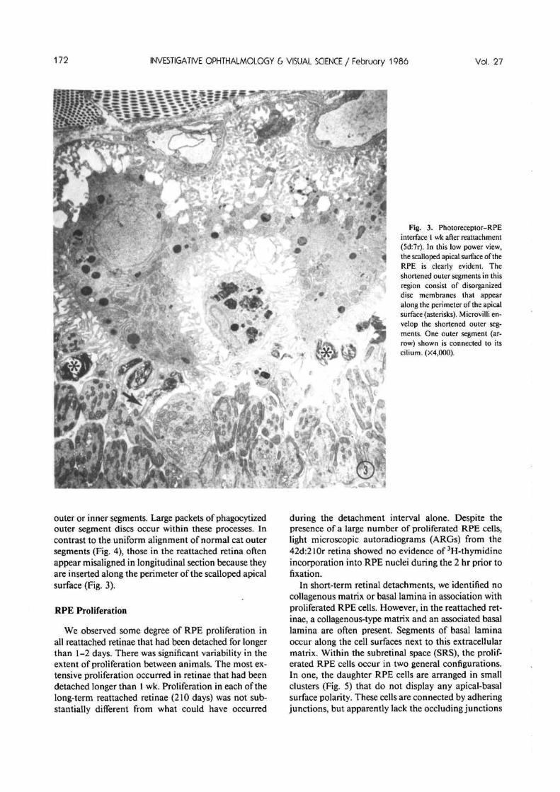
This mounding persists in the reattached retina, andis evident to varying degrees in almost all reattachedregions from both short and long-term reattachments(see Figs. 2-3). Apical processes project from themounded surface and contact the photoreceptor outersegments or, in the absence of outer segments, the distalinner segment tips. In some of the long-term reattach-ments, closely packed, parallel arrays of sheet-like pro-cesses, similar to the sheaths that normally envelop catcone outer segments, emerge from the mounded apicalsurface (Fig. 2). These processes interdigitate with bothrod and cone outer segments and appear more nu-merous than in normal cat retina (Fig. 4). Some arraysfollow long and tortuous routes before they contact the
INVESTIGATIVE OPHTHALMOLOGY & VISUAL SCIENCE / Februory 1986 Vol. 27
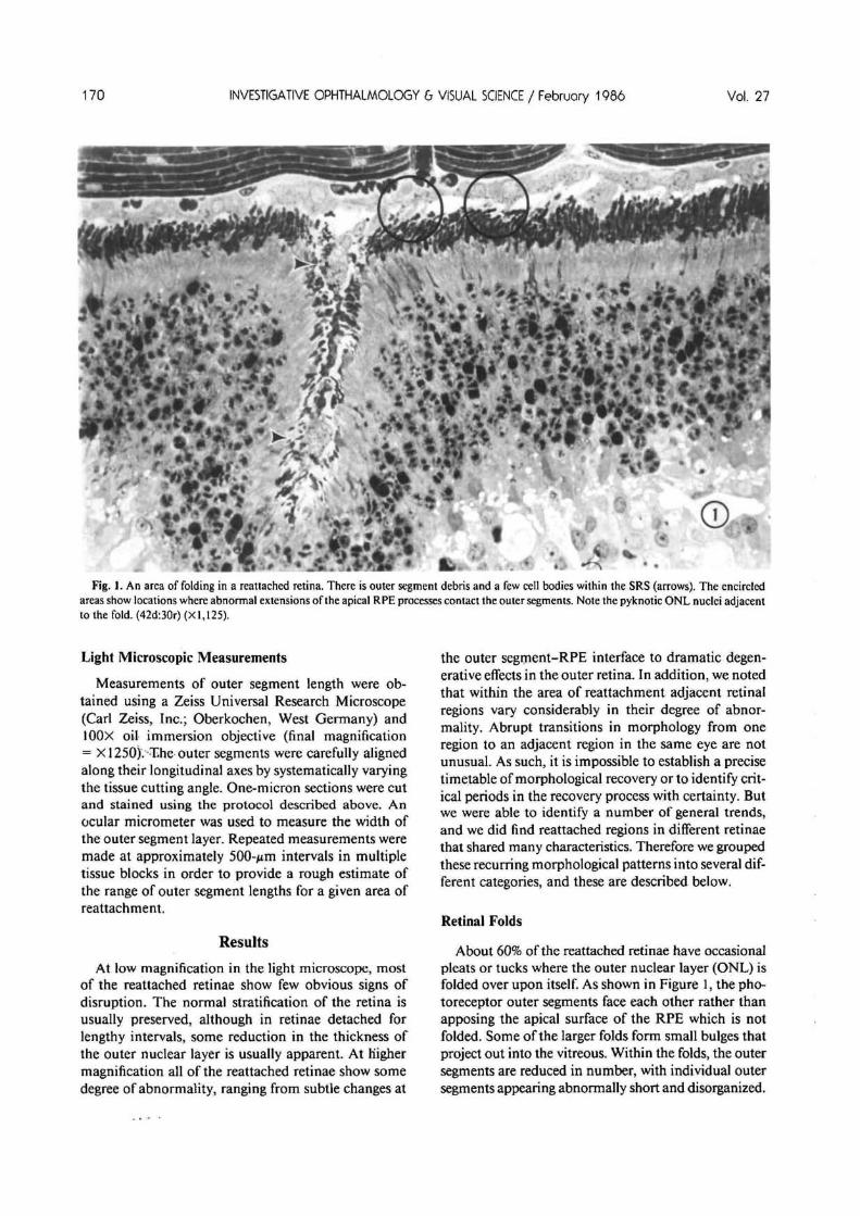
Fig. 3. Photoreceptor-RPEinterface I wk after reattachment(5d:7r). In this low power view,the scalloped apical surface of theRPE is clearly evident. Theshortened outer segments in thisregion consist of disorganizeddisc membranes that appearalong the perimeter of the apicalsurface (asterisks). Microvilli en-velop the shortened outer seg-ments. One outer segment (ar-row) shown is connected to itscilium. (X4,000).
. .4
outer or inner segments. Large packets of phagocytizedouter segment discs occur within these processes. Incontrast to the uniform alignment of normal cat outersegments (Fig. 4), those in the reattached retina oftenappear misaligned in longitudinal section because theyare inserted along the perimeter of the scalloped apicalsurface (Fig. 3).
RPE Proliferation
We observed some degree of RPE proliferation inall reattached retinae that had been detached for longerthan 1-2 days. There was significant variability in theextent of proliferation between animals. The most ex-tensive proliferation occurred in retinae that had beendetached longer than 1 wk. Proliferation in each of thelong-term reattached retinae (210 days) was not sub-stantially different from what could have occurred
during the detachment interval alone. Despite thepresence of a large number of proliferated RPE cells,light microscopic autoradiograms (ARGs) from the42d:210r retina showed no evidence of 3H-thymidineincorporation into RPE nuclei during the 2 hr prior tofixation.
In short-term retinal detachments, we identified nocollagenous matrix or basal lamina in association withproliferated RPE cells. However, in the reattached ret-inae, a collagenous-type matrix and an associated basallamina are often present. Segments of basal laminaoccur along the cell surfaces next to this extracellularmatrix. Within the subretinal space (SRS), the prolif-erated RPE cells occur in two general configurations.In one, the daughter RPE cells are arranged in smallclusters (Fig. 5) that do not display any apical-basalsurface polarity. These cells are connected by adheringjunctions, but apparently lack the occluding junctions
No. 2 RECOVERY AFTER RETINAL REATTACHMENT / Anderson er ol. 173
Fig. 4. Electron micrograph of the photoreceptorand RPE in the normal cat retina. The outer segmerits are oriented along their longitudinal axes iia uniform, densely packed row. Cone outer segments (COS) are somewhat shorter than rods (ROSand are ensheathed by a complex array of apicaprocesses known as the cone sheath (CS). The apicasurface of the RPE is not scalloped or mounded ait is after detachment and reattachment. (x 11,500)
and gap junctions found normally in RPE cells. In theother configuration, the new cells form additionalmonolayers of polarized cells that parallel the originalmonolayer (Figs. 6, 7). The surface polarities of thenew monolayers need not be the same as the originalone. For example, the polarities of the three monolayersshown in Figure 6 (L^-I^) are reversed with respect to
each other. The polarity of L2 is the opposite of theoriginal monolayer (Li) and, similarly, the polarity ofL3 is the reverse of L2. The net result is that the apicalsurfaces of Li and L2 and the basal surfaces of L2 andL3 appose each other.
When the RPE cells assume a multi-layered config-uration and when the additional layer adjacent to the
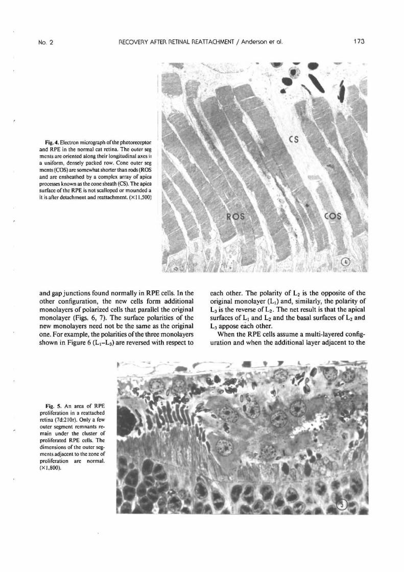
Fig. 5. An area of RPEproliferation in a reattachedretina (7d:210r). Only a fewouter segment remnants re-main under the cluster ofproliferated RPE cells. Thedimensions of the outer seg-ments adjacent to the zone ofproliferation are normal.(X] ,800).

174 INVESTIGATIVE OPHTHALMOLOGY & VI5UAL SCIENCE / February 1986 Vol. 27
Fig. 6. An area of multi-layered RPE in a reattached retina (t4d:30r). Three monolayers of RPE cells are present (Li, L2, L3), each displayingdifferent surface polarity. The apical surfaces of L, and L2 face each other, as do the basal surfaces of L2 and L3. The basal lamina of L2 isclearly evident (arrows). Only outer segment fragments (asterisk) appear near the inner segment tips. A number of pyknotic ONL cell bodiesare also apparent. (X800).
outer segments expresses the correct surface polarity,apical processes from the new monolayer interdigitatewith the outer segments and phagocytize shed packetsof discs. The cells that comprise the new monolayercontain a number of inclusions, including phagosomes,while inclusions are notably absent in the cells thatmake up the original monolayer (Fig. 7).
Outer segment recovery directly underlying a clusterof proliferated RPE cells is usually poor. Rod outer
segments (ROSs) underlying a region of proliferatedcells are usually less than one half the length of ROSsin the immediately adjacent region (Fig. 5). Inner seg-ments remain intact, but the number of mitochondriaand other organelles is reduced when compared tocontrols. The same pattern occurs on a smaller scalenear so-called hyperpigmented RPE cells. These cells,which usually occur singly or in association with nor-mally pigmented RPE cells, are interspersed through-
Mg. /. in tnis reattacnea region, trie second monolayer ot Kft cells (L2) displays normal surface polarity. I tie cells have an even basal borderand long microvillous processes (arrows) extend from the highly mounded apical surface to contact the outer segments. (42d:30r) (X800).
No. 2 RECOVERY AFTER RETINAL REATTACHMENT / Anderson er ol. 175
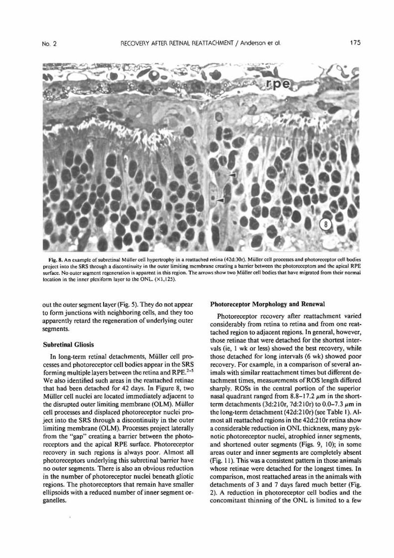
Fig. 8. An example of subretinal Miiller cell hypertrophy in a reattached retina (42d:3Or). Miiller cell processes and photoreceptor cell bodiesproject into the SRS through a discontinuity in the outer limiting membrane creating a barrier between the photoreceptors and the apical RPEsurface. No outer segment regeneration is apparent in this region. The arrows show two Miiller cell bodies that have migrated from their normallocation in the inner plexiform layer to the ONL. (XI, 125).
out the outer segment layer (Fig. 5). They do not appearto form junctions with neighboring cells, and they tooapparently retard the regeneration of underlying outersegments.
Subretinal Gliosis
In long-term retinal detachments, Miiller cell pro-cesses and photoreceptor cell bodies appear in the SRSforming multiple layers between the retina and RPE.2"5
We also identified such areas in the reattached retinaethat had been detached for 42 days. In Figure 8, twoMiiller cell nuclei are located immediately adjacent tothe disrupted outer limiting membrane (OLM). Miillercell processes and displaced photoreceptor nuclei pro-ject into the SRS through a discontinuity in the outerlimiting membrane (OLM). Processes project laterallyfrom the "gap" creating a barrier between the photo-receptors and the apical RPE surface. Photoreceptorrecovery in such regions is always poor. Almost allphotoreceptors underlying this subretinal barrier haveno outer segments. There is also an obvious reductionin the number of photoreceptor nuclei beneath glioticregions. The photoreceptors that remain have smallerellipsoids with a reduced number of inner segment or-ganelles.
Photoreceptor Morphology and Renewal
Photoreceptor recovery after reattachment variedconsiderably from retina to retina and from one reat-tached region to adjacent regions. In general, however,those retinae that were detached for the shortest inter-vals (ie, 1 wk or less) showed the best recovery, whilethose detached for long intervals (6 wk) showed poorrecovery. For example, in a comparison of several an-imals with similar reattachment times but different de-tachment times, measurements of ROS length differedsharply. ROSs in the central portion of the superiornasal quadrant ranged from 8.8-17.2 pm in the short-term detachments (3d:210r, 7d:210r) to 0.0-7.3 /im inthe long-term detachment (42d:2 lOr) (see Table 1). Al-most all reattached regions in the 42d:2 lOr retina showa considerable reduction in ONL thickness, many pyk-notic photoreceptor nuclei, atrophied inner segments,and shortened outer segments (Figs. 9, 10); in someareas outer and inner segments are completely absent(Fig. 11). This was a consistent pattern in those animalswhose retinae were detached for the longest times. Incomparison, most reattached areas in the animals withdetachments of 3 and 7 days fared much better (Fig.2). A reduction in photoreceptor cell bodies and theconcomitant thinning of the ONL is limited to a few

176 INVESTIGATIVE OPHTHALMOLOGY & VISUAL SCIENCE / February 1986 Vol. 27
Table 1. Range of outer segment lengths afterretinal reattachment
Detachmentperiod
0 (normal)5 (days)58
103
144237
42
Reattachmentperiod
07 (days)777
303030
210210210
Outer segment length(
Rods
range)
Cones
11.0-18.2 (am) 8.8-13.1 (nm)5.5-7.31.8-9.17.3-14.63.5-7.09.1-14.65.1-13.93.6-14.68.8-17.2
12.8-16.40.0-7.3
2.2-3.6*
7.0-8.0*
9.0-10.02.8-3.5
*8.8-13.13.6-7.3
•
* Measurements unavailable because of disruption of the outer segment layer.
locations only. In some areas, ROS length as well asouter segment structure appears to be normal. Electronmicroscope autoradiograms in the 3d:210r animalshow an advancing front of radiolabeled protein po-sitioned 4-5 fttn from the outer segment bases 48 hrafter an intravitreal injection of 3H-Leucine (Fig. 12).A similar displacement occurred in ROSs from theopposite control eye. In an adjacent area from the sameanimal (3d:210r) (Fig. 13), the outer segments aretruncated and disorganized. No discrete band of labeledprotein can be identified, but the silver grains over theouter segment fragments indicate that newly synthe-sized protein was incorporated into the outer segmentsduring the preceding 48 hr. A comparison between theanimals whose retinae were reattached for 30 daysshows similar results. ROS length in the retina detachedfor 3 days is normal in most regions (Fig. 14A), butmany ROSs in the retina detached for 42 days are
shortened, and there is evidence of pyknosis in the ONL(Fig. 14B).
ROSs are also abnormally short in those animalsreattached for brief periods (1 wk). Measurements fromthe central area of the superior nasal quadrant in 4such animals (l0d:7r, 8d:7r, 5d:7r(2)) yielded ROSranges of 3.5-7.0 ftm, 7.3-14.6 jim, 1.8-9.1 fim, and5.5-7.3 jtm respectively (Table 1) (see Figure 14C-E).This compares with values of 11.0-18.2 nm for ROSsat similar locations in normal control eyes. Each of thefour animals was injected with 3H-Leucine intraocu-larly 24 hr prior to fixation. Light microscopic ARGsfrom the reattached areas show a concentration of la-beled protein at the expected location close to the outersegment bases.
Although ROS length is normal in several of thereattached retinae, photoreceptor inner and outer seg-ments often retain subtle ultrastructural anomalies. Insome cases, an extension of the rod inner segment ap-pears alongside the outer segment plasma membranecreating an asymmetrical expansion of the ellipsoid.In other cases, there is a slight reduction in inner seg-ment mitochondrial density, or an apparent elongationof the ellipsoid.
In many of the reattached retinae, cone outer seg-ments (COS) were virtually impossible to identify be-cause of the misalignment and disorganization of theouter segment layer. Therefore, we were able to obtainCOS length data only from reattached retinae wherethe outer segment layer was not disrupted. In severalof these retinae, we noted that the COSs appearedshorter than normal, with wide variability in theirlengths. In the superior retina outside of the area cen-tralis, COSs in normal cat retinae are about 60% ofthe ROS length, approximately 6-7 nm in the peripheryand 10-13 nm nearer the posterior pole (in Araldite-
Fig. 9. After lengthy detachment episodes followed by long-term reattachment, the outer segments are usually shortened, and the number ofnuclei in the ONL is reduced. The apical RPE surface retains the mounded profile acquired after detachment. (42:210 (X800).

No. 2 RECOVERY AFTER RETINAL REATTACHMENT / Anderson er ol. 177
Fig. 10. In an electronmicrograph from the areashown in Figure 9 (42d:210r)the regenerated outer seg-ments are shortened sub-stantially and inner segmentmitochondria appear lessnumerous. However the ba-sic organization of the discstack, its ensheathment bythe apical processes and itsrelationship to the connect-ing cilium are still intact(X6,600).
Fig. 11. An electron mi-crograph from the retina de-scribed in Figures 9, 10 (42d:21 Or), There is no evidenceof any outer or inner seg-ments in this region. Only thephotoreceptor cell bodies,including the nucleus and asmall amount of cytoplasm,remain. The junctions whichcomprise the outer limitingmembrane are apposed to theapical RPE surface <X3,2OO).

178 INVESTIGATIVE OPHTHALMOLOGY G VISUAL SCIENCE / February 1986 Vol. 27
Fig. 12. Electron mi-croscope autoradiogram ofROSs in a reattached retina(3d:2lOr). Forty-eight hoursprior to fixation, 3H-Leucinewas injected intraocularly.An advancing front of labeledprotein is positioned 4-5 ^mfrom the ROS bases<X7,5OO).
embedded tissue). The values are somewhat broaderin the reattached retinae (Table 1). In two of the ani-mals (5d:7r, 14d:30r) COSs are quite short (2.2-3.6 /*m) (Fig. 14D), whereas in two others (3d:l80r,3d:3Or) their length appears near normal (8.8-13.1
Short COSs may retain a normal cylindrical organi-zation, but contain many fewer discs than normal outersegments. The disc membrane at the outer segmentbase, the site of disc morphogenesis, is structurallynormal. The most basal discs are continuous with the
Fig. 13. Electron mi-croscope autoradiogram ofROSs in an area adjacent tothat in Figure 12. No patternis observed, but the labelingof the disorganized discmembranes indicates thatnewly synthesized proteinwas incorporated into theouter segments during thepreceding 48 hr (X6,000).

No. 2 RECOVERY AFTER RETINAL REATTACHMENT / Anderson er ol. 179
Fig. 14. (A) The photorecep-tor-RPE interface after short-term detachment and 1-monthreattachment. After short de-tachment intervals the outer seg-ments can reattain normal lengthin a relatively short time frame.Discrete areas of disruption,however, can still be identified.Note the migrating cone nucleusin the center of the field (arrow)(3d:3Or) (X800). (B) In contrastto 14A, lengthy detachment in-tervals (42 days) coupled witha 30-day reattachment period(42d:3Or) result in distinctly in-ferior outer segment regenera-tion. Note the pyknotic photo-receptor nuclei in the ONL(X800). (C) Light micrographfrom a retina detached for S daysand reattached for 7 days (Sd:7r).There is virtually no evidence ofouter segment regrowth in thisregion. The apical RPE processesextend down to the inner seg-ment tips. This probably repre-sents one of the earliest stages inthe recovery process (X800). (D)Light micrograph from a secondanimal whose retina was de-tached for S days and reattachedfor 7 days (5d:7r). In this case theROSs are approximately one-halfnormal length. The 3 COSsshown in the field (arrows) arealso abnormally short (X800).(E) The photoreceptors and RPEin a retina detached for 8 daysand reattached for 7 days (8d:7r).In this region there is a widerange of outer segment lengths.The outer segment tips are po-sitioned along the perimeter ofthe scalloped surface, and thatmay account for the unevenspacing between outer segments.Displaced cone nucleus (arrow)(X800).
;• a .>;*• t
plasma membrane on the centric face of the connectingcilium (Fig. 15). Sheet-like RPE processes contact theshortened outer segments. Cilia with associated striatedrootlets and basal bodies can sometimes be identifiedwithin these RPE processes. When the COS is not pres-ent or limited to only a few disorganized disc mem-
branes, the processes ensheath the distal portion of thecone inner segment.
The distribution and number of organdies in suchcones are also affected. Organelles that are usually seg-regated in the myoid and ellipsoid regions are distrib-uted throughout a single, smaller compartment. Mi-

INVESTIGATIVE OPHTHALMOLOGY & VISUAL SCIENCE / February 1986 Vol. 27
Fig. 15. Cone photoreceptor ini retina detached for 7 days andeattached for 210 days. TheX)S is structurally normal, butt contains many fewer discs thanisual. The inner segment is:omposed of a single compart-nent that includes the nucleusNu), the remaining mitochon-lria of the ellipsoid, Golgi ap->arati (g) and rough endoplasmiceticulum (X7,000).
tochondrial density is much reduced, and many conenuclei are displaced into the inner segment from theirnormal locations just vitread to the outer limitingmembrane (Fig. 15). Electron microscope autoradio-grams show that, 48 hr after intravitreal injection of3H-leucine, most of the labeling is found over the conenuclei in these cells whereas cones from the controleye show most labeling over the disc membranes withvery little nuclear labeling.
Discussion
Experimental studies of reattachment have been re-ported previously in the rabbit,7 owl moneky,8"91' andrhesus monkey10 retinas. In the previous monkey stud-ies, detachments were produced by injecting a solution
of hyaluronidase into the vitreous cavity. After me-chanical disruption and repeated aspiration of the vit-reous, 0.2 cc of digested vitreous was injected with suf-ficient pressure to produce a retinal hole and local de-tachment, which usually progressed to a totaldetachment.10 We used a different method to producedetachments. After a core vitrectomy, a glass micro-pipette was used to inject a dilute solution of sodiumhyaluronate (Healon; 5 mg/ml, Pharmacia; Uppsala,Sweden) directly into the subretinal space. Hyaluronicacid is a naturally occurring component of the inter-photoreceptor matrix,21 and is commonly found inhuman subretinal fluid samples in a wide range of con-centrations (0.01-33.5 mg/ml).22 We cannot rule outthe posibility that injected Na-hyaluronate influences

No. 2 RECOVERY AFTER RETINAL REATTACHMENT / Anderson er ol. 181
the process of reattachment and the resulting mor-phology. In some locations, it can be observed histo-logically in the subretinal space after brief detachmentepisodes. In reattachments, it was rarely identified bylight or electron microscopy, so it is presumably me-tabolized or otherwise removed from the subretinalspace during the detachment/reattachment interval.Therefore, Na-hyaluronate appears to be an appropri-ate substance to use where experimental control is re-quired over detachment duration, size, and retinal lo-cation.
A comparison of previous studies with the presentone yields a number of similarities as well as someinteresting differences. The retinae of all species ex-amined had reattached regions where the outer retinallayers appear folded or buckled. In the owl monkey,this occurred in animals that had been detached forlong intervals (8-12 wk).8"9 In the cat, we found nofirm correlation between detachment duration andretinal folding. In the owl monkey, detachments of lessthan 7 days showed little RPE mounding upon reat-tachment.8 Cat retinae detached for shorter intervals(ie, 3 days) show pronounced mounding that persistsafter reattachment. Irregularities in and "thinning" ofthe nuclear layers are present in the monkey reattach-ments detached for ^8 wk.8 In the cat reattachments,there is a definite reduction in the number of photo-receptor nuclei in retinas detached ^ 1 month, in areasof folded retina, or in regions of RPE or Miiller cellproliferation; intermittent areas of pyknosis occur atthe shortest detachment intervals examined. In the owlmonkey pyknotic nuclei were not identified after twodays reattachment irrespective of the detachment in-terval.8 Kroll and Machemer10 observed disc mem-brane-like profiles within the distal inner segments ofrhesus monkey rods and cones that they interpreted asan aberration of disc morphogenesis. In the cat wefound no instances of disc membranes in the innersegment cytoplasm.
There is very little data on the rates of disc synthesisor disposal after reattachment. One study" suggeststhat the outer segments undergo a phase of acclerateddisc synthesis within the first 24 hr, but the disc dis-placement rate could not be measured directly due toouter segment disruption. The same limitation appliesto the cat retinae reattached for brief intervals. How-ever, several relevant inferences can be drawn from thecat retinae reattached for 1 wk. ARGs from these an-imals show a band of labeled protein at the ROS basesat the normal time, 24 hr after injection of 3H-aminoacids. Estimates of ROS length 1 wk after reattachmentare clearly less than those of the controls (see Table 1;Fig. 14C-E). This suggests that regenerating outer seg-ments must take longer than 1 wk to reattain normallength. If ROSs were to maintain new disc synthesisat the normal rate (2.3 /xm/day)23 immediately after
reattachment, the outer segments could achieve normallength (12-18 ixm) about 1 wk later—but only in theabsence of any disc shedding. Because maintenance ofconstant outer segment length is a function of both thedisc synthesis and disposal rates,24 regenerating outersegments probably take several weeks or longer beforethey achieve equilibrium. The fact that some outersegments are abnormally short many months afterreattachment suggests that their reduced length maybe sustained indefinitely.
Secondly, there is a surprisingly wide range of ROSlengths within individual retinae and between differentretinae with similar detachment/reattachment times.This may reflect inherent variability in the detachment/reattachment process itself. Some areas may lag behindothers in forming a close photoreceptor-RPE apposi-tion. The extent of separation between the retina andRPE during detachment could also influence the re-covery process.
The regenerative capacity of COSs may be inferiorto that in rods, although the evidence is equivocal.Rhesus COSs, but not ROSs, reportedly show ultra-structural abnormalities one month after reattach-ment.10 In the cat, COSs in early reattachments aresubstantially shorter than control outer segments inthe same or opposite eye. In addition, nuclear dis-placement and the disruption of inner segment com-partmentalization is limited almost exclusively tocones. This displacement of photoreceptor organellesis morphologically identical to the early stages of aphenomenon recently identified in normal rat and hu-man retinae where photoreceptor cells are eventuallydisplaced into the subretinal space.25"26 It is possiblethat these changes are early stages in cell degenerationand, therefore, reflect a selective loss of cone photo-receptors in the reattached retina. We expect that reat-tachment studies currently underway in the primatemacula will help resolve this issue.
Whether or not photoreceptors can recover com-pletely from an episode of detachment has not beenresolved. Previous studies in experimental animalsreached different conclusions. Nakamura7 reportedlimited and variable outer segment recovery in rabbitretinae that spontaneously reattached without requiringany surgical manipulations. In contrast, Kroll andMachemer9"10 concluded that ". . . the prominentchanges of a detached retina are reversible, so that areattached retina can look normal after approximately4 wk. This is true for all detachments of up to 12-wkduration." At the light microscope level, the reattachedcat retina can sometimes appear histologically normal.However, ultrastructural morphology does not returnto the pre-detachment state even after brief episodesof detachment coupled with prolonged recovery peri-ods. The photoreceptor mosaic in the reattached catretina is a literal microscopic patchwork of different

182 INVESTIGATIVE OPHTHALMOLOGY G VISUAL SCIENCE / February 1986 Vol. 27
regions each with different characteristics. The fact thatsome rod and cone outer segments do not reattain nor-mal length, even after prolonged reattachment periods,implies some maintained imbalance in the assemblyand/or disposal phases of the outer segment renewalprocess.
The tendency of both RPE and Miiller cells to pro-liferate after detachment results in similar detrimentaleffects after reattachment. Miiller cell processes ex-tending into the SRS act as a cellular barrier that pre-vents RPE-photoreceptor reapposition. ProliferatedRPE cells also produce an abnormal layer of interven-ing cellular material. Outer segment recovery whereMiiller cell processes have invaded the SRS is invariablypoor, but in areas of RPE proliferation the extent ofrecovery depends upon the configuration and surfacepolarity of the proliferated cells. Morphologically, sub-retinal proliferation is very similar in the cat and hu-man retinas.27"28 Future efforts aimed at suppressingproliferation and hypertrophy in these two cell typescould lead to improved visual recovery after reattach-ment.
Re-establishment of the Photoreceptor-RPEInterface: Early Events
The initial molecular interactions that occur betweenthe photoreceptors and RPE cells after reattachmentremain obscure. The photoreceptors are not reapposedto their original loci on the apical RPE surface. Howthen is the ensheathment of individual rod and coneouter segments by the apical processes re-establishedand, secondly, how is the regrowth of the outer seg-ments regulated? At present, the specific events in thisprocess as well as the precise time course are mattersfor speculation. But our results in the reattached retinasuggest a number of parallels between initial photo-receptor development and the recovery that ensues afterreattachment.
Immediately after reattachment, the tips of the pho-toreceptor inner segments are juxtaposed to themounded and undifferentiated apical surface of theRPE (see Fig. 14C). Undefined metabolic processeswhich promote RPE-photoreceptor adhesion tend tomaintain that apposition.29"30 The apposition of thedetached retina must engage a sequence of molecularevents that elicits and controls the re-differentiation ofthe RPE apical surface and the re-ensheathment of thephotoreceptors by the apical processes. Because theensheathment of rod and cone outer segments is dif-ferent in many mammals,31 there must be a mechanismfor regulating the configuration of apical microvilli tothe two photoreceptor classes. This is virtually the sameprocess that occurs during initial development.
Results in the reattached retina, as well as in thedeveloping retina,32 confirm that the apical processes
appear before the outer segments are present. They arelonger and more numerous in the reattached comparedto normal retina. The point at which the outer segmentscease to elongate during the recovery phase must de-pend upon the net difference in the rates of disc pro-duction and disposal, just as it does in the developingretina.24 In the reattached retina, however, persistingabnormalities in either or both of these processes maylead to substantial regional variation in outer segmentlengths. Furthermore, regenerated outer segments arenot oriented normally in a single parallel row. Instead,they appear irregularly oriented in longitudinal sectionbecause the topography of the apical RPE surface hasbeen permanently altered during detachment.
Functional Implications
The morphological changes described above couldhave significant consequences for the recovery of visionafter reattachment. A widespread loss of photoreceptorcells, especially cones, could have obvious and irre-versible effects on acuity, color vision, photopic sen-sitivity, and stereopsis could be affected secondarily.Misalignment of regenerated outer segments could af-fect aspects of visual sensitivity.17 Many of the mor-phological changes we observed affect underlying pho-toreceptor cells in many localized regions—perhapsproducing small multiple defects in the visual field. Inexperimentally detached retinae, these phenomenatend to occur with limited frequency or in small cir-cumscribed regions, especially at short detachmentdurations. Therefore, their functional effects may besimilarly restricted to small retinal regions. Althoughreattached retinae may be interlaced with many suchregions where photoreceptors and perhaps other retinalcells are functionally abnormal or degenerate, the def-icits may be insignificant from the standpoint of overallvisual capacity.
On the other hand, such changes could have pro-found effects on visual recovery if they occur withinthe macula. In humans, the area centralis has a di-ameter of 5.5 mm, and the foveola measures approx-imately 350 nm in diameter.33 Retinal changes thatmight otherwise be inconsequential in the peripheralretina may be quite significant if they occur in thissmall, but highly specialized, retinal location.
The presence of a macular detachment is associatedwith persistently reduced acuity, metamorphopsia, andwith chronic disturbances in color vision (primarilytritanomalous defects) after reattachment.6141618"19
Recent evidence strongly suggests that most variationin final acuity is attributable to macular detachmentduration.19 Because different mammalian retinae re-spond similarly to experimental detachment, we expectthat the morphological changes identified in the reat-tached cat retina also occur in the reattached primate
No. 2 RECOVERY AFTER RETINAL REATTACHMENT / Anderson er ol. 183
macula. Our initial results in the detached and reat-tached rhesus monkey fovea support that conclusion.*These changes at the photoreceptor-RPE interface be-gin to provide an explanation, at the cellular level, forthe poor visual recovery associated with lengthy de-tachment durations, and for the lack of complete visualrecovery after macular detachment in humans.
In summary, we conclude that a modified versionof the normal intercellular relationship between thephotoreceptors and the RPE cells is re-established afterreattachment. The morphological abnormalities are nothomogeneous across the retina. They range from subtleultrastructural anomalies at the outer segment-RPEinterface to dramatic degenerative and proliferativechanges in the outer retina. The degree of recoverydepends broadly upon detachment and reattachmentparameters. The best recovery is associated with shortdetachment periods, but a return to completely normalretinal morphology is probably unattainable. If we as-sume that this modified relationship between the pho-toreceptors and the RPE also occurs after retinal reat-tachment in humans,* it is apparently sufficient tosubserve near normal vision under ideal circumstances.Key words: reattachment, detachment, photoreceptor, RPE,retina
References1. Kroll AJ and Machemer R: Experimental retinal detachment in
the owl monkey: III. Electron microscopy of retina and pigmentepithelium. Am J Ophthalmol 66:410, 1968.
2. Machemer R and Laqua H: Pigment epithelial proliferation inretinal detachment (massive periretinal proliferation). Am JOphthalmol 80:1, 1975.
3. Anderson DH, Stern WH, Fisher SK, Erickson PA, and BorgulaGA: The onset of pigment epithelial proliferation after retinaldetachment. Invest Ophthalmol Vis Sci 21:10, 1981.
4. Anderson DH, Stern WH, Fisher SK, Erickson PA, and BorgulaGA: Retinal detachment in the cat: The pigment epithelial-pho-toreceptor interface. Invest Ophthalmol Vis Sci 24:906, 1983.
5. Erickson PA, Fisher SK, Anderson DH, Stern WH, and BorgulaGA: Retinal detachment in the cat: The outer nuclear and outerplexiform layers. Invest Ophthalmol Vis Sci 24:927, 1983.
6. McPherson AR, O'Malley RE, Butner RW, and Beltangady SS:Visual acuity after surgery for retinal detachment with macularinvolvement. Ann Ophthalmol 14:639, 1982.
7. Nakamura S: Studies on experimental retinal detachment. Mor-phological observations. Part 2. Further observations of experi-mentally detached retina as well as observations of reattachedretina. Acta Societatis Ophthalmologica Japanicae 71:520, 1967.
8. Machemer R: Experimental retinal detachment in the owl mon-key. IV. The reattached retina. Am J Ophthalmol 66:1075, 1968.
9. Kroll AJ and Machemer R: Experimental retinal detachment inthe owl monkey. V. Electron microscopy of the reattached retina.Am J Ophthalmol 67:117, 1969a.
10. Kroll AJ and Machemer R: Experimental retinal detachment
* Our unpublished results in the Rhesus monkey strongly suggestthat virtually all of the changes described in the cat also occur in thereattached primate macula.
and reattachment in the rhesus monkey. Am J Ophthalmol 68:58, 1969b.
11. Kroll AJ and Machemer R: Experimental retinal detachmentand reattachment in the owl monkey. VIII. Photoreceptor proteinrenewal in early retinal reattachment. Am J Ophthalmol 72:356,1971.
12. Gundy MF and Davies EWG: Recovery of visual acuity afterretinal detachment surgery. Am J Ophthalmol 77:310, 1974.
13. Grupposso SS: Visual acuity following surgery for retinal de-tachment. Arch Ophthalmol 93:327, 1975.
14. Chisholm I A, McClure E, and Foulds WS: Functional recoveryof the retina after retinal detachment. Trans Ophthalmol SocUK 95:167, 1975.
15. Kressig I, Lincoff H, and Witassek B: Color vision and otherparameters of macular function after retinal reattachment. DevOphthalmol 2:77, 1977.
16. deary PE and Leaver PK: Macular abnormalities in the reat-tached retina. Br J Ophthalmol 62:595, 1978.
17. Fitzgerald CR, Enoch JM, Birch DG, Benedetto MD, TemmeLA, and Dawson WW: Anomalous pigment epithelial photo-receptor relationships and receptor orientation. Invest Ophthal-mol Vis Sci 19:956, 1980.
18. Tani P, Robertson DM, and Langworthy A: Prognosis for centralvision and anatomic reattachment in rhegmatogenous retinaldetachment with macula detached. Am J Ophthalmol 92:611,1981.
19. Burton TC: Recovery of visual acuity after retinal detachmentinvolving the macula. Tr Am Ophthalmol Soc 80:475, 1982.
20. Young RW and Droz B: The renewal of protein in retinal rodsand cones. J Cell Biol 39:169, 1968.
21. Bach G and Berman ER: Amino sugar-containing compoundsof the retina. I. Isolation and identification. Biochim BiophysActa 252:453, 1971.
22. Sweeny DB: Biochemistry of subretinal fluid. In ControversialAspects of the Management of Retinal Detachment, SchepensCL and Regan CD, editors. Boston, MA, Little Brown and Co.,1965, pp. 315-316.
23. Fisher SK, Pfeffer BA, and Anderson DH: Both rod and conedisc shedding are related to light onset in the cat. Invest Ophthal-mol Vis Sci 24:844, 1983.
24. LaVail MM: Kinetics of rod outer segment renewal in the de-veloping mouse retina. J Cell Biol 58:650, 1973.
25. Lai YL: Outward movement of photoreceptor cells in normalrat retina. Invest Ophthalmol Vis Sci 19:849, 1980.
26. Lai YL, Masuda K, Mangum MD, Lug R, Macrae DW, FletcherG, and Liu YP: Subretinal displacement of photoreceptor nucleiin human retina. Exp Eye Res 34:219, 1982.
27. Sternberg P and Machemer R: Subretinal proliferation. Am JOphthalmol 98:456, 1984.
28. Federman JL, Folberg R, Ridley M, and Arbizo VA: Subretinalcellular bands. Trans Am Ophthalmol Soc 81:172, 1983.
29. Marmor MF, Abdul-Rahim AS, and Cohen DS: The effect ofmetabolic inhibitors on retinal adhesion and subretinal fluid re-sorption. Invest Ophthalmol Vis Sci 19:893, 1980.
30. Zauberman H: Adhesive forces between the retinal pigment ep-ithelium and sensory retina. In The Retinal Pigment Epithelium,Zinn KM and Marmor MF, editors. Cambridge, MA, HarvardUniv Press, 1979, p. 226.
31. Fisher SK and Steinberg RH: Origin and organization of pigmentepithelial apical projections to cones in cat retina. J Comp Neurol206:131, 1982.
32. Hollenberg MJ and Spira AW: Human retinal development: ul-trastructure of the outer retina. Am J Anat 137:357, 1973.
33. Hogan MJ, Alvarado JA, and Weddell JE: In Histology of theHuman Eye, Ch 9, Philadelphia. WB Saunders Co, 1971, p. 491.