Embed Size (px)
Citation preview

Lymphatic System• Tissues, organs, vessels
and a fluid called lymph– similar to interstitial fluid
• Diffuse lymphatic tissue– Tonsils, mucosa associated
lymph tissue and red bone marrow
• Organs involved– thymus
– spleen
– lymph nodes
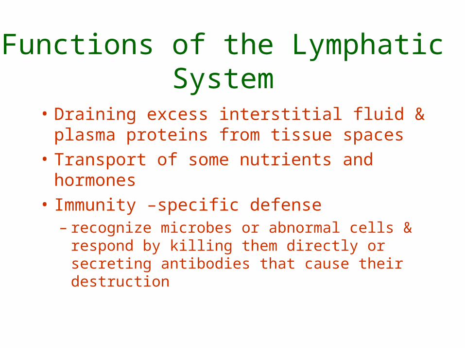
Functions of the Lymphatic System
• Draining excess interstitial fluid & plasma proteins from tissue spaces
• Transport of some nutrients and hormones
• Immunity –specific defense– recognize microbes or abnormal cells &
respond by killing them directly or secreting antibodies that cause their destruction
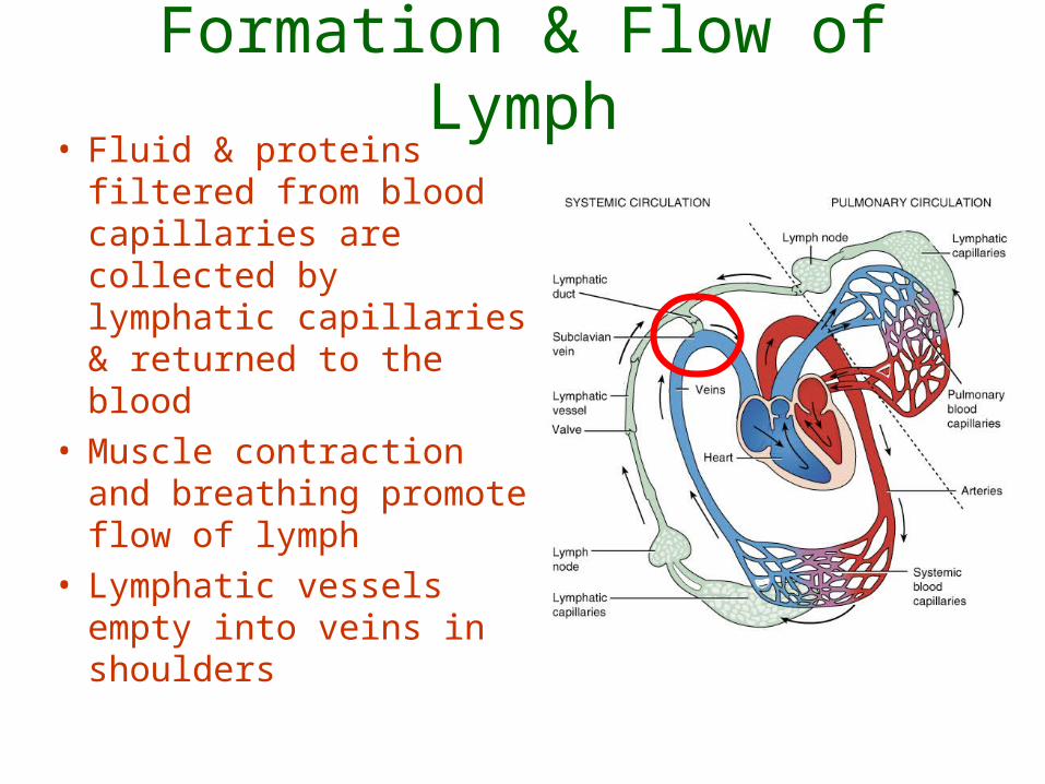
Formation & Flow of Lymph• Fluid & proteins filtered
from blood capillaries are collected by lymphatic capillaries & returned to the blood
• Muscle contraction and breathing promote flow of lymph
• Lymphatic vessels empty into veins in shoulders

Lymph Circulation • Lymphatic capillaries
– Capillaries are closed-ended tubes in tissues– One-way valves– Pickup fluid (lymph) from Tissues

Lymph Circulation
• LymphaticVessels– Lymph passes from capillaries into lymph vessels– Resemble veins with thin walls & valves– Afferrent lymphatics carry lymph to nodes– Efferent lymphatics carry lymph from nodes
• Lymphatic Trunks – Lymph passes from lymphatic vessels into trunks– *Bronchomediastinal, jugular, subclavian, intestinal
and lumbar trunks
Lymph Circulation
• Lymphatic ducts– Two ducts drain lymph from lymphatic trunks
into the subclavian veins – Thoracic Duct: Larger one that drains most of
body lymph into the left subclavian vein– Right Lymphatic Duct: Smaller one that
drains right side of head, right shoulder and right arm into the right subclavian vein
Lymph Circulation
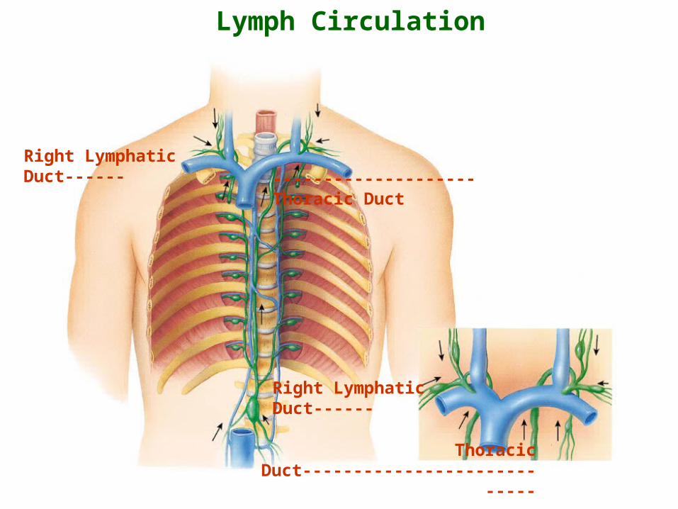
Right Lymphatic Duct--------------------------Thoracic Duct
Right Lymphatic Duct------
Thoracic Duct----------------------------
Lymphatic Pathways
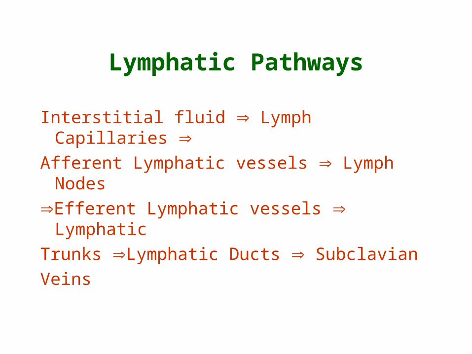
Interstitial fluid Lymph Capillaries Afferent Lymphatic vessels Lymph Nodes
Efferent Lymphatic vessels Lymphatic
Trunks Lymphatic Ducts Subclavian
Veins
Lymphocytes
• B-cell lymphocytes– Respond to antigens by dividing to produce
• Plasma cells: Antibody secreting cells• Memory B-cells
• T-cell lymphocytes – Respond to antigens by dividing to produce
• Cytotoxic T-cells that kill antigen-bearing cells• Helper T-cells that help activate other T-cells and B-cells• Memory T-cells
• Natural killer (NK) lymphocytes can kill invading cells and tumor cells without need to respond to antigens – nonspecific defense
Diffuse Lymphatic Tissues
• Lymphatic nodules – MALT (Mucosa Associated Lymph Tissues) – lymphatic nodules within the digestive and
respiratory systems – Small intestine -Peyer's patches – Appendix – Bronchi of respiratory tract
Diffuse Lymphatic Tissues
• Tonsils– Located in and around throat– Tonsilar crypts– Functions
• Crypts Trap microbes• Mount immune response against inhaled and
ingested microbes
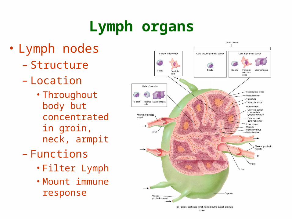
Lymph organs
• Lymph nodes – Structure– Location
• Throughout body but concentrated in groin, neck, armpit
– Functions • Filter Lymph
• Mount immune response
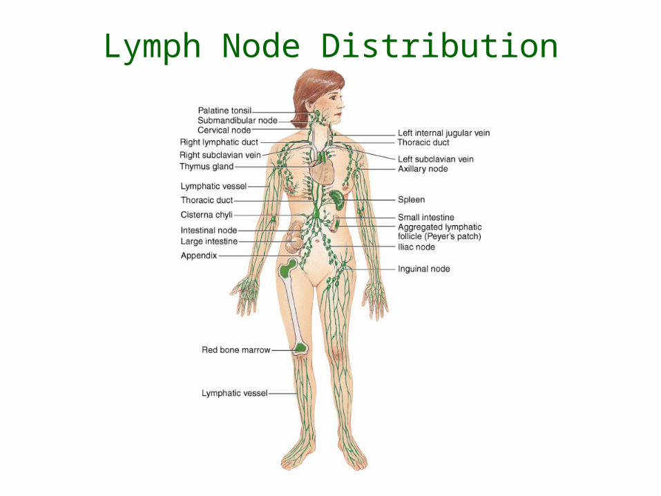
Lymph Node Distribution
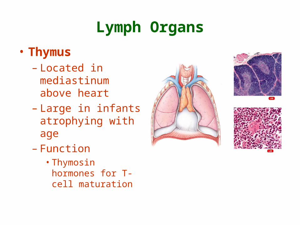
Lymph Organs
• Thymus – Located in
mediastinum above heart
– Large in infants atrophying with age
– Function• Thymosin hormones
for T-cell maturation

Spleen
• Located in upper left quadrant to left of stomach
• Functions – Filters blood– Produces
lymphocytes– Stores iron and
platelets
Defense
• Non-specific Defense– First Line Defense: External
• Skin
• Mucous Membranes
• Tears
• Saliva
• Stomach acid
Non-specific Defense
• Second Line Defense: Internal• Antimicrobial proteins
– Interferons – antiviral– Complement – immunity, allergies and inflammation
• Natural killer (NK) cells– Lymphocytes
– kill microbes and tumor cells
• Phagocytes– Wandering phagocytes– Fixed phagocytes
Non-specific Defense
• Second Line Defense: Internal
• Inflammation– Mast cells and basophils release chemicals
– Arterioles vasodilate – more blood to site
– Increased capillary permeability – more fluid into tissues
Non-specific Defense
• Inflammation results in– Redness
– Heat
– Swelling (edema)
– Pain
– Healing
Specific Defense: Immunnity
• Study of immunity is immunology
• Immunity versus non-specific defense– Specificity: responds to specific antigens– Memory: Second exposure to antigen causes a
stronger response
• Antigens– Foreign (non-self) chemicals– Cause immune response
Immunity
• B-cell and T-cell lymphocytes responsible for immunity
• Two types of immune responses– Cell mediated immunity: T-cells respond to
intracellular antigens such as virus infected cells and tumor cells
– Antibody mediated immunity: B-cells respond to extracellular antigens such as bacteria
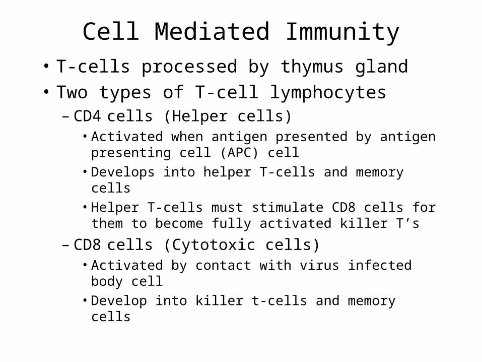
Cell Mediated Immunity
• T-cells processed by thymus gland
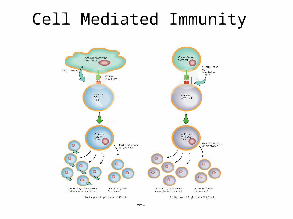
• Two types of T-cell lymphocytes– CD4 cells (Helper cells)
• Activated when antigen presented by antigen presenting cell (APC) cell
• Develops into helper T-cells and memory cells
• Helper T-cells must stimulate CD8 cells for them to become fully activated killer T’s
– CD8 cells (Cytotoxic cells) • Activated by contact with virus infected body cell
• Develop into killer t-cells and memory cells
Cell Mediated Immunity
• Cytotoxic T’s leave lymphatic tissue to search for and destroy virus infected cells, tumor cells and tissue transplant cells on contact
Cell Mediated Immunity

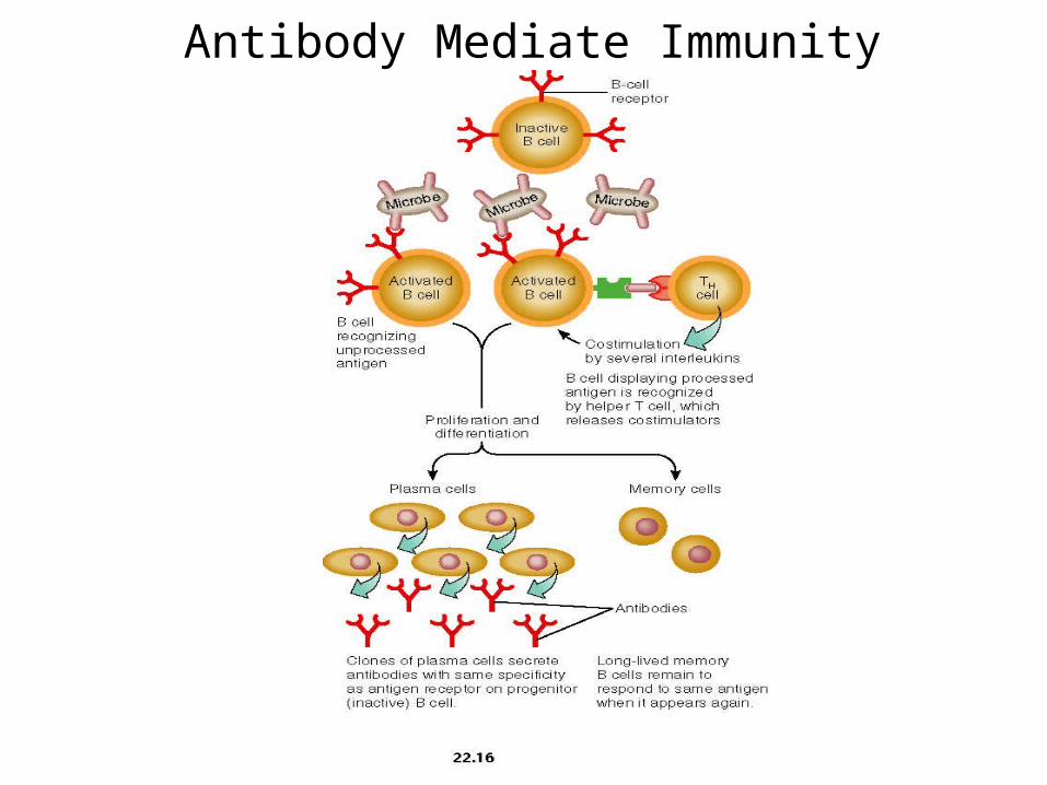
Antibody Mediated Immunity
• B-cell lymphocytes stay in lymph tissues
• Extracellular antigen enters lymph tissue and binds to B-cell receptors
• B-cells become activated – B-cells divide (clone) to form plasma cells and
memory cells– Helper T-cells bind to antigen on B-cells and
“help” stimulate plasma cell and memory cell formation
Antibody Mediated Immunity
• Plasma cells secrete various types of antibodies
• Antibodies bind to the specific antigen that activated its parent B-cells
• Antibodies cause the destruction of the antigen
Antibody Mediated Immunity
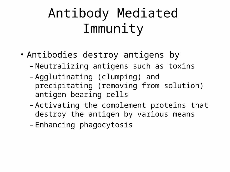
• Antibodies destroy antigens by– Neutralizing antigens such as toxins– Agglutinating (clumping) and precipitating
(removing from solution) antigen bearing cells– Activating the complement proteins that
destroy the antigen by various means– Enhancing phagocytosis
Antibody Mediate Immunity
Immune Response
Primary Response
• After an initial exposure to an antigen a slow rise in antibodies production occur first as immunoglobulin M (IgM) then (IgG)
Secondary Response
• After a subsequent exposure the antibodies production is far greater and is mainly (IgG)

Summary of Cell and Antibody Mediated Immunity