Embed Size (px)
Citation preview

Lymphatic System
• Lymphatic Vessels – transport only in the direction toward the heart .– Pick up the “extra” tissue fluid that capillaries leave– Lymph – the interstitial fluid carried by lymphatics
• Lymphatic Organs – “filtration” devices staged at various important junction points in the body


Lymph Vessels• Lymphatic capillaries – diffuse, highly permeable due to
presence of:– Minivalves loosely overlapping endothelial cells
– Collagen fiber attachment to surrounding structures prevents collapse of capillary
• Lymphatic collecting vessels like veins but thinner walled w/ more valves and anastomoses
• Lymphatic trunks – lumbar, bronchomediastinal, subclavian, jugular and intestinal
• Lymphatic ducts – Right Lymphatic Duct & Thoracic Duct


Lymphatic Capillaries• During inflammation, lymph capillaries can
absorb:– Cell debris– Pathogens– Cancer cells
• Cells in the lymph nodes cleanse and “examine” this debris
• Lacteals – specialized lymph capillaries present in intestinal mucosa– Absorb digested fat and deliver chyle to the blood

Lymphatic Trunks
• Lymph is delivered into one of two large trunks– Right lymphatic duct – drains the right upper
arm and the right side of the head and thorax– Thoracic duct – arises from the cisterna chyli
and drains the rest of the body


Lymph Transport
• The lymphatic system lacks a pumping organ
• Vessels are low-pressure conduits
• Uses the same methods as veins to propel lymph:– Pulsations of nearby arteries– Contractions of smooth muscle in the walls of
the lymphatics

Lymphoid Cells
• Lymphocytes are the main cells involved in the immune response
• Two main varieties:– T cells– B cells

Lymphocytes
• T cells and B cells protect the body against antigens
• Antigen – anything the body perceives as foreign– Bacteria and their toxins; viruses– Mismatched RBCs or cancer cells

Lymphocytes
• T cells – Manage the immune response– Attack and destroy foreign cells
• B cells – Produce plasma cells, which secrete antibodies– Antibodies immobilize antigens

Other Lymphoid Cells
• Macrophages – phagocytize foreign substances and help activate T cells
• Dendritic cells – spiny-looking cells with functions similar to macrophages
• Reticular cells – fibroblast–like cells that produce a stroma, or network, that supports other cell types in lymphoid organs

Lymphoid Tissue
• Diffuse lymphatic tissue – scattered reticular tissue elements in every body organ– Larger collections appear in the lamina propria of
mucous membranes and lymphoid organs
• Lymphatic follicles (nodules) – solid, spherical bodies consisting of tightly packed reticular elements and cells– Germinal center composed of dendritic and B cells– Found in isolation and as part of larger lymphoid
organs

Figure 20.4a
Lymph Nodes• Principal lymphoid organs of the body
• Embedded in connective tissue and clustered along lymphatic vessels
• Aggregations of these nodes occur near the body surface in inguinal, axillary, and cervical regions of the body

Lymph Nodes
• Two basic functions:– Filtration – macrophages destroy
microorganisms and debris– Immune system activation – monitor for
antigens and mount an attack against them

Figure 20.4a
Structure of a Lymph Node• Nodes are bean shaped and surrounded by a
fibrous capsule• Trabeculae extended inward from the
capsule and divide the node into compartments
• Nodes have two histologically distinct regions: a cortex and a medulla
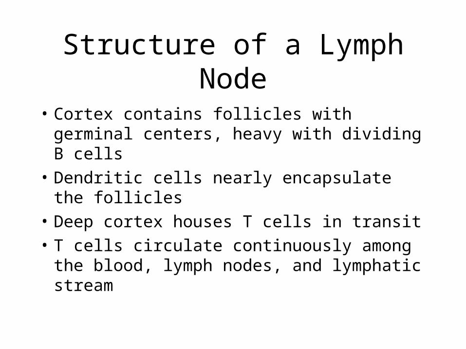
Structure of a Lymph Node
• Cortex contains follicles with germinal centers, heavy with dividing B cells
• Dendritic cells nearly encapsulate the follicles
• Deep cortex houses T cells in transit
• T cells circulate continuously among the blood, lymph nodes, and lymphatic stream

Structure of a Lymph Node
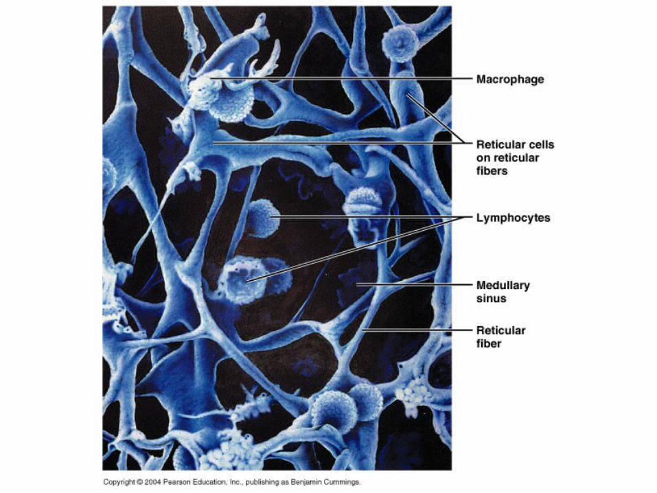
• Medullary cords extend from the cortex and contain B cells, T cells, and plasma cells
• Throughout the node are lymph sinuses crisscrossed by reticular fibers
• Macrophages reside on these fibers and phagocytize foreign matter

Structure of a Lymph Node
Figure 20.4a, b

Circulation in the Lymph Nodes• Lymph enters via afferent lymphatic vessels• It then enters a large subcapsular sinus and
travels into smaller sinuses• It meanders through these sinuses and exits
the node at the hilus via efferent vessels• Because there are fewer efferent vessels,
lymph stagnates somewhat in the node• This allows lymphocytes and macrophages
time to carry out protective functions

Lymph Vessel & Transport Disorders
• Lymphangitis – inflammation of the larger lymphatics vasa vasorum which congest with blood
• Lymphedema – collection of fluid in the interstitial, abdominal, and pleural spaces– blockage of drainage or loss after cancer
surgery– hypoproteinemia


Other Lymphoid Organs
• The spleen, thymus gland, and tonsils
• lymphatic tissue scattered in connective tissue
• All are composed of connective tissue
• All help protect the body
• Only lymph nodes filter lymph

Spleen
• Largest lymphoid organ, located on the left side of the abdominal cavity beneath the diaphragm
• It is served by the splenic artery and vein, which enter and exit at the hilus
• Functions:– Site of lymphocyte proliferation– Immune surveillance and response– Cleanses the blood

Additional Spleen Functions
• Stores breakdown products of RBCs for later reuse– Spleen macrophages salvage and store iron for
later use by bone marrow
• Site of fetal erythrocyte production (normally ceases after birth)
• Stores blood platelets

Structure of the Spleen• Surrounded by a fibrous capsule, it has
trabeculae that extend inward and contains lymphocytes, macrophages, and huge numbers of erythrocytes
• Two distinct areas:– White pulp – containing mostly lymphocytes
suspended on reticular fibers and involved in immune functions
– Red pulp – remaining splenic tissue concerned with disposing of worn-out RBCs and bloodborne pathogens


Thymus
• Young individuals
• Aging reduces it to fibrous/fatty tissue
• Creates immunocompetent T-cells– Unique since it DOESN’T get involved in the
immune reaction to antigens– Thymocytes reside in epithelial tissue rather
than reticulocytes

Thymus• A bilobed organ that secretes hormones
(thymosin and thymopoietin) that cause T lymphocytes to become immunocompetent
• Size of the thymus varies with age:– In infants, it is found in the inferior neck and
extends into the mediastinum where it partially overlies the heart
– It increases in size and is most active during childhood
– It stops growing during adolescence and then gradually atrophies

Internal Anatomy of the Thymus
• Thymic lobes contain an outer cortex and inner medulla
• Cortex contains densely packed lymphocytes and scattered macrophages
• Medulla contains fewer lymphocytes and thymic (Hassall’s) corpuscles

Thymus
• The thymus differs from other lymphoid organs in important ways– It functions strictly in T lymphocyte maturation– It does not directly fight antigens
• The stroma of the thymus consists of star-shaped epithelial cells (not reticular fibers)
• These thymocytes secrete the hormones that stimulate lymphocytes to become immunocompetent

Tonsils
• Simplest lymphoid organs; form a ring of lymphatic tissue around the pharynx
• Location:– Palatine tonsils – either side of the posterior end of
the oral cavity– Lingual tonsils – lie at the base of the tongue– Pharyngeal tonsil – posterior wall of the
nasopharynx– Tubal tonsils – surround the openings of the
auditory tubes into the pharynx
Tonsils
• Lymphoid tissue of tonsils contains follicles with germinal centers
• Tonsil masses are not fully encapsulated
• Epithelial tissue overlying tonsil masses invaginates, forming blind-ended crypts
• Crypts trap and destroy bacteria and particulate matter
Aggregates of Lymphoid Follicles
• Peyer’s patches – isolated clusters of lymphoid tissue, similar to tonsils– Found in the wall of the distal portion of the small
intestine– Similar structures are found in the appendix
• Peyer’s patches and the appendix:– Destroy bacteria, preventing them from breaching
the intestinal wall– Generate “memory” lymphocytes for long-term
immunity