Embed Size (px)
Citation preview
SCUOLA DI INGEGNERIA E ARCHITETTURA
Lunedì 23 Maggio, ore 9:00 – 11:00, Aula TA11, Via Terracini 28, Bologna Doppio seminario: Metodi avanzati per lo studio della meccanica del tessuto osseo
Dr. Gianluca Tozzi University of Portsmouth, UK
Digital Volume Correlation for the experimental investigation of bone tissue
and biomaterials MicroCT-based digital volume correlation (DVC) is becoming very popular in orthopaedic research. DVC enables the measurement of 3D full-field displacements and strains throughout the interior of a material/structure undergoing motion or deformation. This technique was originally developed by Bay et al. (1999) to investigate the strain distribution in trabecular bone. However, as DVC itself is relatively new as an experimental technique, a number of studies aimed (and are still aiming) at obtaining a better understanding of its reliability in the mechanical analysis of biological tissues and biomaterials. Moreover, an evaluation of how DVC could be potentially established as a diagnostic tool in combination with clinical CT has been recently considered. This may open new avenues for clinical CT diagnostics to promote a reliable and efficient use of DVC in clinical practice.
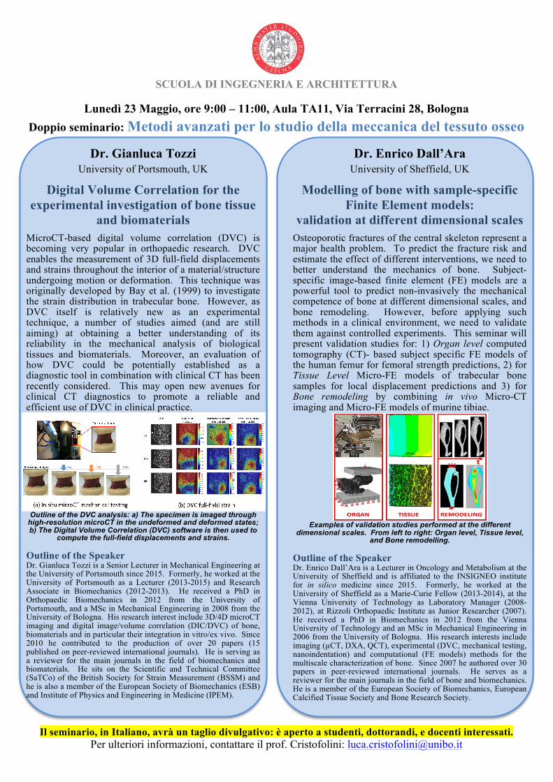
Outline of the DVC analysis: a) The specimen is imaged through
high-resolution microCT in the undeformed and deformed states; b) The Digital Volume Correlation (DVC) software is then used to
compute the full-field displacements and strains.
Outline of the Speaker Dr. Gianluca Tozzi is a Senior Lecturer in Mechanical Engineering at the University of Portsmouth since 2015. Formerly, he worked at the University of Portsmouth as a Lecturer (2013-2015) and Research Associate in Biomechanics (2012-2013). He received a PhD in Orthopaedic Biomechanics in 2012 from the University of Portsmouth, and a MSc in Mechanical Engineering in 2008 from the University of Bologna. His research interest include 3D/4D microCT imaging and digital image/volume correlation (DIC/DVC) of bone, biomaterials and in particular their integration in vitro/ex vivo. Since 2010 he contributed to the production of over 20 papers (15 published on peer-reviewed international journals). He is serving as a reviewer for the main journals in the field of biomechanics and biomaterials. He sits on the Scientific and Technical Committee (SaTCo) of the British Society for Strain Measurement (BSSM) and he is also a member of the European Society of Biomechanics (ESB) and Institute of Physics and Engineering in Medicine (IPEM).
Dr. Enrico Dall’Ara University of Sheffield, UK
Modelling of bone with sample-specific Finite Element models:
validation at different dimensional scales Osteoporotic fractures of the central skeleton represent a major health problem. To predict the fracture risk and estimate the effect of different interventions, we need to better understand the mechanics of bone. Subject-specific image-based finite element (FE) models are a powerful tool to predict non-invasively the mechanical competence of bone at different dimensional scales, and bone remodeling. However, before applying such methods in a clinical environment, we need to validate them against controlled experiments. This seminar will present validation studies for: 1) Organ level computed tomography (CT)- based subject specific FE models of the human femur for femoral strength predictions, 2) for Tissue Level Micro-FE models of trabecular bone samples for local displacement predictions and 3) for Bone remodeling by combining in vivo Micro-CT imaging and Micro-FE models of murine tibiae.
Examples of validation studies performed at the different
dimensional scales. From left to right: Organ level, Tissue level, and Bone remodelling.
Outline of the Speaker Dr. Enrico Dall’Ara is a Lecturer in Oncology and Metabolism at the University of Sheffield and is affiliated to the INSIGNEO institute for in silico medicine since 2015. Formerly, he worked at the University of Sheffield as a Marie-Curie Fellow (2013-2014), at the Vienna University of Technology as Laboratory Manager (2008-2012), at Rizzoli Orthopaedic Institute as Junior Researcher (2007). He received a PhD in Biomechanics in 2012 from the Vienna University of Technology and an MSc in Mechanical Engineering in 2006 from the University of Bologna. His research interests include imaging (µCT, DXA, QCT), experimental (DVC, mechanical testing, nanoindentation) and computational (FE models) methods for the multiscale characterization of bone. Since 2007 he authored over 30 papers in peer-reviewed international journals. He serves as a reviewer for the main journals in the field of bone and biomechanics. He is a member of the European Society of Biomechanics, European Calcified Tissue Society and Bone Research Society.
Il seminario, in Italiano, avrà un taglio divulgativo: è aperto a studenti, dottorandi, e docenti interessati. Per ulteriori informazioni, contattare il prof. Cristofolini: [email protected]