Embed Size (px)
Citation preview
The Journal of Applied Research • Vol. 4, No. 1, 2004 127
esterase, lipoprotein lipase were investi-gated.
Results: BCH concentrations in thetumor tissue at 2, 6, 8, and 14 hoursafter the intracerebral administrationwere 27.70, 12.51, 9.13, and 5.47 µgBCH/gm of tissue, respectively. Theratio of BCH between tumor tissue andnormal tissue at 2, 6, 8, and 14 hourswere 5.46, 1.56, 1.12, and 1.01, respec-tively. The affinity study showed thatBCH concentration in tumor tissueswas 4.45 µg/gm of tissue and in the nor-mal tissue was 3.91 µg /gm of tissue,when both samples were taken 2 mmaway from the BCH injection site in theopposite direction. Compared with thecontrol, BCH had good stability in thepresence of cholesterol esterase andlipoprotein lipase.
Conclusion: BCH formulated in lipo-somes distributed from the site ofintracerebral injection rapidly. The ade-quate radiation time window appearedto be 2 hours or less. A small but statis-tically insignificant difference was foundbetween the BCH concentrations in
Intracerebral Diffusion of NewCholesterol-Based AnticancerConjugate in Tumor-Bearing Rat ModelFars Alanazi, PhD*
David S. Halpern†
D. Robert Lu, PhD‡
*Department of Pharmaceutical and Biomedical Sciences, College of Pharmacy, University ofGeorgia, Athens, Georgia†Isotron Inc, Alpharetta, Georgia‡Department of Pharmaceutical Sciences, School of Pharmacy, Temple University, Philadelphia,Pennsylvania
KEY WO R D S : brain tumor, c h o l e s-teryl conjugate, tissue drug diffusion,l i p o s o m e, cholesterol esterase, l i p o p r o-tein lipase
ABSTRACTP u rp o s e:To study the diffusion andaffinity in brain of a new cholesterol-based boron anticancer conjugate (cho-lesteryl 1,12-dicarba-closo-dodecaborane1- carboxylate, BCH) using a tumor-bearing rat model and to examine thestability of BCH in the presence of cho-lesterol esterase and lipoprotein lipase.
Methods: The tumor-bearing rat modelwas produced by intracerebral implanta-tion of rat 9L glioma cells. BCH was for-mulated in liposomes to enhance theaqueous solubility due to its extremelyhydrophobic nature. Ten µl of BCH lipo-somes containing 150 µg BCH/mL wasadministrated to tumor-bearing ratsintracerebrally and the BCH concentra-tions in tumor and normal tissues wereexamined at different time points. Thedegradation profiles of BCH and thecontrol, cholesterol oleate, in cholesterol
LuJARWin04 3/18/04 6:46 PM Page 127
tumor tissue and normal tissue, bothsampled with the same distance to theBCH injection site. BCH was stable inpresence of cholesterol esterase andlipoprotein lipase, when compared withcholesterol oleate as the control.
INTRODUCTIONEach year more than 17,000 people inthe United States are diagnosed withbrain tumors.1 The prognosis for braintumor is poor due to the lack of effec-tive treatment options. For thechemotherapeutic option, it is critical todeliver a sufficient amount of therapeu-tic agents to the brain tumor site.However, delivery of therapeutic agentsto brain is technically challenging due topresence of blood brain barrier (BBB).2
Many attempts of special drug deliveryhave been made to overcome this obsta-cle. One approach is to intracerebrallyadminister the agents within brainparenchyma through local delivery totumor tissue.3 The therapeutic moleculesthen diffuse from the site of administra-tion to nearby cancer cells. This processoffers the advantage of having high drugconcentrations at the tumor site withlimited exposure to normal tissues andorgans in the rest of the body. Recently,our laboratory has developed a choles-terol-based anticancer agent containingcarborane as the anticancer unit forboron neutron capture therapy. The newcompound (Figure 1), cholesteryl 1,12-dicarba-closo-dodecaborane 1-carboxy-late (BCH) mimics the nativecholesteryl ester in structure and wasfound to be effectively taken up by
brain glioma cells in cell culture stud-ies.4,5 The intracerebral administration ofBCH in animal models, however, hadnot been studied.
Therefore, the purpose of the pres-ent study is to investigate the in vivo dif-fusion and tissue affinity of BCH in thebrain after intracerebral BCH adminis-tration using a tumor-bearing rat model.The ratio of BCH concentrations intumor versus surrounding normal tissueat different time points is an importantfactor for determination of the neutronradiation time window and it was inves-tigated after intracerebral BCH adminis-tration. BCH stability in brain hydrolyticenzymes, including cholesterol esterase(CE) and lipoprotein lipase (LPL), wasalso examined.
EXPERIMENTAL SECTIONMaterialsBCH was synthesized and purifiedaccording to a previously publishedmethod.6 Cholesterol Dipalmitoyl DL-!-phosphotidylcholine (DPPC), CE, andLPL were purchased from SigmaChemical Co. (St.Louis, Mo). All otherchemicals were analytical grade.
Preparation of BCH LiposomalFormulation Similar to native cholesteryl esters, BCHis extremely hydrophobic and thus, wasformulated in liposomal formulation toenhance its aqueous solubility. The BCHliposomal formulation was prepared bythin film hydration method as describedby Alanazi et al.7 Briefly, cholesterol,phospholipid and BCH were dissolvedin a 2:1 mixture of chloroform andmethanol in a round bottom flask. TheBCH to lipid ratio was 1:50 (w/w) andthe total lipid (cholesterol and phospho-lipid) to water ratio was 1:38 (w/w).Cholesterol and phospholipid ratio was0.33:1 molar ratio.7 The solvents wereevaporated under reduced pressure anda thin film of lipid was formed on the
Vol. 4, No. 1, 2004 • The Journal of Applied Research128
Figure 1. Cholesteryl 1,12-dicarba-closo-dodecarborane 1-carboxylate (BCH).
LuJARWin04 3/18/04 6:46 PM Page 128
bottom of the flask. The film was hydrat-ed at a temperature above the phasetransition temperature of the lipids withdeionized water and glass beads (5 mmdiameter) were added to ease the disso-lution. The hydration process was car-ried out for several hours using ashaking water bath. Liposomes wereseparated from the glass beads by filtra-tion through a Buchner funnel. The sizeof the liposomes was reduced usingEmulsiflex B3 (Avestin, Ottawa,Canada) and measured at 25oC using aNicomp Submicron Particle Sizer(Model 380, Nicomp, Calif).
Tumor-Bearing Rat Model Male Fisher rats weighing about 250 gwere used throughout the experiment.Rats were kept in standard cages (2 ratsper cage) without restriction to waterand food during the experiment. Rat 9Lglioma cell line was used to introducebrain tumor in the animal. The cellswere propagated in Dulbecco’sModified Eagle’s Medium (DMEM,Fisher, Pittsburgh, Pa) supplementedwith 10% fetal bovine serum (FBS,Sigma, St. Louis, Mo), 100 µg/mL strep-tomycin, and 100 U/mL of penicillin(Sigma, St. Louis, Mo) at 37oC and 5%CO2 in 150 mm2 flasks (Corning,Corning, NY). When the cells reachedconfluence, they were washed withDMEM, harvested using trypsin, cen-trifuged, re-suspended with phosphatebuffer, counted, and kept in an ice bathfor immediate use. The animals wereanesthetized intramuscularly with keta-mine (40 mg/kg) and xylazine (2.7mg/kg). The rat scalps were shaved andsterilized with 70% ethyl alcohol. Therats were placed on a stereotaxic appa-ratus (Stoelting, Wood Date, Ill) and amiddle incision was performed. Theskull was exposed and a small burr holewas made 1 mm anterior to Lambdoidsuture and 3 mm lateral (right) toSagittal suture.8 Four ml of the rat 9 L
glioma cell suspension (4 x 10⁴ cells) wasinjected intracerebrally 5 mm deep fromthe surface of the skull using a Hamiltonsyringe. The injection was performedover a period of 2 minutes and the nee-dle was withdrawn slowly to minimizethe backflow of cells in the needle track.The hole was filled with bone wax andthe wound was closed with running silksutures. The animals were watched overuntil they recovered from anesthesiaand then returned to their cages.
Histological Verification of Tumor-Bearing Rat Model To validate the tumor-bearing rat model,after 14 days of post-tumor implanta-tion, brain tissues were obtained, fixedimmediately with 10% buffered forma-lin, and sliced into coronal sections (3 to5 mm in thickness). Tissue slides wereplaced in a paraffin tank, consolidatedinto a single unit using a ShandonHistoCenter 2 and cut into 4 µm sec-tions using a Finesse Rotary Microtome(Thermo Shandon Inc, Pittsburgh, Pa).Micrometer sections were placed on aglass slide and subjected to washing with70% to 100% of ethanol and 100%xylene to dehydrate sample and toremove the paraffin layer. The sampleswere stained with Hematoxylin andEosin, using a robotic staining system(Leica Autostainer XL, LeciaMicrosystems Inc, Bannockburn, Ill),and examined under light microscopy.
Diffusion and Affinity Studies AfterIntracerebral BCH Administration The rats were ready for BCH diffusionstudies when the tumor reached a rela-tively large size (about 14 to 16 daysafter implantation of tumor cells). Theanimals were injected intracerebrallywith BCH liposomal formulation at thetumor site using the same procedure asdescribed above for tumor implantation.Ten µl of BCH liposomal formulation(150 µg BCH/mL) was administrated for
The Journal of Applied Research • Vol. 4, No. 1, 2004 129
LuJARWin04 3/18/04 6:46 PM Page 129
each rat. To study BCH diffusion fromthe tumor site, 4 groups of rats (n = 3 foreach time point) were sacrificed at 2, 6,8, and 14 hours post-administration.Tumor tissue was carefully separatedfrom the surrounding normal tissue andboth tissue samples were prepared. Inorder to study BCH affinity to tumorand normal tissues in vivo, BCH liposo-mal formulation was injected intracere-brally 2 mm away from the tumor site.Rats (n =3) were sacrificed at 18 hourspost-administration. A tumor tissue sam-ple and a normal tissue sample weretaken in opposite directions and bothsampling sites were equidistant from theBCH administration site. All tissue sam-ples were homogenized, digested in aTeflon-lined acid bomb, and quantita-tively analyzed for BCH concentrationusing ICP-MS (inductively coupled plas-ma-mass spectrometry). ICP-MS provid-ed a more sensitive method for analyzingthe low BCH concentrations in tissues a m p l e s.
BCH Stability in Cholesterol Esteraseand Lipoprotein Lipase Ten mg of CE or 4 mg LPL were dis-solved in 7.5 mL of phosphate bufferedsaline (pH 7.4). The concentrations ofboth enzymes were determined basedon the recommendation of the supplier(Sigma). BCH liposomal formulation(0.673 µmole of BCH) was added andthe mixture incubated at 37oC. At appro-priate time intervals, 200 µL sampleswere withdrawn and immediately addedto 600 µL acetonitrile kept in an ice bathto precipitate the enzyme. The sampleswere vacuum dried (Speed Vac plus,Savant, NY) at low temperature, recon-stituted with HPLC (high performanceliquid chromatography) mobile phasesolution (methanol to isopropanol ratioof 50:50), vortexed, and centrifuged at3000 RPM for 10 minutes. The super-natant was analyzed with a reversephase HPLC system based on an exist-
ing method.9 A liposomal formulation ofcholesterol oleate was prepared as acontrol and underwent the same processas described. The HPLC method specifi-cally separated BCH and cholesterololeate from their degradation product,cholesterol, and thus was suitable for thestability studies.9
RESULTSThe size of the BCH liposomes wasmeasured by photon correlation spec-troscopy (number weighted mean) and abimodal distribution was observed.Approximately 91% of the vesicles had amean diameter of 49.31 ± 6.30 nm andthe remaining 9% of the vesicles had amean diameter of 170.10 ± 21.32 nm. Th emean BCH concentration in the liposo-mal formulation was about 150 µ g / m L .
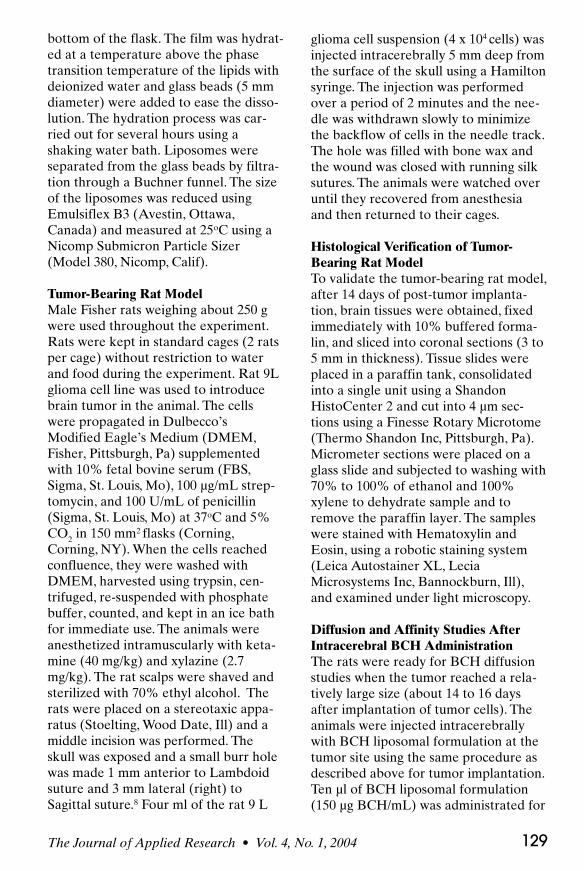
After 14 days post-implantation oftumor cells in the rat brains, the tumorwas readily visualized (Figure 2).Histological examination of the braintissue indicated the presence of largepleomorphic nuclei and abnormal mitot-ic figures (Figure 3). Physical changeswere also observed in the tumor-bearingrats, including weight loss and abnormalyellowish nasal excretion, further con-firming the presence of brain tumor.The concentration of BCH in tumor tis-sues after intracerebral administrationat the tumor site was 27.70 ± 5.54, 12.51
Vol. 4, No. 1, 2004 • The Journal of Applied Research130
Figure 2. Picture of a rat brain with devel-oped brain tumor after implantation of 9Lglioma cells (16 days after implantation). Thetumor tissue was located on the upper partof the right hemisphere.
LuJARWin04 3/18/04 6:46 PM Page 130
The Journal of Applied Research • Vol. 4, No. 1, 2004 131
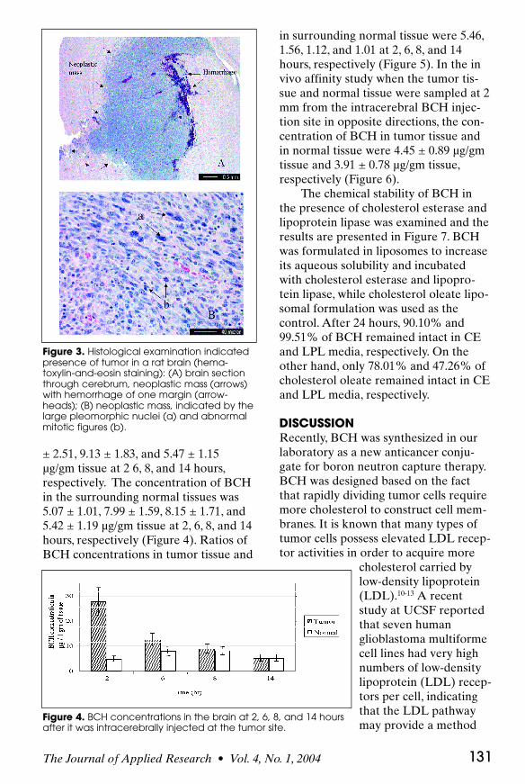
± 2.51, 9.13 ± 1.83, and 5.47 ± 1.15µg/gm tissue at 2 6, 8, and 14 hours,respectively. The concentration of BCHin the surrounding normal tissues was5.07 ± 1.01, 7.99 ± 1.59, 8.15 ± 1.71, and5.42 ± 1.19 µg/gm tissue at 2, 6, 8, and 14hours, respectively (Figure 4). Ratios ofBCH concentrations in tumor tissue and
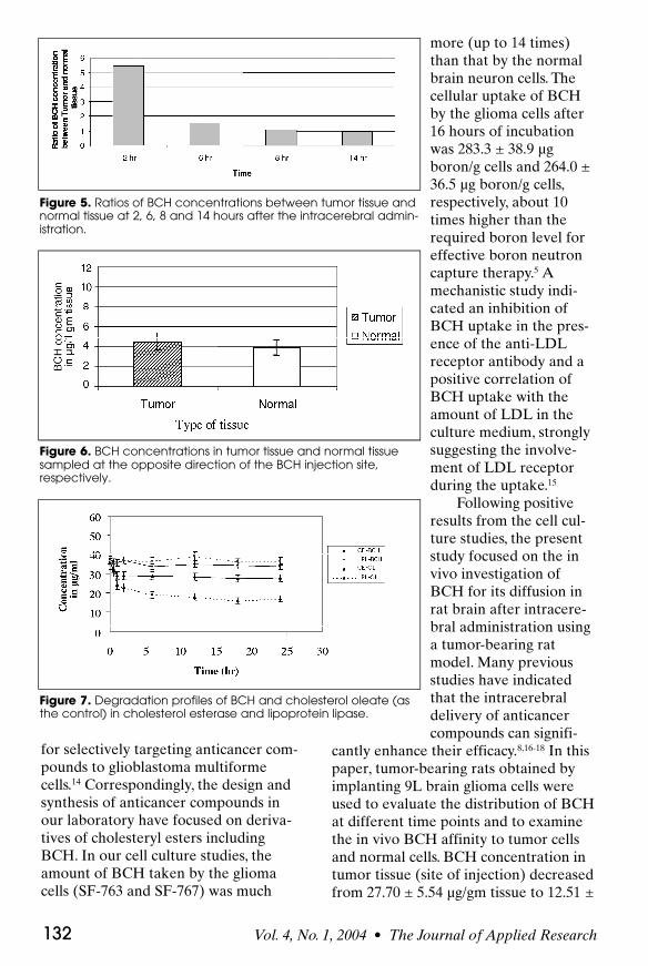
in surrounding normal tissue were 5.46,1.56, 1.12, and 1.01 at 2, 6, 8, and 14hours, respectively (Figure 5). In the invivo affinity study when the tumor tis-sue and normal tissue were sampled at 2mm from the intracerebral BCH injec-tion site in opposite directions, the con-centration of BCH in tumor tissue andin normal tissue were 4.45 ± 0.89 µg/gmtissue and 3.91 ± 0.78 µg/gm tissue,respectively (Figure 6).
The chemical stability of BCH inthe presence of cholesterol esterase andlipoprotein lipase was examined and theresults are presented in Figure 7. BCHwas formulated in liposomes to increaseits aqueous solubility and incubatedwith cholesterol esterase and lipopro-tein lipase, while cholesterol oleate lipo-somal formulation was used as thecontrol. After 24 hours, 90.10% and99.51% of BCH remained intact in CEand LPL media, respectively. On theother hand, only 78.01% and 47.26% ofcholesterol oleate remained intact in CEand LPL media, respectively.
DISCUSSIONRecently, BCH was synthesized in ourlaboratory as a new anticancer conju-gate for boron neutron capture therapy.BCH was designed based on the factthat rapidly dividing tumor cells requiremore cholesterol to construct cell mem-branes. It is known that many types oftumor cells possess elevated LDL recep-tor activities in order to acquire more
cholesterol carried bylow-density lipoprotein(LDL).10-13 A recentstudy at UCSF reportedthat seven humanglioblastoma multiformecell lines had very highnumbers of low-densitylipoprotein (LDL) recep-tors per cell, indicatingthat the LDL pathwaymay provide a method
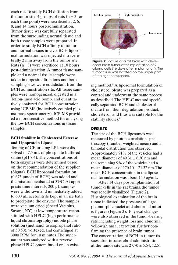
Figure 3. Histological examination indicatedpresence of tumor in a rat brain (hema-toxylin-and-eosin staining): (A) brain sectionthrough cerebrum, neoplastic mass (arrows)with hemorrhage of one margin (arrow-heads); (B) neoplastic mass, indicated by thelarge pleomorphic nuclei (a) and abnormalmitotic figures (b).
Figure 4. BCH concentrations in the brain at 2, 6, 8, and 14 hoursafter it was intracerebrally injected at the tumor site.
LuJARWin04 3/18/04 6:46 PM Page 131
for selectively targeting anticancer com-pounds to glioblastoma multiformecells.14 Correspondingly, the design andsynthesis of anticancer compounds inour laboratory have focused on deriva-tives of cholesteryl esters includingBCH. In our cell culture studies, theamount of BCH taken by the gliomacells (SF-763 and SF-767) was much
more (up to 14 times)than that by the normalbrain neuron cells. Thecellular uptake of BCHby the glioma cells after16 hours of incubationwas 283.3 ± 38.9 µgboron/g cells and 264.0 ±36.5 µg boron/g cells,respectively, about 10times higher than therequired boron level foreffective boron neutroncapture therapy.5 Amechanistic study indi-cated an inhibition ofBCH uptake in the pres-ence of the anti-LDLreceptor antibody and apositive correlation ofBCH uptake with theamount of LDL in theculture medium, stronglysuggesting the involve-ment of LDL receptorduring the uptake.15
Following positiveresults from the cell cul-ture studies, the presentstudy focused on the invivo investigation ofBCH for its diffusion inrat brain after intracere-bral administration usinga tumor-bearing ratmodel. Many previousstudies have indicatedthat the intracerebraldelivery of anticancercompounds can signifi-
cantly enhance their efficacy.8,16-18 In thispaper, tumor-bearing rats obtained byimplanting 9L brain glioma cells wereused to evaluate the distribution of BCHat different time points and to examinethe in vivo BCH affinity to tumor cellsand normal cells. BCH concentration intumor tissue (site of injection) decreasedfrom 27.70 ± 5.54 µg/gm tissue to 12.51 ±
Vol. 4, No. 1, 2004 • The Journal of Applied Research132
Figure 5. Ratios of BCH concentrations between tumor tissue andnormal tissue at 2, 6, 8 and 14 hours after the intracerebral admin-istration.
Figure 6. BCH concentrations in tumor tissue and normal tissuesampled at the opposite direction of the BCH injection site,respectively.
Figure 7. Degradation profiles of BCH and cholesterol oleate (asthe control) in cholesterol esterase and lipoprotein lipase.
LuJARWin04 3/18/04 6:46 PM Page 132
2.51 µg/gm tissue at 2 hours and 6 hours,r e s p e c t i v e l y. The decline of BCH concen-t r a t i o n , greater than 54% within 4 hours,indicates that BCH diffused from the siteof injection rapidly. In addition, the BCHconcentration in neighboring normal tis-sues increased from 5.07 ± 1.01 µg / g mtissue to 7.99 ± 1.59 µg/gm tissue at 2hours and 6 hours, r e s p e c t i v e l y, due tothe diffusion of BCH from the injectionsite to the neighboring normal tissues.The result is in agreement with the braindistribution pattern of boroncaptate intumor bearing rats after intracerebrali n j e c t i o n .8 Based on the diffusion study,the results also provided suggestions fora suitable irradiation time window toallow BCH to diffuse to most of tumorcells but keep the ratio of BCH concen-trations in tumor tissue and normal tis-sue sufficiently high. Due to the rapidBCH diffusion in brain, an adequateradiation window appeared to be lessthan 2 hours. In our study regarding thein vivo affinity of BCH to tumor andnormal tissues, the BCH concentration intumor tissues and normal tissues, b o t hsampled 2 mm away from the BCHinjection site, was 4.45 ± 0.89 µg/gm tis-sue and 3.91 ± 0.78 µg/gm tissue, c o r r e-s p o n d i n g l y. The small difference in BCHc o n c e n t r a t i o n s, h o w e v e r, did not yield astatistical significance at 95% confidencelevel (student’s t t e s t ) , which againappeared to contribute to the rapid dif-fusion of BCH in brain tissues.
Because BCH is composed of a cho-lesterol unit linked to the anticancercarborane moiety via an ester bond, itsstability in the presence of hydrolyticenzymes, such as CE and LPL, in thebrain may be questionable19,20-22 andneeds to be examined in parallel withthe diffusion study. The suitability ofusing phospholipid vesicles for thehydrolytic study of cholesteryl ester hasbeen demonstrated.23,24 Our results indi-cated that BCH in liposomal formula-tion was relatively stable after incubated
with CE and LPL, with 90.10% and99.51% of BCH remaining intact,respectively. In contrast, during the con-trol study, cholesterol oleate in liposo-mal formulation showed only 78.01%and 47.26% remaining intact for CE andLPL, respectively, under the same condi-tions (Figure 7). The presence of a bulkygroup near to the ester bond is knownto provide protection from hydrolysis bysteric hindrance.25,26 Thus, BCH stabilityin the presence of CE and LPL could beattributed to the existence of the bulkycarborane group next to the ester bond.Absence of a bulky group in cholesterololeate apparently led to its instability.The hydrolytic effect of CE and LPL,however, was significantly different onBCH and cholesterol oleate. The stabili-ty difference between BCH and choles-terol oleate in the presence of CE was12.09%. The stability difference in thepresence of LPL, however, was muchgreater (52.25%).
In conclusion, the new cholesterylester anticancer conjugate, BCH, wasinvestigated for its in vivo diffusion andaffinity in brain using a tumor-bearingrat model. The chemical stability ofBCH in brain hydrolytic enzymes, CEand LPL, was also examined. The resultsindicated that BCH formulated in lipo-somes distributed from the site of intrac-erebral injection rapidly. The adequateradiation time window appeared to beless than 2 hours. A small but statistical-ly insignificant difference was foundbetween the BCH concentrations intumor tissue and normal tissue, bothsampled equidistant to the BCH injec-tion site. BCH was stable in presence ofcholesterol esterase and lipoproteinlipase, when compared with cholesterololeate as the control.
ACKNOWLEDGMENTThis study was supported in part by NIHResearch Grant CA 67352(DRL) andby Isotron Inc. The authors would also
The Journal of Applied Research • Vol. 4, No. 1, 2004 133
LuJARWin04 3/18/04 6:46 PM Page 133
Vol. 4, No. 1, 2004 • The Journal of Applied Research134
like to thank Dr Barry G. Harmon,Department of Veterinary Pathology,College of Veterinary Medicine, UGA,for his assistance with the histopatholo-gy examination.
REFERENCES 1. Anderson RN. Deaths: leading causes for
2000. Natl Vital Stat Rep. 2002;50:1-85.
2. Zee-Cheng RK, Cheng CC. Delivery of anti-cancer drugs. Methods Find Exp ClinPharmacol 1989;11:439-529.
3. Lu DR, Mehta SC, Chen W. Selective borondrug delivery to brain tumors for boron neu-tron capture therapy. Adv Drug Deliv Rev.1997;26:231-247.
4. Peacock G, Ji B, Wang CK, Lu DR. Cell cul-tural studies for a carborane cholesteryl esterwith conventional and PEG liposomes. DrugDelivery. 2003;10:29-34.
5. Peacock G, Sidwell R, Pan G, Oie S, Lu DR.In vitro uptake of a new cholesteryl carbo-rane ester compound in human glioma celllines. J Pharm Sci. 2004;93:13-19.
6. Ji B, Peacock G, Lu DR. Synthesis of choles-terol-carborane conjugate for targeted drugdelivery. Bioorganic Med Chem Lett.2002;12:2455-2458.
7. Alanazi F, Li H, Halpern DS, Øie S, Lu DR.Synthesis, preformulation and liposomal for-mulation of cholesteryl carborane esters withvarious fatty chains. Int J Pharm.2003;225:189-197.
8. Ji B, Chen W, Lu DR, Halpern DS. Cell cul-ture and animal studies for intracerebraldelivery of borocaptate in liposomal formula-tion. Drug Deliv. 2001;8:13-17.
9. Sidwell R. Extraction and analysis by HPLCof the novel compound BCH in formulations,cell culture, and animal tissues [thesis].Athens, Ga: University of Georgia; 2002.
10. Callahan DE, Forte TM, Afzal SM, et al.Boronated protoporphyrin (BOPP): localiza-tion in lysosomes of the human glioma cellline SF-767 with uptake modulated bylipoprotein levels. Int J Radiat Oncol BiolPhys. 1999;45:761-771.
11. Murakami M, Ushio Y, Mihara Y et al.Cholesterol uptake by human glioma-cell viareceptor-mediated endocytosis of Low-densi-ty lipoprotein. J Neurosurg. 1990;73:760-767.
12. Rudling MJ, Angelin B, Peterson CO, CollinsVP. Low-density lipoprotein receptor activityin human intracranial tumors and its relationto the cholesterol requirement. Cancer Res.1990;50:483-487.
13. Lombardi P, Norata G, Maggi FM et al.
Assimilation of LDL by experimental-tumorsin mice. Biochim Biophys Acta.1989;1003:301-306.
14. Maletinska L, Blakely EA, Bjornstad KA, etal. Human glioblastoma cell lines: Levels oflow-density lipoprotein receptor and low-density lipoprotein receptor-related protein.Cancer Res. 2000;60:2300-2303.
15. Pan G. In vitro gene and drug delivery andtargeting to human glioma cells by lipopro-tein mimics [thesis]. Athens, Ga: University ofGeorgia; 2003.
16. McKeran RO, Firth G, Oliver S, Uttley D,O’Laoire S. A potential application for theintracerebral injection of drugs entrappedwithin liposomes in the treatment of humancerebral gliomas. J Neurol NeurosurgPsychiatry. 1985;48:1213-1219.
1 7 . Firth G, Oliver A S, M c Keran RO. Studies onthe intracerebral injection of bleomycin freeand entrapped within liposomes in the rat. JNeurol Neurosurg Psych i a t r y. 1 9 8 4 ; 4 7 : 5 8 5 - 5 8 9 .
18. Oliver AS, Firth G, McKeran RO. Studies onthe intracerebral injection of vincristine freeand entrapped within liposomes in the rat. JNeurol Sci. 1985;68:25-30.
19. Johnson RC, Shah SM. Cholesterol estermetabolizing enzymes in human brain: prop-erties, subcellular distribution and relativelevels in various diseases conditions.
20. Nunez M, Peinado-Onsurbe J, Vilaro S,Llobera M. Lipoprotein lipase activity inDeveloping Rat brain areas. Biol Neonate.1995;68:119-127.
21. Ogino T, Suzuki K. Specificities of human andrat brain enzymes of cholesterol ester metab-olism toward very long chain acids: implica-tion for biochemical pathogenesis ofAdrenoleukodystrophy. J Neurochem.1981;36:776-779.
22. Roussea A, Dubois G, Gatt S. Subcelluardistribution of diacylglycerol lipase in rat andmouse brain. Neurochem Res. 1983;8:417-422.
23. Brecher P, Chobanian J, Small DM,Chobanian AV. The use of phospholipid vesi-cles for in vitro studies on cholesteryl esterhydrolysis. J Lipid Res. 1976;17:239-247.
24. Ishii I, Onozaki R, Takahashi E, et al.Regulation of neutral cholesterol esteraseactivity by phospholipids containing negativecharges in substrate liposome. J Lipid Res.1995;36:2303-2310.
25. VanDort M, Schwendner SW, Skinner RW,Gross MD, Counsell RE. Potential tumor ororgan-imaging agents. 24. Radioidinatedpregenolone esters. Steroid. 1984;44:85-93.
26. Morrison RT, Body RN. Organic chemistry.3rd ed. Boston, Mass: Allan and Bacon Inc;1973:603.
LuJARWin04 3/18/04 6:46 PM Page 134