Embed Size (px)
Citation preview

WOUND CARE TODAY
www.woundcare-today.com
In association with
WOUND CARE TODAY
For what matters in practice
Volume 2Number 1
2015
Are pressure ulcers in danger of landing nurses in the dock?
WCT Cover 15.indd 1 4/23/15 1:30 PM
© 2015
Wou
nd C
are P
eople
Ltd
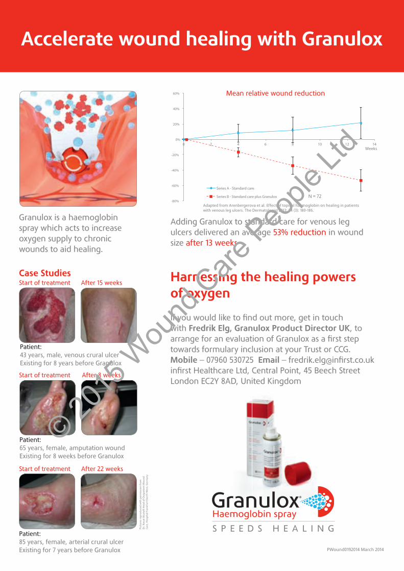
Accelerate wound healing with Granulox
Start of treatment After 15 weeks
Patient: 43 years, male, venous crural ulcer Existing for 8 years before Granulox
Pict
ure
s re
pro
du
ced
wit
h p
erm
issi
on
fro
m
Dr.
Nes
at M
ust
afi H
ead
of
Ou
tpat
ien
t W
ou
nd
C
are,
Ho
spit
al F
ran
kfu
rt N
ort
h-W
est,
Ger
man
y
Haemoglobin spray
S P E E D S H E A L I N G
If you would like to fi nd out more, get in touch with Fredrik Elg, Granulox Product Director UK, to arrange for an evaluation of Granulox as a fi rst step towards formulary inclusion at your Trust or CCG. Mobile – 07960 530725 Email – fredrik.elg@infi rst.co.uk infi rst Healthcare Ltd, Central Point, 45 Beech Street London EC2Y 8AD, United Kingdom
PWound0192014 March 2014
Harnessing the healing powers of oxygen
Granulox is a haemoglobin spray which acts to increase oxygen supply to chronic wounds to aid healing.
Adding Granulox to standard care for venous leg ulcers delivered an average 53% reduction in wound size after 13 weeks
-80%
-60%
-40%
-20%
0%
20%
40%
60%
0 2 4 6 8 10 12 14
Series A - Standard care
Series B - Standard care plus Granulox
Weeks
Mean relative wound reduction
Case Studies
N = 72
Adapted from Arenbergerova et al. Effect of topical haemoglobin on healing in patients with venous leg ulcers. The Dermatologist 2013; 64 (3): 180-186.
Start of treatment After 8 weeks
Patient: 65 years, female, amputation woundExisting for 8 weeks before Granulox
Start of treatment After 22 weeks
Patient: 85 years, female, arterial crural ulcerExisting for 7 years before Granulox
16462_Granulox_AltADv7.indd 1 03/04/2014 15:06
© 2015
Wou
nd C
are P
eople
Ltd

WOUND CARE TODAY 2015, Vol 2, No 1 3
EDITORIAL
This could be a defining year for wound care in the UK. As well as the annual European Wound Management Association (EWMA) meeting coming to London on 13–15 May, there is also the usual
run of conferences and study days (including a full programme from the WCAUK), and the little matter of a general election, which will have a major impact on the future of the NHS and the way in which services such as wound care are delivered. So far the contest looks pretty neck-and-neck, but whoever wins out on 7 May one thing will remain as true in 2015 as in any other year — whatever the prevailing political, social or policy agenda, good clinical care is a constant.
In this issue we have provided you with a set of articles that get to the bottom of what ‘good care’ is — a set of fundamental principles, delivered well and in a timely fashion. We’ve tried to cover the basics — dressing choice; pressure ulcers; leg ulcers; oedema — to give you all you need to keep up to date with the latest clinical information. It’s not all about the clinical though — we also take a look at the growing trend for litigation in pressure ulcers, an issue that is costing the health service millions and remains a grey area for nurses (see ‘Wound watch’ on pp.6–7).
As ever, I hope you enjoy reading and I wish you a productive and fulfilling 2015, whoever wins the keys to number 10.
Jason Beckford-Ball, editor, Wound Care Today
Managing directorNicola [email protected] [email protected] [email protected] managerAlec O’[email protected] 282827
Opinions expressed in the articles are those of the authors and do not necessarily reflect those of the publishers. Any products referred to by the authors should only be used as recommended by manufacturers’ data sheets.
2015 is a big year for wound care in the UK
WOUND CARE TODAY
&
© Wound Care People Limited 2015Finials House, The Square, Stow-on-the-Wold, Gloucestershire GL54 1AF
ISSN 2054-9636
t: +44(0) 1451 870310e: [email protected]://www.woundcare-today.com
All rights reserved. No part of the Wound Care Today journal may be reproduced, stored in a retrieval system or transmitted by any means electronic or mechanical, photocopied or otherwise without the prior written permission of Wound Care People Limited.
Printed in England by Blackmore Ltd, Shaftesbury
Contents5 Will the general election
put wound care on top of the agenda?Jackie Stephen-Haynes
6 Wound watch: are pressure ulcers in danger of landing nurses in the dock?
8 How to choose the correct dressing... Kirsty Mahoney
16 The causes of oedema and managing any associated complicationsJeanette Milne
30 How to categorise and assess pressure ulcers Menna Lloyd Jones
40 Management of moisture-related skin damageRosie Callaghan, Jackie Stephen-Haynes
50 Compression hosiery to manage venous leg ulcers and oedema Jackie Stephen-Haynes, Rosie Callaghan
Edit-Contents-Flannel.indd 35 23/04/2015 14:12
© 2015
Wou
nd C
are P
eople
Ltd

4 WOUND CARE TODAY 2015, Vol 2, No 1
EDITORIAL BOARD›It is recognised that healthcare assistants have an important role to play in the delivery of wound care, particularly in the primary care setting.
The articles published in this journal will enhance the care they give through the provision of appropriate education and support.Menna Lloyd Jones
Wound Care Today is aimed at a wide range of practitioners and we are confident that you will find the articles both interesting and reflective of
contemporary evidence-based practice. Our aim is to provide you with the knowledge you require to deliver high quality care for individuals with, or at risk of, a breakdown in the integrity of their skin.Louise Toner
An important part of any specialist nurses’ role is to share knowledge and skills. Educating and empowering clinicians to effectively
assess and treat patients with a wound ensures positive clinical outcomes and continuity in care. Wound Care Today aims to reach all healthcare professionals caring for patients with wounds and inform clinical practice.Lorraine Grothier
Welcome to the 2015 edition of Wound Care Today. The year has started on an encouraging note for the Wound Care Alliance UK, with positive feedback about our
achievements last year. The new journal was very well received and as always, we welcome your comments.Jackie Stephen-Haynes
It is an exciting time at the Wound Care Today journal. This journal seeks to provide wound care knowledge from evidence-based practice
faced in daily nursing. I hope the journal will enable clinicians to share and enrich practical knowledge in caring for individuals with wounds and improve patient outcomes.Julie Evans
Wound care and tissue viability can be challenging. As a member of the editorial board, I am committed to help produce a journal which is
researched-based, relevant, clinically useful and educational and, in addition, very readable for those looking for support.Jackie Griffin
WOUND CARE TODAY
› Board membersJackie Stephen-Haynes Chair, Wound Care Alliance UK; Professor and consultant nurse in tissue viability, Birmingham City University and Worcestershire Health and Care Trust
Julie Evans Vice chair, Wound Care Alliance UK; Tissue viability nurse, Abertawe Bro Morgawwng University Health Board, Swansea
Rosie CallaghanTissue viability nurse specialist, Worcester Health and Care Trust
Michelle CarolanCare home quality lead, NHS Sandwell and West Birmingham CCG
Jackie GriffinTissue viability clinical nurse specialist, Montgomery County Infirmary, Powys Health Board
Lorraine GrothierClinical nurse specialist tissue viability/lymphoedema manager, Provide, St Peter’s Hospital, Maldon
Jane JamesTissue viability lead nurse, Hywel Dda University Health Board, Carmarthen
Jola MerrickSecretary, Wound Care Alliance UK; Clinical nurse manager
Jeanette Milne Tissue viability nurse specialist, South Tyneside Foundation Trust, Tyne and Wear
Louise TonerEvents organiser, Wound Care Alliance UK; Associate dean, Birmingham City University
Richard WhiteProfessor of tissue viability, University of Worcester
Board members page.indd 2 23/04/2015 14:23
© 2015
Wou
nd C
are P
eople
Ltd

WOUND CARE TODAY 2015, Vol 2, No 1 5
WOUND CARE ALLIANCE UK
Welcome to the 2015 edition of Wound Care Today. The year has started on an encouraging note for the Wound Care Alliance UK, with positive feedback about our achievements last year. The new journal was very well received and as always, we
welcome your comments. A big ‘thank you’ must be extended to all the trustees — who are all members of the editorial board on Wound Care Today — and also to Wound Care People, who have helped to make our transition to a new publishing house so easy.
The Wound Care Alliance UK has also received praise for its partnership with the Welsh Lymphoedema Network and the successful Wound Care Alliance UK conference in Swansea,Wales.
So a huge thank you to our Welsh trustees — Julie Evans, Jackie Griffin and Jane James for leading on this.
September 2014 also saw the launch of our ‘skills day’, with the focus on key wound care areas and the development of the skills necessary to support clinical delivery. A big thank you to all the trustees for supporting this event and the development of the posters including, Menna Lloyd Jones, Rosie Callaghan, Jola Merrick, Michelle Greenwood, Julie Evans and Jackie Griffin. If you were unable to attend and have not received a paper copy, they are available on the Wound Care Alliance UK website (www.wcauk.org). Once again it has been a pleasure to work with Tim, Nicola and all the team at Mole Productions and if you haven’t yet seen the video of the 2014 conference, do take a look on the website (www.wcauk.org/annual-conference). I would also like to thank Rosie Callaghan for her support with the website and the analysis of the Wound Care Alliance UK’s membership.
Perhaps like me your thoughts have turned to the forthcoming election, and whichever party (or parties) win-out there are sure to be changes to the NHS. Personally, I welcome a greater focus on the prevention, assessment, and management of tissue viability. One thing that has struck me already this year is the discussion about accurate dementia diagnosis and who should be responsible for this. An accurate diagnosis is always needed to support care delivery and tissue viability needs to be similarly high on the agenda irrespective of who forms the next government.
Jackie Stephen-Haynes, Chair of the Wound Care AllianceApril, 2015
Will the general election put wound care on top of the agenda?
The Wound Care Alliance UK would like to thank their sponsors for their ongoing support:
3M
JSH EditC/final.indd 35 23/04/2015 13:12
© 2015
Wou
nd C
are P
eople
Ltd

In each issue of Wound Care Today we investigate a hot topic in wound care. In this issue, Jason Beckford-Ball asks...
There are certain clichés that seem to find a special place in the general public’s
perception of health services — for example, that every spare inch in A&E is filled with patients being looked after on trolleys; or that nurses always need to be told to wash their hands. No matter how much fact there is in these accepted ‘truths’, once they are established, no amount of good care or PR can shift them.
Are pressure ulcers in danger of landing nurses in the dock?
Unfortunately for those of us involved in wound care, the widespread development of pressure ulcers is also in danger of becoming a popular myth, with press reports focusing on payouts awarded to patients through poor care in hospitals and nursing homes (‘NHS: 700 victims of bed sores through hospital neglect receive £16m in payouts’ — Daily Mirror).
Not only are pressure ulcers now acknowledged as a sign of sub-
standard care in an organisation (‘Nurses face new focus on reducing pressure ulcers’ — Nursing Times), there are also campaigns to eradicate avoidable pressure ulcers altogether (‘Stop the Pressure’ — http://nhs.stopthepressure.co.uk).
As well as the distressing implications for patients, much of this new drive against pressure ulcers is driven by cost — as recently as October last year, the health secretary Jeremy Hunt was bemoaning the increase of litigation in the NHS, (‘NHS errors costing billions a year - Jeremy Hunt’ — BBC News), stating that the NHS had spent £1.3 billion on payouts after being sued by patients over poor care. The Department of Health identified four major areas of patient safety — falls and trips; pressure ulcers; urinary infections; and deep vein thrombosis (DVT).
Now that the problem has been identified, there are significant moves to monitor the incidence of pressure ulcers, and it is incumbent upon health services to record pressure ulcer rates using mechanisms like the ‘Patient safety thermometer’ (www.safetythermometer.nhs.uk). There are even penalties for those trusts who fail to cut the number of pressure ulcers by half (‘Trusts to be told to halve pressure ulcers or face fines’ — Health Service Journal).
But, how did pressure ulcers come to be such a drain on NHS resources and how has such an obvious manifestation of poor care become so widespread? Unfortunately, it has to be said that
6 WOUND CARE TODAY 2015, Vol 2, No 1
WOUND WATCH
Wound Watch.indd 2 23/04/2015 12:31
© 2015
Wou
nd C
are P
eople
Ltd

lack of knowledge among nurses is undoubtedly a factor, with gaps in post-registration education and the difficulty of obtaining time off for study leave particularly to blame.
Another huge issue that features in many cases is documentation, where clinicians had failed to record the steps they took to prevent or manage ulceration (‘Pressure ulcers, negligence and litigation’ — Wounds UK). There is also confusion around what represents an ‘avoidable’ or an ‘unavoidable’ pressure ulcer, with legal opinion generally following that if action was not taken to prevent an ‘avoidable’ pressure ulcer, then an element of negligence is involved.
The recent NHS Patient Safety First campaign (www.patientsafetyfirst.nhs.uk) stated that an avoidable pressure ulcer is where ‘the person receiving care developed a pressure ulcer and the provider of care did not do one of the following: evaluate the person’s
clinical condition and pressure ulcer risk factors; plan and implement interventions that are consistent with the person’s needs and goals, and recognised standards of practice; monitor and evaluate the impact of the interventions; or revise the interventions as appropriate’.
In the future, what is important is that nurses not only understand the clinical implications of pressure ulceration and how it develops — can you tell the difference between a category two and four ulcer, for example? — but also the legal implications of an area that is putting wound care clinicians increasingly under the spotlight.
What many of you will want to know is — ‘Can I be liable?’ While it is unlikely that an individual nurse would ever be held liable for the development of a pressure ulcer due to the difficulty in apportioning personal blame (‘Pressure ulcers and litigation’ — Nursing Times),
WOUND CARE TODAY 2015, Vol 2, No 1 7
WCT
Some district nurses across the country are beginning to feel demoralised every time a patient on their caseload develops a category three or four pressure ulcer, thereby requiring them to attend a ‘safeguarding vulnerable adults’ meeting and a case conference. This is despite the fact that in most instances there are no safeguarding issues involved with the development of the pressure ulcer and, if there were any, the attending district nurses would have highlighted them.
Edwin T Chamanga, Tissue viability service lead, Hounslow and Richmond Community Healthcare NHS Trust
WOUND WATCH
it is still important that nurses understand best practice and how to implement it, as well as following some basic rules:
Use recognised pressure ulcer assessment scales and always perform skin assessmentAlways try and follow local policy — if you can’t, make sure you record why notDocument any discussions with other practitionersRecord any clinical changes, particularly in the skinRecord any interventions, such as turning or dressings applied (‘Pressure ulcers, negligence and litigation’ — Wounds UK).
While it is unlikely that any of us are going to end up in court any time soon, the litigation bill for pressure ulcers is rising fast. We owe it to ourselves and our patients to be mindful not only of the financial cost of these wounds, but also the impact on the health and wellbeing of our patients.
Every nurse will be aware of being under the pressure ulcer spotlight. While the emphasis from organisations is often about robust data collection and accurate reporting, the frontline nurse wants to ensure that they prevent patients from sustaining an ‘avoidable’ wound. The reality is that pressure ulcer prevention is relatively easy to achieve if we are given time to nurse our patients instead of repeatedly ‘ticking boxes’ to comply with policy. Some patients will need more support than others. As nurses we should be establishing patient need and risk based on professional judgement and act accordingly.
Michael Ellis, clinical nurse specialist in tissue viability; lecturer in healthcare practice, Royal Devon and Exeter NHS Trust
Wound Watch.indd 3 23/04/2015 12:31
© 2015
Wou
nd C
are P
eople
Ltd

Due to technological advances there has been an explosion in new wound care dressings in recent years, which can leave nurses baffled as to which dressing to use on which wound. Here, Kirsty Mahoney takes us through the various dressing options and outlines the criteria that should be used when trying to choose the right product for a particular wound type.
How to choose the correct dressing...
BACKGROUND
Before 1960, wound dressings were mainly limited to Gamgee and dry gauze (Jones, 2006). It was believed that allowing the wound to dry and form an eschar was an effective environment for healing (Turner et al, 1986), and that a dry wound would facilitate the death of bacteria (Hinman and Maibach, 1963).
These beliefs were challenged by the publication of a study by Winter (1962), into how a moist wound- healing environment had accelerated healing rates in an acute animal model. Winter’s research attracted a lot of interest and his findings became the basis for the concept of moist wound healing; one which has influenced many manufacturers to develop the range of modern dressings (Jones, 2005).
Since Winter’s findings, our understanding of the wound-healing process has continued to expand and there has been a huge increase in the number of products designed to support wounds along the healing continuum. However, the pure variety and amount of dressings that clinicians can be faced with can make choosing the right one a daunting task.
With the NHS under constant pressure to provide cost-effective treatment (Hamilton, 2008), it is extremely important for nurses to be able to justify the use of wound care products and ensure that they are used correctly and appropriately.
However, there is a lack of good quality evidence to aid decision-making around dressing selection, (National Prescribing Centre, 2010) and to recommend one dressing over another (Palfreyman et al, 2006). Dressing selection should, therefore be based on promoting moist wound healing, addressing issues identified within the wound bed and surrounding skin, and using the least costly dressing to meet the requirements of the wound (Jeffcoate et al, 2009).
However, most health organisations have a designated wound dressing formulary and decision-making is influenced by the products available and the local prescribing guidelines.
AssessmentThe cornerstone to treating a patient with a wound is to undertake an holistic assessment. Without an assessment, as well as the identification of any underlying pathologies or potential barriers to healing, wound care can become a series of ritualistic dressing changes, and become costly and ineffective.
A comprehensive wound assessment can be divided into patient-related factors and wound-related factors. Patient related factors start with identifying any underlying conditions or circumstances that may need to be addressed before the wound will start to heal — these may include vascular or venous disease, rheumatoid arthritis, diabetes, immunosuppressive or autoimmune disorders, anaemia, poor nutrition, smoking, interactions with medication, immobility, and psychological status.
Wound-related factors include how the wound occurred, its
measurements, the identification of tissue within the wound bed, assessment of surrounding skin, pain, exudate, and odour.
Accurate documentation and reassessment of these elements will assist in determining the progression of the wound and alert the nurse to any circumstances — such as an increase in the volume of exudate or failure of the wound to decrease in size — that may require the dressing to be reviewed.
TIMEOverall, selecting an appropriate wound dressing can be a complex process and requires a good knowledge of the wound, the dressing and the patient. To help clinicians identify the type of wound bed environment they were dealing with, Schultz et al (2003) suggested an approach known as wound bed preparation. Using the acronym
✔ Wound care facts...
✔ The role of a dressing is not necessarily to heal the wound, but to create an environment within the wound bed that will assist the healing process.
✔ Dressings do not always need to be changed daily. Most dressings can be left in place for seven days, depending on the level of exudate and what you want the dressing to achieve.
✔ It is important to consult the manufacturer’s recommendations, as well as assessing the wound, before deciding the frequency of dressing change.
❝
Kirsty Mahoney, clinical nurse specialist, Cardiff and Vale University Health Board
8 WOUND CARE TODAY 2015, Vol 2, No 1
DRESSING CHOICE›
Mahoney - dressing choiceC3.indd 2 24/04/2015 12:21
© 2015
Wou
nd C
are P
eople
Ltd

Advancis Medical
@AdvancisMedical
+44 (0)1623 751500
MAR382
References (references relate to Advancis Manuka honey)* taken from ‘International consensus: Appropriate use of silver dressings in wounds’ - Wounds International 2012
Effective exudate management1
Maintains a moist wound healing enviroment
Optimum Moisture Vapour Transfer Rate1
Unique range of sizes
The use of
Activon Manuka Honey®
and Eclypse®
Viral and bacterial backing barrier
Strike-through barrier1
Up to 7 days wear time
The perfect combination
Eclypse vs Honey UPDATED V10.indd 1 07/10/2014 12:35
© 2015
Wou
nd C
are P
eople
Ltd

DRESSING CHOICE›
10 WOUND CARE TODAY 2015, Vol 2, No 1
TIME, Schultz et al (2003) described a framework that provides a structured approach to wound assessment. This approach can help the nurse select the correct dressing to optimise the wound environment.
The TIME framework consists of four basic concepts that nurses should consider when attempting dressing selection and treatment (see Box 1) (Schultz et al, 2003; European Wound Management Association [EWMA], 2004)› T: tissue, non-viable or deficient› I: infection or inflammation› M: moisture imbalance› E: edge of the wound — non-
advancing or undermined.
DRESSING TYPES
Alginates PropertiesAlginates are made from brown seaweed and are considered highly absorbent and biodegradable. On contact with sodium-rich exudate, an exchange of sodium and calcium ions occurs leading to the alginate forming a gel.
The action of the various alginate dressings differs slightly according to the amounts of calcium/sodium salts within the product, which mean that some have a faster gelling action and form a softer gel; whereas others form a firmer gel that can be removed from the wound in one piece. Some alginates have haemostatic (stopping the flow of blood) properties due to the level of calcium ions that are released.
When to useAlginates are generally recommended for use in moderate-to-highly exuding wounds and are also suitable for cavity wounds, however they will require a secondary dressing. They come in flat sheets and ribbons, and some are silver-impregnated and may be used on infected wounds.
ConsiderationsAlginates are not recommended for dry or necrotic wounds as they may adhere to the wound bed, causing trauma and pain on removal (Vuolo, 2009).
Examples include: Sorbsan® (Aspen Medical); Kaltostat® (ConvaTec).
Foams Properties Foams are available either as polyurethane or silicone dressings. They are recommended for low-to-moderately exuding wounds, however the ability to manage exudate will depend on the moisture vapour transfer rate (MVTR) (a measure of the passage of water vapour through a substance) of the dressing and performance does vary between brands.
When to useFoams are suitable for granulating wounds with low-to-moderate exudate. They can also be used as a secondary dressing. Foams generally conform well to the body shape and create an ideal moist wound environment. The silicone formulation of the foam is recommended for fragile skin, however it is slightly more expensive than the polyurethane equivalent, which should be taken into consideration when selecting foam dressings. Foams are available as cavity dressings and some are silver-impregnated for use on infected wounds.
ConsiderationsCaution should be used when using adhesive foam dressings, as they may cause skin-stripping. Using a foam dressing on a wound that is too wet, may cause maceration or erythema (White et al, 2012). Foams are not recommended for necrotic wounds unless they are to be used in conjunction with a debriding agent. Not all foams are suitable for use under compression and the manufacturer’s recommendations should always be checked before use.
Examples include: Allevyn® (Smith and Nephew); Mepilex® (Mölnlycke Health Care); Activheal® (Advanced Medical Solutions).
HydrocolloidsPropertiesHydrocolloids are made from carboxymethyl cellulose and
comprise self-adhesive conformable wafers, which are occlusive (air- and water-tight) or semi-occlusive. Hydrocolloids turn into a gel when absorbing exudate and this creates an ideal moist environment, which in turn promotes autolytic debridement.
When to useHydrocolloids are suitable for low-to-moderately exuding wounds. They can be used to debride slough and necrosis, and are also suitable for use on shallow granulating wounds. There may be a slight odour present when the dressing is removed.
ConsiderationsHydrocolloids may cause maceration if the wound is too moist. They can also cause stripping if used on fragile skin. Due to the occlusive property of these dressings, hydrocolloids should be used with caution on infected wounds.
Examples include: Comfeel®
(Coloplast); Duoderm® (ConvaTec); Granuflex® (ConvaTec).
Hydrogels PropertiesHydrogels have a high water content — sometimes as much as 96% — and are mainly composed of starch compounds or carboxymethyl cellulose. They have excellent biocompatibility (where a synthetic or
› Practice point
Properties of the ideal dressing (Morgan, 1999):› Bacteria-resistant
› Permits gaseous exchange
› Maintains a moist wound environment
› Non-adherent
› Fibre- and toxin-free
› Hypoallergenic
› Maintain haemostasis and optimum temperature
› Is acceptable to patients
› Cost-effective.
Mahoney - dressing choiceC3.indd 4 24/04/2015 12:21
© 2015
Wou
nd C
are P
eople
Ltd

DRESSING CHOICE
WOUND CARE TODAY 2015, Vol 2, No 1 11
Box 1
Choosing the right dressing
TISSUE TYPE Exudatelevel Dressing suggestion
Slough: requires a dressing to debride the wound bed
Low to moderate
Hydrogel/hydrofibre/hydrocolloid/honey
Low to moderate
Hydrofibre/honey
Necrosis: requires a dressing to debride the wound bed
Low Hydrogel/hydrocolloid/honeySurgical debridement may be required
Granulation: needs to be protected Low
Moderate
INFECTION/INFLAMMATION Exudatelevel Dressing suggestion
Infected wounds may require systemic antibioticsDressings should only be used for short periods
Low Iodine /silver wound contact layer/silver foam/PHMB contact layer or PHMB gel/honey
High Silver alginate/silver hydrofibre/dacc dressings/Honey
MOISTURE Dressing suggestionIf a wound is too wet, maceration and skin damage may occurDressing selection should assist in exudate management
Hydrofibres/alginates/superabsorbants Underlying cause of exudate should be identified during holistic assessment
If wounds are to dry it may prevent the migration of fibroblasts. Dressings that donate moisture are required
EDGES Dressing suggestionNon-advancing A non-advancing wound indicates non healing and referral to
specialist wound teams may be appropriate
natural material functions alongside living tissue, in this case the wound bed). The main function of hydrogels is to donate moisture to the wound and facilitate autolytic debridement of slough and necrosis. Hydrogels are available in a gel or sheet formulation.
When to useHydrogels are useful for dry, sloughy or necrotic wounds. There is some evidence that hydrogel sheets may assist in the management of painful wounds (Young and Hampton, 2005).
ConsiderationsMaceration of the surrounding skin may occur with hydrogels and dressing changes may need to be more frequent. For this reason, hydrogels are not usually recommended for use on highly exuding wounds. If larval therapy is to be used, any trace of the hydrogel will need to be fully irrigated from the wound, as the propylene glycol contained in most hydrogels (with the exception of Purilon® [ConvaTec]) is toxic to larvae.
Examples include: Intrasite® Gel (Smith and Nephew); Purilon®
(ConvaTec); Actiform Cool® (Activa Healthcare).
FilmsPropertiesFilm dressings are composed of a thin non-absorbent polyurethane material. They are semi-permeable to oxygen and water vapour, but impermeable to bacteria, therefore creating an ideal wound-healing environment (Sussman, 2010).
When to useFilm dressings are useful for low-exuding superficial wounds and because they are transparent they allow the nurse to view the wound bed and monitor the wound’s progress. Films are also helpful as secondary dressings, however they are not recommended for use over foam dressings as this may affect the overall ability of the foam to handle exudate.
ConsiderationsFilms are not suitable for highly exuding wounds as they have no
Mahoney - dressing choiceC3.indd 5 24/04/2015 12:21
© 2015
Wou
nd C
are P
eople
Ltd

DRESSING CHOICE
12 WOUND CARE TODAY 2015, Vol 2, No 1
Box 1 cont.
EDGES cont. Dressing suggestionUndermining – dressing selected will be a rope or ribbon that is suitable for use in cavities
Hydrofibre ribbon/alginate ribbon/honey ribbon, Dacc ribbon dressing
Epithelial – epithelial tissue within the wound will require a dressing that protects this delicate tissue
Wound contact layer/film dressing /foam
ability to absorb. Care should also be taken when applying or removing films from fragile skin as the adherence of the product may cause skin-stripping (Sussman, 2010).
Examples include: Opsite® (Smith and Nephew); Tegaderm® (3M); Mepore® (Mölnlycke Health Care).
Honey dressingsPropertiesHoney’s high osmolality and slow release of hydrogen peroxide has been shown to inhibit bacterial growth (Gethin and Cowman, 2008).
When to useHoney can be used on a wide variety of wounds due to its antimicrobial and anti-inflammatory properties. Honey also facilitates debridement and can be used on slough or necrotic wounds. Honey also may deodorise odourous wounds, promote a moist wound environment, and stimulate the formation of granulation tissue.
ConsiderationsHoney may initially increase exudate production, which may cause maceration and require an increase in dressing-change frequency. There have also been reports that honey can increase pain due to its high osmolality (Coulborn et al, 2009). Caution should be used on patients with sensitivity to bee venom.
Honey is available in several presentations, such as tulle dressings, alginate dressings, and ointments.
Wound contact layersPropertiesThese dressings are simple inexpensive dressings which are designed to be in direct contact with the wound bed. They are primarily made from knitted viscose or polyester. Newer versions have been impregnated with hydrocolloids, petroleum jelly and silver.
When to useWound contact layers are useful to protect granulation tissue and preventing adherence. They can be used under compression (Palfreyman et al, 2006), as well as in combination with an absorptive secondary dressing, allowing exudate to flow through the weave of the fabric into the secondary layer.
ConsiderationsWound contact layers have no absorptive properties and are not recommended for use on highly exudating wounds. There is some evidence to show that granulation tissue may grow through the weave of the dressing if left in place for too long, resulting in potentially traumatic removal (World Union of Wound Healing Societies [WUWHS], 2007).
HydrofibresPropertiesHydrofibre dressings are often confused with alginates due to similarities in appearance and gelling properties. Hydrofibre dressings consist of sodium carboxymethyl cellulose and
hollow fibres — this allows for the hydrophilic action of the dressing, which absorbs high quantities of exudate by vertical wicking.
Hydrofibres create a moist wound-healing environment as well as promoting autolysis (destruction of cells through the action of their own enzymes). There is also some evidence to indicate that hydrofibres are able to bind harmful proteases that may prevent healing, thus protecting the wound bed.
When to useHydrofibres are recommended for highly exuding wounds with or without slough. Formulations include ribbon dressings and a silver version, making the product suitable for cavity and infected wounds.
ConsiderationsHydrofibres require a secondary dressing and are not considered suitable for dry or necrotic wounds.
Examples include: Aquacel®
(ConvaTec); Durafiber® (Smith and Nephew); Exufiber® (Mölnlycke Health Care).
AntimicrobialsPropertiesAntimicrobial dressings are variously impregnated with non-selective agents that kill bacteria or inhibit their growth. There are a wide range of products that come under the antimicrobial umbrella, including those which contain:
IodineSilverHoneyPolyhexamethylene biguanide (PHMB)Dialkylcarbamoyl chloride (DACC technology).
There is an increased interest in topical antimicrobials due to the issues surrounding antibiotic resistance, and in some cases the use of topical antimicrobial therapy negates the need for treatment with antibiotics. Some areas of controversy around the use of topical antimicrobials lie in the cost of the products (Michaels et al, 2009), and inappropriate usage (Mahoney, 2014).
Mahoney - dressing choiceC3.indd 6 24/04/2015 12:21
© 2015
Wou
nd C
are P
eople
Ltd

In some healthcare organisations topical antimicrobials are restricted or banned, particularly silver products where cost effectiveness has been an issue (Michaels et al, 2009). It is, therefore important for clinicians to be able to correctly identify wound infection to ensure that these products are used appropriately and for the right length of time. Antimicrobial dressings are not appropriate for use throughout the healing trajectory to wound closure and should be stopped as soon as any infection has resolved.
It has been suggested that nurses use a ‘two-week challenge’ to ensure that silver dressings are used for an appropriate time scale. The dressings should be used for two weeks then the wound reassessed. If infection has resolved, the dressings should be discontinued; if there is no improvement the product should be stopped and other interventions considered. If there is improvement but infection has not fully resolved, the antimicrobial should be continued for another two weeks then reassessed (Wounds International, 2012).
Considerations for use› Silver: silver dressings contain
the silver ion Ag+ which is either released into the wound bed or draws the bacteria into the wound. Silver products have a broad spectrum of antimicrobial action and a low toxicity, as well as being available in a wide variety of formats, including hydrofibres, wound contact layers, foams and alginates. Selection of a silver product should be based on exudate levels, condition of the wound bed and what is available on local formulary. Silver should not be applied for long periods or on patients with known allergies and should be removed prior to X-ray or radiotherapy treatment. Some resistance to silver has been identified in-vitro (Gottrup et al, 2013).
› Iodine: iodine is a chemical element available in two forms — cadexomer iodine and povidone iodine. Iodine products do not have the capacity
DRESSING CHOICE ›
WCT
to absorb exudate and may be rendered ineffective if it is heavy. Cadexomer iodine has the added ability of being able to deslough wounds. Iodine preparations are not recommended during pregnancy, breast-feeding or in babies aged under six months. Long-term iodine use should be avoided in patients with impaired thyroid function (Boothman, 2010).
› PHMB: PHMB is a disinfectant that is available in a variety of formations, which include gels, solutions, wound contact layers and foams. At present no resistance has been identified and PHMB is considered to be non toxic to cells as well as being hypoallergenic (Wounds UK, 2010).
› DACC: DACC is a fatty acid derivative and a strongly hydrophobic substance. Some dressings are coated with DACC and as bacteria and fungi also display hydrophobic properties they are attracted to and irreversibly binded into the dressing. The product is safe to use on a wide variety of patients as no chemicals are donated into the wound (Probst et al, 2012).
CONCLUSION
Correct dressing selection will help nurses create an ideal environment for wound healing and is an essential part of treating a patient with a wound.
Successful treatment is partly dependent on nurses’ skills in assessment and their ability to correctly identify and treat any underlying pathologies.
If performed correctly, the assessment process will ensure that dressing selection is both cost-effective and appropriate.
REFERENCES
Benbow M (2004) J Comm Nurs 18(3): 26–32
Boothman S (2009) Iodine White Paper: the use of iodine in wound therapy. Available at: http://www.systagenix.com/cms/
uploads/Iodine-White-Paper.pdf (accessed 21 April, 2009)
Coulborn, A Hampton S Tade M (2009) J Comm Nurs 23(6): 25–8
EWMA (2004) MEP Ltd, London
Gethin G Cowman S (2008) J Wound Care 17(6): 241–7
Gottrup F, Apelqvist J, Bjansholt T, et al (2013) J Wound Care 22(5 Suppl): S1–92
Hamilton C (2008) J Wound Care 17(8): 359–63
Hinman C, Maibach H (1963) Nature 200: 377–8
Jeffcoate, W Price P, Phillist C, et al (2009) Health Technol Assess 13(54): 1–86
Jones V (2005) Int Wound J 3(2): 79–88
Jones J (2006) J Wound Care 14(6): 273–6
Mahoney K (2014) Poster presentation Wounds UK, Harrogate
Michaels J Campbell W King B, et al (2009) Br J Surg 96(10): 1147–56
Morgan D (1999) Pharmaceutical J 263(7072): 820–5
National Prescribing Centre (2010) Merec Bulletin 21(1)
Palfreyman S Nelson, E Lochiel R Michaels J (2006) Cochrane Database of Systematic Reviews 3: CD0011003
Probst A, Norris R, Cutting K (2012) Wounds International 3(2): 1–6
Schultz G Sibbald R , Falanga V, et al (2003) Wound Repair Regen 11(Suppl 2): S1–28
Sussman G (2010) Wounds Int 1(14): 23–5
Turner T, Schmidt R, Harding K (1986) Wiley and Sons, Chichester
Vuolo J (2009) Lippincott Williams & Wilkins, London
White R, Gardner S, Cutting K, Waring M (2012) Wounds UK 8(3): 21–4
Winter G (1962) Formation of the scab and the rate of epithelisation of superficial wounds in the skin of the young domestic pig. Nature 193: 293–4
Wounds International (2012) Appropriate Use of Silver Dressings in Wounds: an expert working group consensus. Wounds International, London
Wounds UK (2010) PHMB and its Potential Contribution to Wound Management. Wounds UK, Aberdeen
WUWHS (2007) Principles of Best Practice: wound exudate and the role of dressings. A consensus document. MEP Ltd, London
Young S, Hampton S (2005) Wounds UK 1(3): 95–101
WOUND CARE TODAY 2015, Vol 2, No 1 13
Mahoney - dressing choiceC3.indd 7 24/04/2015 12:21
© 2015
Wou
nd C
are P
eople
Ltd

EXHIBITION & STUDY DAYS 2015FREE LOCAL EDUCATION FOR ALL THOSE WORKING IN PRIMARY CARE
NEW AND IMPROVED PROGRAMMEExtended exhibition viewing time, with companies representing
stoma care, wound care, continence, nutrition,and equipment, to name but a few.
event venue date
Blackpool De Vere Village Hotel Wednesday 11 February
Cardiff De Vere Village Hotel Wednesday 11 March
Newcastle De Vere Village Hotel Wednesday 25 March
Coventry De Vere Village Hotel Wednesday 29 April
Peterborough Holiday Inn Wednesday 3 June
Swindon De Vere Village Hotel Wednesday 10 June
Leeds De Vere Village Hotel Wednesday 24 June
Bournemouth De Vere Village Hotel Wednesday 8 July
Elstree Holiday Inn Wednesday 9 September
Ashford Ashford International Hotel Wednesday 14 October
Sheffi eld Doubletree by Hilton Wednesday 18 November
Norwich Norwich City Football Club Wednesday 2 December
2015EVENTS CALENDAR
At JCN study days at Village hotels, all delegates will receive a free day pass to use their leisure facilities on another dayFree entry
and parking
at all events
‘I haven’t been to an event in a while, but this is so well-organised and up to date, and the speakers were very entertaining and informative.' Shirley Wallington, community nurse
‘Absolutely brilliant day — so informative.' Mary McGhee, practice nurse
AS WELL AS THE MAIN EDUCATIONAL SESSIONS PRESENTED BY CLINICAL SPECIALISTS, THE EVENTS ALSO INCLUDE:
Learning zones: three practical, hands-on sessions that focus on important areas of your caseload
Regional sessions: hosted by local NHS organisations,these sessions cover topics that matter to your community
The exhibition also provides the opportunity to see and discuss latest products, treatments and techniques,and to catch up with colleagues and enjoy a free lunch
WHO SHOULD ATTEND?
• District nurses • Community nurses • Practice nurses • Nursing home and school nurses • Health visitors • Primary care trusts • Healthcare assistants
To register for the JCN event of your choice andsee the full day’s programme, go to:
www.jcn.co.uk and follow the link from the home page, or email: [email protected]
www.jcn.co.uk @jcnreport
‘Having recently moved into practice nursing from a hospital setting, it was really helpful to have the chance to meet the people who are producing and designing products at the JCN exhibition, rather than simply learning about what is available from hearsay.' Jennifer Brown, practice nurse
Roadshows DPS 15.indd 2-3 10/10/14 4:56 PM
© 2015
Wou
nd C
are P
eople
Ltd

16 WOUND CARE TODAY 2015, Vol 2, No 1
FOCUS ON OEDEMA
Chronic oedema is a term used for swelling that has been present for at least
three months in a limb or limbs and/or mid-line structures, such as the trunk, head, neck or genitalia (Moffatt et al, 2003).
Chronic oedema of the lower limb is not always dealt with promptly or effectively, which can be due to lack of knowledge and inexperience in clinicians, or delay in patient contact (Morgan et al, 2005). Chronic oedema can result in leg ulceration, delayed healing and can greatly reduce patient quality of life as a result of increased exudate, pain, immobility, difficulty with clothing and footwear, social isolation, and the susceptibility to wound infections and cellulitis (Williams, 2003).
The causes of oedema and managing any associated complications
If not recognised and treated properly, oedema will steadily worsen and symptoms will increase in severity and become irreversible. Unfortunately, the condition is all-too-often misdiagnosed or
diagnosis and prompt early treatment to reduce the impact that chronic oedema has on patient quality of life, both physically and psychosocially.
CIRCULATION AND LYMPHATICS
Clinicians need a clear understanding of how the lymphatic and venous systems function, and how problems can lead to the build up of oedema. When the circulatory and lymphatic systems are working correctly, blood passes through the capillaries (a process known as filtration) and fluid leaks out through the semi-permeable walls and
into the interstitial space that lies between the capillary wall
and the tissues. This fluid is known as interstitial fluid. The movement of nutrients, waste, fluid, electrolytes, and proteins between the vascular and lymphatic systems and tissue cells occurs in the interstitial fluid. Normally, the amount of fluid passing
Jeanette Milne, tissue viability nurse specialist, South Tyneside Foundation Trust
IN BRIEF KEY WORDS:
Jeanette Milne
Figure 1. Patient with oedema at the ankle.
Top tip:
When oedema has been present for at least three months, it is considered to be chronic.
undiagnosed, which in turn leads to ineffective and costly treatments. It is essential that a correct diagnosis is established before treatment is initiated. If the signs and symptoms of oedema are spotted early, treatment of the underlying cause can be started, so that disease progression can be halted or slowed.
Better understanding of the different causes of oedema helps clinicians provide effective treatment. This article will highlight the causes and presenting features that aid
Cre
dit:
John
Cam
pbel
l @fli
ckr.c
om
Milne - oedema - read by BM/JBB.indd 36 22/04/2015 18:23
© 2015
Wou
nd C
are P
eople
Ltd

30% MOREABSORBENT
Advancis Medical
@AdvancisMedical
+44 (0)1623 751500
Eclypse®
Super absorbent dressing
MAR389
Prevents maceration
Reduced dressing changes
Effective exudate management
Breathable, strike through and bacteria-proof backing
Absorbs excess MMP’s & factors inhibiting wound healing
not your budget
Eclypse Advert 2014 - V7.indd 1 03/12/2014 10:08
© 2015
Wou
nd C
are P
eople
Ltd

18 WOUND CARE TODAY 2015, Vol 2, No 1
FOCUS ON OEDEMA
between the tissues, the blood and the lymphatic system is balanced so that the tissues retain their usual appearance and function.
The venous and lymph systems play an essential role in maintaining fluid balance. The former carries deoxygenated blood and by-products of cellular activity back to the heart via the liver and kidneys; whereas the lymph system carries fluids, fats and proteins back into general circulation from the tissues so that they do not accumulate in the interstitial spaces (Green, 2007). Any increase of fluid into the interstitial spaces will also result in excess fluid accumulation if not reabsorbed.
Certain diseases and/or conditions affect the body’s ability to
balance fluid and waste production with reabsorption. Oedema may also occur due to a number of more ‘systemic’ problems such as heart failure, hepatic cirrhosis and hypothyroidism. It may also be caused by drugs such as the calcium-channel blocker amlodipine, non-steroidal anti inflammatory medicines, and steroids (Keeley, 2008). The delicate balance of filtration and reabsorbtion is key to the maintenance of healthy skin (Williams, 2003).
VENOUS SYSTEM
Return of deoxygenated blood from the legs to the heart when the body is in an upright position takes place against the force of gravity. The movement of the ankle and knee
activate the calf muscle pump, which propels the blood in the deep veins up towards the heart (see box left).
LYMPHATIC SYSTEM
The lymphatic system comprises lymph capillaries, lymph vessels and ducts, as well as other structures such as ducts and nodes, which house vast numbers of immune cells to fight infection. Lymph fluid circulates in the lymphatic system and it has an important immunological function, carrying foreign particles and cellular debris to the lymph nodes (Green, 2007). Lymph capillaries are present in the tissues of all organs.
The initial lymphatics are blind-ended, with a single cell wall, and flaps supported by anchoring filaments open and shut in response to changes in tissue pressures, permitting extracellular fluid to pass through (Starr et al, 2008).
Like veins, the larger lymph vessels also have smooth muscle in their walls and another similarity, although more relevant to the lymphatics, is that breathing and muscle movement help to propel lymph through the lymph vessels. Lymph nodes are located at intervals along the lymph vessels and white blood cells (macrophages) within the nodes help to clear the lymph of bacteria, debris and other substances.
The lymph vessels eventually converge at collecting ducts where lymph fluid drains into the veins in the lower neck. In this way, the cleansed lymph fluid is returned to the circulation (Starr et al, 2008).
PATIENT ASSESSMENT
It is essential that all patients who
Top tip:
Once oedema has been reduced, a follow-up programme must be initiated to monitor any recurrence, control disease progression and prevent the complications of ulceration, exudate and lymphorrhea.
Table 1: Scale for the assessment and categorisation of lymphoedema
Stage DescriptionStage I This represents early onset of the condition where there is accumulation
of tissue fluid that subsides with limb elevation. The oedema may be pitting at this stage
Stage II Limb elevation alone rarely reduces swelling and pitting is manifest at late stage II
Stage II late There may or may not be pitting as tissue fibrosis is more evident
Stage III The tissue is hard (fibrotic) and pitting is absent. Skin changes such as thickening, hyperpigmentation, increased skin folds, fat deposits and warty overgrowths develop
THE SCIENCE — THE VENOUS SYSTEM
The venous valves open up as soon as blood is pushed upwards and close when the muscles relax (Partsch et al, 2006). Chronic venous insufficiency occurs when these valves become damaged, allowing the blood to flow backwards. Valve damage may occur as the result of aging, reduced mobility, extended periods of sitting or standing, or a combination of these. The most common cause is damage following deep venous
thrombosis (DVT) (post-thrombotic syndrome). The backflow of blood increases venous pressure, resulting in venous hypertension. Exposing the connecting and superficial veins to this constant high pressure also increases the filtration rate into the interstitial spaces and leads to an increased amount of interstitial fluid which may overload the lymphatic system. As lymphatic drainage becomes further compromised, tissue fibrosis can develop as a result of the inflammatory processes seen in chronic oedema (Green and Mason, 2006).
Milne - oedema - read by BM/JBB.indd 38 22/04/2015 18:23
© 2015
Wou
nd C
are P
eople
Ltd

3MTM CobanTM 2 Compression System
For more information on Coban 2 compression system, visit
www.3m.co.uk/coban
Two layers instead of four
■ Simple to apply
■ Less bulky than traditional 4 layer systems
■ 98% patient concordance with treatment1
■ Less bandage slippage2
■ 72% of patients preferred the Coban 2 compression system2
■ An improvement in physical symptoms* leads to better patientquality of life2
An improvement on existing compression bandages
3M and Coban are trademarks of the 3M Company. ©3M 201 .
*Significant difference in physical symptoms/daily living quality of life scores in the first four weeks of therapy2
References1. Stephens C, Arrowsmith M et al. Positive Experience of
UK Patients and Clinicians with a Novel Two Layer
Compression System: A Multi-site Case Series. Presented at
the Third Congress of the World Union of Wound Healing
Societies, Toronto, Canada 4-8 June 2008.
2. Moffatt CJ, et al. A randomised controlled 8-week
crossover clinical evaluation of the 3MTM CobanTM 2 LayerCompression System versus ProforeTM to evaluate the
product performance in patients with venous leg. Int Wound
J 2008; 5:267–279.
3
Engineeredforcomfort
for real lifeDesigned
The 3MTM CobanTM 2 Compression System has been specifically
developed to overcome some of the challenges associated with other
compression systems. It offers your patients an improved level of comfort,
mobility and freedom.
Untitled-2.indd 2 14/04/2015 09:39
© 2015
Wou
nd C
are P
eople
Ltd

20 WOUND CARE TODAY 2015, Vol 2, No 1
FOCUS ON OEDEMA›of the lymphatic system, leading to poor lymph drainage; whereas secondary lymphoedema is caused by damage to the lymph system through an extrinsic process, e.g. radiotherapy, infection such as cellulitis, obesity, surgery, trauma and lymphadenopathy due to cancer.
Lymphoedema swelling is a mixture of both fibro-fatty tissue and fluid. The early stages of lymphoedema are often ignored by both patient and clinicians since there are no distinctive clinical signs and the initial swelling can be relieved with limb elevation during the day or overnight (Williams, 2003).
However, as the fluid and waste products accumulate in the tissues they becomes harder and non-pitting (‘pitting’ is where an indentation caused by pressing with a finger persists for some time after the release of the pressure), and any swelling will not reduce on elevation.
The International Society for Lymphology (2003) has developed a scale for the assessment and categorisation of lymphoedema (Table 1)
Venous oedema (hypertension)If the venous system is damaged or not working correctly, this will
cause venous insufficiency, where the blood flows back down into the veins leading to an increase in blood volume and pressure. As a result, the walls of the veins stretch and the pores in the capillary wall enlarge, allowing fluid, red cells and protein to leak out into the tissues. This increased load also puts extra pressure on the perforating and superficial veins. As these veins stretch, their valves do not close properly so they cannot prevent a backflow of blood into the lower limb.
Veins can be damaged by deep vein thrombosis (DVT), which results in scarring and which may hamper venous return. The valves inside the veins may not work because of traumatic damage, e.g. surgery or fracture; or will be unable to close because the vein is swollen due to congestion.
This situation can be made worse if the foot and calf muscle pump are not working effectively. For example, when the muscle is weak or has limited movement in people who are immobile or elderly, it will not compress the veins sufficiently to propel the blood up towards the heart (Moffatt, 2000).
As the volume of blood within the veins increases, fluid begins to leak out. Chronic venous hypertension that has not been treated will lead to venous oedema in the lower limbs (Green and Mason, 2006). The continuous unrelieved pressure on the venous circulation overloads the lymphatic system, reducing its capacity to remove fluid from the tissues, thus causing oedema. Initially, the tissue will be soft and pitting and elevation can help to reduce the swelling.
present with oedema undergo a comprehensive assessment to establish the underlying cause of oedema. Simply treating the clinical signs and symptoms, e.g. dressing a venous leg ulcer but not applying compression that supports venous return and lymph drainage will only palliate the symptoms.
Different types of oedema have their own symptoms and accurate diagnosis can be difficult. The common types of oedema include:› Lymphoedema› Venous oedema› Dependency oedema› Cardiac oedema› Oedema with obesity› Oedema of multiple aetiology.
LymphoedemaLymphoedema occurs as a result of an accumulation of fluid and other elements (i.e. proteins) in the interstitial spaces due to a failure of lymph drainage. Lymphoedema arises from congenital malformation of the lymphatic system, or damage/loss of lymphatic vessels and/or lymph nodes (International Society of Lymphology, 2003).
Lymphoedema can be divided into primary and secondary classes. Primary lymphoedema occurs due to an abnormality in the development
› Wound facts...
Holistic assessment is the basis for identification of the underlying pathology so that effective treatment can be implemented in concordance with the patient.
Table 2: Classification of venous disease: CEAP (clinical, aetiology, anatomy, pathophysiology) (Berridge et al, 2010)
Classification (category) Description Recommendations
C0 › No visible or palpable signs of venous disease
› None
C1 › Telangiectasias or reticular veins (spider veins)
› Lifestyle advice exercise, elevation, and compression hosiery
C2 › Plus varicose veins (over 3mm) › As above
C3 › Plus oedema › As above — if no improvement refer appropriately
C4 › Changes in skin and subcutaneous tissue, including pigmentation, eczema, lipodermatosclerosis, or atrophie blanch
› Refer to vascular surgeon for full clinical and duplex ultrasound assessment
C5 › Plus healed venous ulcer › As above
C6 › Active venous ulcer that has failed to heal within two weeks
› Urgent referral within two weeks. Note: urgent referrals also apply if there is bleeding, varicosities, or superficial thrombophlebitis
Milne - oedema - read by BMJBB.indd 40 24/04/2015 12:42
© 2015
Wou
nd C
are P
eople
Ltd

Registered Charity Number 1130912
Membership free until April 2016Wound Care Alliance UK is ready to welcome new and existing members
Our key objectives:
• Provide, publish and distribute educational material for our members, patients and carers
• Act as a signifi cant voice for tissue viability• Provide advice and guidance for membersVisit our website: www.wcauk.orgwww.wcauk.org
where you will fi nd:
• Educational materials,• A discussion forum for members
Our trustees
Our charity’s trustees are all clinical leaders in the specialty of tissue viability
New members:
Apply online
So what do you get for your membership?
• Online video footage from previous conferences• Downloadable posters and twice-yearly journal• Opportunity to attend WCAUK conferences for £5 registration fee
Don’t delay, visit: www.wcauk.org... we look forward to welcoming new members.
WCA UK membership NEW15.indd 1 1/30/15 5:12 PM
© 2015
Wou
nd C
are P
eople
Ltd

22 WOUND CARE TODAY 2015, Vol 2, No 1
FOCUS ON OEDEMA›as the protein-rich fluid provides an ideal medium for bacteria.
Elevating the legs so that they are higher than the heart improves venous flow and decreases fluid volume. In practice, many patients are unable to elevate their legs high enough to achieve real results, although any elevation is better than none. Simple exercises performed regularly throughout the day such as flexion, dorsiflexion and circular movements of the ankle can help to encourage venous return and lymphatic drainage (Davies et al, 2008).
Where possible, short periods of standing in conjunction with small steps are beneficial for oedema reduction. Limb elevation while sitting should also be encouraged. Wheelchair-bound individuals need external compression in order to prevent or reduce swelling and induration (increase in fibrous elements in tissue) as long as immobility exists.
However, some patients — such as those who are frail or have dementia — may not be able to perform these exercises. For those who are bed-bound or unable to lift their legs easily, the foot of the bed can be elevated to help venous flow.
Holistic assessment of dependency oedema must include arterial investigation, initially conducted by Doppler. This key investigation for establishing vascular status measures the ankle brachial pressure index (ABPI).
ABPI is the index used to assign a numerical value to help objectively assess the level of arterial occlusion present in a patient. This is calculated by assessing the systolic pressure of the brachial pulses and dividing this with the systolic pressure reading for the ankle. Doppler assessment should always be combined with the clinical judgement of the practitioner before any garments or bandages are prescribed (Doherty et al, 2006).
Assessment of pedal pulses in patients with chronic oedema can be difficult due to the volumes of fluid
A good patient history together with the clinical signs can help to determine the presence and severity of venous oedema. Oedema is an early clinical sign that indicates the presence of chronic venous hypertension. Skin changes should also raise suspicion of possible underlying venous problems (Table 2). These include:› Ankle flare› Varicose eczema› Lipodermatosclerosis (an
inflammation of the subcutaneous fat causing fibrosis)
› Skin described as ‘hard’ and ‘tight’› Visible varicose veins› Pigmentation changes,
particularly in the gaiter area› Venous eczema› Hyperkeratosis.
If left untreated, the subsequent build up of inflammation and swelling can lead to ulceration.
Dependency oedemaDependency oedema, or ‘armchair legs’ as it is sometimes known, is seen in those who are immobile and spend much of their time sitting in a chair with their limbs hanging down.
As the patient is not moving or weight-bearing, the calf/foot muscle pump stops being effective, causing venous hypertension and increased capillary leakage, which results in fluid pooling in the lower limb.
For this reason, it is also known as ‘gravitational oedema’. Some of the worst cases are found in patients who do not go to bed at night but stay sitting up in a chair.
In dependency oedema, the skin appears translucent and shiny, and the tissue is usually soft and pitting, but can become firmer over time. In the early stages, the oedema will resolve overnight after elevation. However, as the disease progresses, the situation worsens and can lead to lymphorrhoea (‘leaky or wet legs’) if not treated early enough (Beldon, 2009).
This is a severe sign which can be uncomfortable and distressing to the patient as well as putting the limb at risk of complications such as cellulitis,
present in the tissue (Doherty et al, 2006). Simple palpation techniques can be carried out but are essentially flawed due to the distortion of the pulse signal through the oedematous tissue. This can also be true when attempting to use Doppler in very oedematous limbs (Doherty et al, 2006).
Early management is advisable before significant limb distortion occurs. Meticulous skin care is also essential to prevent breakdown and infection.
Compression with bandaging or hosiery should be considered for dependency oedema if the arterial blood flow permits it (i.e. if there is an ABPI of greater than or equal to 0.8). Compression is the mainstay of treatment, although older people often have limited dexterity and need help when applying compression garments.
Also, if patients have neurological problems and/or numbness in the limb, they may not be able to feel any tissue injury caused by compression garments.
Cardiac oedemaPatients may develop peripheral oedema as a result of acute heart failure following on from a myocardial infarction. However, many patients have chronic heart failure which leads to persistently swollen legs — the oedema is typically present in both legs and can extend to the thigh, genitalia and sacral area. Heart failure is usually managed medically with drugs such as diuretics, ACE inhibitors and digoxin.
However, patients with chronic heart failure may benefit from light compression therapy to their legs, although medical assessment should be sought to see if this is advisable, as compression treatment may exacerbate acute heart failure. If light compression is used, it should be applied to one leg to minimise fluid movement (Lymphoedema Framework, 2006). Patients need to be closely monitored and skin care and ankle exercises can make a difference if compression is contraindicated.
Milne - oedema - read by BM/JBB.indd 42 22/04/2015 18:25
© 2015
Wou
nd C
are P
eople
Ltd

Read the new, Wound Care Today journal on the move viayour tablet and mobile with our page-turning edition.
All the features and clinical content of the print journal available online in an easy-to-read digital format.
WOUND CARE TODAYalso online
visit: www.jcn.co.uk/woundcare-today/reader
WOUND CARE TODAY
www.woundcare-today.com
In association with
WOUND CARE TODAY
For what mattersin practice
Volume 2Number 1
2015
iPad
100%9:41 AM
WCT on the go 15.indd 1 4/23/15 2:35 PM
© 2015
Wou
nd C
are P
eople
Ltd

24 WOUND CARE TODAY 2015, Vol 2, No1
FOCUS ON OEDEMA›If oedema is found in the thigh
and sacrum area, patients should be referred to their GP for reassessment.
Oedema and obesityPatients who are morbidly obese are at risk of developing oedema, possibly due to impaired cardiac, respiratory and/or renal function, chronic venous insufficiency, and the additional burden that being overweight places on the lymphatic system, particularly in the groin (Keeley, 2009). The restricted mobility of this patient group is also a factor.
Oedema usually appears in the legs, although patients with an abdominal ‘apron’ of fat may develop oedema there.
Managing oedema in obese patients can be complex as applying compression is difficult. Encouraging patients to lose weight will help to reduce the swelling, although conversely it is the oedema itself which contributes to their immobility and prevents them from taking the exercise needed to lose weight.
An individual approach needs to be taken with realistic goals including achievable exercise programmes, referral to obesity clinics, appropriate diet, advice on living aids and home adaptations, analgesia, and psychosocial support. Advice on skin care is also important as skin folds are susceptible to infection.
Oedema of multiple aetiologyIn many patients there is not one single cause for their oedema — for instance, a single patient may have undergone a hip replacement operation, had DVT in the same leg, become immobile from arthritis and developed varicose veins (Keeley, 2009).
These different factors can be identified by taking a full patient history, but the extent to which each condition contributes to oedema is hard to quantify. As with other types of oedema, reducing the swelling is the main aim of treatment together with good skin care, reducing the incidence of infection and improving the patient’s quality of life.
Acute oedemaAs well as underlying vascular and/or lymphatic failure, acute oedema can be caused by pregnancy, hormonal changes, minor injuries such as sprains, long periods of immobility, high salt intake, or medication such as corticosteroids or those for high blood pressure. Once the factor that has caused the swelling has passed, the oedema will usually resolve.
ASSESSMENT
Thorough assessment of patients with oedema is essential to identify the underlying cause of the swelling and start the correct treatment. Oedema should never be viewed as being part of the aging process, and assessment should go beyond just examining the swollen limb and involve a full holistic approach.
History-takingA full history should identify any conditions that may affect the outcome of treatment, such as venous insufficiency, DVT and cardiac disease, and renal disease (Scottish Intercollegiate Guidelines Network [SIGN], 2010). Any recent operations should be recorded, as should any surgery and subsequent infection, all of which are risk factors for the development of oedema.
It is also important to establish if the patient is currently on any medication that may cause oedema, such as steroids, non-steroidal anti-inflammatories and calcium channel blockers (Keeley, 2008).
Assessment of people with chronic lower limb oedema must include Doppler assessment.
Blood tests may also be needed to help with the diagnosis, for example if electrolyte imbalance is suspected blood results will be essential to inform treatment choices. If wound infection is present, a wound swab should be taken.
Psychological considerations should also form part of the assessment — asking open-ended questions and actively listening to
patients will provide an opportunity for them to explain how oedema affects their quality of life (Quéré and Sneddon, 2009).
If the whole limb is affected by oedema, measurements to assess the degree of swelling should be recorded at regular intervals. Using a ruler and tape, measurements should be taken at 4cm intervals along the limb from a predetermined, fixed starting point and a calculation made to determine limb volume.
Oedema is considered to be present if the volume of the affected limb is more than 10% greater than the contralateral limb (it is important to note which of the limbs is the patient’s ‘dominant limb’, as this can be up to 9% larger than the non-dominant limb, even in non-affected patients). In bilateral oedema, the measurements of both limbs are used to track progress.
Skin assessmentAttention must also be paid to the skin in patients with chronic oedema — as the disease progresses the skin becomes increasingly vulnerable. The following changes should be noted when assessing the skin (Lymphoedema Framework, 2006):› Dry skin› Known sensitivities to topical
treatment or bandages› Signs of cellulitis (infection of the
deeper layers of the skin), such as increased temperature, redness, pain, tenderness or blisters
› Changes in pigmentation/lipodermatosclerosis
› Fungal infections, commonly occurring in skin folds
› Fragility of the skin: check for eczema, contact dermatitis, papillomatosis (warts), lymphorrhoea (leakage of lymph fluid onto the skin), lymphangioma (malformations of the lymphatic system resulting in thin-walled cysts)
› Hyperkeratosis (thickening of the stratum corneum)
› If a wound is present on the limb, a full wound assessment should be carried out.
Once a diagnosis of oedema has been arrived at and it has been
Milne - oedema - read by BM/JBB.indd 44 22/04/2015 18:33
© 2015
Wou
nd C
are P
eople
Ltd

WOUND CARE TODAY 2015, Vol 2, No1 25
FOCUS ON OEDEMA ›established that there are no sub-clinical arterial problems, treatment can commence.
MANAGEMENT OF OEDEMA
The Lymphoedema Framework (2006) recommend four cornerstones for the management of oedema:› Skin care› Exercise/lifestyle advice› Compression therapy› Lymphatic drainage.
Skin careEffective skin care is essential to help reduce the risk of hard dry skin, ulceration and infection. Patients should be encouraged to inspect their skin for irregularities and apply emollients or soap substitutes. Emollient therapy can help to maintain the skin barrier function and has the following benefits (Ersser et al, 2007):› Occlusive — trapping water in
the stratum corneum › Active — moving water from the
dermis to the epidermis› Exfoliative› Anti-inflammatory› Antimitotic› Antipruritic› Accelerates regeneration of
skin barrier.
Exercise/lifestyle adviceExercise and mobility will generally help to improve the calf muscle pump action, which, in turn, aids venous return and lymph drainage. In addition, referral for weight management and or surgical intervention such as gastric bypass may be appropriate, as may various venous interventions such as endovenous laser therapy or foam sclerotherapy.
Compression therapyEffective compression should provide a balance between exerting too little and too much pressure. Too little pressure is ineffective in reducing oedema, while too much may not be tolerated by the patient and can cause damage to the arterial system and/or bony/prominent parts of the limb (Lymphoedema Framework, 2006).
After a set of measurements has been taken and once a
compression system (usually bandages or hosiery garments) has been chosen by the nurse and patient, it should be applied according to the manufacturer’s instructions. To achieve optimum results for the patient it is essential that practitioners have a working knowledge of how the chosen compression system works. One of the key aims of management is to reduce the limb size/volume and then maintain it.
Compression should be used with caution under specialist supervision in patients with arterial insufficiency (demonstrated by an ABPI less than or equal to 0.8) (SIGN, 2010). It is also important to consider the results of the holistic assessment alongside those of the vascular assessment.
Lymphatic drainageManual lymphatic drainage (MLD) is a technique which uses massage to reduce swelling by encouraging lymphatic flow (Quéré and Sneddon, 2009). Together with compression, this technique provides psychological benefits and symptom relief. MLD and compression should be used in combination to achieve the best outcome.
Patients should also be encouraged to control their weight, take exercise and maintain good skin care.
CONCLUSION
Chronic oedema is an incurable and lifelong condition for which there may be more than one cause and which may be exacerbated by other medical conditions.
In the early stages of oedema (i.e. before it becomes chronic), the patient’s skin will be soft and pitting. Once recognised, treatment with elevation and compression therapy can help to reduce the swelling. The development of oedema is such that the signs and symptoms increase in severity as the disease progresses.
The management of chronic oedema must be holistic,
and involve the patient and the multidisciplinary team. Management should also include treatment of any underlying medical conditions; reduction of oedema through leg elevation; exercise; the application of effective compression; and skin care to prevent breaks and possible infection and cellulitis. WCT
REFERENCES
Beldon, P( 2009) Skills for Practice: management of chronic oedema in the community. Wounds UK, Aberdeen
Berridge D, Bradbury AW, Davies AH et al (2010) Venous Forum of the Royal Society of Medicine, London
Davies J, Bull R, Farrelly I, Wakelin M (2008) Wounds UK 4(3): 48–55
Doherty DC, Morgan PA, Moffatt CJ (2006) MEP Ltd, London
Ersser S, Maguire S, Nicol N, Penzer R, Peters J (2007) Dermatology UK, Aberdeen
Green T, Mason W (2006) Br J Comm Nurs 11(Suppl 4): S8–16
Green T (2007) Wounds Essentials 2: 46–58 International Society of Lymphology
(2003) Lymphology 36(2): 84–91 Keeley V (2008) J Lymphoedema 3(1): 57–65 Keeley V (2009) Wounds
International Available at: www.woundsinternational.com/media/issues/240/files/content_206.pdf (accessed 14 April, 2015)
Lymphoedema Framework (2006) MEP Ltd, London
Moffatt CJ (2000) J Community Nurs 14: 26–36
Moffatt CJ, Franks PJ, Doherty DC et al (2003) QJM 96(10): 731–8
Morgan PA, Moody M, Franks PJ, et al (2005) Br J Nurs 14(1): 8–13
Partsch H, Clark M, Bassez S et al (2006) Dermatol Surg 32(2): 224–33
Quéré I, Sneddon M (2009) Available at: http://www.lympho.org/mod_turbolead/upload/file/Resources/Compression%20bandaging%20-%20final.pdf (accessed 14 April, 2015)
SIGN (2010) Available at: www.sign.ac.uk/guidelines/fulltext/120/section3.html (accessed 14 April, 2015)
Starr C, Taggart R, Evers C, Starr L (2008) Brooke Cole, Belmont: 287–415
Williams A (2003) Available at: http://bit.ly/11G8z60 (accessed 14 April, 2015)
Milne - oedema - read by BM/JBB.indd 45 22/04/2015 18:33
© 2015
Wou
nd C
are P
eople
Ltd

STERILE
Medical adhesive removerAppeel® Sterile is available via NHS Supply Chain or direct from CliniMed®. Appeel® and CliniMed® are registered trademarks of CliniMed (Holdings) Ltd. CliniMed Ltd, a company registered in England number 01646927. Cavell House, Knaves Beech Way, Loudwater, High Wycombe, Bucks. HP10 9QY. ©2015 CliniMed Ltd. PID 1587.
From one cry baby to another
Appeel Sterile medical adhesive remover helps
to remove dressings and adhesive appliances
from delicate skin easily, whilst reducing pain and
skin stripping. So, by using our unique range of
applications, now including a single patient multi-use
spray, you can make tearful dressing changes history.
For more information please call
the CliniMed Careline on: 0800 036 0100 or visit www.clinimed.co.uk
7817 Appeel Range Ad A3 AW.indd All Pages 13/04/2015 17:21
© 2015
Wou
nd C
are P
eople
Ltd

Leg
ulce
r ass
essm
ent
Clin
ical
com
panion1
Com
plet
e a
Hol
istic
Pa
tient
Ass
essm
ent
Con
side
r co
mor
bidi
ties
that
may
impa
ct h
ealin
g su
ch a
s pe
riph
eral
ar
teri
al d
isea
se, d
iabe
tes,
sy
stem
ic v
ascu
litis
, rh
eum
atoi
d ar
thri
tis,
rena
l dis
ease
If c
ondi
tion
is u
nsta
ble,
co
nsid
er s
peci
alis
t ad
vice
/ref
erra
lA
lso
cons
ider
ext
ra d
iagn
ostic
ex
amin
atio
ns, s
uch
as to
e br
achi
al p
ress
ure
inde
x,
mon
ofila
men
t tes
t, bl
oods
, i.e
. ES
R a
nd U
&Es
Obt
ain
an a
nkle
bra
chia
l pr
essu
re in
dex
(ABP
I)
ABPI
<0.6
ABPI
0.6–
0.8 or
LESS
TO
LERA
NT
ABPI
0.8–
1.2
Follo
w lo
cal g
uide
lines
fo
r re
ferr
al to
a le
g ul
cer
spec
ialis
t, tis
sue
viab
ility
te
am o
r ge
nera
l pra
ctiti
oner
. If
pat
ient
is s
uita
ble
for
reva
scul
aris
atio
n, c
onsi
der
refe
rral
to v
ascu
lar
spec
ialis
t
Use
a li
ght o
r re
duce
d co
mpr
essi
on s
yste
m
If p
atie
nt is
less
tole
rant
, tr
y an
d tr
ansi
tion
to
full
com
pres
sion
with
su
ppor
t and
adv
ice
Use
a fu
ll co
mpr
essi
on
syst
em
Incr
ease
d im
port
ance
to
be a
war
e of
ski
n hy
gien
e,
hiti
itTh
ih
ft
Cid
fll
lt
Thi
hf
t
Com
plet
e a H
olist
ic Pa
tient
Ass
essm
ent
Yes
Obt
ain
an a
nkle
bra
chia
l pre
ssur
e in
dex
(ABP
I)
No
© 2015
Wou
nd C
are P
eople
Ltd

This
doc
umen
t is
a gu
ide
only
and
doe
s no
t dim
inis
h th
e re
quir
emen
t to
exer
cise
clin
ical
judg
emen
t and
follo
w lo
cal p
olic
y. T
he p
ublis
hers
and
3M
Hea
lth C
are
cann
ot a
ccep
t re
spon
sibi
lity
for
the
use
of th
is in
form
atio
n in
clin
ical
pra
ctic
e.
spon
sore
d by
For m
ore
info
rmat
ion
and
to d
ownl
oad
this
pos
ter,
go to
: ww
w.jc
n.co
.uk,
or w
ww
.3m
.co.
uk/h
ealth
care
3MIncr
ease
d im
port
ance
to
be a
war
e of
ski
n hy
gien
e,
hype
rsen
sitiv
ity, e
.g.
para
bens
in s
ome
emol
lient
s,
smok
ing
cess
atio
n ad
vice
an
d nu
triti
onal
adt
e
Thig
h, fo
ot or
toe o
edem
a?C
onsi
der
full
leg
syst
eman
d/or
toe
boot
Thig
h, fo
ot or
toe o
edem
a?
Use
T.I.
M.E
todi
rect
wou
nd th
erap
y
Rev
iew
dia
gnos
is?
Rev
iew
dre
ssin
g re
gim
en?
Is p
atie
nt c
onco
rdan
t?
Use
T.I.
M.E
todi
rect
wou
nd
ther
apy
Ulc
er im
prov
ing?
Ulc
er im
prov
ing?
Con
tinue
ther
apy
Con
tinue
ther
apy
Ulc
er h
eale
dU
lcer
hea
led
Ulc
er re
occu
rsan
d/or
lim
b be
com
es o
edem
atou
s
To p
reve
nt re
curr
ence
of t
he u
lcer
:U
se s
uita
ble
com
pres
sion
hos
iery
(pre
scri
be
repl
acem
ent h
osie
ry a
ccor
ding
to
man
ufac
ture
r’s g
uide
lines
)C
ontin
ued
skin
car
e (c
lean
se a
nd
moi
stur
ise
leg
daily
)Ex
erci
se a
nd d
iet i
nfor
mat
ion
Rea
sses
s D
oppl
er y
earl
y, o
r ac
cord
ing
to lo
cal g
uide
lines
Yes
Yes
No
No No
No No
No
Yes
Yes
Yes
Yes
Acro
nym
TIM
E rep
resen
ts:T-
Tis
sue
I- In
fect
ion
or in
flam
mat
ion
M -
Moi
stur
eE-
Edg
es o
r epi
thel
ialis
atio
n
It is
impo
rtan
t to
be
awar
e of
ski
n hy
gien
e an
d hy
pers
ensi
tivity
, e.g
. pa
rabe
ns in
som
e em
ollie
nts,
sm
okin
g ce
ssat
ion
advi
ce
and
nutr
ition
al a
dvic
e
3M le
g ulc
er p
oste
rC2
FINA
L.ind
d 1
23/0
4/20
15
13:1
7
© 2015
Wou
nd C
are P
eople
Ltd
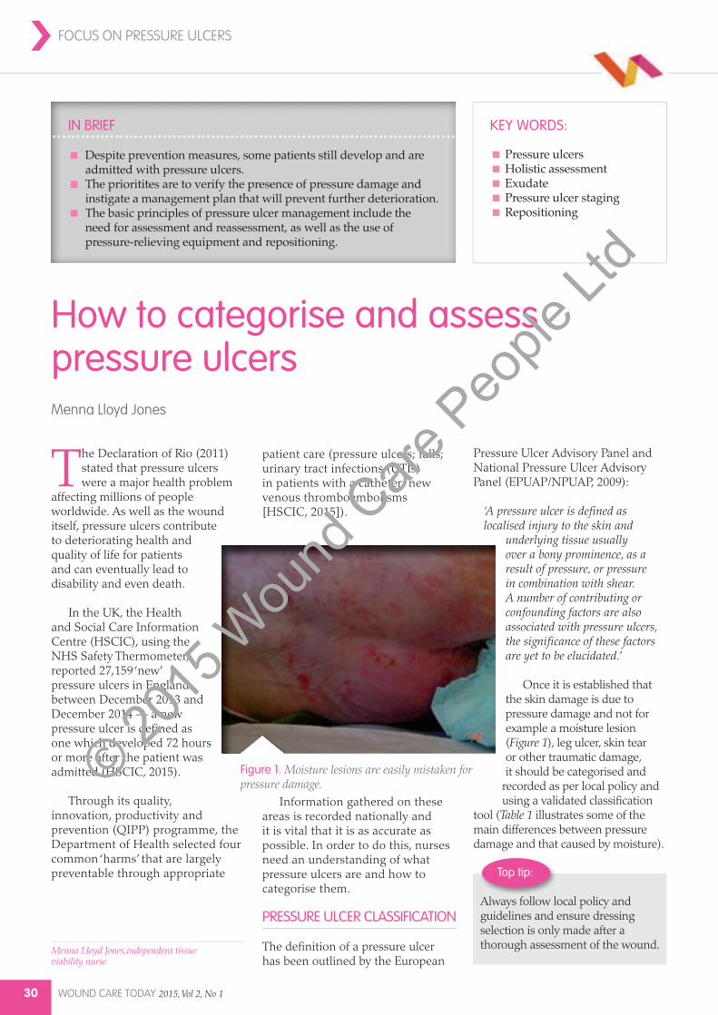
30 WOUND CARE TODAY 2015, Vol 2, No 1
FOCUS ON PRESSURE ULCERS
The Declaration of Rio (2011) stated that pressure ulcers were a major health problem
affecting millions of people worldwide. As well as the wound itself, pressure ulcers contribute to deteriorating health and quality of life for patients and can eventually lead to disability and even death.
In the UK, the Health and Social Care Information Centre (HSCIC), using the NHS Safety Thermometer, reported 27,159 ‘new’ pressure ulcers in England between December 2013 and December 2014 — a new pressure ulcer is defined as one which developed 72 hours or more after the patient was admitted (HSCIC, 2015).
Through its quality, innovation, productivity and prevention (QIPP) programme, the Department of Health selected four common ‘harms’ that are largely preventable through appropriate
How to categorise and assess pressure ulcers
patient care (pressure ulcers; falls; urinary tract infections (UTIs) in patients with a catheter; new venous thromboembolisms [HSCIC, 2015]).
Pressure Ulcer Advisory Panel and National Pressure Ulcer Advisory Panel (EPUAP/NPUAP, 2009):
‘A pressure ulcer is defined as localised injury to the skin and
underlying tissue usually over a bony prominence, as a result of pressure, or pressure in combination with shear. A number of contributing or confounding factors are also associated with pressure ulcers, the significance of these factors are yet to be elucidated.’
Once it is established that the skin damage is due to pressure damage and not for example a moisture lesion (Figure 1), leg ulcer, skin tear or other traumatic damage, it should be categorised and
recorded as per local policy and using a validated classification
tool (Table 1 illustrates some of the main differences between pressure damage and that caused by moisture).
Menna Lloyd Jones,independent tissue viability nurse
Menna Lloyd Jones
Figure 1. Moisture lesions are easily mistaken for pressure damage.
Top tip:
Always follow local policy and guidelines and ensure dressing selection is only made after a thorough assessment of the wound.
Information gathered on these areas is recorded nationally and it is vital that it is as accurate as possible. In order to do this, nurses need an understanding of what pressure ulcers are and how to categorise them.
PRESSURE ULCER CLASSIFICATION
The definition of a pressure ulcer has been outlined by the European
IN BRIEF
Despite prevention measures, some patients still develop and are admitted with pressure ulcers. The prioritites are to verify the presence of pressure damage and instigate a management plan that will prevent further deterioration.The basic principles of pressure ulcer management include the need for assessment and reassessment, as well as the use of pressure-relieving equipment and repositioning.
KEY WORDS:
Pressure ulcers Holistic assessment Exudate Pressure ulcer staging Repositioning
Menna Lloyd-Jones - PUs - read by JBB/BM.indd 36 22/04/2015 18:15
© 2015
Wou
nd C
are P
eople
Ltd

References 1. SMTL method TM-404 for Free Swell Absorbtion and Retention. Test performed at SMTL, UK.
Laboratory report 20140806-001.
Molnlycke Health Care, Arenson Centre, Arenson Way, Dunstable, Bedfordshire, LU5 5UL.
Telephone: 0800 917 4918. Email: [email protected].
The Mölnlycke Health Care, Exufiber® and Safetac® trademarks, names and logo types are registered
globally to one or more of the Mölnlycke Health Care Group of Companies.
© 2015 Mölnlycke Health Care AB. All rights reserved.
Superior retention1
Easier removal
Exufiber® - the future of fiber dressingsNow
on Dru
g
Tariff
Exufiber is a unique gelling fibre dressing with Hydrolock® Technology which
addresses the challenges of highly exuding wounds.
• Superior retention capacity1 reduces risk of leakage and maceration.
• Excellent tensile strength enables removal in one piece.
• Together with a secondary dressing with Safetac® technology, Exufiber creates a
unique treatment option for highly exuding wounds.
Visit www.molnlycke.co.uk/exufiber to find out more and request free samples.
Exufiber ad A4.indd 1 02/03/2015 11:08
© 2015
Wou
nd C
are P
eople
Ltd
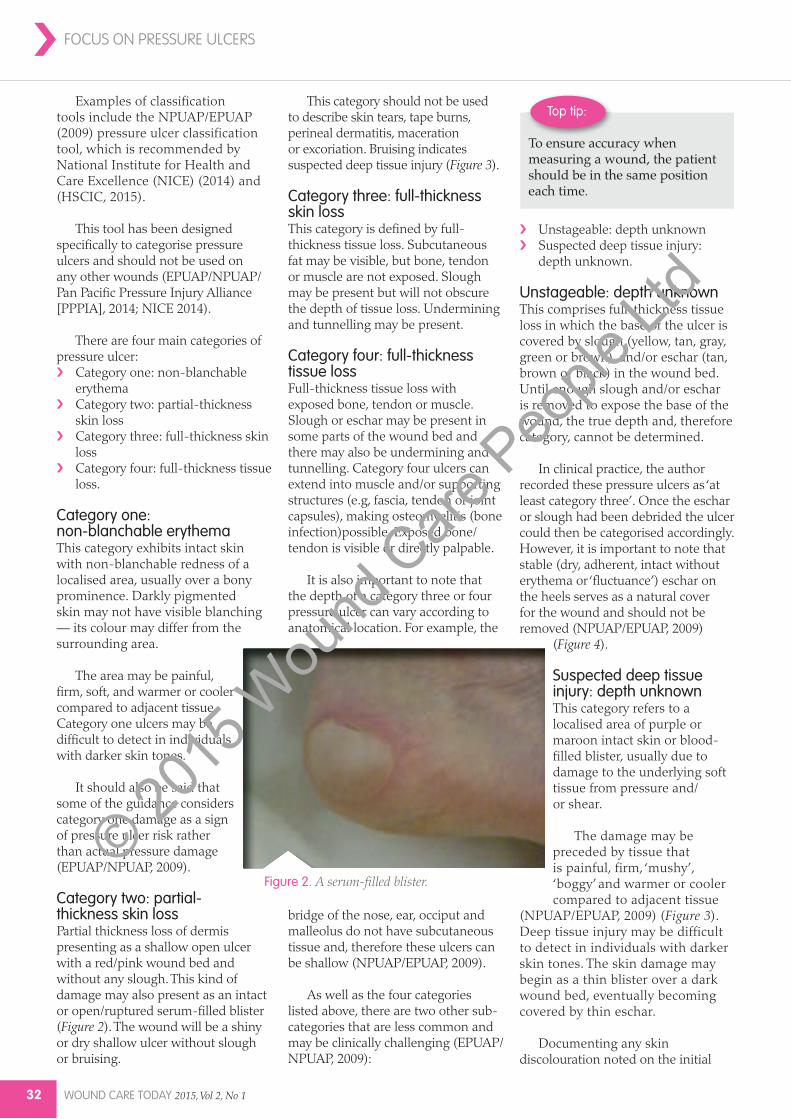
32 WOUND CARE TODAY 2015, Vol 2, No 1
FOCUS ON PRESSURE ULCERS
Examples of classification tools include the NPUAP/EPUAP (2009) pressure ulcer classification tool, which is recommended by National Institute for Health and Care Excellence (NICE) (2014) and (HSCIC, 2015).
This tool has been designed specifically to categorise pressure ulcers and should not be used on any other wounds (EPUAP/NPUAP/Pan Pacific Pressure Injury Alliance [PPPIA], 2014; NICE 2014).
There are four main categories of pressure ulcer:
Category one: non-blanchable erythemaCategory two: partial-thickness skin loss Category three: full-thickness skin lossCategory four: full-thickness tissue loss.
Category one: non-blanchable erythema This category exhibits intact skin with non-blanchable redness of a localised area, usually over a bony prominence. Darkly pigmented skin may not have visible blanching — its colour may differ from the surrounding area.
The area may be painful, firm, soft, and warmer or cooler compared to adjacent tissue. Category one ulcers may be difficult to detect in individuals with darker skin tones.
It should also be said that some of the guidance considers category one damage as a sign of pressure ulcer risk rather than actual pressure damage (EPUAP/NPUAP, 2009).
Category two: partial-thickness skin loss Partial thickness loss of dermis presenting as a shallow open ulcer with a red/pink wound bed and without any slough. This kind of damage may also present as an intact or open/ruptured serum-filled blister (Figure 2). The wound will be a shiny or dry shallow ulcer without slough or bruising.
This category should not be used to describe skin tears, tape burns, perineal dermatitis, maceration or excoriation. Bruising indicates suspected deep tissue injury (Figure 3).
Category three: full-thickness skin loss This category is defined by full-thickness tissue loss. Subcutaneous fat may be visible, but bone, tendon or muscle are not exposed. Slough may be present but will not obscure the depth of tissue loss. Undermining and tunnelling may be present.
Category four: full-thickness tissue loss Full-thickness tissue loss with exposed bone, tendon or muscle. Slough or eschar may be present in some parts of the wound bed and there may also be undermining and tunnelling. Category four ulcers can extend into muscle and/or supporting structures (e.g, fascia, tendon or joint capsules), making osteomyelitis (bone infection)possible. Exposed bone/tendon is visible or directly palpable.
It is also important to note that the depth of a category three or four pressure ulcer can vary according to anatomical location. For example, the
bridge of the nose, ear, occiput and malleolus do not have subcutaneous tissue and, therefore these ulcers can be shallow (NPUAP/EPUAP, 2009).
As well as the four categories listed above, there are two other sub-categories that are less common and may be clinically challenging (EPUAP/NPUAP, 2009):
Unstageable: depth unknown Suspected deep tissue injury: depth unknown.
Unstageable: depth unknown This comprises full-thickness tissue loss in which the base of the ulcer is covered by slough (yellow, tan, gray, green or brown), and/or eschar (tan, brown or black) in the wound bed. Until enough slough and/or eschar is removed to expose the base of the wound, the true depth and, therefore category, cannot be determined.
In clinical practice, the author recorded these pressure ulcers as ‘at least category three’. Once the eschar or slough had been debrided the ulcer could then be categorised accordingly. However, it is important to note that stable (dry, adherent, intact without erythema or ‘fluctuance’) eschar on the heels serves as a natural cover for the wound and should not be removed (NPUAP/EPUAP, 2009)
(Figure 4).
Suspected deep tissue injury: depth unknown This category refers to a localised area of purple or maroon intact skin or blood-filled blister, usually due to damage to the underlying soft tissue from pressure and/or shear.
The damage may be preceded by tissue that is painful, firm, ‘mushy’, ‘boggy’ and warmer or cooler compared to adjacent tissue
(NPUAP/EPUAP, 2009) (Figure 3). Deep tissue injury may be difficult to detect in individuals with darker skin tones. The skin damage may begin as a thin blister over a dark wound bed, eventually becoming covered by thin eschar.
Documenting any skin discolouration noted on the initial
Top tip:
To ensure accuracy when measuring a wound, the patient should be in the same position each time.
Figure 2. A serum-filled blister.
Menna Lloyd-Jones - PUs - read by JBB/BM.indd 38 23/04/2015 13:01
© 2015
Wou
nd C
are P
eople
Ltd

Do you need an evidenced-based source of practical, ‘need-to-know’,key information at your fi ngertips?
The ‘Care Today’ series of annual journals cover specifi c areas of care vital for day-to-day practice:
✓ Wound Care Today (now in its second volume)
✓ Skin Care Today
✓ Respiratory Care Today
Their easy-to-read style combines clinical education with practical tips through:
› Point-specifi c posters — your clinical companions for everyday practice
› Clinical features — written by experts in their fi eld
› Debate and discussion — on what matters most to provide patient-centred care
Visit www.jcn.co.uk for information on forthcoming journals in the series....
... as keeping up to date has never been easier for all those working in the community.
Available in print and online via:
www.jcn.co.uk
Don’t wait for tomorrow — start caring today …. ✓
WOUND CARE TODAY
www.woundcare-today.com
In association with
WOUND CARE TODAY
For what matters in practice
Volume 2Number 1
2015
Are pressure ulcers in danger of landing nurses in the dock?
WCT Cover 15.indd 1 4/23/15 1:30 PM
All Today ad.indd 1 4/23/15 2:46 PM
© 2015
Wou
nd C
are P
eople
Ltd
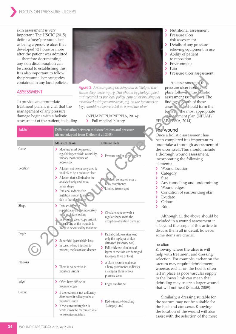
34 WOUND CARE TODAY 2015, Vol 2, No 1
FOCUS ON PRESSURE ULCERS
(NPUAP/EPUAP/PPPIA, 2014):Full medical history
Nutritional assessment Pressure ulcer risk assessment Details of any pressure- relieving equipment in useAbility of patient to repositionEnvironment PainPressure ulcer assessment.
An assessment of the pressure ulcer itself takes place following the holistic assessment (see below). The findings of both of these assessments should form the basis for the most appropriate management plan (NPUAP/
EPUAP/PPPIA, 2014).
The wound Once a holistic assessment has been completed it is important to undertake a thorough assessment of the ulcer itself. This should include a thorough wound assessment, incorporating the following elements:
Wound location CategorySizeAny tunnelling and undermining Wound edges Condition of surrounding skinExudateOdourPain.
Although all the above should be included in a wound assessment it is beyond the scope of this article to discuss them all in detail, however some items are crucial.
LocationKnowing where the ulcer is will help with treatment and dressing selection. For example, eschar on the sacrum may require debridement; whereas eschar on the heel is often left in place as poor vascular supply to the lower limb can mean that debriding may create a larger wound that will not heal (Suzuki, 2009).
Similarly, a dressing suitable for the sacrum may not be suitable for the heel and vice versa. Knowing the location of the wound will also assist with the selection of the most
skin assessment is very important. The HSCIC (2015) define a ‘new’ pressure ulcer as being a pressure ulcer that developed 72 hours or more after the patient was admitted — therefore documenting any skin discolouration can be crucial to establishing this. It is also important to follow the pressure ulcer categories contained in any local policies.
ASSESSMENT
To provide an appropriate treatment plan, it is vital that the management of any pressure damage begins with a holistic assessment of the patient, including
Table 1:
Cause Moisture must be present, e.g. shining, wet skin caused by urinary incontinence or loose stool
Pressure and/or shear present
Location A lesion not over a bony area is unlikely to be a pressure ulcerA lesion that is limited to the anal cleft only and has a linear shapePeri-anal redness/skin irritation is most likely to be due to faecal irritation
Tends to be located over a bony prominenceLimited to one spot
Shape Diffuse: different superficial spots are more likely to be moisture lesionsIn a kissing ulcer (copy lesion), at least one of the wounds is likely to be caused by moisture
Circular shape or with a regular shape (with the exception of friction damage)
Depth
Superficial (partial skin loss)In cases where infection is present, the lesion can deepen
Partial-thickness skin loss: only the top layer of skin damaged (category two)Full-thickness skin loss: all layers of the skin are damaged (category three or four)
NecrosisThere is no necrosis in moisture lesions
A black necrotic scab over a bony prominence indicates a category three or four pressure ulcer
Edge Often have diffuse or irregular edges
Edges are distinct
Colour If the redness is not uniformly distributed it is likely to be a moisture lesionIf the surrounding skin is white it may be macerated due to excessive moisture
Red skin non-blanching (category one)
Figure 3. An example of bruising that is likely to con-stitute deep tissue injury. This should be photographed and recorded as per local policy. Any other bruising not associated with pressure areas, e.g. on the forearms or legs, should not be recorded as a pressure ulcer.
Menna Lloyd-Jones - PUs - read by JBB/BM.indd 40 22/04/2015 18:17
© 2015
Wou
nd C
are P
eople
Ltd

WOUND CARE TODAY
http://woundcare-today.com/categories-pyramid
WOUND CARE TODAYWound Care Today’s Product Pyramid
Detailed explanations of the different wound care product categories +
Listings of all products available within each category +
Links to relevant websites + Extended product entries, including: › Specifi cations, how to use, and performance indicators
› Key clinical evidence to underpin product use in clinical practice
= Comprehensive product information to guide formulary decision-making
➥ www.woundcare-today.com
Pro Pyramid ad4.indd 1 9/10/13 7:30 PM
© 2015
Wou
nd C
are P
eople
Ltd

36 WOUND CARE TODAY 2015, Vol 2, No 1
FOCUS ON PRESSURE ULCERS
done by finding the cause of the excessive exudate and the use of appropriate dressings. For example, infection or the rehydration of necrotic tissue could increase the level of exudate — the treatment for necrotic tissue would be to debride. This can be done in several ways, including sharp and surgical debridement or autolytic debridement, or, as in the majority of cases, the use of dressings (autolytic debridement) (NICE, 2014). Some dressings such as
hydrofibres or hydrogels can be used to aid autolytic debridement (World Union of Wound Healing Societies (WUWHS), 2007).
Dressings that absorb exudate are available as primary and/or secondary dressings. As a minimum, any dressing selected should be able to absorb and contain exudate and enhance moist wound-healing environment.
Dressing manufacturers usually provide literature demonstrating the amount of exudate that a dressing will absorb and the recommended wear time.
However, in the case of excessive exudate it is not advisable to use a dressing such as a hydrogel, which will donate fluid to the wound (WUWHS, 2007).
appropriate pressure-relieving equipment.
SizeMeasuring the size of the ulcer and checking for any tunnelling or undermining is important, not only to chart the ulcer’s progress, but also to select the correct type and size of dressing.
To ensure that wound measurement is consistent, the patient should be in the same position for each assessment and the same method for measuring the size and depth of the wound should be used (NPUAP/EPUAP/PPPIA, 2014). All assessments and reassessments should be recorded as per local policy.
Condition of surrounding skin Checking the condition of the surrounding skin is important when seeking to choose the correct dressing.
However, the condition of the periwound skin can also be an indication of the level of exudate and the efficiency of the dressing currently being used. For example, a heavily exuding wound will cause damage to the surrounding skin — changing to a more absorbent dressing and applying a barrier product to the surrounding skin can help with this.
ExudateWound exudate is an essential component in the normal wound-healing process. However, excessive exudate can be a major challenge for clinicians and a source of embarrassment and discomfort for the patient.
The aim is to provide a wound-healing environment that maintains the optimal amount of exudate to promote healing. This is can be
InfectionIn the case of infection, debridement may also be required and where appropriate an antimicrobial dressing may address this. Some antimicrobial dressings also have autolytic properties which can aid debridement and address the wound infection simultaneously. Antimicrobial dressings should always be used in accordance with local policies (Jones, 2014).
FURTHER MANAGEMENT
Pressure-relievingequipment and repositioningIf a patient with a pressure ulcer is already being managed on pressure-relieving equipment such as a pressure-relieving mattresses, cushions, or chairs with integral cushions, these will need to be reviewed as will any repositioning schedule.
If the equipment is not effective it may need to be changed, for example from a high-specification mattress to a dynamic mattress. This is especially important where the patient has a category three or four ulcer (NICE, 2014; NPUAP/EPUAP/PPPIA, 2014).
In order to aid healing of the ulcer and prevent deterioration, it is important to be able to identify the underlying cause of the ulcer and, where possible, treat this. For example, if a patient with a pressure ulcer on his or her sacrum has limited mobility and sits in a chair all day, it is probable that the cause of the ulcer is the lack of mobility plus the amount of time spent sitting.
In this case, management would include pressure-relieving equipment, a repositioning schedule and, depending on the category of
Top tip:
Never leave slings or manual-handling equipment underneath a patient.
Top tip:
Provide supplements to patients with pressure ulcers who are
Figure 4. A heel pressure ulcer demonstrating significant eschar.
Menna Lloyd-Jones - PUs - read by JBB/BM.indd 42 22/04/2015 18:21
© 2015
Wou
nd C
are P
eople
Ltd

WOUND CARE TODAY 2015, Vol 2, No1 37
FOCUS ON PRESSURE ULCERS
pressure ulcer, bed rest (NICE, 2014; NPUAP/EPUAP/PPIA, 2014). NICE (2014) recommends that where possible patients should not lie or sit on an existing pressure ulcer.
Even if patients are being managed on dynamic mattresses it is still important to reposition or keep them mobile. Where they are able to do so, patients should be encouraged to move themselves as much as possible.
However, immobile patients or those with cognitive impairment (who do not have the capacity to understand the importance of maintaining mobility) will have to be repositioned, where possible with their agreement (EPUAP/NPUAP/PPPIA, 2014).
All repositioning methods and equipment used should be recorded, as should the appropriate use of manual-handling techniques and equipment. Slings, sleeves or other parts of the manual-handling equipment should never be left underneath the patient.
Similarly, before leaving the patient the nurse should ensure that he or she is comfortable and not lying on any equipment such as catheters and tubes (NICE, 2005; EPUAP/NPUAP, 2009; Wilson, 2012).
NutritionNutrition is an important factor in both the prevention and treatment of pressure ulcers (Johnston, 2007). NICE (2014) recommends that all patients with a pressure ulcer should be offered a nutritional assessment undertaken by a dietician or other healthcare professional with the necessary skills.
Nutritional supplements should only be offered to those patients who have been assessed as nutritional deficient (NICE, 2014).
PainPressure ulcers are painful and it should be remembered that some patients will be living with chronic pain. It is, therefore important to assess the patient using a validated tool and incorporate the findings into an appropriate management plan. For example, where possible the patient should not be positioned on the pressure ulcer.
It is also vital to ensure that adequate pain relief has been provided before any procedures are undertaken, such as dressing changes.
The nurse should select dressings that promote a warm moist-healing environment and which do not cause trauma to the wound or surrounding skin.
It is also important to encourage patients to say if they are in pain and involve the multidisciplinary team if necessary (NPUAP/EPUAP/PPPIA, 2014).
CONCLUSION
There is no doubt that pressure ulcers are a major health problem, affecting thousands of people in the UK. Management begins with ensuring that any skin damage is in fact a pressure ulcer.
The NPUAP/EPUAP (2009) has developed a validated tool for categorising pressure ulcers and also offer advice on those ulcers which are difficult to categorise.
The basic principles of pressure ulcer management include the need
for assessment and reassessment as well as the use of pressure-relieving equipment and repositioning. Patients with pressure ulcers also require nutritional and pain assessment. To promote the prompt healing of pressure ulcers, an holistic assessment of the patient and thorough wound assessment should be undertaken and only then should a management plan be written. WCT
REFERENCES
Declaration of Rio (2011) Available at: www.epuap.org (accessed 16 April, 2015)
Defloor T, Schoonhoven L, Fletcher J, et al (2005) EPUAP Review 6(3): 81–4
EPUAP/NPUAP (2009) Available at www.epuap.org (accessed 16 April, 2015)
EPUAP, NPUAP, PPPIA (2014) Available at: www.epuap.org (accessed 16 April, 2015)
HSCIC (2015) Available at: www.hscic.gov.uk/catalogue/PUB16329/NHS%20Safety%20Thermometer%20Report%20-%20December%202013%20to%20December%202014.pdf (accessed 16 April, 2015)
Johnston E (2007) Wound Essentials 2:10–21
Jones ML (2014) Br J Comm Nurs. June (supplement): s18–23
NICE (2005) The Management of Pressure Ulcers in Primary and Secondary Care. NICE, London
NICE (2014) Pressure Ulcers: prevention and management of pressure ulcers. NICE, London
Suzuki K (2009) Available at: http://www.podiatrytoday.com/current-insights-on-treating-heel-pressure-ulcers (accessed 20 April, 2015)
Wilson M (2012) Wound Essentials 7(1):53–5
WUWHS (2007) MEP Ltd, London
THE SCIENCEA pressure ulcer is defined as localised injury to the skin and
underlying tissue usually over a bony prominence, as a result of pressure, or pressure in combination with shear. A number of contributing or confounding factors are also associated with pressure ulcers, although the significance of these is yet to be elucidated (EPUAP/NPUAP, 2009).
Did you know: The longer pressure is exerted over a bony prominence, the higher it becomes and the more likely the patient is to develop pressure ulcers.
Menna Lloyd-Jones - PUs - read by JBB/BM.indd 43 22/04/2015 18:21
© 2015
Wou
nd C
are P
eople
Ltd

How
to m
anag
e ex
udat
e C
linic
alco
mpanion2
Read
ing
the
sign
s
Look
ing
at th
e pa
tient
Whi
le a
cer
tain
am
ount
of e
xuda
te is
nec
essa
ry to
hea
l the
w
ound
, the
pro
blem
s of
a h
ighl
y ex
udin
g w
ound
are
num
erou
s,
incl
udin
g w
et d
ress
ings
, con
tam
inat
ed c
loth
ing,
mal
odou
r an
d em
barr
assm
ent.
All
of th
ese
fact
ors
can
lead
to p
oor
qual
ity o
f life
an
d it
is v
ital t
hat c
linic
ians
are
abl
e to
ass
ess
patie
nts,
incl
udin
g3 :Id
entif
ying
any
con
ditio
ns th
at m
ay b
e co
ntri
butin
g to
the
exud
ate,
suc
h as
oed
ema
Look
ing
at th
e st
ate
of th
e w
ound
and
the
leve
l and
type
of
wou
nd e
xuda
te b
eing
pro
duce
dId
entif
ying
any
pot
entia
l dan
ger
to th
e pe
riw
ound
ski
n, fo
r ex
ampl
e is
ther
e le
akag
e of
exu
date
from
the
wou
nd; i
s th
e sk
in
red
(exc
oria
ted)
, or
does
it lo
ok w
hite
and
bog
gy (m
acer
ated
)?
Wha
t is
exud
ate?
Exud
ate
is th
e flu
id re
leas
ed fr
om a
wou
nd in
a c
ompl
icat
ed
inte
ract
ion
resu
lting
from
wou
nd a
etio
logy
, hea
ling,
env
iron
men
t, an
d co
mpo
undi
ng p
atho
logi
cal p
roce
sses
. It i
nclu
des
a m
ix o
f co
mpo
nent
s th
at e
ncou
rage
hea
ling,
incl
udin
g1 , 2 :
Elec
trol
ytes
M
atri
x m
etal
lopr
otei
nase
s (M
MP
s)
Vari
ous
cells
, suc
h as
leuk
ocyt
es, m
acro
phag
es, n
eutr
ophi
ls
and
plat
elet
sM
icro
orga
nism
sN
utri
ents
Gro
wth
fact
ors
Infla
mm
ator
y m
edia
tors
.
Cons
iste
ncy
Colo
urO
dour
Volu
me
Thic
ker,
visc
ous
and
som
etim
es
‘stic
ky’ e
xuda
te
is o
ften
due
to
infe
ctio
n, d
evita
lised
(n
ecro
tic) t
issu
e or
re
tain
ed d
ress
ing
fibre
sTh
inne
r exu
date
ca
n be
an
indi
catio
n th
atm
alnu
triti
onor
Hea
lthy
exud
ate
that
is
requ
ired
for
heal
ing
is a
‘s
traw
’-lik
e co
lour
Gre
en, o
r ‘m
ilky’
exud
ate
can
be a
sig
n of
infe
ctio
nPi
nk o
r red
exu
date
sh
ould
pro
mpt
the
nurs
e to
che
ck fo
r bl
eedi
ng in
the
wou
ndbe
d
A s
tron
g un
plea
sant
odo
ur
can
be a
sig
n of
ba
cter
ial g
row
th,
infe
ctio
n or
ne
cros
is
Odo
ur is
als
o of
ten
a fe
atur
e of
fu
ngat
ing
wou
nds
and
fistu
lae
In a
‘hea
lthy’
wou
nd-h
ealin
g tra
ject
ory,
the
amou
nt o
f ex
udat
e w
ill re
duce
as
the
wou
nd h
eals
Som
e w
ound
s, s
uch
as
burn
s an
d ve
nous
leg
ulce
rs, m
ay p
rodu
ce
mor
e ex
udat
eLo
ok o
ut fo
r a
sudd
en
incr
ease
in e
xuda
te —
this
m
aybe
due
toin
fect
ion
in
An
unde
rsta
ndin
g of
the
pres
enta
tion
of e
xuda
te is
requ
ired
in o
rder
to id
entif
y w
hat t
his
mea
ns fo
r th
e w
ound
and
the
patie
nt:
© 2015
Wou
nd C
are P
eople
Ltd

spon
sore
d by
Zetu
vit®
Plu
s sup
er-a
bsor
bent
dre
ssin
g pa
d is
just
one
of t
he d
ress
ings
ava
ilabl
e fro
m H
ART
MA
NN
for e
ffect
ive
exud
ate
man
agem
ent.
Des
igne
d fo
r hig
h vo
lum
es o
f wou
nd fl
uid5 , i
t rap
idly
bin
ds a
nd
relia
bly
reta
ins
exud
ate5
Soft,
con
form
able
and
com
fort
able
5, 6
to w
ear
Cos
t-ef
fect
ive7 .
For m
ore
info
rmat
ion
and
to d
ownl
oad
this
pos
ter,
go to
:w
ww
.jcn.
co.u
k,or
ww
w.h
artm
ann.
co.u
k/11
5595
.php
Man
agin
g a
high
ly e
xudi
ng w
ound
Whe
n tr
eatin
g a
patie
nt w
ith a
hig
hly
exud
ing
wou
nd, t
he
follo
win
g el
emen
ts a
re im
port
ant3 :
An
initi
al m
edic
al a
sses
smen
t of t
he p
atie
nt, t
o id
entif
y co
mor
bidi
ties
Join
t dec
isio
n-m
akin
g on
trea
tmen
t bet
wee
n nu
rse
and
patie
nt
Spec
ific
wou
nd a
nd e
xuda
te a
sses
smen
t by
a kn
owle
dgea
ble
and
com
pete
nt n
urse
Use
of a
n ab
sorb
ent d
ress
ing
whi
ch is
com
patib
le w
ith o
ther
tr
eatm
ent s
uch
as c
ompr
essi
on b
anda
ging
Any
dre
ssin
g m
ust r
efle
ct th
e pa
tient
’s s
kin
asse
ssm
ent a
nd
not c
ause
irri
tatio
nC
oncu
rren
t ski
n pr
otec
tion
trea
tmen
t with
was
hing
, m
oist
uris
ing
and
emol
lient
ther
apy
Reg
ular
eva
luat
ion
and
reas
sess
men
t of t
reat
men
t.
Choo
sing
the
right
dre
ssin
gA
wou
nd d
ress
ing
that
is c
hose
n to
man
age
high
vol
umes
of
exud
ate
shou
ld h
ave
som
e or
all
of th
e fo
llow
ing
attr
ibut
es4 :
Hig
h-ab
sorb
ency
: hel
ps to
redu
ce d
ress
ing
freq
uenc
yA
bilit
y to
‘lock
aw
ay’ e
xuda
te, p
reve
ntin
g le
akag
e on
to th
e pe
riwou
nd s
kin
and
avoi
ding
mac
erat
ion/
exco
riatio
nC
an b
e us
ed u
nder
com
pres
sion
ban
dagi
ng w
ithou
t bec
omin
g bu
lky
thro
ugh
exud
ate
abso
rptio
nA
trau
mat
ic o
n re
mov
alC
onfo
rms
wel
l to
the
patie
nt’s
body
and
the
wou
nd s
ite
Prov
ides
the
clin
icia
n w
ith a
cos
t-ef
fect
ive
optio
n.
This
doc
umen
t is
a gu
ide
only
and
doe
s no
t dim
inis
h th
e re
quir
emen
t to
exer
cise
clin
ical
judg
emen
t and
follo
w lo
cal p
olic
y. T
he p
ublis
hers
and
HA
RTM
AN
N c
anno
t acc
ept
resp
onsi
bilit
y fo
r th
e us
e of
this
info
rmat
ion
in c
linic
al p
ract
ice.
fibre
sTh
inne
r exu
date
ca
n be
an
indi
catio
n th
at m
alnu
triti
on o
r he
art d
isea
se a
re
an is
sue
shou
ld p
rom
pt th
e nu
rse
to c
heck
for
blee
ding
in th
e w
ound
bed
ofte
n a
feat
ure
of
fung
atin
g w
ound
s an
d fis
tula
e
mor
e ex
udat
eLo
ok o
ut fo
r a
sudd
en
incr
ease
in e
xuda
te —
this
m
ay b
e du
e to
infe
ctio
n in
th
e w
ound
or
a w
orse
ning
m
edic
al p
robl
em
1. W
UW
HS
(200
7) W
ound
s Int
erna
tiona
l, Lo
ndon
2. W
hite
R, C
uttin
g K
C (2
006)
Ava
ilabl
e at
:ht
tp://
ww
w.w
orld
wid
ewou
nds.c
om3.
Bel
don
P (2
014)
Wou
nd C
are T
oday
1(1)
: 38–
444.
Ste
phen
-Hay
nes J
(201
1)Br
J Co
mm
Nur
s 16(
3 Su
ppl):
S44
–49
5. K
aspa
r D (2
009)
Inte
rnal
pub
licat
ion.
Ava
ilabl
e fro
m H
artm
ann
6. N
esta
S, M
cKay
S (2
012)
Inte
rnal
pub
licat
ion.
A
vaila
ble
from
Har
tman
n7.
Dru
g Ta
riff a
nd N
HS
Supp
ly C
hain
Pric
e co
mpa
rison
s Apr
il 20
15
Exud
ate
poste
r Har
tman
n - v
ersio
n 2.
indd
123
/04/
2015
13
:44
© 2015
Wou
nd C
are P
eople
Ltd
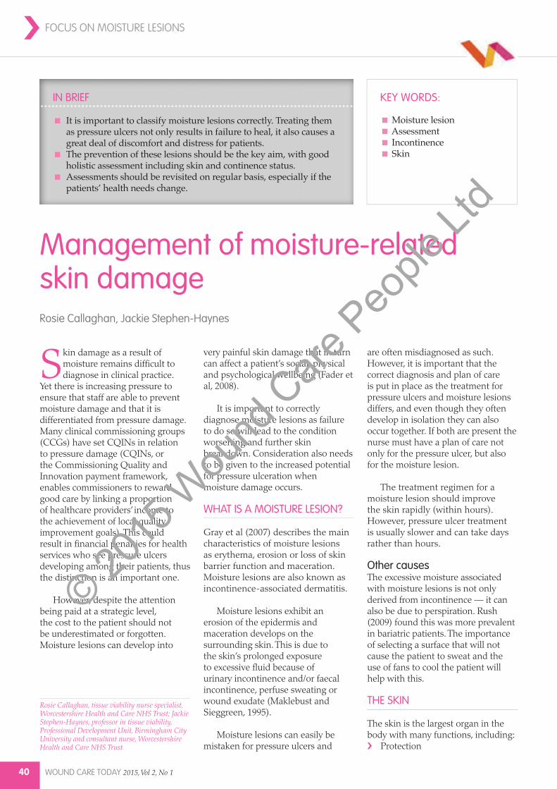
40 WOUND CARE TODAY 2015, Vol 2, No 1
FOCUS ON MOISTURE LESIONS
Skin damage as a result of moisture remains difficult to diagnose in clinical practice.
Yet there is increasing pressure to ensure that staff are able to prevent moisture damage and that it is differentiated from pressure damage. Many clinical commissioning groups (CCGs) have set CQINs in relation to pressure damage (CQINs, or the Commissioning Quality and Innovation payment framework, enables commissioners to reward good care by linking a proportion of healthcare providers’ income to the achievement of local quality improvement goals). This could result in financial penalties for health services who see pressure ulcers developing among their patients, thus the distinction is an important one.
However, despite the attention being paid at a strategic level, the cost to the patient should not be underestimated or forgotten. Moisture lesions can develop into
Management of moisture-related skin damage
are often misdiagnosed as such. However, it is important that the correct diagnosis and plan of care is put in place as the treatment for pressure ulcers and moisture lesions differs, and even though they often develop in isolation they can also occur together. If both are present the nurse must have a plan of care not only for the pressure ulcer, but also for the moisture lesion.
The treatment regimen for a moisture lesion should improve the skin rapidly (within hours). However, pressure ulcer treatment is usually slower and can take days rather than hours.
Other causesThe excessive moisture associated with moisture lesions is not only derived from incontinence — it can also be due to perspiration. Rush (2009) found this was more prevalent in bariatric patients. The importance of selecting a surface that will not cause the patient to sweat and the use of fans to cool the patient will help with this.
THE SKIN
The skin is the largest organ in the body with many functions, including:
Protection
Rosie Callaghan, tissue viability nurse specialist, Worcestershire Health and Care NHS Trust; Jackie Stephen-Haynes, professor in tissue viability, Professional Development Unit, Birmingham City University and consultant nurse, Worcestershire Health and Care NHS Trust
IN BRIEF
It is important to classify moisture lesions correctly. Treating them as pressure ulcers not only results in failure to heal, it also causes a great deal of discomfort and distress for patients.The prevention of these lesions should be the key aim, with good holistic assessment including skin and continence status.Assessments should be revisited on regular basis, especially if the patients’ health needs change.
KEY WORDS:
Moisture lesion Assessment Incontinence Skin
Rosie Callaghan, Jackie Stephen-Haynes
very painful skin damage that in turn can affect a patient’s social, physical and psychological wellbeing (Fader et al, 2008).
It is important to correctly diagnose moisture lesions as failure to do so will lead to the condition worsening and further skin breakdown. Consideration also needs to be given to the increased potential for pressure ulceration when moisture damage occurs.
WHAT IS A MOISTURE LESION?
Gray et al (2007) describes the main characteristics of moisture lesions as erythema, erosion or loss of skin barrier function and maceration. Moisture lesions are also known as incontinence-associated dermatitis.
Moisture lesions exhibit an erosion of the epidermis and maceration develops on the surrounding skin. This is due to the skin’s prolonged exposure to excessive fluid because of urinary incontinence and/or faecal incontinence, perfuse sweating or wound exudate (Maklebust and Sieggreen, 1995).
Moisture lesions can easily be mistaken for pressure ulcers and
Callaghan - moisture lesions - read by JBB/BM.indd 36 22/04/2015 18:36
© 2015
Wou
nd C
are P
eople
Ltd

Unbeatable protection for even the most sensitive skin
The LBF® No Sting Barrier Film and LBF® Barrier Cream product range can save skin as well as money. To protect vulnerable areas from the harmful effects of bodily fl uids, friction and adhesive trauma, there’s a choice that includes cream, spray, foam applicator and wipes. A range designed to save skin as well as budgets.
LBF® is a registered trademark of CliniMed (Holdings) Ltd. CliniMed Ltd, a company registered in England number 01646927. Cavell House, Knaves Beech Way, Loudwater, High Wycombe, Bucks. HP10 9QY. ©2015 CliniMed Ltd. PID 1341
Save more than just skin
CliniMed careline: 0800 036 0100 www.clinimed.co.uk
7654 LBF HCP Range Ad_A4_AW.indd 1 25/02/2015 12:13
© 2015
Wou
nd C
are P
eople
Ltd

42 WOUND CARE TODAY 2015, Vol 2, No 1
FOCUS ON MOISTURE LESIONS
SensationThermoregulation ExcretionCommunication.
The skin is made up of distinct layers — epidermis, dermis and hypodermis — that function as one unit. Sebum is an oily substance produced by the sebaceous glands and which helps prevent hair and skin from drying out. The production of sebum enables the skin to maintain an acidic pH of between 4.5–6.2.
However, incontinence can result in a mixing of urine and faeces which in turn creates an alkaline (ammonia), raising the pH of the skin. This raised pH increases protease and lipase activity, thereby causing skin irritation (Le Lievre, 2001). The skin becomes more permeable and allows for fluid to be retained, thus leading to excoriation (damage or removal of part of the surface of the skin).
The increased moisture combined with this bacterial and enzymatic
activity leads to breakdown of the skin and the development of moisture lesions (Figure 1). This is particularly seen in the very young or elderly.
The skin is our body’s main line of defence against potential environmental hazards, with intact skin playing a major part in our body’s immune system. Therefore, it is vital that any break in the skin, such as a moisture lesion, is prevented.
URINARY INCONTINENCE
Urinary incontinence affects between three to six million people in the UK (Irwin et al, 2006). The incidence increases with age and it is known that a high proportion of residents in nursing homes have
incontinence problems (Bale et al, 2004). Urinary incontinence in the general population affects 31% of older women and 23% of older men (Bale et al, 2004), with the incidence of faecal incontinence also rising with age and affecting about 12% of older people (Goode et al, 2005).
With people living longer through medical advances, continence is a problem that is only going to increase. However, it should also be said that incontinence is not an issue restricted to the older population and it can affect anyone at any age. As such, nurses must not assume incontinence in old age is inevitable and should always ensure that they investigate the causes of a patient’s incontinence rather than attributing it to his or her age.
Any episode of incontinence needs to be accurately recorded to establish the severity, with advice sought from a continence specialist where possible.
ASSESSMENT
There are several assessment tools that can be used to identify moisture lesions, including
The ‘perineal assessment tool’ (Nix, 2002)The ‘perirectal skin assessment tool’ (Storer-Brown, 1993) The National Association of Tissue Viability Nurses Scotland’s ‘skin excoriation tool’ (NATVNS, 2008).
The ‘perineal assessment tool’ (Nix, 2002) is a picture guide with score for specific characteristics and
Top tip:
Use a barrier film or cream to protect the skin from excess moisture.
THE SCIENCE — WHAT IS INCONTINENCE?
People can suffer from both urinary and faecal incontinence. Urinary incontinence is more common and up to six million people in the UK are thought to experience urinary incontinence, which is defined as the unintentional passing of urine. There are several types of urinary incontinence, but the most common are:
Stress incontinence: urine leaks when the bladder is under pressure, such as during
coughing or laughingUrge incontinence: a sudden, intense urge to pass urineMixed: a mixture of both stress and urge urinary incontinence.
Faecal incontinence is an inability to control bowel movements, resulting in the involuntary passage of stools. The most common causes are:
Rectal problems: severe constipation (resulting in overflow diarrhoea) or diarrhoea itself mean that stools cannot be retained properly Sphincter muscles: events such as childbirth can damage the muscles at the base of the rectumNerves: nerve damage through conditions such as diabetes or multiple sclerosis may mean that nerve signals sent from the rectum may not reach the brain in time.
Source: NHS Choices: www.nhs.uk
Cre
dit:
Nic
olas
Ale
jand
ro @
flick
r
Figure 1. The development of a moisture lesion.
Normalskin pH4.5–6.2
Mixing ofurine and
faecesmakes
ammonia
Prolongedmoisture
Skinbreakdown
Callaghan - moisture lesions - read by JBB/BM.indd 38 22/04/2015 18:37
© 2015
Wou
nd C
are P
eople
Ltd

PRODUCT INFORMATION
Balneum CreamIngredients: Urea 5 %, Ceramide NP, Aqua, Glycine Soya Oil, Propylene Glycol, Cetearyl Alcohol, Liquid Paraffi n, Isohexadecane, Sodium Lactate, Lactic Acid, PEG-20 Stearate, Polysorbate 60, Squalane, Stearic Acid, Disodium EDTA, Lecithin, Tocopherol, Ascorbyl Palmitate, Hydrogenated Palm Glycerides Citrate. Balneum Cream is free of fragrances, colourants and preservatives. Pharmaceutical form: White cream. Indications: Balneum Cream is formulated to protect the skin, to maintain skin moisture and to restore the impaired skin barrier in conditions giving rise to dry skin. It contains urea, ceramide NP and physiological lipids. Urea is known to increase the skins ability to retain water and maintain skin hydration. Ceramide NP is known to help restore the barrier function of skin. Balneum Cream is designed for daily use to relieve dry and very dry skin conditions including eczema, psoriasis and other dermatological conditions. Method of administration: Using clean hands, apply the cream to the skin once or twice daily. Contraindications: Patients with known hypersensitivity to any of the ingredients, soya or peanut. Warnings and precautions: For topical use only. Do not use on broken or infl amed skin. Caution should be exercised with concomitant treatment with other medicated topical products. If the condition becomes worse or if patients experience side effects/adverse incidents, discontinue treatment with Balneum Cream and seek medical advice. Adverse incidents: Although very few adverse incidents have been reported, the most frequent are local skin reactions. Special precautions for storage: Do not store above 25°C. Use within 6 months of fi rst opening Pack sizes: Available in 50g (£2.85) and 500g (£9.97) pump dispensers. CE marking held by: Almirall Hermal GmbH, Scholtzstraße 3, 21465 Reinbek, Germany Distributed in the UK by: Almirall Ltd, 1 The Square, Stockley Park, Uxbridge, UB11 1TD Date of Revision: 02/2015. Item code: UKSOY3039.
Adverse incidents should be reported. Online reporting forms and information can be found at www.mhra.gov.uk/yellowcard. Adverse incidents should also be reported to Almirall Ltd.
Adverse events should be reported. Reporting forms and information can be found at www.mhra.gov.uk/yellowcard. Adverse events should also be reported to Almirall Ltd.
Solutions with you in mind
PRESCRIBING INFORMATION(Please consult the Summary of Product Characteristics (SmPC) before prescribing.)
Balneum Plus Cream Lauromacrogols 3.0 % w/w and urea 5.0 % w/w. Active Ingredient: lauromacrogols 3.0 % w/wand urea 5.0 % w/w. Indication: For the treatment of pruritus, eczema, dermatitis and scaling skin conditions where an antipruritic and/or hydrating effect is required. It may also be used for the continued treatment and follow-up treatment of these skin diseases. Dosage and Administration: Adults, the elderly and children: Apply to each affected area twice a day. The duration of treatment depends on the clinical response. For external use only. Contraindications, Warnings, etc: Contraindications: Patients with known hypersensitivity to any of the ingredients. It should not be used to treat acute erythroderma, acute infl ammatory, oozing or infected skin lesions. Precautions: May cause irritation if applied to broken or infl amed skin. Interactions: Noneknown. Pregnancy and lactation: No clinical data on exposed pregnancies are available. Animal studies do not indicate direct or indirect harmful effects with respect to pregnancy, embryonal/foetal development, parturition or postnatal development. There are no specifi c restrictions concerning its use during pregnancy, but it is not to be used on the breasts immediately prior to breast feeding during lactation. Ability to drive and use machines: None. Adverse Effects: Balneum Plus Cream has been reported to cause a burning sensation, erythema, pruritus or the formation of pustules, aggravation of eczema if applied to infl amed skin areas. Contact allergy has also been reported. Legal Category: GSL. Marketing Authorisation Number(s): PL 33016/0010. NHS Cost: (excluding VAT). Tube containing 100g – £3.29. Pump pack containing 500g – £14.99. Marketing Authorisation Holder: Almirall Hermal GmbH, Scholtzstrasse 3, 21465 Reinbek, Germany. Further information is available from: Almirall Limited, 1 The Square, Stockley Park, Uxbridge, Middlesex, UB11 1TD, UK. Tel: (0) 207 160 2500. Fax: (0) 208 7563 888. Email: [email protected]. Date of Revision: 10/2012. Item code: UKSOY1402.
Urea 5% and ceramide NPUrea 5% and lauromacrogols 3%
CARING FOR OLDER SKINAgeing skin suffers from decreased lipids, urea, ceramides and lactic acid compared to younger skin.1–4
BALNEUM OFFERS TWO SOLUTIONS
RELIEVES & REHYDRATES
ITCH RELIEVER
Rapid relief within 5 minutes5
Well tolerated treatment6,7
Reduces poly-pharmacy and topical steroid use by up to 74%7
REHYDRATES & RESTORES
DRY SKIN RESTORER
Once daily can provide 24hr skin hydration8,9
Improves skin barrier function and skin integrity8
Once daily can offer a time saving and cost-effective treatment10
REFERENCES1. Harding C, Bartolone J, Rawlings A. Effects of Natural Moisturizing Factor and Lactic Acid Isomers on Skin Function. In: Dry skin and Moisturizers: Chemistry and Function (Loden M, Maibach H, eds). Boca Raton: CRC Press, 2000:229–41. 2. Seyfarth F, Schliemann S, Antonov D, Elsner P. Dry skin, barrier function, and irritant contact dermatitis in the elderly. Clinics in Dermatology 2011;29:31–6. 3. Stamatas GN, Nikolovski J, Mack MC, Kollias N. Infant skin physiology and development during fi rst years of life: a review of recent fi ndings based on in vivo studies. Int J Cosmet Sci 2011;33:17–24. 4. Cork M, Danby S. Skin barrier breakdown: a renaissance in emollient therapy. British Journal of Nursing 2009;18(14):872–877. 5. Kunzel J, Walter B, et al. How Effective is local Treatment in Acute Itch? Third International Workshop for the Study of Itch held in Heidelberg, Germany September 25–27 2005. 6. Balneum Plus Cream. Summary of Product Characteristics September 2012. 7. Schommer (2007) ‘Effi cacy of a polidocanol-urea combination in dry itchy skin’. Presented at EADV in Vienna 2007; Centre of Dermatology Research: Poster. 8. Brown K, Higgs-Bayliss T, Chittock J, Cork MJ & Danbu SG. The effect of an emollient cream containing 5% urea on the biophysical properties of aged skin. Poster 2013; Presented at European Epidermal Barrier Research Network: 1. 9. Almirall. Data on File BAL01. 10. MIMS September 2014.
UKSOY2999
February 2015
AR2784 - Almirall - Balneum Ad Wound Care Today 297mm x 210mm (April 2015) V1_PRINT.indd 1 20/04/2015 16:48
© 2015
Wou
nd C
are P
eople
Ltd

44 WOUND CARE TODAY 2015, Vol 2, No 1
FOCUS ON MOISTURE LESIONS›
Reassessment needs to take place regularly every 24–48 hours, and sometimes more frequently depending on the severity of the skin damage and the patient’s environment. For example, those in hospitals and care homes will have 24-hour access to staff, making observations easier than for those at home. This is particularly important in patients with incontinence and any skin changes must be documented and a robust plan to protect the skin put in place.
MANAGEMENT
IncontinenceA key factor in the prevention of moisture lesions is the accurate assessment and management of incontinence. If the cause of the incontinence cannot be treated, a variety of continence products are available, including: › Body-worn incontinence pads› Urinary sheaths and
containment devices
› Urinary catheters› Urinals and absorbent gels› Anal plugs› Faecal collectors› Faecal management systems.
Each patient should be individually assessed to ascertain the best product for his or her needs. This should be reassessed regularly to ensure the product remains the most appropriate.
NutritionEffective management of the nutritional status of the patient can improve the strength of the skin and reduce the risk of skin breakdown. Encourage the patient to drink enough to maintain good hydration as this will also dilute the urine (concentrated urine has an increased amount of waste products and a higher pH and can be detrimental to the skin).
A nutritional assessment should be carried out in those patients who already have moisture lesions as well as those thought to be at risk.
Skin careA good skin care regimen is very important and should be performed immediately after soiled and wet skin has been cleaned. It is also important to avoid the over-hydration that can be caused by excessive washing (Ersser et al, 2005).
The skin should be cleansed following each episode of incontinence with a low pH soap and warm water. Normal soap can dehydrate the skin and contains surfactants that can irritate the skin, which can make it more susceptible to breakdown (Bale et al, 2004).
like the ‘perirectal skin assessment tool’ (Storer-Brown, 1993) considers specific areas in the sacral/buttock area. The latter is the easiest tool to use as it splits the excoriation into mild, moderate, and severe, and has pictures to aid the clinician.
There are clear differences between moisture lesions and pressure ulcers and by working through these differences it enables the practitioner to establish the correct diagnosis (Table 1).
Holistic assessment An holistic assessment of the patient should include an accurate skin assessment with particular attention paid to the perianal area, natal cleft, and between the thighs, buttocks and scrotum/labia, as all these areas can be affected by moisture lesions.
In severe cases, the erythema may extend to the lower abdomen and sacrum (Beldon, 2008).
› Wound facts...
Uncontrolled diarrhoea can cause dehydration, gradually draining the body of the fluids it needs to function properly. Dehydration is particularly dangerous in children and older people, and must be treated quickly to avoid serious health complications.
Table 1: Differences between moisture lesions and pressure ulcers (Evans and Stephen-Haynes, 2007)
Moisture lesion Pressure ulcerCause › Moisture › Pressure and/or shear and friction
Location › Natal cleft or on the wider buttock not over a bony prominence
› Over bony prominence
Shape › Mirror image, linear in shape› Diffuse in several superficial spots
› Takes on shape of the causative pressure, often limited to one spot
Depth › Superficial › Superficial or deep
Edges › Diffuse or irregular edges › Distinct edges
Colour › Non-uniform redness› Blanchable or non-blanchable erythema› Pink or white surrounding skin due to maceration
› Uniform redness
Necrosis › No necrosis › Necrosis can be present
› Incontinence and the skin...
The normal pH of the skin is between 4.5 and 6.2. To keep the skin intact it is essential to maintain the acidic pH of the skin. With incontinence, ammonia is produced as a result of the breakdown of urinary urea and aggravated by faecal urease, resulting in an increase in the pH of the skin. The skin becomes more permeable when the pH rises, which is exacerbated by excessive moisture and eventually causes invisible breaches on the skin’s surface.
Callaghan - moisture lesions - read by JBB/BM.indd 40 22/04/2015 18:37
© 2015
Wou
nd C
are P
eople
Ltd

Read JCN’s newoptimised versionfor tablets and smartphones
Visit: http://w
ww.jcn.co.uk
JCN has been around for a while,but read it now 21st century style,via tablets and smart phones,online and in print
Visit: www.jcn.co.uk
ipad & JCN.indd 1 1/23/14 6:24 PM
© 2015
Wou
nd C
are P
eople
Ltd

46 WOUND CARE TODAY 2015, Vol 2, No 1
FOCUS ON MOISTURE LESIONS
Creams are applied to intact skin, while spray foam applicators and wipes can be applied to either intact or broken skin. It is important to ensure that these are applied to dry, clean skin — if the skin is contaminated with urine or faeces, these products will only serve to seal them against the skin where they will continue inflict damage. These products should always be applied in accordance with the manufacturer’s instructions.
Talcum powder or creams should not to be used underneath these products. Whichever products are chosen, it is important to establish if they are compatible with any incontinence pads being worn.
Incontinence pads can become clogged with various products that prevent them from adequately absorbing and containing urine (for example, some creams designed for use with infants can cause absorbency problems). However, because many barrier products include dimethicone (a silicone oil), they do not have this effect.
InfectionIf the skin is broken then the possibility of infection should be considered, particularly as the sacral/buttock area is susceptible to fungal infections.
Nurses should consider the use of an anti-fungal cream for local infections. Candida albicansis a common fungus that thrives in environments such as moisture lesions. Diagnosis of Candida albicans is usually made by observation rather than skin ‘scrapings’, and wound swabs will not normally identify it.
Infection with Candida albicans is more likely when the patient has other skin problems or comorbidities. If Candida albicans is identified, the nurse should wash the skin regularly and dry it carefully afterwards (overweight people or their carers should be careful to dry any skin folds). Antifungal cream should be applied, remembering always to wash the hands carefully after touching an infected area and after applying any cream.
Similarly, the alkaline nature of normal soap raises the pH balance of the skin when in contact with urine and faeces, and this in turn facilitates the growth of bacteria.
There are skin cleansers available that can be used as an alternative to soap, including:
3-in-1 preparationsFoamsSpraysWipes.
These cleansers can have a soothing effect on the skin. It is important that the patient and family/carers are aware that these products are designed for the adult skin and do not contain alcohol. The use of baby wipes is not recommended on adult skin as many contain alcohol, which has a drying effect.
After washing, the skin should be patted and not rubbed dry as this not only causes friction and skin damage, but can also be very painful.
Barrier creams/filmsEarly identification of at-risk patients and the use of an appropriate barrier cream/film is important (see below).
PainManagement of the pain associated with moisture lesions is a top priority, and the patient’s pain level should be assessed using a visual analogue scale, before appropriate analgesia is prescribed.
The use of barrier products can reduce the erythema by protecting the skin and this in turn may help to reduce the pain. The aim of a barrier film or cream is to protect, repair, restore or prevent skin damage by mimicking the skin’s natural barrier function. Barrier protection is available as creams and films as well as sprays, foam applicators and wipes.
CONCLUSION
It is important to classify moisture lesions correctly. Treating them as pressure ulcers not only results in failure to heal, it also causes a great deal of discomfort and distress.
The prevention of these lesions should be the key aim, with good holistic assessment including skin and continence status. Nurses should consider carefully the appropriate use of barrier creams and incontinence aids to ensure that they are patient-specific. Assessments should be revisited on a regular basis, especially if the patient’s health changes or drugs/treatment such as antibiotics are administered that may affect the patient’s continence status. WCT
REFERENCES
Bale S, Tebble N, Jones V, Price P (2004) JTiss Viabil 14(2): 44–50
Beldon P (2008) Br J Comm Nur 13(Suppl12): S6–10
Ersser SJ, Getliffe K. Voegeli D, Regan S (2005) Int J Nurs Stud 42(7): 823–35
Evans J, Stephen-Haynes J (2007) J Wound Care 16(2): 54–6
Fader M, Cottenden AM, Getliffe K (2008) Cochrane Database Syst Rev 8(4):CD007408
Gray M, Bliss DZ, Doughty DB et al (2007) J Wound Ostomy Continence Nurs34(1): 45–54
Goode PS, Burgio KL, Halli AD et al (2005) J Am Geriatr Soc 53(4): 629–35
Irwin DE, Milsom I, Hunskaar S, et al (2006) European Urology 50(6): 1306–14
Le Lievre S (2001) Br J Community Nurs6(4): 180–5
Maklebust J, Sieggreen M (1995) Springhouse Corporation, Pennsylvania
Nix DH (2002) Ostomy Wound Manage48(2): 43–9
NATVNS (2008) Available at: www.patientsafetyfederation.nhs.uk/MediaLibrary/Downloads/Workstreams/NoNeedlessSkinBreakdown/ExcoriationTool.pdf (accessed 16 April, 2015)
Rush A (2009) Wound Essentials 4: 66–75
Storer-Brown D (1993) Ostomy Wound Manage 39(7): 28–31
Top tip:
Use appropriate urinary incontinence products and/or a faecal management system if there is profuse diarrhoea.
Callaghan - moisture lesions - read by JBB/BM.indd 42 22/04/2015 18:38
© 2015
Wou
nd C
are P
eople
Ltd

JOURNAL OF COMMUNITY NURSING
Welcome to JCN’s learning zone...
JCN’s online resource, which, together with the learning zone in the Journal of Community Nursing, helps you to develop your knowledge in vital areas of care, to keep up to date with clinical practice.
Read the article
Reflect on what you have learnt
Review your knowledge with the online test
... Then, download your certificate to show that you have completed this e-learning unit and gained competency in this area of clinical practice.
JCN’s learning zone — an essential educational resource for all busy nurses working in the community.
E-learning LZ.indd 1 10/29/13 12:15 PM
© 2015
Wou
nd C
are P
eople
Ltd

Trea
ting
pilo
nida
l sin
us
Clin
ical
com
panion3
Clin
ical
pre
sent
atio
n T
he m
ost c
omm
on p
rese
ntat
ion
in th
e cl
inic
al s
ettin
g is
a s
wol
len,
usu
ally
pai
nful
lesi
on in
the
sacr
al a
rea
clos
e to
the
anus
(4–5
cm a
way
). P
atie
nts
will
typi
cally
pre
sent
with
an
absc
ess
and
clin
ical
sig
ns in
clud
ing
loca
l war
mth
, red
ness
, loc
al p
ain
and
tend
erne
ss, a
nd p
ossi
ble
indu
ratio
n (in
crea
se in
fibr
ous
tissu
e). H
air
may
be
seen
pro
ject
ing
from
the
site
of t
he s
inus
.4
Wha
t is
a pi
loni
dal s
inus
?A
‘chan
nel’ o
r sin
us w
hich
deve
lops i
n the
natal
cleft
be
twee
n the
butto
cks a
nd be
hind
the a
nus.
It
com
mon
ly in
volve
s a ha
ir, wh
ich ha
s pier
ced t
he
skin
caus
ing a
tiny c
yst; o
r the
diste
nsion
of a
hair
follic
le, w
hich
then
beco
mes
bloc
ked.1
can d
evelo
p, lea
ding
to a
woun
d if le
ft un
treate
d.W
hen
the s
inus
requ
ires s
urge
ry, th
e con
ditio
n ca
n re
sult
in a
pain
ful w
ound
that
may
take
mon
ths t
o fu
lly h
eal.2
Wha
t cau
ses
a pi
loni
dal s
inus
?Pi
loni
dal s
inus
es h
ave t
wo m
ain ca
uses
:Co
ngen
ital: w
here
the p
atien
t is p
redi
spos
ed d
ue to
a ‘d
impl
ing’
of th
e na
tal cl
eft, m
eani
ng h
air an
d de
bris
such
as d
ead
skin
cells
can
colle
ct2, 3
Acqu
ired:
a re
sult
of in
-gro
win
g hair
s, w
hich
lead
to th
e hair
folli
cle
beco
min
g sw
ollen
with
kera
tin an
d in
fected
due
to th
e acc
umul
ation
of
hair,
cellu
lar d
ebris
and
bacte
ria. T
his c
an le
ad to
can l
ead t
o rup
ture
of
the f
ollicl
e and
infec
tion.1,
2
They
can
also b
e cau
sed
by ‘s
tretch
ing’
and
‘pul
ling’
in th
e dee
per l
evels
of
tiss
ue in
the n
atal c
left, c
ausin
g disr
uptio
n an
d ev
entu
al ru
ptur
e of
the h
air fo
llicle
. Pr
esen
tatio
n is
mor
e com
mon
in m
ales t
han
female
s and
is of
ten se
en in
yo
unge
r peo
ple d
ue to
hor
mon
al ch
ange
s affe
cting
the s
kin.
2
Sign
s/sy
mpt
oms5
Com
plic
atio
nsPr
even
tion
Pain
at t
he w
ound
site
Swel
ling
Dis
char
ge o
f pus
or
bloo
d fr
om th
e w
ound
Infla
mm
atio
nG
ener
al m
alai
sePy
rexi
a (f
ever
).
Infe
ctio
n H
aem
orrh
age
at th
e w
ound
site
Del
ayed
hea
ling
Wou
nd b
reak
dow
n W
ound
recu
rren
ce P
sych
olog
ical
issu
es, p
artic
ular
ly
embr
assm
ent a
nd s
tigm
a du
e to
th
e po
sitio
ning
of t
he w
ound
and
as
soci
atio
ns w
ith p
oor
hygi
ene.
The
nat
al c
left
requ
ires
spe
cial
at
tent
ion
with
rega
rds
clea
ning
an
d sh
ould
be
kept
as
free
from
ha
ir a
s po
ssib
le W
eekl
y sh
avin
g or
the
use
of a
ha
ir re
mov
al c
ream
up
to th
e ag
e of
30
is re
com
men
ded
to
prev
ent r
ecur
renc
e.
Credit: Nartoun@wikicommons
© 2015
Wou
nd C
are P
eople
Ltd

Nur
sing
and
wou
nd m
anag
emen
t N
ursi
ng m
anag
emen
t of a
pilo
nida
l sin
us in
clud
es:
Man
agem
ent o
f pai
n is
impo
rtan
t, pa
rtic
ular
ly in
pat
ient
s ad
mitt
ed a
s an
em
erge
ncy3
Nur
ses
shou
ld g
ive
cons
ider
atio
n to
any
und
erly
ing
cond
ition
s th
at m
ay b
e a
cont
ribu
ting
fact
or o
r gi
ve r
ise
to c
ompl
icat
ions
, suc
h as
di
abet
es, u
se o
f ste
roid
s, w
eak
imm
une
syst
em, e
tc2
It is
als
o ve
ry im
port
ant t
hat c
linic
ians
rem
embe
r th
at p
atie
nts
may
be
unpr
epar
ed fo
r th
e si
ze o
f the
wou
nd th
at m
ay b
e un
cove
red,
as
the
open
ing
of th
e ab
sces
s its
elf m
ay a
ppea
r sm
all
The
patie
nt’s
vita
l sig
ns, s
uch
as te
mpe
ratu
re, a
re a
lso
impo
rtan
t as
an in
dica
tor
of p
oten
tial i
nfec
tion2
Spec
ific
wou
nd c
are
cons
ider
atio
ns w
hen
deal
ing
with
a p
iloni
dal s
inus
incl
ude:
Wou
nds
in th
is a
rea
can
heal
qui
ckly
as
they
are
wel
l per
fuse
d, h
owev
er h
ealin
g ca
n be
affe
cted
by
orga
nism
s re
gula
rly fo
und
in th
e na
tal c
left6
Due
to th
e fr
eque
ncy
of d
ress
ing
chan
ges
requ
ired
— a
nd th
e pa
in in
volv
ed6 —
dre
ssin
gs th
at n
ot o
nly
conf
orm
to th
e w
ound
, but
whi
ch
are
non-
trau
mat
ic o
n re
mov
al (i
.e. g
ellin
g fib
re d
ress
ings
), sh
ould
be
cons
ider
ed in
this
are
aD
ue to
the
delic
acy
of th
e pe
riw
ound
ski
n an
d th
e lo
catio
n of
thes
e w
ound
s, a
dre
ssin
g th
at c
an a
bsor
b ex
udat
e, p
reve
ntin
g le
akag
e, a
nd
conf
orm
wel
l to
the
wou
nd b
ed is
des
irab
le6
Shav
ing
arou
nd th
e pe
riw
ound
ski
n ca
n al
so h
elp
with
atr
aum
atic
rem
oval
of d
ress
ings
, as
wel
l as
prev
entin
g re
curr
ence
.6
The
publ
ishe
rs a
nd M
ölnl
ycke
Hea
lth C
are
cann
ot a
ccep
t res
pons
ibili
ty fo
r the
use
of t
his i
nfor
mat
ion
in c
linic
al p
ract
ice,
whi
ch d
oes n
ot d
imin
ish
the
requ
irem
ent t
o ex
erci
secl
inic
al ju
dgem
ent a
nd fo
llow
loca
l pol
icy.
spon
sore
d by
1. G
rant
T (2
009)
Wou
nds
UK
5(1)
: 78–
802.
Ste
phen
-Hay
nes J
(200
8) W
ound
Ess
entia
ls3:
128–
333.
Tim
mon
s J (
2007
) Nur
s St
and
21(5
2): 4
8–56
4. L
anig
an M
D, D
yne
PL (2
015)
Med
scap
eA
vaila
ble
at: e
med
icin
e.m
edsc
ape.
com
5. N
ursi
ng S
tand
ard
(200
3) N
urs
Stan
d99
(14)
: 28
6. B
radl
ey L
(200
6) W
ound
s U
K 2
(1):
45–5
3
Exuf
iber
® is
a g
ellin
g fib
re d
ress
ing
with
Hyd
rolo
ck®
Tech
nolo
gy fo
r us
e on
exu
ding
wou
nds s
uch
as
pilo
nida
l sin
uses
. For
sam
ples
an
d in
form
atio
n, v
isit:
w
ww
.mol
nlyc
ke.c
o.uk
/exu
fiber
To d
ownl
oad
this
pos
ter,
go to
:w
ww
.jcn.
co.u
k
Trea
tmen
tTh
ere
are
seve
ral t
reat
men
t str
ateg
ies
for
pilo
nida
l sin
us, i
nclu
ding
2 :C
onse
rvat
ive:
par
ticul
arly
use
d in
non
-sev
ere
pres
enta
tions
and
invo
lves
ant
ibio
tics
to c
lear
any
infe
ctio
n an
d pr
even
t sur
gery
, as
wel
l as
prev
enta
tive
mea
sure
s su
ch a
s re
gula
r cl
eans
ing
and
shav
ing
of th
e ar
ea
Min
or s
urge
ry: i
n sm
alle
r no
n-in
fect
ed c
ases
any
hai
r ca
n be
rem
oved
from
the
absc
ess/
folli
cle
and
the
trac
k cl
eane
d ou
tSu
rger
y: in
clud
ing
exci
sion
of t
he a
ffect
ed ti
ssue
— in
clud
ing
absc
esse
s an
d in
flam
mat
ion
— fo
llow
ed b
y pr
imar
y cl
osur
e; a
nd w
ide
exci
sion
, whe
re th
e si
nus
is ‘l
aid
open
’ and
hea
ls b
y se
cond
ary
inte
ntio
n.
y(
)g
the
posi
tioni
ng o
f the
wou
nd a
nd
asso
ciat
ions
with
poo
r hy
gien
e.
g prev
ent r
ecur
renc
e.
Credit: Nartoun
Moln
lycke
pos
ter v
2.ind
d 1
22/0
4/20
15
13:4
9
© 2015
Wou
nd C
are P
eople
Ltd

50 WOUND CARE TODAY 2015, Vol 2, No 1
FOCUS ON COMPRESSION HOSIERY
The use of compression hosiery is widespread within the community and
is an essential component of the prevention of lower limb problems, as well as management following a comprehensive assessment to determine the aetiology of any ulceration.
Compression hosiery is a valid alternative to multilayer compression bandaging (Ashby et al, 2014) and wound care clinicians should be familiar with the types of hosiery available and become skilled in making the correct choice for their patients. The focus of the article will be on the practical considerations of using compression hosiery, including selection, application, removal and how to help patients use the garments.
OBJECTIVES OF COMPRESSION THERAPY
Compression therapy is the application of graduated pressure to the lower leg, with higher pressure at the ankle reducing to below the knee.
Compression hosiery to manage venous leg ulcers and oedema
treatment is prescribed (Clinical Resource Efficiency Support Team [CREST], 1998; RCN, 2006; Scottish Intercollegiate Guidelines Network [SIGN], 2010). Venous insufficiency affects up to 50% of the adult population (Venous Forum, 2011) and an estimated 1% of the population will have a leg ulcer in their lifetime, with venous disease accounting for 60–80% of leg ulceration (Callam, 1992).
Additionally, chronic oedema poses a challenge for the nurse, although it can be managed with hosiery. Chronic oedema is an accumulation of fluid, proteins and other macro-molecules leading to tissue swelling, skin changes and fibrosclerosis. It is a symptom of many different clinical conditions and affects over 100,000 people in the UK (Moffatt et al, 2007). Chronic oedema has become an increasing issue for nurses and appropriate management can be highly effective in improving the patient’s quality of life.
Compression hosiery can be used to achieve several things:
Prevention of venous leg ulcer recurrenceHealing of venous ulcersPrimary prevention of leg ulcers where varicose veins are present or patients have known risk factors
Jackie Stephen-Haynes, professor in tissue viability, Professional Development Unit, Birmingham City University and consultant nurse, Worcestershire Health and Care NHS Trust; Rosie Callaghan, tissue viability nurse specialist, Worcestershire Health and Care NHS Trust
IN BRIEF
Compression therapy is the application of graduated pressure to the lower leg, with the higher pressure at the ankle reducing to below the knee.Compression is provided in a number of ways, most commonly using bandaging and hosiery. A sound knowledge of the options will support the clinician when selecting and supplying the most appropriate hosiery.
KEY WORDS:
Compression Hosiery Lower limb
Jackie Stephen-Haynes, Rosie Callaghan
Compression supports the superficial veins and counteracts raised capillary pressure and is provided in a number of ways, most commonly using bandaging and hosiery (Moffatt et al, 2007).
Compression hosiery is suitable for patients with chronic venous insufficiency, post-thrombotic syndrome, varicose veins, venous eczema, lipodermatosclerosis and swelling in the legs associated with pregnancy (National Institute of Health and Care Excellence [NICE], 2012). Compression hosiery offers cost-effective therapy that promotes healing in venous ulceration (Ashby et al, 2014). It can prevent recurrence and can contain chronic oedema; thus significantly improving a patient’s clinical outcomes (Osborne, 2009).
Annual costs for leg ulcer care and management are conservatively estimated to be in the region of £200 million and, with age featuring as a key predisposing factor, the demographic profile of the UK will result in a continually increasing financial burden (Moffatt et al, 2004; Persoon et al, 2004; Posnett and Franks, 2008).
The key factor when treating people with leg ulcers is to establish the underlying aetiology to ensure that the most appropriate
JSH - hosieryBMC.indd 36 23/04/2015 12:33
© 2015
Wou
nd C
are P
eople
Ltd

UCS™ DebridementOptimum care for the wound and the whole limb
• Unique, convenient and safe way to clean the leg and prepare the wound
• Safe and simple to use
• Save time and improve skin-care
www.mediuk.co.uk medi. i feel better.
Pre-moistened cleaning and debridement clothClean & Prepare
Watch the UCS Debridement in action!
medi UK LimitedPlough Lane, Hereford, HR4 0EL
T 01432 373500
Part IXA of the Drug Tariff, Physical Debridement Devices
10 Sachetsper Box
Product Code: DT500PIP Code: 384-4271
00000_AZ_A4_210x297_UCS_Debridement_UK_12_2014_low3.indd 1 19.12.14 09:06
© 2015
Wou
nd C
are P
eople
Ltd

52 WOUND CARE TODAY 2015, Vol 2, No 1
FOCUS ON COMPRESSION HOSIERY›› Prevention of deep vein
thrombosis (DVT) in patients who have mobility difficulties
› Prevention of complications following DVT
› Maintenance and reduction of chronic oedema/lymphoedema.
In one 40-patient study, Stephen-Haynes and Sykes (2013) identified the following reasons for hosiery prescription: › 83% (n=35) of respondents used
hosiery for prevention of ulcer recurrence
› 72% (n=30) used hosiery for the management of chronic oedema
› 56% (n=24) used hosiery for prevention of venous leg ulceration
› 50% (n=21) used hosiery to actively treat leg ulceration
› 31% (n=13) used hosiery for the management of varicose veins.
COMPRESSION HOSIERY OPTIONS
It is important to select the appropriate hosiery as it will lead to improved clinical and financial outcomes. In the UK, hosiery is classified by the yarn used and by the amount of compression the garment exerts. Hosiery is either flat-knit or circular-knit.
Flat-knit hosieryThese garments are thicker and are sewn together. The fabric is stiffer so they are more suitable for distorted limbs and controlling oedema. Made-to-measure garments are usually flat-knit.
Circular-knitThese garments are thinner and finer and are knitted on a cylinder with no seam. They are suitable for low-risk patients with mild or little oedema. Being lighter, they are often more cosmetically acceptable to patients.
Hosiery is also classified according to the level of compression it provides. The World Union of Wound Healing Societies (WUWHS) (2008) offers the following classification: › Mild: less than 20mmHg› Moderate: 20–40mmHg› Strong: 40–60mmHg › Very strong: greater than
60mmHg.
Clinicians can choose between British and European standard hosiery and the choice is usually dependent on the levels of oedema or lymphoedema present.
British Standard classificationBritish Standard has three classes and these can be used variously for varicose veins, DVT prevention, treatment and prevention of venous leg ulcers, post-thrombotic venous insufficiency, and lymphoedema (Table 1).
Hosiery treatment kits are also available within the British Standard category that combine a 10mmHg liner and class 3 (25–35mmHg) garments; thus providing 40mmHg of therapeutic compression to aid ulcer healing. This is an alternative option to compression bandaging. It is suitable for those with a small ulcer and exudate that can be managed with a primary wound dressing. It
offers a clinically viable alternative as well as positive financial outcomes (Ashby et al, 2014).
European StandardEuropean Standard classification garments, particularly those with a higher stiffness index, are commonly accepted as the most suitable hosiery choice for oedema management (Lawrence, 2006; Lymphoedema Framework Project, 2006). They are available in four classes (Table 2).
Style and fitCompression hosiery is available as: › Standard fit › Made-to-measure hosiery› Leg ulcer treatment kits.
It is available in different styles such as thigh length and below-knee stockings, socks, and tights. Factors to consider when prescribing hosiery include: › Size› Colour › Open/closed toe › Style.
PREVENTING THE RECURRENCE OF LEG ULCERATION
Dowsett (2011) identified a reduction in recurrence of leg ulceration from 18–20% to 5.8% following a structured plan for the introduction of RAL hosiery (a German quality standard) within one UK primary care trust, demonstrating that hosiery has the potential to reduce leg ulceration recurrence.
HOSIERY IN THE TREATMENT OF VENOUS LEG ULCERATION
Where it is clinically appropriate, the use of compression hosiery in the care of patients with active venous leg ulceration achieves similar healing rates to those achieved with compression bandages (Burgess and Robinson, 1993; Samson and Showalter, 1996; Ashby et al, 2014). The venous leg ulcer study performed by Ashby et al (2014) was a randomised control trial (RCT) that investigated compression hosiery versus four-layer compression bandaging in the treatment of venous leg ulcers in 457 patients.
Table 1: British Standard drug tariff classification for compression hosiery
Class Pressure Support Indications for use
Class1 14–17mmHg Light › Early varicose veins › Mild ankle oedema
Class 2 18–24mmHg Medium
› Medium severity varicose veins › Post-thrombotic limb › Mild oedema › To prevent recurrence of venous ulcers
Class 3 25–35mmHg Strong
› Severe varicose veins › Gross oedema › Chronic insufficiency › To prevent recurrence of venous ulcers
JSH - hosieryBMC.indd 38 23/04/2015 12:33
© 2015
Wou
nd C
are P
eople
Ltd

Your patients are unique...
...their compression hosiery should be too
AltiMed, an Urgo Medical Company - Sullington Road, Shepshed, Loughborough LE12 9JG
01509 501720 01509 501721 [email protected] www.altimed.co.uk
Find out why our Made to Measure hosiery fits perfectly everytime by visiting www.altimed.co.uk© 20
15 W
ound
Care
Peo
ple Lt
d

54 WOUND CARE TODAY 2015, Vol 2, No 1
FOCUS ON COMPRESSION HOSIERY›
as many as they liked in order of preference, with the following results: › 92% (n=36) selected hosiery based
upon patient concordance › 86% (n=36) selected hosiery based
upon level of compression › 83% (n=35) selected hosiery due
to sizing and fit › 64% (n=27) were influenced
by the local wound management formulary.
When prescribing hosiery, it is important to advise the patients that they may have to wear hosiery as long as their venous disease persists. Nurses should consult with the patient when choosing the type of hosiery used, especially regarding colour and style, and whether the hosiery is open- or closed-toe, below-knee or thigh-length.
Concordance can be an issue. Rather than using ready-made hosiery, a made-to-measure garment
may help with this and nurses should aim for the optimum level of compression while recognising that any compression is better than none, particularly if the patient has difficulty coping with garments that exert a higher pressure.
British Standard hosiery will need replacing every three months whereas European Standard will need to replaced every six months.
Hosiery that is suitable for oedema should be prescribed when swelling is present. A stiffer garment would be the most appropriate to use in this case.
CONCLUSION
A sound knowledge of the options will help the nurse when selecting the most appropriate hosiery. Hosiery can be convenient for patients and is often tolerated where multilayer bandaging may not be. It can provide consistent pressures and makes efficient use of resources as it can be washed several times while still providing effective compression.
The selection of appropriate hosiery in collaboration with the patient can result in considerable cost savings and, more importantly, improve the patient’s quality of life (Ashby et al, 2014). WCT
REFERENCES
Ashby RL, Gabe R, Ali S, et al (2014) Lancet 383(9920): 871–9
Burgess B, Robinson B (1993) Nurs Times 89(13): 90–2
The study looked at patient concordance, quality of life, healing and cost. Researchers showed that the median time to healing in the hosiery kits and the four-layer bandages was roughly similar, however the recurrence rate was only 4% in the hosiery kit group, compared to 23% in the four-layer bandage group. The annual cost of hosiery was £1,493.90, a saving of £300 on the cost of four-layer bandaging.
PRESCRIBING
The importance of competence for all practitioners involved in the assessment, selection and fitting of hosiery and the impact on clinical and financial outcomes is significant.
In a 40-patient study, Stephen-Haynes and Sykes (2013) sought to identify factors that influence hosiery selection by asking nurses to fill in a checklist. Nurses were given a list of options and were asked to tick
› Top tips...
› Hosiery should have a soft and silky feel but can be damaged by direct heat and washing at high temperatures.
› Hosiery should be washed following the manufacturer’s instructions, usually by hand at 40 degrees, and dried flat away from direct heat.
› Hosiery must not be tumble-dried, ironed, bleached or dry-cleaned. › Hosiery can be damaged by fingernails and toenails, and jewellery, so
care must be taken when putting garments on.
Table 2: European Standard Drug Tariff classification for compression hosiery
Class Pressure Support Indications for use
Class1 18–21mmHg Light › Early varicose veins › Mild-ankle oedema
Class 2 25–32mmHg Medium
› Medium severity varicose veins › Post-thrombotic limb › Mild oedema › To prevent recurrence of venous ulcers
Class 3 36–46mmHg Strong
› Severe varicose veins › Gross oedema › Chronic insufficiency › To prevent recurrence of venous ulcers
Class 4 More than 59mmHg
Very strong› Gross oedema› Lymphoedema› Elephantiasis
› Further information
To find out more about the issues covered in this article and to access relevant clinical information, try the industry websites below: › AltiMed: http://altimed.co.uk/
products/altiform› Medi UK: www.mediuk.co.uk› Activa Healthcare:
www.activahealthcare.co.uk/compression-hosiery
News About Us Product Pyramid The Knowledge Wound Watch Forums Diary
Search
Register today www.woundcare-today.com
The website that brings everyone and everythingin wound care together
Wound Care Todaysaves you ti me...We digest wound care informati on from all media sources and tell you what they say in a quick and easy to read format.
about everythingyou want to knowWe cater for all wound care people, and because we recognise your interests vary, you can tailor your Wound Care Today content so that it is specifi c to you.
and keeps you informed...with all the latest nati onal and global wound care news, updates and in-depth reports on technology, innovati on, market intelligence, clinical practi ce, policy, products, people and places.
Half page Launching WCTb.indd 1 9/9/13 5:32 PMJSH - hosieryBMC.indd 40 23/04/2015 12:40
© 2015
Wou
nd C
are P
eople
Ltd
WOUND CARE TODAY 2015, Vol 2, No1 55
FOCUS ON COMPRESSION HOSIERY ›
for lower limbs. Poster presentation. Wounds UK Conference, Harrogate.
Lymphoedema Framework Project (2006) Best Practice for the Management of Lymphoedema. International Consensus. MEP Ltd, London
Moffatt C, Franks P, Doherty D et al (2004) QJM 97(7): 431–7
Moffatt C, Martin R, Smithdale R (2007) Leg ulcer management. Essential Skills for Nursing. Blackwell Science, Oxford
Callam M (1992) Phlebology 7(Suppl): S6–12
CREST (1998) Clinical Resource Efficiency Support Team Guidelines for the Assessment and Management of Leg Ulceration. CREST, Belfast
Dowsett C (2011) Wounds UK 7(1): 115 –9
Horakova M, Partsch H (1994) Phlebologie 47: 53
Lawrence (2006) Evaluation of new lymphoedema drug tariff garments
NICE (2012) Available at: http://cks.nice.org.uk/compression-stockings#! topicsummary (accessed 21 April, 2015)
NICE (2014) Available at: http://cks.nice.org.uk/varicose-veins#!topicsummary (accessed 21 April, 2015)
Persoon A, Heinen M, van der Vleuten C et al (2004) J Clin Nurs 13: 341–54
Posnett J, Franks P (2008) Nurs Times. 104(3): 44–5
RCN (2006) Clinical Practice Guidelines: the nursing management of patients with venous leg ulcers. 2nd edn. RCN, London
Osborne K (2009) Available at: www.woundsinternational.com/media/issues/240/files/content_206.pdf (accessed 4.03.2015)
Samson RH, Showalter DP (1996) Dermatol Surg 22(4): 373–6
SIGN (2010) Management of Chronic Venous Leg Ulcers. A national Clinical Guideline. SIGN, Edinburgh
Stephen-Haynes J, Sykes R (2013) Wounds UK 9(1): 16–20
Venous Forum of the Royal Society of Medicine (2011) Phlebology 26(3): 9–3
WUWHS (2008) Principles of Best Practice: compression in venous leg ulcers. MEP Ltd, London
› Application and removal...
The following tips will help with application:› A hosiery applicator can be used to aid application and concordance› Ideally, hosiery measurements should be taken in the morning
to reduce the impact of any oedema › Apply hosiery first thing in the morning when the leg is the
least swollen› A liner can be used to assist with application. Hosiery should be removed if the patient experiences any of the following:› Leg becomes painful› Leg become itchy › Hosiery feels tighter than previously› There is a loss of sensation in the leg.
News About Us Product Pyramid The Knowledge Wound Watch Forums Diary
Search
Register today www.woundcare-today.com
The website that brings everyone and everythingin wound care together
Wound Care Todaysaves you ti me...We digest wound care informati on from all media sources and tell you what they say in a quick and easy to read format.
about everythingyou want to knowWe cater for all wound care people, and because we recognise your interests vary, you can tailor your Wound Care Today content so that it is specifi c to you.
and keeps you informed...with all the latest nati onal and global wound care news, updates and in-depth reports on technology, innovati on, market intelligence, clinical practi ce, policy, products, people and places.
Half page Launching WCTb.indd 1 9/9/13 5:32 PMJSH - hosieryBMC.indd 41 23/04/2015 12:40
© 2015
Wou
nd C
are P
eople
Ltd
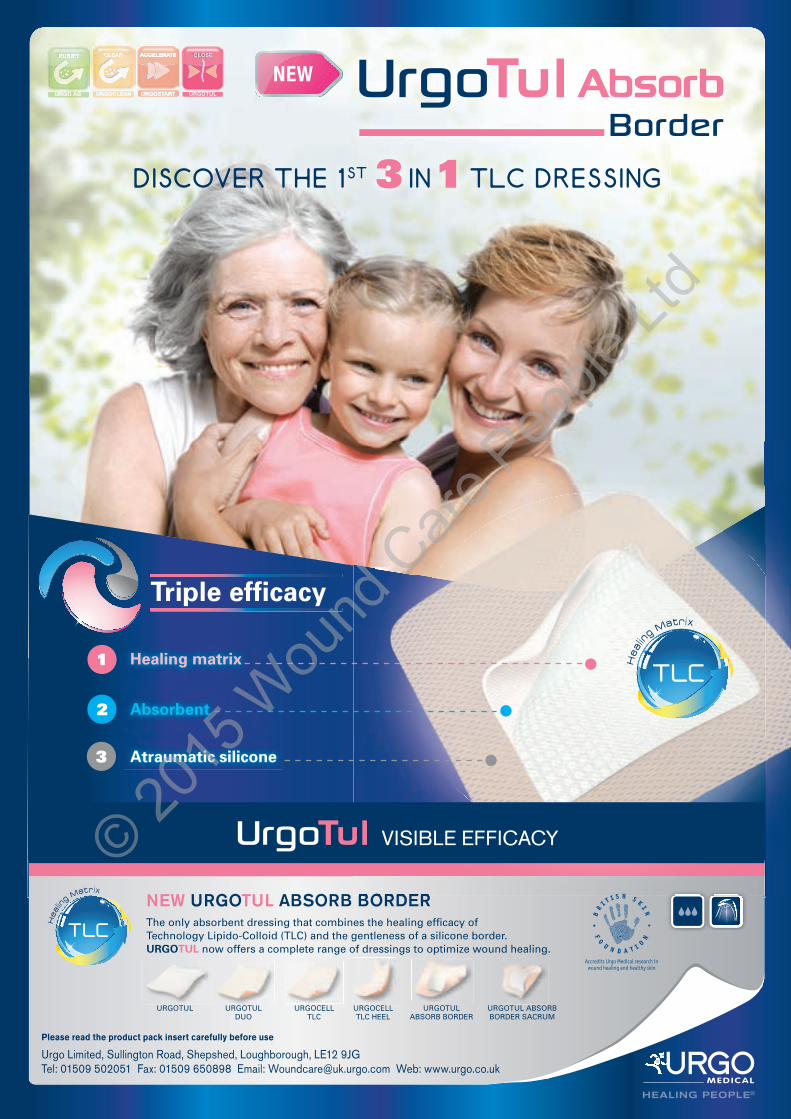
Healing matrix
Absorbent
Atraumatic silicone
The only absorbent dressing that combines the healing effi cacy of Technology Lipido-Colloid (TLC) and the gentleness of a silicone border.URGOTUL now offers a complete range of dressings to optimize wound healing.
NEW URGOTUL ABSORB BORDER
URGOTULABSORB BORDER
URGOTUL ABSORB BORDER SACRUM
URGOCELL TLC
URGOCELL TLC HEEL
URGOTUL DUO
URGOTUL
DISCOVER THE 1ST 3 IN 1 TLC DRESSING
1
2
3
VISIBLE EFFICACY
URGO AG
PURIFY
URGOCLEAN
CLEAN
URGOTUL
CLOSE
URGOSTART
ACCELERATE PREPARE HEAL
URGOREPAIR
REPAIR
KTWO
COMPRESSION
Please read the product pack insert carefully before use
Urgo Limited, Sullington Road, Shepshed, Loughborough, LE12 9JGTel: 01509 502051 Fax: 01509 650898 Email: [email protected] Web: www.urgo.co.uk
© 2015
Wou
nd C
are P
eople
Ltd