Embed Size (px)
Citation preview
© Copyright 2018 Bako Diagnostics
Lower Extremity Biopsy Techniques
PRACTICE ESSENTIALS™
Biopsy OverviewExam Room SuppliesPunch BiopsyEpidermal Nerve Fiber Density Punch BiopsyShave BiopsySaucerization BiopsyToenail Specimen Collection for OnychodystrophyNail Unit Biopsy & Ulcer BiopsyCurettage BiopsyFine Needle Aspiration
Contents01020304
050607
080910
01 Biopsy Overview
© Copyright 2018 Bako Diagnostics
● Biopsy is a small sampling of tissue to send to a laboratory
● Used to determine presence and extent of a disease
● Reduces differential diagnoses and delays of treatment
● Provides independent objective laboratory histopathological diagnosis
● Should be done BEFORE excising a skin, nail, or soft tissue lesion
● Guides targeted pharmacological therapy for dermatitis and other conditions of the lower extremity
● Offers stepwise plastic surgical treatment options for neoplasms
WHAT IS A BIOPSY?
BIOPSY TIPS ● Biopsy the newest, darkest, most elevated area of the lesion
● Use caution around major blood vessels and nerves
● Be gentle with obtaining and handling all biopsy specimens
● Ablative procedures are contraindicated without a biopsy
● Do not let skin biopsy specimens dry out, place in formalin
● Gout crystals should be sent in reagent alcohol (ETOH), not formalin
MEDICAL DOCUMENTATION ● Consider obtaining an informed consent
● Take clinical photographs of pathology
● Document the medical necessity for performing a biopsy
● Describe pain, color change, rapid growing, suspicion of malignancy
● Describe location, size, shape, color
● Create a procedure note for the biopsy
● Document the specific laboratory test ordered
● Confirm review of laboratory report and review the diagnosis with the patient
SENDING A BIOPSY TO BAKO DIAGNOSTICS
● Include a clinical description with all specimens
● Send clinical photos to laboratory by visiting bakodx.com and clicking the image upload button at the top right of the home page
● Multiple biopsies from the same condition can be submitted in one specimen vial (one lab fee)
● Multiple biopsies from different locations should be submitted in different specimen vials (multiple lab fees)
● Multiple biopsies should be labeled anatomically site specific
© Copyright 2018 Bako Diagnostics
● 70% isopropyl alcohol wipe ● Local anesthesia
› Lidocaine with (or without) epinephrine, pre-filled 1cc or 3cc syringes, 27g or 30g needle
● Sterile biopsy instruments › 2mm and 3mm punch biopsy, #10 blade scalpel,
Miltex® BiopBlade™, curette, 10cc syringe with 18 gauge needle for needle aspiration biopsy, toenail nipper, forceps, needle holder, scissors
● Suture › Optional for punch biopsy >3mm or packing material
such as Gelfoam®
› Hemostatic agent (35% aluminum chloride or Monsel’s solution) to control bleeding
● Bandage ● Formalin fixative vials
› For skin, soft tissue mass, and needle aspiration specimens
● Reagent alcohol (ETOH) vials › For gout specimens and needle
aspiration specimens
● Dry keratin bags › For toenail specimens (onychomycosis) and dry skin
specimens (keratin only)
AVAILABLE FROM BAKO DIAGNOSTICS
● Epidermal Nerve Fiber Density (ENFD) biopsy kit
› For assessing small fiber peripheral neuropathy
● Post-biopsy instructions for patients
● Biopsy refusal form
● Biopsy consent forms
● Bako patient education materials
● Bako Diagnostics requisition forms
● Shipping supplies
Exam Room SuppliesStock each exam room with the following to reduce prep time and improve efficiency when performing biopsy procedures:
02
© Copyright 2018 Bako Diagnostics
● 2mm or 3mm punch biopsy with or without plunger
● 70% isopropyl alcohol wipe
● 1cc lidocaine with epinephrine in a 3cc syringe, 27g or 30g needle
● Hemostatic agent (35% aluminum chloride or Monsel’s solution)
● Topical antibiotic
● Gauze pad
● Bandage
MATERIALS NEEDED
DIFFERENTIAL DIAGNOSIS
CarcinomasDermatitisDiabetic UlcersEczema
MelanomaNeoplasmNeoplastic UlcersPigmented Lesions
PsoriasisTinea PedisTumorsVerruca
PROCEDURE
A multiple 2mm punch approach is warranted for large lesions and non-specific dermatitis as two small punches allow for more rapid wound-healing, better tissue sampling, and usually does not need suture closure. A single 3mm punch can be used on a smaller pigmented lesion.
1. Prepare biopsy site with 70% isopropyl alcohol wipe for 10 seconds.
2. Administer local anesthesia as a raised wheal at the biopsy site.
› 1cc lidocaine with epinephrine in a 3cc syringe, 27g or 30g needle.
3. Gently press the punch instrument onto the skin to ensure sampling dermal and subcutaneous tissue.
4. Rotate punch clockwise/counterclockwise and cut to the level of the subcutaneous fat.
5. Pull biopsy out, gently grasp and distract the biopsy core, connective tissue may need to be cut.
6. Place the biopsy into formalin fixative.
7. Apply hemostatic agent (35% aluminum chloride) to biopsy site or suture defect closure (if needed).
8. Apply topical antibiotic and bandage.
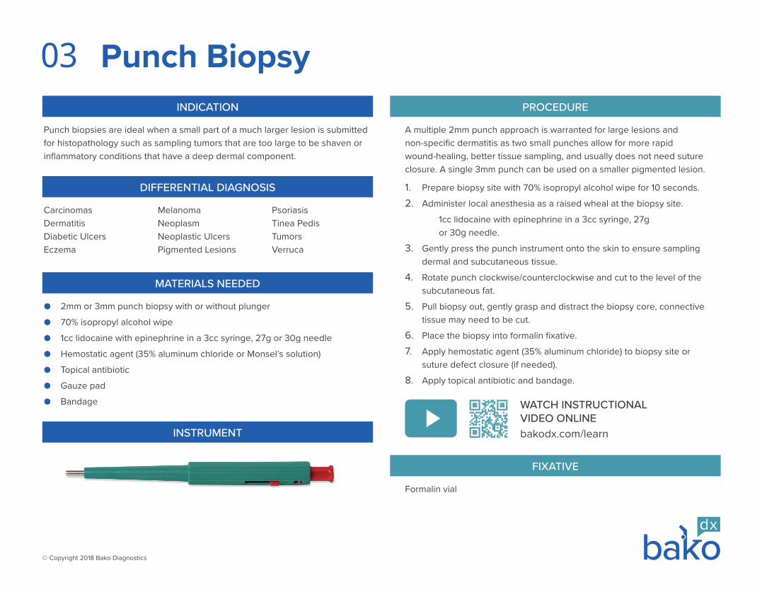
INSTRUMENT
Formalin vial
FIXATIVE
Punch Biopsy
Punch biopsies are ideal when a small part of a much larger lesion is submitted for histopathology such as sampling tumors that are too large to be shaven or inflammatory conditions that have a deep dermal component.
INDICATION
WATCH INSTRUCTIONAL VIDEO ONLINEbakodx.com/learn
03
© Copyright 2018 Bako Diagnostics
Epidermal Nerve Fiber Density Punch BiopsyPROCEDURE
The most studied ENFD biopsy location is the leg at 10cm proximal to the lateral malleolus as normative values have been established at this location.1. Prep with alcohol wipe, infiltrate lidocaine with epinephrine proximal
to, but not directly at the biopsy site using an inverted “V” pattern.2. Perform 3mm punch biopsy - 10cm proximal to lateral malleolus,
keeping the instrument perpendicular with the skin surface and enter the skin to the level of the subcutis cutting through the epidermis and dermis.
3. Apply hemostatic agent, topical antibiotic, gauze, bandage.
Epidermal Nerve Fiber Density (ENFD) biopsy provides a definitive diagnosis of small fiber peripheral neuropathy and an assessment of its degree of severity (normal, borderline, mild, moderate, severe). Symptoms suggestive of small fiber neuropathy of the foot or leg include: pain, burning, tingling, prickling, paresthesia. Affects the feet first and progresses upward with a “stocking-and-glove” distribution. Can be caused by external triggers such as foot pain when wearing socks or touching bedsheets. Muscle strength, reflexes, nerve conduction velocity test (NCVT), and electromyography (EMG) may be normal and unaffected.
INDICATION
● ENFD Kit from Bako Diagnostics › Kit includes 3mm punch, forceps, scissors, alcohol wipe, gauze,
bandage, Zamboni’s fixative, buffer rinse, cryoprotectant, cold pack, shipping supplies
● 1cc lidocaine with epinephrine in a 3cc syringe, 27g or 30g needle
MATERIALS NEEDED
DIFFERENTIAL DIAGNOSIS
● Metabolic - diabetes mellitus, metabolic syndrome, hyperlipidemia
● Amylidosis - non-inherited forms, lymphoma, plasma cell dyscrasias
● Autoimmune - Sjogren’s syndrome, vasculitis/polyarteritis nodosa
● Idiopathic - there is no identifiable cause
● Infectious - HIV, hepatitis C, Lyme disease
● Inherited - Fabry’s or Tangier’s disease, familial amyloid polyneuropathy
● Toxic - chemotherapy, alcoholism, solvent exposure
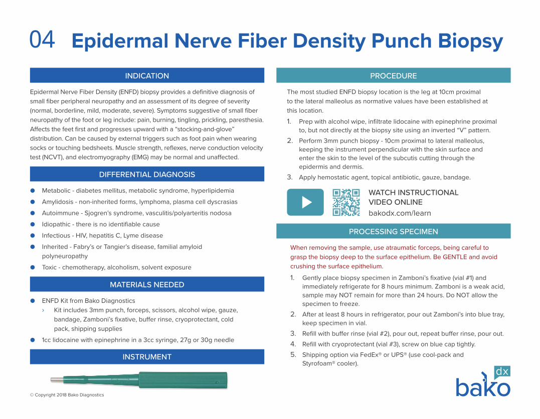
INSTRUMENT
When removing the sample, use atraumatic forceps, being careful to grasp the biopsy deep to the surface epithelium. Be GENTLE and avoid crushing the surface epithelium.
1. Gently place biopsy specimen in Zamboni’s fixative (vial #1) and immediately refrigerate for 8 hours minimum. Zamboni is a weak acid, sample may NOT remain for more than 24 hours. Do NOT allow the specimen to freeze.
2. After at least 8 hours in refrigerator, pour out Zamboni’s into blue tray, keep specimen in vial.
3. Refill with buffer rinse (vial #2), pour out, repeat buffer rinse, pour out.4. Refill with cryoprotectant (vial #3), screw on blue cap tightly.5. Shipping option via FedEx® or UPS® (use cool-pack and
Styrofoam® cooler).
PROCESSING SPECIMEN
WATCH INSTRUCTIONAL VIDEO ONLINEbakodx.com/learn
04
© Copyright 2018 Bako Diagnostics
Shave Biopsy
Shave biopsies are the most effective method for sampling superficially located pathologic processes (epidermal or dermal lesion), such as small to intermediate-sized pigmented macules, verrucous lesions, pyogenic granuloma like lesions, and other papular (small elevations) and macular (small flat areas of altered pigment) lesions.
INDICATION
● #10 scalpel or #15 scalpel
● Forceps
● 70% isopropyl alcohol wipe
● 1cc lidocaine with epinephrine in a 3cc syringe, 27g or 30g needle
● Hemostatic agent (35% aluminum chloride or Monsel’s solution)
● Topical antibiotic
● Gauze pad
● Bandage
MATERIALS NEEDED
DIFFERENTIAL DIAGNOSIS
CarcinomasKaposi’s Sarcoma Macules (flat lesions)
MelanomaNeoplasmPapules (elevated lesions)
Pigmented LesionsTumorsVerruca
PROCEDURE
The chief advantage of a shave biopsy is the ability to better sample the dermal-epidermal junction. Where a punch biopsy typically samples 2-3mm length of epidermis, a shave biopsy most commonly samples 5mm to 2cm of junctional tissue.
1. Prepare biopsy site with 70% isopropyl alcohol wipe for 10 seconds.
2. Administer local anesthesia fanned out into the dermis beneath the lesion and biopsy site, 1cc lidocaine with epinephrine in a 3cc syringe, 27g or 30g needle.
3. For papules (elevated lesions) - shave across the lesion creating a superficial plane of section beginning immediately adjacent to the lesion to be biopsied.
4. For macules (flat lesions) - tangentially score the adjacent epidermis thereby creating a free epidermal lip.
5. Enter skin at 10-15% angle and create a smooth divot, continuously use a sawing motion, moving blade underneath lesion.
6. Grasp the epidermal lip with forceps and remove the lesion using a scalpel to create a superficial dermal sample of section.
7. Place the biopsy into formalin fixative.
8. Apply hemostatic agent (35% aluminum chloride) to biopsy site.
9. Apply topical antibiotic and bandage.
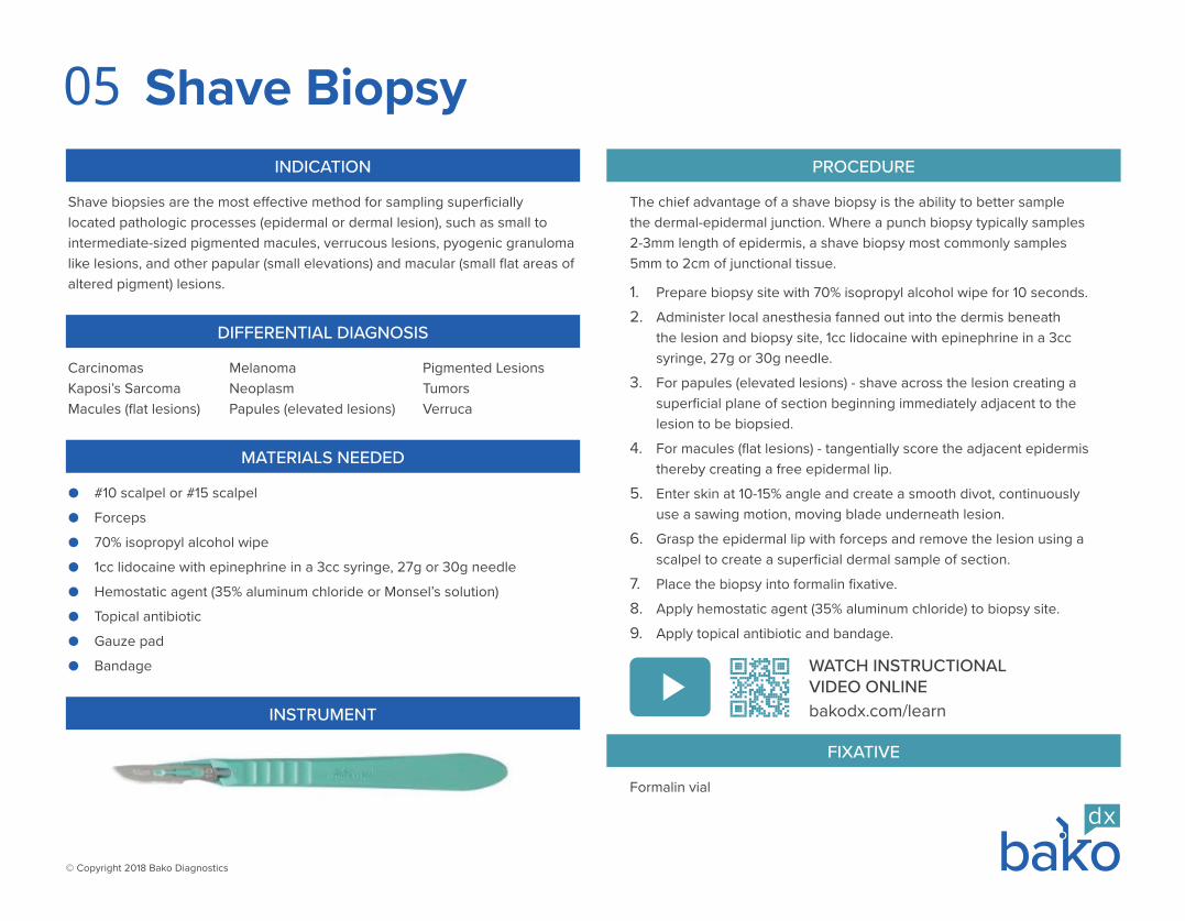
INSTRUMENT
Formalin vial
FIXATIVE
WATCH INSTRUCTIONAL VIDEO ONLINEbakodx.com/learn
05

© Copyright 2018 Bako Diagnostics
Saucerization Biopsy
Saucerization is a form of shave biopsy that may be used in place of a scalpel when superficial skin lesions, such as large flat tumors or infiltrative tumors (macules or papules), must be scooped out.
INDICATION
● Miltex® BiopBlade™ (bendable blade)
● Forceps
● 70% isopropyl alcohol wipe
● 1cc lidocaine with epinephrine in a 3cc syringe, 27g or 30g needle
● Hemostatic agent (35% aluminum chloride or Monsel’s solution)
● Topical antibiotic
● Gauze pad
● Bandage
MATERIALS NEEDED
DIFFERENTIAL DIAGNOSIS
CarcinomasKaposi’s Sarcoma Macules (flat lesions)
MelanomaNeoplasmPapules (elevated lesions)
Pigmented LesionsTumorsVerruca
PROCEDURE
This technique is thought of as a more aggressive method of sampling and thus can lend itself more to complete excision than can a standard shave biopsy. Use Miltex® BiopBlade™ to scoop out lesion.
1. Prepare biopsy site with 70% isopropyl alcohol wipe for 10 seconds.
2. Administer local anesthesia fanned out into the dermis beneath the lesion and biopsy site with 1cc lidocaine with epinephrine in a 3cc syringe, 27g or 30g needle.
3. Hold sides of BiopBlade™ with thumb and middle finger, use index finger for stabilization and forward movement.
4. Light squeeze = shallow transverse cut.
Hard squeeze = deeper concave cut.
5. Enter skin at 10-15% angle and create a smooth divot, continuously use a sawing motion, moving blade underneath lesion.
6. Rotate hand 90° to exit skin without leaving a ledge.
7. Place the biopsy into formalin fixative.
8. Apply hemostatic agent (35% aluminum chloride) to biopsy site.
9. Apply topical antibiotic and bandage.
INSTRUMENT
Formalin vial
FIXATIVE
WATCH INSTRUCTIONAL VIDEO ONLINEbakodx.com/learn
06
© Copyright 2018 Bako Diagnostics
●
Toenail Specimen Collection for Onychodystrophy
● 70% isopropyl alcohol wipe
● Nail Nippers
● Curette
MATERIALS NEEDED
DIFFERENTIAL DIAGNOSIS
Lichen PlanusMelanonychiaMicrotrauma
Pigmented MoldOnychogryphosisOnycholysis
OnychomycosisPseudomonasPsoriasis
Minimum specimen size is 3mm of nail and subungual debris for ordering a single test such as PAS or DNA. However, when ordering multiple tests such as PAS, GMS, FM, and DNA at the same time, 6mm or more of specimen is preferred.
ONYCHODYSTROPHY LABORATORY TESTS
● PERIODIC ACID–SCHIFF (PAS) › Sensitive test for staining cell walls of fungi magenta to identify
living fungi ● GOMORI METHENAMINE SILVER (GMS)
› Sensitive test for staining degenerated fungal organisms ● FONTANA-MASSON (FM)
› Sensitive test for melanin pigment (melanoma) and pigmented saprophytes
● ONYCHODYSTROPHY DNA TEST (PCR) › Specific test that identifies genus and species with molecular
genetic testing and rapid detection of both dermatophytes and non-dermatophytes by detecting DNA genetic material of dermatophytes, saprophytes, yeasts, and pseudomonas
INDICATION
Toenail specimen collection for onychodystrophy/onychomycosis augments the physician’s physical examination and is used to diagnose the presence of nail disease. Additionally, laboratory testing provides independent objective data that can assist physicians to determine a precise course of targeted patient treatment.
PROCEDURE
1. Wipe toenail collection site with 70% isopropyl alcohol.
2. Local anesthesia may or may not be required.
3. Debride and discard distal nail clippings.
4. Obtain specimen from most proximal area of nail and hyponychium.
5. Use curette to obtain additional subungual debris.
6. Place nail and subungual debris into dry keratin bag.
No fixative required, use dry keratin bag.
FIXATIVE
07
Anatomic pathology testing (PAS, GMS, FM) are sensitive tests designed to detect fungi, melanin, and allows for a diagnosis of microtrauma in cases of non-infectious nail dystrophy. DNA (PCR) testing is a specific test designed to identify the genus and species of causative organisms in cases of infectious nail dystrophy.Combination testing (PAS, GMS, FM, DNA) provides the most comprehensive evaluation of nail unit dystrophy, incorporates both the highest sensitivity and highest specificity, and enables precise targeted therapy for the underlying etiology.
© Copyright 2018 Bako Diagnostics
Nail Unit Biopsy Ulcer Biopsy
● 2mm or 3mm punch biopsy (see 03 Punch Biopsy)
MATERIALS NEEDED
DIFFERENTIAL DIAGNOSIS
Acral Lentiginous MelanomaLongitudinal Melanonychia
Malignant MelanomaNail Bed Tumor
Nail DystrophyNail Psoriasis
● 2mm or 3mm punch biopsy (see page 03 - Punch Biopsy)
● Curette (see page 09 - Curettage Biopsy)
MATERIALS NEEDED
DIFFERENTIAL DIAGNOSIS
Basal Cell CarcinomaDermatitis
Malignant MelanomaPyoderma Gangrenosum
Squamous Cell CarcinomaVasculitis
INDICATION
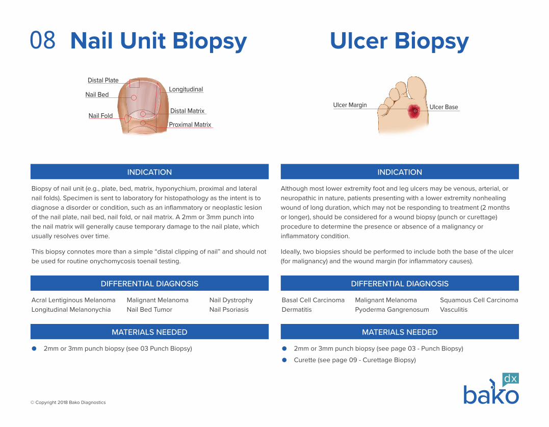
Biopsy of nail unit (e.g., plate, bed, matrix, hyponychium, proximal and lateral nail folds). Specimen is sent to laboratory for histopathology as the intent is to diagnose a disorder or condition, such as an inflammatory or neoplastic lesion of the nail plate, nail bed, nail fold, or nail matrix. A 2mm or 3mm punch into the nail matrix will generally cause temporary damage to the nail plate, which usually resolves over time.
This biopsy connotes more than a simple “distal clipping of nail” and should not be used for routine onychomycosis toenail testing.
INDICATION
Although most lower extremity foot and leg ulcers may be venous, arterial, or neuropathic in nature, patients presenting with a lower extremity nonhealing wound of long duration, which may not be responding to treatment (2 months or longer), should be considered for a wound biopsy (punch or curettage) procedure to determine the presence or absence of a malignancy or inflammatory condition.
Ideally, two biopsies should be performed to include both the base of the ulcer (for malignancy) and the wound margin (for inflammatory causes).
Distal Plate
Nail Bed
Nail Fold
Longitudinal
Distal Matrix
Proximal Matrix
Ulcer BaseUlcer Margin
08
© Copyright 2018 Bako Diagnostics
Curettage Biopsy
Curettage can be used for sampling superficial lesions which can be “scraped off” the skin surface such as small superficial scaly lesions, interdigital web space tissue to rule in/out superficial infections, or ulcer bases to rule in/out neoplasia.
INDICATION
● Dermal curette (2mm, 3mm, 4mm)
● Optional Items
› 70% isopropyl alcohol wipe
› 1cc lidocaine with epinephrine in a 3cc syringe, 27g or 30g needle
› Hemostatic agent (35% aluminum chloride or Monsel’s solution)
› Topical antibiotic
› Gauze pad
› Bandage
MATERIALS NEEDED
DIFFERENTIAL DIAGNOSIS
Actinic KeratosisSkin Tumors
Squamous Cell CarcinomaUlcers
VerrucaWeb Space Maceration
PROCEDURE
1. Anesthesia may or may not be necessary.
2. Biopsy site may or may not need preparation.
3. Direct attention to the leading edge of the condition.
4. Hold like pencil, index finger on top flat textured area.
5. Gently scrape curette across biopsy site, collecting tissue within loop.
Warning – too much pressure on curette will gouge patient.
6. One to four scrapes may be required.
7. For web space, scrape off a sample of macerated epithelium.
8. Shake specimen into formalin fixative.
› PCR Assay must be submitted dry.
9. Hemostatic agent, antibiotic, bandage may or may not be required.
INSTRUMENT
Formalin vial
FIXATIVE
WATCH INSTRUCTIONAL VIDEO ONLINEbakodx.com/learn
09
© Copyright 2018 Bako Diagnostics
Fine Needle Aspiration
Fine needle aspiration biopsy samples fluid and/or cells from a deep-seated subcutaneous mass for diagnosis, such as lipoma, ganglion cyst, sarcoma, soft tissue tumors, or lesions which may resemble epidermal inclusion cysts.
If skin moves over soft tissue mass - perform needle aspiration biopsy. If soft tissue mass moves with skin - perform punch biopsy.
Do not surgically remove a soft tissue mass without establishing a definitive diagnosis for what the mass is, otherwise the risk of amputation increases.
INDICATION
● 70% isopropyl alcohol wipe
● 3cc lidocaine in a 3cc syringe, 27g
● 10cc syringe
● 18g to 21g needle (21g preferred)
● Bandage
MATERIALS NEEDED
DIFFERENTIAL DIAGNOSIS
Cysts (fluid filled)Epidermal Inclusion CystsGanglion Cyst
Giant Cell TumorKaposi SarcomaLymphoma
Nodule (solid)SarcomaSoft Tissue Mass
IMPORTANT
A syringe body containing the sample with the needle REMOVED is acceptable, but formalin or cytology fixative is preferred.
Do not submit any “needles” as a patient sample.
PROCEDURE
Formalin vial or reagent alcohol (ETOH) vials or empty vial (for larger fluid samples)
Reagent alcohol (ETOH) vials MUST be used when gout is suspected
FIXATIVE
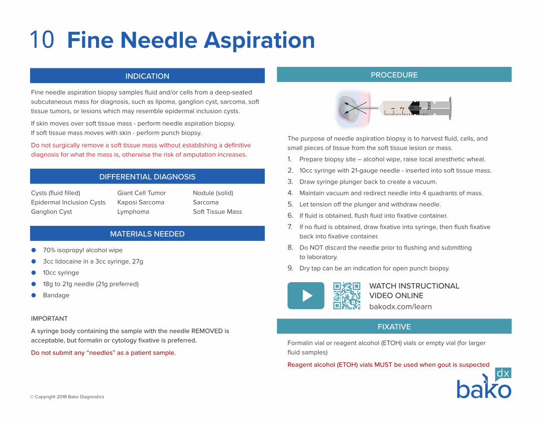
1. Prepare biopsy site – alcohol wipe, raise local anesthetic wheal.
2. 10cc syringe with 21-gauge needle - inserted into soft tissue mass.
3. Draw syringe plunger back to create a vacuum.
4. Maintain vacuum and redirect needle into 4 quadrants of mass.
5. Let tension off the plunger and withdraw needle.
6. If fluid is obtained, flush fluid into fixative container.
7. If no fluid is obtained, draw fixative into syringe, then flush fixative back into fixative container.
8. Do NOT discard the needle prior to flushing and submitting to laboratory.
9. Dry tap can be an indication for open punch biopsy.
The purpose of needle aspiration biopsy is to harvest fluid, cells, and small pieces of tissue from the soft tissue lesion or mass.
WATCH INSTRUCTIONAL VIDEO ONLINEbakodx.com/learn
10
BKMDLEBP0001680119
© Copyright 2018 Bako Diagnostics
All product names, logos, and brands are property of their respective owners. All company, product and service names used in this document are for identification purposes only. Use of these names, logos, and brands does not imply endorsement. [email protected]
6240 Shiloh Road, Alpharetta, GA 30005 bakodx.com