Embed Size (px)
Citation preview
This is an Open Access article distributed under the terms of the Creative Commons At-tribution Non-Commercial License (https://creativecommons.org/licenses/by-nc/4.0/) which permits unrestricted non-commercial use, distribution, and reproduction in any medium, provided the original work is properly cited.
Copyright © 2021 Korean Society of Exercise Rehabilitation https://www.e-jer.org pISSN 2288-176XeISSN 2288-1778
324
*Corresponding author: Youn-Jung Kim https://orcid.org/0000-0003-4991-0825Department of Basic Nursing Science, College of Nursing Science, Kyung Hee University, 26 Kyunghee-daero, Dongdaemun-gu, Seoul 02447, KoreaEmail: [email protected]†These authors contributed equally to this study as co-first authors.Received: August 12, 2021 / Accepted: September 18, 2021
Low-intensity treadmill exercise protects cognitive impairment by enhancing cerebellar mitochondrial calcium retention capacity in a rat model of chronic cerebral hypoperfusionJae-Min Lee1,†, Jongmin Park2,†, Joo-Hee Lee1, Hyo-Bum Kwak3, Mi-Hyun No3, Jun-Won Heo3, Youn-Jung Kim1,*
1Department of Basic Nursing Science, College of Nursing Science, Kyung Hee University, Seoul, Korea2Research Institute of Nursing Science, College of Nursing, Pusan National University, Yangsan, Korea3Department of Kinesiology, Inha University, Incheon, Korea
Chronic cerebral hypoperfusion (CCH) is caused by reduced blood flow to the brain representing gradually cognitive impairment. CCH induces mitochondrial dysfunction and neuronal cell death in the brain. Exercise is known to have a neuroprotective effect on brain damage and cogni-tive dysfunction. This study aimed to clarify the neuroprotective effect of low-intensity treadmill exercise (LITE) by enhancing cerebellar mito-chondrial calcium retention capacity in an animal model of CCH. Wistar rats were divided into the sham group, the bilateral common carotid ar-teries occlusion (BCCAO) group, and the BCCAO and treadmill exercise (BCCAO+Ex) group. BCCAO+Ex group engaged the LITE on a treadmill for 30 min once a day for 8 weeks before the BCCAO surgery to investi-gate the protective effect of LITE on cognitive impairment. CCH induced by BCCAO resulted in mitochondrial dysfunction in the cerebellum, in-
cluding impaired calcium homeostasis. CCH also decreased cerebellar Purkinje cells including of calbindin D28k and parvalbumin, resulting in cognitive impairment. The impairment of mitochondrial function, loss of cerebellar Purkinje cells, and cognitive dysfunction ameliorated by ex-ercise. The present study showed that LITE hindered the deficit of spatial working memory and loss of Purkinje cell in the cerebellum induced by CCH. We confirmed that the protective effect of LITE on Purkinje cell by enhanced the mitochondrial calcium retention capacity. We suggest that LITE may protect against cognitive impairment, and further studies are needed to develop the intervention for patients who suffered from CCH.
Keywords: Exercise, Cerebral hypoperfusion Cognition, Cerebellum, Mitochondria, Calcium
INTRODUCTION
Chronic cerebral hypoperfusion (CCH), the main reason for vas-cular dementia (VaD), is caused by the thromboembolic events of the brain, results in progressive cognitive impairment (Iadecola, 2013). The cerebral cortex, hippocampus, and white matter are susceptible to hypoperfusion-induced lesions. A reduction of cere-bral blood flow leads to hypoxia and oxidative stress. The hypox-ia-induced oxidative stress causes mitochondrial dysfunction and neuronal cell death via the release of reactive oxygen species (Ven-
kat et al., 2015). Although it is essential to understand the patho-physiology of the CCH for establishing the proper animal model to develop treatments, the pathology and mechanism of CCH have not yet been fully understood. Hence, it needs a new approach to understand the mechanism better.
The role of the cerebellum has recently been reestablished to represent a cognitive function. The description of the cerebellar cognitive affective syndrome (CCAS) and topographical study of the cerebellum indicate the cerebellar engagement into neural cir-cuits relevant to cognition and emotion (Jacobs et al., 2018). CCAS
https://doi.org/10.12965/jer.2142544.272
Original Article
Journal of Exercise Rehabilitation 2021;17(5):324-330
https://www.e-jer.org 325https://doi.org/10.12965/jer.2142544.272
Lee JM, et al. • Exercise protects cognitive impairment
signifies a deficit in the cognitive domain of executive function, spatial memory, and language caused by the cerebellar damage (Schmahmann and Sherman, 1998). The cerebellum is mainly re-lated to cognitive function, especially related to spatial navigation, situated in the posterior lobe of the cerebellum. Damage to the posterior lobes of the cerebellum is associated with impairment of spatial working memory and knockout mice with cerebellar dys-function have been shown to exhibit spatial navigation disorders (Tomlinson et al., 2014). Our previous study was confirmed that the loss of Purkinje cell that has contained plenty of calcium- binding proteins in the posterior lobe caused by CCH affects spa-tial navigation impairment (Lee et al., 2018).
It has been reported that the loss of Purkinje cells in the cere-bellum is caused by glutamate release and excitotoxicity due to intracellular calcium imbalance (Welsh et al., 2002). Intracellular calcium homeostasis is vital in maintaining calcium-related sig-naling pathways and nerve function in healthy brains (Pchitskaya et al., 2018). Calcium channel, endoplasmic reticulum, calcium- binding protein, and mitochondria perform a buffer system to pre-vent calcium overload. Mitochondria are essential calcium modu-lators that absorb calcium to reduce cellular calcium levels. Mito-chondria are regulating synaptic transmission, brain function, and cognitive function. Mitochondrial dysfunction has been found to be involved in the pathology of neurodegenerative diseases such as amyotrophic lateral sclerosis, Alzheimer disease, and Parkinson disease (Marambaud et al., 2009). Many diseases impaired neuro-plasticity, which leads to cell dysfunction, or even cell death. Re-cently previous study, exercise strengthened mitochondria in the brain by increasing neuroplasticity and suppressing cell death (Seo et al., 2019).
Enhancing mitochondrial function may be a therapeutic ap-proach to prevent the loss of Purkinje cells. It is a well-known in-tervention that treadmill exercise improves cognitive function by reducing the neuronal apoptosis and the responsiveness of astro-cytes in the cerebellum following global ischemia and attenuates age-related Purkinje cell loss (Larsen et al., 2000; Seo et al., 2010). The study aimed to examine whether CCH induces a loss of Pur-kinje cells, and to investigate the effect of low-intensity treadmill exercise (LITE) on cognitive function by enhancing the mitochon-drial calcium retention capacity in the cerebellum.
MATERIALS AND METHODS
AnimalsWe used adult male Wistar rats (body weight 80±10 g, 4 weeks
old) in this experiment. We randomly divided the rats into three groups: the sham group (n=8), the bilateral common carotid ar-teries occlusion (BCCAO) group (n=8), and the BCCAO and treadmill exercise (BCCAO+Ex) group (n=8). All animals were inhabited under controlled conditions of temperature (25°C±2°C), humidity (50%–55%), and light (12-hr light-dark cycles), with access to food and water ad libitum. All animal experimental pro-cedures were approved by the National Institutes of Health and the guidelines of the Korean Academy of Medical Science. This study obtained approval by the Institutional Animal Care and Use Committee of Kyung Hee University (KHUASP [SE]-17-138).
Bilateral common carotid arteries occlusionWe anesthetized the rats in 3% halothane in fresh gas flow
composed of an N2O:O2 (70:30). A ventral right incision in the neck was performed to expose the right common carotid artery, which was carefully separated to avoid damage of surrounding tis-sues particularly near a vagus nerve. The right common carotid artery was ligated doubly with 3-0 silk (Ailee, Seoul, Korea) be-low the carotid bifurcation. A left common carotid artery occlu-sion was performed in the same procedure at 1 week after the right common carotid artery surgery. Sham-operated animals un-derwent the same operation process without the vessel ligation.
LITE protocolBefore the surgery, the rats were engaged on a treadmill for
30 min for 8 weeks, according to the previously described method (Heo et al., 2014). The LITE procedure was made up running at 2 m/min for the first of 5 min, at 3 m/min for the next 5 min, and then at 5 m/min for the last 20 min at 0° of inclination. The rats in the nonexercise groups were put in the treadmill without run-ning for the same period as the exercise group.
Radial arm maze testThe radial 8-arm maze test was performed to assess spatial working
memory ability after 3 weeks of BCCAO. The radial 8-arm maze consists of eight equally spaced arms (60 cm×12 cm×12 cm) radi-ating from a central octagonal platform, placing the rat on the cen-tral octagonal platform and collecting water hidden at the tip of the arm. On the first day of training, rats can enter each arm arbitrarily to collect water hidden in each arm for 8 min. After 24 hr, the test day scores the arm selection when all four paws of the rat into the arm. On test day after 24 hr, rats will go into each arm to find wa-ter, and revisiting a previously visited arm will result in an error record. The number of correct selections before the first error was
https://doi.org/10.12965/jer.2142544.272
Lee JM, et al. • Exercise protects cognitive impairment
326 https://www.e-jer.org
measured, and the error was counted when they revisited the arms.
ImmunohistochemistrySerial sagittal sections of 40-µm thickness were obtained using
a freezing microtome (Leica, Nussloch, Germany). Immunohisto-chemistry was performed to investigate the expression of calbindin D28k and parvalbumin. Free-floating sections were initially incu-bated in 3% H2O2 for 30 min at room temperature. After being blocked with 10% normal horse and rabbit serum for 1 hr, the sections were incubated overnight at 4°C with calbindin D28k antibody (1:500; Abcam, Cambridge, UK), and parvalbumin an-tibody (1:1,000; Abcam). The sections were then incubated for 2 hr with the biotinylated rat and mouse secondary antibody (1:200; Vector Laboratories, Burlingame, CA, USA) for 1 hr at room temperature. The bound secondary antibody was then am-plified using a Vector Elite ABC kit (Vector Laboratories) for 1 hr at room temperature. For staining, the sections were incubated in a solution consisting of 0.02% diaminobenzidine and 0.03% H2O2 in 50 mM Tris-HCl (pH, 7.6) for approximately 5 min, following which they were washed with phosphate-buffered saline and mount-ed onto gelatin-coated slides. Cover slips were mounted using Permount (Fisher Scientific, Waltham, MA, USA). The number of positive cells per section was counted in 5 random fields from every specimen with a Nikon Eclipse 80i microscope (magnifica-tion, ×40; Nikon Corporation, Tokyo, Japan).
Mitochondrial calcium retention capacityWhen the mitochondria are no longer able to retain calcium,
calcium ion (Ca2+) exit the mitochondria through the permeabili-ty transition pore (PTP) opening of the mitochondria, we can
measure the Ca2+ retention capacity by the susceptibility to mito-chondrial PTP opening. After homogenizing the cerebellum tis-sue, we continuous measured response of calcium green 5-N fluo-rescence using a Spex Fluoromax 4 (HORIBA, Edsion, NJ, USA) at 37°C state four respiration (10-μg/mL oligomycin). After set-ting the background, and then Ca2+ (12.5 nM) was injected peri-odically at 506 nm (excitation wavelength) and 532 nm (emission wavelength) to measure and analyze the reaction. The maximum Ca2+ uptake capacity of the mitochondria before PTP opening (Ca2+ release) was represented as picomoles/sec/mg tissue weight.
Statistical analysisWe expressed data mean±standard error of the mean. IBM
SPSS Statistics ver. 25.0 (IBM Co., Armonk, NY, USA) was used for statistical analysis. Statistical analysis was performed using one-way analysis of variance followed by Tukey post hoc test. Dif-ferences between groups were regarded as significant at P<0.05.
RESULTS
Protected cognitive performanceThe radial arm maze test was used to confirm the establishment
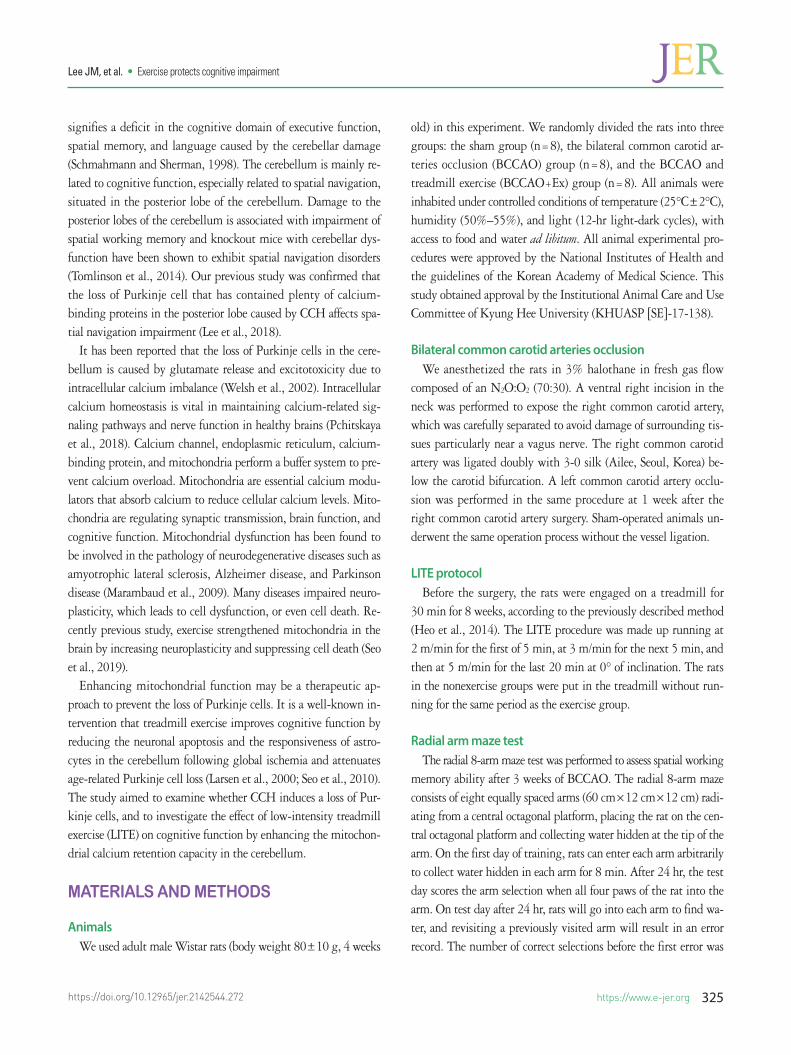
of VaD animal model and evaluate the effect of low-intensity ex-ercise on the cognition dysfunction induced by CCH. We verified the well-established animal model through the results that the BCCAO group (3.5±0.4) was decreased correct performance com-pared to the sham group (6.4±0.5) (P<0.05) (Fig. 1A). BCCAO+ Ex group (5.3±0.6) showed significantly better performance as compared with the BCCAO group (P<0.05) (Fig. 1A). Error per-formance also showed the same result (Fig. 1B). We confirmed
Fig. 1. Effect of low-intensity treadmill exercise on spatial working memory. (A) Corrects measured prior to the first error. (B) Errors were measured when rats revisit-ed the arms. The results are presented as the mean± standard error of the mean. *P< 0.05 compared with sham group. #P< 0.05 compared with bilateral common carotid arteries occlusion group. Sham, sham group; BCCAO, bilateral common carotid arteries occlusion group; BCCAO+Ex, BCCAO and treadmill exercise group.
20
15
10
5
0Sham BCCAO BCCAO+Ex
#
*
Num
ber o
f erro
r
B
8
6
4
2
0Sham BCCAO BCCAO+Ex
#
*
Num
ber o
f cor
rect
A
https://www.e-jer.org 327https://doi.org/10.12965/jer.2142544.272
Lee JM, et al. • Exercise protects cognitive impairment
the improved cognitive performance by LITE.
Decreased loss of Purkinje cellCalbindin D28k and parvalbumin are proteins that are partici-
pated in Ca2+ signaling, protects neuronal cells through reduced
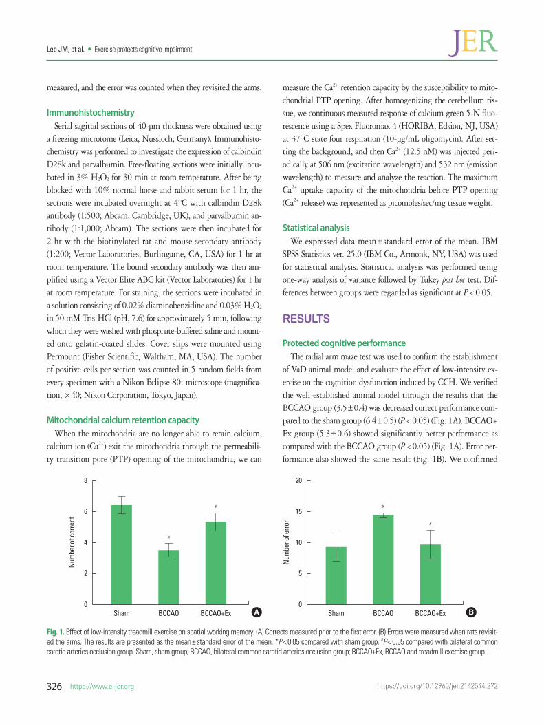
intracellular Ca2+ concentrations to maintain Ca2+ homeostasis, mainly locate in Purkinje cells and molecular layer interneurons in the cerebellum. The number of calbindin-positive Purkinje cells in the BCCAO+Ex group (41.50±2.66) was also significantly higher than the BCCAO group (31.00±1.67) (P<0.05) (Fig. 2A).
Fig. 2. Effect of low-intensity treadmill exercise attenuates Purkinje cell loss induced by chronic cerebral hypoperfusion in the posterior lobe of the cerebellum. (A) Calbindin D28K positive cells. Upper panel: photomicrographs of immunostaining of calbindin D28k positive cells. The scale bars represent 50 μm. Lower panel: num-ber of calbindin D28k positive cells. (B) Parvalbumin-positive cells. Upper panel: photomicrographs of immunostaining of parvalbumin-positive cells. The scale bars represent 50 μm. Lower panel: number of parvalbumin-positive cells. The results are presented as the mean± standard error of the mean. *P< 0.05 compared with sham group. #P< 0.05 compared with bilateral common carotid arteries occlusion group. Sham, sham group; BCCAO, bilateral common carotid arteries occlusion group; BCCAO+Ex, BCCAO and treadmill exercise group.
60
40
20
0Sham BCCAO BCCAO+Ex
#
*
Calb
indi
n po
sitive
cel
ls
A
Sham BCCAO BCCAO+Ex
60
50
40
30
20
10
0Sham BCCAO BCCAO+Ex
#
*
Parv
albu
min
pos
itive
cel
ls
B
Sham BCCAO BCCAO+Ex
https://doi.org/10.12965/jer.2142544.272
Lee JM, et al. • Exercise protects cognitive impairment
328 https://www.e-jer.org
The BCCAO+Ex group (29.83±1.01) showed significantly in-creased the number of parvalbumin-positive Purkinje cells com-pared with the BCCAO group (19.67±1.43) (P<0.05) (Fig. 2B). These results indicate that LITE protects the neuronal cell viabili-ty in the cerebellum.
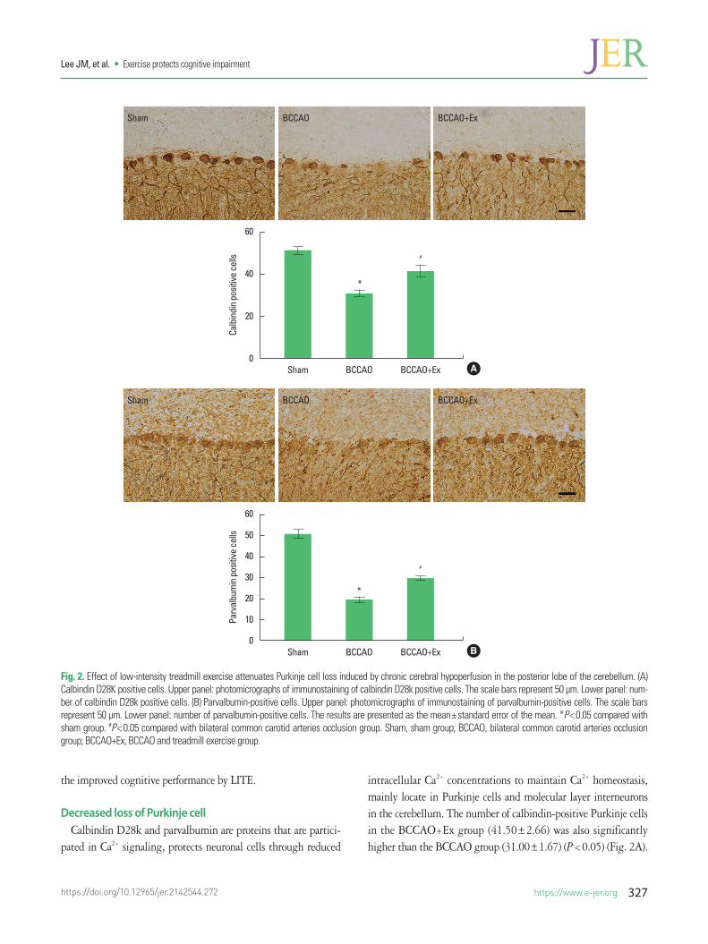
Enhanced mitochondrial calcium retention capacityOne of major role of mitochondria is retention of calcium when
they exceed the normal range of intracellular calcium level. In our study, the BCCAO+Ex group (751.28±52.97) showed significantly increased calcium retention capacity compared with the BCCAO group (424.19±28.46) (P<0.05) (Fig. 3). This result shows that low-intensity exercise has protective effect in the cerebellum by increasing calcium retention capacity.
DISCUSSION
The present study demonstrated that LITE protects the cogni-tive impairment induced by CCH through neuronal cell viability in the cerebellum and increasing the cerebellar mitochondrial cal-
cium-binding capacity. The cerebellum has emerged a critical role in cognitive function as a connection between cerebrum and cere-bellum, including cerebello-thalamo-cortical and cortico-ponto- cerebellar pathways (Palesi et al., 2017). It has been hypothesized that dysfunction of the cerebellum may play an important role in the pathology of VaD (Sui and Zhang, 2012). However, little re-search has been conducted in the cerebellum compared to the oth-er brain regions to uncover the mechanism of CCH. The increase in studies of the cerebellum will be an opportunity for us to estab-lish an essential basis for developing interventions that protect or restore cognitive health.
Most subjects who have suffered from CCH are old aged, so it is necessary to develop feasible and safe interventions for frail el-derly (Buchner and Coleman, 1994). In this study, we applied the LITE to the CCH animal model. The low-intensity exercise showed a maximum oxygen uptake of 54% to 58% and better exercise adherence compared to the high-intensity exercise (Schefer and Talan, 1996). Moreover, the exercise reduced in injury risk and was generally more easily accessible to older adults (Marques-Aleixo et al., 2015). We observed the LITE prevented impaired cognitive
Fig. 3. Effect of low-intensity treadmill exercise on mitochondrial calcium retention capacity in the cerebellum. (A) Sham group. (B) Bilateral common carotid arteries occlusion group. (C) Bilateral common carotid arteries occlusion and treadmill exercise group. (D) Quantified total mitochondrial calcium retention capacity. Experi-mental traces representing increases in calcium green 5-N fluorescence in response to 1 μM Ca2+. The results are presented as the mean± standard error of the mean. *P< 0.05 compared with sham group. #P< 0.05 compared with bilateral common carotid arteries occlusion group. CPS, counts per second; Sham, sham group; BCCAO, bilateral common carotid arteries occlusion group; BCCAO+Ex, BCCAO and treadmill exercise group.
1,000
800
600
400
200
0Sham BCCAO BCCAO+Ex
#
*
Mito
chon
dria
l cal
cium
rete
ntio
n ca
paci
ty (p
mol
/mg)
D
800,000
600,000
400,000
200,000
01 201 401 601
Time (sec)
Inte
nsity
(CPS
)
A
800,000
600,000
400,000
200,000
01 201 401
Time (sec)
Inte
nsity
(CPS
)
C
800,000
600,000
400,000
200,000
01 201 401
Time (sec)
Inte
nsity
(CPS
)
B
https://www.e-jer.org 329https://doi.org/10.12965/jer.2142544.272
Lee JM, et al. • Exercise protects cognitive impairment
performance induced by CCH. These results are consistent with previous studies showed that treadmill exercise ameliorated spatial memory performance using the 8-arm maze and Morris water maze test induced by CCH (Lee et al., 2017; Lee et al., 2018).
A previous study found that treadmill exercise in CCH-induced animal models has a preventive effect on the loss of Purkinje cells in the cerebellum via the suppression of glial cells and apoptosis (Lee et al., 2018). Our study enhanced the mitochondrial calcium retention capacity by LITE protected the loss of Purkinje cell in the cerebellum. Diminished Purkinje cells showed the spatial working memory deficit by acting minor or indirect role (Martin et al., 2004). Purkinje cells also express plenty of calcium-binding proteins, such as calbindin-D28k and parvalbumin. These proteins have engaged the regulation of cell cycle and intracellular calcium concentration related to apoptosis (Zhao et al., 2008). One of the main causes of loss of Purkinje cell has been known the mitochon-drial dysfunction.
Mitochondrial dysfunction is regarded as one of the major causes of neuronal injury in VaD induced by CCH (Venkat et al., 2015). Dysfunction of mitochondria represents diminished production of adenosine triphosphate, impaired calcium buffering capacity, and increased reactive oxygen species (Zorov et al., 2014). The mito-chondrial PTP and forms a transmembrane pore that is large enough to allow release of cytochrome c. Mitochondrial calcium retention capacity was checked by measuring the PTP opening of mitochon-dria. Notably, the decline of calcium retention capacity affects the mitochondrial calcium homeostasis, which results in apoptosis through the caspase signaling cascade (Hajnóczky et al., 2006). Intracellular calcium overload activates the caspase that the pro-teolytic activity of caspases has the biochemically vital role of apop-tosis (Zhivotovsky and Orrenius, 2011). In this study, we observed the mitochondrial calcium retention capacity in VaD group was decreased compared to sham group. Exercise improved the mito-chondrial calcium retention capacity in the brain (Seo et al., 2019). Especially, low-intensity exercise improved physical and cognitive health (Tse et al., 2015). In accord with our result, the previous research showed that physical exercise increased manganese-de-pendent superoxide dismutase activity and calcium retention ca-pacity in the cerebellum (Marques-Aleixo et al., 2015).
In conclusion, we showed that LITE might exert a neuroprotec-tive effect against the loss of Purkinje cells via increasing the mi-tochondrial calcium retention capacity in the cerebellum. There-fore, LITE may have potential as a therapeutic intervention strategy for the attenuation of cognitive impairment in patients with CCH.
CONFLICT OF INTEREST
No potential conflict of interest relevant to this article was re-ported.
ACKNOWLEDGMENTS
This study was supported by the Ministry of Education of the Republic of Korea and the National Research Foundation of Korea (NRF-2016S1A5B5A01022873 and NRF-2019R1I1A1A010- 60991).
REFERENCES
Buchner DM, Coleman EA. Exercise considerations in older adults: inten-sity, fall prevention, and safety. Phys Med Rehabil Clin N Am 1994;5: 357-375.
Hajnóczky G, Csordás G, Das S, Garcia-Perez C, Saotome M, Roy SS, Yi M. Mitochondrial calcium signalling and cell death: Approaches for assessing the role of mitochondrial Ca2+ uptake in apoptosis. Cell Cal-cium 2006;40:553-560.
Heo YM, Shin MS, Kim SH, Kim TW, Baek SB, Baek SS. Treadmill exercise ameliorates disturbance of spatial learning ability in scopolamine-in-duced amnesia rats. J Exerc Rehabil 2014;10:155-161.
Iadecola C. The pathobiology of vascular dementia. Neuron 2013;80:844-866.
Jacobs HI, Hopkins DA, Mayrhofer HC, Bruner E, van Leeuwen FW, Raaijmakers W, Schmahmann JD. The cerebellum in Alzheimer’s dis-ease: evaluating its role in cognitive decline. Brain 2018;141:37-47.
Larsen JO, Skalicky M, Viidik A. Does long-term physical exercise coun-teract age-related Purkinje cell loss A stereological study of rat cere-bellum. J Comp Neurol 2000;428:213-222.
Lee JM, Kim CJ, Park JM, Song MK, Kim YJ. Effect of treadmill exercise on spatial navigation impairment associated with cerebellar Purkinje cell loss following chronic cerebral hypoperfusion. Mol Med Rep 2018; 17:8121-8128.
Lee JM, Park JM, Song MK, Oh YJ, Kim CJ, Kim YJ. The ameliorative ef-fects of exercise on cognitive impairment and white matter injury from blood-brain barrier disruption induced by chronic cerebral hypoper-fusion in adolescent rats. Neurosci Lett 2017;638:83-89.
Marambaud P, Dreses-Werringloer U, Vingtdeux V. Calcium signaling in neurodegeneration. Mol Neurodegener 2009;4:20.
Marques-Aleixo I, Santos-Alves E, Balca M, Rizo-Roca D, Moreira P, Ol-iveira P, Ascensão A. Physical exercise improves brain cortex and cer-ebellum mitochondrial bioenergetics and alters apoptotic, dynamic
https://doi.org/10.12965/jer.2142544.272
Lee JM, et al. • Exercise protects cognitive impairment
330 https://www.e-jer.org
and auto(mito)phagy markers. Neuroscience 2015;301:480-495.Martin LA, Escher T, Goldowitz D, Mittleman G. A relationship between
cerebellar purkinje cells and spatial working memory demonstrated in a lurcher/chimera mouse model system. Genes Brain Behav 2004;3: 158-166.
Palesi F, De Rinaldis A, Castellazzi G, Calamante F, Muhlert N, Chard D, Tournier D, Magenes G, D’Angelo E, Wheeler-Kingshott CAG. Con-tralateral cortico-ponto-cerebellar pathways reconstruction in humans in vivo: implications for reciprocal cerebro-cerebellar structural con-nectivity in motor and non-motor areas. Sci Rep 2017;7:12841.
Pchitskaya E, Popugaeva E, Bezprozvanny I. Calcium signaling and mo-lecular mechanisms underlying neurodegenerative diseases. Cell Cal-cium 2018;70:87-94.
Schefer V, Talan MI. Oxygen consumption in adult and AGED C57BL/6J mice during acute treadmill exercise of different intensity. Exp Geron-tol 1996;31:387-392.
Schmahmann JD, Sherman JC. The cerebellar cognitive affective syndrome. Brain 1998;121(Pt4):561-579.
Seo JH, Park HS, Park SS, Kim CJ, Kim DH, Kim TW. Physical exercise ameliorates psychiatric disorders and cognitive dysfunctions by hip-pocampal mitochondrial function and neuroplasticity in post-trau-matic stress disorder. Exp Neurol 2019;322:113043.
Seo TB, Kim BK, Ko IG, Kim DH, Shin MS, Kim CJ, Yoon JH, Kim H. Ef-fect of treadmill exercise on Purkinje cell loss and astrocytic reaction
in the cerebellum after traumatic brain injury. Neurosci Lett 2010;481: 178-182.
Sui R, Zhang L. Cerebellar dysfunction may play an important role in vascular dementia. Med Hypotheses 2012;78:162-165.
Tomlinson SP, Davis NJ, Morgan HM, Bracewell RM. Cerebellar contri-butions to spatial memory. Neurosci Lett 2014;578:182-186.
Tse AC, Wong TW, Lee PH. Effect of low-intensity exercise on physical and cognitive health in older adults: a systematic review. Sports Med Open 2015;1:37.
Venkat P, Chopp M, Chen J. Models and mechanisms of vascular demen-tia. Exp Neurol 2015;272:97-108.
Welsh JP, Yuen G, Placantonakis DG, Vu TQ, Haiss F, O’Hearn E. Molliver ME, Aicher SA. Why do Purkinje cells die so easily after global brain ischemia? Aldolase C, EAAT4 and the cerebellar contribution to post-hypoxic myoclonus. Adv Neurol 2002;89:331-359.
Zhao S, Chen N, Yang Z, Huang L, Zhu Y, Guan S, Chen Q, Wang JH. Ischemia deteriorates the spike encoding of rat cerebellar Purkinje cells by raising intracellular Ca2+. Biochem Biophys Res Commun 2008;366:401-407.
Zhivotovsky B, Orrenius S. Calcium and cell death mechanisms: a per-spective from the cell death community. Cell Calcium 2011;50:211-221.
Zorov DB, Juhaszova M, Sollott SJ. Mitochondrial reactive oxygen species (ROS) and ROS-induced ROS release. Physiol Rev 2014;94:909-950.