Embed Size (px)
Citation preview
Research ArticleThe Treadmill Exercise Protects against Dopaminergic NeuronLoss and Brain Oxidative Stress in Parkinsonian Rats
Roberta Oliveira da Costa,1 Carlos Vinicius Jataí Gadelha-Filho,1
Ayane Edwiges Moura da Costa,1 Mariana Lima Feitosa,1 Dayane Pessoa de Araújo,1
Jalles Dantas de Lucena,1 Pedro Everson Alexandre de Aquino,1
Francisco Arnaldo Viana Lima,1 Kelly Rose Tavares Neves,1 andGlauce Socorro de Barros Viana1,2
1Faculty of Medicine of the Federal University of Ceará (UFC), Fortaleza, CE, Brazil2Faculty of Medicine Estácio of Juazeiro do Norte (Estácio/FMJ), Juazeiro do Norte, CE, Brazil
Correspondence should be addressed to Glauce Socorro de Barros Viana; [email protected]
Received 7 February 2017; Revised 23 April 2017; Accepted 26 April 2017; Published 21 June 2017
Academic Editor: Rodrigo Franco
Copyright © 2017 Roberta Oliveira da Costa et al. This is an open access article distributed under the Creative CommonsAttribution License, which permits unrestricted use, distribution, and reproduction in any medium, provided the originalwork is properly cited.
Parkinson’s disease (PD), a progressive neurological pathology, presents motor and nonmotor impairments. The objectives were tosupport data on exercise benefits to PD. Male Wistar rats were distributed into sham-operated (SO) and 6-OHDA-lesioned, bothgroups without and with exercise. The animals were subjected to treadmill exercises (14 days), 24 h after the stereotaxic surgery andstriatal 6-OHDA injection. Those from no-exercise groups stayed on the treadmill for the same period and, afterwards, weresubjected to behavioral tests and euthanized for neurochemical and immunohistochemical assays. The data, analyzed byANOVA and Tukey post hoc test, were considered significant for p < 0 05. The results showed behavioral change improvementsin the 6-OHDA group, after the treadmill exercise, evaluated by apomorphine rotational behavior, open field, and rota rod tests.The exercise reduced striatal dopaminergic neuronal loss and decreased the oxidative stress. In addition, significant increases inBDNF contents and in immunoreactive cells to TH and DAT were also observed, in striata of the 6-OHDA group with exercise,relatively to those with no exercise. We conclude that exercise improves behavior and dopaminergic neurotransmission in 6-OHDA-lesioned animals. The increased oxidative stress and decreased BDNF contents were also reversed, emphasizing theimportance of exercise for the PD management.
1. Introduction
The rising of the older population worldwide is expected tolead to a high prevalence of age-related diseases. Thus, neu-rodegenerative pathologies like Parkinson’s disease (PD) aregoing to increase, as life expectancy is getting longer,impacting public health-care costs [1]. Exercise is animportant part of daily life and, for PD patients, besidesbeing healthy, it is also a vital component for maintainingbalance, mobility, and daily activities. In this sense, exercisecan be beneficial in symptom management and also possi-bly slowing disease progression. Furthermore, to reach thepathology recovery, a prominent goal in PD research is
finding a neuroprotective treatment that, when appliedprior to the onset of the disease, will decrease its risk orseverity. One such treatment which has a potential to be aneuroprotective agent in PD is exercise [2].
Neurodegenerative diseases are characterized by a pro-gressive deterioration of brain function, with a consequentand significant decrease in the quality of life of patientsand their families. In PD, the progressive loss of dopami-nergic neurons in the substantia nigra pars compacta leadsto motor dysfunction. Thus, evidence [3] indicates thattreadmill exercise enhances the survival of dopaminergicneurons in the substantia nigra, as well as the fiber projec-tion to the striatum.
HindawiOxidative Medicine and Cellular LongevityVolume 2017, Article ID 2138169, 10 pageshttps://doi.org/10.1155/2017/2138169
It is largely accepted that the exposure to an enrichedenvironment increases neurogenesis in the dentate gyrus ofadult rodents [4–7]. Furthermore, evidence has indicated animportant role of physical exercise as a potent enhancer ofadult hippocampal neurogenesis, pointing it as a potentialtherapeutic strategy for reducing cognitive decline [8, 9].
Oxidative stress is known to play an important role in thedegeneration of dopaminergic neurons in PD. The DAmetabolism itself is known to contribute to oxidative stress,as well as mitochondrial dysfunction and the consequentincrease in reactive oxygen species (ROS). Evidences fromclinical trials have failed to demonstrate any benefit of theoxidative stress for decreasing PD progression. However,recent findings on mechanisms related to PD gene productsand neuronal response to stress may provide new targetstowards neuroprotection [10].
The objectives of the present work were to add some newinformation on the role of exercise and oxidative stress in aPD model in rats. We focus on behavioral testing, neuro-chemical determination of DA and its main metabolites,DOPAC and HVA. In addition, striatal BDNF contents andimmunohistochemical assays for tyrosine hydroxylase (TH)and the dopamine transporter (DAT) were also carried out.
2. Materials and Methods
2.1. Drugs and Reagents. 6-hydroxydopamine (6-OHDA),apomorphine, and HPLC standards were from Sigma-Aldrich (St. Louis, MO, USA); ketamine and xylazine werefrom Konig do Brasil (Santana de Parnaíba, São Paulo,Brazil). The BDNF kit for ELISAwas fromAbcam (Cambridge,UK), and antibodies for immunohistochemistry assays werefrom Santa Cruz Biotechnology (Dallas, TX, USA) or MerckMillipore (Darmstadt, Germany). All other reagents were ofanalytical grade.
2.2. Animals. Male Wistar rats (200–250 g) were maintainedat a 24± 2°C temperature, in a 12 h dark/12 h light cycle,with standard food and water ad libitum. The study wassubmitted to the Ethical Committee for Animal Experimen-tation of the Faculty of Medicine of the Federal Universityof Ceará (Brazil) and was approved under the number90/2014. All experiments followed the ethical principles
established in the Guide for the Care and Use of LaboratoryAnimals, USA, 2011.
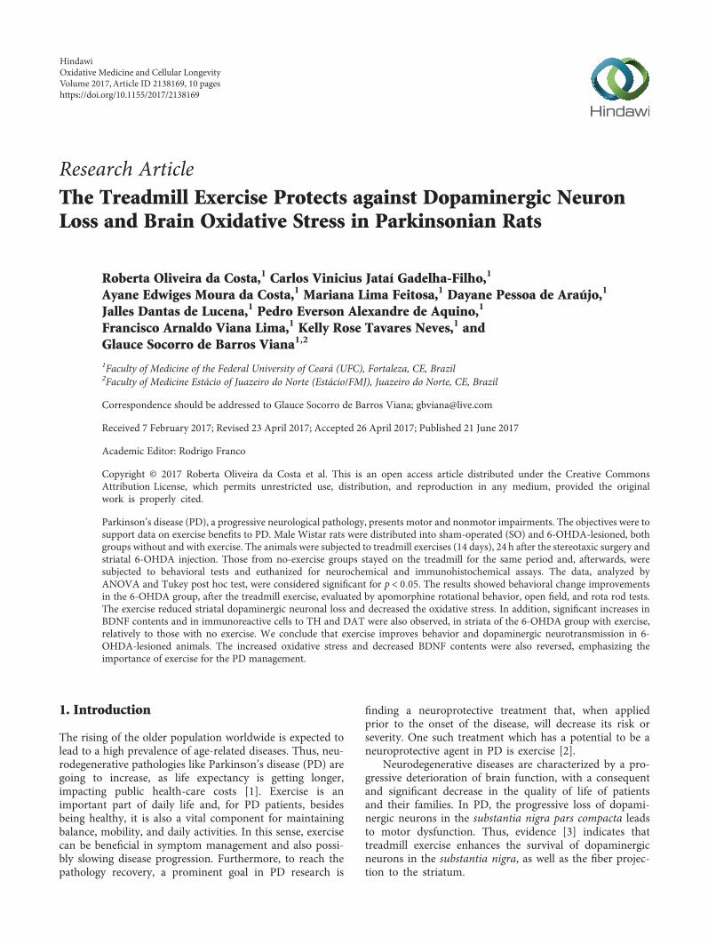
2.3. Experimental Procedure for the Treadmill Exercise. MaleWistar rats (200–250 g) were distributed into the followinggroups (6–10 animals each): sham-operated (SO) with orwithout exercise and 6-OHDA-treated also with or withoutexercise. The animals were subjected to treadmill physicalexercise (30min, at a speed of 20 cm/s), once a day, for 14consecutive days. This procedure started 24h after the stereo-taxic surgery and the striatal 6-OHDA injection. For the no-exercise groups, the animals stayed on the treadmill for thesame period of time [11, 12]. One hour later, the animalswere subjected to the apomorphine-induced rotation testand, in the next day (day 15), to the open field, followed bythe rota rod test and, then, euthanized for neurochemicaland immunohistochemical assays (see Figure 1).
2.4. The 6-OHDA Model of PD and the ExperimentalProtocol. This is a widely used model of Parkinson disease.The intrastriatal injection of 6-OHDA causes a progressiveretrograde and dose-dependent neuronal degeneration inthe nigrostriatal pathway [13, 14]. The degree of lesion canbe intensified if 6-OHDA is injected into different striatalsites [15]. In the present study, the animals were anesthetizedwith the association of xylazine (10mg/kg, i.p.) and ketamine(80mg/kg, i.p.) and, after head shaving, fixed to the stereo-taxic frame by their ear canals. A longitudinal midline inci-sion was made, and the tissues were separated for bregmavisualization. Then, a thin hole was performed in the skullover the target area, and a 1μL solution containing 6μg 6-OHDA was injected into two different points. The followingcoordinates were used: 1st point (AP,+0.5; ML,−2.5;DV,+5.0) and 2nd point (AP,−0.9; ML,−3.7; DV,+6.5). Thesyringe stayed in place for 5min, to assure the solution diffu-sion, and, then, the incision was sutured. The sham-operated(SO) animals were subjected to all procedures, except thatsaline was injected into the two points. Afterwards, the ani-mals returned to their cages for recovery. They were dividedinto the following groups: SO (with exercise), SO (withoutexercise), 6-OHDA-lesioned (with exercise), and 6-OHDA-lesioned (without exercise).
�e SO and6-OHDA groups(2 of each) were
subjected tostereotaxic
surgery
24 h later
�e treadmill startedfor the two exercise
groups
Apomorphine-induced rotationaltest for all groups
Open �eld and rota rod tests forall groups. Four hours later, the
animals were euthanized forneurochemical and
immunohistochemical assays.
24 h later
1st day
2nd day14th day
15th day
Figure 1: Experimental protocol design.
2 Oxidative Medicine and Cellular Longevity
2.5. Rotational Behavior. The apomorphine induction ofrotational (circling) behavior is widely used for assessingthe effects of lesions to the dopaminergic system and thesuccess of treatment strategies, in rat models of Parkinson’sdisease. The number of rotations under apomorphine isrelated to the extent of dopamine depletion, after the unilat-eral 6-hydroxydopamine lesion. The contralateral rotations(opposite to the lesioned right side) induced by apomorphine(3mg/kg, s.c.) were monitored for 1 h. The cause for thisapomorphine-induced rotational behavior is related to theunbalance, in the nigrostriatal dopaminergic pathways,between the right (lesioned) and left (unlesioned) brainhemispheres. This asymmetric circling behavior, after theapomorphine administration, is a quantifiable motor deficitand an important paradigm in this model of PD [16, 17].This test was performed at the 14th day (1 h after the tread-mill exercise).
2.6. Open Field Test. This test evaluates a stimulant or depres-sant drug activity and may also indicate an anxiolytic action.The arena was made of wood, whose dimensions were50 cm× 50 cm× 30 cm (length, width, and height). The floorwas divided into 4 quadrants of equal size. At the time ofthe experiment, the apparatus was illuminated by a red lightand was afterwards cleaned with a 70% alcohol solution foravoiding odor interference in the test response. The numberof crossings with the four paws from one quadrant toanother, for 5min (parameter for measuring the locomotorspontaneous activity), was determined. This test was per-formed at the 15th day (24 h after the apomorphine-inducedrotational behavior).
2.7. Rota Rod Test. The rota rod is a standard test of motorcoordination, balance, and fatigue in rodents and is especiallysensitive in detecting cerebellar dysfunction. Motor deficitsare usually observed in the Parkinson’s disease model inrodents. Basically, the animal is placed on a rotating bar,under continuous speed (12 rpm/min), and the timelatency/min to fall from the bar is recorded [18]. This testwas performed at day 15 (after the open field test).
2.8. Neurochemical Determinations of DA, DOPAC and HVAby HPLC. At the 15th day after the treadmill exercise and theapomorphine-induced rotational test, the animals were sub-jected to the open field and rota rod tests and, 4 h later, eutha-nized for decapitation and striatal tissue dissection. Thestriatal contents of DA, DOPAC and HVA were determinedby HPLC. Homogenates were prepared in 10% HClO4 andcentrifuged at 4°C (15,000 rpm, 15min). The supernatantswere filtered, and 20μL was injected into the HPLC column.For that, an electrochemical detector (model L-ECD-6A,from Shimadzu, Japan) coupled to a column (Shim-PakCLC-ODS, 25 cm) with a flux of 0.6mL/min was employed.A mobile phase was prepared with monohydrated citric acid(150mM), sodium octyl sulfate (67mM), 2% tetrahydrofu-ran, and 4% acetonitrile, in deionized water. The mobilephase pH was adjusted to 3.0 with NAOH (10mM). Mono-amines were quantified by comparison with standards,
processed the same manner as the samples. The results areexpressed as ng/g tissue.
2.9. Determination of Nitrite Contents. In this assay, theGriess reagent (1 part 0.1% naphthylethylenediamine dihy-drochloride in distilled water plus 1 part 1% sulfanilamidein 5%H3PO4) indicates the presence of nitrites in the sample.Striatal homogenates (10% in KCl buffer) were centrifuged(12,000 rpm for 10min), and 100μL supernatants wereadded to 100μL Griess reagent; this mixture stayed onRT for 10min. The standard NaNO2 curve was obtained(in spectrophotometer, at 520 nm) and used for calculatingthe results expressed as μmol nitrite per g tissue [19].
2.10. Determination of Lipid Peroxidation by ThiobarbituricAcid Reactive Substances (TBARS). Lipid peroxidationexpresses oxidative stress induced by ROS reactivity. A largelyused method for measuring it is the determination of malon-dihaldehyde (MDA) in biological samples [20]. Although thelipid peroxidation products are MDA and 4-hydroxy-2-nonenal (4-HNE), MDA is a good biomarker of oxidativestress and an end product of lipid peroxidation [21, 22]. Stria-tal homogenates (10%) in 1.15% KCl were added (250μL) to1mL 10% TCA, followed by addition of 1mL 0.6% thiobarbi-turic acid. After agitation, this mixture was maintained in awater bath (95–100°C) for 15min. Then, the mixture wascooled on ice and centrifuged (4000 rpm/5min). The TBARScontent was determined in a plate reader, at 540 nm, withresults expressed in μmolMDA per g tissue. A standard curvewith several MDA concentrations was also performed.
2.11. BDNF Measurements in the Rat Striata. Quantificationof endogenous brain-derived neurotrophic factor (BDNF)can be performed with an enzyme-linked immunosorbentassay (ELISA). The rat striata were homogenized in PBS(pH7.4), with the addition of protease inhibitors (Sigma-Aldrich, USA), according to the manufacturer’s instructions.The results were expressed as pg/g tissue.
2.12. Immunohistochemistry Assays. Brain striatal sections(5μm) were fixed in 10% buffered formol, for 24 h, followedby a 70% ethanol solution. The sections were embedded intoparaffin wax for slice processing on appropriate glass slides.These were placed in the oven at 58°C, for 10min, followedby deparaffinization in xylol and rehydration in alcohol atdecreasing concentrations, and washed in distilled waterand PBS (0.1M sodium phosphate buffer, pH7.2), for10min. The endogenous peroxidase was blocked with a 3%hydrogen peroxide solution, followed by incubation withthe appropriate primary anti-antibody, for tyrosine hydroxy-lase (TH) and dopamine transporter (DAT), and dilutedaccording to the manufacturer’s instructions (Santa Cruz orMillipore, USA), for 2 h, at room temperature in a moistchamber. The glass slides were then washed with PBS(3 times, 5min each) and incubated with the biotinylated sec-ondary antibody, for 1 h, at room temperature. Then, theywere washed again in PBS and incubated with streptavidin-peroxidase, for 30min, at room temperature. After anotherwash in PBS, they were incubated in 0.1% DAB solution(in 3% hydrogen peroxide). Finally, the glass slides were
3Oxidative Medicine and Cellular Longevity
washed in distilled water and counterstained with Mayer’shematoxylin, washed in tap water, dehydrated in alcohol(at increasing concentrations), diaphanized in xylol, andmounted on Entelan® for optic microscopy examination.The immunostaining intensity was quantified by the ImageJ software (National Institute of Health, USA), and theresults were expressed as relative optical density.
3. Statistical Analyses
For statistical analyses, one-way ANOVA, followed byTukey as the post hoc test, was used for multiple compar-isons. Whenever needed, the two-tailed paired or unpairedStudent’s t-tests were used for comparisons between twomeans. The photomicrograph data were quantified by theImage J software (NIH, USA). Differences were consideredsignificant at p < 0 05.
4. Results
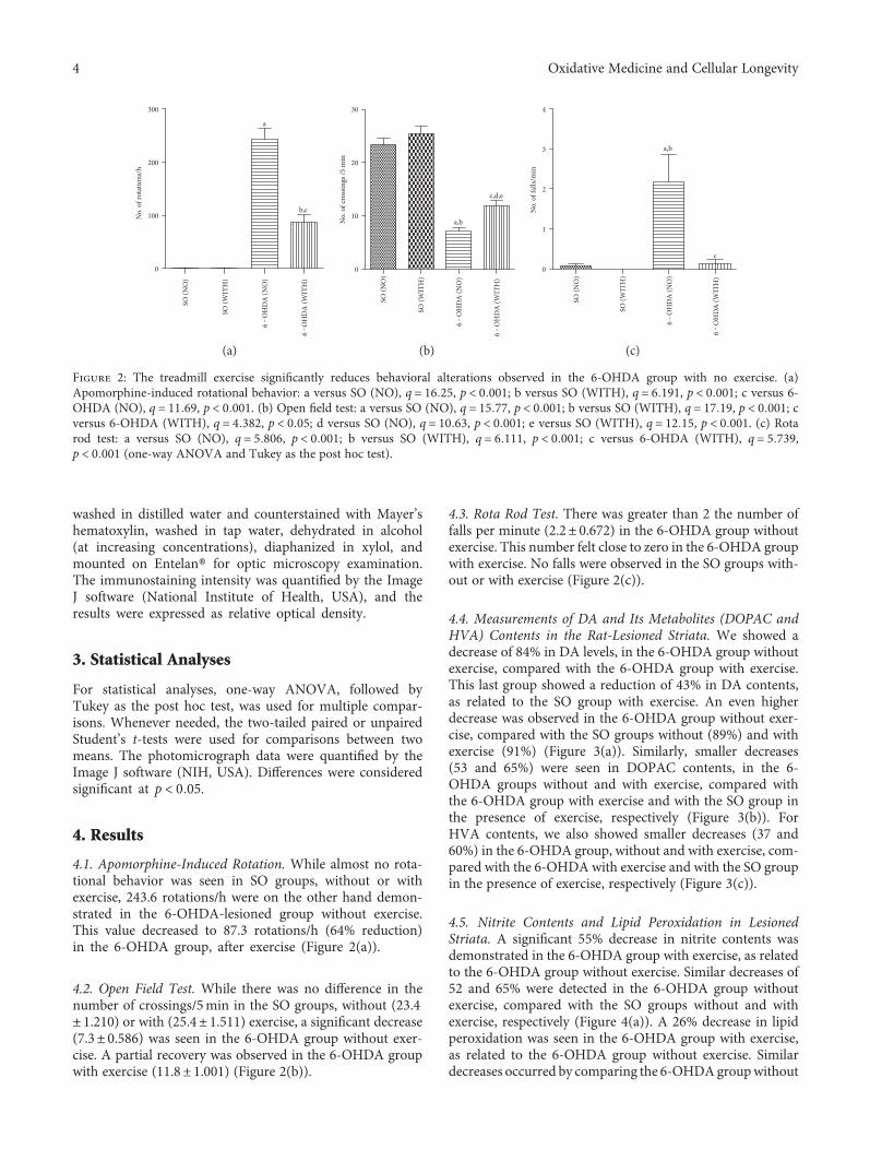
4.1. Apomorphine-Induced Rotation. While almost no rota-tional behavior was seen in SO groups, without or withexercise, 243.6 rotations/h were on the other hand demon-strated in the 6-OHDA-lesioned group without exercise.This value decreased to 87.3 rotations/h (64% reduction)in the 6-OHDA group, after exercise (Figure 2(a)).
4.2. Open Field Test. While there was no difference in thenumber of crossings/5min in the SO groups, without (23.4± 1.210) or with (25.4± 1.511) exercise, a significant decrease(7.3± 0.586) was seen in the 6-OHDA group without exer-cise. A partial recovery was observed in the 6-OHDA groupwith exercise (11.8± 1.001) (Figure 2(b)).
4.3. Rota Rod Test. There was greater than 2 the number offalls per minute (2.2± 0.672) in the 6-OHDA group withoutexercise. This number felt close to zero in the 6-OHDA groupwith exercise. No falls were observed in the SO groups with-out or with exercise (Figure 2(c)).
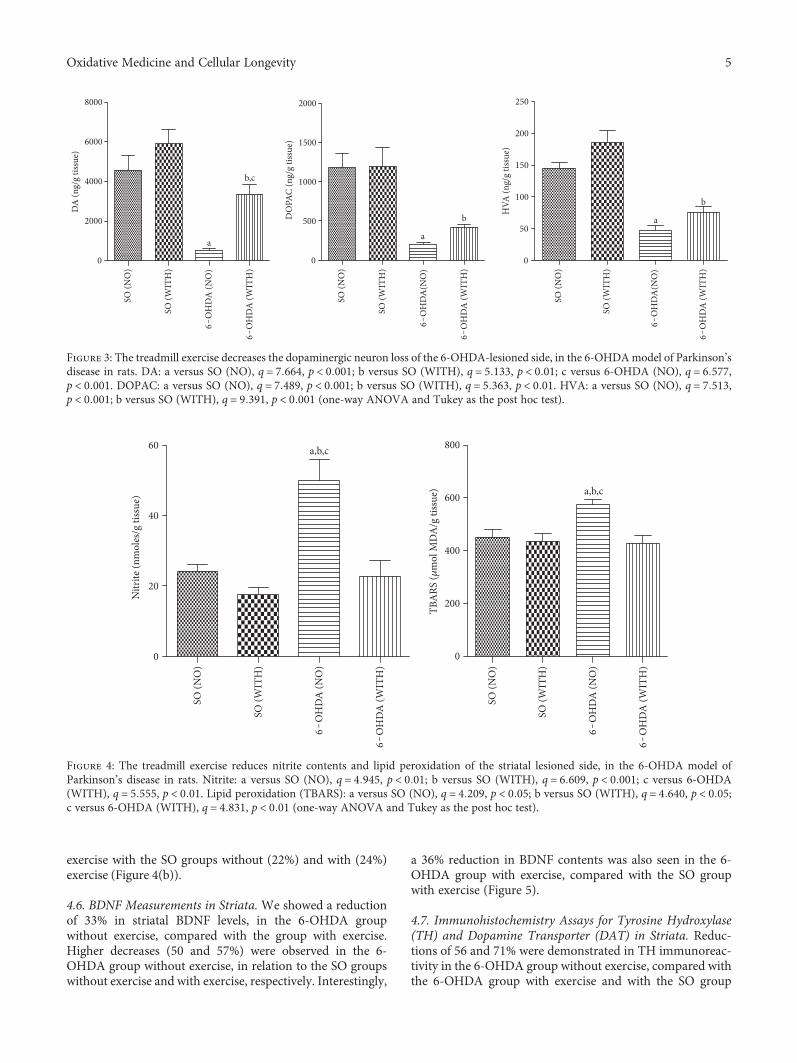
4.4. Measurements of DA and Its Metabolites (DOPAC andHVA) Contents in the Rat-Lesioned Striata. We showed adecrease of 84% in DA levels, in the 6-OHDA group withoutexercise, compared with the 6-OHDA group with exercise.This last group showed a reduction of 43% in DA contents,as related to the SO group with exercise. An even higherdecrease was observed in the 6-OHDA group without exer-cise, compared with the SO groups without (89%) and withexercise (91%) (Figure 3(a)). Similarly, smaller decreases(53 and 65%) were seen in DOPAC contents, in the 6-OHDA groups without and with exercise, compared withthe 6-OHDA group with exercise and with the SO group inthe presence of exercise, respectively (Figure 3(b)). ForHVA contents, we also showed smaller decreases (37 and60%) in the 6-OHDA group, without and with exercise, com-pared with the 6-OHDA with exercise and with the SO groupin the presence of exercise, respectively (Figure 3(c)).
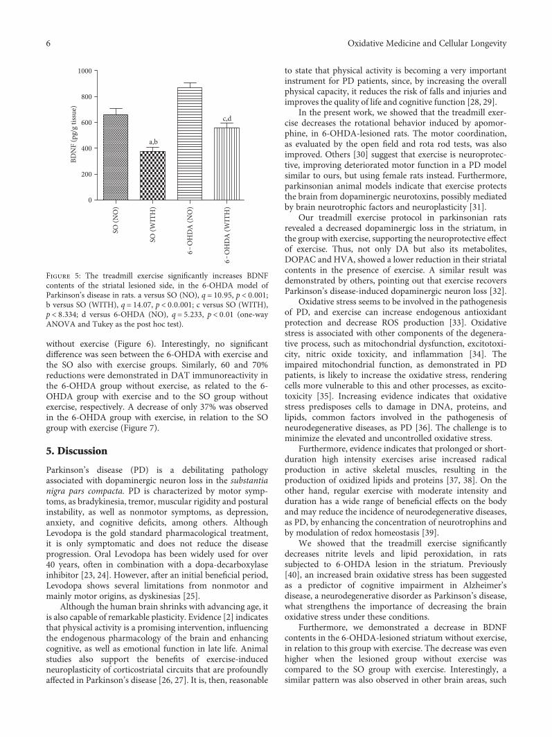
4.5. Nitrite Contents and Lipid Peroxidation in LesionedStriata. A significant 55% decrease in nitrite contents wasdemonstrated in the 6-OHDA group with exercise, as relatedto the 6-OHDA group without exercise. Similar decreases of52 and 65% were detected in the 6-OHDA group withoutexercise, compared with the SO groups without and withexercise, respectively (Figure 4(a)). A 26% decrease in lipidperoxidation was seen in the 6-OHDA group with exercise,as related to the 6-OHDA group without exercise. Similardecreases occurred by comparing the 6-OHDAgroupwithout
SO (N
O)
SO (W
ITH
)
6‒
OH
DA
(NO
)
6‒
OH
DA
(WIT
H)
0
100
200
300
a
b,c
No.
of r
otat
ions
/h
(a)
SO (N
O)
SO (W
ITH
)
6‒
OH
DA
(NO
)
6‒
OH
DA
(WIT
H)
0
10
20
30
a,b
c,d,e
No.
of c
ross
ings
/5 m
in(b)
SO (N
O)
SO (W
ITH
)
6‒
OH
DA
(NO
)
6‒
OH
DA
(WIT
H)
0
1
2
3
4
a,b
c
No.
of f
alls/
min
(c)
Figure 2: The treadmill exercise significantly reduces behavioral alterations observed in the 6-OHDA group with no exercise. (a)Apomorphine-induced rotational behavior: a versus SO (NO), q = 16 25, p < 0 001; b versus SO (WITH), q = 6 191, p < 0 001; c versus 6-OHDA (NO), q = 11 69, p < 0 001. (b) Open field test: a versus SO (NO), q = 15 77, p < 0 001; b versus SO (WITH), q = 17 19, p < 0 001; cversus 6-OHDA (WITH), q = 4 382, p < 0 05; d versus SO (NO), q = 10 63, p < 0 001; e versus SO (WITH), q = 12 15, p < 0 001. (c) Rotarod test: a versus SO (NO), q = 5 806, p < 0 001; b versus SO (WITH), q = 6 111, p < 0 001; c versus 6-OHDA (WITH), q = 5 739,p < 0 001 (one-way ANOVA and Tukey as the post hoc test).
4 Oxidative Medicine and Cellular Longevity
exercise with the SO groups without (22%) and with (24%)exercise (Figure 4(b)).
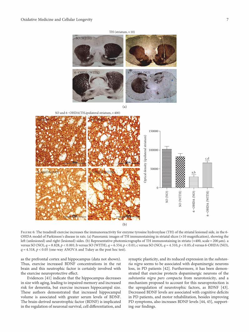
4.6. BDNF Measurements in Striata. We showed a reductionof 33% in striatal BDNF levels, in the 6-OHDA groupwithout exercise, compared with the group with exercise.Higher decreases (50 and 57%) were observed in the 6-OHDA group without exercise, in relation to the SO groupswithout exercise and with exercise, respectively. Interestingly,
a 36% reduction in BDNF contents was also seen in the 6-OHDA group with exercise, compared with the SO groupwith exercise (Figure 5).
4.7. Immunohistochemistry Assays for Tyrosine Hydroxylase(TH) and Dopamine Transporter (DAT) in Striata. Reduc-tions of 56 and 71% were demonstrated in TH immunoreac-tivity in the 6-OHDA group without exercise, compared withthe 6-OHDA group with exercise and with the SO group
SO (N
O)
SO (W
ITH
)
6‒O
HD
A (N
O)
6‒O
HD
A (W
ITH
)0
2000
4000
6000
8000
a
b,c
DA
(ng/
g tis
sue)
SO (N
O)
SO (W
ITH
)
6‒O
HD
A(N
O)
6‒O
HD
A (W
ITH
)
0
500
1000
1500
2000
a
bDO
PAC
(ng/
g tis
sue)
SO (N
O)
SO (W
ITH
)
6‒O
HD
A(N
O)
6‒O
HD
A (W
ITH
)
0
50
100
150
200
250
a
b
HVA
(ng/
g tis
sue)
Figure 3: The treadmill exercise decreases the dopaminergic neuron loss of the 6-OHDA-lesioned side, in the 6-OHDAmodel of Parkinson’sdisease in rats. DA: a versus SO (NO), q = 7 664, p < 0 001; b versus SO (WITH), q = 5 133, p < 0 01; c versus 6-OHDA (NO), q = 6 577,p < 0 001. DOPAC: a versus SO (NO), q = 7 489, p < 0 001; b versus SO (WITH), q = 5 363, p < 0 01. HVA: a versus SO (NO), q = 7 513,p < 0 001; b versus SO (WITH), q = 9 391, p < 0 001 (one-way ANOVA and Tukey as the post hoc test).
SO (N
O)
SO (W
ITH
)
6‒O
HD
A (N
O)
6‒O
HD
A (W
ITH
)
0
20
40
60 a,b,c
Nitr
ite (n
mol
es/g
tiss
ue)
SO (N
O)
SO (W
ITH
)
6‒O
HD
A (N
O)
6‒O
HD
A (W
ITH
)0
200
400
600
800
a,b,c
TBA
RS (�휇
mol
MD
A/g
tiss
ue)
Figure 4: The treadmill exercise reduces nitrite contents and lipid peroxidation of the striatal lesioned side, in the 6-OHDA model ofParkinson’s disease in rats. Nitrite: a versus SO (NO), q = 4 945, p < 0 01; b versus SO (WITH), q = 6 609, p < 0 001; c versus 6-OHDA(WITH), q = 5 555, p < 0 01. Lipid peroxidation (TBARS): a versus SO (NO), q = 4 209, p < 0 05; b versus SO (WITH), q = 4 640, p < 0 05;c versus 6-OHDA (WITH), q = 4 831, p < 0 01 (one-way ANOVA and Tukey as the post hoc test).
5Oxidative Medicine and Cellular Longevity
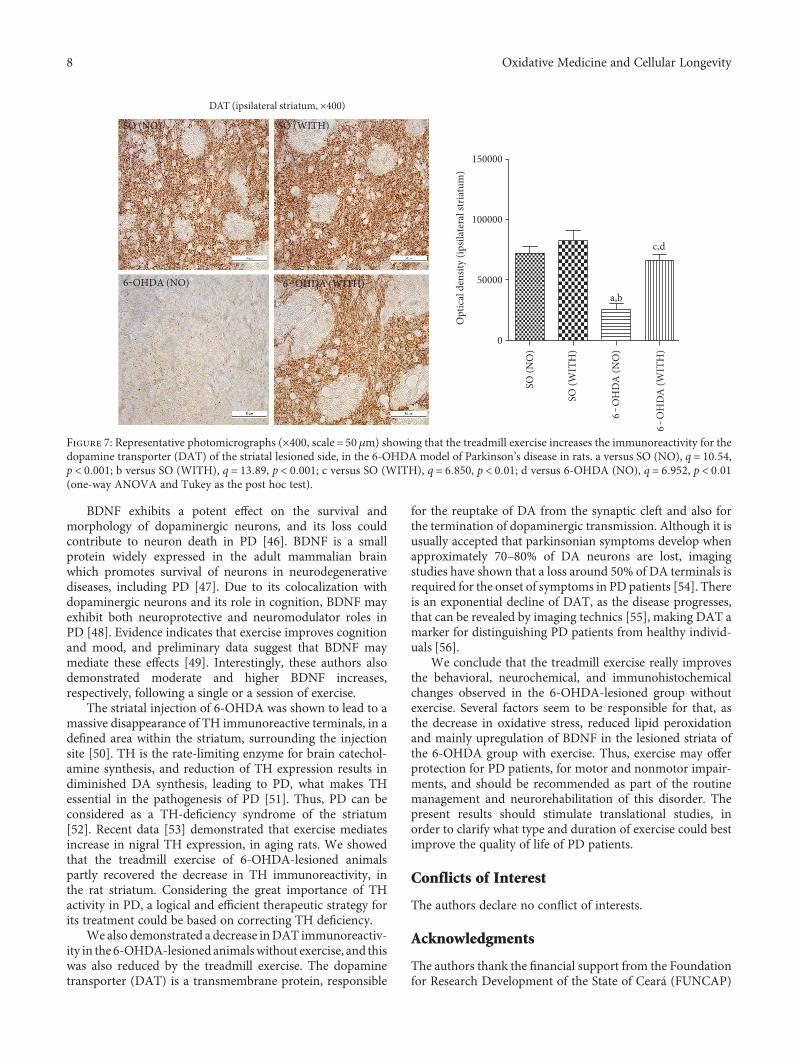
without exercise (Figure 6). Interestingly, no significantdifference was seen between the 6-OHDA with exercise andthe SO also with exercise groups. Similarly, 60 and 70%reductions were demonstrated in DAT immunoreactivity inthe 6-OHDA group without exercise, as related to the 6-OHDA group with exercise and to the SO group withoutexercise, respectively. A decrease of only 37% was observedin the 6-OHDA group with exercise, in relation to the SOgroup with exercise (Figure 7).
5. Discussion
Parkinson’s disease (PD) is a debilitating pathologyassociated with dopaminergic neuron loss in the substantianigra pars compacta. PD is characterized by motor symp-toms, as bradykinesia, tremor, muscular rigidity and posturalinstability, as well as nonmotor symptoms, as depression,anxiety, and cognitive deficits, among others. AlthoughLevodopa is the gold standard pharmacological treatment,it is only symptomatic and does not reduce the diseaseprogression. Oral Levodopa has been widely used for over40 years, often in combination with a dopa-decarboxylaseinhibitor [23, 24]. However, after an initial beneficial period,Levodopa shows several limitations from nonmotor andmainly motor origins, as dyskinesias [25].
Although the human brain shrinks with advancing age, itis also capable of remarkable plasticity. Evidence [2] indicatesthat physical activity is a promising intervention, influencingthe endogenous pharmacology of the brain and enhancingcognitive, as well as emotional function in late life. Animalstudies also support the benefits of exercise-inducedneuroplasticity of corticostriatal circuits that are profoundlyaffected in Parkinson’s disease [26, 27]. It is, then, reasonable
to state that physical activity is becoming a very importantinstrument for PD patients, since, by increasing the overallphysical capacity, it reduces the risk of falls and injuries andimproves the quality of life and cognitive function [28, 29].
In the present work, we showed that the treadmill exer-cise decreases the rotational behavior induced by apomor-phine, in 6-OHDA-lesioned rats. The motor coordination,as evaluated by the open field and rota rod tests, was alsoimproved. Others [30] suggest that exercise is neuroprotec-tive, improving deteriorated motor function in a PD modelsimilar to ours, but using female rats instead. Furthermore,parkinsonian animal models indicate that exercise protectsthe brain from dopaminergic neurotoxins, possibly mediatedby brain neurotrophic factors and neuroplasticity [31].
Our treadmill exercise protocol in parkinsonian ratsrevealed a decreased dopaminergic loss in the striatum, inthe group with exercise, supporting the neuroprotective effectof exercise. Thus, not only DA but also its metabolites,DOPAC and HVA, showed a lower reduction in their striatalcontents in the presence of exercise. A similar result wasdemonstrated by others, pointing out that exercise recoversParkinson’s disease-induced dopaminergic neuron loss [32].
Oxidative stress seems to be involved in the pathogenesisof PD, and exercise can increase endogenous antioxidantprotection and decrease ROS production [33]. Oxidativestress is associated with other components of the degenera-tive process, such as mitochondrial dysfunction, excitotoxi-city, nitric oxide toxicity, and inflammation [34]. Theimpaired mitochondrial function, as demonstrated in PDpatients, is likely to increase the oxidative stress, renderingcells more vulnerable to this and other processes, as excito-toxicity [35]. Increasing evidence indicates that oxidativestress predisposes cells to damage in DNA, proteins, andlipids, common factors involved in the pathogenesis ofneurodegenerative diseases, as PD [36]. The challenge is tominimize the elevated and uncontrolled oxidative stress.
Furthermore, evidence indicates that prolonged or short-duration high intensity exercises arise increased radicalproduction in active skeletal muscles, resulting in theproduction of oxidized lipids and proteins [37, 38]. On theother hand, regular exercise with moderate intensity andduration has a wide range of beneficial effects on the bodyand may reduce the incidence of neurodegenerative diseases,as PD, by enhancing the concentration of neurotrophins andby modulation of redox homeostasis [39].
We showed that the treadmill exercise significantlydecreases nitrite levels and lipid peroxidation, in ratssubjected to 6-OHDA lesion in the striatum. Previously[40], an increased brain oxidative stress has been suggestedas a predictor of cognitive impairment in Alzheimer’sdisease, a neurodegenerative disorder as Parkinson’s disease,what strengthens the importance of decreasing the brainoxidative stress under these conditions.
Furthermore, we demonstrated a decrease in BDNFcontents in the 6-OHDA-lesioned striatum without exercise,in relation to this group with exercise. The decrease was evenhigher when the lesioned group without exercise wascompared to the SO group with exercise. Interestingly, asimilar pattern was also observed in other brain areas, such
0
200
400
600
800
1000
a,b
c,d
BDN
F (p
g/g
tissu
e)
SO (N
O)
SO (W
ITH
)
6‒O
HD
A (N
O)
6‒O
HD
A (W
ITH
)
Figure 5: The treadmill exercise significantly increases BDNFcontents of the striatal lesioned side, in the 6-OHDA model ofParkinson’s disease in rats. a versus SO (NO), q = 10 95, p < 0 001;b versus SO (WITH), q = 14 07, p < 0 0 001; c versus SO (WITH),p < 8 334; d versus 6-OHDA (NO), q = 5 233, p < 0 01 (one-wayANOVA and Tukey as the post hoc test).
6 Oxidative Medicine and Cellular Longevity
as the prefrontal cortex and hippocampus (data not shown).Thus, exercise increased BDNF concentrations in the ratbrain and this neutrophic factor is certainly involved withthe exercise neuroprotective effect.
Evidences [41] indicate that the hippocampus decreasesin size with aging, leading to impaired memory and increasedrisk for dementia, but exercise increases hippocampal size.These authors demonstrated that increased hippocampalvolume is associated with greater serum levels of BDNF.The brain-derived neurotrophic factor (BDNF) is implicatedin the regulation of neuronal survival, cell differentiation, and
synaptic plasticity, and its reduced expression in the substan-tia nigra seems to be associated with dopaminergic neuronsloss, in PD patients [42]. Furthermore, it has been demon-strated that exercise protects dopaminergic neurons of thesubstantia nigra pars compacta from neurotoxicity, and amechanism proposed to account for this neuroprotection isthe upregulation of neurotrophic factors, as BDNF [43].Decreased BDNF levels are associated with cognitive deficitsin PD patients, and motor rehabilitation, besides improvingPD symptoms, also increases BDNF levels [44, 45], support-ing our findings.
TH (striatum,×10)
SO (WITH) SO (NO)
6 ‒ OHDA (NO)6‒OHDA (WITH)
(a)
SO (NO) SO (WITH)
6‒OHDA (NO) 6‒OHDA (WITH)
SO and 6‒OHDA(TH,ipsilateral striatum,×400)
0
150000
100000
50000a,b
c,d
Opt
ical
den
sity
(ipsil
ater
al st
riatu
m)
SO (N
O)
SO (W
ITH
)
6‒O
HD
A (N
O)
6‒O
HD
A (W
ITH
)
(b)
Figure 6: The treadmill exercise increases the immunoreactivity for enzyme tyrosine hydroxylase (TH) of the striatal lesioned side, in the 6-OHDA model of Parkinson’s disease in rats. (a) Panoramic images of TH immunostaining in striatal slices (×10 magnification), showing theleft (unlesioned) and right (lesioned) sides. (b) Representative photomicrographs of TH immunostaining in striata (×400, scale = 200 μm). aversus SO (NO), q = 8 828, p < 0 001; b versus SO (WITH), q = 6 554; p < 0 01; c versus SO (NO), q = 4 310, p < 0 05; d versus 6-OHDA (NO),q = 4 518, p < 0 05 (one-way ANOVA and Tukey as the post hoc test).
7Oxidative Medicine and Cellular Longevity
BDNF exhibits a potent effect on the survival andmorphology of dopaminergic neurons, and its loss couldcontribute to neuron death in PD [46]. BDNF is a smallprotein widely expressed in the adult mammalian brainwhich promotes survival of neurons in neurodegenerativediseases, including PD [47]. Due to its colocalization withdopaminergic neurons and its role in cognition, BDNF mayexhibit both neuroprotective and neuromodulator roles inPD [48]. Evidence indicates that exercise improves cognitionand mood, and preliminary data suggest that BDNF maymediate these effects [49]. Interestingly, these authors alsodemonstrated moderate and higher BDNF increases,respectively, following a single or a session of exercise.
The striatal injection of 6-OHDA was shown to lead to amassive disappearance of TH immunoreactive terminals, in adefined area within the striatum, surrounding the injectionsite [50]. TH is the rate-limiting enzyme for brain catechol-amine synthesis, and reduction of TH expression results indiminished DA synthesis, leading to PD, what makes THessential in the pathogenesis of PD [51]. Thus, PD can beconsidered as a TH-deficiency syndrome of the striatum[52]. Recent data [53] demonstrated that exercise mediatesincrease in nigral TH expression, in aging rats. We showedthat the treadmill exercise of 6-OHDA-lesioned animalspartly recovered the decrease in TH immunoreactivity, inthe rat striatum. Considering the great importance of THactivity in PD, a logical and efficient therapeutic strategy forits treatment could be based on correcting TH deficiency.
We also demonstrated a decrease inDAT immunoreactiv-ity in the 6-OHDA-lesioned animalswithout exercise, and thiswas also reduced by the treadmill exercise. The dopaminetransporter (DAT) is a transmembrane protein, responsible
for the reuptake of DA from the synaptic cleft and also forthe termination of dopaminergic transmission. Although it isusually accepted that parkinsonian symptoms develop whenapproximately 70–80% of DA neurons are lost, imagingstudies have shown that a loss around 50% of DA terminals isrequired for the onset of symptoms in PD patients [54]. Thereis an exponential decline of DAT, as the disease progresses,that can be revealed by imaging technics [55], making DAT amarker for distinguishing PD patients from healthy individ-uals [56].
We conclude that the treadmill exercise really improvesthe behavioral, neurochemical, and immunohistochemicalchanges observed in the 6-OHDA-lesioned group withoutexercise. Several factors seem to be responsible for that, asthe decrease in oxidative stress, reduced lipid peroxidationand mainly upregulation of BDNF in the lesioned striata ofthe 6-OHDA group with exercise. Thus, exercise may offerprotection for PD patients, for motor and nonmotor impair-ments, and should be recommended as part of the routinemanagement and neurorehabilitation of this disorder. Thepresent results should stimulate translational studies, inorder to clarify what type and duration of exercise could bestimprove the quality of life of PD patients.
Conflicts of Interest
The authors declare no conflict of interests.
Acknowledgments
The authors thank the financial support from the Foundationfor Research Development of the State of Ceará (FUNCAP)
SO (NO) SO (WITH)
6‒OHDA (NO) 6‒OHDA (WITH)
0
150000
100000
50000a,b
c,d
Opt
ical
den
sity
(ipsil
ater
al st
riatu
m)
SO (N
O)
SO (W
ITH
)
6‒O
HD
A (N
O)
6‒O
HD
A (W
ITH
)
DAT (ipsilateral striatum, ×400)
Figure 7: Representative photomicrographs (×400, scale = 50 μm) showing that the treadmill exercise increases the immunoreactivity for thedopamine transporter (DAT) of the striatal lesioned side, in the 6-OHDA model of Parkinson’s disease in rats. a versus SO (NO), q = 10 54,p < 0 001; b versus SO (WITH), q = 13 89, p < 0 001; c versus SO (WITH), q = 6 850, p < 0 01; d versus 6-OHDA (NO), q = 6 952, p < 0 01(one-way ANOVA and Tukey as the post hoc test).
8 Oxidative Medicine and Cellular Longevity
and the Brazilian National Research Council (CNPq). Theauthors are also grateful to Professor M.O.L. Viana for theorthographic revision of the manuscript.
References
[1] A. Reeve, E. Simcox, and D. Turnbull, “Ageing and Parkin-son’s disease: why is advancing age the biggest risk factor?”Ageing Research Reviews, vol. 14, pp. 19–30, 2014.
[2] K. I. Erickson, A. G. Gildengers, and M. A. Butters, “Physicalactivity and brain plasticity in late adulthood,” Dialogues inClinical Neuroscience, vol. 15, no. 1, pp. 99–108, 2013.
[3] M. C. Yoon, M. S. Shin, T. S. Kim et al., “Treadmill exercisesuppresses nigrostriatal dopaminergic neuronal loss in 6-hydroxydopamine-induced Parkinson’s rats,” NeuroscienceLetters, vol. 423, no. 1, pp. 12–17, 2007.
[4] H. van Praag, T. Schubert, C. Zhao, and F. H. Gage, “Exerciseenhances learning and hippocampal neurogenesis in agedmice,” The Journal of Neuroscience, vol. 25, no. 38, pp. 8680–8685, 2005.
[5] S. Hattori, R. Hashimoto, T. Miyakawa et al., “Enriched envi-ronment influence depression-related behavior in adult miceand the survival of newborn cells in their hippocampi,” Behav-ioural Brain Research, vol. 180, no. 1, pp. 69–76, 2007.
[6] B. M. Monteiro, F. A. Moreira, A. R. Massensini, M. F. Moraes,and G. S. Pereira, “Enriched environment increases neurogen-esis and improves social memory persistence in socially iso-lated adult mice,” Hippocampus, vol. 24, no. 2, pp. 239–248,2014.
[7] M. S. Nokia, S. Lensu, J. P. Ahtiainen et al., “Physical exerciseincreases adult hippocampal neurogenesis in male rats pro-vided it is aerobic and sustained,” The Journal of Physiology,vol. 594, no. 7, pp. 1855–1873, 2016.
[8] D. K. Murray, M. A. Sacheli, and J. J. Eng, “The effects of exer-cise on cognition in Parkinson’s disease: a systematic review,”Translational Neurodegeneration, vol. 3, no. 5, 2014.
[9] S. Y. Yau, J. Gil-Mohapel, B. R. Christie, and S. Kuok-fai,“Physical exercise-induced adult neurogenesis: a good strategyto prevent cognitive decline in neurodegenerative diseases?”BioMed Research International, vol. 2014, Article ID 403120,20 pages, 2014.
[10] V. Dias, E. Junn, and M. M. Mouradian, “The role of oxidativestress in Parkinson’s disease,” Journal of Parkinson's disease,vol. 3, no. 4, pp. 461–491, 2013.
[11] H.-S. Cho,M.-S. Shin,W. Song et al., “Treadmill exercise allevi-ates short-term memory impairment in 6-hydroxydopamine-induced Parkinson’s rats,” Journal of Exercise Rehabilitation,vol. 9, no. 3, pp. 354–361, 2013.
[12] J. L. Tillerson, W. M. Caudle, M. E. Reveron, and G. W. Miller,“Exercise induces behavioral recovery and attenuates neuro-chemical deficits in rodent models of Parkinson’s disease,”Neuroscience, vol. 119, no. 3, pp. 899–911, 2003.
[13] H. Sauer and W. H. Oertel, “Progressive degeneration ofnigrostriatal dopamine neurons following intrastriatal termi-nal lesions with 6-hydroxydopamine: a combined retrogradetracing and immunocytochemical study in the rat,” Neurosci-ence, vol. 59, no. 2, pp. 401–415, 1994.
[14] S. Przedborski, M. Levivier, H. Jiang et al., “Dose-dependentlesions of the dopaminergic nigrostriatal pathway induced byintrastriatal injection of 6-hydroxydopamine,” Neuroscience,vol. 67, no. 3, pp. 631–647, 1995.
[15] A. M. Penttinen, I. Suleymanova, K. Albert, J. Anttila, M. H.Voutilainen, and M. Airavaara, “Characterization of a newlow-dose 6-hydroxydopamine model of Parkinson’s diseasein rat,” Journal of Neuroscience Research, vol. 94, no. 4,pp. 318–328, 2016.
[16] J. L. Waddington, A. J. Cross, A. Longden, F. Owen, and M.Poulter, “Apomorphine-induced rotation in the unilateral 6-OHDA-lesioned rat: relationship to changes in striataladenylate cyclase activity and 3H-spiperone binding,” Neuro-pharmacology, vol. 18, no. 7, pp. 643–645, 1979.
[17] G.A.Metz andI.Q.Whishaw, “Drug-induced rotation intensityin unilateral dopamine-depleted rats is not correlated with endpoint or qualitative measures of forelimb or hindlimb motorperformance,”Neuroscience, vol. 111, no. 2, pp. 325–336, 2002.
[18] H. Shiotsuki, K. Yoshimi, Y. Shimo et al., “A rotarod test forevaluation of motor skill learning,” Journal of NeuroscienceMethods, vol. 189, no. 2, pp. 180–185, 2016.
[19] L. C. Green, D. A. Wagner, J. Glogowski, P. L. Skipper, J. S.Wishnok, and S. R. Tannenbaum, “Analysis of nitrate, nitrite,and [15N] nitrate in biological fluids,” Analytical Biochemistry,vol. 126, no. 1, pp. 131–138, 1982.
[20] H. H. Draper and M. Hadley, “Malondialdehyde determina-tion as index of lipid peroxidation,” Methods in Enzymology,vol. 186, pp. 421–431, 1990.
[21] D. Grotto, L. SantaMaria, J. Valentini et al., “Importance of thelipid peroxidation biomarkers and methodological aspects formalondialdehyde quantification,” Quimica Nova, vol. 32,no. 1, pp. 169–174, 2009.
[22] A. Ayala, M. F. Muñoz, and S. Argüelles, “Lipid peroxidation:production, metabolism, and signaling mechanisms of malon-dialdehyde and 4-Hydroxy-2-Nonenal,” Oxidative Medicineand Cellular Longevity, vol. 2014, Article ID 360438, 31 pages,2014.
[23] S. Fahn and Parkinson Study Group, “Does levodopa slow orhasten the rate of progression of Parkinson’s disease,” Journalof Neurology, vol. 252, Supplement 4, pp. IV37–IV42, 2005.
[24] D. Salat and E. Tolosa, “Levodopa in the treatment of Parkin-son’s disease: current status and new developments,” Journalof Parkinson's disease, vol. 3, no. 3, pp. 255–269, 2013.
[25] B. R. Thanvi and T. C. N. Lo, “Long term motor complicationsof levodopa: clinical features, mechanisms, and managementstrategies,” Postgraduate Medicine, vol. 80, no. 946, pp. 452–458, 2004.
[26] K. I. Erickson, M. W. Voss, R. S. Prakash et al., “Exercise train-ing increases size of hippocampus and improves memory,”Proceedings of the National Academy of Sciences, vol. 108,no. 7, pp. 3012–3022, 2011.
[27] F. Gomez-Pinilla and C. Hillman, “The influence of exerciseon cognitive abilities,” Comprehensive Physiology, vol. 3,no. 1, pp. 403–428, 2013.
[28] G. M. Petzinger, D. P. Holschneider, and B. E. Fischer, “Theeffects of exercise on dopamine neurotransmission in Parkin-son’s disease: targeting neuroplasticity to modulate basal gan-glia circuitry,” Brain Plasticity, vol. 1, no. 1, pp. 29–39, 2015.
[29] P. Borrione, E. Tranchita, P. Sansone, and A. Parisi, “Effects ofphysical activity in Parkinson’s disease: a new tool forrehabilitation,” World Journal of Methodology, vol. 4,no. 3, pp. 133–143, 2014.
[30] N. Tajiri, T. Yasuhara, T. Shingo et al., “Exercise exerts neuro-protective effects on Parkinson’s disease model of rats,” BrainResearch, vol. 1310, pp. 200–207, 2010.
9Oxidative Medicine and Cellular Longevity
[31] J. E. Ahlskog, “Does vigorous exercise have a neuroprotectiveeffect in Parkinson’s disease?” Neurology, vol. 77, no. 3,pp. 288–294, 2011.
[32] R. J. Bloomer, B. K. Schilling, and R. E. Karlage, “Effect ofresistance training on blood oxidative stress in Parkinsondisease,” Medicine and Science in Sports and Exercise, vol. 40,no. 8, pp. 1385–1389, 2008.
[33] P. Jenner, “Oxidative stress in Parkinson’s disease,” Annals ofNeurology, vol. 53, Supplement 53, pp. S26–S38, 2003.
[34] C. Henchcliffe and M. Flint Beal, “Mitochondrial biology andoxidative stress in Parkinson disease pathogenesis,” NatureClinical Practice. Neurology, vol. 4, no. 11, pp. 600–609, 2008.
[35] E. T. Ang, A. Tai, S. Q. Lo, R. Seet, and T.-W. Soong, “Neuro-degenerative diseases: exercising toward neurogenesis andneuroregeneration,” Frontiers in Aging Neuroscience, vol. 2,2010.
[36] S. K. Powers and M. J. Jackson, “Exercise-induced oxida-tive stress: cellular mechanisms and impact on muscleforce production,” Physiological Reviews, vol. 88, no. 4,pp. 1243–1276, 2008.
[37] S. K. Powers, Z. Radak, and L. L. Ji, “Exercise-induced oxida-tive stress: past, present and future,” The Journal of Physiology,vol. 594, no. 18, pp. 5081–5092, 2016.
[38] Z. Radak, S. Kumagai, A. W. Taylor, H. Naito, and S. Goto,“Effects of exercise on brain function: role of free radicals,”Applied Physiology, Nutrition, and Metabolism, vol. 32, no. 5,pp. 942–946, 2007.
[39] D. Praticò, C. M. Clark, F. Liun, J. Rokach, V. W. Lee,and J. Q. Trojanowski, “Increase of brain oxidative stressin mild cognitive impairment,” Archives of Neurology,vol. 59, pp. 972–976, 2002.
[40] Z. C. Baquet, P. C. Bickford, and K. R. Jones, “Brain-derivedneurotrophic factor is required for the establishment of theproper number of dopaminergic neurons in the substantianigra pars compacta,” The Journal of Neuroscience, vol. 25,no. 26, pp. 6251–6259, 2005.
[41] K. M. Gerecke, Y. Jiao, V. Pagala, and R. J. Smeyne, “Exercisedoes not protect against MPTP-induced neurotoxicity inBDNF happloinsufficient mice,” PLoS One, vol. 7, no. 8, 2012.
[42] A. Costa, A. Peppe, G. A. Carlesimo et al., “Brain-derivedneurotrophic factor serum levels correlate with cognitiveperformance in Parkinson’s disease patients with mildcognitive impairment,” Frontiers in Behavioral Neuroscience,vol. 9, p. 253, 2015.
[43] S.-Y. Wu, T-F Wang, L. Yu et al., “Running exercise protectsthe substantia nigra dopaminergic neurons againstinflammation-induced degeneration via the activation ofBDNF signaling pathway,” Brain, Behavior, and Immunity,vol. 25, no. 1, pp. 135–146, 2011.
[44] Y. Wang, H. Liu, B. S. Zhang, J. C. Soares, and X. Y. Zhang,“Low BDNF is associated with cognitive impairments inpatients with Parkinson's disease,” Parkinsonism & RelatedDisorders, vol. 29, pp. 66–71, 2016.
[45] F. Angelucci, J. Piermaria, F. Gelfo et al., “The effects of motorrehabilitation training on clinical symptoms and serum BDNFlevels in Parkinson’s disease subjects,” Canadian Journal ofPhysiology and Pharmacology, vol. 94, no. 4, pp. 455–461,2016.
[46] D. M. Howells, M. J. Porritt, J. Y. Wong et al., “Reduced BDNFmRNA expression in the Parkinson’s disease substantia nigra,”Experimental Neurology, vol. 166, no. 1, pp. 127–135, 2000.
[47] M. G. Murer, Q. Yan, and R. Raisman-Vozani, “Brain-derivedneurotrophic factor in the control human brain, and inAlzheimer’s disease and Parkinson’s disease,” Progress inNeurobiology, vol. 63, no. 1, pp. 71–124, 2001.
[48] F. Fumagalli, G. Racagni, and M. A. Riva, “Shedding light intothe role of BDNF in the pharmacotherapy of Parkinson’sdisease,” The Pharmacogenomics Journal, vol. 6, no. 2,pp. 95–104, 2006.
[49] K. L. Szuhany, M. Bugatti, and M. W. Otto, “A meta-analyticreview of the effects of exercise on brain-derived neurotrophicfactor,” Journal of Psychiatric Research, vol. 60, pp. 56–64,2015.
[50] R. W. P. Rodrigues, V. C. Gomide, and G. Chadi, “Striatalinjection of 6-hydroxydopamine induces retrograde degenera-tion and glial activation in the nigrostriatal pathway,” ActaCirurgica Brasileira, vol. 18, no. 4, pp. 272–282, 2003.
[51] Y. Zhu, J. Zhang, and Y. Zeng, “Overview of tyrosinehydroxylase in Parkinson’s disease,” CNS & NeurologicalDisorders Drug Targets, vol. 11, no. 4, pp. 350–358, 2012.
[52] S. Tabrez, N. R. Jabir, S. Shakil et al., “A synopsis on the role oftyrosine hydroxylase in Parkinson’s disease,” CNS&Neurolog-ical Disorders Drug Targets, vol. 11, no. 4, pp. 395–409, 2012.
[53] J. C. Arnold and M. F. Salvatore, “Exercise-mediated increasein nigral tyrosine hydroxylase is accompanied by increasednigral GFR-α1 and EAAC1 expression in aging rats,” ACSChemical Neuroscience, vol. 7, no. 2, pp. 227–239, 2016.
[54] A. Varrone and C. Halldin, “Molecular imaging of thedopamine transporter,” Journal of Nuclear Medicine, vol. 51,no. 9, pp. 1331–1334, 2010.
[55] A. Antonini and R. Biundo, “Parkinson disease: can dopaminetransporter imaging define early PD?” Nature ReviewsNeurology, vol. 10, no. 8, pp. 432-433, 2014.
[56] E. Bor-Seng-Shu, A. C. Felicio, P. Braga-Neto et al., “Dopa-mine transporter imaging using 99mTc-TRODAT-1 SPECTin Parkinson’s disease,” Medical Science Monitor, vol. 20,pp. 1413–1418, 2014.
10 Oxidative Medicine and Cellular Longevity

Submit your manuscripts athttps://www.hindawi.com
Stem CellsInternational
Hindawi Publishing Corporationhttp://www.hindawi.com Volume 2014
Hindawi Publishing Corporationhttp://www.hindawi.com Volume 2014
MEDIATORSINFLAMMATION
of
Hindawi Publishing Corporationhttp://www.hindawi.com Volume 2014
Behavioural Neurology
EndocrinologyInternational Journal of
Hindawi Publishing Corporationhttp://www.hindawi.com Volume 2014
Hindawi Publishing Corporationhttp://www.hindawi.com Volume 2014
Disease Markers
Hindawi Publishing Corporationhttp://www.hindawi.com Volume 2014
BioMed Research International
OncologyJournal of
Hindawi Publishing Corporationhttp://www.hindawi.com Volume 2014
Hindawi Publishing Corporationhttp://www.hindawi.com Volume 2014
Oxidative Medicine and Cellular Longevity
Hindawi Publishing Corporationhttp://www.hindawi.com Volume 2014
PPAR Research
The Scientific World JournalHindawi Publishing Corporation http://www.hindawi.com Volume 2014
Immunology ResearchHindawi Publishing Corporationhttp://www.hindawi.com Volume 2014
Journal of
ObesityJournal of
Hindawi Publishing Corporationhttp://www.hindawi.com Volume 2014
Hindawi Publishing Corporationhttp://www.hindawi.com Volume 2014
Computational and Mathematical Methods in Medicine
OphthalmologyJournal of
Hindawi Publishing Corporationhttp://www.hindawi.com Volume 2014
Diabetes ResearchJournal of
Hindawi Publishing Corporationhttp://www.hindawi.com Volume 2014
Hindawi Publishing Corporationhttp://www.hindawi.com Volume 2014
Research and TreatmentAIDS
Hindawi Publishing Corporationhttp://www.hindawi.com Volume 2014
Gastroenterology Research and Practice
Hindawi Publishing Corporationhttp://www.hindawi.com Volume 2014
Parkinson’s Disease
Evidence-Based Complementary and Alternative Medicine
Volume 2014Hindawi Publishing Corporationhttp://www.hindawi.com