Embed Size (px)
Citation preview

Loss of Heterozygosity Occurs Predominantly, But NotExclusively, in the Epithelial Compartment ofPleomorphic Adenoma
Micaela Poetsch*, Anett Zimmermann*, Eduard Wolf y and Britta Kleist z
*Institute of Forensic Medicine, University of Greifswald, Greifswald, Germany; yInstitute of Pathology,Regional Hospital, Stralsund, Germany; zInstitute of Pathology, University of Greifswald, Greifswald, Germany
Abstract
Pleomorphic adenoma (PA), being the most common
benign tumor of the salivary glands, is composed of
epithelial and mesenchymal compartments. In this
study, we analyzed 19 microsatellite markers from
chromosomal arms 6q, 8q, 9p, 12q, and 17p in 31 PAs
and 3 carcinoma ex pleomorphic adenomas (CXPAs)
as well as 11 other non–PA-related carcinomas of the
salivary gland for comparison. In our analysis, we
differentiated between epithelial and mesenchymal
tissues. Loss of heterozygosity (LOH) in PAs was
most often found in 8q (32%) and 12q (29%). Two of
the three CXPAs displayed allelic loss at all chro-
mosomal arms investigated, whereas the results of
the non–PA-related carcinomas were rather hetero-
geneous. LOH could not only be detected in the epi-
thelial, but also in the mesenchymal, compartments of
a subset of PAs, especially at chromosomal arm 8q.
Concerning the CXPAs, we were able to demonstrate
allelic losses not only in the malignant epithelial com-
partment, but also in the residual adenoma parts. Our
data give further evidence that alterations in 8q may
be an early event in PA tumorigenesis, whereas LOH in
12q may characterize cells with the potential to trans-
form in CXPAs.
Neoplasia 7, 688–695
Keywords: pleomorphic adenoma, epithelial and mesenchymal compart-ments, allelic imbalance, CXPA.
Introduction
Pleomorphic adenoma (PA) is the most common salivary
gland neoplasm, arising predominantly in the parotid gland
[1,2]. This tumor of variable capsulation is characterized by
architectural, rather than cellular, pleomorphism. Epithelial
and modified myoepithelial elements intermingle with mes-
enchymal tissues (stromas) of mucoid, myxoid, or chondroid
appearance without a clear-cut boundary between tissue
components [3]. The histogenesis of PA is still unclear. In
general, the important role of modified myoepithelial cells
for the development of PA is widely accepted [4,5]. How-
ever, the origin of these cells is controversially discussed and
could be related either to ductal basal cells [4,5] or to a ductal
acinus unit [6].
Besides the incompletely understood pathogenesis of
PA, the background of its extraordinary biologic behavior
seems worthy of further investigations. After tumor enucle-
ation, tumor recurrence rates varying from 20% to 45% have
been described [7]. Tumor recurrence after lateral or total
parotidectomy is to be expected in 1% to 5% of cases [8].
The transformation of PA to carcinoma [carcinoma ex pleo-
morphic adenoma (CXPA)] occurs at rates between 2% and
9% [2,9,10]. It has been hypothesized that translocations
within the chromosomal regions 3p, 8q, and 12q activate
proto-oncogenes, thereby leading to PA development and pro-
gression [11,12]. Especially an involvement of overexpression
of mdm2 on 12q has been proposed in this process [13]. In
addition, deregulation of the cell mobility inhibitor, maspin [14],
or the matricellular protein, tenascin [15], has been discussed
as a contributor to PA progression.
Microsatellites are short nucleotide repeat sequences dis-
tributed throughout the genome. Their high degree of sequence
length variability and their suitability for polymerase chain re-
action (PCR) amplification have led to their widespread use
as markers for chromosomal aberrations. Only limited loss of
heterozygosity (LOH) studies on PA have been published so
far [12,16–18]. Besides few approaches to analyze certain
chromosomal aberrations in different compartments of PA
[19,20], this is the first study that investigates LOH separately
in both mesenchymal and epithelial areas of PA. To assess the
impact of our LOH data for salivary gland tumor progression,
we analyzed also 3 CXPAs and 11 non–PA-related carcino-
mas of the salivary gland.
Abbreviations: PA, pleomorphic adenoma; CXPA, carcinoma ex pleomorphic adenoma; AEM,
adenoma of the classic variant; AE, cell-rich adenoma; AM, stroma-rich adenoma
Address all correspondence to: Dr. Micaela Poetsch, Institute of Forensic Medicine, Ernst
Moritz Arndt University, Kuhstrasse 30, Greifswald D-17489, Germany.
E-mail: [email protected]
Received 1 February 2005; Revised 22 March 2005; Accepted 22 March 2005.
Copyright D 2005 Neoplasia Press, Inc. All rights reserved 1522-8002/05/$25.00
DOI 10.1593/neo.05163
Neoplasia . Vol. 7, No. 7, July 2005, pp. 688 – 695 688
www.neoplasia.com
RESEARCH ARTICLE

Materials and Methods
Patients and Specimens
Thirty-one PAs of the salivary gland and corresponding
normal tissues (nonsalivary) of 31 patients (18 females and
13 males) were investigated. The median age at surgery
was 49.4 years (range 13–71 years). We subclassified the
adenomas/mixed tumors on the basis of the proportion and
differentiation of the epithelial cells and the areas of mesen-
chymal differentiation (stroma) according to the proposal of
Seifert et al. [3]: 19 adenomas of the classic variant (AEM)
which epithelial and mesenchymal tissues could be easily
isolated (30–50% stroma), 5 cell-rich adenomas (AE) (20%
mesenchymal components), and 7 stroma-rich adenomas
(AM) (80% mesenchymal components). Furthermore, we
included in this study adherent non-neoplastic salivary tissue
which surrounded each PA (range <1mm to 15 mm; Table 1).
Three CXPA from three patients (according to the defini-
tion of Ellis and Auclair [2]) were also analyzed in this study.
In all of them, residual adenoma elements could be identified
between malignant epithelia, which are defined by cytologic
and histologic characteristics of anaplasia, abnormal mitosis
(Figure 1), progressive course, and infiltrative growth. In all
CXPA, the primary tumor was available for investigation.
Furthermore, in one case (CXPA2), a recurrent tumor and, in
another case (CXPA3), two metastases could be analyzed.
For comparison, 11 other non–PA-related carcinomas of
the salivary gland comprising two polymorphous low-grade
adenocarcinomas, two adenoid cystic carcinomas, four
acinic cell carcinomas, one oncocytic carcinoma, one myo-
epithelial carcinoma, and one mucoepidermoid carcinoma
and adherent nontumorous tissues from each carcinoma
were also included in the study.
DNA Isolation
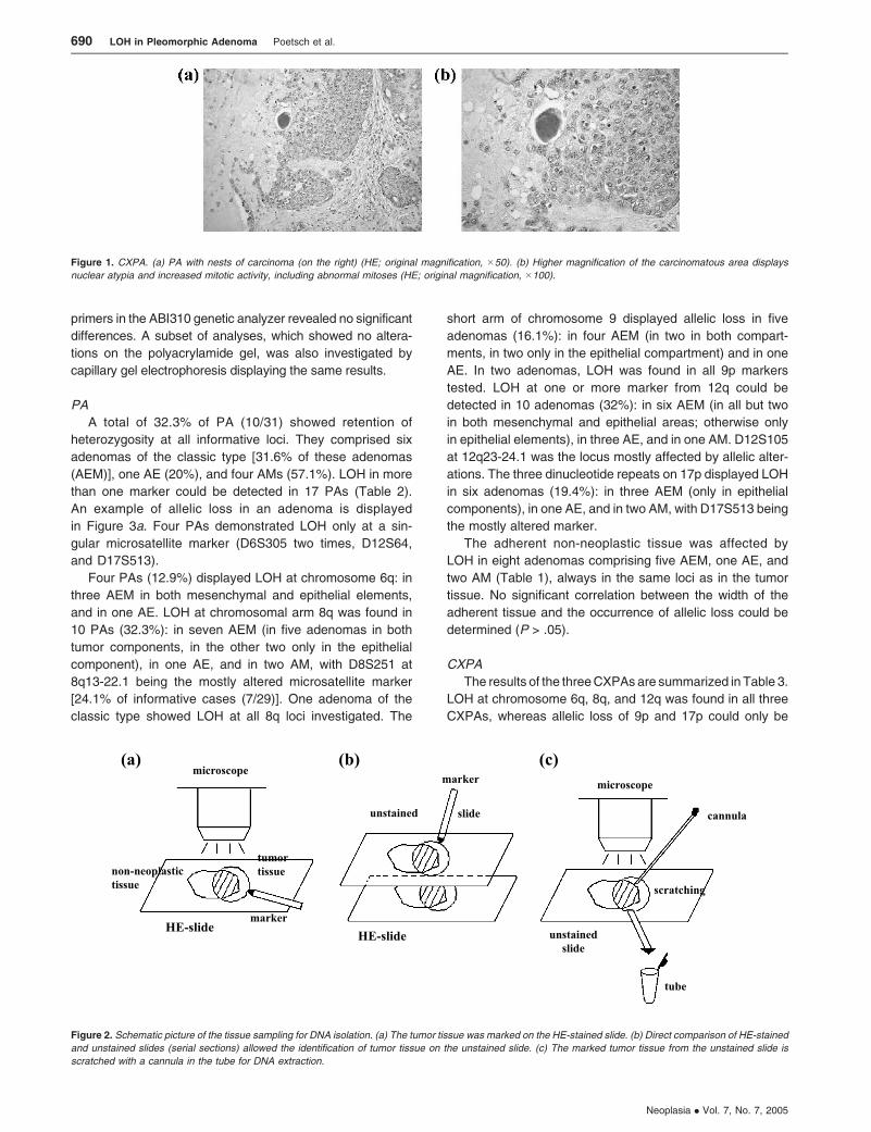
First, hematoxylin and eosin–stained slides were care-
fully inspected by light microscopy to identify areas that carry
a sufficient amount (at least 3 mm2) of tumor measured by a
scaled optical adjustment. This same area was then identi-
fied on the unstained 10-mm dewaxed, rehydrated, and air-
dried tissue section, which was fixed in an optical installation,
allowing the separate isolation of predominantly epithelial or
mesenchymal tissue without adherent nontumorous structures
under microscopic control with a cannula (used for intravenous
injections) (Figure 2). DNA isolation from paraffin-embedded
tissue was performed as previously described [21] with the
High Pure PCR Template Preparation Kit (Roche Molecular
Biochemicals, Mannheim, Germany).
Molecular Genetic Analysis
A panel of 19 PCR primer pairs amplifying informative
dinucleotide repeat microsatellite loci, comprising D6S308
(6q21-23), D6S311 (6q24), and D6S305 (6q25.2-27);
D8S166 (8q11.2-12), D8S251, and D8S164 (8q13-22.1); and
D8S199 (8q24.1), D9S161, D9S286, D9S162, and D9S171
(9p21, covering thep16 region); D12S64 (12q13-14),D12S101
(12q14), D12S1706 (12q23), D12S105 (12q23.1-24), and
D12S184 (12q24); and D17S513 (17p13), D17S786, and
D17S952 (17p, around p53), was investigated for allelic im-
balances. Primer sequences and cytogenetic locations were
obtained from Genome Data Base (http://www.gdb.org).
PCR amplification was performed in 12.5-ml sample volumes
with 2 to 5 ng of genomic tumor or normal DNA as template in
15 mM Tris/HCl and 50mMKCl, with 200 mMdNTPs, 1.5 mM
MgCl2, 0.3 nM primers, 5% formamide, and 1 U of HotStart
Taq Polymerase (Qiagen, Hilden, Germany). An initial dena-
turation and activation step of 12 minutes at 95jC was
followed by 30 to 35 cycles of 1 minute at 95jC, 1 minute at
55jC, and 3 minutes at 72jC, and a 30-minute final elonga-
tion step at 72jC. PCR products were electrophoresed on
denaturing (8 M urea) 10% polyacrylamide gels and visual-
ized by silver staining. The band patterns produced from
each normal and tumor tissue pair were visually assessed
for allelic alterations in the tumor DNA. In case of suspected
LOH or allelic shifts, the PCR of this normal/tumor pair at
this locus was repeated twice with fluorochrom-labeled pri-
mers (6-FAM, JOE, or TAMRA) and analyzed on a ABI310
genetic analyzer (Applied Biosystems, Weiterstadt, Ger-
many) with ROX-labeled internal lane standard, mostly in
multiplex assays. LOH was scored if one allele was >50%
decreased in tumorDNAwhen comparedwith the sameallele
in normal control DNA. The following equation was used to
calculate LOH:
ðpeak height of normal allele 2=
LOH ¼ peak height of tumor allele 1Þðpeak height of tumor allele 2=peak height of tumor allele 1Þ
Allelic loss is indicated by an LOH value of less than 0.5 or
higher than 2.0.
Statistical Analysis
A possible correlation between the width of the adherent
non-neoplastic tissue and the occurrence of allelic loss was
evaluated with the Cochrane-Armitage test. A P value of .05
or less was considered as statistically significant.
Results
Microsatellite Analysis
A comparison between the 10%denaturing polyacrylamide
gel approach and the analysis with fluorescence-labeled
Table 1. Adherent Nontumorous Tissue in Pleomorphic Adenomas.
Cases with Adherent
Nontumorous Tissue: Depth
AEM
(n = 19)
AE
(n = 5)
AM
(n = 7)
<1 mm without alterations 2 2 2
<1 mm with alterations 1 0 1
1–3 mm without alterations 1 1 1
1–3 mm with alterations 1 1 1
3–6 mm without alterations 9 1 1
3–6 mm with alterations 1 0 0
>6 mm without alterations 2 0 1
>6 mm with alterations 2 0 0
LOH in Pleomorphic Adenoma Poetsch et al. 689
Neoplasia . Vol. 7, No. 7, 2005
primers in the ABI310 genetic analyzer revealed no significant
differences. A subset of analyses, which showed no altera-
tions on the polyacrylamide gel, was also investigated by
capillary gel electrophoresis displaying the same results.
PA
A total of 32.3% of PA (10/31) showed retention of
heterozygosity at all informative loci. They comprised six
adenomas of the classic type [31.6% of these adenomas
(AEM)], one AE (20%), and four AMs (57.1%). LOH in more
than one marker could be detected in 17 PAs (Table 2).
An example of allelic loss in an adenoma is displayed
in Figure 3a. Four PAs demonstrated LOH only at a sin-
gular microsatellite marker (D6S305 two times, D12S64,
and D17S513).
Four PAs (12.9%) displayed LOH at chromosome 6q: in
three AEM in both mesenchymal and epithelial elements,
and in one AE. LOH at chromosomal arm 8q was found in
10 PAs (32.3%): in seven AEM (in five adenomas in both
tumor components, in the other two only in the epithelial
component), in one AE, and in two AM, with D8S251 at
8q13-22.1 being the mostly altered microsatellite marker
[24.1% of informative cases (7/29)]. One adenoma of the
classic type showed LOH at all 8q loci investigated. The
short arm of chromosome 9 displayed allelic loss in five
adenomas (16.1%): in four AEM (in two in both compart-
ments, in two only in the epithelial compartment) and in one
AE. In two adenomas, LOH was found in all 9p markers
tested. LOH at one or more marker from 12q could be
detected in 10 adenomas (32%): in six AEM (in all but two
in both mesenchymal and epithelial areas; otherwise only
in epithelial elements), in three AE, and in one AM. D12S105
at 12q23-24.1 was the locus mostly affected by allelic alter-
ations. The three dinucleotide repeats on 17p displayed LOH
in six adenomas (19.4%): in three AEM (only in epithelial
components), in one AE, and in two AM, with D17S513 being
the mostly altered marker.
The adherent non-neoplastic tissue was affected by
LOH in eight adenomas comprising five AEM, one AE, and
two AM (Table 1), always in the same loci as in the tumor
tissue. No significant correlation between the width of the
adherent tissue and the occurrence of allelic loss could be
determined (P > .05).
CXPA
The results of the threeCXPAs are summarized in Table 3.
LOH at chromosome 6q, 8q, and 12q was found in all three
CXPAs, whereas allelic loss of 9p and 17p could only be
Figure 2. Schematic picture of the tissue sampling for DNA isolation. (a) The tumor tissue was marked on the HE-stained slide. (b) Direct comparison of HE-stained
and unstained slides (serial sections) allowed the identification of tumor tissue on the unstained slide. (c) The marked tumor tissue from the unstained slide is
scratched with a cannula in the tube for DNA extraction.
Figure 1. CXPA. (a) PA with nests of carcinoma (on the right) (HE; original magnification, �50). (b) Higher magnification of the carcinomatous area displays
nuclear atypia and increased mitotic activity, including abnormal mitoses (HE; original magnification, �100).
690 LOH in Pleomorphic Adenoma Poetsch et al.
Neoplasia . Vol. 7, No. 7, 2005

demonstrated in two CXPAs each. The recurrence of CXPA2
and the metastases of CXPA3 displayed at least all alter-
ations found in the primary tumor. The results concerning
LOH in the stroma diverge in the three CXPAs. In CXPA3, the
residual adenoma compartments showed no alterations at
all, whereas in CXPA1, LOH could only be found in two loci in
these areas (out of seven with LOH in the epithelial malignant
tissue). We found nearly identical LOH in the residual ade-
noma parts and the epithelial malignant tissues of the primary
tumor from CXPA2, but the mesenchymal compartment of
the recurrent tumor showed no loss. In two of the CXPAs, the
adherent non-neoplastic tissue (1–6 mm wide) also showed
alterations in one or more of the affected markers (in tumor
tissue). An example for allelic loss in a CXPA could be found
in Figure 3b.
Other Non–PA-Related Carcinomas of the Salivary Gland
The LOH results for the other non–PA-related carcino-
mas of the salivary gland are summarized in Table 4. In
general, the polymorphous low-grade adenocarcinomas and
the myoepithelial carcinoma displayed hardly any LOH at the
chosen microsatellite markers, and in the adenoid cystic
carcinomas, allelic alterations were mostly restricted to the
long arm of chromosome 6. In contrast, the oncocytic and the
mucoepidermoid carcinoma with its recurrences showed
LOH at nearly all chromosomal arms investigated here. In
two of four acinic cell carcinomas, no alterations were found,
whereas in the other two tumors, LOH could be demon-
strated especially in 8q. The adherent non-neoplastic tissue
was affected only in the oncocytic carcinoma (1–3 mm wide)
and in one acinic cell carcinoma (<1 mm wide).
Discussion
In the salivary glands, the mechanisms for the establishment
of a PA with its epithelial and mesenchymal compartments
are still under discussion.
In contrast to a wide variety of cytogenetically character-
ized PAs [11,22–25], only a few studies investigated allelic
losses in PAs [12,17,18,26]. Chromosomal analyses have
mostly revealed translocations involving chromosomes 8q,
12q, and 3p. LOH in PA has especially been demonstrated at
the long arms of chromosomes 8 and 12 [12,17,18,26]. In
addition, alterations of the p16 region on 9p have been
shown in PA [19] and CXPA [27,28], as well as aberrations
on chromosome 17p in CXPA [12] or mutations of p53 in PA
[20] or CXPA [28]. Allelic losses of the long arm of chromo-
some 6 have been described for a variety of salivary gland
tumors [29–31]. Therefore, we used microsatellite markers
from these chromosomal localizations for our study of the
epithelial and mesenchymal components of PA.
We found allelic losses mostly at chromosomal arms 8q
and 12q in 32% of PAs each, which is roughly in line with
results from Gillenwater et al. [18] using the same micro-
satellite markers in 8q and two of our markers in 12q, and
with data reported by El-Naggar et al. [12]. LOH at 8q has
also been reported in oral premalignant lesions and oral
cancer [32], in high percentages in squamous cell larynx
cancer [33], and was found at 8q11-12 in one polymorphous
low-grade adenocarcinoma and at 8q13-22 in two acinic
cell carcinomas and the mucoepidermoid carcinoma of this
study. These data support the thesis of a tumor-suppressor
gene at 8q13-22.
The region mostly affected by LOH in 12q in PAs was
12q23.1-24. Allelic losses in this region have already been
demonstrated in gastric cancer [34,35] or pancreatic cancer
cell lines [36]. Here, a potential tumor-suppressor gene may
be thymine DNA glycosylase (TDG) at 12q23.3, or the newly
discovered TU12B1-TY with reduced expression in pancre-
atic cancer cell lines [37]. LOH at chromosomal arm 17p
occurred less in PA, but in two of the three CXPAs. This is
consistent with results demonstrating losses at 17p, or
alterations of p53 as a characteristic for more advanced
tumors [17,26,38], especially CXPAs [16] and in a variety
Table 2. Pleomorphic Adenoma with Allelic Alterations in More Than One Microsatellite Marker.
LOH 6q LOH 8q LOH 9p LOH 12q LOH 17pCase
No.
Sex/Age Classification
mesenchymal epithelial mesenchymal epithelial mesenchymal epithelial mesenchymal epithelial mesenchymal epithelial
A1 F/66 Classic type � � + + � � � + � �A2 M/41 Classic type � � + + � � (+) + � �A3 M/62 Classic type � � � � � � � � + +
A4 M/52 Classic type + + (+) + (+) + � + � �A5 M/65 Classic type � � � � � � � + � �A6 F/66 Classic type � � � + � + � � � �A7 F/69 Classic type � � � � � � + + � �A8 M/70 Classic type � � � � � � � + � �A9 F/63 Classic type � � � + � + � � � �A10 M/41 Classic type � � + + + + � � � +
A11 F/21 Classic type + + + + � � � � + +
A12 F/58 Cell-rich + + + � �A13 F/52 Cell-rich � � � + +
A14 F/56 Cell-rich � � � + �A15 M/72 Cell-rich � � � + �A16 F/44 Stroma-rich � + � � +
A17 F/45 Stroma-rich � + � + �
Mesenchymal, mesenchymal compartment; +, allelic loss >70%; (+), allelic loss >50% but <70%; �, retention of heterozygosity or not informative.
LOH in Pleomorphic Adenoma Poetsch et al. 691
Neoplasia . Vol. 7, No. 7, 2005

Figure 3. Electropherograms of two investigated loci with the y-axis representing the peak height in fluorescence units (a). PA of the classic type (A2) with LOH at
D12S105 and retention of heterozygosity at D17S513. The arrow marks the lost allele. (b) CXPA (CXPA3) with LOH at D9S171 and D6S311. The arrows mark the
lost alleles.
692 LOH in Pleomorphic Adenoma Poetsch et al.
Neoplasia . Vol. 7, No. 7, 2005

of non–PA-related carcinomas in this study. LOH at chro-
mosome 6q has frequently been found in non–PA-related
carcinomas of the salivary gland (e.g., in 6q23-27 in adenoid
cystic carcinomas [30,31,39,40], in 6q24-25 in muco-
epidermoid carcinomas [31], and also in both of our adenoid
cystic carcinomas). The fact that we were able to demon-
strate LOH in some cases in the adherent non-neoplastic
tissue even in margins measuring more than 6 mm wide
justifies the currently used surgical resection technique with
wide margins of surrounding salivary gland tissues.
Our LOH investigations differentiate between the epithe-
lial compartment and the areas of mesenchymal differentia-
tion, which has not been done in the analysis of microsatellite
alterations before. There are a few investigations that per-
formed microdissection between these two compartments
and demonstrated alterations of p14 and p16, as well as
mutations of p53 only in the epithelial tissue [19,20]. In con-
trast, in our study, LOH at chromosomal arms 6q, 8q, 9p,
12q, and 17p could be demonstrated in some adenomas
not only in the epithelial compartment but also in the mes-
enchymal compartment. Despite the fact that we took great
care in the preparation of the two different components, we
could not completely rule out the possibility that some
epithelial cells have been included in the mesenchymal
fraction. Nevertheless, there may be other explanations for
our results. The cellular origin of the mesenchymal (stroma)
and epithelial cells of PA is still not completely understood,
although the old theory of different cellular origins [6] could
not be maintained due to immunohistochemical and ultra-
structural evidence [41]. In a salivary gland carcinosarcoma,
Gotte et al. [42] reported LOH at 17p13.1, 17q21.3, and
18q21.3 in carcinomatous as well as spindle cell– like sarco-
matous components, thus supporting the theory of a mono-
cellular origin of this tumor. If mesenchymal components as
well as epithelial cells derived from the same precursor cell,
alterations displayed by both compartments may be very
early events in the histogenesis of the adenoma. In 36.8%
of the adenomas of the classic type, LOH at one or more loci
in 8q could be detected and, in more than 70% of them, even
in the mesenchymal compartment. In contrast, although
31.6% of AEM displayed LOH at 12q, only in one third were
the areas of mesenchymal differentiation also affected.
Allelic loss at chromosomal arm 12q was found in 60% of
the AEs, but only in 14.3% of the AMs. In this context, it is
Table 4. Non–PA-Related Carcinomas of the Salivary Gland.
Case Number Sex/Age Classification Location TNM Status LOH
C1 M/42 Polymorphous low-grade adenocarcinoma Palate pT1NxMx 8q11-12
C2 M/66 Polymorphous low-grade adenocarcinoma Palate pT3NxMx None
C3 M/70 Adenoid cystic carcinoma Maxillary sinus pT2NxMx 6q21-27, 8q24
C4 M/73 Adenoid cystic carcinoma Buccal pT1N0Mx 6q25-27
C5 M/64 Acinic cell carcinoma Parotid pT4N1Mx 8q13-22, 9p22
C5a Metastasis Lymph node 8q13-24, 9p22
C6 F/81 Acinic cell carcinoma Parotid pT1NxMx None
C7 F/76 Acinic cell carcinoma recurrence Parotid pT2N0Mx (primary) None
C8 M/40 Acinic cell carcinoma recurrence Parotid nd 6q21-23, 8q13-22, 12q14-24
C9 F/79 Oncocytic carcinoma Parotid pT1NxMx 9p22, 12q23-24, 17p
C10 F/55 Myoepithelial carcinoma Parotid pT3NxMx 17p
C11 F/46 Mucoepidermoid carcinoma Parotid pT2N0Mx 8q11-22, 9p21-22, 12q24, 17p
C11a Mucoepidermoid carcinoma, first recurrent tumor 8q11-22, 9p21, 12q13-24, 17p
C11b Mucoepidermoid carcinoma, second recurrent tumor 8q11-22, 9p21, 12q13-24, 17p
Table 3. Clinical, Histopathologic, and Molecular Genetic Data of the CXPA.
Case No. Sex/Age TNM Status
D
6
S
3
0
8
D
6
S
3
1
1
D
6
S
3
0
5
D
8
S
1
6
6
D
8
S
2
5
1
D
8
S
1
6
4
D
8
S
1
9
9
D
9
S
1
6
1
D
9
S
2
8
6
D
9
S
1
6
2
D
9
S
1
7
1
D
1
2
S
6
4
D
1
2
S
1
0
1
D
1
2
S
1
7
0
6
D
1
2
S
1
0
5
D
1
2
S
2
1
8
4
D
1
7
S
5
1
3
D
1
7
S
7
8
6
D
1
7
S
9
5
2
CXPA1p m/70 residual adenoma pT3NxMx n nCXPA1p malignant epithelial nCXPA2p w/47 residual adenoma pT3N0Mx n nCXPA2p malignant epithelial n n nCXPA2r residual adenoma n n nCXPA2r malignant epithelial
CXPA3p w/72 residual adenoma pT3N26M1
CXPA3p malignant epithelial
CXPA3 m1 lymph node
CXPA3 m2 skin
P, primary tumor; r, recurrent tumor; m, metastasis; black, LOH; grey, noninformative; white, retention of heterozygosity.
LOH in Pleomorphic Adenoma Poetsch et al. 693
Neoplasia . Vol. 7, No. 7, 2005

noteworthy that loss from the long arm of chromosome
8 has also been described as a possible early event by other
work groups [18,43]. In addition, El-Naggar et al. [12] pro-
posed that LOH at 8q may lead to the development of PA
with a benign course of the disease, whereas LOH at 12q
may characterize adenomas with a potential to transform in
CXPA. Here, our results may be understood as an additional
hint that the epithelial cells are the only compartment to
undergo transformation into a carcinoma. All three CXPAs
of this study displayed LOH at the long arm of chromosome
12. The additional investigation of some cases of pure epi-
thelial and pure mesenchymal neoplasms, including basal
cell adenomas, canalicular adenomas, papillomas, or myo-
epitheliomas, could help support these theories.
Conclusion
Our data give further evidence that alterations in 8q may be
an early event in PA tumorigenesis, occurring before a sub-
set of cells transforms into mesenchymal cells. In contrast,
allelic losses in 12q may characterize (epithelial) cells al-
ready bearing the potential to progress to CXPAs.
References[1] Su L, Morgan PR, Harrison DL, Waseem A, and Lane EB (1993). Ex-
pression of keratin mRNA in normal salivary gland epithelium and pleo-
morphic adenoma. J Pathol 171, 173–181.
[2] Ellis GL and Auclair PL (1996). Tumors of the Salivary Glands. Atlas of
Tumors Pathology, 3rd series, Fascicle 17, pp. 39–57. Armed Forced
Institute of Pathology, Washington, DC.
[3 ] Se i fer t G, Mieh lke A, Haubr ich J, and Chi l la R (1984) .
Speicheldrusenkrankheiten, p. 192ff. Georg Thieme Verlag, Stuttgart.
[4] Mori M, Takai Y, and Sumitomo S (1992). Salivary gland tumors: a
possible origin of modified myoepithelial cells in ductal basal cells.
Cancer 5, 316–320.
[5] Erlandson RA, Cardon-Cardo C, and Higgins PJ (1984). Histogenesis
of benign pleomorphic adenoma (mixed tumor) of the major salivary
glands. An ultrastructural and immunohistochemical study. Am J Surg
Pathol 8, 803–820.
[6] Dardick I, Nostrand AWP, van Jeans MTD, Rippstein P, and Edwards V
(1983). Pleomorphic adenoma: I. Ultrastructural organization of ‘‘epi-
thelial’’ regions. II. Ultrastructural organization of ‘‘stromal’’ regions.
Hum Pathol 14, 780–809.
[7] Stennert E, Guntinas-Lichius O, Klussmann JP, and Arnold G (2001).
Histopathology of pleomorphic adenoma in the parotid gland: a prospec-
tive unselected series of 100 cases. Laryngoscope 111, 2195–2200.
[8] Douglas JG, Einck J, Austin-Seymour M, Koh W-J, and Laramore GE
(2001). Neutron radiotherapy for recurrent pleomorphic adenomas of
major salivary glands. Head Neck 23, 1037–1042.
[9] Chen KTK (1978). Metastasizing pleomorphic adenoma of the salivary
gland. Cancer 42, 2407–2411.
[10] Gnepp DR (1993). Malignant mixed tumors of the salivary glands: a
review. Pathol Annu 28, 279–328.
[11] Mark J and Dahlenfors R (1986). Cytogenetical observations in 100 hu-
man benign pleomorphic adenomas: specificity of the chromosomal
aberrations and their relationship to sites of localized oncogenes. Anti-
cancer Res 6, 299–308.
[12] El-Naggar A, Callender D, Coombes MM, Hurr K, Luna MA, and
Batsakis JG (2000). Molecular genetic alterations in carcinoma ex-
pleomorphic adenoma: a putative progression model? Genes Chro-
mosomes Cancer 27, 162–168.
[13] Mantesso A, Loducca SV, Bendit I, Garicochea B, Nunes FD, and de
Araujo VC (2004). Mdm2 mRNA expression in salivary gland tumour
cell lines. J Oral Pathol Med 33, 96–101.
[14] de Navarro RL, Martins MT, and de Araujo VC (2004). Maspin expres-
sion in normal and neoplastic salivary gland. J Oral Pathol Med 33,
435–440.
[15] Felix A, Rosa JC, Fonseca I, Cidadao A, and Soares J (2004). Pleo-
morphic adenoma and carcinoma ex pleomorphic adenoma: immuno-
histochemical demonstration of the association between tenascin
expression and malignancy. Histopathology 45, 187–192.
[16] Yamamoto Y, Kishimoto Y, Virmani AK, Smith A, Vuitch F, Albores-
Saavedra J, and Gazdar AF (1996). Mutations associated with carci-
nomas arising from pleomorphic adenomas of the salivary glands. Hum
Pathol 27, 782–786.
[17] El-Naggar AK, Hurr K, Kagan J, Gillenwater A, Callender D, Luna MA,
and Batsakis JG (1997). Genotypic alterations in benign and malignant
salivary gland tumors: histogenetic and clinical implications. Am J Surg
Pathol 21, 691–697.
[18] Gillenwater A, Hurr K, Wolf P, Batsakis JG, Goepfert H, and El-Naggar
AK (1997). Microsatellite alterations at chromosome 8q loci in pleomor-
phic adenoma. Otolaryngol Head Neck Surg 117, 448–452.
[19] Weber A, Langhanki L, Schutz A, Wittekind C, Bootz F, and Tannapfel
A (2002). Alterations of the INK4a-ARF gene locus in pleomorphic
adenoma of the parotid gland. J Pathol 198, 326–334.
[20] Weber A, Langhanki L, Schutz A, Gerstner A, Bootz F, Wittekind C, and
Tannapfel A (2002). Expression profiles of p53, p63, and p73 in benign
salivary gland tumors. Virchows Arch 441, 428–436.
[21] Poetsch M, Dittberner T, and Woenckhaus C (2001). PTEN/MMAC1 in
malignant melanoma and its importance for tumor progression. Cancer
Genet Cytogenet 125, 21–26.
[22] Bullerdiek J, Bartnitzke S, Weinberg M, Chilla R, Haubrich J, and
Schloot W (1987). Rearrangements of chromosome region 12q13-15
in pleomorphic adenomas of the human salivary gland. Cytogenetic
Cell Genet 45, 184–190.
[23] Bullerdiek J, Raabe G, Bartnitzke S, Boschen C, and Schloot W
(1987). Structural rearrangements of chromosome 8 involving 8q12: a
primary event in pleomorphic adenomas of the parotid gland. Genetica
72, 85–92.
[24] Bullerdiek J,Wobst G, Meyer-Bolte K, Chilla R, Haubrich J, Thode B, and
Bartnitzke S (1993). Cytogenetic subtyping of 220 salivary gland pleo-
morphic adenomas: correlations to occurrence, histological subtype, and
in vitro cellular behavior. Cancer Genet Cytogenet 65, 27–31.
[25] Mark HF, Hanna I, and Gnepp DR (1996). Cytogenetic analysis of
salivary gland type tumors. Oral Surg Oral Med Oral Pathol Oral Radiol
Endo 82, 187–192.
[26] Johns MM III, Westra WH, Califano JA, Eisele D, Koch WM, and
Sidransky D (1996). Allelotype of salivary gland tumors. Cancer Res
56, 1151–1154.
[27] Suzuki H and Fujioka Y (1998). Deletion of the p16 gene and micro-
satellite instability in carcinoma arising in pleomorphic adenoma of the
parotid gland. Diagn Mol Pathol 7, 224–231.
[28] Kishi M, Nakamura M, Nishimine M, Ikuta M, Kirita T, and Konishi N
(2005). Genetic and epigenetic alteration profiles for multiple genes in
salivary gland carcinomas. Oral Oncol 41, 161–169.
[29] El-Naggar AK, Abdul-Karim FW, Hurr K, Callender D, Luna MA, and
Batsakis JG (1998). Genetic alterations in acinic cell carcinoma of
the parotid gland determined by microsatellite analysis. Cancer Genet
Cytogenet 102, 19–24.
[30] Queimado L, Reis A, Fonseca I, Martins C, Lovett M, Soares J, and
Parreira L (1998). A refined localisation of two deleted regions in chro-
mosome 6q associated with salivary gland carcinomas. Oncogene 16,
83–88.
[31] Kishi M, Nakamura M, Nishimine M, Ishida E, Shimada K, Kirita T, and
Konishi N (2003). Loss of heterozygosity on chromosome 6q correlates
with decreased thrombospondin-2 expression in human salivary gland
carcinomas. Cancer Sci 94, 530–535.
[32] Garnis C, Coe BP, Ishkanian A, Zhang L, Rosin MP, and Lam WL
(2004). Novel regions of amplification on 8q distinct from theMYC locus
and frequently altered in oral dysplasia and cancer. Genes Chromo-
somes Cancer 39, 93–98.
[33] Sasiadek M, Stembalska-Kozlowska A, Smigiel R, Krecicki T, Blin N,
and Mirghomizadeh F (2001). Microsatellite and chromosome instability
in squamous cell laryngeal carcinoma. Int J Oncol 19, 401–405.
[34] Schmutte C, Baffa R, Veronese LM, Murakumo Y, and Fishel R (1997).
Human thymine–DNA glycosylase maps at chromosome 12q22-q24.1:
a region of high loss of heterozygosity in gastric cancer. Cancer Res 57,
3010–3015.
[35] Schneider BG, Rha SY, Chung HC, Bravo JC, Mera R, Torres JC,
Plaisance KT Jr, Schlegel R, McBride CM, Reveles XT, et al. (2003).
Regions of allelic imbalance in the distal portion of chromosome 12q in
gastric cancer. Mol Pathol 56, 141–149.
[36] Yatsuoka T, Furukawa T, Abe T, Yokoyama T, Sunamura M, Kobari M,
Matsuno S, and Horii A (1999). Genomic analysis of the thymine–DNA
694 LOH in Pleomorphic Adenoma Poetsch et al.
Neoplasia . Vol. 7, No. 7, 2005

glycosylase (TDG) gene on 12q22-q24.1 in human pancreatic ductal
adenocarcinoma. Int J Pancreatol 25, 97–102.
[37] Yatsuoka T, Furukawa T, Sunamura M, Matsuno S, and Horii A (2004).
TU12B1-TY, a novel gene in the region at 12q22-q23.1 frequently de-
leted in pancreatic cancer, shows reduced expression in pancreatic
cancer cells. Oncol Rep 12, 1263–1268.
[38] Ohki K, Kumamoto H, Ichinohasama R, Suzuki M, Yamaguchi T,
Echigo S, Motegi K, and Ooya K (2001). Genetic analysis of DNA
microsatellite loci in salivary gland tumours: comparison with immuno-
histochemical detection of hMSH2 and p53 proteins. Int J Oral Maxil-
lofac Surg 30, 538–544.
[39] Stallmach I, Zenklusen P, Komminoth P, Schmid S, Perren A, Roos M,
Jianming Z, Heitz PU, and Pfaltz M (2002). Loss of heterozygosity at
chromosome 6q23-25 correlates with clinical and histologic parameters
in salivary gland adenoid cystic carcinoma. Virchows Arch 440, 77–84.
[40] El-Rifai W, Rutherford S, Knuutila S, Frierson HF Jr, and Moskaluk CA
(2001). Novel DNA copy number losses in chromosome 12q12-q13 in
adenoid cystic carcinoma. Neoplasia 3, 173–178.
[41] Dardick I, Ostrynski VL, Ekem JK, Leung R, and Burford-Mason AP
(1992). Immunohistochemical and ultrastructural correlates of muscle-
actin expression in pleomorphic adenomas and myoepitheliomas
based on comparison of formalin and methanol fixation. Virchows Arch
421, 95–104.
[42] Gotte K, Riedel F, Coy JF, Spahn V, and Hormann K (2000). Salivary
gland carcinosarcoma: immunohistochemical, molecular genetic and
electron microscopic findings. Oral Oncol 36, 360–364.
[43] Kazmierczak B, Thode B, Bartnitzke S, Bullerdiek S, and Schloot W
(1992). Pleomorphic adenoma cells vary in their susceptibility to SV40
transformation depending on the initial karyotype. Genes Chromo-
somes Cancer 5, 35–39.
LOH in Pleomorphic Adenoma Poetsch et al. 695
Neoplasia . Vol. 7, No. 7, 2005