Embed Size (px)
Citation preview
Copyright © ESPGHAN and NASPGHAN. All rights reserved.
Journal of Pediatric Gastroenterology and Nutrition Publish Ahead of Print
DOI: 10.1097/MPG.0000000000001787
Long Title:
Wilson’s Disease in Children: A position paper by the European Society for
Paediatric Gastroenterology, Hepatology and Nutrition Committee
Short Title:
Wilson’s Disease in Children:
A position paper by the ESPGHAN Hepatology Committee
Piotr Socha1*, Wojciech Janczyk1*, Anil Dhawan2, Ulrich Baumann 3, Lorenzo
D’Antiga4, Stuart Tanner 5, Raffaele Iorio6, Pietro Vajro7, Roderick Houwen8, Björn
Fischler9,
Antal Dezsofi10, Nedim Hadzic11, Loreto Hierro12, Jörg Jahnel13, Valérie McLin14,
Valerio Nobili15, Francoise Smets16, Henkjan J. Verkade17, Dominique Debray18.
*equally contributed to this paper
Copyright © ESPGHAN and NASPGHAN. All rights reserved.
1 Department of Gastroenterology, Hepatology, Nutritional Disorders and Pediatrics, The
Children’s Memorial Health Institute, Warsaw, Poland
2 Paediatric Liver GI and Nutrition Center, King's College Hospital, London
3 Div of Paediatric Gastroenterology and Hepatology, Dept of Paediatric Kidney, Liver
and Metabolic Diseases, Hannover Medical School, Hannover, Germany
4 Paediatric Hepatology, Gastroenterology and Transplantation, Hospital Papa Giovanni
XXIII Bergamo, Italy
5 Children’s Hospital, Sheffield, UK
6 Department of Translational Medical Science, Section of Pediatrics, University of
Naples Federico II, Italy
7 Dipartimento di Medicina e Chirurgia, University of Salerno, Italy
8 Dept of Paediatrics, Wilhelmina Children’s Hospital, University Medical Center
Utrecht, The Netherlands
9 Dept of Paediatrics, CLINTEC, Karolinska Institute, Karolinska University Hospital,
Stockholm, Sweden
10 First Dept of Paediatrics, Semmelweis University, Budapest, Hungary
11 Paediatric Centre for Hepatology, Gastroenterology and Nutrition, King’s College
Hospital, London, UK
12 Paediatric Hepatology Service, Hospital Infantil Universitario ‘‘La Paz,’’ Madrid,
Spain
13 Dept of Paediatrics and Adolescent Medicine, Medical University of Graz, Austria
Copyright © ESPGHAN and NASPGHAN. All rights reserved.
14 Paediatric Gastroenterology Unit, Department of Pediatrics, University Hospitals
Geneva, Switzerland
15 Hepatometabolic Unit, Bambino Gesu Children’s Hospital, Rome, Italy
16 Université Catholique de Louvain-IREC-Cliniques universitaires Saint-Luc-Pediatric
gastroenterology and hepatology unit, Brussels, Belgium
17 Dept of Paediatrics, University of Groningen, University Medical Center Groningen,
Groningen, The Netherlands
18 Pediatric Hepatology Unit, Centre national de référence de la maladie de Wilson,
Hôpital Necker-APHP, Paris, France
Corresponding authors:
Wojciech Janczyk: [email protected]
& Piotr Socha: [email protected]
Department of Gastroenterology, Hepatology, Nutritional Disorders and Pediatrics
The Children’s Memorial Health Institute, Warsaw, Poland
Al. Dzieci Polskich 20
04-730 Warszawa
Poland
.
Conflicts of Interest: none declared
Copyright © ESPGHAN and NASPGHAN. All rights reserved.
ABSTRACT
Background. Clinical presentations of Wilson’s disease (WD) in childhood ranges from
asymptomatic liver disease to cirrhosis or acute liver failure, while neurological and
psychiatric symptoms are rare. The basic diagnostic approach includes serum
ceruloplasmin and 24h-urinary copper excretion. Final diagnosis of WD can be
established using a diagnostic scoring system based on symptoms, biochemical tests
assessing copper metabolism and molecular analysis of mutations in the ATP7B gene.
Pharmacological treatment is life-long and aims at removal of copper excess by chelating
agents as D-penicillamine, trientine or inhibition of intestinal copper absorption with zinc
salts. Acute liver failure often requires liver transplantation. This publication aims to
provide recommendations for diagnosis, treatment and follow-up of WD in children.
Methods. Questions addressing the diagnosis, treatment and follow-up of WD in children
were formulated by a core group of ESPGHAN members. A systematic literature search
on WD using MEDLINE, EMBASE, Cochrane Database from 1990 to 2016 was
peformed focusing on prospective and retrospective studies in children. Quality of
evidence was assessed according to the GRADE system. Expert opinion supported
recommendations where the evidence was regarded as weak. The ESPGHAN core group
and ESPGHAN Hepatology Committee members voted on each recommendation, using
the nominal voting technique.
Key words: Wilson‘s disease, hepatitis, liver, diagnosis, treatment, children.
Copyright © ESPGHAN and NASPGHAN. All rights reserved.
What is known:
Guidelines on diagnosis and treatment of Wilson’s disease concerning
mainly adults
What is new:
The most updated systematic review of literature related mainly to
management of Wilson’s disease in childhood
Specific criteria for diagnosis of Wilson’s disease in children, including
diagnosis in early childhood and screening
Recommendations on choice of therapy depending on age and severity of
liver damage in children with Wilson’s disease
Copyright © ESPGHAN and NASPGHAN. All rights reserved.
INTRODUCTION
Wilson’s disease (WD) is an autosomal recessive genetic disorder of copper metabolism
with an estimated prevalence of about 1:30,000 (1). It is caused by mutations in the
ATP7B gene encoding a copper transporting P-type ATPase required for copper excretion
into the bile (2). This defect results in progressive toxic accumulation of copper in the
liver that begins in infancy when copper-containing solids are introduced in the diet. With
increasing copper overload over time, deposition of copper in other organs, such as the
nervous system, corneas, kidneys and heart, occurs usually during the second decade or
later. If WD is not recognized and adequately treated, the progression of liver disease to
cirrhosis and liver failure can be very rapid or irreversible brain damage can occur.
Diagnosis of WD is difficult in children because they are often asymptomatic and
conventional criteria established for adults may not be appropriate (3, 4).
The aim of this position paper was to recommend appropriate steps for diagnosis,
treatment and follow up of children with WD.
METHODS
A core group of ESPGHAN members (PS, WJ, AD, LD’A, ST, RI, PV, RH) formulated
questions relevant for the diagnosis and treatment of WD in children, which were agreed
by the ESPGHAN Hepatology Committee (BF, AD, NH, LH, JJ, VM, VN, FS, HV, UB,
DD). To approach these questions, systematic reviews, prospective and retrospective
cohort or controlled studies from 1986 to 2016 in children <18 years and adults if
Copyright © ESPGHAN and NASPGHAN. All rights reserved.
evidence in children was lacking were searched in EMBASE, MEDLINE, Cochrane
Database of Systematic Reviews and Cochrane Central Register of Controlled Clinical
Trials. The following key words, Wilson’s disease, symptoms, diagnosis, liver,
ceruloplasmin, copper, treatment, penicillamine, zinc, trientine, children, were used to
identify relevant papers. Articles in languages other than English and French, animal
studies and abstracts presented only during conference proceedings were excluded.
Using the GRADE system (1 for strong recommendation; 2 for weak recommendation),
the quality of evidence of each recommandation was graded as follows (5):
1. High (A): Further research is unlikely to change our confidence in the estimate of
effect.
2. Moderate (B): Further research is likely to have impact on our confidence in the
estimate of effect and may change the estimate.
3. Low (C) Any estimate of effect is very uncertain.
Consensus Meeting and Voting
A first draft of the position paper was discussed with the ESPGHAN Hepatology
Committee members..The core group and Hepatology Committee members voted on each
recommendation, using the nominal voting technique. Expert opinion supported
recommendations where the evidence was regarded as weak.
Recommendations were accepted if they received >75% positive votes and are presented
in table 1.
Copyright © ESPGHAN and NASPGHAN. All rights reserved.
WHEN SHOULD WD BE SUSPECTED IN CHILDREN ?
The accumulation of copper in diverse organs accounts for the wide range of
clinical manifestations shown in table 1. Most children present with liver disease (6)
ranging from incidental finding of increased serum transaminases in otherwise
asymptomatic children > 1 year of age (7), acute hepatitis, hepatomegaly, hyperechogenic
liver on ultrasound to acute liver failure (ALF) or cirrhosis (8-12).,WD may present at
any age between 3 to 74 years (average 13.2 years), but WD is rarely symptomatic before
5 years of age (13, 14). In a paediatric series of 100 children from Bangladesh, chronic
liver disease (76%), most often limited to increased serum transaminases, was the most
common presenting feature (15). The finding of another possible cause of liver
dysfunction, such as acute viral hepatitis A, non-alcoholic fatty liver disease (NAFLD) or
non-alcoholic steatohepatitis (NASH) (16), and autoimmune hepatitis (AIH) should not
exclude WD. Low-titre autoantibodies (mainly antinuclear antibodies) are commonly
found in WD patients (17), but cases of WD and concomitant AIH have been reported
(17, 18).
Although neurological/psychiatric symptoms usually develop in the second or third
decade of life, they may occasionally be seen before 10 years of age (19-22) (Table 1)
and have been reported in 4% to 6% of paediatric cases with hepatic onset (9, 10, 15).
However, mild cognitive impairment such as working memory and language difficulties
seems quite frequent (23). Kayser–Fleischer (K-F) rings, caused by copper deposition on
Descemet’s membrane, are usually not seen on slit lamp examination in children with
asymptomatic or mild liver disease, but are almost always present in children with
neurological involvement (11).
Copyright © ESPGHAN and NASPGHAN. All rights reserved.
Acute haemolysis has been described as the initial presentation of WD, sometimes
apparently precipitated by infection or drugs, and is prominent in fulminant WD. The
prevalence of acute haemolysis was 6.9% in a recent retrospective study of 321 patients
with WD with an average onset of 12.6 years (ranging from 7 to 20 years) (24). But, an
earlier onset at 3 years of age has been reported (14). Several other extrahepatic
manifestations have also been described in children, usually as case reports (Table 2).
Recommendations of the ESPGHAN Hepatology Committee are shown in table 1.
DIAGNOSTIC TESTS FOR WILSON’S DISEASE IN CHILDREN
The diagnosis of WD is usually straightforward in children with advanced liver disease,
as the classical biochemical features of disturbed copper metabolism (Table 3) are usually
present. However, establishing a diagnosis of WD in young asymptomatic children with
mild liver disease is often challenging given that ceruloplasmin levels and urinary copper
excretion may be normal, and K-F rings absent.
Liver function tests.. In acute presentation of WD with liver failure, typical findings are
total high bilirubin levels (>300 micromol/l, >17.5 mg/dL) combined with relatively low
serum transaminases levels (100–500 IU/l), low serum alkaline phosphatase level thought
to result from zinc deficiency, and a low alkaline phosphatase (IU/l) - to - total bilirubin
(mg/dl) ratio < 1 (37, 38). However, these findings are not pathognomonic of WD.
Ceruloplasmin is a copper-carrying protein that is bound to 90% of the circulating
copper in normal individuals. Serum ceruloplasmin concentration is low in neonates, then
gradually rises with age and peaks in mid childhood before declining slightly during
puberty (39). The youngest age suitable for testing serum ceruloplasmin for the diagnosis
of WD is 1 year. The concentration of ceruloplasmin is reduced below 20 mg/dl in most
Copyright © ESPGHAN and NASPGHAN. All rights reserved.
patients with WD because of its impaired biosynthesis and the short half-life of the
copper-free molecule apoceruloplasmin. However, decreased levels of serum
ceruloplasmin are also found in approximately 20% of heterozygous carriers, patients
with liver failure, malabsorption, glycosylation disorders, Menkes disease, protein caloric
malnutrition, nephrotic syndrome, protein-losing enteropathy, acquired copper
deficiency, and hereditary aceruloplasminemia (40-44). Two main studies aimed at
evaluating the diagnostic accuracy of serum ceruloplasmin for the diagnosis of WD (40,
44). The best WD diagnostic threshold of serum ceruloplasmin was below 14 mg/dl
(sensitivity 93% and specificity100%) in a series of 57 WD adults and children with liver
dysfunction and/or neurological deficits (40), and below 20 mg/dl (sensitivity 95% and
specificity 84.5%) in a series of 40 clinically asymptomatic children with elevated serum
transaminases (44).
On the other hand, up to 20% of children and adults with WD may have normal serum
ceruloplasmin levels, as reported in patients carrying bi-allelic missenses mutations of the
ATPB7 gene (45, 46). Ceruloplasmin levels may increase in WD patients with
histologically active chronic hepatitis, in pregnant women or women on estrogens. In
addition, misleadingly elevated serum levels may be seen when using the immunological-
nephelometric assay which measures both ceruloplasmin and the biologically inactive
apo-form (45, 46), the reason why the enzymatic assay measuring oxidase activity should
be the preferred method (46).
Total serum copper (which includes non-ceruloplasmin bound copper or « free copper »
and copper incorporated in ceruloplasmin) is usually decreased in proportion to the
decreased serum ceruloplasmin. However, in WD patients with severe liver injury, serum
Copyright © ESPGHAN and NASPGHAN. All rights reserved.
copper may be within the normal range or markedly elevated in the setting of ALF due to
the release of copper from liver tissue stores and the increase in free copper in the blood.
The serum non–ceruloplasmin bound copper concentration can be estimated from the
serum copper and serum ceruloplasmin levels, but is dependent on the adequacy of the
methods for measuring both serum copper and ceruloplasmin (3, 4). Total serum copper
has a poor diagnostic value but could be more valuable for monitoring of
pharmacotherapy. Very low values may signal systemic copper depletion that can occur
in some patients with prolonged treatment.
Urinary copper excretion. In asymptomatic children or children with mild liver disease,
urinary copper values are often normal. The reported optimal basal urinary copper
diagnostic cut-off value is 40 �g /24 hours (0.65 �mol)/ 24 hrs) with a sensitivity of
78.9% and a specificity of 87.9% (44). The penicillamine challenge test (i.e 0.5 g D-
penicillamine given at the beginning of the 24-h urine collection and 12 h later) is
unreliable to rule out the diagnosis in asymptomatic children with a sensitivity of 12%
and 46% at the established cut-off diagnostic value of 1575 �g/24 hrs (25 �mol/24 hrs)
(44, 47). Lowering the cut-off to 5 times the upper normal limit of basal-urinary copper
excretion (200 �g/24 hrs; 3.2 �mol/24 hrs) increased the sensitivity to 88% respectively
at the cost of considerable loss in specificity (24.1%) (44). Importantly, plastic or acid
washed glass containers should be used for urine collection to avoid contamination with
copper.
Mutation analysis. Over 500 mutations within the ATP7B gene (locus 13q14.3.) have
been identified and most affected individuals are compound heterozygotes (48).
Predominant mutations have been reported in specific populations, such as in Eastern
Copyright © ESPGHAN and NASPGHAN. All rights reserved.
Europe (H1069Q), Spain (Met645Arg), Sardinia (c-441 427del15), Japan (229insC,
Arg778Leu), Costa Rica (Asp1279Ser) and China, Korea, Taiwan (Arg778Leu)
facilitating molecular diagnosis (49-53). Next-Generation Sequencing (NGS) can identify
both mutant alleles in 95% of affected subjects, but several limitations should be
considered (54). First, molecular defects outside the coding regions and in adjacent
intron/exon junctions of the gene and deletions can still be missed using these techniques.
Second, their high yield comes with the risk of identifying Variants of Unknown
Significance (VUS) which pose diagnostic difficulties.
Liver biopsy and liver copper content- In equivocal cases, measurement of liver copper
content is recommended as the next step for diagnosis of WD. A copper content greater
than 250 µg/g dry weight (normal <50 µg/g dry weight) in non-cholestatic patients is
considered diagnostic for WD in adult series. Lower concentrations are reported in up to
20% of WD patients possibly related to sampling error because the distribution of copper
in the liver is not homogeneous (3, 4, 11, 55-58). The accuracy of liver copper
measurement is improved with an adequately sized specimen (preferably >1 cm long,
min. 0.5 cm) that should be placed on a small piece of paper for drying, and in a dry
plastic copper-free container for atomic absorption analysis on fresh tissue. It has also
been proposed that two liver biopsy passes be performed and that an entire core of the
sample be used for copper determination (57, 58). The most comprehensive study
analyzed liver samples from 691 patients with various liver diseases, including 178 with
WD (58). Mean liver copper content was significantly higher in WD patients with liver
dysfunction than in asymptomatic patients or patients with neurological dysfunction
without signs of liver disease (p=0.001). All WD patients with liver dysfunction had liver
Copyright © ESPGHAN and NASPGHAN. All rights reserved.
copper levels over 250 μg/g dry weight, but a high proportion (47.8%) of patients with
primary biliary cirrhosis or primary sclerosing cholangitis also had liver copper values
≥250 μg/g dry weight.
There have been only a few studies evaluating the diagnostic accuracy of liver copper
content in WD children (44). Liver copper content is increased physiologically in early
infancy up to 14 months of age (59), in healthy heterozygotes and in cases of chronic
cholestastic diseases such as biliary atresia (60, 61). Nicastro et al reported an increase in
liver copper > 250 µg/g dry weight in 28 of 30 WD children with mild liver disease
(mean 813 µg/g dry weight) (44). Among WD patients, 2 children (7%) had liver copper
level < 75 μg/g dry weight while 4 (6%) of 24 controls had liver copper levels > 50 µg/g
dry weight. Liver copper exceeded 250 µg/g of dry weight in 2 children with congenital
disorders of glycosylation mimicking WD.
Liver histology alone cannot be used to establish the diagnosis of WD. The main features
are non-specific and include microvesicular and macrovesicular fatty deposition, mallory
hyaline glycogen-containing vacuoles in the nuclei, portal fibrosis and inflammation
resembling AIH with interportal fibrous bridging or cirrhosis (62). Copper deposition
may be demonstrable by rhodanine, orcein or rubeanic acid staining but has limited
diagnostic value: negative staining does not exclude increased liver copper content (62),
while positive staining is seen in many liver diseases associated with impaired bile
secretion (63).
Scoring system. In 2001, an international consensus of experts proposed a scoring system
to facilitate the diagnosis of WD (referred to as the Ferenci score), using the previously
discussed biochemical parameters and molecular diagnostics. It was subsequently
Copyright © ESPGHAN and NASPGHAN. All rights reserved.
adopted for the Eurowilson database (Table 4) (64). The identification of only one
disease-causing mutation appears adequate to confirm the diagnosis of WD only in the
presence of definite clinical symptoms and biochemical signs of impaired copper
metabolism. Otherwise in asymptomatic children, identification of 2 disease-causing
mutations becomes necessary to confirm the diagnosis of WD with certainty (64, 65). In
children the Ferenci score provided a relatively good combination of sensitivity and
specificity for the diagnosis of WD in children - 98.14% and 96.59% respectively in one
study (66), and 90% and 91.6% respectively in the other (44). In this latest study,
considering 40 µg/24 hours instead of 100 µg/24 hours as the urinary copper excretion
cut-off increased the sensitivity of the scoring system to 93% with no change in the
specificity (44).
Other tests are being used in some centers to improve diagnosis. These include the
measurement of the incorporation of radiolabelled copper into ceruloplasmin which is
impaired in WD (67), and the measurement of serum exchangeable copper (68, 69).
Exchangeable copper (CuEXC) corresponds to the labile fraction of copper in the serum
complexed to albumin and other peptides. Recently, relative exchangeable copper
(namely exchangeable copper-to-total copper ratio) has been reported to provide 100%
sensitivity and 100% specificity for the diagnosis of WD in adults with a cut off value of
15% and also showed promising results for family screening of asymptomatic patients
(68, 70). Further studies are needed to evaluate its diagnostic accuracy in children with
liver disease.
Recommendations of the ESPGHAN Hepatology Committee are shown in table 1.
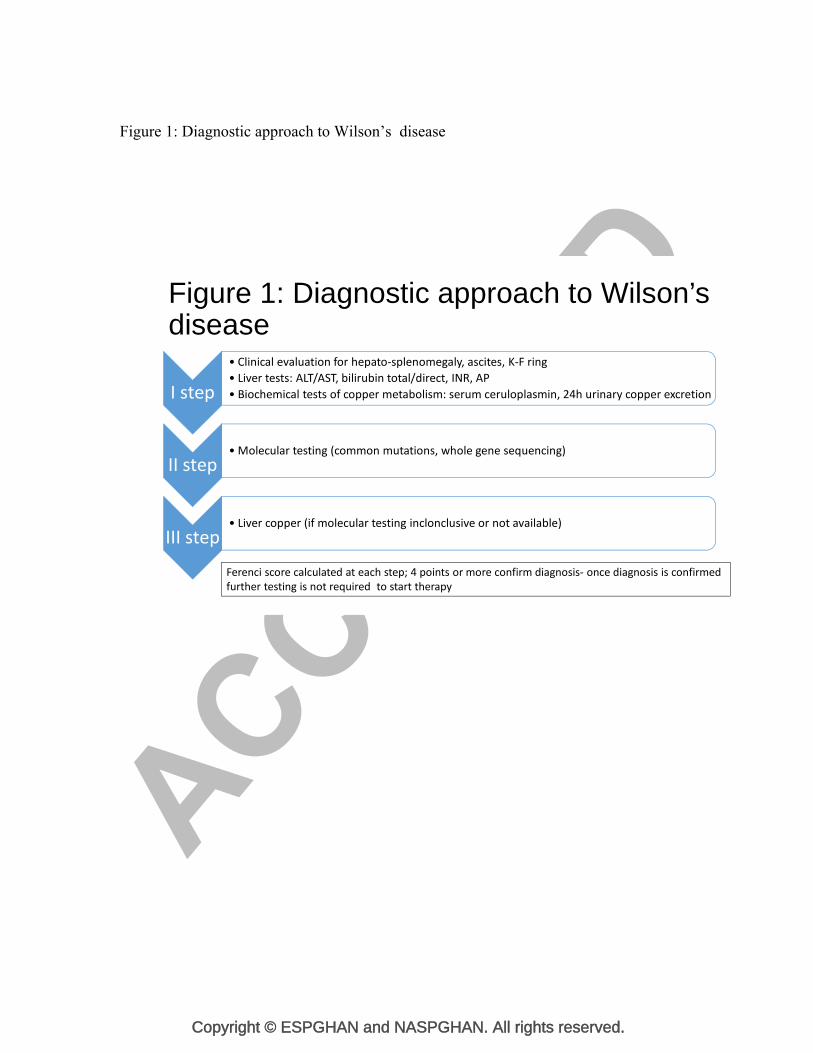
The diagnostic approach is illustrated on figure 1.
Copyright © ESPGHAN and NASPGHAN. All rights reserved.
THE IMPORTANCE OF FAMILY SCREENING FOR WD
Genetic counseling is essential for families of WD patients, and screening first degree
relatives is recommended by both European and American guidelines (3, 4).
It is essential to screen siblings of any patient newly diagnosed with WD because the
chance of being a homozygote and developing clinical disease is 25%. Assessment
should include physical examination; serum ceruloplasmin, liver function tests and
molecular testing for ATP7B mutations or haplotype studies if not available. Newborn
screening is not warranted and screening may be delayed until 1 to 2 years of age.
The occurrence of WD in 2 consecutive generations has been reported in apparently non-
consanguineous families suggesting a benefit for screening WD in offspring of an
affected parent (71-74). Moreover, the risk of occurrence of WD in offspring is increased
in consanguineous families and in specific populations with a high carrier frequency.
Finally, considering the possibility of late onset of WD, parents of a child newly
diagnosed with WD should also be screened by performing liver tests, explorations of
copper metabolism, and suitable genetic testing, as illustrated in a recently reported
family (75).
TREATMENT OF CHILDREN WITH WILSON’S DISEASE
Treatment is based on the removal of copper excess by chelating agents such as D-
penicillamine, trientine or by blocking the intestinal copper absorption with zinc salts.
Dietary copper restriction does not prevent accumulation in WD, and there is a lack of
evidence that it improves the outcome once chelators are initiated. However, avoiding
copper-rich food (shellfish, nuts, chocolate, mushrooms, and organ meats) is advised
Copyright © ESPGHAN and NASPGHAN. All rights reserved.
until remission of symptoms and biochemical abnormalities (3). Treatment should be
initiated upon diagnosis in pre-symptomatic children identified by family screening as
soon as 2 to 3 years of age, promptly in symptomatic children to prevent progression of
liver and/or neurological disease. High-quality evidence is lacking to estimate the optimal
first line treatment choice in WD. Treatment is life-long as well as the monitoring of
compliance and early detection of complications. Prognosis is excellent provided
compliance to therapy is adequate.
Treatment options
D-penicillamine was introduced in 1956, and remains the standard treatment for WD. It
chelates copper and favors its urinary excretion. Experimentally, D-penicillamine also
has a copper “detoxifying” effect by inducing the endogenous hepatic metallothionein, a
cytosolic metal-binding protein which sequesters copper and thereby limiting damage to
the liver.
D-penicillamine has been shown to efficiently prevent the progression of disease in
asymptomatic children. This drug improved liver symptoms in over 80% of symptomatic
children within a mean time of 16 months (28), including those presenting with liver
failure but no hepatic encephalopathy (76). However, worsening of neurologic symptoms
has been reported (77, 78).
Significant adverse effects are reported with the use of D-penicillamine resulting in drug
withdrawal in up to 30% of cases in children or adults (3, 12, 28, 79, 80). Early adverse
effects include sensitivity reactions characterized by fever and cutaneous eruptions,
neutropenia or thrombocytopenia, lymphadenopathy, and proteinuria. Other adverse
effects that may occur at any time in the medium and long term include a lupus-like
Copyright © ESPGHAN and NASPGHAN. All rights reserved.
syndrome characterized by hematuria, proteinuria, arthralgia, bone marrow toxicity with
severe thrombocytopenia or aplasia, and skin changes related to D-penicillamine’s anti-
collagen effects such as elastosis perforans serpiginosa, cutis laxa, pemphigus, lichen
planus, and apthous stomatitis (81). Elevations in serum antinuclear antibodies are
frequent, but there is no clear correlation with the development of immune-mediated
diseases (82).
In children, the dose of D-penicillamine is usually increased progressively to 20
mg/kg/day given in 2 or 3 doses, with close follow-up for the occurrence of adverse
events such as hypersensitivity and proteinuria, hematologic toxicity warranting
immediate discontinuation and switching to trientine or zinc salts (83) (Table 5). As food
inhibits the absorption of D-penicillamine, this drug should be administered one hour
before or two hours after meals. There is lack of recent evidence as to pyridoxine
deficiency in patients receiving penicillamine hence there has been variable personal
practice in its supplementation. Moreover, pyridoxine intake is relatively high as many
food products are supplemented with water soluble vitamins.
Trientine, triethylene tetramine hydrochloride, was initially introduced in 1969 as a
second line chelating agent in WD patients who developed adverse events related to D-
penicillamine (84). There have been only rare reports of allergic reactions, arthralgias,
muscle cramps, and sideroblastic anemia induced by trientine (85-87). Given its safety
profile it is being increasingly used as first line chelation therapy, although there is a lack
of robust clinical studies evaluating the efficiency of trientine in treating WD compared
to D-penicillamine. Similar efficacy of trientine compared to D-penicillamine was shown
Copyright © ESPGHAN and NASPGHAN. All rights reserved.
in one large study in WD adults with liver symptoms (88). However, first line treatment
with trientine was associated with a higher risk of neurologic worsening of symptomatic
neurologic patients compared to D-penicillamine. The only available paediatric study
analysed the efficacy of trientine as second-line therapy in 16 children with D-
penicilllamine intolerance or adverse events. Liver function normalized in the majority of
children, but trientine did not improve accompanying neurological or psychiatric
symptoms (86).
The recommended dose of trientine in children is 20 mg/kg/day in 2-3 divided doses.
Recently, a small prospective pilot study in adults with WD has suggested that a once
daily trientine regimen at a dose of 15 mg/kg provided good efficacy and safety but it
should be further evaluated as maintenance therapy to improve compliance (89).
Trientine also chelates iron, and therefore if iron supplementation is necessary, it should
be administered at a different time of the day. The drug is best given 1 hour before or 2-3
hours after food for optimal absorption (Table 5). Trientine tablets must be kept
refrigerated, which could be a problem for patients residing in or travelling to hot
countries.
Zinc salts are being increasingly used as first line therapy for the treatment of pre-
symptomatic patients and for maintenance therapy after initial de-coppering with a
chelator, but the efficacy of zinc monotherapy in symptomatic patients with liver disease
is still under debate (90-96). The postulated mode of action of zinc is the induction of
metallothionein in enterocytes (97). Copper absorbed in the small intestine is thereby
sequestered in enterocytes which at the end of their life cycle carry copper into the lumen.
Zinc also induces hepatocyte metallothionein, and as D-penicillamine may have copper
Copyright © ESPGHAN and NASPGHAN. All rights reserved.
detoxifying effect. Most studies evaluating the clinical efficacy of zinc when applied as
first line monotherapy in the various clinical presentations of WD showed that zinc had a
better tolerance profile than penicillamine and could be safely used for treatment of
presymptomatic children (90, 92-94). Treatment failure was however reported in
symptomatic children presenting with liver disease (79, 91, 95, 96) and patients who
relapsed on zinc improved after reintroduction of a chelating agent (91). Initiation of
therapy with zinc salts presents also risk of neurological deterioration as is observed with
other treatment modalities (91, 98).
Different formulations of zinc salts are available: zinc sulphate, zinc acetate and zinc
gluconate. Gastrointestinal problems, such as nausea, vomiting, epigastric pain,
gastric/duodenal mucosal ulceration or erosion have been reported mainly with zinc
sulphate, and may particularly alter the child’s quality of life and lead to poor adherence
(95, 99). Gastrointestinal symptoms may resolve when switching formulation from zinc
sulphate to zinc acetate. Anemia related to iron deficiency, isolated increase of serum
amylase and lipase levels (zinc containing enzymes) without clinical and radiological
features of pancreatitis may also be observed.
The recommended dosage is 25 mg twice daily of elemental zinc in children under 5
years of age, 75 mg/day (if body weight < 50 kg) or 150 mg/day (if body weight >50 kg)
in three divided doses in children over 5 years of age (3, 92, 94). Zinc should not be taken
with food because it interferes with its absorption, and dietary copper restriction is not
recommended because zinc blocks copper absorption from the intestinal track (Table 5).
Copyright © ESPGHAN and NASPGHAN. All rights reserved.
Treatment strategy
Treatment should be individually tailored to the clinical condition of the child defined by
the type and severity of organ involvement. A clinically relevant limitation in the long-
term use of D-penicillamine is the occurrence of severe adverse events. Lack of
adherence and underdosage are the main risk factors for an unfavorable clinical course.
To improve adherence to the life-long therapy, the treatment scheme should be as simple
as possible.
Paediatric hepatologists vary in the approaches they use in the care for children with WD
(100). In asymptomatic children or children with only mild liver symptoms, all available
treatments have proven effective (28, 86, 90, 92-94). In symptomatic WD patients,
current guidelines favor the use of chelating agents (D-penicillamine, trientine) as first
line therapy (3, 4).
A sequential treatment in WD could be proposed based on the hypothesis that after a
treatment phase with the more effective chelating agents, smaller dosage or alternative
treatment with zinc in de-coppered patients might be sufficient to upkeep copper
homeostasis. However, only very limited data are available to decide when and under
which conditions a patient can be switched to zinc maintenance therapy and vice versa.
Combination therapy using zinc in conjunction with a chelating agent (administered at
widely spaced intervals during the day to avoid interference- and given one hour before
or two hours after meals) has a theoretical basis in both blocking copper uptake and
eliminating excess copper, and thus might be synergistic effect. Reports have been
limited to patients who present with decompensated chronic liver disease, and suggest a
favorable outcome for combination therapy with D-penicillamine and zinc (8, 76) or with
Copyright © ESPGHAN and NASPGHAN. All rights reserved.
trientine and zinc (79, 101). Recommendations of the ESPGHAN Hepatology Committee
are shown in table 1. INDICATIONS FOR LIVER TRANSPLANTATION IN CHILDREN WITH
WD
Indications for LT are rare (<1%), including patients with ALF or those with progression
of liver dysfunction to liver failure despite drug therapy (102). Excellent post-LT
outcomes are reported (103, 104). Actual patient survival rates were 87% at 5, 10, and 15
years in a French series of 75 adults and 46 children (median age: 14 years, range 7 to 17
yrs) transplanted between 1985 and 2009 for ALF (53%), decompensated cirrhosis
(41%), or severe neurological disease (6%) (103). In another study analysing the United
Network for Organ Sharing (UNOS) database including 170 WD children who
underwent LT between 1987 and 2008, one and five year survival were 90.1% and 89%
respectively (104). In both studies, patients transplanted for end stage chronic liver
disease had better long term survival than patients transplanted for ALF. Extracorporeal
liver support systems as a bridge to LT may improve the outcome (105, 106).
Neurological and especially psychiatric involvement may show little improvement with
transplantation but LT cannot be considered as a therapy for patients with severe
neuropsychiatric involvement (107, 108).
Children presenting with decompensated liver cirrhosis with liver failure but no hepatic
encephalopathy can be often rescued with chelation treatment. Response to medical
treatment may take time with improvement of PT after a minimum of 1 month and
normalization within 3 months to 1 year or more (76). Close follow-up and monitoring of
clinical status for hepatic encephalopathy, ascites, sepsis and liver function tests is
required in specialized LT units to timely listing the child on the transplant list, a decision
Copyright © ESPGHAN and NASPGHAN. All rights reserved.
that is extremely challenging. In 1986, Nazer et al devised a scoring system to predict the
outcome of patients including adults and children with hepatic decompensation in the
setting of WD (109). In 2005, the score was re-examined in the paediatric population by
Dhawan et al who proposed a new scoring system (King’s Wilson index (WI)) that had a
better positive predictive value for mortality without transplantation (Table 6) (8, 102).
The WI is reported to be 93% sensitive, 98% specific, with a positive predictive value of
93% (8). However, although useful, the WI may not be entirely accurate and continued
investigation of predictors of outcome in WD is necessary.
Recommendations of the ESPGHAN Hepatology Committee are shown in table 1.
MONITORING EFFICACY, SAFETY AND COMPLIANCE TO TREATMENT
Treatment of WD should aim at normal physical examination and normal liver function
tests. Patients must avoid alcohol consumption and potential hepatotoxic drug therapy.
Monitoring should be performed once a week at initiation of therapy, especially while
increasing penicillamine dosages, every 1 to 3 months until remission and every 3 to 6
months afterwards. Non-adherence to therapy can lead to life-threatening deterioration
and monitoring intervals should be shorter, especially in adolescents, in whom
compliance is uncertain.
Monitoring includes physical examination (search for new WD symptoms or related to
therapeutic adverse events of therapy) and liver function tests which should normalize
progressively within 3 to 12 months. Twenty-four hour urinary copper excretion should
increase after initiation of therapy with D-penicillamine or trientine, followed by a
decrease once liver function tests return to normal indicating a reduction in the body load
Copyright © ESPGHAN and NASPGHAN. All rights reserved.
of copper. In general pre-symptomatic children excrete less copper than those with
symptomatic disease. Urinary copper excretion should lie between 200-500 μg/24 h
during maintenance therapy with D-penicillamine or trientine. Monitoring of patients on
zinc therapy must ensure a reduction in urinary copper excretion (initially below 100
μg/24h, and between 30 - 75 μg/24h on maintenance therapy); low levels below 30
μg/24h suggest zinc overdosage. Additionally, serum zinc levels and urinary zinc
excretion should be maintained above 125 µg/dL and 1.5-2 g/d respectively; lower levels
generally indicate poor compliance to therapy (110).
It is important to also monitor blood cell counts, and screen any therapeutic related
adverse events (such as proteinuria). Neutropenia and anemia may be due to failed iron
mobilization, with transaminase elevation due to increased hepatic iron accompanied by
increased ferritin indicating over treatment. Temporary discontinuation of therapy, with
close observation, is warranted followed by reintroduction of therapy at a reduced dose
(see Table 6).
Yearly slit-lamp examination should document fading or disappearance in patients with
KF rings if they are being adequately "de-coppered". Appearance of KF rings in patients
with persisting abnormalities of liver function tests on maintenance therapy indicates non
compliance. Search for appearance of changes at magnetic resonance imaging of the
brain may be useful in patients non-compliant to therapy (111).
Copyright © ESPGHAN and NASPGHAN. All rights reserved.
KEY RECOMMENDATIONS
Appropriate steps for diagnosis, treatment and follow up of children with WD were
discussed collectively between the core group and ESPGHAN hepatology members. An
agreement was obtained on the recommendations shown in table 1.
DISCLAIMER
“ESPGHAN is not responsible for the practices of physicians and provides
guidelines and position papers as indicators of best practice only. Diagnosis and
treatment is at the discretion of physicians”.
Copyright © ESPGHAN and NASPGHAN. All rights reserved.
References
1. Reilly M, Daly L, Hutchinson M. An epidemiological study of Wilson's disease in
the Republic of Ireland. J Neurol Neurosurg Psychiatry 1993;56:298-300.
2. Bandmann O, Weiss KH, Kaler SG. Wilson's disease and other neurological
copper disorders. Lancet Neurol 2015;14:103-13.
3. Roberts EA, Schilsky ML. Diagnosis and treatment of Wilson disease: an update.
Hepatology 2008;47:2089-111.
4. EASL Clinical Practice Guidelines: Wilson's disease. J Hepatol 2012;56:671-85.
5. Guyatt GH, Oxman AD, Schunemann HJ, et al. GRADE guidelines: a new series
of articles in the Journal of Clinical Epidemiology. J Clin Epidemiol 2011;64:380-2.
6. Iorio R, D'Ambrosi M, Mazzarella G, et al. Early occurrence of
hypertransaminasemia in a 13-month-old child with Wilson disease. J Pediatr
Gastroenterol Nutr 2003;36:637-8.
7. Iorio R, Sepe A, Giannattasio A, et al. Hypertransaminasemia in childhood as a
marker of genetic liver disorders. J Gastroenterol 2005;40:820-6.
8. Dhawan A, Taylor RM, Cheeseman P, D et al. Wilson's disease in children: 37-
year experience and revised King's score for liver transplantation. Liver Transpl
2005;11:441-8.
9. Iorio R, D'Ambrosi M, Marcellini M, et al. Serum transaminases in children with
Wilson's disease. J Pediatr Gastroenterol Nutr 2004;39:331-6.
Copyright © ESPGHAN and NASPGHAN. All rights reserved.
10. Sanchez-Albisua I, Garde T, Hierro L, et al. A high index of suspicion: the key to
an early diagnosis of Wilson's disease in childhood. J Pediatr Gastroenterol Nutr
1999;28:186-90.
11. Merle U, Schaefer M, Ferenci P, et al. Clinical presentation, diagnosis and long-
term outcome of Wilson's disease: a cohort study. Gut 2007;56:115-20.
12. Giacchino R, Marazzi MG, Barabino A, et al. Syndromic variability of Wilson's
disease in children. Clinical study of 44 cases. Ital J Gastroenterol Hepatol 1997;29:155-
61.
13. Lin LJ, Wang DX, Ding NN, et al. Comprehensive analysis on clinical features of
Wilson's disease: an experience over 28 years with 133 cases. Neurol Res 2014;36:157-
63.
14. Wilson DC, Phillips MJ, Cox DW, et al. Severe hepatic Wilson's disease in
preschool-aged children. J Pediatr 2000;137:719-22.
15. Rukunuzzaman M. Wilson's Disease in Bangladeshi Children: Analysis of 100
Cases. Pediatr Gastroenterol Hepatol Nutr 2015;18:121-7.
16. Roberts EA, Yap J. Nonalcoholic Fatty Liver Disease (NAFLD): Approach in the
Adolescent Patient. Curr Treat Options Gastroenterol 2006;9:423-31.
17. Yener S, Akarsu M, Karacanci C, et al. Wilson's disease with coexisting
autoimmune hepatitis. J Gastroenterol Hepatol 2004;19:114-6.
18. Milkiewicz P, Saksena S, Hubscher SG, et al. Wilson's disease with superimposed
autoimmune features: report of two cases and review. J Gastroenterol Hepatol
2000;15:570-4.
Copyright © ESPGHAN and NASPGHAN. All rights reserved.
19. Machado A, Chien HF, Deguti MM, et al. Neurological manifestations in
Wilson's disease: Report of 119 cases. Mov Disord 2006;21:2192-6.
20. Abdel Ghaffar TY, Elsayed SM, Elnaghy S, et al. Phenotypic and genetic
characterization of a cohort of pediatric Wilson disease patients. BMC Pediatr
2011;11:56.
21. Kalra V, Khurana D, Mittal R. Wilson's disease--early onset and lessons from a
pediatric cohort in India. Indian Pediatr 2000;37:595-601.
22. Dening TR, Berrios GE. Wilson's disease: a longitudinal study of psychiatric
symptoms. Biol Psychiatry 1990;28:255-65.
23. Favre E, Lion-Francois L, Canton M, et al. Cognitive Abilities of Children With
Neurological and Liver Forms of Wilson's Disease. J Pediatr Gastroenterol Nutr
2017;64:436-9.
24. Walshe JM. The acute haemolytic syndrome in Wilson's disease--a review of 22
patients. QJM 2013;106:1003-8.
25. Pendlebury ST, Rothwell PM, Dalton A, et al. Strokelike presentation of Wilson
disease with homozygosity for a novel T766R mutation. Neurology 2004;63:1982-3.
26. Leinweber B, Moller JC, Scherag A, et al. Evaluation of the Unified Wilson's
Disease Rating Scale (UWDRS) in German patients with treated Wilson's disease. Mov
Disord 2008;23:54-62.
27. Ben-Pazi H, Jaworowski S, Shalev RS. Cognitive and psychiatric phenotypes of
movement disorders in children: a systematic review. Dev Med Child Neurol
2011;53:1077-84.
Copyright © ESPGHAN and NASPGHAN. All rights reserved.
28. Manolaki N, Nikolopoulou G, Daikos GL, et al. Wilson disease in children:
analysis of 57 cases. J Pediatr Gastroenterol Nutr 2009;48:72-7.
29. Di Stefano V, Lionetti E, Rotolo N, et al. Hypercalciuria and nephrocalcinosis as
early feature of Wilson disease onset: description of a pediatric case and literature review.
Hepat Mon 2012;12:e6233.
30. Elkiran O, Karakurt C, Selimoglu A, et al. Subclinical diastolic dysfunction in
children with Wilson's disease assessed by tissue Doppler echocardiography: a possible
early predictor of cardiac involvement. Acta Cardiol 2013;68:181-7.
31. Fatima J, Karoli R, Jain V. Hypoparathyroidism in a case of Wilson's disease:
Rare association of a rare disorder. Indian J Endocrinol Metab 2013;17:361-2.
32. Weizman Z, Picard E, Barki Y, et al. Wilson's disease associated with
pancreatitis. J Pediatr Gastroenterol Nutr 1988;7:931-3.
33. Schaefer M, Gotthardt DN, Didion C, Stremmel W, et al. Increased Prevalence of
Subcutaneous Lipomas in Patients With Wilson Disease. J Clin Gastroenterol
2015;49:e61-3.
34. Quemeneur AS, Trocello JM, Ea HK, et al. Miscellaneous non-inflammatory
musculoskeletal conditions. Musculoskeletal conditions associated with Wilson's disease.
Best Pract Res Clin Rheumatol 2011;25:627-36.
35. Weiss KH, Van de Moortele M, Gotthardt DN, et al. Bone demineralisation in a
large cohort of Wilson disease patients. J Inherit Metab Dis 2015;38:949-56.
Copyright © ESPGHAN and NASPGHAN. All rights reserved.
36. Selimoglu MA, Ertekin V, Doneray H, et al. Bone mineral density of children
with Wilson disease: efficacy of penicillamine and zinc therapy. J Clin Gastroenterol
2008;42:194-8.
37. Tissieres P, Chevret L, Debray D, et al. Fulminant Wilson's disease in children:
appraisal of a critical diagnosis. Pediatr Crit Care Med 2003;4:338-43.
38. Sallie R, Katsiyiannakis L, Baldwin D, et al. Failure of simple biochemical
indexes to reliably differentiate fulminant Wilson's disease from other causes of
fulminant liver failure. Hepatology 1992;16:1206-11.
39. Kelly J, Raizman JE, Bevilacqua V, et al. Complex reference value distributions
and partitioned reference intervals across the pediatric age range for 14 specialized
biochemical markers in the CALIPER cohort of healthy community children and
adolescents. Clin Chim Acta 2015;450:196-202.
40. Mak CM, Lam CW, Tam S. Diagnostic accuracy of serum ceruloplasmin in
Wilson disease: determination of sensitivity and specificity by ROC curve analysis
among ATP7B-genotyped subjects. Clin Chem 2008;54:1356-62.
41. Gitlin JD. Aceruloplasminemia. Pediatr Res 1998;44:271-6.
42. Mandato C, Brive L, Miura Y, et al. Cryptogenic liver disease in four children: a
novel congenital disorder of glycosylation. Pediatr Res 2006;59:293-8.
43. Costa LS, Pegler SP, Lellis RF, et al. Menkes disease: importance of diagnosis
with molecular analysis in the neonatal period. Rev Assoc Med Bras (1992) 2015;61:407-
10.
Copyright © ESPGHAN and NASPGHAN. All rights reserved.
44. Nicastro E, Ranucci G, Vajro P, et al. Re-evaluation of the diagnostic criteria for
Wilson disease in children with mild liver disease. Hepatology 2010;52:1948-56.
45. Gromadzka G, Schmidt HH, Genschel J, et al. Frameshift and nonsense mutations
in the gene for ATPase7B are associated with severe impairment of copper metabolism
and with an early clinical manifestation of Wilson's disease. Clin Genet 2005;68:524-32.
46. Merle U, Eisenbach C, Weiss KH, et al. Serum ceruloplasmin oxidase activity is a
sensitive and highly specific diagnostic marker for Wilson's disease. J Hepatol
2009;51:925-30.
47. Muller T, Koppikar S, Taylor RM, et al. Re-evaluation of the penicillamine
challenge test in the diagnosis of Wilson's disease in children. J Hepatol 2007;47:270-6.
48. Kenney SM, Cox DW. Sequence variation database for the Wilson disease copper
transporter, ATP7B. Hum Mutat 2007;28:1171-7.
49. Caca K, Ferenci P, Kuhn HJ, et al. High prevalence of the H1069Q mutation in
East German patients with Wilson disease: rapid detection of mutations by limited
sequencing and phenotype-genotype analysis. J Hepatol 2001;35:575-81.
50. Margarit E, Bach V, Gomez D, et al. Mutation analysis of Wilson disease in the
Spanish population -- identification of a prevalent substitution and eight novel mutations
in the ATP7B gene. Clin Genet 2005;68:61-8.
51. Loudianos G, Dessi V, Lovicu M, et al. Molecular characterization of wilson
disease in the Sardinian population--evidence of a founder effect. Hum Mutat
1999;14:294-303.
Copyright © ESPGHAN and NASPGHAN. All rights reserved.
52. Nanji MS, Nguyen VT, Kawasoe JH, et al. Haplotype and mutation analysis in
Japanese patients with Wilson disease. Am J Hum Genet 1997;60:1423-9.
53. Wu Z, Wang N, Murong S, et al. Identification and analysis of mutations of the
Wilson disease gene in Chinese population. Chin Med J (Engl) 2000;113:40-3.
54. Glenn TC. Field guide to next-generation DNA sequencers. Mol Ecol Resour
2011;11:759-69.
55. Ferenci P, Steindl-Munda P, Vogel W, et al. Diagnostic value of quantitative
hepatic copper determination in patients with Wilson's Disease. Clin Gastroenterol
Hepatol 2005;3:811-8.
56. Gow PJ, Smallwood RA, Angus PW, et al. Diagnosis of Wilson's disease: an
experience over three decades. Gut 2000;46:415-9.
57. Liggi M, Mais C, Demurtas M, et al. Uneven distribution of hepatic copper
concentration and diagnostic value of double-sample biopsy in Wilson's disease. Scand J
Gastroenterol 2013;48:1452-8.
58. Yang X, Tang XP, Zhang YH, et al. Prospective evaluation of the diagnostic
accuracy of hepatic copper content, as determined using the entire core of a liver biopsy
sample. Hepatology 2015;62:1731-41.
59. Araya M, Koletzko B, Uauy R. Copper deficiency and excess in infancy:
developing a research agenda. J Pediatr Gastroenterol Nutr 2003;37:422-9.
60. Sato C, Koyama H, Satoh H, et al. Concentrations of copper and zinc in liver and
serum samples in biliary atresia patients at different stages of traditional surgeries.
Tohoku J Exp Med 2005;207:271-7.
Copyright © ESPGHAN and NASPGHAN. All rights reserved.
61. Bayliss EA, Hambidge KM, Sokol RJ, et al. Hepatic concentrations of zinc,
copper and manganese in infants with extrahepatic biliary atresia. J Trace Elem Med Biol
1995;9:40-3.
62. Johncilla M, Mitchell KA. Pathology of the liver in copper overload. Semin Liver
Dis 2011;31:239-44.
63. Miyamura H, Nakanuma Y, Kono N. Survey of copper granules in liver biopsy
specimens from various liver abnormalities other than Wilson's disease and biliary
diseases. Gastroenterol Jpn 1988;23:633-8.
64. Ferenci P, Caca K, Loudianos G, et al. Diagnosis and phenotypic classification of
Wilson disease. Liver Int 2003;23:139-42.
65. Caprai S, Loudianos G, Massei F, et al. Direct diagnosis of Wilson disease by
molecular genetics. J Pediatr 2006;148:138-40.
66. Koppikar S, Dhawan A. Evaluation of the scoring system for the diagnosis of
Wilson's disease in children. Liver Int 2005;25:680-1.
67. Lyon TD, Fell GS, Gaffney D, et al. Use of a stable copper isotope (65Cu) in the
differential diagnosis of Wilson's disease. Clin Sci (Lond) 1995;88:727-32.
68. El Balkhi S, Trocello JM, Poupon J, et al. Relative exchangeable copper: a new
highly sensitive and highly specific biomarker for Wilson's disease diagnosis. Clin Chim
Acta 2011;412:2254-60.
69. El Balkhi S, Poupon J, Trocello JM, et al. Determination of ultrafiltrable and
exchangeable copper in plasma: stability and reference values in healthy subjects. Anal
Bioanal Chem 2009;394:1477-84.
Copyright © ESPGHAN and NASPGHAN. All rights reserved.
70. Trocello JM, El Balkhi S, Woimant F, et al. Relative exchangeable copper: a
promising tool for family screening in Wilson disease. Mov Disord 2014;29:558-62.
71. Firneisz G, Szonyi L, Ferenci P, et al. The other mutation is found: follow-up of
an exceptional family with Wilson disease. Am J Gastroenterol 2004;99:2504-5.
72. Maier-Dobersberger T, Mannhalter C, Rack S, et al. Diagnosis of Wilson's disease
in an asymptomatic sibling by DNA linkage analysis. Gastroenterology 1995;109:2015-8.
73. Dufernez F, Lachaux A, Chappuis P, et al. Wilson disease in offspring of affected
patients: report of four French families. Clin Res Hepatol Gastroenterol 2013;37:240-5.
74. Dziezyc K, Gromadzka G, Czlonkowska A. Wilson's disease in consecutive
generations of one family. Parkinsonism Relat Disord 2011;17:577-8.
75. Brunet AS, Marotte S, Guillaud O, et al. Familial screening in Wilson's disease:
think at the previous generation! J Hepatol 2012;57:1394-5.
76. Santos Silva EE, Sarles J, Buts JP, et al. Successful medical treatment of severely
decompensated Wilson disease. J Pediatr 1996;128:285-7.
77. Medici V, Trevisan CP, D'Inca R, Barollo M, et al. Diagnosis and management of
Wilson's disease: results of a single center experience. J Clin Gastroenterol 2006;40:936-
41.
78. Kalita J, Kumar V, Chandra S, et al. Worsening of Wilson disease following
penicillamine therapy. Eur Neurol 2014;71:126-31.
79. Wiggelinkhuizen M, Tilanus ME, Bollen CW, et al. Systematic review: clinical
efficacy of chelator agents and zinc in the initial treatment of Wilson disease. Aliment
Pharmacol Ther 2009;29:947-58.
Copyright © ESPGHAN and NASPGHAN. All rights reserved.
80. Czlonkowska A, Litwin T, Karlinski M, et al. D-penicillamine versus zinc sulfate
as first-line therapy for Wilson's disease. Eur J Neurol 2014;21:599-606.
81. Ranucci G, Di Dato F, Leone F, Vet al. Penicillamine-Induced Elastosis Perforans
Serpiginosa in Wilson's Disease: is Useful Switching to Zinc? J Pediatr Gastroenterol
Nutr 2017;64(3):e72-3.
82. Seessle J, Gotthardt DN, Schafer M, et al. Concomitant immune-related events in
Wilson disease: implications for monitoring chelator therapy. J Inherit Metab Dis
2016;39:125-30.
83. Brewer GJ, Yuzbasiyan-Gurkan V, Young AB. Treatment of Wilson's disease.
Semin Neurol 1987;7:209-20.
84. Walshe JM. Treatment of Wilson's disease with trientine (triethylene tetramine)
dihydrochloride. Lancet 1982;1:643-7.
85. Perry AR, Pagliuca A, Fitzsimons EJ, et al. Acquired sideroblastic anaemia
induced by a copper-chelating agent. Int J Hematol 1996;64:69-72.
86. Taylor RM, Chen Y, Dhawan A. Triethylene tetramine dihydrochloride (trientine)
in children with Wilson disease: experience at King's College Hospital and review of the
literature. Eur J Pediatr 2009;168:1061-8.
87. Condamine L, Hermine O, Alvin P, et al. Acquired sideroblastic anaemia during
treatment of Wilson's disease with triethylene tetramine dihydrochloride. Br J Haematol
1993;83:166-8.

Copyright © ESPGHAN and NASPGHAN. All rights reserved.
88. Weiss KH, Thurik F, Gotthardt DN, et al. Efficacy and safety of oral chelators in
treatment of patients with Wilson disease. Clin Gastroenterol Hepatol 2013;11:1028-1035
e1021-2.
89. Ala A, Aliu E, Schilsky ML. Prospective pilot study of a single daily dosage of
trientine for the treatment of Wilson disease. Dig Dis Sci 2015;60:1433-9.
90. Mizuochi T, Kimura A, Shimizu N, et al. Zinc monotherapy from time of
diagnosis for young pediatric patients with presymptomatic Wilson disease. J Pediatr
Gastroenterol Nutr 2011;53:365-7.
91. Weiss KH, Gotthardt DN, Klemm D, et al. Zinc monotherapy is not as effective as
chelating agents in treatment of Wilson disease. Gastroenterology 2011;140:1189-98
e1181.
92. Ranucci G, Di Dato F, Spagnuolo MI, et al. Zinc monotherapy is effective in
Wilson's disease patients with mild liver disease diagnosed in childhood: a retrospective
study. Orphanet J Rare Dis 2014;9:41.
93. Abuduxikuer K, Wang JS. Zinc mono-therapy in pre-symptomatic Chinese
children with Wilson disease: a single center, retrospective study. PLoS One
2014;9:e86168.
94. Marcellini M, Di Ciommo V, Callea F, Devito R, et al. Treatment of Wilson's
disease with zinc from the time of diagnosis in pediatric patients: a single-hospital, 10-
year follow-up study. J Lab Clin Med 2005;145:139-43.
95. Santiago R, Gottrand F, Debray D, et al. Zinc Therapy for Wilson Disease in
Children in French Pediatric Centers. J Pediatr Gastroenterol Nutr 2015;61:613-8.
Copyright © ESPGHAN and NASPGHAN. All rights reserved.
96. Linn FH, Houwen RH, van Hattum J, et al. Long-term exclusive zinc
monotherapy in symptomatic Wilson disease: experience in 17 patients. Hepatology
2009;50:1442-52.
97. Brewer GJ. Zinc therapy induction of intestinal metallothionein in Wilson's
disease. Am J Gastroenterol 1999;94:301-2.
98. Litwin T, Dziezyc K, Karlinski M, et al. Early neurological worsening in patients
with Wilson's disease. J Neurol Sci 2015;355:162-7.
99. Wiernicka A, Janczyk W, Dadalski M, et al. Gastrointestinal side effects in
children with Wilson's disease treated with zinc sulphate. World J Gastroenterol
2013;19:4356-62.
100. Sturm E, Piersma FE, Tanner MS, et al. Controversies and Variation in
Diagnosing and Treating Children With Wilson Disease: Results of an International
Survey. J Pediatr Gastroenterol Nutr 2016;63:82-7.
101. Askari FK, Greenson J, Dick RD, et al. Treatment of Wilson's disease with zinc.
XVIII. Initial treatment of the hepatic decompensation presentation with trientine and
zinc. J Lab Clin Med 2003;142:385-90.
102. Fischer RT, Soltys KA, Squires RH Jr, et al. Prognostic scoring indices in Wilson
disease: a case series and cautionary tale. J Pediatr Gastroenterol Nutr 2011;52:466-9.
103. Guillaud O, Dumortier J, Sobesky R, et al. Long term results of liver
transplantation for Wilson's disease: experience in France. J Hepatol 2014;60:579-89.
Copyright © ESPGHAN and NASPGHAN. All rights reserved.
104. Arnon R, Annunziato R, Schilsky M, et al. Liver transplantation for children with
Wilson disease: comparison of outcomes between children and adults. Clin Transplant
2011;25:E52-60.
105. Nagata Y, Uto H, Hasuike S, Ido A, et al. Bridging use of plasma exchange and
continuous hemodiafiltration before living donor liver transplantation in fulminant
Wilson's disease. Intern Med 2003;42:967-70.
106. Rustom N, Bost M, Cour-Andlauer F, et al. Effect of molecular adsorbents
recirculating system treatment in children with acute liver failure caused by Wilson
disease. J Pediatr Gastroenterol Nutr 2014;58:160-4.
107. Medici V, Mirante VG, Fassati LR, et al. Liver transplantation for Wilson's
disease: The burden of neurological and psychiatric disorders. Liver Transpl
2005;11:1056-63.
108. Yagci MA, Tardu A, Karagul S, et al. Influence of Liver Transplantation on
Neuropsychiatric Manifestations of Wilson Disease. Transplant Proc 2015;47:1469-73.
109. Nazer H, Ede RJ, Mowat AP, et al. Wilson's disease: clinical presentation and use
of prognostic index. Gut 1986;27:1377-81.
110. Brewer GJ, Yuzbasiyan-Gurkan V, Lee DY, et al. Treatment of Wilson's disease
with zinc. VI. Initial treatment studies. J Lab Clin Med 1989;114:633-8.
111. Hermann W. Morphological and functional imaging in neurological and non-
neurological Wilson's patients. Ann N Y Acad Sci 2014;1315:24-9.
Copyright © ESPGHAN and NASPGHAN. All rights reserved.
Figure 1: Diagnostic approach to Wilson’s disease
Figure 1: Diagnostic approach to Wilson’s disease
I step
• Clinical evaluation for hepato‐splenomegaly, ascites, K‐F ring
• Liver tests: ALT/AST, bilirubin total/direct, INR, AP
• Biochemical tests of copper metabolism: serum ceruloplasmin, 24h urinary copper excretion
II step•Molecular testing (common mutations, whole gene sequencing)
III step• Liver copper (if molecular testing inclonclusive or not available)
Ferenci score calculated at each step; 4 points or more confirm diagnosis‐ once diagnosis is confirmedfurther testing is not required to start therapy
Copyright © ESPGHAN and NASPGHAN. All rights reserved.
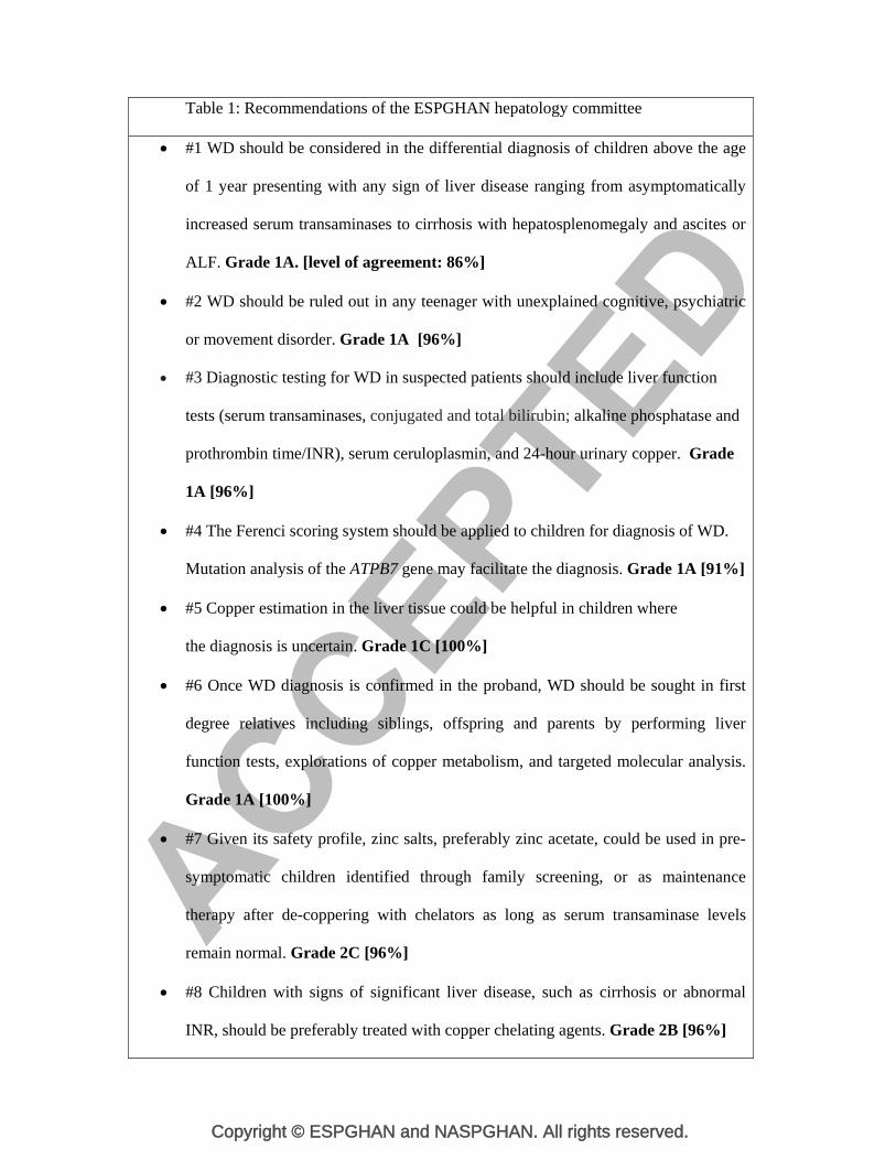
Table 1: Recommendations of the ESPGHAN hepatology committee
#1 WD should be considered in the differential diagnosis of children above the age
of 1 year presenting with any sign of liver disease ranging from asymptomatically
increased serum transaminases to cirrhosis with hepatosplenomegaly and ascites or
ALF. Grade 1A. [level of agreement: 86%]
#2 WD should be ruled out in any teenager with unexplained cognitive, psychiatric
or movement disorder. Grade 1A [96%]
#3 Diagnostic testing for WD in suspected patients should include liver function
tests (serum transaminases, conjugated and total bilirubin; alkaline phosphatase and
prothrombin time/INR), serum ceruloplasmin, and 24-hour urinary copper. Grade
1A [96%]
#4 The Ferenci scoring system should be applied to children for diagnosis of WD.
Mutation analysis of the ATPB7 gene may facilitate the diagnosis. Grade 1A [91%]
#5 Copper estimation in the liver tissue could be helpful in children where
the diagnosis is uncertain. Grade 1C [100%]
#6 Once WD diagnosis is confirmed in the proband, WD should be sought in first
degree relatives including siblings, offspring and parents by performing liver
function tests, explorations of copper metabolism, and targeted molecular analysis.
Grade 1A [100%]
#7 Given its safety profile, zinc salts, preferably zinc acetate, could be used in pre-
symptomatic children identified through family screening, or as maintenance
therapy after de-coppering with chelators as long as serum transaminase levels
remain normal. Grade 2C [96%]
#8 Children with signs of significant liver disease, such as cirrhosis or abnormal
INR, should be preferably treated with copper chelating agents. Grade 2B [96%]
Copyright © ESPGHAN and NASPGHAN. All rights reserved.
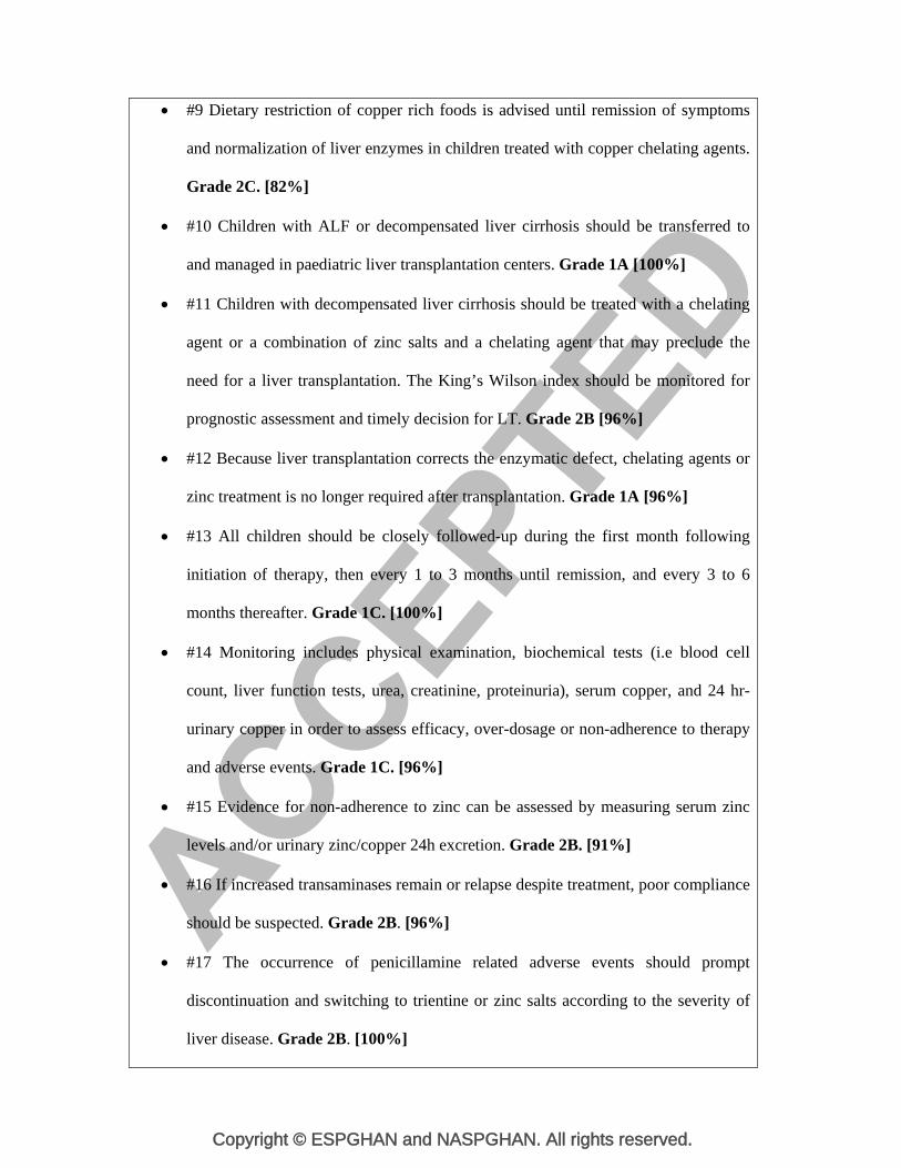
#9 Dietary restriction of copper rich foods is advised until remission of symptoms
and normalization of liver enzymes in children treated with copper chelating agents.
Grade 2C. [82%]
#10 Children with ALF or decompensated liver cirrhosis should be transferred to
and managed in paediatric liver transplantation centers. Grade 1A [100%]
#11 Children with decompensated liver cirrhosis should be treated with a chelating
agent or a combination of zinc salts and a chelating agent that may preclude the
need for a liver transplantation. The King’s Wilson index should be monitored for
prognostic assessment and timely decision for LT. Grade 2B [96%]
#12 Because liver transplantation corrects the enzymatic defect, chelating agents or
zinc treatment is no longer required after transplantation. Grade 1A [96%]
#13 All children should be closely followed-up during the first month following
initiation of therapy, then every 1 to 3 months until remission, and every 3 to 6
months thereafter. Grade 1C. [100%]
#14 Monitoring includes physical examination, biochemical tests (i.e blood cell
count, liver function tests, urea, creatinine, proteinuria), serum copper, and 24 hr-
urinary copper in order to assess efficacy, over-dosage or non-adherence to therapy
and adverse events. Grade 1C. [96%]
#15 Evidence for non-adherence to zinc can be assessed by measuring serum zinc
levels and/or urinary zinc/copper 24h excretion. Grade 2B. [91%]
#16 If increased transaminases remain or relapse despite treatment, poor compliance
should be suspected. Grade 2B. [96%]
#17 The occurrence of penicillamine related adverse events should prompt
discontinuation and switching to trientine or zinc salts according to the severity of
liver disease. Grade 2B. [100%]

Copyright © ESPGHAN and NASPGHAN. All rights reserved.
Legend: Voting results are indicated in brackets for each recommendation .
Copyright © ESPGHAN and NASPGHAN. All rights reserved.
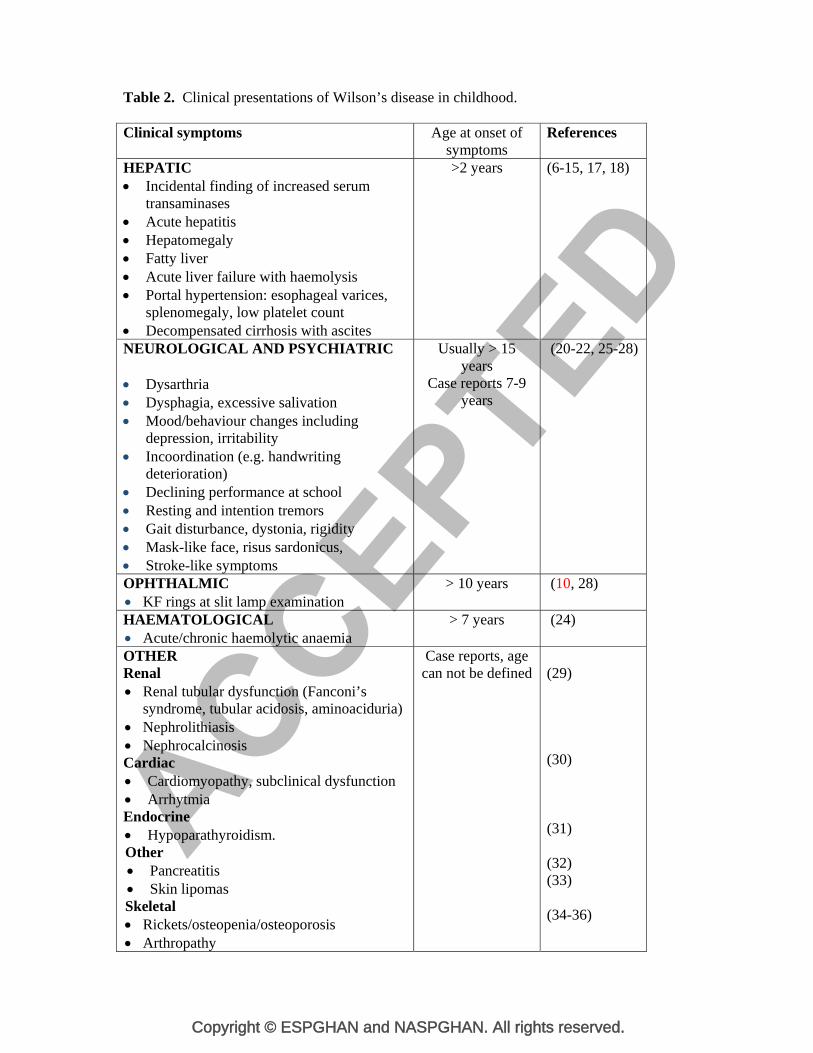
Table 2. Clinical presentations of Wilson’s disease in childhood. Clinical symptoms Age at onset of
symptoms References
HEPATIC Incidental finding of increased serum
transaminases Acute hepatitis Hepatomegaly Fatty liver Acute liver failure with haemolysis Portal hypertension: esophageal varices,
splenomegaly, low platelet count Decompensated cirrhosis with ascites
>2 years (6-15, 17, 18)
NEUROLOGICAL AND PSYCHIATRIC Dysarthria Dysphagia, excessive salivation Mood/behaviour changes including
depression, irritability Incoordination (e.g. handwriting
deterioration) Declining performance at school Resting and intention tremors Gait disturbance, dystonia, rigidity Mask-like face, risus sardonicus, Stroke-like symptoms
Usually > 15 years
Case reports 7-9 years
(20-22, 25-28)
OPHTHALMIC KF rings at slit lamp examination
> 10 years (10, 28)
HAEMATOLOGICAL Acute/chronic haemolytic anaemia
> 7 years (24)
OTHER Renal Renal tubular dysfunction (Fanconi’s
syndrome, tubular acidosis, aminoaciduria) Nephrolithiasis Nephrocalcinosis Cardiac Cardiomyopathy, subclinical dysfunction Arrhytmia Endocrine Hypoparathyroidism. Other Pancreatitis Skin lipomas Skeletal Rickets/osteopenia/osteoporosis Arthropathy
Case reports, age can not be defined
(29) (30) (31) (32) (33) (34-36)
Copyright © ESPGHAN and NASPGHAN. All rights reserved.
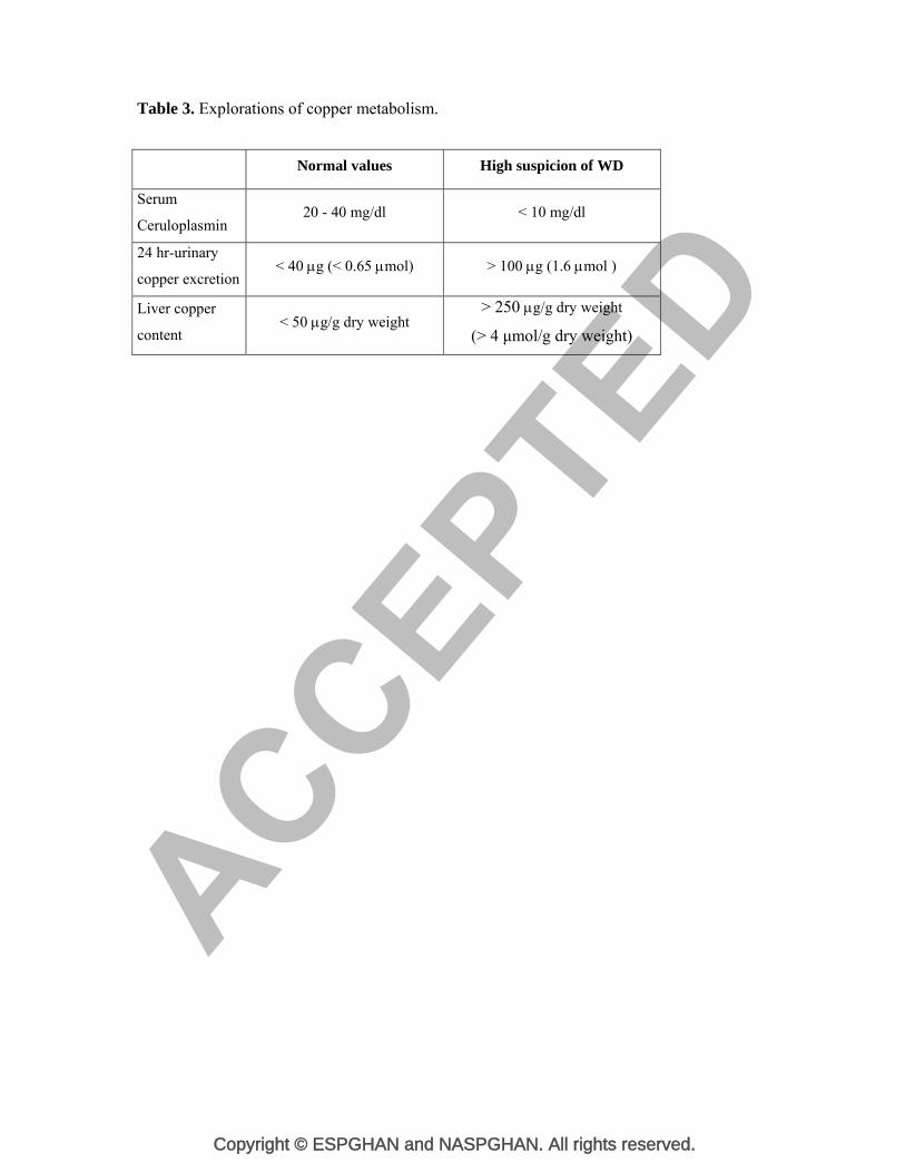
Table 3. Explorations of copper metabolism.
Normal values High suspicion of WD
Serum
Ceruloplasmin 20 - 40 mg/dl < 10 mg/dl
24 hr-urinary
copper excretion < 40 g (< 0.65 mol) > 100 g (1.6 mol )
Liver copper
content < 50 g/g dry weight
> 250 g/g dry weight
(> 4 μmol/g dry weight)
Copyright © ESPGHAN and NASPGHAN. All rights reserved.
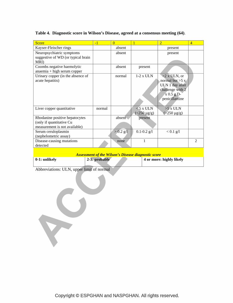
Table 4. Diagnostic score in Wilson’s Disease, agreed at a consensus meeting (64). Score -1 0 1 2 4Kayser-Fleischer rings absent present Neuropsychiatric symptoms suggestive of WD (or typical brain MRI)
absent present
Coombs negative haemolytic anaemia + high serum copper
absent present
Urinary copper (in the absence of acute hepatitis)
normal 1-2 x ULN >2 x ULN, or normal but >5 x ULN 1 day after challenge with 2
x 0.5 g D-penicillamine
Liver copper quantitative normal < 5 x ULN (<250 µg/g)
>5 x ULN (>250 µg/g)
Rhodanine positive hepatocytes (only if quantitative Cu measurement is not available)
absent present
Serum ceruloplasmin (nephelometric assay)
> 0.2 g/l 0.1-0.2 g/l < 0.1 g/l
Disease-causing mutations detected
none 1 2
Assessment of the Wilson’s Disease diagnostic score
0-1: unlikely 2-3: probable
4 or more: highly likely
Abbreviations: ULN, upper limit of normal
Copyright © ESPGHAN and NASPGHAN. All rights reserved.
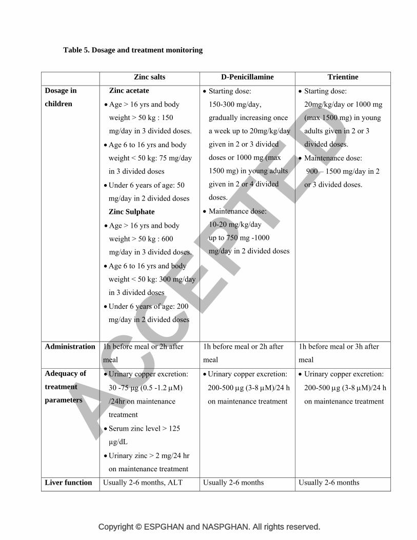
Table 5. Dosage and treatment monitoring
Zinc salts D-Penicillamine Trientine
Dosage in
children
Zinc acetate
Age > 16 yrs and body
weight > 50 kg : 150
mg/day in 3 divided doses.
Age 6 to 16 yrs and body
weight < 50 kg: 75 mg/day
in 3 divided doses
Under 6 years of age: 50
mg/day in 2 divided doses
Zinc Sulphate
Age > 16 yrs and body
weight > 50 kg : 600
mg/day in 3 divided doses.
Age 6 to 16 yrs and body
weight < 50 kg: 300 mg/day
in 3 divided doses
Under 6 years of age: 200
mg/day in 2 divided doses
Starting dose:
150-300 mg/day,
gradually increasing once
a week up to 20mg/kg/day
given in 2 or 3 divided
doses or 1000 mg (max
1500 mg) in young adults
given in 2 or 4 divided
doses.
Maintenance dose:
10-20 mg/kg/day
up to 750 mg -1000
mg/day in 2 divided doses
Starting dose:
20mg/kg/day or 1000 mg
(max 1500 mg) in young
adults given in 2 or 3
divided doses.
Maintenance dose:
900 – 1500 mg/day in 2
or 3 divided doses.
Administration 1h before meal or 2h after
meal
1h before meal or 2h after
meal
1h before meal or 3h after
meal
Adequacy of
treatment
parameters
Urinary copper excretion:
30 -75 μg (0.5 -1.2 M)
/24hr on maintenance
treatment
Serum zinc level > 125
µg/dL
Urinary zinc > 2 mg/24 hr
on maintenance treatment
Urinary copper excretion:
200-500 g (3-8 M)/24 h
on maintenance treatment
Urinary copper excretion:
200-500 g (3-8 M)/24 h
on maintenance treatment
Liver function Usually 2-6 months, ALT Usually 2-6 months Usually 2-6 months
Copyright © ESPGHAN and NASPGHAN. All rights reserved.
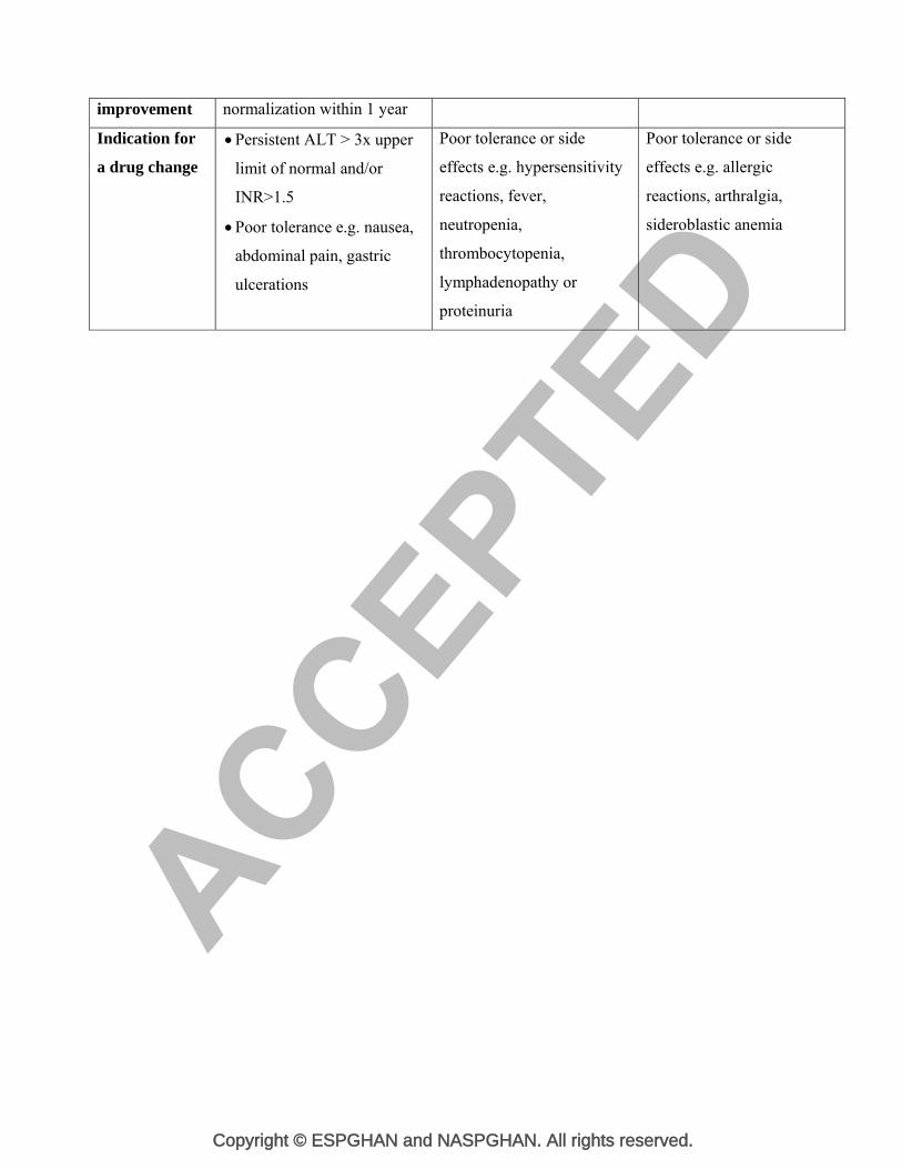
improvement normalization within 1 year
Indication for
a drug change
Persistent ALT > 3x upper
limit of normal and/or
INR>1.5
Poor tolerance e.g. nausea,
abdominal pain, gastric
ulcerations
Poor tolerance or side
effects e.g. hypersensitivity
reactions, fever,
neutropenia,
thrombocytopenia,
lymphadenopathy or
proteinuria
Poor tolerance or side
effects e.g. allergic
reactions, arthralgia,
sideroblastic anemia
Copyright © ESPGHAN and NASPGHAN. All rights reserved.
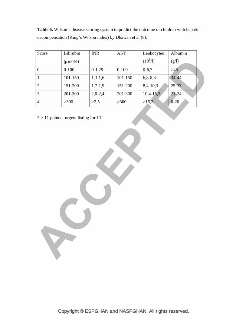
Table 6. Wilson’s disease scoring system to predict the outcome of children with hepatic
decompensation (King’s Wilson index) by Dhawan et al (8).
Score Bilirubin
(mol/l)
INR AST Leukocytes
(109/l)
Albumin
(g/l)
0 0-100 0-1,29 0-100 0-6,7 >45
1 101-150 1,3-1,6 101-150 6,8-8,3 34-44
2 151-200 1,7-1,9 151-200 8,4-10,3 25-33
3 201-300 2,0-2,4 201-300 10.4-15,3 21-24
4 >300 >2,5 >300 >15,3 0-20
* > 11 points - urgent listing for LT