Embed Size (px)
Citation preview
54 ENDOVASCULAR TODAY MAY 2016 VOL. 15, NO. 5
L I M B S A LVA G E
Long Peripheral CTOs:Femoropopliteal to Mid and Distal Tibial Arteries
Peripheral vascular disease (PVD) is an epidemic affecting millions of patients worldwide. The number of patients suffering from PVD is expected to increase by 15% in western coun-
tries and 30% in developing countries, and 1% to 2% of patients with PVD have critical limb ischemia (CLI).1 Endovascular revascularization of patients with CLI is increasingly becoming a first-line strategy, referred to as an endovascular-first approach. Surgical options should always be part of the plan in the event that endovas-cular therapy is not successful. In patients who are not surgical candidates, an endovascular approach is an excellent alternative.
Comorbidities associated with CLI are noteworthy, especially diabetes mellitus, chronic kidney disease, hyperlipidemia, hypertension, and age > 75 years.2 CLI is also known to be associated with multilevel and multi-vessel disease. Chronic total occlusions (CTOs) of above- and/or below-the-knee arteries are very common, which presents a major challenge for revascularization. This is especially challenging when the CTOs are long and involve a proximal CTO cap located above the knee and reconstitutes in the tibial vessels below the knee. These types of CTOs are the most challenging for endovas-cular revascularization; elements of complexity include the severity of the proximal and distal CTO caps, length of the CTO lesions (which tend to be > 50 mm), and involvement of the P3 and proximal tibial junctions.
This article will explore the role of endovascular therapy in long CTOs extending from the superficial femoral arteries (SFA) with reconstitution in the mid to distal tibial arteries.
CLI PATIENT ASSESSMENT Patients with CLI tend to present with advanced
Rutherford class 4 to 6. It is important to document the presence of the common femoral artery (CFA) palpable pulse and absent palpable popliteal and pedal pulses, followed by a foot exam that includes handheld Doppler to look for monophasic, biphasic, or triphasic Doppler signals. Patients with triphasic signals are much better candidates for revascularization than those with mono-
Proper patient assessment and current access and revascularization techniques to treat
challenging CLI anatomy.
BY J.A. MUSTAPHA, MD, AND FADI SAAB, MD
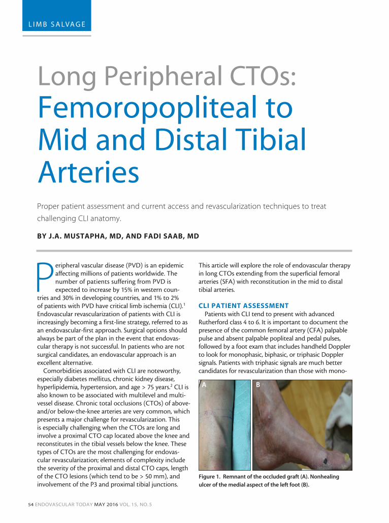
Figure 1. Remnant of the occluded graft (A). Nonhealing
ulcer of the medial aspect of the left foot (B).
A B

VOL. 15, NO. 5 MAY 2016 ENDOVASCULAR TODAY 55
L I M B S A LVA G E
phasic Doppler signals. If wounds are present, they tend to help direct the operator toward choosing the proper and most valuable target artery to open using the so-called angiosome concept. For patients with wounds, detailed wound assessment and documentation (includ-ing photos) is a necessary baseline step to track the prog-ress of treatment.
Noninvasive AssessmentNoninvasive testing should be applied with a good
understanding of its limitations. The value of noninva-sive testing, including ankle-brachial index (ABI), pulse volume recording, and arterial duplex ultrasound, in patients with CLI may be limited due to the nature of the underlying disease process. Severely calcified ves-sels can produce noncompressible ABIs or false-positive values that can provide the wrong diagnosis and may contribute to delayed or inappropriate therapy. The usefulness of arterial duplex ultrasound can vary depend-ing on the expertise of the technicians performing the test and the reader’s experience. However, baseline ABI values can be a good benchmark to use for comparison after an intervention. The same role may apply to pulse volume recordings and arterial duplex ultrasound. Other imaging modalities, such as CT angiography or magnetic resonance angiography, can be beneficial. The value of CT angiography depends on the individual center and varies based on the specific protocols used. The ability to visualize tibial flow on CT angiography is very limited in CLI patients with heavy calcification.
Anatomical AssessmentIt is our opinion that all CLI patients benefit from
thorough diagnostic angiography. This entails obtain-ing a detailed diagnostic angiogram using selective angiography in which a catheter is placed as distally as possible within the limb requiring revascularization. The
information gathered should include lesion locations, CTO length, calcification, and vessel reconstitution. It is important to obtain a lateral view of both pedal arteries during this assessment.
RevascularizationThe revascularization plan must involve a preplanned
access site, alternative access sites, and revascularization modalities. Discussing all revascularization modalities is beyond the scope of this article. However, we believe that proficiency with multiple modalities (including stenting platforms and atherectomy devices) is essential to any procedure’s success. Choosing the right device for the lesion being treated is paramount, while also staying mindful of the cost involved. Patients with multilevel dis-ease may require staged procedures to treat both inflow and outflow disease; this is usually recommended by the operator due to maximum contrast utilization, radiation exposure, and patient comfort considerations.
CLINICAL SCENARIOThe following complex case demonstrates the previously
mentioned steps. A 79-year-old man with a significant past medical history of diabetes, hypertension, and ischemic cardiomyopathy with a ventricular ejection fraction of 35% was referred from another institution. Initially, the patient had a history of claudication, which was treated with femoropopliteal bypass with a synthetic graft (Figure 1A). Surgery was successful; the patient’s claudication resolved, and he was discharged home with Rutherford class 0.
Figure 2. Aortoiliac angiogram (A), occlusion at the CFA (B),
and a hint of distal PT artery flow (C).
Figure 3. Available modified Schmidt access points when
ultrasound is utilized.
A B C
56 ENDOVASCULAR TODAY MAY 2016 VOL. 15, NO. 5
L I M B S A LVA G E
Eight months later, the patient presented again with new symptoms, this time consistent with Rutherford class 4 rest pain. Shortly after, the patient developed a wound involving the medial aspect of the foot, placing him at Rutherford class 5 (Figure 1B). After the hemo-dynamic assessment, the patient underwent peripheral diagnostic angiography (Figure 2), which revealed severe left lower extremity disease, including total occlusion of the recent bypass graft, the SFA/popliteal arteries, and majority of the tibial arteries. This type of severe ana-tomical disease indicated that the patient had limited revascularization options, with the exception of a high-risk surgical redo with bypass into the plantar arteries or complex high-risk endovascular revascularization.
Clinical Presentation and Physical Examination
As noted above, this patient progressed from Rutherford class 3 to 5 over the course of 2 years. Interestingly, the transition from Rutherford class 4 to 5 occurred over a short period of time of < 4 weeks. The initial exam showed a frail patient with normal cardiac exam, and the bilateral com-mon femoral artery pulses were intact. The left popliteal artery pulse was absent, and the left pedal pulses were not palpable. Doppler sig-
nals were weak, with faint monophasic Doppler signals in the posterior tibial (PT) artery and no signal in the dorsalis pedis (DP). The left foot was cool to the touch; the wound had a Wifi (wound, ischemia, and foot infec-tion) grade score of 2. Wound pictures were obtained and stored in electronic medical records, which is a common practice at our institution. The risk of limb loss was imminent.
Noninvasive AssessmentThe noninvasive assessment included an ABI with an
arterial duplex ultrasound. The patient’s ABI was mea-sured at 0.2. Arterial duplex ultrasound was limited due to the presence of calcification, especially intimal calci-
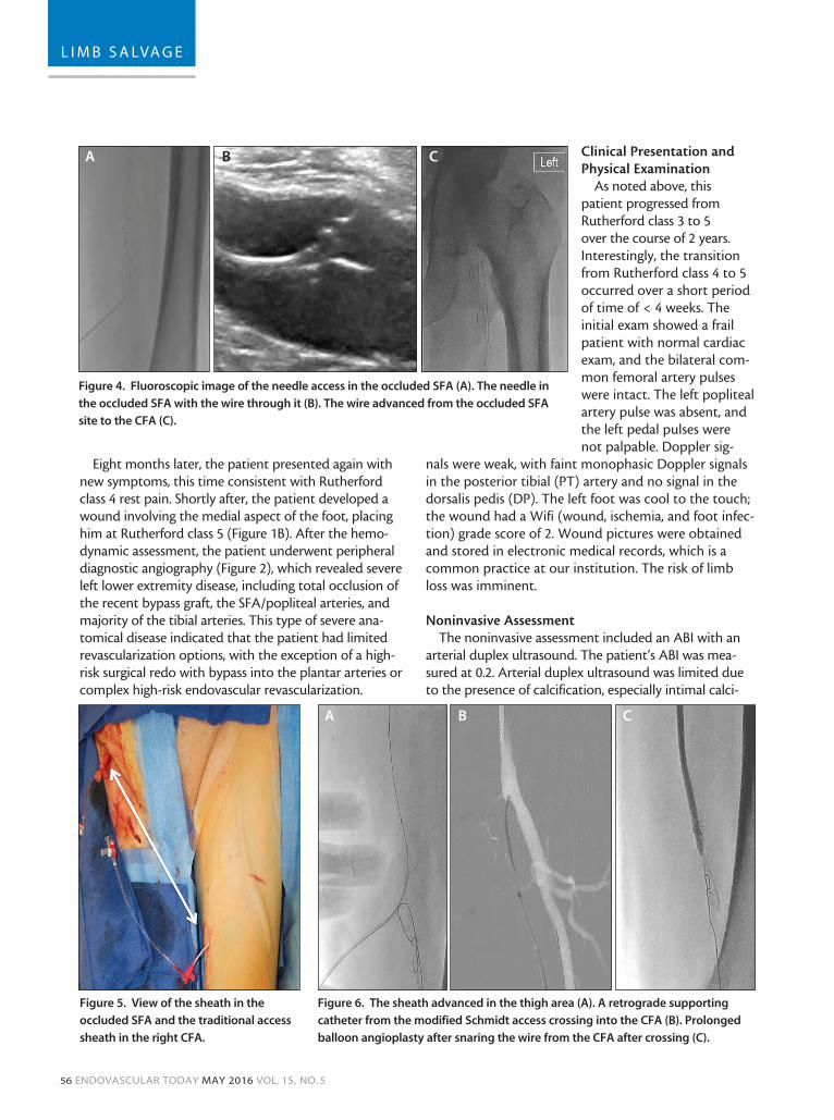
Figure 4. Fluoroscopic image of the needle access in the occluded SFA (A). The needle in
the occluded SFA with the wire through it (B). The wire advanced from the occluded SFA
site to the CFA (C).
Figure 6. The sheath advanced in the thigh area (A). A retrograde supporting
catheter from the modified Schmidt access crossing into the CFA (B). Prolonged
balloon angioplasty after snaring the wire from the CFA after crossing (C).
Figure 5. View of the sheath in the
occluded SFA and the traditional access
sheath in the right CFA.
A
A B C
B C
58 ENDOVASCULAR TODAY MAY 2016 VOL. 15, NO. 5
L I M B S A LVA G E
fication, which created significant acoustic shadowing. Only the distal PT artery showed monophasic flow, with the remainder of the tibial vessels appearing occluded. The SFA was assessed with ultrasound and showed that it had a high takeoff with flush occlusion.
Anatomical Assessment
Selective angiography was performed with a cath-eter placed in the left CFA. The angiogram showed no flow beyond the CFA and the profunda femoral artery, which supplied the collaterals, and in turn connect to the tibiopedal collaterals with flow to the distal PT artery (Figure 2). A long, complex SFA/popliteal/tibial CTO was also identified.
RevascularizationIn complex disease as described in this case, access
becomes extremely important for the success of the pro-cedure. Operators should consider all access options to optimize revascularization success.
Access. Choosing the proper access is a critical step in achieving adequate revascularization. Normally at our institution, antegrade access is the preferred route for the majority of infrainguinal arterial disease including SFA flush occlusion. In this case, antegrade access was not an option due to the fact that the SFA takeoff was too high (Figure 2B); therefore, contralateral access was achieved to avoid an ipsilateral antegrade high stick. In the setting where the SFA takeoff is not too high, one can still obtain antegrade access and deliver successful CTO crossing of flush SFA occlusion provided that there is an opportunity to place an antegrade sheath 2 to 3 cm from the ostium of the occluded SFA.
Alternative access. Alternative access is usually viewed as any access other than retrograde CFA. In this case to ensure success, a decision was made to combine and uti-lize multiple alternative accesses along with contralateral CFA access. This type of approach is necessary to achieve
shorter distance between the access site and the target occluded segments (eg, the CTO) to be crossed and treated. This approach eliminates many of the anatomi-cal challenges and variables. As noted previously, the CTO in this case was from the CFA to the ankle region of the PT artery with a total CTO lesion length of 70 cm. The next step after access is to connect the CFA to the tibiopedal circulation. Occasionally in a long CTO seg-ment, one can find a “hibernating lumen,” defined as a patent segment between two CTO caps. A hibernating lumen was found in this case in the anterior tibial (AT) artery after achieving pedal retrograde access in the left DP to find the mid to distal portion of the AT artery to patent hibernating. This concept is important to keep in mind, especially in long CTO segments and in the SFA.
In this case, a decision was made to perform a modi-fied Schmidt access directly into the mid-left SFA and the left DP. The modified Schmidt technique is defined as access into an occluded segment of any vessel either under ultrasound or fluoroscopy (Figure 3). In this case, an attempt was initially made to cross into the SFA from the CFA without success. This was followed with ultra-sound-guided retrograde SFA access using the modified Schmidt access technique, and a 4-F Pinnacle Precision sheath (Terumo Interventional Systems) was placed (Figures 4 and 5).
Using the newly placed sheath in the SFA, the opera-tor was able to cross the proximal SFA CTO in a retro-grade fashion into the left patent CFA (Figure 6). Once in the CFA, the retrograde SFA wire was then snared and externalized from the right CFA so that there was a flossing wire from the right CFA to the left mid SFA (Figure 6B). To obtain flow to the SFA access point, balloon angioplasty was performed (Figure 6C). With the left CFA connected to the SFA via a patent SFA seg-
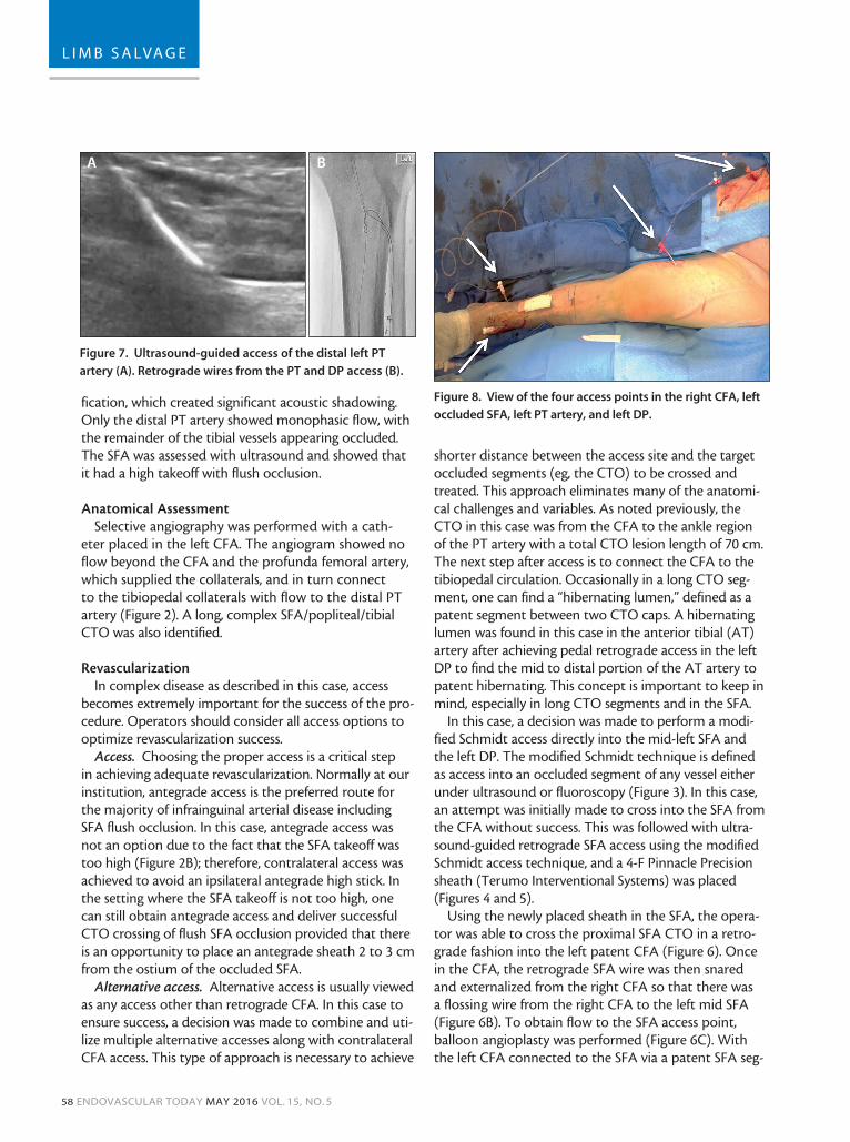
Figure 7. Ultrasound-guided access of the distal left PT
artery (A). Retrograde wires from the PT and DP access (B).
Figure 8. View of the four access points in the right CFA, left
occluded SFA, left PT artery, and left DP.
A B
60 ENDOVASCULAR TODAY MAY 2016 VOL. 15, NO. 5
L I M B S A LVA G E
ment, it was then necessary to cross the rest of the long CTO to reach the anterior and posterior tibiopedal cir-culation. Retrograde PT and DP accesses were achieved using an ultrasound-guided approach. For the DP access, a modified Schmidt technique was used because of the occluded nature of the selected access site (Figure 7). Ultrasound-guided access was achieved in the PT artery at the level above the medial malleolus in a patent arte-rial segment. Both circulations were involved due to the angiosome distribution of the wound, which involved the anterior and posterior tibiopedal arteries. After tib-iopedal access, retrograde CTO crossing of the AT and PT arteries were performed using a wire and catheter technique with the NaviCross (Terumo Interventional Systems) and the Micro18 catheter (Roxwood Medical). Simultaneously, a second operator continued to cross the rest of the SFA/popliteal CTO in an antegrade fashion from the contralateral CFA access and sheath. Once the retrograde catheters met with the antegrade catheters, snaring was performed and flossing between the right CFA and the tibiopedal access was achieved. CTO crossing was successful, requiring four accesses (Figure 8).
Revascularization modalities. The goal of revascular-ization is to deliver adequate blood flow to the distal limb in order to relieve symptoms and achieve wound healing. With wires in the SFA, popliteal, AT, and PT arteries, the next step was to treat all the CTO segments
with prolonged balloon angioplasty with a balloon-to-vessel ratio sizing of 1:1.
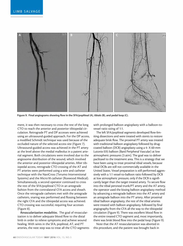
The left SFA/popliteal segments developed flow-lim-iting dissections and were treated with stents to restore adequate brisk flow. The proximal PT artery was treated with traditional balloon angioplasty followed by drug-coated balloon (DCB) angioplasty using a 4- X 60-mm Lutonix 035 balloon (Bard Peripheral Vascular) at low atmospheric pressures (3 atm). The goal was to deliver paclitaxel to the treatment area. This is a strategy that we have been using to treat proximal tibial vessels, because tibial DCBs are still not commercially available in the United States. Vessel preparation is still performed aggres-sively with a 1:1 vessel-to-balloon ratio followed by DCB at low atmospheric pressure, only if the DCB is signifi-cantly larger than the target treated artery. To secure flow into the tibial peroneal trunk/PT artery and the AT artery, the operator used the kissing balloon angioplasty method by advancing a retrograde balloon into the AT artery and an antegrade balloon into the PT artery. After proximal tibial balloon angioplasty, the rest of the tibial arteries were treated with balloon angioplasty, followed by final angiography from the CFA all the way to the tibiopedal circulation (Figure 9). There was excellent blood flow in the entire treated CTO segment and, most importantly, there was brisk blood flow into the pedal loop (Figure 9).
Note that the AT revascularization was aborted in this procedure, and the patient was brought back in
Figure 9. Final angiograms showing flow in the SFA/popliteal (A), tibials (B), and pedal loop (C).
A B C
VOL. 15, NO. 5 MAY 2016 ENDOVASCULAR TODAY 61
L I M B S A LVA G E
48 hours later for stenting in the ostial AT artery with a 4- X 38-mm drug-eluting stent.
DISCUSSIONPatients with a history of PVD and CLI tend to have
a history of prior procedures. Unpublished findings from the Peripheral Registry of Endovascular Outcomes (PRIME) registry showed that 10% of patients had a prior history of bypass. Almost 30% of patients had a prior endovascular procedure. Overall, surgical bypass graft failure still occurs in as many as 50% of patients within 5 years, depending on the type and level of the conduit.3,4 Repeat bypass grafting is associated with worse outcomes compared with primary bypass and is often not feasible because of the patient’s medical risk factors, limited life expectancy, and a lack of adequate bypass conduit or target.5
Due to these limitations, there has been widespread implementation of an endovascular-first approach. This strategy has been adopted by all disciplines, including vascular surgery, interventional radiology, and interven-tional cardiology, with some exceptions. This wave has been fueled by continuous innovations in endovascular techniques and devices.6 CTOs are frequently encoun-tered during endovascular intervention of infrainguinal peripheral artery disease. In diabetic patients, CTOs can constitute up to 55% of all infrainguinal arterial lesions in patients with Rutherford class ≥ 3 symptoms.7 Failure to penetrate the proximal CTO cap, navigate side branches or bridging collaterals, and reenter the distal true lumen are a few mechanisms of failure that prompted the adoption of such advanced techniques. Dense fibrous caps and heavy calcification often char-acterize CTOs and are associated with failure to cross them.8 Crossing of these lesions with traditional guide-wires and catheters can often be challenging, prompting the development of several crossing devices. In addition to using CTO crossing devices, other techniques have been advocated, including tibiopedal access and modi-fied Schmidt access.
The tackling of PVD and CLI is a complex process that involves multiple steps, as highlighted in the case exam-ple. Advanced revascularization techniques need to be available in these complex cases. Having a well-trained team familiar and equipped with multiple devices to treat these patients is essential. In addition, the opera-tor’s experience dictates which techniques can be per-formed and which should be avoided. CLI patients tend to suffer from multiple comorbidities that limit their ability to tolerate procedures. These obstacles prompt the question, “Is it time to create specialized CLI centers that can fulfill the above requirements?”9
CONCLUSIONIn patients with CLI, currently available techniques and
technologies (from alternative access to newer revas-cularization technologies) make limb salvage feasible in cases that were previously deemed not possible. The case presented in this article demonstrates the importance of specialized CLI centers that can provide the full spectrum of care from clinical evaluation to revascularization and, most importantly, delivering care after revascularization including immediate wound care and medical therapy (antiplatelet therapy, statins, ß blockers, and angiotensin-converting enzyme inhibitors as tolerated). n
1. Fowkes FG, Rudan D, Rudan I, et al. Comparison of global estimates of prevalence and risk factors for peripheral artery disease in 2000 and 2010: a systematic review and analysis. Lancet. 2013;382:1329-1340.2. Philip F. 3-year outcomes of the OLIVE registry, a prospective multicenter study of patients with critical limb ischemia. JACC Cardiovasc Interv. 2016;9:201-202. 3. Leather RP, Shah DM, Chang BB, et al. Resurrection of the in situ saphenous vein bypass. 1,000 cases later. Ann Surg. 1988;208:435-442.4. Taylor LM Jr, Edwards JM, Porter JM. Present status of reversed vein bypass grafting: five-year results of a modern series. J Vasc Surg. 1990;11:193-205; discussion 205-206.5. Brewster DC, LaSalle AJ, Robison JG, et al. Femoropopliteal graft failures. Clinical consequences and success of secondary reconstructions. Arch Surg. 1983;118:1043-1047.6. Jaff MR, White CJ, Hiatt WR, et al. An update on methods for revascularization and expansion of the TASC lesion classification to include below-the-knee arteries: a supplement to the Inter-Society Consensus for the Management of Peripheral Arterial Disease (TASC II): the TASC Steering Committee. Ann Vasc Dis. 2015;8:343-357.7. Aziz S, Ramsdale DR. Chronic total occlusions—a stiff challenge requiring a major breakthrough: is there light at the end of the tunnel? Heart. 2005;91(suppl 3):iii42-48.8. Kandzari DE. The challenges of chronic total coronary occlusions: an old problem in a new perspective. J Interv Cardiol. 2004;17:259-267.9. Saab F, Diaz-Sandoval L, Mustapha JA. The nuts and bolts of building a critical limb ischemia program. American College of Cardiology. September 8, 2015. http://www.acc.org/latest-in-cardiology/articles/2015/09/02/13/22/the-nuts-and-bolts-of-building-a-critical-limb-ischemia-program. Accessed April 5, 2016.
J.A. Mustapha, MDDirector of Cardiovascular Catheterization LaboratoriesMetro Health Hospital Wyoming, MichiganClinical Associate Professor of MedicineMichigan State University CHM and COM East Lansing, [email protected]: Consultant to Bard Peripheral Vascular and Terumo Interventional Systems.
Fadi Saab, MDInterventional Cardiologist Metro Health HospitalWyoming, MichiganClinical Assistant Professor of MedicineMichigan State University CHM and COMEast Lansing, MichiganDisclosures: Consultant to Bard Peripheral Vascular and Terumo Interventional Systems.