Embed Size (px)
Citation preview
1
Local and systemic factors impacting on
visual outcome in uveitis
Lazha Ahmed Talat Sharief
August 2016
Submitted for the Doctor of Philosophy degree
University College London

2
ACKNOWLEDGMENTS
My sincere gratitude to my supervisor Professor Sue Lightman (Consultant
Ophthalmologists) and co-supervisor Dr Oren Tomkins-Netzer (Senior Clinical
Teaching Fellow) for their guidance and support in planning and conducting the
studies included in this academic work and in the writing process of this thesis.
I also wish to thank my fellow colleague ophthalmologists at the uveitis clinic for
their encouragement and support throughout the time period required to conduct this
research work. Special gratitude also goes to Sarah Mayhew and Hazel Lawrence;
their kind help and assistance in the administrative process required for this research
will always be remembered with sincere gratitude.
I am most grateful to all the participating patients and all the staff working in the
uveitis clinic and imaging unit at Moorfields Eye Hospital for their help and support
during this research.
Finally, I would like to thank my parents, my sisters, Lava, Lawan, Sham, and my
brother Hasan. Your patience and support through every moment in my life made it
possible to finish this research. I hope I make you proud. I dedicate this thesis to my
parents and my only son, my sun, Mand.
3
DECLARATION
I, LAZHA AHMED TALAT SHARIEF, CONFIRM THAT THE WORK
PRESENTED IN THIS THESIS IS MY OWN. WHERE INFORMATION
HAS BEEN DERIVED FROM OTHER SOURCES, I CONFIRM THAT
THIS HAS BEEN INDICATED IN THE THESIS.
SIGNATURE:
DATE:
NAME: LAZHA A. T. SHARIEF
4
ABSTRACT
Uveitis is the fourth most common cause of blindness among the working age group in
developed countries. The aim of this research is to examine the influence of diabetes
mellitus, cataract surgery, and retinal vasculitis on the visual outcome and prognosis of
eyes with uveitis. The studied cohort included 1169 patients with uveitis attending
Moorfields Eye Hospital between January 2012 and December 2013.
The first study divided uveitis cases with diabetes into two groups; the first group included
99 eyes with diabetes diagnosed prior to uveitis. The second group included 96 eyes with
uveitis later diagnosed with diabetes. Within the first group, 28.2% had vision loss mainly
from maculopathy with the risk of vision loss 4.6 times higher when compared to the
control group of non-diabetic uveitis group. The diagnosis of diabetes in the second group
was associated with a drop in the mean vision over the year post diagnosis. The mean dose
of corticosteroid was lower post diagnosis (15 mg versus 10 mg, p=0.03), and relapses were
significantly less often treated with systemic corticosteroid alone (70.2% vs. 55.6% of the
relapses, p=0.003).
The second study included 236 eyes with retinal vasculitis (121 ischaemic, 115 non-
ischaemic) which was compared to non-vasculitis control group (1022 eyes). Macular
ischaemia increased the risk of vision loss in vasculitis by 4.4 times. Retinal vasculitis had
twice the risk of macular oedema compared to non-vasculitis. Macular oedema and
ischaemia increased risk of vision loss in ischaemic vasculitis while corticosteroids reduce
the risk by 30%. Retinal ischaemia involving ≥ 2 quadrants was associated with increased
risk of NV formation.
The third study included 228 uveitic eyes undergone cataract surgery and was compared to
a control group of 300 phakic eyes with uveitis. The vision continued to improve from the
baseline first postoperative week. However, risk of vision loss and CMO were twice more
in the pseudophakic group compared to the control. The rate of uveitis relapse and rate of
using high dose of corticosteroids was significantly lower postoperatively versus
preoperatively.
5
TABLE OF CONTENTS
ACKNOWLEDGMENTS ..................................................................................................... 2
DECLARATION ................................................................................................................... 3
ABSTRACT ........................................................................................................................... 4
TABLE OF CONTENTS ....................................................................................................... 5
LIST OF ABBREVIATIONS .............................................................................................. 10
LIST OF TABLES AND FIGURES .................................................................................... 14
1 CHAPTER ONE: INTRODUCTION ........................................................................... 19
1.1 General introduction .............................................................................................. 19
1.2 Aims and objectives .............................................................................................. 20
1.3 Background and literature review ......................................................................... 21
1.3.1 Uveitis ............................................................................................................ 21
1.3.1.1 Definition and classification ....................................................................... 21
1.3.1.1.1 Anatomical classification ...................................................................... 22
1.3.1.1.2 Clinical classification ............................................................................ 22
1.3.1.2 Epidemiology ............................................................................................. 23
1.3.1.3 Immunopathology of uveitis ...................................................................... 25
1.3.1.4 Clinical features .......................................................................................... 27
1.3.1.5 Laboratory investigations in uveitis ........................................................... 29
1.3.1.5.1 Routine blood investigations ................................................................ 29
1.3.1.5.2 Disease specific laboratory investigations ............................................ 30
1.3.1.6 Ancillary tests in uveitis ............................................................................. 32
1.3.1.6.1 Ultrasound biomicroscopy .................................................................... 32
1.3.1.6.2 Fundus photography ............................................................................. 33
1.3.1.6.3 Optical coherence tomography (OCT) ................................................. 35
1.3.1.7 Management of uveitis ............................................................................... 36
1.3.1.7.1 Corticosteroids ...................................................................................... 38
1.3.1.7.1.1 Topical corticosteroids ................................................................... 38
1.3.1.7.1.2 Systemic corticosteroids ................................................................. 39
1.3.1.7.1.3 Periocular corticosteroids ............................................................... 40
1.3.1.7.1.4 Intraocular corticosteroids .............................................................. 41
6
1.3.1.7.2 Antimetabolites ..................................................................................... 44
1.3.1.7.2.1 Methotrexate (MTX) ...................................................................... 44
1.3.1.7.2.2 Azathioprine (AZA) ....................................................................... 45
1.3.1.7.2.3 Mycophenolate mofetil (MMF) ...................................................... 46
1.3.1.7.3 Alkylating agents .................................................................................. 46
1.3.1.7.3.1 Cyclophosphamide ......................................................................... 46
1.3.1.7.3.2 Chlorambucil .................................................................................. 47
1.3.1.7.4 Calcineurin inhibitors ........................................................................... 48
1.3.1.7.4.1 Cyclosporine (CSA) ....................................................................... 48
1.3.1.7.4.2 Tacrolimus ...................................................................................... 48
1.3.1.7.5 Tumour necrosis factor inhibitors ......................................................... 49
1.3.1.7.5.1 Infliximab (Remicade®) ................................................................. 49
1.3.1.7.6 Anti-CD 20 monoclonal antibody......................................................... 50
1.3.1.8 Uveitis complications and causes of vision loss......................................... 50
1.3.1.8.1 Potentially reversible causes ................................................................. 51
1.3.1.8.2 Irreversible causes................................................................................. 53
1.3.2 Diabetes mellitus ............................................................................................ 54
1.3.2.1 Definition and classification ....................................................................... 54
1.3.2.1.1 Type 1 diabetes ..................................................................................... 55
1.3.2.1.2 Type 2 diabetes ..................................................................................... 55
1.3.2.2 Epidemiology ............................................................................................. 55
1.3.2.3 Clinical features and complications ........................................................... 56
1.3.2.4 Diagnosis .................................................................................................... 56
1.3.2.5 Management ............................................................................................... 57
1.3.2.6 Diabetic retinopathy ................................................................................... 58
1.3.2.6.1 Definition and classification ................................................................. 58
1.3.2.6.2 Epidemiology ........................................................................................ 62
1.3.2.6.3 Risk factors ........................................................................................... 62
1.3.2.6.4 Pathophysiology of diabetic retinopathy .............................................. 62
1.3.2.6.4.1 Histological changes in early stages of diabetic retinopathy ......... 62
1.3.2.6.4.2 Increased vascular permeability and Late stage diabetic retinopathy
63
7
1.3.2.6.4.3 Neurodegenerative changes in diabetic retinopathy ....................... 63
1.3.2.6.4.4 Role of inflammation in diabetic retinopathy ................................ 64
1.3.2.6.5 Management of diabetic retinopathy .................................................... 65
1.3.2.6.5.1 Management of modifiable risk factors.......................................... 65
1.3.2.6.5.2 Retinal laser photocoagulation ....................................................... 67
1.3.2.6.5.3 Intravitreal anti-VEGF therapy ...................................................... 69
1.3.2.6.5.4 Intravitreal steroids ......................................................................... 72
1.3.3 Retinal vasculitis ............................................................................................ 75
1.3.3.1 Blood retinal barrier and the blood supply of the retina ............................. 75
1.3.3.2 Definition and Clinical presentation of retinal vasculitis ........................... 77
1.3.3.3 Pathophysiology ......................................................................................... 77
1.3.3.4 Classification and aetiology of retinal vasculitis ........................................ 78
1.3.3.4.1 Behçet's Disease .................................................................................... 81
1.3.3.4.2 Sarcoidosis ............................................................................................ 83
1.3.3.4.3 Presumed tuberculous retinal vasculitis ................................................ 84
1.3.3.4.4 Systemic lupus erythematosus .............................................................. 86
1.3.3.4.5 Antiphospholipid syndrome .................................................................. 87
1.3.3.4.6 Multiple sclerosis .................................................................................. 88
1.3.3.4.7 Other causes of occlusive retinal vasculitis .......................................... 89
1.3.3.5 Treatment .................................................................................................... 91
1.3.3.5.1 Systemic immunosuppressants ............................................................. 91
1.3.3.5.2 Biologics ............................................................................................... 91
1.3.3.5.3 Retinal laser photocoagulation and intravitreal anti-VEGF ................. 93
1.3.4 Cataract surgery in uveitis .............................................................................. 96
1.3.4.1 Management of cataract in eyes with uveitis ............................................. 96
1.3.4.1.1 Preoperative management ..................................................................... 96
1.3.4.1.2 Intraoperative management................................................................... 97
1.3.4.1.3 Postoperative management ................................................................... 99
1.3.4.2 Complications of cataract surgery in uveitis .............................................. 99
2 CHAPTER TWO: GENERAL METHODOLOGY ................................................... 102
2.1 Patients and setting .............................................................................................. 102
2.2 Uveitis clinical and demographic data ................................................................ 104
8
2.3 Statistical analysis ............................................................................................... 115
3 CHAPTER THREE: THE INFLUENCE OF DIABETES MELLITUS ON THE
VISUAL OUTCOME AND MANAGEMENT OF PATIENTS WITH UVEITIS ........... 117
3.1 Introduction ......................................................................................................... 117
3.2 Aims and objectives ............................................................................................ 121
3.3 Subjects and method ............................................................................................ 122
3.3.1 Group 1 (diabetic patients diagnosed with uveitis) ...................................... 122
3.3.2 Group 2 (Uveitis patients diagnosed with diabetes) .................................... 123
3.3.3 Statistical analysis ........................................................................................ 125
3.4 Results ................................................................................................................. 126
3.4.1 Group 1 (diabetic patients diagnosed with uveitis) ...................................... 127
3.4.2 Group 2 (Uveitis patients diagnosed with diabetes) .................................... 142
3.5 Discussion ........................................................................................................... 149
3.6 Publications and poster presentations .................................................................. 155
4 CHAPTER FOUR: COMPARISON OF VISUAL OUTCOME IN UVEITIS WITH
AND WITHOUT RETINAL VASCULITIS AND PREDICTORS FOR PROGNOSIS OF
ISCHAEMIC RETINAL VASCULITIS ........................................................................... 156
4.1 Introduction ......................................................................................................... 156
4.2 Aims and objectives ............................................................................................ 160
4.3 Materials and method .......................................................................................... 161
4.3.1 Patient selection ........................................................................................... 161
4.3.2 Data collection ............................................................................................. 162
4.3.3 Fundus fluorescein angiography image analysis.......................................... 163
4.3.4 Statistical analysis ........................................................................................ 164
4.4 Results ................................................................................................................. 166
4.4.1 Visual and clinical outcome of retinal vasculitis versus non-vasculitis ....... 167
4.4.2 Ischaemic compared to non-ischaemic retinal vasculitis ............................. 172
4.4.3 Characteristics of retinal ischaemia ............................................................. 176
4.5 Discussion ........................................................................................................... 178
4.6 Publications and Poster presentations ................................................................. 182
5 CHAPTER FIVE: THE INFLUENCE OF CATARACT SURGERY IN UVEITIS ON
THE VISUAL OUTCOME AND RATE OF INFLAMMATION RELAPSE AND
MANAGEMENT ............................................................................................................... 183
9
5.1 Introduction ......................................................................................................... 183
5.2 Aims and objectives ............................................................................................ 189
5.3 Materials and method .......................................................................................... 190
5.3.1 Statistical analysis ........................................................................................ 192
5.4 Results ................................................................................................................. 193
5.5 Discussion ........................................................................................................... 209
6 CHAPTER SIX: SUMMARY OF THE RESEARCH CONCLUSIONS AND
RECOMMENDATIONS ................................................................................................... 215
7 REFERENCES ........................................................................................................... 222
8 APPENDICES ............................................................................................................ 256
8.1 Appendix 1. Ischaemic retinal vasculitis data collection form........................... 256
8.2 Appendix 2. Cataract surgery in uveitis data collection form ............................ 257
10
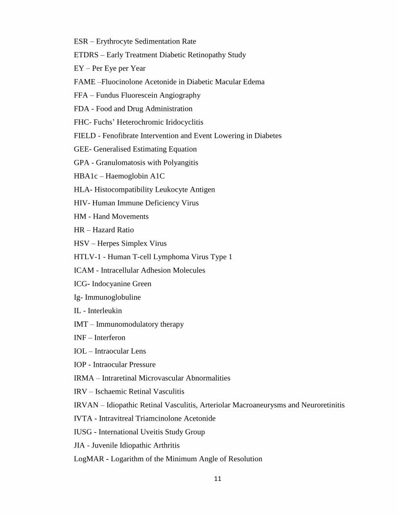
LIST OF ABBREVIATIONS ACE - Angiotensin Converting Enzyme
ACCORD - Action to Control Cardiovascular Risk in Diabetes
ANA- Antinuclear Antibody
ANCA – Antinuclear Cytoplasmic Antibody
APS - Anti-Phospholipid Syndrome
AS- Ankylosing Spondylitis
AZA - Azathioprine
BCG - Bacillus Calmette-Guerin
BCVA - Best Corrected Visual Acuity
BD- Behcet’s Disease
BRAO - Branch Retinal Artery Occlusion
BRB- Blood Retinal Barrier
BRVO - Branch Retinal Vein Occlusion
BSCR- Birdshot Chorioretinopathy
CF - Counting Fingers
CFT – Central Foveal Thickness
CI - Confidence Interval
CMO - Cystoid Macular Oedema
CMV- Cytomegalovirus
CNVM – Choroidal Neovascular Membrane
COX – Cyclooxygenase
CRAO - Central Retinal Artery Occlusion
CSMO- Clinically Significant Macular Oedema
CRVO - Central Retinal Vein Occlusion
DA – Disc Area
DM - Diabetes Mellitus
DMO - Diabetic Macular Oedema
DCCT- Diabetic Control and Complications Trial
DRS- Diabetic Retinopathy Study
ELISA - Enzyme-Linked Immunosorbent Assay
ERM - Epiretinal Membrane
11
ESR – Erythrocyte Sedimentation Rate
ETDRS – Early Treatment Diabetic Retinopathy Study
EY – Per Eye per Year
FAME –Fluocinolone Acetonide in Diabetic Macular Edema
FFA – Fundus Fluorescein Angiography
FDA - Food and Drug Administration
FHC- Fuchs’ Heterochromic Iridocyclitis
FIELD - Fenofibrate Intervention and Event Lowering in Diabetes
GEE- Generalised Estimating Equation
GPA - Granulomatosis with Polyangitis
HBA1c – Haemoglobin A1C
HLA- Histocompatibility Leukocyte Antigen
HIV- Human Immune Deficiency Virus
HM - Hand Movements
HR – Hazard Ratio
HSV – Herpes Simplex Virus
HTLV-1 - Human T-cell Lymphoma Virus Type 1
ICAM - Intracellular Adhesion Molecules
ICG- Indocyanine Green
Ig- Immunoglobuline
IL - Interleukin
IMT – Immunomodulatory therapy
INF – Interferon
IOL – Intraocular Lens
IOP - Intraocular Pressure
IRMA – Intraretinal Microvascular Abnormalities
IRV – Ischaemic Retinal Vasculitis
IRVAN – Idiopathic Retinal Vasculitis, Arteriolar Macroaneurysms and Neuroretinitis
IVTA - Intravitreal Triamcinolone Acetonide
IUSG - International Uveitis Study Group
JIA - Juvenile Idiopathic Arthritis
LogMAR - Logarithm of the Minimum Angle of Resolution
12
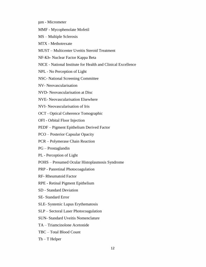
μm - Micrometer
MMF - Mycophenolate Mofetil
MS – Multiple Sclerosis
MTX - Methotrexate
MUST – Multicenter Uveitis Steroid Treatment
NF-Κb- Nuclear Factor Kappa Beta
NICE - National Institute for Health and Clinical Excellence
NPL - No Perception of Light
NSC- National Screening Committee
NV- Neovascularisation
NVD- Neovascularisation at Disc
NVE- Neovascularisation Elsewhere
NVI- Neovascularisation of Iris
OCT - Optical Coherence Tomographic
OFI - Orbital Floor Injection
PEDF – Pigment Epithelium Derived Factor
PCO – Posterior Capsular Opacity
PCR – Polymerase Chain Reaction
PG – Prostaglandin
PL - Perception of Light
POHS – Presumed Ocular Histoplasmosis Syndrome
PRP - Panretinal Photocoagulation
RF- Rheumatoid Factor
RPE - Retinal Pigment Epithelium
SD - Standard Deviation
SE- Standard Error
SLE- Systemic Lupus Erythematosis
SLP – Sectoral Laser Photocoagulation
SUN- Standard Uveitis Nomenclature
TA – Triamcinolone Acetonide
TBC – Total Blood Count
Th – T Helper
13
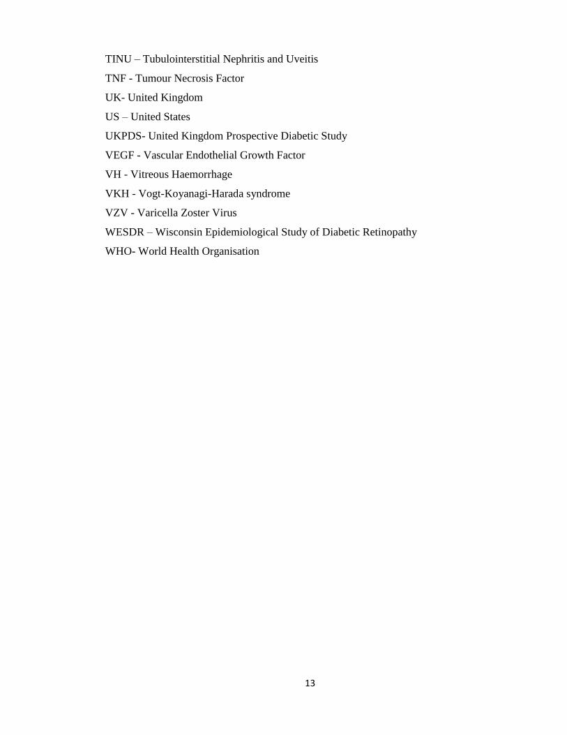
TINU – Tubulointerstitial Nephritis and Uveitis
TNF - Tumour Necrosis Factor
UK- United Kingdom
US – United States
UKPDS- United Kingdom Prospective Diabetic Study
VEGF - Vascular Endothelial Growth Factor
VH - Vitreous Haemorrhage
VKH - Vogt-Koyanagi-Harada syndrome
VZV - Varicella Zoster Virus
WESDR – Wisconsin Epidemiological Study of Diabetic Retinopathy
WHO- World Health Organisation
14
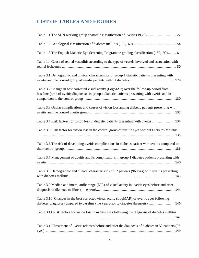
LIST OF TABLES AND FIGURES
Table 1.1 The SUN working group anatomic classification of uveitis (19,20) ................................ 22
Table 1.2 Aetiological classification of diabetes mellitus (159,160) ................................................ 54
Table 1.3 The English Diabetic Eye Screening Programme grading classification (189,190) ......... 61
Table 1.4 Cause of retinal vasculitis according to the type of vessels involved and association with
retinal ischaemia ............................................................................................................................... 80
Table 3.1 Demographic and clinical characteristics of group 1 diabetic patients presenting with
uveitis and the control group of uveitis patients without diabetes. ................................................. 128
Table 3.2 Change in best corrected visual acuity (LogMAR) over the follow-up period from
baseline (time of uveitis diagnosis) in group 1 diabetic patients presenting with uveitis and in
comparison to the control group ..................................................................................................... 130
Table 3.3 Ocular complications and causes of vision loss among diabetic patients presenting with
uveitis and the control uveitis group. .............................................................................................. 132
Table 3.4 Risk factors for vision loss in diabetic patients presenting with uveitis ......................... 134
Table 3.5 Risk factor for vision loss in the control group of uveitic eyes without Diabetes Mellitus
........................................................................................................................................................ 135
Table 3.6 The risk of developing uveitis complications in diabetes patient with uveitis compared to
their control group ........................................................................................................................... 136
Table 3.7 Management of uveitis and its complications in group 1 diabetes patients presenting with
uveitis. ............................................................................................................................................. 140
Table 3.8 Demographic and clinical characteristics of 52 patients (96 eyes) with uveitis presenting
with diabetes mellitus. .................................................................................................................... 143
Table 3.9 Median and interquartile range (IQR) of visual acuity in uveitic eyes before and after
diagnosis of diabetes mellitus (time zero) ....................................................................................... 144
Table 3.10 Changes in the best corrected visual acuity (LogMAR) of uveitic eyes following
diabetes diagnosis compared to baseline (the year prior to diabetes diagnosis) ............................. 146
Table 3.11 Risk factors for vision loss in uveitis eyes following the diagnosis of diabetes mellitus
........................................................................................................................................................ 147
Table 3.12 Treatment of uveitis relapses before and after the diagnosis of diabetes in 52 patients (96
eyes). ............................................................................................................................................... 149
15
Table 4.1 Demographic and clinical comparison of eyes with retinal vasculitis versus non-vasculitis
........................................................................................................................................................ 167
Table 4.2 Mean changes and standard error in best corrected visual acuity in eyes with uveitis,
vasculitis and ischaemic /non ischaemic retinal vasculitis ............................................................. 168
Table 4.3 Risk factors for vision loss in eyes with vasculitis compared to non-vasculitis ............. 171
Table 4.4 Demographic and clinical characteristics of 142 patients with retinal vasculitis ........... 172
Table 4.5 Risk factors for vision loss in ischaemic and non-ischaemic vasculitis .......................... 175
Table 4.6 Risk of neovascularisation following diagnosis of retinal ischaemia and the risk of
neovascularisation relapse following initial retinal laser photocoagulation. .................................. 177
Table 5.1 Demographic and clinical characteristics of pseudophakic uveitic eyes and their phakic
control group ................................................................................................................................... 195
Table 5.2 The mean change in the best corrected visual acuity from baseline (first postoperative
week) among uveitic eyes following cataract surgery .................................................................... 197
Table 5.3 Risk factors for vision loss post cataract surgery in eyes with uveitis ............................ 202
Table 5.4 Changes in central foveal thickness from baseline (within three months preoperatively)
among eyes with uveitis undergoing cataract surgery .................................................................... 203
Table 5.5 Changes in the rate of using topical and systemic steroids after cataract surgery in eyes
with uveitis ...................................................................................................................................... 205
Table 5.6 Risk factors for macular oedema post cataract surgery in eyes with uveitis .................. 207
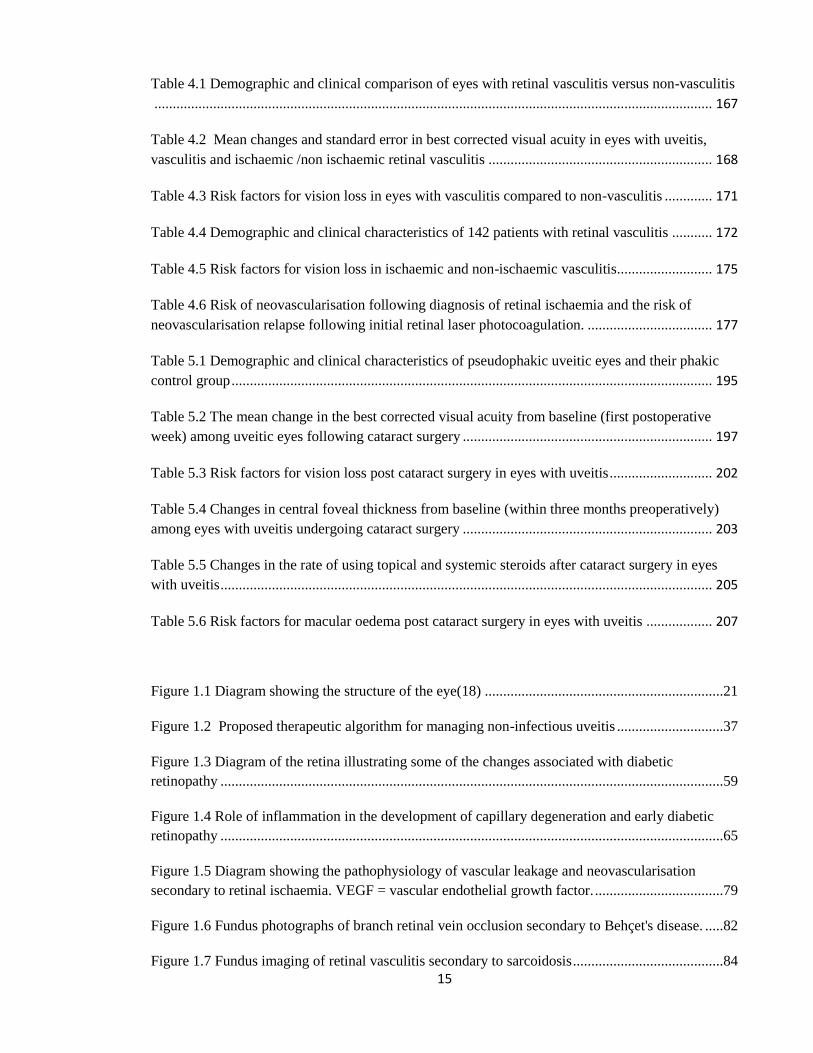
Figure 1.1 Diagram showing the structure of the eye(18) .................................................................21
Figure 1.2 Proposed therapeutic algorithm for managing non-infectious uveitis .............................37
Figure 1.3 Diagram of the retina illustrating some of the changes associated with diabetic
retinopathy .........................................................................................................................................59
Figure 1.4 Role of inflammation in the development of capillary degeneration and early diabetic
retinopathy .........................................................................................................................................65
Figure 1.5 Diagram showing the pathophysiology of vascular leakage and neovascularisation
secondary to retinal ischaemia. VEGF = vascular endothelial growth factor. ...................................79
Figure 1.6 Fundus photographs of branch retinal vein occlusion secondary to Behçet's disease. .....82
Figure 1.7 Fundus imaging of retinal vasculitis secondary to sarcoidosis .........................................84
16
Figure 1.8 Fundus images of tuberculosis associated occlusive retinal vasculitis. ............................85
Figure 1.9 Fundus photographs of SLE associated occlusive retinal vasculitis.................................87
Figure 1.10 Proposed therapeutic algorithm for managing retinal vasculitis ....................................95
Figure 2.1 Screenshot of the uveitis database entered through Microsoft Access datasheet entry
system ..............................................................................................................................................103
Figure 2.2 Normal Topcon 3D Optical Coherence Tomography (OCT) scan (Topcon, Inc) showing
retinal layers from inside out: inner limiting membrane (ILM), nerve fiber layer (NFL), ganglion
cell layer (GCL), inner nuclear layer (INL), inner plexiform layer (IPL), outer nuclear layer (ONL),
outer plexiform layer (OPL), external limiting membrane (ELM), photoreceptor inner segment (IS)
and photoreceptor outer segment (OS) and their IS/OS junction, and the retinal pigment epithelium
layer (RPE).......................................................................................................................................106
Figure 2.3 Normal eye on Heidelberg Spectralis Optical Coherence Tomography (OCT) scan (SD-
OCT; Spectralis, Heidelberg Engineering GmbH) showing retinal layers from inside out: inner
limiting membrane (ILM), nerve fiber layer (NFL), ganglion cell layer (GCL), inner nuclear layer
(INL), inner plexiform layer (IPL), outer nuclear layer (ONL), outer plexiform layer (OPL),
external limiting membrane (ELM), photoreceptor inner segment (IS) and photoreceptor outer
segment (OS) and their IS/OS junction, and the retinal pigment epithelium layer (RPE). ..............106
Figure 2.4 Clinical signs associated with vasculitis as observed on colour fundus images and
fluoresceine angiography. A) Neovascularisation (black arrow). B) Fundus fluoresceine
angiography showing hyperfluoresecence representing neovascularisation (white arrow) at the
border of nonperfusion area representing capillary occlusion (star). C) Fundus fluoresceine
angiography shows active vasculitis with dye leakage over the course of inflamed retinal vessels
(white arrow). D) Active vasculitis with area of retinal haemorrhage (white arrow) and perivascular
sheathing and exudates (black arrow). .............................................................................................108
Figure 2.5 Branch retinal vein occlusion in the right eye of a patient with idiopathic retinal
vasculitis. The colour fundus image (Left) shows perivascular sheething and exudates (black arrow)
together with preretinal haemorrhage (white arrow). The fundus fluorescein angiography (Right)
shows area of hypofluorescence secondary to overlying retinal haemorrhage (white arrow) as well
as areas of hypoperfusion (star). ......................................................................................................108
Figure 2.6 Cystoid macular oedema in a uveitic eye diagnosed using spectralis optical coherence
tomography scan (above) and fundus fluorescein angiography (below). ........................................110
Figure 2.7 Macular ischaemia in a patient with ischaemic retinal vasculitis as seen on fundus
fluorescein angiography of the right eye which shows broken perifoveal capillary ring at the border
of the foveal avascular zone (arrows). .............................................................................................111
Figure 2.8 Epiretinal membrane (arrows) as seen on optical coherence tomography scan (Left) and
on colour fundus image (Right) .......................................................................................................111
Figure 2.9 Fundus images of eyes with nonproliferative diabetic retinopathy (NPDR). .................113
17
Figure 2.10 Right eye of a patient with proliferative diabetic retinopathy. A) Colour fundus image
shows multiple intraretinal haemorrhages (black arrows) together with circinate exudates around the
macular oedema (white arrow). B) Fundus fluorescein angiography of the same eye shows
neovascularisation (black arrow) together with laser scars (white arrow) .......................................113
Figure 2.11 The left eye fundus of a patient with proliferative diabetic retinipathy already treated
with laser photocoagulation (black arrow) and clinical significant macular oedema. Upper image is
a Topcon optical coherence tomography scan showing multiple intraretinal cysts (stars) as well as
subretinal fluid (white arrow). The lower image is fundus fluorescein angiography showing diffuse
area of hyperfluorescence over the macular area (white arrows).....................................................114
Figure 3.1 Flow diagram illustrating the two groups of patients with uveitis and DM together with a
selected control group. .....................................................................................................................126
Figure 3.2 Kaplan-Meier survival curve for the incidence of uveitis following the diagnosis of type
1 and type 2 diabetes mellitus ..........................................................................................................129
Figure 3.3 Kaplan-Meier survival curve for the incidence of vision loss (6/15 or worse) in group 1
(diabetic patients presenting with uveitis) and the control group, showing a higher rate of vision loss
in the diabetic cases when compared to the control group (Log Rank test <0.001). .......................133
Figure 3.4 The mean and standard deviation of systolic blood pressure in diabetic patients at time of
uveitis diagnosis (time zero) and onward up to seven years. N= number of patients ......................138
Figure 3.5 The mean and standard deviation of diastolic blood pressure in diabetic patients at time
of uveitis diagnosis (time zero) and onward up to seven years. N= number of patients ..................138
Figure 3.6 Average number of uveitis relapse in group 1 diabetic patients presented with uveitis. N=
Number of eyes ................................................................................................................................139
Figure 3.7 Kaplan-Meier survival curve for the incidence of cataract surgery in group 1 (diabetic
patients presented with uveitis) ........................................................................................................141
Figure 3.8 Average change in best corrected visual acuity (LogMAR) within five years post
diagnosis of diabetes (Time zero) compared to the year before diagnosis (baseline) (* p<0.01) ....145
Figure 4.1 Fundus Fluoresceine angiography of the right eye with vasculitis showing perivascular
fluorescein leakage (white arrow) suggestive of active inflammation associated with parafoveal
fluorescein staining secondary to macular oedema (black arrow). ..................................................162
Figure 4.2 Montage of combined fundus fluorescein angiography images of the right eye using i2k
retina software program. The image shows inferotemporal retinal hypoperfusion and a single
hyperfluorescent area representing new vessels formation (black arrow). ......................................165
Figure 4.3 Images of merged fundus fluorescein angiography images analysed using ImageJ
software to measure disc area (area 1) in pixel (short arrow) as well as area of retinal ischaemia
(area 2) in pixels (long arrow). Then the area 2 is divided over area 1 to obtain the measured area of
retinal ischaemia in disc areas. .........................................................................................................165
18
Figure 4.4 Flow diagram showing the process of selecting patients with ischaemic retinal vasculitis
.........................................................................................................................................................166
Figure 4.5 Kaplan-Meier Survival analysis graph showing the incidence of vision loss over the
follow up time following diagnosis of uveitis among both groups of retinal vasculitis and non-
vasculitis ..........................................................................................................................................169
Figure 4.6 Kaplan-Meier survival analysis curve showing the incidence of vision loss over the
follow-up time since the diagnosis of ischaemia in eyes with ischaemic retinal vasculitis (IRV) and
since the diagnosis of uveitis in eyes with non-ischaemic retinal vasculitis (non-IRV). .................174
Figure 5.1 Flow diagram showing the process of selecting uveitis patients underwent cataract
surgery and their control group from the uveitis database ...............................................................193
Figure 5.2 Mean changes (and standard error) in the best corrected visual acuity following cataract
surgery from baseline (first postoperative week) in uveitic eyes .....................................................196
Figure 5.3 Kaplan-Meier survival analysis for the incidence of vision loss post cataract surgery in
eyes with anterior uveitis (AU), intermediate uveitis (IU) and panuveitis (PANU). .......................198
Figure 5.4 Kaplan-Meier survival analysis for the incidence of vision loss post cataract surgery
according to the presence of macular oedema. ................................................................................200
Figure 5.5 Kaplan-Meier Survival analysis for the incidence of vision loss post cataract surgery
according to the presence of high intraocular pressure more than 30mmHg. ..................................200
Figure 5.6 Change in central foveal thickness (CFT) over follow-up time compare to preoperative
baseline measurement. The pseudophakic eyes had a significant increase in CFT over the first
month, 3rd month, and first two years postoperatively, unlike the contralateral phakic eye (control)
which did not experience significant change in CFT over follow-up time from baseline. ..............203
Figure 5.7 The mean and standard error bars of uveitis relapse over 10 years before and after
cataract surgery. ...............................................................................................................................204
Figure 5.8 Kaplan-Meier Survival analysis for the incidence of macular oedema following cataract
surgery in 309 eyes with uveitis .......................................................................................................208
Figure 5.9 Kaplan-Meier Survival analysis for the incidence of macular oedema post cataract
surgery in eyes with anterior uveitis (AU), intermediate uveitis (IU) and panuveitis (PANU). ......208
19
1 CHAPTER ONE: INTRODUCTION
1.1 General introduction
The prevalence of uveitis is estimated at 38 cases per 100,000 people and can vary based on
the age group with the prevalence of adult uveitis 93 per 100,000 while the prevalence of
uveitis in children is about 30 cases per 100,000 (1,2). The annual incidence of uveitis
ranges from 4.3 to 6.9 per 100,000 in children, compared with 26 to 102 per 100,000 in
adults (3,4). Uveitis is more prevalent in younger people with a mean age of less than 40
years at the onset of their first symptoms (5).
Uveitis is a potentially blinding condition (6), account for 5–20% of legal blindness in both
the United States and Europe (7,8), and considered the fourth most common cause of
blindness among the working age group in the developed world (9). The most common
cause of visual loss in uveitis is cystoid macular oedema (CMO) and cataract, with one
study documenting visual loss due to CMO or cataract in 64.5% of uveitis patients (10).
Management of macular oedema and active uveitis may require initially a high dose of
systemic glucocorticoids and for prolong duration. Glucocorticoids may induce diabetes
mellitus (DM) as well as worsen glycaemic control in patients with pre-existing diabetes
(11–13). There is a considerably high prevalence of DM world wide, including UK with
2.6 million people diagnosed with DM in 2009, and an estimated doubling of diabetes
prevalence by 2025 (14). Meanwhile, little is known regarding the synergistic effect of DM
and uveitis on the visual outcome and the control of uveitis relapses. The presence of
uncontrolled hyperglycaemia may require reducing the dose of systemic corticosteroids or
the use of other alternative treatments such as local corticosteroid injections; however, the
effect of these modifications on the control of uveitis is not clear. Approach of uveitis
management and its outcome post DM needs to be addressed.
20
Performing cataract surgery for this large cohort of uveitis patients carries a lot of risks,
both during surgery as a result of limited access secondary to posterior synechiae, and after
surgery due to high risk of postoperative inflammatory response. Most studies on cataract
removal among uveitic eyes showed an initial good visual outcome with reduced risk of
intraocular inflammation and postoperative macular oedema following the wide use of
preoperative corticosteroid prophylaxis. However, little is known regarding the long term
effect of cataract removal on the frequency and severity of uveitis compared to the time
prior to the surgery and whether ongoing chronic or recurrent inflammation may sabotage
the initially good visual result obtained post cataract surgery. While both CMO and cataract
are potentially reversible causes of visual loss, ischaemic retinal vasculitis can lead to
permanent loss of vision, either due to macular ischaemia(15,16), or through inducing
retinal neovascularisation (NV) with subsequent complications such as vitreous
haemorrhage and traction resulting in retinal detachment (17).
1.2 Aims and objectives
Examine the influence of diabetes mellitus on the management and visual outcome
in patients with uveitis.
Address the effect of cataract surgery on the visual outcome and course of uveitis.
Study the pattern, management and visual outcome of patients with ischaemic
retinal vasculitis.
21
1.3 Background and literature review
1.3.1 Uveitis
1.3.1.1 Definition and classification
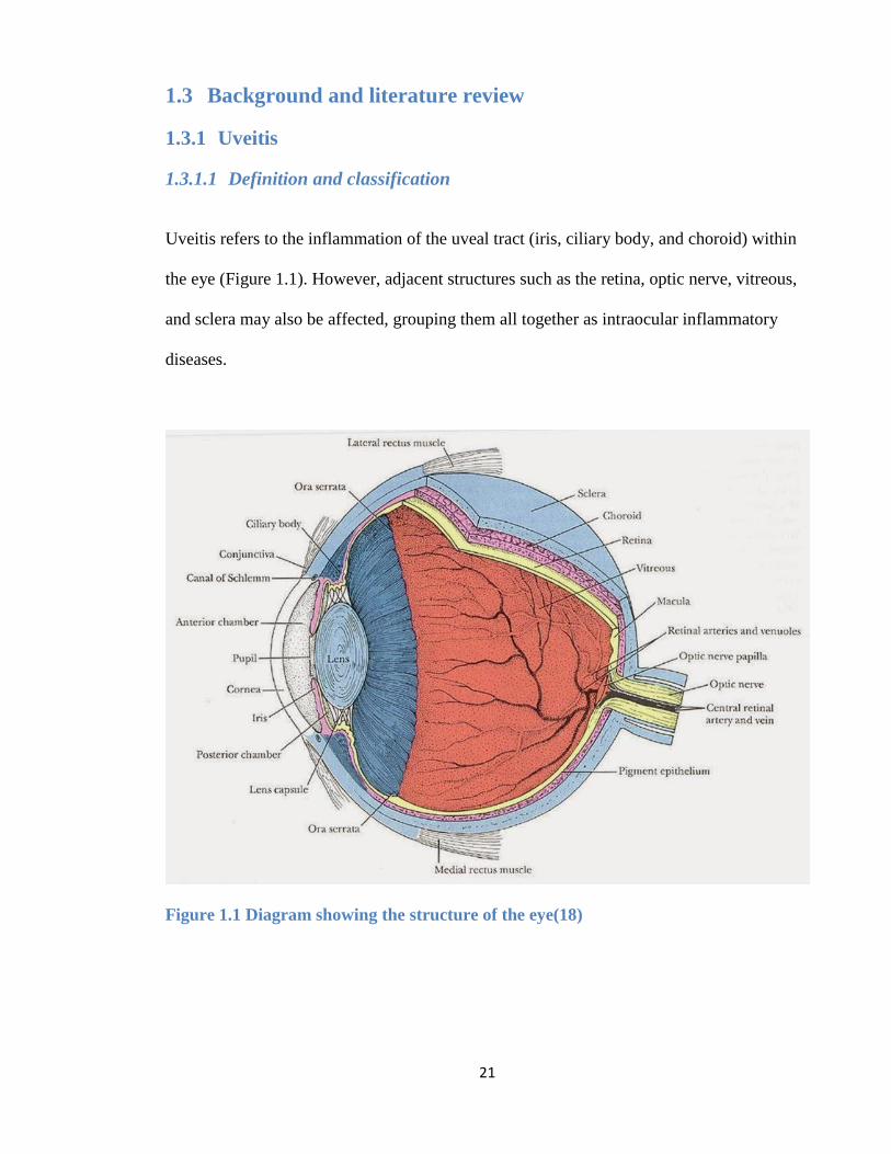
Uveitis refers to the inflammation of the uveal tract (iris, ciliary body, and choroid) within
the eye (Figure 1.1). However, adjacent structures such as the retina, optic nerve, vitreous,
and sclera may also be affected, grouping them all together as intraocular inflammatory
diseases.
Figure 1.1 Diagram showing the structure of the eye(18)
22
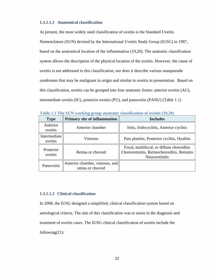
1.3.1.1.1 Anatomical classification
At present, the most widely used classification of uveitis is the Standard Uveitis
Nomenclature (SUN) devised by the International Uveitis Study Group (IUSG) in 1987,
based on the anatomical location of the inflammation (19,20). The anatomic classification
system allows the description of the physical location of the uveitis. However, the cause of
uveitis is not addressed in this classification, nor does it describe various masquerade
syndromes that may be malignant in origin and similar to uveitis in presentation. Based on
this classification, uveitis can be grouped into four anatomic forms: anterior uveitis (AU),
intermediate uveitis (IU), posterior uveitis (PU), and panuveitis (PANU) (Table 1.1)
Table 1.1 The SUN working group anatomic classification of uveitis (19,20)
Type Primary site of inflammation Includes
Anterior
uveitis Anterior chamber Iritis, Iridocyclitis, Anterior cyclitis
Intermediate
uveitis Vitreous Pars planitis, Posterior cyclitis, Hyalitis
Posterior
uveitis Retina or choroid
Focal, multifocal, or diffuse choroiditis
Chorioretinitis, Retinochoroiditis, Retinitis
Neuroretinitis
Panuveitis Anterior chamber, vitreous, and
retina or choroid
1.3.1.1.2 Clinical classification
In 2008, the IUSG designed a simplified, clinical classification system based on
aetiological criteria. The aim of this classification was to assist in the diagnosis and
treatment of uveitis cases. The IUSG clinical classification of uveitis include the
following(21):
23
Infectious (bacterial, viral, fungal, parasitic). Examples are herpes simplex virus (HSV),
varicella zoster virus (VZV), syphilis, cytomegalovirus (CMV), human immune deficiency
virus (HIV), toxoplasma gondii and onchocerciasis.
Non-infectious Known systemic association- Examples are sarcoidosis, Behcet’s disease
(BD), juvenile idiopathic arthritis (JIA), multiple sclerosis (MS), systemic lupus
erythematosis (SLE), granulomatosis with polyangitis (GPA), Vogt-Koyanagi-Harada
(VKH) syndrome, histocompatibility leukocyte antigen (HLA)-B27 related conditions
No known systemic association (inflammation confined to the eye) – Examples are
Birdshot chorioretinopathy (BSCR), sympathetic ophthalmitis, Fuchs’ Heterochromic
iridocyclitis (FHC), idiopathic IU, and Posner–Schlossman syndrome.
Masquerade: Neoplastic and non neoplastic.
1.3.1.2 Epidemiology
The prevalence of uveitis is estimated at 38 cases per 100,000 people (1,2). This can vary
based on the age group with the prevalence of adult uveitis 93 per 100,000 while the
prevalence of uveitis in children is about 30 cases per 100,000.The annual incidence of
uveitis ranges from 4.3 to 6.9 per 100,000 in children, compared with 26 to 102 per
100,000 in adults (3,4). It is more prevalent in younger people with a mean age of less than
40 years at the onset of their first symptoms (5,22).
According to the World Health Organization (WHO), 285 million people were estimated to
be visually impaired; of these, 39 million suffered from blindness, with uveitis as an
underlying cause in approximately 10% of the cases (23). Uveitis also accounts for 10% of
blindness cases among the working age group in the United States and Europe (24,25).
The pattern of uveitis can vary according to the geographic location, which can be related
to many factors such as environmental and genetic predisposing factors.
24
HLA-B27 AU is the most common identifiable cause of AU. It can either be limited to
ocular involvement or, in 49- 84 % of cases, is associated with seronegative
spondyloarthropathies, including ankylosing spondylitis (AS), reactive arthritis, psoriatic
arthritis and arthritis associated with inflammatory bowel disease) (26). HLA-B27
associated AU is significantly more common in western countries (6-29% of AU) compare
to Japan (6-13% of AU), Korea (1% AU) and India (2% of AU cases) (27). BD is a
dominant cause of uveitis in Japan and in the Mediterranean region (28). Onchocerciasis is
a leading cause of uveitis in Africa, such as Sierra Leone (29,30). Presumed ocular
histoplasmosis syndrome (POHS) is mainly found within certain areas in North America,
mainly Ohio and Mississippi River valleys (31). Some causes of uveitis are more prevalent
among specific ethnic groups such as BSCR, which is strongly correlated with the HLA-
A29 class I type, mainly predominant among Caucasian ethnicity. BSCR accounts for
about 8% of PU cases in U.S. (32) while very rare or absent in epidemiological reports of
uveitis in China (33). VKH has a great predilection for dark-skinned populations, such as
Asian, Middle East, Hispanic and Native Americans. With a prevalence of 7%, VKH was
the second most common cause of uveitis after sarcoidosis in a multicentre study in Japan
(28). In paediatric age group, JIA is the most common systemic association of paediatric
uveitis (34), with the prevalence of uveitis among JIA patient varies from 11.6% (35) to
30% (36).
Idiopathic AU, BD, and VKH syndrome are among the most common entities of uveitis in
China with very rare incidences of ocular toxoplasmosis, POHS, and BSCR (33). This is
unlike other countries like Tunisia, where ocular toxoplasmosis is among the most common
causes of uveitis, together with BD, HSV infection and VKH disease (37). The Manchester
Uveitis Clinic study in UK on 3000 new cases of uveitis between 1991 and 2013 found AU
25
to be the most common type of uveitis (46% AU, 11% IU, 21.8% PU and 21% PANU) and
FHC to be the most common diagnosis in their cohort (11.5%) followed by sarcoidosis-
related uveitis (9.7%), idiopathic IU (7.9%), idiopathic acute AU (7.0%), and
toxoplasmosis (6.9%) (38).
1.3.1.3 Immunopathology of uveitis
The initial trigger for the immune reaction leading to uveitis is not always known. In some
cases there is an exogenous stimulus, such as viral or bacterial infection, that may directly
or indirectly trigger inflammation. Other cases can be triggered by an endogenous molecule
within the eye that became exposed under certain circumstances to the immune system and
thus induces inflammation. Furthermore, there are possible environmental and genetic
factors that may lead to variations in the inter-individual and population liability and
manifestations of the ocular inflammation. The role of hereditary factors has been
supported through the association of some forms of uveitis with the presence of identified
HLAs. Examples are the association between idiopathic IU and HLA-DR2 and HLA-
DR15(39), ocular BD with HLA-B51/B5(40), VKH with HLA-DR4/DRB104 (41), BSCR
with HLA-A2901 and HLA-A2902 (42).
The CD4+ T-lymphocyte is pre-eminent in the development of uveitis as observed in
experimental autoimmune uveitis where eye-specific antigens, such as retinal S-antigen or
intraretinal binding protein, are introduced at a site remote from the eye to induce a tissue-
specific immune response (43). Stimulated T-lymphocyte produces cytokines; T-helper cell
1 (Th1) response lead to the release of interferon-gamma (IFN-γ), interleukin-2 (IL-2), IL6,
IL8, IL12, IL18 and tumour necrosis factor alpha (TNFα) which activates macrophages and
cell mediated immunity, while T helper cell 2 (Th2) released IL-4, -5, -10, and -13 that
26
leads to stimulation of B lymphocytes, maturation of plasma cell, immunoglobulin–E (IgE)
formation and eosinophil activation (44). Most of these cytokines have been involved in
the immunopathology of variable causes of non-infectious IU and PU (45).
In BD, both Th1 and Th2 cells are involved (46) with CD8+ T cells observed to be the
predominant intraocular infiltrating cells in active ocular BD(47). High levels of IL8, IL12,
TNFα had been correlated with ocular BD activity(48) including new onset of PU(49). In
cases of SO, the CD4+ helper T-lymphocytes that dominates the early stage of the disease
is replaced with CD8+ T cytotoxic T-lymphocyte which dominates the later course of the
disease(50). B cells are identified in 5-15% of choroidal infiltrates (51). Ocular VKH
disease show infiltration of T-lymphocytes with IL-2 receptors and an increased CD4:CD8
ratio, as compared to peripheral blood. In its late stage, VKH is characterised by choroidal
infiltration with CD8+ T-lymphocyte, B lymphocytes, and plasma cells (52). More recent
study suggests that decreased IL-27 expression in peripheral blood mononuclear cells may
result in a higher Th17 differentiation in active VKH patients (53). Th17 cells, expanded
by IL-2 and inhibited by IFN-γ, has been found in high levels at the peripheral blood in
patients with active uveitis and scleritis (54). The role of Th17 in BSCR has also been
enforced through the detection of significantly higher intraocular levels of IL-17 together
with IL-1β and TNFα compared to their concurrent serum levels (55). This Th-1 cell
mediated inflammation in VKH is possibly triggered through CMV infection which
stimulates an inflammatory response that cross-reacts with molecularly similar melanocyte
peptides, including tyrosinase, in the uvea, skin, inner ear, and central nervous system (56).
It is still unclear why certain individuals are more vulnerable to experiencing uveitis. It is
known that many pathogens and their molecules can have similarities with the host
27
structure and the immune reaction against these molecules can cross-react with host
structures, causing tissue damage and disease. Infection, tissue damage, superantigen
stimulation, or inappropriate activation of CD4+ T lymphocytes can lead to loss of
tolerance to self, allowing autoreactive T and B lymphocytes to become active (57).
1.3.1.4 Clinical features
Uveitis can have a variable course; either an acute uveitis with sudden onset and limited
duration; recurrent when repeated episodes followed by periods of inactivity without
treatment for at least three months; or chronic uveitis when it persists and relapses in less
than three months after discontinuing treatment. Patients with insidious-onset uveitis do not
have symptoms until the development of complications such as poor vision, this is
especially true among children with JIA associated uveitis who can exhibit no symptoms
despite the presence of severe intraocular inflammation (34) wheareas patients with HLA-
B27 associated uveitis are symptomatic even in the presence of mild inflammation.
Symptoms of uveitis include pain and photophobia secondary to ciliary and iris sphincter
muscle spasm, redness, and tearing, as well as floaters and blurred vision secondary to the
inflammatory cells opacities and fibrin deposition in the anterior vitreous. Blurred vision
can also be secondary to other manifestations of intraocular inflammation such as corneal
oedema, corneal band keratopathy, cataract, and CMO.
Keratic precipitates (KPs) are inflammatory cellular deposits on the corneal endothelium,
especially at the lower 1/3 of the cornea except for FHC, which has a diffuse stellate KPs,
and herpetic uveitis. The presence of large KPs with greasy appearance, or “mutton fat”
KPs differentiate the uveitis as granulomatous compared to nongranulomatous uveitis with
no or fine KPs (58). The aqueous fluid is infiltrated with leukocyte cells that are graded
28
based on the inflammatory cell count within a defined set from grade “0” to grade “4”.
Meanwhile, the presence of proteinaceous materials in anterior chamber results in “flare”
with variable grades ranging from “0+” to “4+” according to SUN classification system,
reflecting the severity of the AU(19). Other signs of inflammation include band keratopathy
which is secondary to calcium salt deposition on the coreal surface commonly observed
with chronic uveitis such as JIA associated uveitis. Hypopyon, representing excessive
leukocyte infiltration within the aqueous mainly seen in BD, syphilis and HLA-B27
associated uveitis (59). Signs observed within the iris include iris nodules, associated with
granulmoatous uveitis such as sarcoidosis, and iris atrophy, which can be diffuse as in FHC
or segmental as in herpetic uveitis (58).
IU is characterised by inflammation in the pars plana area of the ciliary body together with
the vitreous base and peripheral retina. Symptoms include blurred vision and floaters while
the main findings consist of variable degrees of vitreous inflammatory cells and haze with
aggregation of lymphocyte cells in the inferior vitreous forming “snowballs” or a more
dense aggregations known as a “snowbank”. Additional findings include retinal vasculitis,
and CMO(60). Complications of IU include secondary glaucoma, cataract, posterior
vitreous detachment, vitreous haemorrhage, and tractional retinal detachment. Around 67-
72% of patients with IU have positive HLA-DR, especially HLA-DR15, which is also
associated with MS (45). MS can be observed in 17 % of patients with IU, while 27 % of
patients with MS patient can present with IU (61).
The inflammation in cases with PU is mainly confined to the the retina and choroid in the
form of retinitis, focal or multifocal choroiditis, retinichoroiditis, chorioretinitis, with or
without an associated retinal vasculitis and neuroretinitis. The choroidal lesions represent
29
focal defects in Bruch membrane may facilitate the development of choroidal neovascular
membrane (CNVM), a major risk factor for visual loss in these cases (62).
1.3.1.5 Laboratory investigations in uveitis
In the presence of a wide range of laboratory tests, both general and specific for a defined
set of pathology, it is important to plan and order for the appropriate tests directed by the
patient’s general history and the symptoms associated with his presentation with uveitis
together with the clinical examination findings.
1.3.1.5.1 Routine blood investigations
These investigations are usually recommended for uveitis patients regardless of the
suspected underlying disease that might be associated with the uveitis. Total blood count
(TBC) and erythrocyte sedimentation rate (ESR) may not be specific enough in defining the
underlying condition with uveitis but it can provide some clues that can direct us toward
additional, more specific tests. Lymphocytosis can be observed in the presence of bacterial
infection such as TB, while lymphopenia can suggest an underlying viral infection or
malignancy. The presence of eosinophilia can be associated with parasitic infections and
has also been observed in patients with Sarcoidosis. Peripheral blood smear is needed to
exclude the presence or leukemia or other forms of myeloblastic malignancies. Abnormal
renal function test can suggests tubulointerstitial nephritis and uveitis (TINU) syndrome.
Renal and Liver function tests, together with TBC are important not only for diagnostic
purposes but also as a baseline investigation and for monitoring patients treated with
systemic corticosteroids and mmunomodulatory therapy (IMT). Elevated C-reactive protein
may not be considered a specific test but it can indicate an active inflammatory process that
may guide toward performing more disease specific tests accordingly.
30
1.3.1.5.2 Disease specific laboratory investigations
Serum angiotensin converting enzyme (ACE) and serum lysozyme can be secreted from the
macrophages within the sarcoid granuloma, making an elevated ACE above its normal
levels highly suggestive of sarcoidosis but not pathognomonic considering that it can be
elevated secondary to variable other conditions. In addition, the interpretation of high ACE
in paediatric age group can be difficult as they tend to have high ACE level in the absence
of underlying sarcoidosis. A recent study of 83 patients with biopsy proven sarcoidosis
associated uveitis demonstrated an elevated ACE (>62 IU/L) in 61.7% of cases while
elevated lysozyme levels (>16.7 mg/L) were observed in 83.9% of cases (63).
The first International Workshop on Ocular Sarcoidosis in Tokyo (58) identified seven
clinical signs suggestive of ocular sarcoidosis; 1) bilateral ocular involvement 2) mutton-fat
KPs and/or iris nodules, 3) trabecular meshwork nodule and/or tent-shaped peripheral
anterior synechiae, 4) vitreous opacities (snowballs), 5) multiple chorioretinal lesions, 6)
nodular or segmental periphlebitis (± candle wax drippings) and/or macroaneurysm, 7)
optic disc nodule/granuloma and/or solitary choroidal nodule. The diagnosis of sarcoidosis
is supported by a group of laboratory findings such as (1) negative tuberculin skin test in a
Bacillus Calmette-Guerin (BCG) vaccinated patient or in a patient having had a positive
tuberculin skin test previously, (2) elevated serum ACE levels and/or elevated serum
lysozyme, (3) bilateral hilar lymphadenopathy on chest X-ray (4) abnormal liver enzyme
tests, and (5) chest computed tomography scan in patients with a negative chest x-ray
result. Among the paediatric age group with granulomatous uveitis, one should exclude
Blau syndrome (Familial juvenile systemic granulomatosis), an autosomal dominant
disorder characterise by skin rash, polyarthritis and granulomatous AU resembling
31
sarcoidosis which can only be differentiated through positive family history and genetic
mutation affecting the CARD15/NOD2 gene on chromosome 16q12 (64).
Interferon gamma release assays had been found to be less sensitive but more specific than
tuberculin skin test in the identification of TB associated uveitis as it is less likely to cross
react with previous BCG vaccination.(65) In syphilis associated uveitis, the veneral disease
research laboratory tests utilised based on the reaction of non specific antibodies produced
by Treponoma pallidum against cardiolipin. Other tests like fluorescent treponomal
antibody absorption test and microhemagglutination Treponoma pallidum test are also used
to detect antibodies against Treponoma pallidum which confirm the diagnosis of syphilis
and also monitor disease response to treatment based on change in antibody titer.
Rheumatoid factor (RF) and antinuclear antibody (ANA) are also performed in patients with
possible JIA associated uveitis and other underlying connective tissue diseases associated
with uveitis. While most of the HLA tests are not required in the investigation process of
uveitis due to lack of specificity, HLA-B27 as well as HLA-A29 are of value in
determining the presence of underlying seronegative arthritis associated uveitis as well as
BSCR, respectively. In patients presenting with vasculitis, the presence of high titers of
antinuclear cytoplasmic antibodies (ANCA) and anticardiolipin antibodies can raise the
suspicion of underlying collagen vascular diseases such as SLE.
Although the diagnosis of most cases of toxoplasmosis chorioretinitis is established through
clinical assessment, laboratory tests may still be needed for cases with atypical
presentation. An acute Toxoplasma gondii infection is associated with positive IgM and
IgG antibodies. IgM usually appears in the first week post infection, reaching its peak at
one month before it disappears by nine months (66).
32
Aqueous sample (0.1 ml) can be obtained from anterior chamber papracentesis to help in
the diagnosis of the causative microorganisms in cases of possible infectious uveitis and
endophthalmitis. It has the advantage of being quick, and can be carried out in outpatient
setting and is of use when there are fears that the vitreous biopsy cannot be performed for
any reason. However, this procedure has higher chance of yielding false-negative results
compare to vitreous biopsy. Samples from aqueous or vitreous can be sent for
microbiologic assessment of bacterial, fungal or acid fast bacilli under microscopic
examination, or for performing culture and sensitivity tests. Polymerase chain reaction
(PCR) and enzyme-linked immunosorbent assay (ELISA) tests for suspected
microorganisms can also be performed to detect viral infections such as HSV, CMV and
VZV infections. The samples can also be sent for cytological assessment in cases with
suspected ocular lymphoma.
1.3.1.6 Ancillary tests in uveitis
1.3.1.6.1 Ultrasound biomicroscopy
Ultrasound biomicroscopy used for ocular examination differs from the conventional
ultrasound in that we use a high frequency waves (35 – 100MHz), allowing for greater
microscopic resolutions of the anterior chamber and ciliary body together with vitreous
structures, peripheral retinal and sclera (67). Ultrasound is also of importance in uveitis
complicated with hypotony to assess ocular structure and exclude the presence of choroidal
effusion.
In eyes with acute AU, ultrasound biomicroscopy in one study had shown a large number
of cells in the anterior and posterior chamber, and oedema and exudates at the iris and
ciliary body, especially within two weeks of acute AU onset, which almost resolve six
weeks from onset of acute AU after initiating steroid therapy (68). The same investigation
33
is of value in eyes with IU as it can detect the vitreous cells or condensations associated
with vitritis in addition to other underlying structural abnormalities that occasionally
missed on clinical examination (69). It can also exclude the presence of vitreoretinal
adhesions and tractional retinal detachment that can occur in association with uveitis, more
commonly in ischaemic retinal vasculitis. Tltrasound biomicroscopy is also of importance
in assessing the posterior sclera thickness in patients with scleritis or VKH and for
excluding significant subretinal fluid or serous retinal detachment. In eyes with disc
swelling suspected to be secondary to optic neuritis, ultrasound is performed to exclude
optic disc drusen or other abnormalities associated with swollen disc. In the presence of
significant media opacity such as cataract that limits the fundus view, ocular ultrasound
would be useful as a diagnostic tool, especially prior to cataract surgery to exclude any
underlying complications that may affect the prognosis such as retinal detachment.
1.3.1.6.2 Fundus photography
Fundus colour images help in viewing and baseline documentation of the abnormalities
observed within the fundus on clinical examination, and is useful in monitoring the
progression or resolution of the uveitis manifestations within the retina and choroid such as
epiretinal membrane (ERM), retinitis, choroiditis and occlusive retinal vasculitis (70).
Fundus fluorescein angiography (FFA) has been widely used as an essential investigation
tool for the diagnosis and monitoring of uveitis cases through identifying any perfusion,
permeability and proliferation abnormality. FFA can show evidence of active inflammation
within the eyes such as the presence of vascular leakage or occlusion or leaking NV
associated with vasculitis. Active retinochoroiditis can be associated with focal or diffuse
hyperfluoresence. The status of the macula can also be assessed with FFA, which can show
parafoveal hyperfluorescence, including the characteristic “petalloid” pattern secondary to
34
CMO. A localised area of leakage with an increasing intensity suggestive of CNVM, or of
increasing pooling of dye as seen in steroid induced central serous chorioretinopathy. FFA
is also useful in assessing any break in the foveal avascular zone circle or an increase in its
diameter, which suggest the presence of macular ischaemia. Optic disc fluorescein leakage
can either be localised from NV at disc or diffuse “hot disc” suggestive of neuritis or VKH
but can also be observed with any form of severe IU and PANU. The small molecules of
free unbound fluorescein dye normally can’t pass through blood-retinal-barrier (BRB) but
can leak out from even minimally damaged BRB secondary to inflamed retinal vessels or
retina which makes it very useful in the diagnosis of retinitis and retinal vasculitis.
However, the limited ability of activated fluorescein molecule under the retinal pigment
epithelium (RPE) layer to emit light through the melanin, xanthenes, and haemoglobin
pigments makes it of limited use in the diagnosis of choroidal pathologies or in the
presence of severe preretinal or intraretinal haemorrhages, for such cases, indocyanine
green (ICG) angiography can aid in the diagnosis.
ICG angiography is based on the emission of fluorescence waves within the infrared
wavelength that can be detected through the RPE which allows for the evaluation of
choroidal structure. The value of ICG angiography in the management of chorioretinal
disease was assessed through a group of retinal expertise in 2003 who reviewed 376 articles
out of which 92 were eventually included. In addition to highly recommending ICG to
identify polypoidal choroidal vasculopathy and other forms of CNVM, they also
recommended ICG with some enthusiasm to identify chronic central serous retinopathy,
MEWDS, vasculitis, AMPEE, VKH and BSCR. Meanwhile, they didn’t find ICG useful in
adding clinical information for the diagnosis and management of BD or sarcoidosis related
uveitis (71).
35
Ultra-widefield images have been increasingly used in uveitis, such as the Optos fundus
camera (Optos, Dunfermline, Scotland, UK) which can capture up to 200-degrees of the
ocular fundus in a single photograph, thus reveals a wider area of the fundus and more
peripheral view than the standard 30- to 50-degree fundus images. A study compared the
wide field images obtained from Optos FFA to a 9-field montage images in eyes with
uveitis demonstrated superiority of the wide-field FFA in capturing the retinal vascular
pathology, in both the periphery and the posterior pole (72).
Campbell et al suggested that the use of ultrawide-field images can eventually alter the
management plan for uveitis for those made based on clinical examination and standard
imaging even though the wide-field imaging was not superior over standard examination
and imaging when it comes to detecting disease activity (73).
1.3.1.6.3 Optical coherence tomography (OCT)
Optical coherence tomography (OCT) is a non-invasive, noncontact, transpupillary imaging
method that can analyse the retina and choroid in cross-sections with 8 to 10μm resolution
(74). Three pattern of CMO in uveitis has been described; diffuse macular oedema seen in
54.8% eyes and presents as sponge-like thickening of the retina with low-reflectivity;
clearly defined intra-retinal cystic spaces seen in 25%; and finally the serous retinal
detachment seen in 5.9% of cases with fluid accumulation between RPE and neurosensory
retina (75).
While FFA is very useful in detecting leakage associated with active uveitis or CMO, OCT
allows for more quantifiable measurement of macular thickness and associated changes
such as intraretinal or subretinal fluid, ERM, pigmented epithelial detachment and RPE
atrophy. The Multicenter Uveitis Steroid Treatment (MUST) trial group compared the
36
effectiveness of FFA versus OCT in the diagnosis of CMO among 479 eyes with uveitis.
They found that both OCT and FFA offered only moderate agreement when it comes to the
diagnosis of CMO. OCT was superior to FFA when it comes to providing more usable
information regarding the CMO characteristics (90% versus 77%, respectively). However,
OCT had a limitation of failing to detect macular leakage required to make modifications to
the uveitis treatment , for which case FFA is recommended in addition to OCT (76).
1.3.1.7 Management of uveitis
The approach in uveitis management can vary depending upon the anatomical location and
aetiology of uveitis, severity of the inflammation, and the associated complications. In most
cases, the treatment of non-infectious uveitis is initiated with the use of corticosteroid
therapy administered topically, through local injection or systematically, aiming to rapidly
control inflammation. In cases where corticosteroid therapy cannot be tapered to low level
without having a uveitis relapse, alternative or second line agents should be considered to
reduce the risk of prolonged corticosteroid use (77). Figure 1.2 illustrates a proposed
therapeutic algorithm for managing non-infectious uveitis.
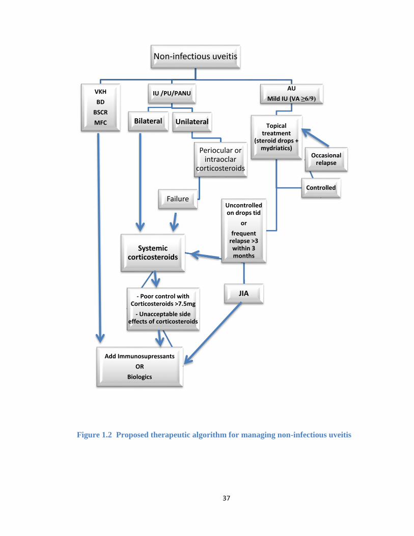
37
Figure 1.2 Proposed therapeutic algorithm for managing non-infectious uveitis
Non-infectious uveitis
VKH
BD
BSCR
MFC
IU /PU/PANU
Bilateral Unilateral
Periocular or intraoclar
corticosteroids
Failure
AU
Mild IU (VA ≥6/9)
Topical treatment
(steroid drops + mydriatics)
Uncontrolled on drops tid
or
frequent relapse >3 within 3 months
JIA
Systemic corticosteroids
- Poor control with Corticosteroids >7.5mg
- Unacceptable side effects of corticosteroids
Add Immunosupressants
OR
Biologics
Controlled
Occasional relapse
38
1.3.1.7.1 Corticosteroids
Corticosteroids are steroid hormones naturally secreted from adrenal gland cortex of
humans and involved in the carbohydrates and protein metabolism as well as controlling
electrolyte levels and body immune reaction against inflammation and stress. In the eye,
steroids acts on inflammation control through inducing phospholipase-A2 inhibitory
proteins which eventually reduce the production of inflammatory mediators such as IL-6
and prostaglandins (78). Furthermore, corticosteroids used in the management of CMO
through reducing vascular and BRB permeability and breakdown through stabilising the
RPE tight junctions (79) and reducing vascular endothelial growth factor (VEGF) level
(80). For the purpose of uveitis management, corticosteroids have been used widely
through variable routes of administration; either topically, as periocular or intraocular
injections, or systemically.
1.3.1.7.1.1 Topical corticosteroids
Topical corticosteroids remain the primary treatment for non-infectious AU as it can
penetrate the cornea to concentrate mainly within the anterior chamber. Commonly used
preparations for managing AU include dexamethasone 0.1% and prednisolone 1% eye
drops. Methylprednisolone 1% eye drops had been shown equal efficacy in treating AU
compared to prednisolone 1% drops (81). Another randomised study found Rimexolone 1%
to be as effective as prednisolone acetate 1% in the management of AU and with no
difference in the risk of increased intraocular pressure (IOP) between the two drops (82).
Topical drops are initiated at frequent intervals depending on the degree of active
inflammation and are then tapered down gradually, aiming to either stop once inflammation
control has been achieved, or reduced to ≤ 3 drops per day as the latter has been shown to
be associated with the minimum risk of side effects, especially cataract formation (83,84).
39
Another adverse effect includes steroid-induced glaucoma, possibly through the
accumulation of extracellular matrices in the trabecular meshwork, resulting in increased
resistance to aqueous outflow in the trabecular meshwork and leading to increased IOP
(85). Other complications include increased risk of secondary ocular infection and corneal
toxicity associated with the preservatives that are present in some steroid drops
formulation.
1.3.1.7.1.2 Systemic corticosteroids
Systemic corticosteroids are preferred route of administration when the uveitis extends
beyond the anterior chamber bilaterally, and in the presence of relative contraindications to
the use of periocular or intraocular corticosteroid injections(86). Prednisone is the most
commonly used oral corticosteroid, with initial dose ranges from 0.5 to 1.5 mg/kg/day then
tapered down until stopped or kept at a maintenance level as low as 7.5mg /day or less.
High-dose intravenous corticosteroids have been used successfully in the management of
acute and severe uveitis episodes (87). However, it doesn’t show superiority in determining
the vision outcome nor on the development of complications when compared to per oral
route of administration (88). This can be significant considering the cost of hospital
admission and the need of medical experts to administer corticosteroids intravenously when
it is not required with the use of steroid tablets. Side effects associated with systemic
corticosteroids include ocular complications such as cataract, increased IOP and central
serous retinopathy, while systemic complications include systemic hypertension,
gastrointestinal disturbance, altered mood, elevated blood sugars, hypokalaemia,
leukocytosis, myopathy, osteoporosis and Cushingoid changes (moon facies, weight gain,
and increased acne). Prolonged use of systemic corticosteroids should be avoided in all
40
patients, especially in children in whom corticosteroid-induced growth retardation is a
significant risk of this therapy (89).
Patients on systemic corticosteroids require regular checking of their blood pressure and
blood glucose levels, at least once every 3 months. Bone mineral density evaluations, blood
cholesterol and lipids should also be monitored on a regular basis (86). The fracture risk
assessment tool have to revise the 10-year probability of fracture by a 15% for individuals
on corticosteroids daily dose of 7.5mg or more (90). Calcium and vitamin D
supplementation is widely recommended for these cases to prevent mineral bone density
loss in patients at high risk of osteoporosis (91).
1.3.1.7.1.3 Periocular corticosteroids
Periocular injections of corticosteroids such as triamcinolone acetonide (TA) and
methylprednisolone can be performed through variable techniques; into the
subconjunctival space, into the subTenon space, into the orbital floor through
transcutaneous or transconjunctival approach, or into the retrobulbar space (92). This
method is especially preferred in the management of CMO when it is unilateral or there is a
relative contraindication against the use of systemic corticosteroid, or it maybe used as an
adjuvant treatment with other systemic therapy. A single injection of periocular TA has
been reported to resolve CMO in 53% of eyes within one month post injection and 22% did
so with more than one injection of TA (93). A major limitation though is the high risk of
cataract formation and increased IOP. In a large study cohort involved 1192 eyes that
received ≥1 periocular injection found about 72% of the eyes achieved complete uveitis
control within 6 months follow up and half of the eyes achieved an improvement in vision
to 6/9 or better. In the same study, the incidence of increased IOP ≥30 mmHg was 15%
41
within one year post injection while the incidence of visually significant cataract formation
was 20% of the cases (94).
1.3.1.7.1.4 Intraocular corticosteroids
Triamcinolone acetonide (TA), a water- insoluble crystalline steroid in a suspension form,
can be injected into the vitreous at a therapeutic dose of typically 2 or 4 mg. The
preservative-free TA approved by the U.S. food and drug administration (FDA) to treat
ocular inflammatory diseases includes Trivaris® and Triesence®. On the other hand,
Kenalog-40® is the most common form used for off-label intravitreal injection. The
intravitreal half-life of TA is approximately 18 days with a therapeutic activity that lasts for
approximately 3 months post injection (95).
Intravitreal injection of TA (IVTA) was first used in human eyes in the late 1990s, since
then it has been commonly used as initial or supplementary therapy for uveitis and its
associated CMO (96). IVTA have been used in the management of CMO among variable
causes of non-infectious uveitis such as BD (97), VKH (98), serpiginous choroiditis (99)
and AS (100). A study compared IVTA injection to subTenon TA and intravitreal
bevacizumab in treating uveitic CMO found all three groups achieved the best
improvement in VA and reduction in central foveal thickness at 4 weeks in all groups then
gradually subsided until 12 weeks. The IVTA injection was associated with a longer period
of effectiveness (30 weeks) compared to intravitreal bevacizumab (16 weeks) and posterior
subTenon TA (12 weeks) (101). In the paediatric age group, a study of 16 eyes with uveitis
receiving IVTA has shown a clinically significant improvement in VA among 56% of
cases that lasted up to 15 months post injection (102).
42
The use of IVTA injection can be limited due to its complications, mainly the high risk of
increasing IOP and cataract formation. An increase in IOP > 10mmHg has been reported to
occur in 43% of eyes following IVTA, especially in patients <40 years of age (96). The risk
of cataract development requiring surgery can vary based on published studies but one
study reported an incidence of 29% of uveitic eyes at an average of two years (12-34
months) post IVTA injection compared to only 5% of eyes that received placebo injection
(103). Other rare adverse events include pseudoendophthalmitis secondary to the
preservatives in off-label TA (Kenalog®) and endophthalmitis. A systematic review of
literature from 1966 to 2004 estimated the prevalence of all forms of endophthalmitis post
IVTA injection to be 1.4% (0.6% were bacterial endophthalmitis) (104). Another two
reports found the incidence of endophthalmitis post IVTA injection ranging from none (97)
to 0.21% (105), respectively. IVTA should be avoided in cases where the possibility of
infectious uveitis hasn’t been excluded, an example is toxoplasmosis chorioretinitis that can
present with fulminant chorioretinitis following IVTA (106).
Dexamethasone intravitreal implant (Ozurdex®, Allergan, Inc., Irvine, CA), known
previously as Posurdex, is a biodegradable device that is inserted into the vitreous using a
23-gauge needle device preloaded with the implant. The implant itself contains a
therapeutic dose of 0.7 mg preservative-free dexamethasone.
Ozurdex has been approved in 2010 by the FDA for the treatment of non-infectious
intermediate and posterior uveitis (107) based on reports of the Dexamethasone drug
delivery system (DDS) phase II study group on 41 eyes with chronic CMO secondary to
uveitis or Irvine-Gass syndrome. Three months after receiving dexamethasone implant,
there was a significant improvement in visual acuity by at least 15 letters in 53.8 % of eyes
on 0.7 mg implant compared to 16.7% of eyes on 0.35mg implant and only 7% in the
43
control group. Improvement in CMO was also observed in 58% of eyes on 0.7 mg implant
compared with 8% in the control group (108). Repeated Ozurdex implantation had also
shown an accumulated effect in a cohort of 38 eyes with uveitis with a relapse rate of 69%
following first intravitreal implant and 48% following second implant(109). Ozurdex
implant is also of benefit in improving vision and managing CMO among the paediatric age
group with non-infectious uveitis (110).
The risk of increased IOP and cataract progression can be seen in eyes with Ozurdex
implant but less frequent compared to those observed secondary to IVTA and Retisert
implant. In eyes with 0.7 mg dexamethasone implant, increased IOP requiring
antiglaucoma drops has been reported in 23% (111) to 38% (108) of cases. Cataract
formation has been observed in 15% of the eyes within six months post 0.7 mg
dexamethasone implant (111). Ozurdex implant should be avoided in aphakic eyes and eyes
with compromised posterior capsule due to the high risk of implant migration into the
anterior chamber resulting in corneal decompensation and requiring implant removal and
occasionally penetrating keratoplasty (112).
Fluocinolone acetonide is one of the most potent and selective glucocorticoids with no
mineralocorticoid effect and has been available for intravitreal implantation in the form of
Retisert® and Iluvien®. Retisert implant consists of a tablet containing 0.59 mg of active
ingredient and needs to be implanted surgically through a sclerotomy incision made at the
level of pars plana. Once implanted, its effectiveness can last for about 2.5 years (30
months).
The MUST trial is an ongoing prospective study set up to compare the visual acuity
outcome in patients with non-infectious intermediate posterior or panuveitis randomised to
44
receive either systemic immunosuppressant or Retisert insertion, in both eyes if necessary
and withdrawal of systemic medication. The two year results of the MUST trial
demonstrated that the visual outcome following Retisert implant was not significantly
different when compared to those on systemic anti-inflammatory medications. The Retisert
implant group had better control of uveitis activity when compared to the group on
systemic therapy with an incidence of active uveitis in 12% versus 29%, respectively. The
group on Retisert implant, however, had a higher risk of cataract surgery (80%), treatment
for elevated IOP (61%) and glaucoma (17%) within 2 years follow up period (113).
A study comparing Ozurdex to Retisert implant found no superiority of one over the other
in terms of uveitis control and vision improvement. However, Retisert implant had higher
rates of cataract progression compared to Ozurdex (100% versus 50%), as well as more
need for glaucoma medications, laser, and surgery (114).
1.3.1.7.2 Antimetabolites
1.3.1.7.2.1 Methotrexate (MTX)
MTX is a folic acid analogue that inhibits dihydrofolate reductase enzyme, leading to
inhibition of DNA replication and the proliferation of rapidly dividing immune cells (115).
MTX has been used in the management of uveitis secondary to JIA, sarcoidosis, and
idiopathic panuveitis (86). It is typically administered in the form of tablets or subcutaneous
injections at a dose between 7.5 and a maximum of 25 mg per week and requires 3 to 6
weeks before reaching its full therapeutic effect. The main serious side effect is reversible
hepatotoxicity thus patient on MTX requires regular monitoring of liver function test (77).
The Systemic Immunosuppressive Therapy for Eye Diseases (SITE) involved 384 patients
with non-infectious uveitis from 4 tertiary ocular inflammation clinics in the United States.
The study reported suppression of ocular inflammation with the use of MTX in 66% of
45
cases within one year; in addition, 58% of the cases had successfully stopped or reduced
their systemic steroids to 10 mg/day or less. MTX was ineffective in controlling
inflammation in 13% of the cases while the side effects resulted in discontinuation of the
medication in 16% of cases (116). These side effects reported in the same study ranged
from gastrointestinal upset (3%), bone marrow suppression (2.6%), elevated liver enzyme
(2.3%), malaise (2.1%), allergy (1.6%), mouth ulcers (1.3%), to less common side effects
such as infection,, hair loss and cirrhosis of liver (each <1%). No increased risk of
neoplasia has been demonstrated with long-term use (116).
1.3.1.7.2.2 Azathioprine (AZA)
AZA is used in inflammation control through its ability to prevent protein synthesis in
lymphoid cells through interfering with purine incorporation into DNA, resulting in
synthesis of nonfunctional DNA sequences. Other mechanisms of action include selective
T-cell inhibition, decreased development of monocyte precursors, and natural killer cell
suppression leading to reduced cytotoxic inflammatory reactions (115,117). AZA is usually
prescribed at an initial dose of 1–3 mg/kg/day orally and then adjusted afterwards
according to the side effects and clinical response, which can take up to 4-12 weeks to
occure. AZA is particularly effective in the management of uveitis secondary to JIA and IU
(115).
The SITE study group reported inflammation control with the use of AZA in 69% of
patients with uveitis within 6 months with uveitis while achieving steroid sparing effect in
32% and 47% of patients by 6 months and 12 months, respectively (118). Side effects of
AZA include gastrointestinal upset (25%), bone marrow suppression, and reversible
hepatotoxicity (<2%) (119).
46
1.3.1.7.2.3 Mycophenolate mofetil (MMF)
MMF is a selective monophosphate dehydrogenase inhibitor. Usual dose for managing
uveitis is 2-3 grams divided over two daily doses. In the SITE study, 236 patients (397
eyes) with ocular inflammation treated with MMF monotherapy. Complete control of
inflammation over at least 28 days was achieved within 6 months in 53% of patients and
within 1 year in 73% of patients. Steroid sparing effect was achieved in 41% and 55% of
patients within 6 months and 1 year, respectively. MMF had to be discontinued in 12% of
cases within a year because of side effects (120). The most common side effects of MMF
include gastrointestinal symptoms (31%) as well as myalgias, fatigue, headache and bone
marrow suppression (119). The latter had been associated with discontinuation of MMF in
1.7% of patients while gastrointestinal symptoms lead to discontinuation of MMF in 2.5%
of patients reported in the SITE study (120).
1.3.1.7.3 Alkylating agents
1.3.1.7.3.1 Cyclophosphamide
Cyclophosphamide is a cytotoxic drug targeting lymphocytes but with a high toxic profile
that limits its use to managing severe ocular inflammation that is resistant to other IMT
medications. The drug is of particular use in controlling inflammation secondary to GPA,
SLE, polyarteritis nodosa, peripheral ulcerative keratitis, and necrotising scleritis (86). The
SITE Cohort Study group on cyclophosphamide reported complete control of inflammation
in 50% and 81% of patients within 6 months and 12 months treatment, respectively. Steroid
sparing effect was achieved in 30% and 61% of patients by 6 months and 12 months,
respectively. The ability to discontinue the medication due to inflammation remission was
achieved in 63% of patients on cyclophosphamide, which is a higher rate achieved when
compared to other IMT. Side effects of cyclophosphamide lead to discontinuation of
47
medication in 33.5% of patients within one year (121). Regular follow up of total blood
count should be performed and the cyclophosphamide should be stopped when white blood
cell counts fall below 2500 cells per µl. The risk of haemorrhagic cystitis and bladder
cancer are major risks associated with cyclophosphamide and thus should only be
considered when the benefits are believed to outweigh the risks when used for short
duration to control active ocular inflammation (115).
1.3.1.7.3.2 Chlorambucil
Chlorambucil has a similar mechanism of action to cyclophosphamide in preventing
inflammation through DNA synthesis in rapidly dividing cells. Initially started at an oral
dose of 0.1 mg/kg/day that can changed based on clinical response and development of side
effect to a maximum dose of 6 to12 mg/day (77). One study looked at the effectiveness of
chlorambucil in 44 patients with refractory uveitis secondary to BD, and observed
inflammation control in 66% of patients(122). Another review found uveitis control in 68%
out of 28 patients with refractory uveitis secondary to BD, JIA, pars planitis, SO, idiopathic
uveitis, Crohn’s disease, and HLA-B27 associated uveitis (123). Side effects include bone
marrow suppression, which can progress to irreversible aplastic anaemia, cutaneous
malignancy, and haematological malignancy (119). Chlorambucil is not as effective as
cyclophosphamide when it comes to controlling uveitis and thus makes it less favourable
choice by ophthalmologists when decision is made to initiate alkylating agents for uveitis
control (115).
48
1.3.1.7.4 Calcineurin inhibitors
1.3.1.7.4.1 Cyclosporine (CSA)
CSA and tacrolimus are termed calcineurin inhibitors due to their ability to bind to the
protein cyclophilin in T lymphocytes leading to calcineurin inhibition. The latter is
responsible for activating the transcription of IL-2 under normal condition as well as
inhibiting the production of TNFα, IFNγ, IL-3, and IL-4 and therefore leads to suppression
of T-cell function (124). CSA dose ranges from 2.5 mg/kg/day to a maximum dose of
7.5 mg/kg/day, given over two equally divided doses. CSA has been effective in the
management non-infectious uveitis (86). One study on the use of CSA for managing ocular
BD found it to be successful in stabilising or improving the vision in 69% of 104 eyes with
BD with no uveitis relapse in over 50% of the eyes over one year follow-up period (125).
The SITE study observed CSA to be successful as steroid-sparing agent (control
inflammation with prednisone ≤10 mg/day) in 22% by six months and 36% within one year
(126). Toxicity secondary to CSA is a major limitation that can lead to discontinuation of
therapy within one year in 10% of the population, especially in patients over 55 years of
age where the risk of stopping the medication due to side effects such as nephrotoxicity and
hypertension is three times more than the younger age group (126).
1.3.1.7.4.2 Tacrolimus
With a mechanism of action similar to that of CSA and with a lower profile of drug
toxicity, tacrolimus has been used for managing uveitis (127). Tacrolimus is usually
administered orally at a dose of 0.05 mg/kg/day. Tacrolimus when used as a second line
medication has shown steroid sparing effect following its use for 14 months in uveitis eyes
while maintaining low incidence of nephrotoxicity and hypertension (128). However, there
is still limited data regarding the effectiveness of tacrolimus in managing variable forms of
49
non-infectious uveitis. In addition, its effectiveness in comparison to other IMT therapies
has yet to be understood.
1.3.1.7.5 Tumour necrosis factor inhibitors
Anti-TNFα such as infliximab, adalimumab, or etanercept have been implemented as off-
labelled drugs in the management of uveitis cases resistant to other local or systemic
immuneosuppressant medications (129). Underlying infections should be excluded and all
patients should be screened for latent TB prior to starting treatment and they may require
Hepatitis B vaccination if they are at risk of infection (130). Anti-TNFα are excluded in
patients with severe heart failure and used with caution in those with mild heart disease.
Blood tests such as TBC and liver function test should be performed at baseline and
afterward at regular intervals (130). Another risk associated with the use of Anti-TNFα is
central nervous system demyelination which has been observed in 0.05 to 0.2% of patients
and should be avoided in cases suspected to have MS (131). Additional side effects include
secondary infection, especially TB and fungal infections, thromboembolism, drug-induced
lupus-type reaction, and possibly malignancy (132).
1.3.1.7.5.1 Infliximab (Remicade®)
Infliximab is a chimeric IgG monoclonal antibody that binds to TNFα and inhibits its
biological function. It has been licensed for the control of systemic manifestations of
variable autoimmune diseases such as JIA, ulcerative colitis, AS and psoriasis. Infliximab
is administered intravenously at a dose of 3–5 mg/kg given at weeks 0, 2, 6 and then every
8 weeks, or as early as every 4-6 weeks, at 5–10 mg/kg and can be given for 2 years once
disease remission is achieved (133). Infliximab has been successfully used to treat difficult
cases of uveitis associated with BD (134), JIA (135), BSCR (136), AS, sarcoidosis, and
Crohn’s disease (137).
50
Adalimumab is a humanised monoclonal IgG antibody against TNFα given subcutaneously
at a usual dose of 40mg every two weeks. It is similar to infliximab on the aspect of
inflammation control and side effects, with less reaction against adalimumab administration
owing to its fully humananised nature. Adalimumab had also been observed to
successfully reduce the rate of AU flare up by 51% in patients with AS (138).
Etanercept acts as TNFα and TNFβ antagonist through acting as a fusion protein of a
human Fc molecule and two p75 TNF receptors. And while there were initial reports
suggesting its effectiveness in controlling uveitis, a small cohort, randomized double
masked, study found no apparent difference between JIA patients treated with etanercept
from placebo in controlling AU (139).
1.3.1.7.6 Anti-CD 20 monoclonal antibody
Rituximab (RTX), a chimeric monoclonal antibody against CD20+ antigen on B-cells, has
been used in the treatment of non-Hodgkin’s lymphoma and rheumatoid arthritis. Studies
did suggest using it as a treatment option in managing severe, sight threatening uveitis
secondary to JIA as well as other forms of non-infectious uveitis that are recalcitrant to
other conventional IMT or TNFα antagonists (140). The treatment dose for managing
uveitis varies but has been used at a dose of 1,000mg given twice at 2 week interval (141).
1.3.1.8 Uveitis complications and causes of vision loss
The WHO defines blindness as the best corrected vision in the better eye of less than 3/60
or a visual field ≤10°, whereas severe visual impairment is defined as the best corrected
visual acuity in the better eye of 3/60 or more, but less than 6/60 or a visual field ≤20°.
Legal blindness can be defined in certain countries as the level of blindness that makes a
person eligible for social support and financial benefits (23).
51
Uveitis is a potentially blinding condition accounting for 5–20% of legal blindness in both
the United States and Europe (7,8,22). The risk of visual loss can vary based on the type
and cause of uveitis and age group. PU has been the most common anatomical type of
uveitis associated with visual loss, followed by anterior uveitis. Among all cases of uveitis,
reports of complications have been as high as 41%, with 19% of cases having resulted in
blindness (142).
Among children with JIA related uveitis, at least 20% develop severe vision-compromising
complications, such as cataracts, band keratopathy, posterior synechiae, hypotony, and
glaucoma (143,144). Studying 75 patients with JIA over a median follow-up of three years
found the rate of vision loss ≤6/12 or worse was 0.1 per eye year and rate of vision loss
≤6/60 was 0.08 per eye year (83). Another study of patients with JIA associated uveitis
found the incidence of any ocular complication to be 0.33 per eye-year (EY) with the rates
of moderate vision loss 0.10/EY and severe vision loss 0.08/EY. The study also found
posterior synechiae, active anterior chamber inflammation, and abnormal intraocular
pressure (IOP) at presentation associated with increased incidence of vision loss (83). Poor
prognostic factors for visual outcome among children with JIA related uveitis include male
gender, anterior chamber flare > or = 1+, positive antinuclear antibody (ANA) and uveitis
onset preceding the development of arthritis symptoms (145,146).
1.3.1.8.1 Potentially reversible causes
Cataract and CMO are the most common cause of visual loss in uveitis. One study found
CMO and cataract, either alone or in combination, to be responsible for visual loss ≤6/18 in
64.5% of the patients with uveitis(10). Rothova et al, investigating causes of visual loss in
582 patients with intraocular inflammation, found CMO to be the most common cause of
decreased vision (26%) followed by cataract (19%) (6). The incidence of cataract in uveitis
52
can vary according to the anatomical and aetiological type of uveitis, severity of
inflammation, use of topical or local steroid therapy, and duration of uveitis with a reported
average of 7.5 years from time of uveitis diagnosis until the eye requires cataract surgery
(147). Cataract formation, especially in the form of posterior subcapsular opacity, is very
common in cases of FHC, with an incidence of 50% (17-75%). (148,149)
CMO is caused by cystic accumulation of intra-retinal fluid in the outer plexiform and inner
nuclear layers of the retina due to breakdown of the BRB (150). The incidence of clinical
CMO varies from 1-2% (151), while angiographic CMO has an incidence range of 9-
19%(152,153). In an attempt to study the impact of CMO on visual outcome using data
from 529 patients (842 eyes), one third of the patients had history of CMO among which
44% resulted in VA ≤ 6/18, making it one of the major causes of visual loss in uveitis
(154). CMO is more commonly observed in cases with IU (25-70%) but can also occur in
AU (20-26%), PU (20%) and PANU (35%) (155).
The incidence of ERM, known also as cellophane maculopathy or macular pucker, can vary
based on the aetiology and duration of uveitis. ERM has been reported in 30% of cases with
pars planitis (156), 17% of patients with ocular BD (157). In a recent study on 1799 eyes
with uveitis, ERM was the third most common cause of moderate vision loss after chronic
CMO and macular scarring, occurring in1.39% of cases with uveitis while ERM caused
severe vision loss in 0.1% of the eyes (147). Vitrectomy with ERM peel in eyes with
uveitis has been available as a management option in cases associated with significant
vision loss but the visual outcome postsurgery is guarded.
53
1.3.1.8.2 Irreversible causes
Irreversible causes of visual loss can occur to a lesser extent compared to cataract and
CMO, but can have dramatic consequences on the quality of life among uveitis patients.
Among 220 patients with uveitis, vision loss ≥ 6/18 was attributed to macular pathology
(scarring, atrophy, hole) in 7.7% of the cases; other causes include glaucoma (5%), optic
neuropathy (5%), and retinal detachment (3.6%) (10). Ocular hypotony may results in
maculopathy, choroidal effusion, optic nerve swelling, irregular astigmatism and eventually
phthisis bulbi (158).
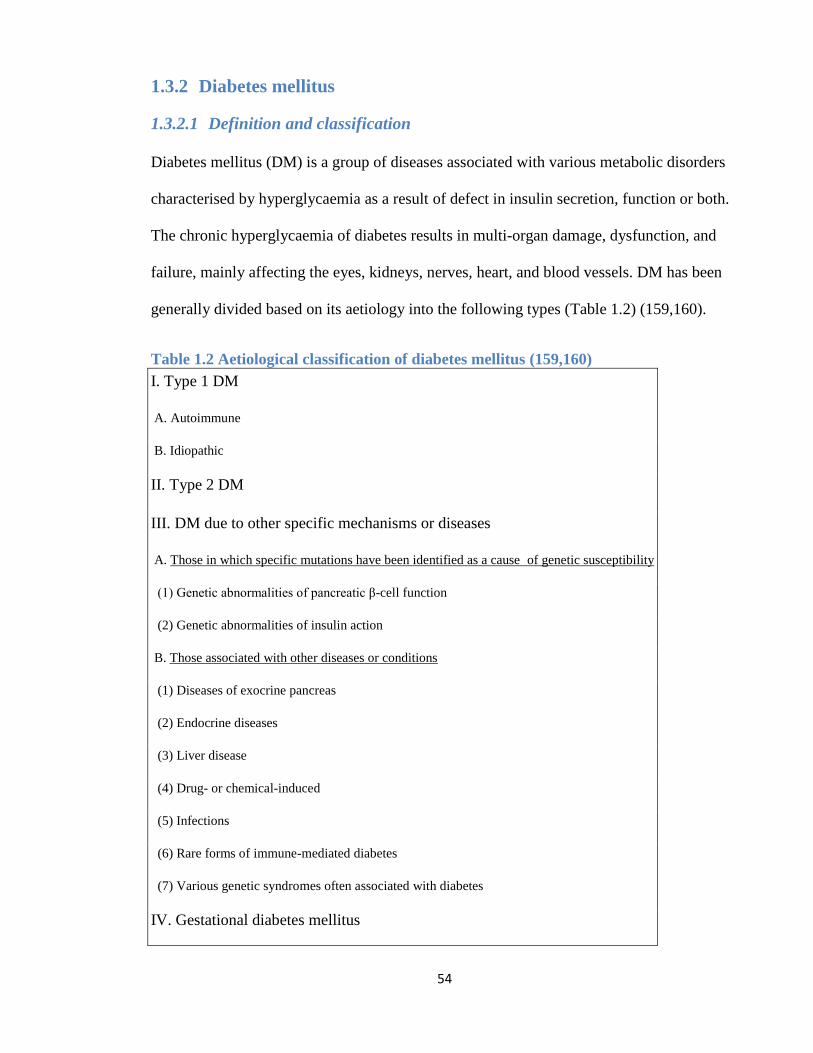
54
1.3.2 Diabetes mellitus
1.3.2.1 Definition and classification
Diabetes mellitus (DM) is a group of diseases associated with various metabolic disorders
characterised by hyperglycaemia as a result of defect in insulin secretion, function or both.
The chronic hyperglycaemia of diabetes results in multi-organ damage, dysfunction, and
failure, mainly affecting the eyes, kidneys, nerves, heart, and blood vessels. DM has been
generally divided based on its aetiology into the following types (Table 1.2) (159,160).
Table 1.2 Aetiological classification of diabetes mellitus (159,160)
I. Type 1 DM
A. Autoimmune
B. Idiopathic
II. Type 2 DM
III. DM due to other specific mechanisms or diseases
A. Those in which specific mutations have been identified as a cause of genetic susceptibility
(1) Genetic abnormalities of pancreatic β-cell function
(2) Genetic abnormalities of insulin action
B. Those associated with other diseases or conditions
(1) Diseases of exocrine pancreas
(2) Endocrine diseases
(3) Liver disease
(4) Drug- or chemical-induced
(5) Infections
(6) Rare forms of immune-mediated diabetes
(7) Various genetic syndromes often associated with diabetes
IV. Gestational diabetes mellitus
55
1.3.2.1.1 Type 1 diabetes
This type accounts for only 5–10% of those with diabetes, known previously as insulin-
dependent diabetes, or juvenile-onset diabetes, caused by cell mediated autoimmune
destruction of pancreatic β-cells. Type 1 diabetes can be either autoimmune mediated,
characterised by the presence of auto-antibodies against islet antigens and pancreatic β-cell
destruction, or idiopathic where patients reach an insulin-dependent status without
identifiable auto-antibodies and in the absence of an underlying cause. Typically it presents
early during childhood or adolescence with ketoacidosis as the first manifestation of the
disease. Others may have moderate increase in the fasting hyperglycaemia that increase in
severity in the presence of stressful condition such as infection.
1.3.2.1.2 Type 2 diabetes
Type 2 DM accounts for around 90–95% of those with diabetes, previously known as non–
insulin-dependent diabetes, or adult-onset diabetes, including those who have insulin
resistance and relative insulin deficiency. Obesity is commonly seen among type 2 DM
which can increase insulin resistance and induce more hyperglycaemia. Ketoacidosis is
rarely seen in this type of diabetes when compared with type 1 DM. However, patients with
type 2 DM are at increased risk of developing macrovascular and microvascular
complications (159). Risk factors for the onset of this type of diabetes include age, obesity,
lack of physical activity, history of gestational DM, hypertension and hyperlipidemia (161–
163).
1.3.2.2 Epidemiology
DM is one of the world's greatest health challenges and its prevalence appears to be
increasing. In the UK some 2.6 million people had diabetes in 2009 with a prevalence of
56
4%, among which 85% of the cases are type 2 DM (14). Among patients with type 2
diabetes in the United Kingdom Prospective Diabetes Study (UKPDS), 37% had
retinopathy (164); 9% neuropathy; and 7.3% nephropathy at the time of diagnosis (165).
1.3.2.3 Clinical features and complications
Patient may present initially with symptoms related to hyperglycaemia such as polyurea,
polydipsia, weight loss, polyphagia, and blurred vision. Some patients may present with
life-threatening complications of hyperglycaemia including ketoacidosis or the nonketotic
hyperosmolar syndrome. Long-term complications of diabetes include microvascular
complications such as diabetic retinopathy with high risk of visual loss(166) and
macrovascular complications such as atherosclerotic cardiovascular, peripheral vascular
and cerebrovascular disease (167). Other complications include nephropathy resulting in
renal failure (168); peripheral neuropathy with risk of foot ulcers, amputations, and Charcot
joints (169); and autonomic neuropathy causing gastrointestinal, genitourinary (170), and
sexual dysfunction (171).
1.3.2.4 Diagnosis
Diagnosis of DM has advanced but the basis for all has been a timed glucose sample such
as a fasting sample, casual sample independent of prandial status, or, a sample taken two
hours following a standardised oral glucose load. Currently, DM is diagnosed based on the
recommendations made by the Expert Committee on the Diagnosis and Classification of
Diabetes Mellitus in 2003 (172). Accordingly, diagnosis of DM is made when fasting
plasma glucose is ≥126 mg/dl (7.0 mmol/L) while normal value is defined as <110 mg/dL
(6.1 mmol/L) and impaired fasting glucose test is for those with fasting glucose measured
between 6.1-6.9 mmol/L. Another more recent report by the same committee agreed on
57
recommending the use of haemoglobin A1C (HbA1c ) in the diagnosed of DM when it is
>6.5% (173). Also, random blood glucose levels of ≥200 mg/dL (11.1 mmol/L) should be
considered suspicious for the presence of diabetes, especially when associated with
classical symptoms of DM. For those with impaired fasting glucose test, the WHO
recommended performing oral glucose tolerance test for diagnosing DM and the test is
abnormal when plasma glucose measures ≥200 mg/dL (11.1 mmol/L) two hours following
the administration of 75 g oral glucose (174).
1.3.2.5 Management
The main aim in managing patients with DM is to eliminate symptoms and to prevent or
delay the development of complications. The risk of diabetic retinopathy and nephropathy
can be reduced through good glycaemic control and by maintaining normal blood pressure.
Cardiovascular and cerebrovascular complications can be reduced through treating
hyperlipidemia and hypertension if exists, as well as smoking cessation and the use of
aspirin. According to NICE guidelines for diabetes control, it is recommended for patients
with DM to agree with their healthcare professional on a documented personalised HbA1c
targer, usually between 48 mmol/mol and 58 mmol/mol (6.5% and 7.5%) (175).
The first step in managing a patient with DM involves life style intervention such as diet
modification, activity composition, glucose self-monitoring, and structured patient
education with emphasis on cessation of smoking (176,177). There is still a need to initiate
a glucose lowering agent, typically Metformin, at the initial management of those patients.
In case of failure to achieve HbA1c < 7% or if high dose of Metformin cannot be tolerated,
a second medication may be added, either insulin or a sulfonylurea (178,179). In type 1
DM, insulin replacement is the main line of management (180–182) while for type 2 DM,
58
glucose-lowering agents have been used for providing glycaemic control, whether a single
agent or combination of medication. Common glucose lowering agents used include
biguanide (metformin) (183), sulfonylureas (e.g. glibenclamide, gliclazide, glimepiride, and
glipizide) (184), and glinides (185). The choice of glucose-lowering agents used to achieve
diabetes control must be tailored for each patient, based on its capacity to reduce HbA1c
with a consideration to medication side effects, ease of use, and patient compliance.
1.3.2.6 Diabetic retinopathy
1.3.2.6.1 Definition and classification
Diabetic retinopathy (DR) is an important neurovascular complication of diabetes that may
develop into sight threatening disease with devastating visual impairment (166). The
spectrum of DR can range from mild and moderate non-proliferative diabetic retinopathy
(NPDR), or background DR to pre-proliferative or severe-NPDR that can advance further
into proliferative diabetic retinopathy (PDR).
According to the Early Treatment Diabetic Retinopathy Study (ETDRS) definitions of
retinal lesions (186), one of the earliest features of background DR is formation of
microaneurysms, which are the consequence of localised capillary closure with swelling of
the adjacent patent capillaries or weakness in the capillary wall. Leakage from the
microaneurysms results in the formation of dot haemorrhage and exudates (Figure 1.3).
Blot haemorrhages occur as a consequence of capillary closure and deep retinal infarction
and are localised mainly in the outer plexiform layer of the retina, unlike flame shape
haemorrhages which localise superficially within the nerve fiber layer of the retina. Cotton
wool spots, a non specific feature of DR, occurs secondary to obliteration of axoplasmic
flow within the axons of the retinal nerve fiber layer (186).
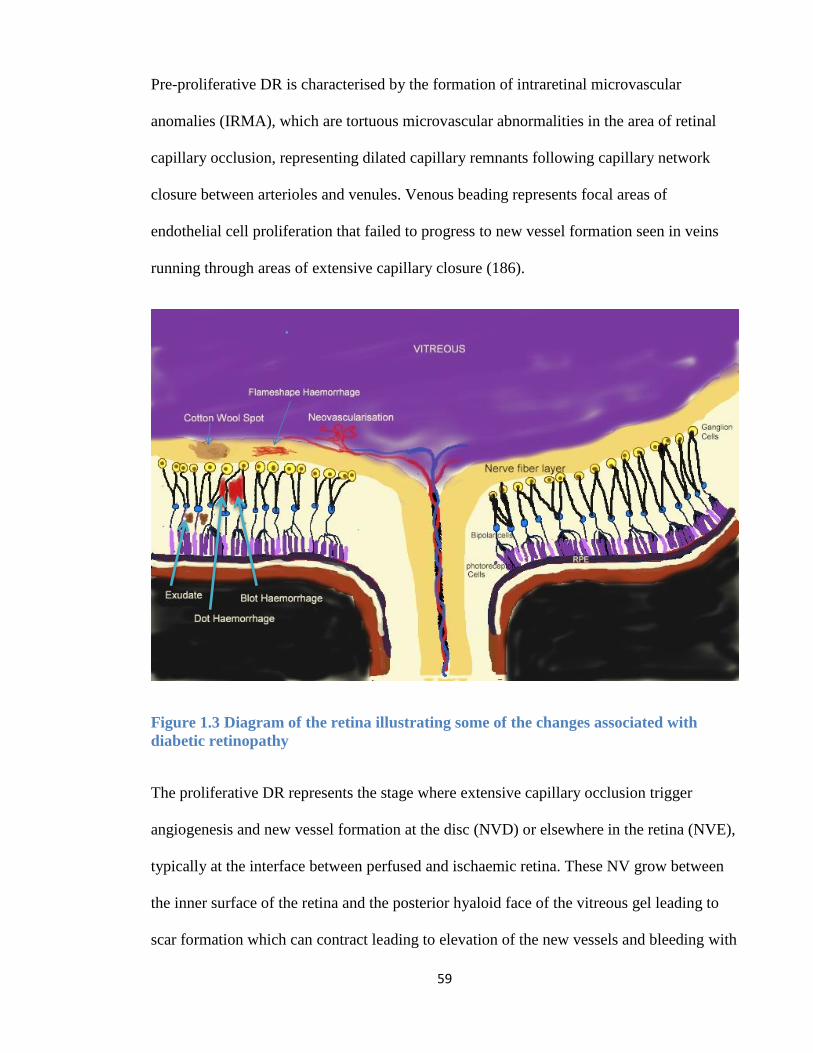
59
Pre-proliferative DR is characterised by the formation of intraretinal microvascular
anomalies (IRMA), which are tortuous microvascular abnormalities in the area of retinal
capillary occlusion, representing dilated capillary remnants following capillary network
closure between arterioles and venules. Venous beading represents focal areas of
endothelial cell proliferation that failed to progress to new vessel formation seen in veins
running through areas of extensive capillary closure (186).
Figure 1.3 Diagram of the retina illustrating some of the changes associated with
diabetic retinopathy
The proliferative DR represents the stage where extensive capillary occlusion trigger
angiogenesis and new vessel formation at the disc (NVD) or elsewhere in the retina (NVE),
typically at the interface between perfused and ischaemic retina. These NV grow between
the inner surface of the retina and the posterior hyaloid face of the vitreous gel leading to
scar formation which can contract leading to elevation of the new vessels and bleeding with
60
either preretinal or subhyaloid haemorrhage, or into the vitreous gel itself resulting in
vitreous haemorrhage (VH). The same scars secondary to NV formation can contract and
pull the vitreous away from the retina and occasionally are complicated with tractional
retinal detachment. Extensive retinal ischaemia may also lead to iris neovascularisation
(NVI) resulting in neovascular glaucoma. Diabetic papillopathy may occur in diabetic
patients independent of the DR status. It is associated with reduced visual acuity without
causing severe vision loss, and would require to be differentiated from ischaemic optic
neuropathy and NVD (186).
Diabetic maculopathy could be in the form of focal or diffuse diabetic macular oedema
(DMO) that can progress into clinically significant macular oedema (CSMO), or macular
ischaemia, or a mixture of all of these. The DMO could be focal, when the retinal
thickening and exudates is secondary to leakage from isolated or clusters of
microaneurysm, or diffuse oedema secondary to widespread leakage from retinal
capillaries. DMO may also occur secondary to RPE dysfunction and ischaemia, which
damage the outer BRB and result in accumulation of exudative fluid (extracellular oedema)
or secondary to hypoxia causing fluid accumulation within retinal cells (intracellular
oedema).
Table 1.3 gives the English and nternational clinical classification of DR and CSMO
adopted by the Royal College of Ophthalmologists and the American Academy of
Ophthalmology based on the ETDRS definitions of retinal lesions (187,188). In the UK, the
National Screening Committee (NSC) have used this classification in the Diabetic Eye
Screening Programme which provides annual screening with digital fundus photography to
all diabetic patients age 12 years or older. Once a potentially sight-threatening retinopathy
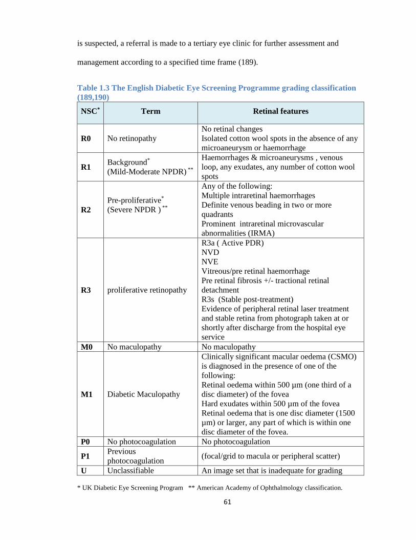
61
is suspected, a referral is made to a tertiary eye clinic for further assessment and
management according to a specified time frame (189).
Table 1.3 The English Diabetic Eye Screening Programme grading classification
(189,190)
NSC* Term Retinal features
R0 No retinopathy
No retinal changes
Isolated cotton wool spots in the absence of any
microaneurysm or haemorrhage
R1 Background*
(Mild-Moderate NPDR) **
Haemorrhages & microaneurysms , venous
loop, any exudates, any number of cotton wool
spots
R2
Pre-proliferative*
(Severe NPDR ) **
Any of the following:
Multiple intraretinal haemorrhages
Definite venous beading in two or more
quadrants
Prominent intraretinal microvascular
abnormalities (IRMA)
R3 proliferative retinopathy
R3a ( Active PDR)
NVD
NVE
Vitreous/pre retinal haemorrhage
Pre retinal fibrosis +/- tractional retinal
detachment
R3s (Stable post-treatment)
Evidence of peripheral retinal laser treatment
and stable retina from photograph taken at or
shortly after discharge from the hospital eye
service
M0 No maculopathy No maculopathy
M1 Diabetic Maculopathy
Clinically significant macular oedema (CSMO)
is diagnosed in the presence of one of the
following:
Retinal oedema within 500 µm (one third of a
disc diameter) of the fovea
Hard exudates within 500 µm of the fovea
Retinal oedema that is one disc diameter (1500
µm) or larger, any part of which is within one
disc diameter of the fovea.
P0 No photocoagulation No photocoagulation
P1 Previous
photocoagulation (focal/grid to macula or peripheral scatter)
U Unclassifiable An image set that is inadequate for grading
* UK Diabetic Eye Screening Program ** American Academy of Ophthalmology classification.
62
1.3.2.6.2 Epidemiology
Internationally, there are around 93 million people with DR, 17 million with proliferative
diabetic retinopathy, 21 million with diabetic macular oedema and 28 million with vision-
threatening diabetic retinopathy globally (191). The overall prevalence of retinopathy in
diabetes is 38%, and 85-90% of all diabetic patients have some manifestations of
retinopathy after 25 years of DM (166). But the Wisconsin epidemiological study of DR
(WESDR) has documented a higher rate in those with earlier age of onset of type I
diabetes, approaching 98% after 15 years of disease (192).
1.3.2.6.3 Risk factors
The duration of diabetes is one of the most important risk factors for the development of
retinopathy. Other risk factors, such as poor metabolic control, hypertension, high blood
cholesterol, nephropathy, age, sex, smoking, and genetic disposition are other factors that
may play a role in the development of DR but the exact mechanism is not fully understood
(193).
1.3.2.6.4 Pathophysiology of diabetic retinopathy
1.3.2.6.4.1 Histological changes in early stages of diabetic retinopathy
The pathogenesis behind the development of DR has been attributed to many factors. A
persistent increase in blood glucose levels shunts excess glucose into the aldose reductase
pathway in certain tissues, which converts sugars into alcohol (eg, glucose into sorbitol,
galactose to dulcitol). Intramural pericytes of retinal capillaries seem to be affected by this
increased level of sorbitol, eventually leading to the loss of its ability to auto-regulate
retinal capillaries. Loss of function of pericytes results in weakness and eventual out-
pouching of capillary walls (microaneurysms). In addition, capillary occlusion and
degeneration represent reduced retinal perfusion. Possible mechanisms leading to the
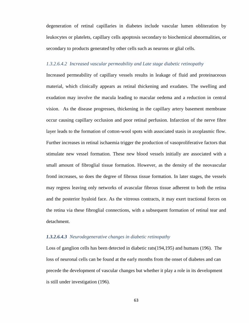
63
degeneration of retinal capillaries in diabetes include vascular lumen obliteration by
leukocytes or platelets, capillary cells apoptosis secondary to biochemical abnormalities, or
secondary to products generated by other cells such as neurons or glial cells.
1.3.2.6.4.2 Increased vascular permeability and Late stage diabetic retinopathy
Increased permeability of capillary vessels results in leakage of fluid and proteinaceous
material, which clinically appears as retinal thickening and exudates. The swelling and
exudation may involve the macula leading to macular oedema and a reduction in central
vision. As the disease progresses, thickening in the capillary artery basement membrane
occur causing capillary occlusion and poor retinal perfusion. Infarction of the nerve fibre
layer leads to the formation of cotton-wool spots with associated stasis in axoplasmic flow.
Further increases in retinal ischaemia trigger the production of vasoproliferative factors that
stimulate new vessel formation. These new blood vessels initially are associated with a
small amount of fibroglial tissue formation. However, as the density of the neovascular
frond increases, so does the degree of fibrous tissue formation. In later stages, the vessels
may regress leaving only networks of avascular fibrous tissue adherent to both the retina
and the posterior hyaloid face. As the vitreous contracts, it may exert tractional forces on
the retina via these fibroglial connections, with a subsequent formation of retinal tear and
detachment.
1.3.2.6.4.3 Neurodegenerative changes in diabetic retinopathy
Loss of ganglion cells has been detected in diabetic rats(194,195) and humans (196). The
loss of neuronal cells can be found at the early months from the onset of diabetes and can
precede the development of vascular changes but whether it play a role in its development
is still under investigation (196).
64
1.3.2.6.4.4 Role of inflammation in diabetic retinopathy
There is more evidence now supporting the role of inflammatory cells, cytokines and other
inflammatory products in the development of DR (197–199). An animal study showed
retinal capillary occlusions by leukocytes, mainly monocytes, endothelial cell damage,
extravascular macrophage accumulation, and areas of NV at the sites of extravascular
leukocytes accumulation (200). The possible role of inflammation in the development of
capillary degeneration and early diabetic retinopathy is demonstrated in Figure 1.4. The
high glucose result in the release of advanced gycation end products (AGEs), AGEs,
together with other factors such as oxidative stress and aldose reductase result in the release
of nuclear factor kappa beta (NF-κB). The latter is a transcription factor that regulates many
genes involved in inflammatory and immune responses, proliferation and apoptosis. NF-κB
was found to be activated in retinal endothelial cells or pericytes when subjected to high
glucose levels (201,202). NF-κB increases expression of intracellular adhesion molecules
(ICAM) in the endothelium of retinal vessels of animals and humans. The ICAM interact
with the CD18 adhesion molecule on monocytes and neutrophils, resulting in increase in
leukocyte aggregation within retinal blood vessels in diabetes (203). Furthermore, NF-κB
activation leads to the expression of cyclooxygenase (COX). COX-2 rather than COX-1 is
mainly involved in the production of prostaglandin-E2 (PGE2) and VEGF in the retina of
diabetic rats (204,205). VEGF is produced by many retinal cells, such as ganglion cells,
Müller cells, and pericytes in response to retinal hypoxia and COM-2 production in patients
with diabetes. VEGF has an important role in retinal NV and increased vascular
permeability (206). Other growth factors that play a role in the pathogenesis of DR include
insulin-like growth factors I and II (207), transforming growth factor β (208), and high ratio
of VEGF to pigment epithelium-derived factor (PEDF) (209,210). Cytokines also play a
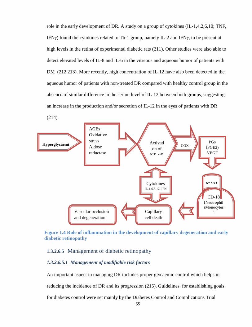
65
role in the early development of DR. A study on a group of cytokines (IL-1,4,2,6,10; TNF,
IFNγ) found the cytokines related to Th-1 group, namely IL-2 and IFNγ, to be present at
high levels in the retina of experimental diabetic rats (211). Other studies were also able to
detect elevated levels of IL-8 and IL-6 in the vitreous and aqueous humor of patients with
DM (212,213). More recently, high concentration of IL-12 have also been detected in the
aqueous humor of patients with non-treated DR compared with healthy control group in the
absence of similar difference in the serum level of IL-12 between both groups, suggesting
an increase in the production and/or secretion of IL-12 in the eyes of patients with DR
(214).
1.3.2.6.5 Management of diabetic retinopathy
1.3.2.6.5.1 Management of modifiable risk factors
An important aspect in managing DR includes proper glycaemic control which helps in
reducing the incidence of DR and its progression (215). Guidelines for establishing goals
for diabetes control were set mainly by the Diabetes Control and Complications Trial
CD-18
(Neutrophil
sMonocytes
)
Hyperglycaemi
a
AGEs
Oxidative
stress
Aldose
reductase
Activati
on of
NF-κB
COX-
2
PGs
(PGE2)
VEGF
ICAM
Capillary
cell death
Vascular occlusion
and degeneration
Cytokines IL-1,6,8,12; IFN
Figure 1.4 Role of inflammation in the development of capillary degeneration and early
diabetic retinopathy
66
(DCCT) for type 1 DM (216), and the UKPDS for type 2 DM (217,218). The goal is to
maintain blood glucose at near-normal levels (fasting glucose level of 5.0 to 7.2 mmol/L
and HbA1c levels between 48-58mmol/mol (6.5-7.5%), together with maintaining normal
blood pressure and serum lipid. Aggressive glucose lowering may not be advised in some
cases with established cardiovascular events and among older age group(219). Meanwhile,
tight glucose control had a better effect in reducing the risk of microvascular complications,
including diabetic retinopathy, with less effect on preventing macrovascular complications
(220,221). In case of type 1 diabetes, the DCCT research showed that each 1% decrease in
HbA1c reduces the risk of DR by 39% (222). In type 2 DM, the UKPDS found a 25%
reduction in the risk of microvascular complications for every 1% decline in HbA1c (223).
While the blood pressure control has a little influence in altering the course of DR among
normotensive patients, it can be of importance in reducing visual loss secondary to DR
among hypertensive patients with diabetes. The UKPDS showed that each 10 mmHg
decrease in systolic blood pressure reduces the risk of microvascular complications by
13%, independent of glycaemic control (224). The benefit of blood pressure treatment in
normotensive diabetic patients is less clear. The effect of controlling blood lipid levels in
reducing the risk of retinopathy is supported by the finding of the Action to Control
Cardiovascular Risk in Diabetes (ACCORD) study which demonstrate reduced risk of DR
progression by about a 40% over 4 years following the use of lipid therapy with fenofibrate
and simvastatin (225). Similarly, the Fenofibrate Intervention and Event Lowering in
Diabetes (FIELD) study showed a significant effect of fenofibrate in reducing the need for
laser photocoagulation (226). In both the FIELD and ACCORD-Eye studies, no
relationship was found between the lipid effects of fenofibrate and the appearance or
67
progression of DR. Thus, the beneficial effects of fenofibrate on DR are unrelated to
quantitative change in serum lipid level.
1.3.2.6.5.2 Retinal laser photocoagulation
The main principle of applying retinal laser photocoagulation in DR is to induce thermal
destruction of RPE cells and its adjacent photoreceptor, thus reducing the production of
VEGF and its angiogenesis effect and regression of the NV (227). The retina contain three
main pigmented substances with variable light wavelengths’ absorbtion capacity; these
include the xanthophylls (420-500 nm) within the neurosensory layer, melanin (400-1000
nm) within the RPE cells and choroidal melanocytes, and hemoglobin (450 to 550 nm)
within red blood cells within the blood vessels or within areas of blood leakage (228). The
melanin and haemoglobin both can absorb the green (495-570 nm) and yellow (570-590
nm) laser wavelengths, making them ideal for use in photocoagulation of macular
disorders. The green wavelength laser can be produced by argon laser (514.5 nm), Krypton
laser (530.9 nm) diode and PASCAL frequency-doubled Yttrium Aluminum Garnet (YAG)
laser (532nm), while the yellow wavelength, which has the advantage of using less power
and better absorbtion by the haemoglobin, can be provided by krypton laser (568 nm) as
well as dye and diode laser (577 nm). So far there has been no proven superiority of yellow
focal/grid laser treatment over the traditional green laser therapy in managing CSMO (228).
The more recent laser technologies include subthreshold diode-laser micropulse technology
works within the infrared spectrum of light (810nm) and is designed to deliver repeated
laser shots for about 0.1-0.3 seconds separated by intervals of 1.7 to 1.9 seconds, allowing
for heat dissipation and hence prevent collateral damage and localise the photothermal
effect to the targeted RPE with little intraretinal damage or ophthalmologically visible
scarring during or after treatment. A recent randomised trial found a better improvement in
68
visual acuity in eyes treated with the high density micropulse laser (0.25LogMAR)
compare to modified ETDRS laser (0.08LogMAR) and no improvement with the normal
density micropulse laser (0.03LogMAR) (229).
Panretinal photocoagulation (PRP) can be carried out in a single or multiple sessions of
laser application, starting initially with about 2000 burns of 500 μm spot size, or more with
smaller burn size. Retreatment can be performed if necessary within one to three months of
initial laser treatment. Macular laser photocoagulation reduces central macular thickness
and improves the vision in eyes with CSMO as shown in the ETDRS study where macular
laser therapy reduced the risk of moderate vision loss from 24% to 12% over 3 years follow
up period (230). The laser application in the ETDRS study was performed as follows; 1)
Focal treatment of microaneuysms and other sites of focal leakage with a 50 -100μm spot
size to obtain definite whitening around the area of leakage. 2) Diffuse leakage and areas of
capillary closure (that were not contiguous with the foveal avascular zone) within two discs
diameters of the centre were treated in a grid fashion using spot sizes of 50 - 200μm, a
space of 1 burn width apart. 3) Lesions within 500μm of fovea were not treated initially but
treatment was allowed to within 300μm of the fovea on repeated sessions, as needed (230).
The long term efficacy of macular laser therapy has also been studied through the diabetic
retinopathy clinical trial research network (DRCR.net) which compared the IVTA therapy
against traditional laser therapy in the management of CSMO (231). In the DRCR trial
group, a modified ETDRS focal green laser treatment was performed as follows: 1) All
leaking microaneurysms 500 to 3000μm from fovea treated directly with 50μm spot size,
duration 0.05 - 0.1s. 2) Direct whitening of the micronaneurysm was not required, but a
greyish reaction beneath the microaneurysm was needed. Grid treatment was performed to
areas of retinal thickening. 3) Grid was performed from 500 to 3000μm superiorly and
69
inferiorly and to 3500μm temporally. The spots were 2 burn widths apart and no burns were
performed within 500μm of the disc. Possible complications that can be associated with
macular grid laser therapy include paracentral scotoma and colour vision impairment,
accidental foveal photocoagulation, subfoveal fibrosis, hard exudates, and CNVM at area of
laser scar (232).
1.3.2.6.5.3 Intravitreal anti-VEGF therapy
While initially the focal/grid laser photocoagulation used to be the main standard of care in
managing DMO, the use of repeated intravitreal injection of anti-VEGF has the potential to
revolutionise the treatment strategy for DMO and can provide additional benefit over the
use of focal/grid laser treatment in reducing central retinal thickness and improving vision
(233). The latest guidelines from the National Institute for health and Clinical Excellence
(NICE) recommend the use of ranibizumab in treating visual impairment secondary to
DMO with central retinal thickness of 400μm or more at the start of the treatment. For
those who currently receiving ranibizumab for treating visual impairment due to DMO and
didn’t meet the above criteria, the NICE still recommend them to continue with the
treatment until it is stopped according to clinical response (234). The Safety and Efficacy of
Ranibizumab in Diabetic Macular Edema (RESOLVE) trial found a significant
improvement in vision by a mean of 10 letters and gained ≥10 letters in 60% of eyes treated
with repeated ranibizumab of the cases compare to the sham group (235). In the
Ranibizumab Monotherapy or Combined With Laser vs Laser Monotherapy for Diabetic
Macular Edema (RESTORE) trial, 345 eyes with DMO were randomised into receiving
either ranibizumab alone, ranibizumab with macular laser therapy, or laser therapy alone.
By one year, there was an improvement in vision by about six letters in both groups
receiving ranibizumab compare to an average of one letter gained in eyes treated with laser
70
alone (236). An extension of the RESTORE study observed the outcome of the three
treatment groups for a further two years. By the third year of follow-up, eyes treated with
ranibizumab were able to maintain the gain they had in their vision over the first year,
while the group with previous laser treatment managed to further gain vision from baseline
from a mean of 2.3 letters gain at the first year to six letters gain by the third year of follow-
up (237).
Despite the advantages of ranibizumab intravitreal injection in managing DMO, it can still
be insufficient alone as an ultimate treatment option. This was observed in the RISE
(Ranibizumab Injection in Subjects with Clinically Significant Macular Edema with Center
Involvement Secondary to Diabetes Mellitus) and RIDE (Ranibizumab Injection in
Subjects with Clinically Significant Macular Edema with Center Involvement Secondary to
Diabetes Mellitus) trials which found 30.4% and 43.2% of patients received 0.3 or 0.5 mg
intravitreal ranibizumab still failed to have improvement in vision after 3 years of treatment
(238). Also the DRCR.net study, failure to gain ≥10 letters was observed after 3 years
follow up period in 57.6% of patients treated with combined ranibizumab and prompt laser
group and in 44.2% of patients on ranibizumab and deferred laser (≥24 weeks) group and
there was vision loss of ≥15 letters in 6% and 3%, respectively (239). For this reason,
further development of treatment strategies is still required when managing cases with
DMO.
While ranibizumab is the main anti-VEGF licensed for use in eyes with DMO, other anti-
VEGF therapies have also been used as an off-label treatment in selected cases.
Bevacizumab has a similar mechanism of action to ranibizumab but is much cheaper and
has shown potential benefit in managing DMO as well as in managing vasoproliferative
71
complications associated with proliferative DR. The prospective randomised BOLT trial of
intravitreal bevacizumab or laser therapy in the management of DMO compared these two
treatment methods over two years. Patients within the bevacizumab group gained ≥10
letters in 49% of cases and gained ≥15 letters in 32% of cases compare to 7% and 4% of the
laser group, respectively (240,241).
Pegaptanib is another anti-VEGF that has been used in the management of DMO when
given at a dose of 0.3mg intravitreal injection given every six weeks. A multicentre
randomised trial found 36.8% of the patients from the pegaptanib group experienced an
improvement in vision by ≥10 letters over two years follow-up compared to only 19.7%
from the sham group (242).
Aflibercept is administered as a single 2mg intravitreal injection that can be given every
month for five consecuative months then every two months onward up to one year before
the interval is changed based on clinical response. Aflibercept has been recently
recommended by the NICE guidelines as a treatment option for eyes with DMO and central
retinal thickness of 400μm or more at start of treatment (243). The initial report from the
da Vinci multicentre study compared the use of intravitreal aflibercept given in four
different doses and/or interval periods in comparison to macular laser photocoagulation
(244). Over the first year, the maximum improvement in vision by ≥15 letters was observed
in eyes receiving 2mg intravitreal aflibercept every 4 weeks (45%) compared to other
treatment protocols and compared to those received macular laser treatment (11.4%) (244).
A more recent randomised phase 3 trial comparing intravitreal aflibercept to macular laser
therapy among 872 patients found a significant superiority of aflibercept given in 2mg dose
72
every 4 weeks or every 8 weeks (following 5 initial monthly doses) in comparison to the
macular laser therapy over a one year follow-up period (245).
The Protocol-T multicentre study by the DRCR network compared the effect of all three
anti-VEGF intravitreal injection; aflibercept, bevacizumab, ranibizumab in managing
DMO. Patients were randomized to receive one of these therapies every 4 weeks. After one
year, aflibercept group had an overall better vision outcome, with the mean visual acuity
letter score improving by 13.3, vs. 9.7 with bevacizumab and 11.2 with ranibizumab, and
this was mainly observed in eyes with worse visual acuity at baseline (246).
1.3.2.6.5.4 Intravitreal steroids
Intravitreal injection of steroids has been used in the management of CSMO aiming to
reduce capillary permeability and intraretinal fluid accumulation, together with controlling
cytokine formation and inflammatory mediated reaction. Intravitreal dexamethasone,
fluocinolone acetonide and TA have been licensed for the management DMO. Managing
DMO with focal/grid laser treatment has been shown to be more effective when compared
to the use of IVTA as suggested by the results of the DRCR.net group randomised trial on
eyes with DMO. In this study, there was initial improvement in the visual outcome among
eyes treated with 4mg IVTA compared to both the 1mg IVTA and the focal/grid laser
groups. However, no significant difference in the visual outcome existed between the three
groups after one year follow-up. However, by the end of two years follow-up the focal/grid
laser photocoagulation group had a better visual outcome and fewer side effect profile
when compared to those receiving IVTA (247).
The dexamethazone implant (Ozurdex®) has shown effectiveness in managing DMO as
shown in the outcomes of Dexamethasone DDS Phase II Study which found an
73
improvement in vision by ≥10 letters in about one third of eyes following the
administration of 0.7 mg dexamethasone implant. This was also associated with a
significant reduction in the central retinal thickness and FFA leakage, making the
dexamethasone implant a recommended treatment option in managing persistent DMO
(248). The safety and efficacy of dexamethasone implant in combination with laser
focal/grid photocoagulation was addressed in the PLACID study. Within the first nine
months of the study, there was a significant improvement in vision among eyes receiving
the combined dexamethasone implant and laser therapy compared to the group receiving
laser therapy alone, although this advantage became less apparent by the end of the one
year follow-up period (249). The efficacy and long term safety of dexamethasone implant
was further studied through the MEAD group who conducted a randomised, multicentre,
phase III clinical trial including 1048 patients with DMO divided into three groups to
receive either 0.7 mg implant, 0.35 mg implant, or sham procedure. After 3 years of follow-
up, a significant proportion of patients on 0.7mg (22%) and 0.35mg (18.4%) implants did
achieve 15 letters improvement compared to the sham group (12%). The average reduction
in central retinal thickness was considerably greater in eyes that received dexamethasone
implant compared to the sham group. By the end of the follow-up period, adverse effects in
the form of cataract formation was observed in about 68% of eyes with the 0.7mg
dexamethasone implant compared to 64% in the 0.35mg group and 20.4% for the sham
group. Only two patients (0.6%) with the 0.7mg dexamethasone implant required
trabeculectomy for managing glaucoma (250). Based on these results, the NICE guidelines
in UK recommend the use of dexamethasone intravitreal implant as a treatment option for
managing DMO only if the oedema does not respond to non-corticosteroid therapy, or such
treatment is unsuitable, and that the implant is to be used only in pseudophakic eyes (251).
74
Fluocinolone acetonide intravitreal implant (Iluvien) is a non-erodible implant which
contains fluocinolone acetonide at a dose of 190 µg and can be injected into the vitreous
using a preloaded needle device. Once in the vitreous, the medication is released at a rate of
0.2 µg /day and continues to be released at a steady rate for up to 36 months post injection.
Its effectiveness over at least 3 years post injection had been examined by the Fluocinolone
Acetonide in Diabetic Macular Edema (FAME) study group which found 28% of patients
with DMO successfully gained ≥15 letters compared to only 19% of patients on sham
intervention. Cataract is the main side effect associated with fluocinolone acetonide
implant, with almost all phakic patients developing cataract by the end of the FAME study
period. Meanwhile, increased IOP and secondary glaucoma is an additional risk factor, with
4.8% of the patients requiring glaucoma surgery by the third year following fluocinolone
acetonide intravitreal injection (252). The effectiveness of the implant is even more
prominent in eyes with chronic DMO (duration of diagnosis ≥3 years) who failed to
respond to at least one session of macular laser therapy. The FAME study showed that
patients with chronic DMO had a greater percentage with improved vision by ≥15 letters
while on 0.2 µg /day of intravitreal fluocinolone acetonide compare to the sham group
(34% versus 13.4%), whereas no significant difference was observed between patients with
non chronic DMO receiving the implant compare to the sham group (22.3% versus
27.8%).(253) The findings of the FAME clinical trials and other related research formed
the base for the UK NICE guidelines which recommended the use of fluocinolone
acetonide intravitreal implant as a treatment option for managing chronic DMO only if they
showed insufficient response to other available therapy and that the implant is to be used
only in psedophakic eyes (254). However, a recent study from the FAME group found the
long term outcomes were not worse in eyes with DMO that had cataract surgery after
75
receiving 0.2 µg/day of fluocinolone acetonide implant compared to pseudophakic eyes
receiving the implant (255).
1.3.3 Retinal vasculitis
1.3.3.1 Blood retinal barrier and the blood supply of the retina
The retina is composed of the outer pigmented layer, the retinal pigmented epithelium
(RPE), and an inner neurosensory layer. The latter is subdivided further into nine layers
arranged from the inside out as follows; internal limiting membrane, nerve fibre layer,
ganglion cell layer, inner plexiform layer, inner nuclear layer, outer plexiform layer, outer
nuclear layer, external limiting membrane and photoreceptor layer. The posterior pole of
the retina is vulnerable to develop oedema due to a high cell count, increased metabolic
activity, shifting of Henle’s fibre layer away from central fovea, thin and loose connection
of inner connecting fibres in the outer plexiform layer leaving a large reservoir for the
accumulation of extravascular fluid, and finally the central avascular zone creating a
watershed arrangement between the choroidal and retinal circulation, thus decreasing
resorption of extracellular fluid. On the other hand, variable mechanisms prevent an
accumulation of extracellular intraretinal fluid and proteins in the retina; these include
osmotic forces, hydrostatic forces, capillary permeability and tissue compliance. These
mechanisms provide a balance between the rate of capillary filtration and the rate of fluid
removal from the extracellular retinal tissue. To maintain this physiological balance, an
intact BRB formed by intercellular junctions is required to separate blood from surrounding
retinal tissues, and to regulate the transfer of ions, cells and protein, in addition to leukocyte
extravasation during inflammation. In the inner retinal circulation, the inner BRB is formed
by tight junctions (zonulae occludentes). Intercellular communication is realised by
adherens junctions (zonulae adherentes) and gap junctions (maculae communicantes),
76
joining the endothelium of retinal capillaries. In the outer retinal circulation, the tight
junctions between the RPE cells maintain the outer BRB as well as adherent junctions and
desmosomes (maculae adherentes). Movement of fluid from the vitreous into the subretinal
space is limited by the external limiting membrane, formed by the zonulae adherents
between photoreceptors and Müller cells. But, this junction is less tight than the zonulae
occludentes of the RPE and retinal capillaries. As a consequence, they can only partially
limit the movement of large molecules such as fluoresceinated albumin from subretinal
space to the vitreous cavity and vice versa. On the other hand, the internal limiting
membrane may have no significant role in preventing water movement but may keep large
molecules such as proteins in the retina, maintaining osmotic movements. Any pathological
conditions leading to a breakdown of the BRB can result in the retention of proteins within
the retinal tissue, leading to water retention by osmosis and the development of DMO
(256).
The central retinal artery is the first branch of the ophthalmic artery entering the eye
through the lamina cribrosa. Soon after entering the globe, it splits into four branches,
superior and inferior nasal and temporal branches, each supplying one quadrant of the
retina. Sometimes, a cilioretinal artery, branching from the ciliary circulation, supplies part
of the inner retina between optic nerve and the centre of the macula. At the tissue level, the
inner two third of the retina is supplied by two layers of capillaries, one superficial in the
ganglion and nerve fiber layer, and one deeper in the inner nuclear layer. The outer one
third of the retina beyond the inner nuclear layer is avascular and receives its oxygen and
nutrition from the choriocapillaries made of a fenestrated capillary system of the choroidal
arteries that branch from the ciliary arteries, supplying 95% of the oxygen used in the
77
fundus. Blood collected from the capillaries accumulates within a branch retinal vein that
ends in the central retinal vein (257).
1.3.3.2 Definition and Clinical presentation of retinal vasculitis
The definition of retinal vasculitis has been a controversial topic. The SUN working group
in 2004 considered the eye to have retinal vasculitis in the presence of perivascular
sheathing and vascular leakage or occlusion on FFA as evidence of retinal vascular
disease(19). Clinically, patients with retinal vasculitis may be asymptomatic or present
with blurred vision and floaters associated with IU, PU and PANU. Reduced visual acuity
in retinal vasculitis might occur due to combined factors including severe vitritis, optic
neuritis, ERM and CMO. In one study, vascular sheathing was observed in 85% of the eyes
with vasculitis in the absence of cotton wool spots and intraretinal haemorrhage. Combined
vascular sheathing and intraretinal haemorrhage occurred in 11% of cases while vascular
sheathing combined with cotton wool spots were only observed in 4% of cases. The same
study found ERM to be more common in association with cotton wool spots and
intraretinal haemorrhage compared to those with vascular sheathing (258). In eyes with
retinal ischaemia, vision loss can also occur secondary to macular ischaemia or due to the
complications of NV such as vitreous haemorrhage, tractional retinal detachment and
neovascular glaucoma.
1.3.3.3 Pathophysiology
Based on histological studies, vascular changes in uveitis are characterised by perivascular
infiltration of lymphocytes resulting in perivasculitis rather than a true vasculitis of the
vessel wall (259,260). Cell-mediated immunity also plays a role in the pathology of retinal
vasculitis, with CD4+ T cells documented within and around the retinal vessels. The
78
pathogenesis of ischaemia in retinal vasculitis is not clear but is suggested to be either
thrombotic or obliterative secondary to the infiltration of inflammatory cells. Thrombotic
vascular changes can occur due to local endothelial injury or increased prothrombin activity
as observed in BD (261). The retina has a uniquely high metabolic demand for oxygen that
is normally met by a highly efficient vascular supply. Insufficiency of the retinal circulation
causes neuroretinal dysfunction and degeneration. Focal retinal ischaemia results in
selective damage to specific subpopulations of retinal neurons and can result in cellular
death by apoptosis or necrosis with dysfunction and degeneration of the inner retina and
eventually visual loss. Retinal vascular obstruction can also promote the production of
VEGF (Figure 1.5) which increases vascular permeability and results in macular oedema
and induce new vessels formation (262). These fragile new vessels bleed easily resulting in
VH, fibrovascular proliferation and subsequent tractional retinal detachment.
1.3.3.4 Classification and aetiology of retinal vasculitis
Retinal vasculitis is a sight-threatening inflammatory condition, occurring in approximately
one in every eight eyes with uveitis (263). Based on the aetiology, retinal vasculitis may be
classified as either idiopathic or secondary to infection, neoplasia or a systemic
inflammatory disease (264,265). Retinal vasculitis can be classified either as idiopathic,
when there is no associated systemic disease, or secondary vasculitis in the presence of
other systemic conditions, the most common being BD, collagen vascular disease and
systemic granulomatous diseases (sarcoidosis and TB).(266)
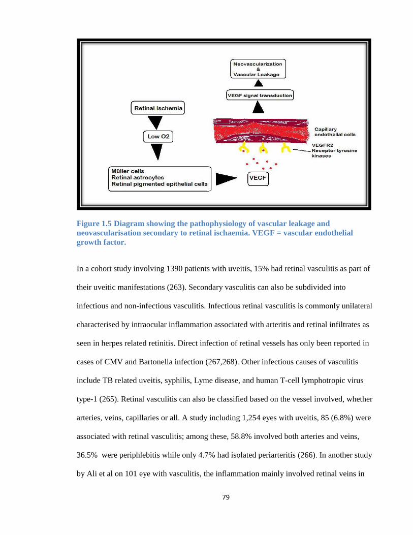
79
Figure 1.5 Diagram showing the pathophysiology of vascular leakage and
neovascularisation secondary to retinal ischaemia. VEGF = vascular endothelial
growth factor.
In a cohort study involving 1390 patients with uveitis, 15% had retinal vasculitis as part of
their uveitic manifestations (263). Secondary vasculitis can also be subdivided into
infectious and non-infectious vasculitis. Infectious retinal vasculitis is commonly unilateral
characterised by intraocular inflammation associated with arteritis and retinal infiltrates as
seen in herpes related retinitis. Direct infection of retinal vessels has only been reported in
cases of CMV and Bartonella infection (267,268). Other infectious causes of vasculitis
include TB related uveitis, syphilis, Lyme disease, and human T-cell lymphotropic virus
type-1 (265). Retinal vasculitis can also be classified based on the vessel involved, whether
arteries, veins, capillaries or all. A study including 1,254 eyes with uveitis, 85 (6.8%) were
associated with retinal vasculitis; among these, 58.8% involved both arteries and veins,
36.5% were periphlebitis while only 4.7% had isolated periarteritis (266). In another study
by Ali et al on 101 eye with vasculitis, the inflammation mainly involved retinal veins in
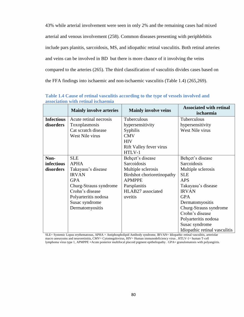
80
43% while arterial involvement were seen in only 2% and the remaining cases had mixed
arterial and venous involvement (258). Common diseases presenting with periphlebitis
include pars planitis, sarcoidosis, MS, and idiopathic retinal vasculitis. Both retinal arteries
and veins can be involved in BD but there is more chance of it involving the veins
compared to the arteries (265). The third classification of vasculitis divides cases based on
the FFA findings into ischaemic and non-ischaemic vasculitis (Table 1.4) (265,269).
Table 1.4 Cause of retinal vasculitis according to the type of vessels involved and
association with retinal ischaemia
Mainly involve arteries Mainly involve veins
Associated with retinal
ischaemia
Infectious
disorders
Acute retinal necrosis
Toxoplasmosis
Cat scratch disease
West Nile virus
Tuberculous
hypersensitivity
Syphilis
CMV
HIV
Rift Valley fever virus
HTLV-1
Tuberculous
hypersensitivity
West Nile virus
Non-
infectious
disorders
SLE
APHA
Takayasu’s disease
IRVAN
GPA
Churg-Strauss syndrome
Crohn’s disease
Polyarteritis nodosa
Susac syndrome
Dermatomyositis
Behçet’s disease
Sarcoidosis
Multiple sclerosis
Birdshot chorioretinopathy
APMPPE
Parsplanitis
HLAB27 associated
uveitis
Behçet’s disease
Sarcoidosis
Multiple sclerosis
SLE
APS
Takayasu’s disease
IRVAN
GPA
Dermatomyositis
Churg-Strauss syndrome
Crohn’s disease
Polyarteritis nodosa
Susac syndrome
Idiopathic retinal vasculitis SLE= Systemic Lopus erythematosus, APHA = Antiphospholipid Antibody syndrome, IRVAN= Idiopathic retinal vasculitis, arteriolar
macro aneurysms and neuroretinitis, CMV= Cytomegalovirus, HIV= Human immunodeficiency virus , HTLV-1= human T-cell
lymphoma virus type 1, APMPPE =Acute posterior multifocal placoid pigment epitheliopathy . GPA= granulomatosis with polyangiitis.

81
1.3.3.4.1 Behçet's Disease
BD is a chronic, relapsing multi-systemic inflammation of the blood vessels of all sizes
characterised by oral and genital mucous ulcerations, skin lesions, and uveitis (270). BD
affects young patients aged between 20 and 40 years with more prevalence in male gender
within the Eastern Mediterranean region and in female gender within Northern
Europe(271). The incidence of ocular involvement in BD is high that can reach up to 80%
of patients with BD (272) and thus ocular involvement had been one of the key features
within the scoring system that guide toward the diagnosis of BD (273). Uveitis is often the
dominant manifestation of this disease and occurs mainly in the form of chronic, episodic
PANU that is characteristically bilateral and associated with hypopyon affecting 20 % of
patients. Other ocular manifestations include PU, retinal vasculitis, retinal vein or artery
occlusions with sight threatening complications such as optic neuritis, macular ischaemia
and NV formation (274).
Retinal vasculitis in ocular BD most commonly manifests as vitritis with diffuse vascular
leakage on FFA due to inflammatory hyperpermeability. This may be accompanied by
capillary nonperfusion secondary to occlusive vasculitis resulting in NV. Both retinal
arteries and veins can be involved in BD though venous involvement is more common
(265). BRVO with intraretinal hemorrhages and CMO are frequently seen and these are
often central in the retina with a high risk of significant visual loss (Figure 1.6). BRVO and
ischaemic retinal vasculitis have been reported as the first presentation of ocular BD in 28%
and 21%, respectively, while central vein (4%) and artery (1%) occlusions are less common
presentations (275). Macular ischaemia, a predictor of poor visual outcome, has also been
reported in cases with BD. In a recent retrospective study of 120 eyes of patients with BD,
macular ischaemia was seen in one eye (0.8%) at initial visit, while three eyes (2.5%)
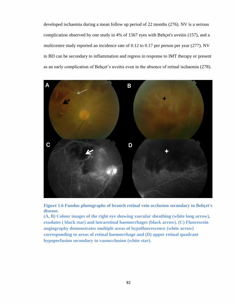
82
developed ischaemia during a mean follow up period of 22 months (276). NV is a serious
complication observed by one study in 4% of 1567 eyes with Behçet's uveitis (157), and a
multicentre study reported an incidence rate of 0.12 to 0.17 per person per year (277). NV
in BD can be secondary to inflammation and regress in response to IMT therapy or present
as an early complication of Behçet’s uveitis even in the absence of retinal ischaemia (278).
Figure 1.6 Fundus photographs of branch retinal vein occlusion secondary to Behçet's
disease.
(A, B) Colour images of the right eye showing vascular sheathing (white long arrow),
exudates ( black star) and intraretinal haemorrhages (black arrow). (C) Fluorescein
angiography demonstrates multiple areas of hypofluorescence (white arrow)
corresponding to areas of retinal haemorrhage and (D) upper retinal quadrant
hypoperfusion secondary to vasoocclusion (white star).
83
1.3.3.4.2 Sarcoidosis
Sarcoidosis is a chronic granulomatous disease affecting multiple organs, with ocular
involvement in 25-60% of patients, mainly manifesting as uveitis (30–70%) and
conjunctival nodules (40%) (279,280). The anterior chamber is most commonly affected,
with up to 66 % of patients with ocular sarcoidosis presenting with iridocyclitis while the
posterior chamber is involved in 25 % of cases (281). Retinal vasculitis in the form of
multifocal periphlebitis has been reported in 37% of patients with ocular sarcoidosis (266).
Retinal periphlebitis is a common ocular manifestation and was considered by the first
International Workshop on Ocular Sarcoidosis as one of seven clinical signs that comprise
the diagnosis of ocular sarcoidosis (282). Although ocular sarcoidosis is typically
associated with non-obstructive vasculitis, ischaemic retinal vasculitis has rarely been
reported in patients with sarcoidosis. Typical features of the involved vessels include
segmental cuffing or extensive sheathing and perivenous exudates, known as “candle wax
drippings” associated with vasculitis on FFA that mainly involves midperipheral retinal
veins. Additional vascular features include the presence of macroaneurysms, peripheral
vessel closure and NV (Figure 1.7) (269,283).
The exact underlying pathology of retinal vasculitis in these cases is not clear. One case
report documented the presence of non-caseating granulomas around retinal blood vessels
following a post-mortem examination of a patient with known idiopathic ischaemic retinal
vasculitis. Even though such histological finding was suggestive of ocular sarcoidosis, there
was no similar findings in the blood vessels elsewhere and no features of systemic
sarcoidosis (15).
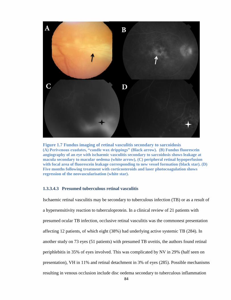
84
Figure 1.7 Fundus imaging of retinal vasculitis secondary to sarcoidosis (A) Perivenous exudates, “candle wax drippings” (Black arrow). (B) Fundus fluorescein
angiography of an eye with ischaemic vasculitis secondary to sarcoidosis shows leakage at
macula secondary to macular oedema (white arrow), (C) peripheral retinal hypoperfusion
with focal area of fluorescein leakage corresponding to new vessel formation (black star). (D)
Five months following treatment with corticosteroids and laser photocoagulation shows
regression of the neovascularisation (white star).
1.3.3.4.3 Presumed tuberculous retinal vasculitis
Ischaemic retinal vasculitis may be secondary to tuberculous infection (TB) or as a result of
a hypersensitivity reaction to tuberculoprotein. In a clinical review of 21 patients with
presumed ocular TB infection, occlusive retinal vasculitis was the commonest presentation
affecting 12 patients, of which eight (38%) had underlying active systemic TB (284). In
another study on 73 eyes (51 patients) with presumed TB uveitis, the authors found retinal
periphlebitis in 35% of eyes involved. This was complicated by NV in 29% (half seen on
presentation), VH in 11% and retinal detachment in 3% of eyes (285). Possible mechanisms
resulting in venous occlusion include disc oedema secondary to tuberculous inflammation
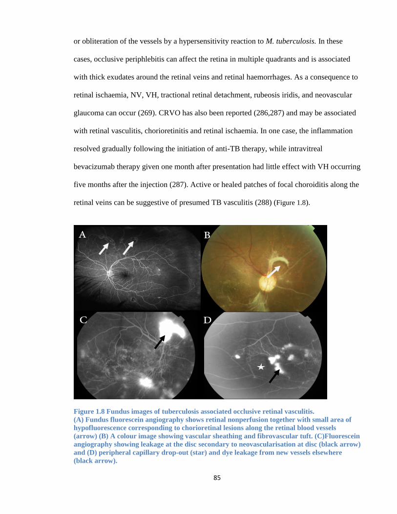
85
or obliteration of the vessels by a hypersensitivity reaction to M. tuberculosis. In these
cases, occlusive periphlebitis can affect the retina in multiple quadrants and is associated
with thick exudates around the retinal veins and retinal haemorrhages. As a consequence to
retinal ischaemia, NV, VH, tractional retinal detachment, rubeosis iridis, and neovascular
glaucoma can occur (269). CRVO has also been reported (286,287) and may be associated
with retinal vasculitis, chorioretinitis and retinal ischaemia. In one case, the inflammation
resolved gradually following the initiation of anti-TB therapy, while intravitreal
bevacizumab therapy given one month after presentation had little effect with VH occurring
five months after the injection (287). Active or healed patches of focal choroiditis along the
retinal veins can be suggestive of presumed TB vasculitis (288) (Figure 1.8).
Figure 1.8 Fundus images of tuberculosis associated occlusive retinal vasculitis.
(A) Fundus fluorescein angiography shows retinal nonperfusion together with small area of
hypofluorescence corresponding to chorioretinal lesions along the retinal blood vessels
(arrow) (B) A colour image showing vascular sheathing and fibrovascular tuft. (C)Fluorescein
angiography showing leakage at the disc secondary to neovascularisation at disc (black arrow)
and (D) peripheral capillary drop-out (star) and dye leakage from new vessels elsewhere
(black arrow).
86
1.3.3.4.4 Systemic lupus erythematosus
The incidence of retinopathy in patients with SLE ranges from 3% to 29% (289–291)
depending on the studied population and associated risk factors for SLE retinopathy such as
the presence of anticardiolipin antibodies, central nervous system involvement, serum
creatinine level, and SLE activity (289,292). Retinal vasculopathy and associated vascular
occlusion is a sight threatening manifestation of SLE retinopathy, reported to cause severe
visual loss in 55% of patients (293). The main factor affecting visual outcome in these
cases is the occurrence of NV with or without VH, reported in about 40% of the cases
(290), as well as an increased risk of developing retinal vein occlusion (294).
Vasoocclusive retinopathy can be the primary manifestation that leads to the diagnosis of
SLE (295). The exact pathogenesis of vascular occlusion is not clear, but there have been
proposed theories on the role of immune-complex deposition and complement activation
with fibrinoid degeneration of the vascular wall as factors contributing to the vascular
damage seen in these cases (296,297). Occlusive retinal vasculopathy involving the retinal
arterioles may present with cotton-wool spots, predominantly in the posterior pole,
representing retinal micro-infarctions. On FFA (Figure 1.9), vascular occlusion can
manifest as widespread arteriolar or branch retinal artery occlusion (BRAO) with severe
retinal ischaemia and NV (290). Larger retinal vessels may be occluded leading to retinal
and optic disc infarction that may also result in NV (298). Central retinal artery occlusion
(CRAO) and central retinal vein occlusion (CRVO), while very rarely seen in other causes
of retinal vasculitis, have been reported secondary to SLE (299–301). In one report
involving 71 patients with SLE and retinal vasculopathy, three (6.3%) of the patients had
either CRAO, CRVO or ischaemic optic neuropathy (266).
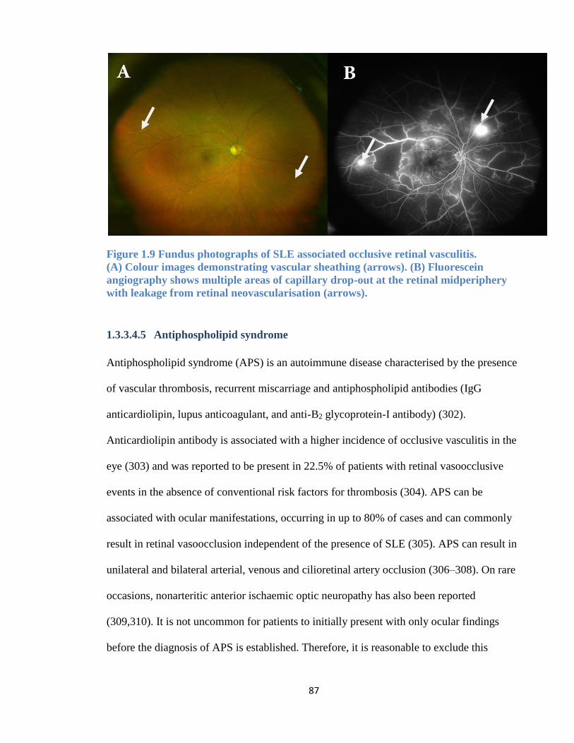
87
Figure 1.9 Fundus photographs of SLE associated occlusive retinal vasculitis.
(A) Colour images demonstrating vascular sheathing (arrows). (B) Fluorescein
angiography shows multiple areas of capillary drop-out at the retinal midperiphery
with leakage from retinal neovascularisation (arrows).
1.3.3.4.5 Antiphospholipid syndrome
Antiphospholipid syndrome (APS) is an autoimmune disease characterised by the presence
of vascular thrombosis, recurrent miscarriage and antiphospholipid antibodies (IgG
anticardiolipin, lupus anticoagulant, and anti-B2 glycoprotein-I antibody) (302).
Anticardiolipin antibody is associated with a higher incidence of occlusive vasculitis in the
eye (303) and was reported to be present in 22.5% of patients with retinal vasoocclusive
events in the absence of conventional risk factors for thrombosis (304). APS can be
associated with ocular manifestations, occurring in up to 80% of cases and can commonly
result in retinal vasoocclusion independent of the presence of SLE (305). APS can result in
unilateral and bilateral arterial, venous and cilioretinal artery occlusion (306–308). On rare
occasions, nonarteritic anterior ischaemic optic neuropathy has also been reported
(309,310). It is not uncommon for patients to initially present with only ocular findings
before the diagnosis of APS is established. Therefore, it is reasonable to exclude this
88
condition in younger patients presenting with occlusive vasculitis in the absence of known
systemic risk factors (311).
1.3.3.4.6 Multiple sclerosis
The risk of uveitis in patients with MS is ten times higher compared to the general
population, commonly in the form of intermediate uveitis (312). However, the presence of
peripheral periphlebitis was described in the early case reports of MS related uveitis
(313,314). A review of 1254 uveitis case records at a tertiary eye centre found 14 (1.3%) to
be MS related uveitis, with more than half of the cases associated with vasculitis (61).
Periphlebitis has been suggested to be a risk factor for the development of neurological
manifestations of MS, including optic neuritis (156,315).
Many theories have been proposed to explain the pathophysiological correlation between
MS and the presence of periphlebitis (316). In an autopsy series of 93 eyes from patients
with an established diagnosis of MS, seven showed segmental perivenular infiltrates of
lymphocytes and plasma cells (317), lymphocyte and plasma cells were also concomitantly
observed around retinal and central nervous system veins in two patients with MS, leading
to the conclusion that periphlebitis is an early event that may lead to plaque formation in
the brain (318). While periphlebitis has been reported in 20% of eyes (319), occlusive
vasculitis and NV are rare complications in MS related uveitis (320–324). In a case series
of 16 patients with MS related uveitis, eight suffered from ischaemic retinal vasculitis with
NV requiring SLP, while three eyes had unresolved VH secondary to NV requiring
vitrectomy (323). Peripheral retinal ischaemia can be severe and had been reported to cause
bilateral rubeosis iridis and neovascular glaucoma. While the rubeotic vessels regressed
following treatment with oral corticosteroids and SLP, one eye required trabeculectomy to
manage the glaucoma. No steroid sparing drugs were required in this case (325). Although
89
the presence of VH in uveitis can be highly suspicious of ocular BD or sarcoidosis, the
presence of MS may also need to be excluded in patients with IU that develop VH. In a
series of 25 patients with MS related IU, six (24%) had periphlebitis associated with retinal
ischaemia and VH and four had NV on angiography. VH occurred at an average of five
years following onset of uveitis, while it was the initial presenting manifestation in two
patients (320). The visual prognosis of MS related uveitis is generally good (61); however,
in those with occlusive vasculitis and NV the visual prognosis may vary. In one report, two
of six patients with retinal ischaemia and VH had a final vision of 20/80 five years after
onset of VH (320).
1.3.3.4.7 Other causes of occlusive retinal vasculitis
Idiopathic retinal vasculitis, arteriolar macro aneurysms and neuroretinitis (IRVAN) is
characterised by recurrent multiple branch retinal arterial occlusions of unknown cause in
one or both eyes of healthy middle-aged patients with no associated ocular or systemic
aetiology. An important cause of visual loss in IRVAN is chronic CMO with hard exudate
accumulation in the fovea. Vision loss also occurs secondary to peripheral capillary non-
perfusion leading to NV and tractional retinal detachment (326).
West Nile virus infection has been associated with chorioretinitis as its most common
ocular finding; whereas occlusive retinal vasculitis is an uncommon finding reported to date
in eight cases. Findings include perivascular sheathing, microaneurysms, cotton wool spots,
intraretinal haemorrhages and NV with or without macular ischaemia. Interestingly, six of
these cases with established West Nile virus infection also suffered from DM (327,328).
Progressive outer retinal necrosis, a viral retinitis most commonly caused by VZV, can
rarely be associated with occlusive vasculitis. In a recent report, a 45 years old HIV
90
infected patient on highly active antiretroviral therapy presented with bilateral progressive
retinal necrosis and was treated with combined systemic and intravitreal ganciclovir
therapy, which was repeated a week later. Despite all the management efforts, he
progressed to occlusive vasculitis and CRVO within a week after the second intravitreal
injection. Even following the application of PRP, which acts poorly on necrotic retina, the
condition progressed with the occurrence of VH and eventually retinal detachment (329).
Haemorrhagic occlusive retinal vasculitis is rare but potentially aggressive form of retinal
vasculitis, predominantly phlebitis, occuring after uncomplicated cataract surgery
associated with prophylactic intracameral injection of antibiotics. While the exact
mechanism is not clear, it is believed to be caused by delayed immune reaction against the
antibiotics Intraocular injection of aminoglycosides (amikacin and gentamycin) were
reported to be associated with severe intraocular inflammation 1-3 days post surgery
together with macular ischaemia (330,331). A case series of 11 eyes were reported with a
diagnosis of haemorrhagic occlusive retinal vasculitis, all occurred following intracameral
vancomycin injection in uncomplicated cataract surgery. Treatment involved topical and
systemic corticosteroids together with PRP and anti-VEGF intravitreal injection. In
addition, four cases received antiviral medications, four received intravitreal antibiotics,
and four underwent vitrectomy. Despite all the treatments, eight of the 11 eyes had a final
visual acuity worse than 6/60 (332). Crohn's disease has been reported to be associated with
ischaemic retinal vasculitis, NV (333), NV glaucoma (334) and CRAO (335).
91
1.3.3.5 Treatment
1.3.3.5.1 Systemic immunosuppressants
Severe retinal vasculitis requires adequate inflammation control using corticosteroids and,
in non-infectious vasculitis, may need the addition of IMT (336). IMT of choice include
MMF, MTX and Cyclosporine A. The latter has shown to be effective in providing long-
term inflammatory control but can be associated with renal toxicity (337). Meanwhile,
AZA in BD with retinal vasculitis may not be very effective in producing complete
resolution and relapse prevention during corticosteroid tapering(338). In SLE vasculopathy,
systemic corticosteroids and IMT, such as cyclophosphamide and MMF, are established
treatments that can reduce vasculopathy and resolve cotton wool spots (339), though there
is little evidence supporting their role in preventing the progression of retinal vasoocclusion
(290). In presumed TB vasculitis, commencing systemic anti-TB therapy is useful in
controlling the inflammation by suppressing the active TB focus, which causes immune
activation and triggers uveitis. In addition, adjunctive use of systemic corticosteroid therapy
may be required in the management of these cases to prevent damage to ocular tissues
especially from delayed hypersensitivity.
1.3.3.5.2 Biologics
Anti TNFα drugs such as infliximab and adalimumab have been used successfully in the
management of sight threatening retinal vasculitis. In severe ocular BD, anti-TNFα can be
considered as first-line IMT (133) or used in cases refractory to other IMT to reduce the
risk of severe visual loss and promote long term remission of uveitis(275,340,341). BD
vasculitis with NVD has been reported to regress, recurrent VH resolve, and vision improve
following extended treatment with infliximab (342,343). Anti- TNFα is used successfully
in treating refractive cases of sarcoidosis with retinal vasculitis, especially infliximab
92
(137,344) and adalimumab (345,346). Clinical reports on the use of infliximab to control
ischaemic retinal vasculitis secondary to sarcoidosis have shown good results, especially in
cases where ocular symptoms manifest despite the use of IMT (347). Meanwhile,
etanercept is not only less effective in managing sarcoidosis, it is also reported to induce
sarcoid IU and PANU (348,349). It should be noted that anti-TNF, often used in the
management of severe non-infectious uveitis, should be avoided in treating MS related
uveitis as it may precipitate or exacerbate nerve demyelination and worsen the neurological
manifestations of this disease (350). Infliximab used in patients with IRVAN was very
successful in inducing dramatic resolution of ocular inflammation, reduction of retinal
exudation, improving nerve leakage and vision improvement after the first dose of
infliximab therapy. However, it was not useful in preventing NV formation which occurred
months later requiring laser therapy(351). The use of rituximab, a chimeric monoclonal
antibody against CD20+ B cells, demonstrated some benefit in treating severe cases of SLE
in uncontrolled studies but failed to prove superiority against placebo groups in a
randomised controlled trial (352). Rituximab combined with cyclophosphamide infusions
was shown to result in rapid resolution of retinal vasoocclusion in a paediatric group of
SLE patients when used early in the course of the disease (353).
Interferon alfa (INFα) therapies have been used in selected conditions to control
inflammation. In ocular BD, INFα-2a therapy was reported to provide long lasting
remission in up to 55% of cases even after discontinuation of therapy (354). In a
retrospective study, INFα-2a was effective in controlling retinal vasculitis in 36/38 eyes
with BD, and in 18/22 eyes with other causes of retinal vasculitis (355). INFα-2a may also
result in reperfusion of vasoocclusion (356) and induce NVD regression among BD
vasculitis even in the absence of concomitant SLP (357). In a retrospective review, five
93
patients with BD and unilateral ischaemic NVD received SLP; three had resolution of NVD
following laser treatment while the other two patients responded only following additional
treatment with INF-α-2a therapy (278).
The role of INFβ, an established treatment for MS, needs to be further studied to examine
its effectiveness in controlling MS with retinal vasculitis. In a small retrospective study of
13 patients with MS related uveitis, ten of which were associated with retinal vasculitis,
showed promising results with improvement of visual acuity in 71% of the eyes while a
corticosteroid sparing effect was achieved in all cases (358).
1.3.3.5.3 Retinal laser photocoagulation and intravitreal anti-VEGF
SLP is the main approach in managing NV that form secondary to occlusive vasculitis. In
patients with presumed TB vasculitis, SLP was found to be very effective in inducing
involution of NV. In a case series of 21 eyes with presumed TB vasculitis that received
SLP for NV, there was no recurrence of VH or NV formation within a mean follow up
period of 18 months (285). In BD, SLP is useful in inducing regression of NV and
preventing further complications such as NV glaucoma (359). In patients with IRVAN,
SLP has been recommended in the presence of retinal ischaemia before or shortly after the
formation of NV regardless of the extent of vascular closure in order to prevent its
progression and maintain good visual outcome (360). Another study suggested using SLP
only in eyes with retinal ischaemia involving more than two quadrants (361). In addition to
SLP, other treatment options for IRVAN include macular grid laser, vitrectomy and anti-
TNFα agents with a smaller role for corticosteroids (351,362).
The primary treatment of retinal NV among patients with SLE and APS vasculopathy
involves the use of SLP to the ischaemic area with or without intravitreal anti-VEGF agents
94
(308). Unlike cases with presumed TB vasculitis, SLP is less effective in causing regression
of NV in SLE and APS vasculopathy. In a systematic review of literature, SLP was
performed on 22 eyes, causing regression of the NV and stabilisation of vision in only 54%
of the cases (290). Thus, it is not uncommon to see NV formation with subsequent VH and
vitreoretinal traction even after retinal laser application (295). In the absence of randomised
clinical trials, it is difficult to assess the role of SLP alone in controlling NV due to the
concomitant use of IMT in most cases. Intravitreal bevacizumab can be used in eyes with
recurrent or persistent NV following SLP. A reported case of SLE with NVE that
progressed despite the use of IMT and fill-in laser, did respond to one intravitreal injection
of bevacizumab resulting in NVE regression with no new bleeding over three months
follow up (363). However, bevacizumab itself can reduce retinal perfusion and worsen
retinal ischaemia and therefore should be administered concomitantly with SLP. In a report
of two patients with SLE, one received bevacizumab combined with SLP that resulted in
halting the progression of the vascular occlusion with regression of the NVD. The second
patient, who did not have laser, had progression of retinal ischaemia with secondary NVE
within a month of injecting bevacizumab (364). In rare cases, intravitreal bevacizumab was
reported to aggravate capillary nonperfusion within a day following injection despite
previous administration of SLP (365).
Based on the treatment options described above, a proposed treatment pathway or protocol
for managing retinal vasculitis is illustrated in Figure 1.10
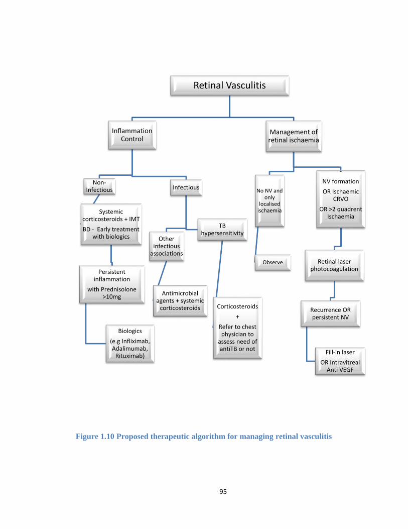
95
Figure 1.10 Proposed therapeutic algorithm for managing retinal vasculitis
Retinal Vasculitis
Inflammation Control
Non-Infectious
Systemic corticosteroids + IMT
BD - Early treatment with biologics
Persistent inflammation
with Prednisolone >10mg
Biologics
(e.g Infliximab, Adalimumab,
Rituximab)
Infectious
Other infectious
associations
Antimicrobial agents + systemic
corticosteroids
TB hypersensitivity
Corticosteroids
+
Refer to chest physician to
assess need of antiTB or not
Management of retinal ischaemia
No NV and only
localised ischaemia
Observe
-
NV formation
OR Ischaemic CRVO
OR >2 quadrent Ischaemia
Retinal laser photocoagulation
Recurrence OR persistent NV
Fill-in laser
OR Intravitreal Anti VEGF
96
1.3.4 Cataract surgery in uveitis
1.3.4.1 Management of cataract in eyes with uveitis
The decision and timing of cataract surgery is subject to many factors including the visual
potential, uveitis activity, associated ocular comorbidity, and the risk of amblyopia among
paediatric age group with cataract. The main indications for cataract surgery in eyes with
uveitis is to achieve a better visual outcome, or as part of managing the uveitis secondary to
leakage of lens proteins (phacoantigenic uveitis). Cataract surgery is also required to obtain
a better fundus view necessary in managing certain retinal pathologies such as assessment
of fundus disorders e.g. diabetic retinopathy or glaucomatous optic neuropathy, or for
performing vitreoretinal surgeries or applying retinal laser therapy (366).
1.3.4.1.1 Preoperative management
Preoperative assessment is essential to decide on the visual potential of the eye if cataract
surgery is to be performed and whether any associated complications may have an impact
on the visual outcome postoperatively. The macular and optic nerve structure and function
should be assessed prior to surgery to exclude the presence of any coexisting complications
that may jeopardise the outcome of surgery, such as the presence of macular scar or
atrophy, macular ischaemia, and chronic CMO; as well as the presence of pre-existing optic
neuropathy or atrophy. In cases where there is poor view of the fundus secondary to a dens
cataract, B-scan ultrasonography is essential to exclude the presence of retinal detachment
as a complication of uveitis.
Preoperative inflammation control is essential in preventing complications associated with
cataract surgery in uveitis with most of the literature recommending the eye be quiescent
for a minimum of three months prior to surgical intervention. In certain situations, such as
previous history of CMO or severe panuveitis, prophylactic anti-inflammatory medication
97
is required to be initiated 7-14 days prior to surgery. The most common prophylactic
therapy is in the form of systemic prednisolone given for adults at a dose of 40mg once a
day, or 0.5 to 1mg/kg, for two weeks until the day of surgery. Alternatively, preoperative
periocular or intraocular steroids, such as IVTA, can be given preoperatively (367). Case
reports exists which describe the successful use of dexamethasone intravitreal implants
(Ozurdex®) preoperatively as a prophylactic measure in JIA related uveitis with the eye
continuing to be quiescent over the 10 month follow-up period (368).
1.3.4.1.2 Intraoperative management
Since the beginning of the new millennium, phacoemulsification with in-the-bag
intraocular lens (IOL) implantation has become the preferred surgical procedure for
cataract removal in most people with uveitis (369). Cataract surgery in uveitic eyes can be
difficult due to limited access secondary to the possible presence of band keratopathy,
posterior synechiae, miotic pupil, and pupillary fibrinous membranes over the lens. A poor
view may require managing the corneal band karatopathy first before advancing with the
cataract surgery, either through phototherapeutic keratectomy or chemical chelation with
ethylenediaminetetraacetic acid. Efforts may also be needed to release the
adhesions/synechiae and expand the pupil size through variable approaches such as the use
of viscoelastics and iris hooks (367). In some cases with IU or FHC associated with
significant vitritis, cataract removal is combined with pars plana vitrectomy which helps
not only to improve vision and reducing floaters, but also to reduce the postoperative
inflammation and the incidence of postsurgical CMO (370).
In addition to anterior capsulotomy and lens aspiration within the sac, cataract removal with
IOL implantation in children with uveitis often requires an additional step of posterior
capsulotomy combined with anterior vitrectomy to minimise the chance of posterior
98
capsular opacity (PCO) and fibrinous deposition over the IOL (371). In the presence of
concomitant glaucoma and cataract, it is recommended to avoid combined glaucoma and
cataract surgery due to the high risk of bleb failure in these cases, and thus it is better to
remove the cataract first prior to planning for glaucoma surgery (372).
The choice of optimum IOL associated with minimal postoperative adverse effect has been
addressed in several studies. Acrylic IOLs in some studies show superiority over other
types of IOL materials, such as silicon lenses, and was associated with the lowest level of
postoperative uveitis and lowest rate of PCO within the first 6 months of follow up
postoperatively (369). However, a recent review paper found no enough evidence that
acrylic lenses may perform better than silicon lenses (373). The anterior chamber IOL in a
recent study was not associated with a significant increase in the incidence of intraoperative
and postoperative complications among uveitic eyes over five years follow up period when
compared to a control group without uveitis. However, the uveitis flare-up attributed to the
IOL and the risk of ERM formation were indeed more in uveitic eyes with anterior chamber
IOL compare to non uveitic eyes with same lens type (374). When another study made a
comparison to posterior chamber IOL in uveitic eyes, the anterior chamber IOL did not
have a significant difference in the risk of long-term complications and can still be
associated with a significant improvement in the visual acuity over 3 years follow up period
postoperatively (375).
Overall, uveitic eyes undergoing cataract surgery with IOL implantation have a better
visual outcome than eyes with no IOL placement as the latter appeared in a systematic
review to only achieve 6/12 vision or better in 23% of cases (376). In some cases of chronic
AU, mainly children with JIA associated uveitis, the decision to insert an IOL can be
controversial. IOL insertion in these cases may be associated with variable postoperative
99
complications such as fibrin deposition and synechiae, pupillary membrane formation,
hypotony, and secondary lens opacity, with some cases requiring IOL explantation.
However, several more recent studies have demonstrated no significant worsening of
uveitis outcome post cataract removal and IOL implantation among cases with JIA
associated uveitis. One study examined the long term outcome of 48 eyes in children with
JIA associated uveitis over 16 years follow up period and found no significant difference in
the incidence of postoperative complications such as elevated IOL, glaucoma, and CMO
between pseudophakic eyes and aphakic ones. Moreover, the pseudophakic group
experience a better visual outcome over seven years follow-up compared to the aphakic
eyes, with no incidence of phthisis or hypotony (377).
1.3.4.1.3 Postoperative management
Postoperatively, local and systemic steroids are tapered gradually based on the degree of
postoperative inflammation. In eyes with flare up of uveitis postoperatively, adequate
administration of topical steroids and mydriatic eye drops should be initiated. Systemic or
periocular/intraocular steroid injection can be administered in the presence of severe uveitis
not responding to topical drops or in the presence of postoperative CMO. Other
postoperative complications require management accordingly such as PCO and elevated
IOP.
1.3.4.2 Complications of cataract surgery in uveitis
Prior to the era of using systemic corticosteroids to control inflammation, cataract surgery
in uveitic eyes was inevitably associated with significant preoperative and postoperative
complications that could sometimes progress to permanent vision loss (378,379). The
incidence of complications associated with cataract surgery in uveitic eyes has reduced over
the past years, mainly due to the improved ability to control inflammation preoperatively
100
and the recent developments in implementing new systemic and local anti-inflammatory
therapies, as well as the rapid evolution in microsurgical techniques over the past few years
(372).
Complications of cataract surgery in uveitis include retained cortical and nuclear lens
matter, hyphaema, severe or persistent postoperative inflammation, CMO, elevated IOP,
pupillary membrane, PCO, hypotony and endophthalmitis. A recent study looked at the
outcome of phacoemulsification in uveitic eyes with more than three years follow up
period. Intraoperative complications included posterior capsule rupture (1.7%) whereas
long term complications postoperatively include CMO in 10.2% with an incidence rate of
0.02 per EY which was highly related to the presence of chronic postoperative
inflammation and also in those with more than one postoperative relapse per year. PCO
occurred in 27% with an incidence rate of 0.07 per EY with no significant association with
the presence of chronic postoperative inflammation. Elevated IOP more than 21mmHg
observed in 10.2% of cases with an incidence of 0.04 per EY. Hyoptony on the other hand
was related to the presence of chronic inflammation postoperatively and mainly confined to
eyes with AU, occurring in 5.1% of cases and an incidence of 0.02 per EY (380). Cataract
surgery in uveitic eyes can also increase risk of hypotony by seven fold, especially in
aphakic eyes. Even with phacoemulsification, which carries a lower risk of hypotony, there
is five fold increased risk when performed on uveitic eyes (381). The incidence of
hyphaema in FHC post cataract surgery can vary from 3.6% to 76%, with recent reports
showing a reduced incidence mainly following the use of improved microsurgical
techniques (372). The incidence of PCO following cataract surgery in FHC has been
reported to occur in 14.6% of eyes within a mean follow up period of 17 months after
surgery (148). Meanwhile, the most significant sight threatening complication of cataract
101
surgery in FHC cases is glaucoma, reported in 3 to 35% of cases, with almost 2/3 of cases
eventually requiring glaucoma surgery (382). Cataract surgery in general is associated with
a small risk of endophthalmitis, about 1 in 1000 cases, and an overall incidence rate of
0.12%, leading to permanent vision loss in most of the cases (383). The risk of
postoperative endophthalmitis that can be reduced with the use of intraoperative
prophylactic intracameral injection of antibiotics, such as cefuroxime (1 mg/0.1 ml) (384).
102
2 CHAPTER TWO: GENERAL METHODOLOGY
2.1 Patients and setting
The initial stage of the research involved reviewing hospital case notes of all patients with
uveitis who attended the uveitis clinic of a single consultant (S.L.) at Moorfields Eye
Hospital, London, United Kingdom, between January 2012 and December 2013. The study
received institutional review board approval (ethical approval for data collection:
LIGS10201, visual loss in uveitis; Clinical Trials registry no., NCT01983488). Collected
data from these case-notes were eventually entered into a Microsoft Access database
designed to be used as a general database for uveitis patients (Figure 2.1). This eventually
led to the collection of 1169 patients with uveitis. From this collected cohort of uveitis
patients, eyes from diabetic patients, retinal vasculitis and those underwent cataract
surgeries who met the inclusion criteria were selected for three separate studies and were
further analysed as discussed in the methodology of chapter 3, 4 and 5.
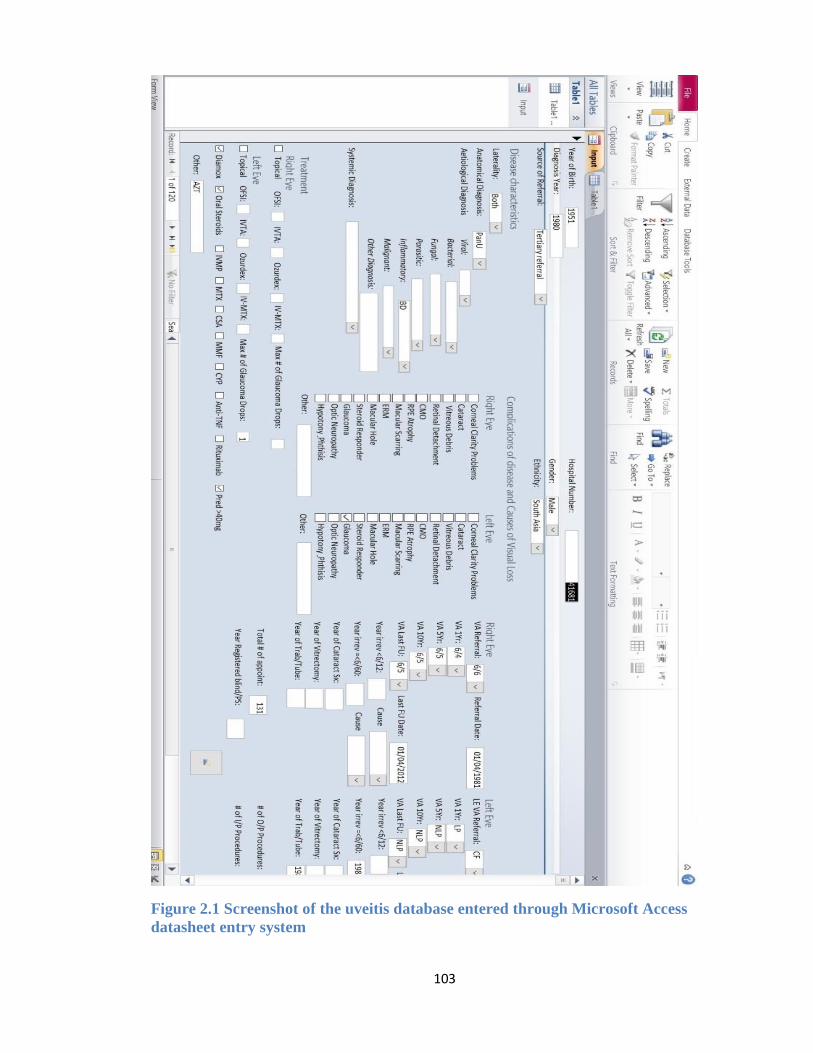
103
Figure 2.1 Screenshot of the uveitis database entered through Microsoft Access
datasheet entry system
104
2.2 Uveitis clinical and demographic data
Patient demographic data such as gender and age at time of uveitis diagnosis were
documented. Uveitis was classified based on the SUN Working Group classification of
uveitis as AU (inflammation confined to the anterior segment), IU (inflammatory cells
observed in the anterior vitreous and commonly associated with snowballs or snowbanks),
PU (inflammation mainly affecting the retina and/or choroid), and PANU (inflammation is
diffuse involving the anterior chamber, vitreous and retina or choroid (19). In addition, the
aetiology of uveitis if known was recorded including infectious and non-infectious
aetiology together with any localised ocular pathology or systemic diseases associated with
uveitis development. Other data of interest were the first and last clinic visit date and
changes in the clinical data over the follow up period. Uveitis management approach was
recorded including the use of topical, periocular and intraocular injections as well as the use
of systemic prednisolone and IMT therapy. Additional data included the use of laser
therapy and the type and timing of surgical management of uveitis complications e.g.
cataract surgery, aqueous shunt and vitrectomy.
All patients had their best corrected visual acuity (BCVA) measured separately in each eye
during the clinic visits. This was done using an illuminated Snellen chart at a distance of six
metres. The BCVA was obtained from measuring the vision while looking through a
pinhole or with patient’s glasses prescription and the result was recorded in Snellen acuity
format. For eyes that were unable to see the top line at a distance of six metres, they were
brought up closer to the chart by one metre a time. If there was still no view at one metre,
then ability to view finger counting, hand movement, or light perception were assessed at a
distance of 1 meter. For the purpose of continuous or longitudinal data analysis, the BCVA
measurements were converted from Snellen acuity into the negative value of the decadal
105
logarithm of the minimal angle of resolution (LogMAR) (385). For visual acuity of
counting fingers or worse, the following conversion was used: counting fingers = 2.0
LogMAR, hand motion = 2.3 LogMAR, light perception = 2.6 LogMAR, and no light
perception = 2.9 LogMAR.(386,387) Vision loss was defined as final BCVA of ≤ 6/15
(<0.3 LogMAR) or loss of > 2 snellen lines in eyes with baseline BCVA of 6/24 or better.
Vision loss were described as either moderate vision loss (MVL) in eyes with BCVA
between 6/15 and 6/36 or severe vision loss (SVL) in eyes with BCVA ≤ 6/60 according to
the SUN working group criteria (19).
Patients attending the clinic had a full ophthalmological examination including slit-lamp
examination, funduscopy using indirect ophthalmoscopy after pupil dilation and IOP check
using Goldman applanation tonometry. Ancillary tests were requested as appropriate to the
history and the clinical examination such as OCT using Topcon 3D OCT (Topcon, Inc)
(Error! Reference source not found. ) or Heidelberg Spectralis (SD-OCT; Spectralis,
Heidelberg Engineering GmbH) (Figure 2.3), FFA and ICG taken through the digital retinal
camera system either as 30-50 degree images (Topcon TRC 50IX; Topcon Medical
Systems Inc, Paramus, NJ) or as ultra-widefield images (Optos PLC, Dunfermline,
Scotland) (Figure 1.9), as well as performing orbital B-Scan ultrasound,
electrophysiological and laboratory tests.
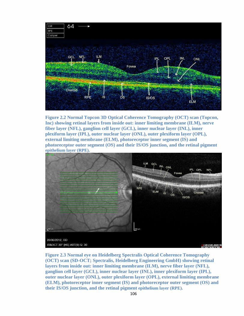
106
Figure 2.2 Normal Topcon 3D Optical Coherence Tomography (OCT) scan (Topcon,
Inc) showing retinal layers from inside out: inner limiting membrane (ILM), nerve
fiber layer (NFL), ganglion cell layer (GCL), inner nuclear layer (INL), inner
plexiform layer (IPL), outer nuclear layer (ONL), outer plexiform layer (OPL),
external limiting membrane (ELM), photoreceptor inner segment (IS) and
photoreceptor outer segment (OS) and their IS/OS junction, and the retinal pigment
epithelium layer (RPE).
Figure 2.3 Normal eye on Heidelberg Spectralis Optical Coherence Tomography
(OCT) scan (SD-OCT; Spectralis, Heidelberg Engineering GmbH) showing retinal
layers from inside out: inner limiting membrane (ILM), nerve fiber layer (NFL),
ganglion cell layer (GCL), inner nuclear layer (INL), inner plexiform layer (IPL),
outer nuclear layer (ONL), outer plexiform layer (OPL), external limiting membrane
(ELM), photoreceptor inner segment (IS) and photoreceptor outer segment (OS) and
their IS/OS junction, and the retinal pigment epithelium layer (RPE).
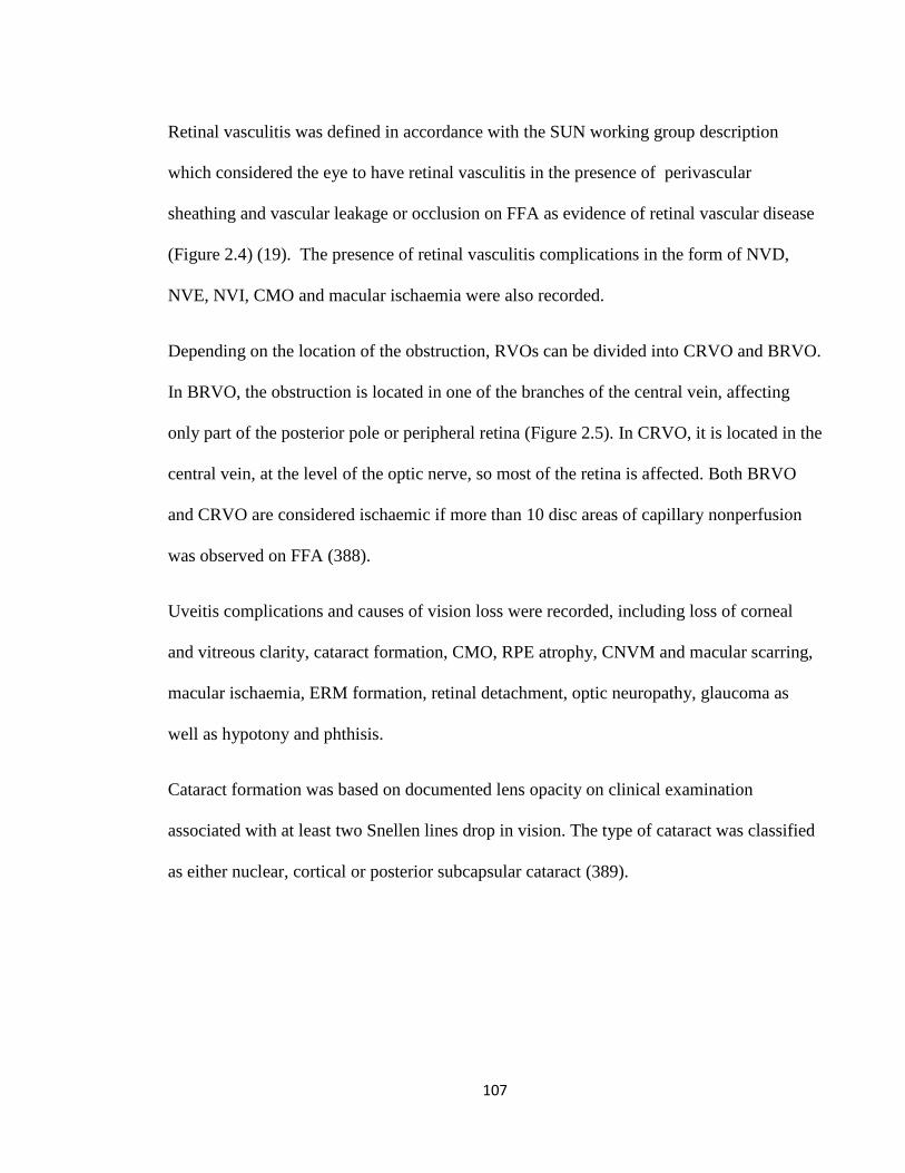
107
Retinal vasculitis was defined in accordance with the SUN working group description
which considered the eye to have retinal vasculitis in the presence of perivascular
sheathing and vascular leakage or occlusion on FFA as evidence of retinal vascular disease
(Figure 2.4) (19). The presence of retinal vasculitis complications in the form of NVD,
NVE, NVI, CMO and macular ischaemia were also recorded.
Depending on the location of the obstruction, RVOs can be divided into CRVO and BRVO.
In BRVO, the obstruction is located in one of the branches of the central vein, affecting
only part of the posterior pole or peripheral retina (Figure 2.5). In CRVO, it is located in the
central vein, at the level of the optic nerve, so most of the retina is affected. Both BRVO
and CRVO are considered ischaemic if more than 10 disc areas of capillary nonperfusion
was observed on FFA (388).
Uveitis complications and causes of vision loss were recorded, including loss of corneal
and vitreous clarity, cataract formation, CMO, RPE atrophy, CNVM and macular scarring,
macular ischaemia, ERM formation, retinal detachment, optic neuropathy, glaucoma as
well as hypotony and phthisis.
Cataract formation was based on documented lens opacity on clinical examination
associated with at least two Snellen lines drop in vision. The type of cataract was classified
as either nuclear, cortical or posterior subcapsular cataract (389).
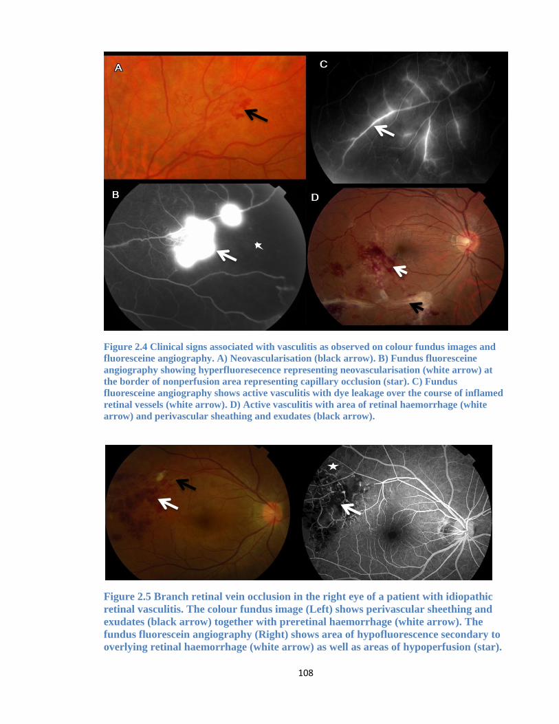
108
Figure 2.4 Clinical signs associated with vasculitis as observed on colour fundus images and
fluoresceine angiography. A) Neovascularisation (black arrow). B) Fundus fluoresceine
angiography showing hyperfluoresecence representing neovascularisation (white arrow) at
the border of nonperfusion area representing capillary occlusion (star). C) Fundus
fluoresceine angiography shows active vasculitis with dye leakage over the course of inflamed
retinal vessels (white arrow). D) Active vasculitis with area of retinal haemorrhage (white
arrow) and perivascular sheathing and exudates (black arrow).
Figure 2.5 Branch retinal vein occlusion in the right eye of a patient with idiopathic
retinal vasculitis. The colour fundus image (Left) shows perivascular sheething and
exudates (black arrow) together with preretinal haemorrhage (white arrow). The
fundus fluorescein angiography (Right) shows area of hypofluorescence secondary to
overlying retinal haemorrhage (white arrow) as well as areas of hypoperfusion (star).
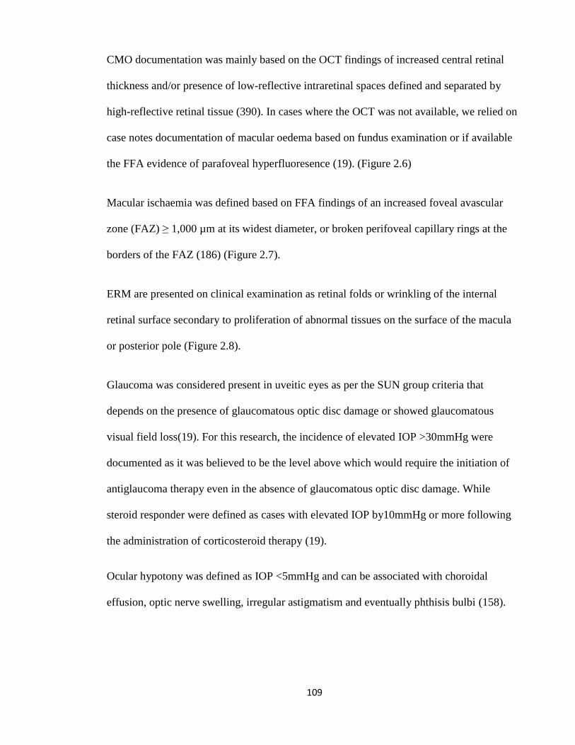
109
CMO documentation was mainly based on the OCT findings of increased central retinal
thickness and/or presence of low-reflective intraretinal spaces defined and separated by
high-reflective retinal tissue (390). In cases where the OCT was not available, we relied on
case notes documentation of macular oedema based on fundus examination or if available
the FFA evidence of parafoveal hyperfluoresence (19). (Figure 2.6)
Macular ischaemia was defined based on FFA findings of an increased foveal avascular
zone (FAZ) ≥ 1,000 µm at its widest diameter, or broken perifoveal capillary rings at the
borders of the FAZ (186) (Figure 2.7).
ERM are presented on clinical examination as retinal folds or wrinkling of the internal
retinal surface secondary to proliferation of abnormal tissues on the surface of the macula
or posterior pole (Figure 2.8).
Glaucoma was considered present in uveitic eyes as per the SUN group criteria that
depends on the presence of glaucomatous optic disc damage or showed glaucomatous
visual field loss(19). For this research, the incidence of elevated IOP >30mmHg were
documented as it was believed to be the level above which would require the initiation of
antiglaucoma therapy even in the absence of glaucomatous optic disc damage. While
steroid responder were defined as cases with elevated IOP by10mmHg or more following
the administration of corticosteroid therapy (19).
Ocular hypotony was defined as IOP <5mmHg and can be associated with choroidal
effusion, optic nerve swelling, irregular astigmatism and eventually phthisis bulbi (158).
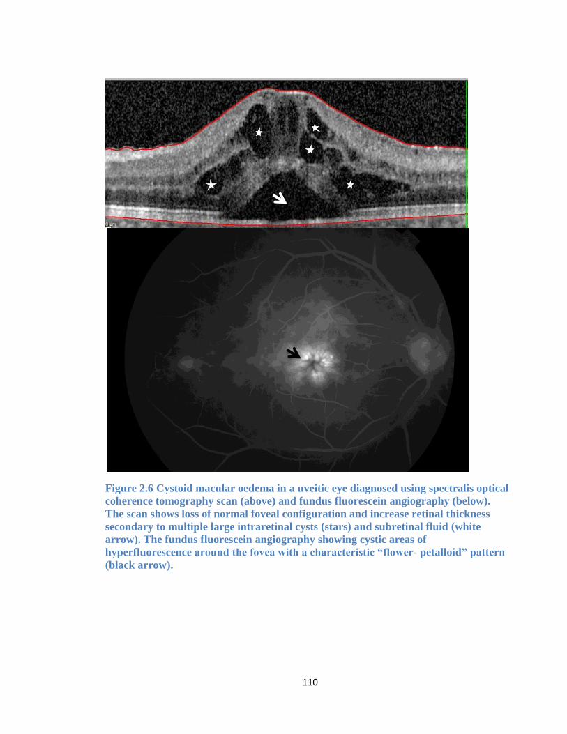
110
Figure 2.6 Cystoid macular oedema in a uveitic eye diagnosed using spectralis optical
coherence tomography scan (above) and fundus fluorescein angiography (below).
The scan shows loss of normal foveal configuration and increase retinal thickness
secondary to multiple large intraretinal cysts (stars) and subretinal fluid (white
arrow). The fundus fluorescein angiography showing cystic areas of
hyperfluorescence around the fovea with a characteristic “flower- petalloid” pattern
(black arrow).
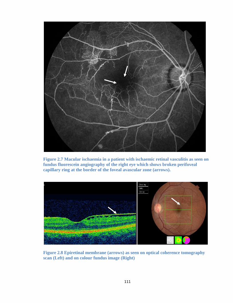
111
Figure 2.7 Macular ischaemia in a patient with ischaemic retinal vasculitis as seen on
fundus fluorescein angiography of the right eye which shows broken perifoveal
capillary ring at the border of the foveal avascular zone (arrows).
Figure 2.8 Epiretinal membrane (arrows) as seen on optical coherence tomography
scan (Left) and on colour fundus image (Right)
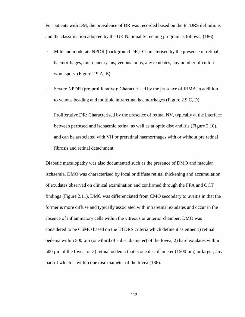
112
For patients with DM, the prevalence of DR was recorded based on the ETDRS definitions
and the classification adopted by the UK National Screening program as follows; (186)
- Mild and moderate NPDR (background DR): Characterised by the presence of retinal
haemorrhages, microaneurysms, venous loops, any exudates, any number of cotton
wool spots. (Figure 2.9 A, B)
- Severe NPDR (pre-proliferative): Characterised by the presence of IRMA in addition
to venous beading and multiple intraretinal haemorrhages (Figure 2.9 C, D)
- Proliferative DR: Characterised by the presence of retinal NV, typically at the interface
between perfused and ischaemic retina, as well as at optic disc and iris (Figure 2.10),
and can be associated with VH or preretinal haemorrhages with or without pre retinal
fibrosis and retinal detachment.
Diabetic maculopathy was also documented such as the presence of DMO and macular
ischaemia. DMO was characterised by focal or diffuse retinal thickening and accumulation
of exudates observed on clinical examination and confirmed through the FFA and OCT
findings (Figure 2.11). DMO was differenciated from CMO secondary to uveitis in that the
former is more diffuse and typically associated with intraretinal exudates and occur in the
absence of inflammatory cells within the vitreous or anterior chamber. DMO was
considered to be CSMO based on the ETDRS criteria which define it as either 1) retinal
oedema within 500 µm (one third of a disc diameter) of the fovea, 2) hard exudates within
500 µm of the fovea, or 3) retinal oedema that is one disc diameter (1500 µm) or larger, any
part of which is within one disc diameter of the fovea (186).
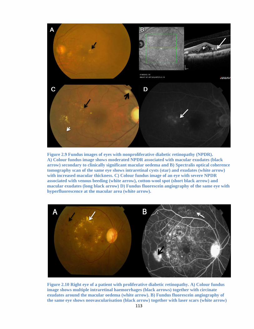
113
Figure 2.9 Fundus images of eyes with nonproliferative diabetic retinopathy (NPDR).
A) Colour fundus image shows moderated NPDR associated with macular exudates (black
arrow) secondary to clinically significant macular oedema and B) Spectralis optical coherence
tomography scan of the same eye shows intraretinal cysts (star) and exudates (white arrow)
with increased macular thickness. C) Colour fundus image of an eye with severe NPDR
associated with venous beeding (white arrow), cotton-wool spot (short black arrow) and
macular exudates (long black arrow) D) Fundus fluorescein angiography of the same eye with
hyperfluorescence at the macular area (white arrow).
Figure 2.10 Right eye of a patient with proliferative diabetic retinopathy. A) Colour fundus
image shows multiple intraretinal haemorrhages (black arrows) together with circinate
exudates around the macular oedema (white arrow). B) Fundus fluorescein angiography of
the same eye shows neovascularisation (black arrow) together with laser scars (white arrow)
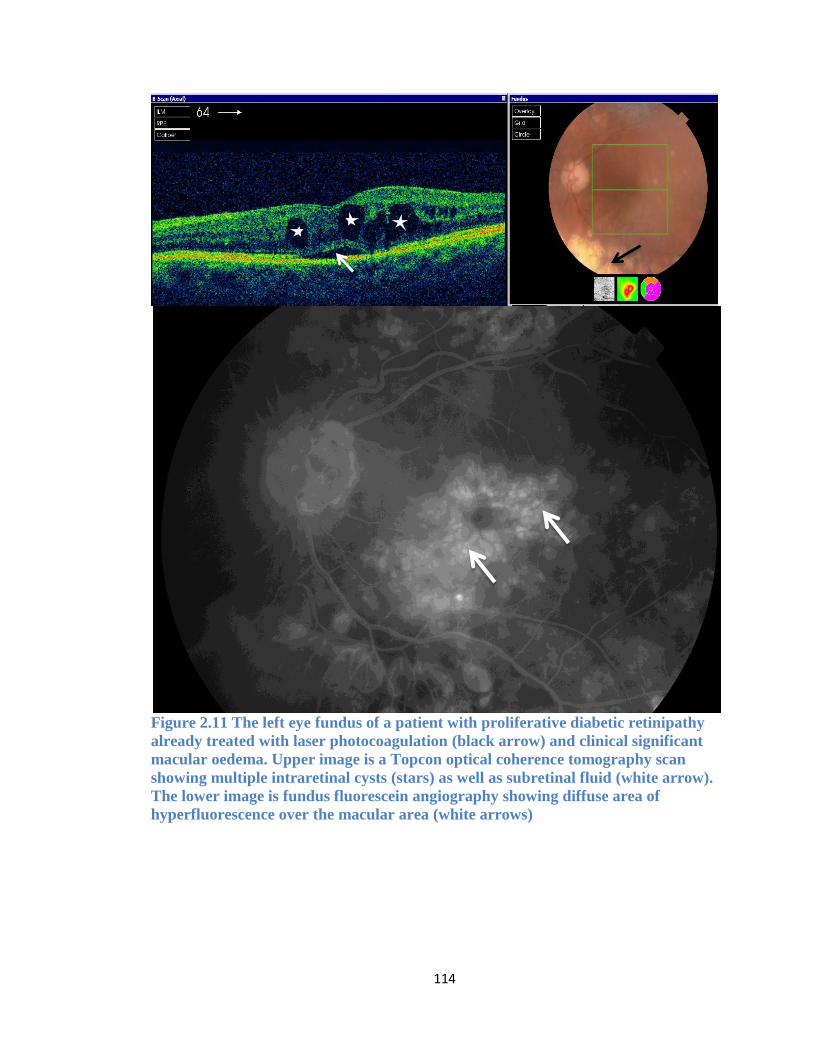
114
Figure 2.11 The left eye fundus of a patient with proliferative diabetic retinipathy
already treated with laser photocoagulation (black arrow) and clinical significant
macular oedema. Upper image is a Topcon optical coherence tomography scan
showing multiple intraretinal cysts (stars) as well as subretinal fluid (white arrow).
The lower image is fundus fluorescein angiography showing diffuse area of
hyperfluorescence over the macular area (white arrows)
115
2.3 Statistical analysis
All the continuous data measured in the research were tested for normality of distribution
through plotting of the data together with normality test using Shapiro-Wilk test which
considered the data to be not normally distributed if the p value was <0.05. Baseline
demographic and clinical parameters were described using mean and its standard error (SE)
for continuous data and percentages for categorical data, except when lack of normality in
distribution was observed for which case it was reported as median and interquantile range
(IQR). Continuous data between two dependent groups were compared using dependent t-
test for normally distributing data, or Wilcoxin signed rank test for data which lack
normality in distribution. Chi-square test or Fisher’s exact test was used, as appropriate, to
compare categorical variables among independent groups while using McNeemar test for
similar analysis between dependent groups. Repeated measurements were analysed using a
multivariate linear regression method to assess the mean difference (MD) in BCVA and
corresponding 95% confidence intervals (C.I.) over the follow up period compared to the
baseline measurement. This was performed using generalised estimating equation (GEE)
methods to account for correlations between the eyes at multiple intervals (391).
Kaplan-Meier survival analysis with Log-Rank (Mantel-Cox) test were applied together
with survival curve graphs to compare the survival rates for the incidence of some uveitis
outcomes such as vision loss, CMO and NV formation among studied subgroups. The
hazard ratio (HR) and its corresponding 95% C.I for vision loss and uveitis complications
were measured using Cox proportional hazards regression analysis.
The incidence rate per eye per year (EY) and 95% C.I for uveitis relapse and use of topical
and systemic corticosteroids were calculated using negative binominal regression with log

116
link model. The General Linear Model test was used to calculate the probability of
Type II (Beta) error, or the power of the study, aiming to have a power level of 80%
or more. Statistical analyses utilised the Statistical Package for the Social Sciences (SPSS)
software, version 21 (SPSS®, Chicago, USA). The level of statistical significance was set at
p<0.05.
117
3 CHAPTER THREE: THE INFLUENCE OF DIABETES
MELLITUS ON THE VISUAL OUTCOME AND
MANAGEMENT OF PATIENTS WITH UVEITIS
3.1 Introduction
DR is a well-known cause of newly acquired vision loss among the working age group
between 20 to 74 years old, and accounts for approximately 12% of all cases of blindness in
Western countries (392). In the UK, DR is the leading cause of blindness among the
working age group with an estimate of 4,200 blind people in England due to DR and an
annual 1,280 new cases each year (393). DR and diabetic maculopathy account for 6.3% of
registered cases of blindness in the UK and is the third most common cause of blindness
after degenerative macular changes (58.6%) and glaucoma (8.4%). It is also the second
most common cause of partial sight impairment in UK, accounting for 7.6% of cases (394).
One out of five patients with type 2 diabetes is diagnosed with DR within one year after
diabetes diagnosis in UK general practices (395).
Since 1868 when Noyes suggested that diabetes can cause iritis, many authors have
subsequently presented various hypothesis that supported (396–398) or disputed (399,400)
such an association. Few studies and case reviews have examined the association between
DM and uveitis, focusing mainly on diabetes as a risk factor for the development of AU
(25,401–404). Guy et al reported iritis in 30% of patients with type 1 DM associated with
autonomic neuropathy, suggesting that an immunological mechanism may be involved in
the development of uveitis as well as diabetic neuropathy (397). Another study of 340
patients with AU found 6% to be associated with DM. This was more prevalent in patients
with idiopathic AU, among which 12.5% had DM, compared to only 1.9% of AU patients
with an underlying ocular diagnosis (405). Rothova et al while studying the association of
118
uveitis with systemic diseases in 865 patients with uveitis, found 29 (3.3%) to have DM; 20
cases were idiopathic AU and nine cases were associated with an underlying aetiology
(398). DM is associated with alteration in blood-aqueous barrier permeability, resulting in
an increase in the aqueous protein concentration, or flare, in a degree parallel with the
progression of the DR (406).
The interaction between DM and uveitis can occur in two scenarios; either a patient known
to have DM presents with uveitis, or a uveitic patient is subsequently diagnosed with DM,
such as type 2 DM or steroid-induced DM. Oswal et al initially reported the clinical
characteristics of 36 diabetic patients (48 eyes) that presented with their first episodes of
uveitis (403). In this report, all of the cases were type 2 DM except for one case with type 1
DM. The uveitis occurred after a mean period of 6.8 years from time of diagnosis with DM.
Most of the uveitis cases were idiopathic and mainly in the form of AU (66%) followed by
PANU (27%) and IU (6%). An underlying infectious disease was associated with the
uveitis in 12.5% of the eyes, mainly toxoplasmosis, TB and syphilis associated uveitis
(403). The same group of patients were further studied through another longitudinal
retrospective research with a mean follow up period of 4.4 years. The study reported a final
visual outcome of 6/18 or worse in 15.5% of the eyes, mainly secondary to cataract (37%),
glaucoma (29%) and CMO (17%). In addition, DR was observed in 65.5% of the eyes,
including 6 eyes with CSMO and nine eyes had PDR. Progression of DR to proliferative
disease occurred in seven eyes over a mean of 4.4 years from onset of uveitis (407).
While the role of DM in inducing or aggravating ocular inflammatory diseases is still
controversial, the risk of uveitis therapy in inducing DM or jeopardising glycaemic control
is a well known adverse effect, especially with the use of systemic corticosteroids.
119
Systemic corticosteroids have been widely used in the management of sight threatening
uveitis, especially when topical or local corticosteroid use is ineffective or inapplicable and
in the presence of severe ocular inflammation behind the lens and for managing associated
complications such as CMO, optic neuritis or retinal vasculitis (86). However, systemic
corticosteroids can be associated with variable adverse effects including hyperglycaemia
and steroid induced DM (89). Corticosteroids can induce DM through inducing
gluconeogenesis as well as increasing peripheral insulin resistance (408). More recent in-
vitro studies suggested additional mechanisms, such as the inhibition of insulin secretion
through increasing the expression of α-2 adrenergic receptors (409), decreased cAMP
levels and pancreatic β-cell apoptosis through activating calcineurin phosphatase and
corticosteroid receptors as well as reducing the secretion and the insulinotropic effects of
the incretin glucagon-like peptide-1 (GLP-1) (410). In addition to systemic corticosteroid
therapy, some IMT therapy used in the management of uveitis can also have a diabetogenic
effect. Calcineurin inhibitors such as CSA and tacrolimus are associated with increasing
risk of hyperglycaemia or causing drug-induced DM through suppression of insulin
secretion and transcriptional regulations in pancreatic β-cells (411). Meanwhile, other IMT
such as MMF have not proven to carry similar diabetogenic effect (412).
The incidence and risk factors of corticosteroid-induced hyperglycaemia have been
addressed in the SITE multicentre retrospective study on 2,073 patients with inflammatory
eye diseases treated with oral corticosteroids (413). Steroid induced hyperglycaemia
requiring hypoglycaemic medications was observed in 25 patients (1.21%) on oral
corticosteroids compare to 0.19% of patients who were not on similar treatment. The use of
oral corticosteroid in the uveitis cohort was associated with four fold increase in risk of
steroid-induced hyperglycaemia compared to those who did not receive oral corticosteroids.
120
The risk is even higher in patients with higher initial dose of corticosteroids of >40mg per
day compare to those on lower initial dose. Other risk factors for the development of steroid
induced hyperglycaemia were older age and African-American race group. Interestingly,
62.7% of the uveitis patients on hypoglycaemic medications did subsequently manage to
discontinue these medications within one year after starting them (413). The study though
did not focus on the synergistic effect of IMT if used on the incidence of hyperglycaemia
secondary to oral corticosteroids, nor did it address the effect of the newly acquired
hyperglycaemia on the rate of uveitis activity or its influence, if any, on the management of
uveitis relapse.
Both macular oedema and secondary cataract are common causes of visual loss in uveitis
(22,414) and DM (415,416). However, few studies have assessed the cumulative effect of
uveitis and DM on visual outcome and uveitis management strategies. Furthermore, the rate
of development of DR in the setting of uveitis is also another research question of interest.
121
3.2 Aims and objectives
1) In uveitis eyes with newly diagnosed DM, the aim was to measure the average
change in BCVA taken at time of diagnosis of diabetes and onward compared to
the baseline (one year prior to diagnosis of diabetes) as well as the risk factors
of vision loss if present.
2) In the same group, another objective was to evaluate any change in the treatment
strategy used to control active ocular inflammation and the effectiveness in
treating uveitis relapses following the diagnosis of DM compared to the period
preceding the DM diagnosis.
3) In patients with DM presenting with newly diagnosed uveitis, the aim was to
describe the clinical and demographic characteristics of these patients as well as
risk factors for vision loss if present and the rate of uveitis complications
compared to uveitis patients without diabetes.
4) Examine the rate of developing diabetic retinopathy in the setting of combined
DM and uveitis.
122
3.3 Subjects and method
The study setting and ethical approval were previously described in Chapter 2. Patients
with DM were selected from the uveitis cohort and were divided into two groups based on
the timing of DM diagnosis in relation to the uveitis onset.
3.3.1 Group 1 (diabetic patients diagnosed with uveitis)
Patients were included in this group if they had an established diagnosis of DM by the
patient’s general health practitioner, prior to experiencing new onset of uveitis. A minimum
of one-year follow-up visits post uveitis diagnosis was required as well.
Patient demographic data were collected including gender, age at time of uveitis diagnosis
and uveitis classification and its aetiology if known. The date of DM diagnosis and its
duration prior to the diagnosis of uveitis were documented. The type of DM (type 1 and
type 2) together with the use of hypoglycaemic medications at time of uveitis diagnosis and
any alteration in the DM therapy by the final visit were recorded. The random blood
glucose levels as well as systolic and diastolic blood pressure were collected if documented
in the case notes at time of uveitis diagnosis and annually over the follow-up period.
Diabetic patients were considered to have hypertension if they were on antihypertensive
medications initiated by their general health care provider. The level of diabetes control
was assessed according to the NICE guidelines which agree on a HBA1c target between 48
to 58 mmol/mol (6.5% to 7.5%) (175). The DM in this study was considered poorly
controlled if the patient had an abnormal HbA1c level >7.5% or had abnormally high
random blood glucose levels ≥200 mg/dL (11.1 mmol/L) on more than one occasion
observed during the follow-up time with uveitis.
123
The BCVA were measured at time of uveitis diagnosis and onward on an annual basis until
last visit together with the incidence of vision loss if occurred.
After the DM group with uveitis were collected, a control group of uveitis patients with no
prior or subsequent diagnosis of DM were also collected and matched according to the
uveitis aetiology and other demographic characteristics.
3.3.2 Group 2 (Uveitis patients diagnosed with diabetes)
The second group include uveitis patients presenting with newly diagnosed DM. Patients
were included if they had uveitis followed by the diagnosis of type 2 DM, including drug-
induced DM, with at least one year follow up period before and after diabetes diagnosis.
Patients were excluded if they were already on hypoglycaemic medication or had
documented medical history with type 1 or type 2 DM before the diagnosis of uveitis.
In group 2 patients with uveitis, the diagnosis of DM was mainly done through detecting
elevated random blood glucose level during routine assessment of uveitis patients made
prior to starting them on systemic corticosteroids or IMT or during follow-up visits while
on these medications. In these patients, the blood glucose level was measured by the nurse
using finger-stick testing and the clinic glucometer. Occasionally, the abnormal blood
glucose levels were detected during preoperative assessment of uveitis patients undergoing
ocular surgery. DM was suspected when the patient show abnormally high random blood
glucose levels ≥200 mg/dL (11.1 mmol/L), or abnormal HbA1c > 6.5% (174). In these
cases, the ophthalmologist would refer the patient to the general practitioner who would
confirm the diagnosis and manage them accordingly. Occasionally, uveitis patients were
diagnosed with DM as part of regular monitoring by the patients’ general practitioners or
other healthcare providers outside MEH. The DM was considered steroid-induced if the
124
diagnosis of DM occurred within 12 months after starting them on systemic
corticosteroids(413).
Patient demographic data were collected including gender, age at time of uveitis diagnosis,
age at time of DM diagnosis, uveitis classification and its aetiology if known. The random
glucose levels as well as systolic and diastolic blood pressures were collected if
documented at time of DM diagnosis and annually afterward.
The BCVA measured at time of DM diagnosis and onward at an annual interval were
compared to the BCVA at baseline (the year prior to the diagnosis of DM). The prevalence
of vision loss (≥ 6/15) was recorded together with uveitis complications that occurred prior
to DM diagnosis and afterward.
Strategies for uveitis treatment and successful control of relapses using local corticosteroid
injections, systemic corticosteroids and IMT were compared pre and post diagnosis of
diabetes. Successful relapse control was defined as reduction in anterior chamber cells and
flare scores to ≤ 0.5+ and/ or an improvement in the vitritis haze score to < 1+ within three
months of starting an intervention.(139,417) Successful CMO treatment was defined as a
reduction in the increased central retinal thickness and/or return of the normal foveal
configuration following the elimination or reduction of the intraretinal cysts on OCT (418).
In cases where OCT was not available, we relied on case note documentation of resolving
CMO on fundus examination and improvement in BCVA to pre relapse levels within three
months following intervention.
For both groups of patients with DM, the prevalence of DR was recorded based on the
ETDRS definitions and the classification (186) into either no DR, mild and moderate
125
NPDR (background DR), severe NPDR (pre-proliferative), and PDR together with
reporting the presence or absence of CSMO based on the criteria described in Chapter 2.
Uveitis complications as well as complications associated with DR were documented.
CMO secondary to uveitis was distinguished from that due to DM by the presence of
associated vitritis or other signs of uveitis activity and/or the absence of other features
suggestive of DR such as retinal haemorrhages, microaneurysms, hard exudates, venous
beading and intraretinal microvascular abnormalities. Strategies for uveitis treatment using
local corticosteroid injections, systemic corticosteroids and IMT were documented.
3.3.3 Statistical analysis
Detailed description of the statistical method used was presented earlier in chapter 2.
Repeated measurements analysis of the BCVA measurement and its difference from
baseline was done using a multivariate linear regression method obtained from GEE test.
Kaplan-Meier survival analysis with Log-Rank (Mantel-Cox) test were applied together
with survival curve graphs to compare the survival rates for the incidence of vision loss,
uveitis following diagnosis of DM, as well as incidence of cataract surgery among diabetic
patients with uveitis. The HR and 95% C.I for vision loss and uveitis complications were
measured using Cox proportional hazards regression analysis.
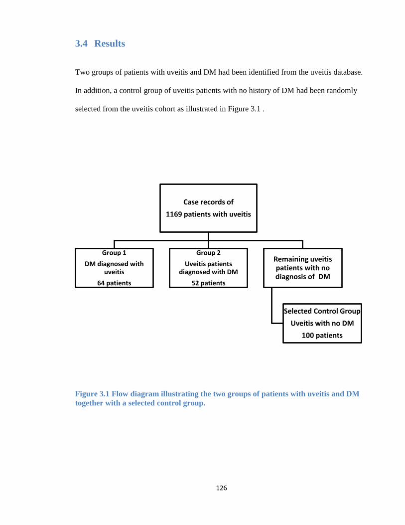
126
3.4 Results
Two groups of patients with uveitis and DM had been identified from the uveitis database.
In addition, a control group of uveitis patients with no history of DM had been randomly
selected from the uveitis cohort as illustrated in Figure 3.1 .
Figure 3.1 Flow diagram illustrating the two groups of patients with uveitis and DM
together with a selected control group.
Case records of
1169 patients with uveitis
Group 1
DM diagnosed with uveitis
64 patients
Group 2
Uveitis patients diagnosed with DM
52 patients
Remaining uveitis patients with no diagnosis of DM
Selected Control Group
Uveitis with no DM
100 patients
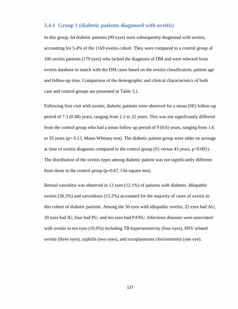
127
3.4.1 Group 1 (diabetic patients diagnosed with uveitis)
In this group, 64 diabetic patients (99 eyes) were subsequently diagnosed with uveitis,
accounting for 5.4% of the 1169 uveitis cohort. They were compared to a control group of
100 uveitis patients (170 eyes) who lacked the diagnosis of DM and were selected from
uveitis database to match with the DM cases based on the uveitis classification, patient age
and follow-up time. Comparison of the demographic and clinical characteristics of both
case and control groups are presented in Table 3.1.
Following first visit with uveitis, diabetic patients were observed for a mean (SE) follow-up
period of 7.3 (0.48) years, ranging from 1.2 to 22 years. This was not significantly different
from the control group who had a mean follow up period of 9 (0.6) years, ranging from 1.6
to 35 years (p= 0.13, Mann-Whitney test). The diabetic patient group were older on average
at time of uveitis diagnosis compared to the control group (55 versus 43 years, p<0.001).
The distribution of the uveitis types among diabetic patient was not significantly different
from those in the control group (p=0.67, Chi-square test).
Retinal vasculitis was observed in 12 eyes (12.1%) of patients with diabetes. Idiopathic
uveitis (56.5%) and sarcoidosis (15.2%) accounted for the majority of cases of uveitis in
this cohort of diabetic patients. Among the 56 eyes with idiopathic uveitis, 22 eyes had AU,
20 eyes had IU, four had PU, and ten eyes had PANU. Infectious diseases were associated
with uveitis in ten eyes (10.0%) including TB hypersensitivity (four eyes), HSV related
uveitis (three eyes), syphilis (two eyes), and toxoplasmosis chorioretinitis (one eye).
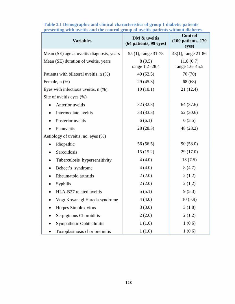
128
Table 3.1 Demographic and clinical characteristics of group 1 diabetic patients
presenting with uveitis and the control group of uveitis patients without diabetes.
Variables DM & uveitis
(64 patients, 99 eyes)
Control
(100 patients, 170
eyes)
Mean (SE) age at uveitis diagnosis, years 55 (1), range 31-78 43(1), range 21-86
Mean (SE) duration of uveitis, years 8 (0.5)
range 1.2 -28.4
11.8 (0.7)
range 1.6- 45.5
Patients with bilateral uveitis, n (%) 40 (62.5) 70 (70)
Female, n (%) 29 (45.3) 68 (68)
Eyes with infectious uveitis, n (%) 10 (10.1) 21 (12.4)
Site of uveitis eyes (%)
Anterior uveitis 32 (32.3) 64 (37.6)
Intermediate uveitis 33 (33.3) 52 (30.6)
Posterior uveitis 6 (6.1) 6 (3.5)
Panuveitis 28 (28.3) 48 (28.2)
Aetiology of uveitis, no. eyes (%)
Idiopathic 56 (56.5) 90 (53.0)
Sarcoidosis 15 (15.2) 29 (17.0)
Tuberculosis hypersensitivity 4 (4.0) 13 (7.5)
Behcet’s syndrome 4 (4.0) 8 (4.7)
Rheumatoid arthritis 2 (2.0) 2 (1.2)
Syphilis 2 (2.0) 2 (1.2)
HLA-B27 related uveitis 5 (5.1) 9 (5.3)
Vogt Koyanagi Harada syndrome 4 (4.0) 10 (5.9)
Herpes Simplex virus 3 (3.0) 3 (1.8)
Serpiginous Choroiditis 2 (2.0) 2 (1.2)
Sympathetic Ophthalmitis 1 (1.0) 1 (0.6)
Toxoplasmosis chorioretinitis 1 (1.0) 1 (0.6)
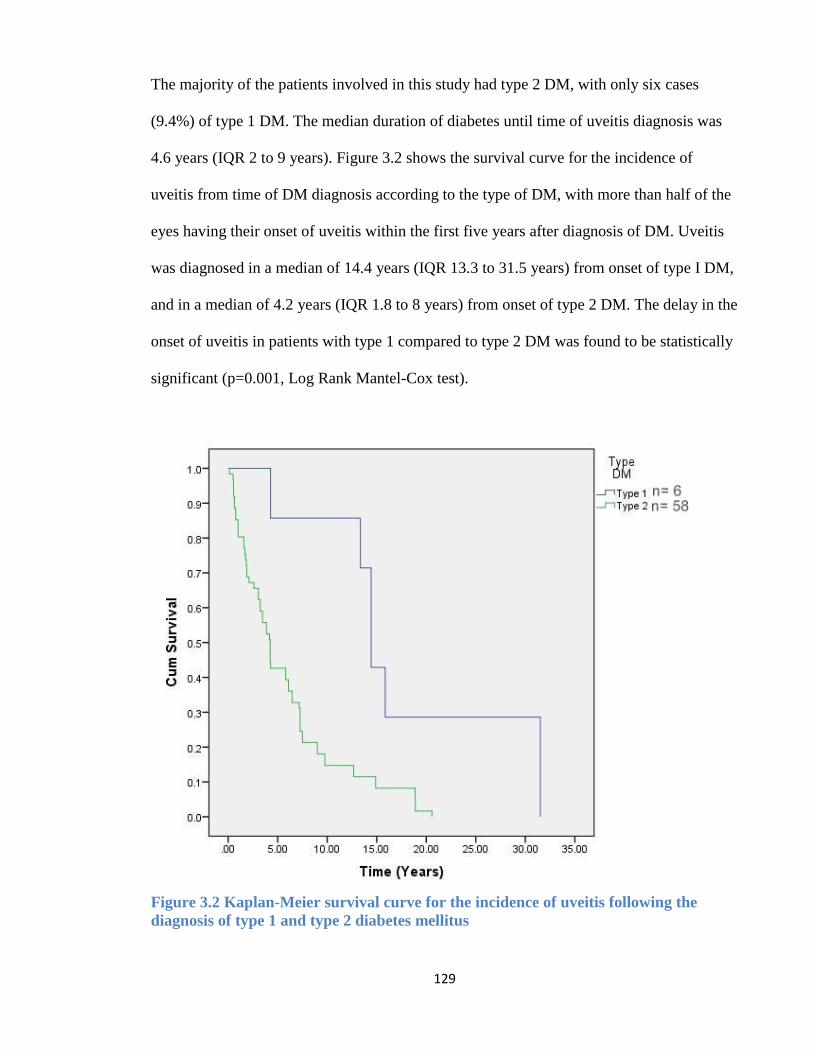
129
The majority of the patients involved in this study had type 2 DM, with only six cases
(9.4%) of type 1 DM. The median duration of diabetes until time of uveitis diagnosis was
4.6 years (IQR 2 to 9 years). Figure 3.2 shows the survival curve for the incidence of
uveitis from time of DM diagnosis according to the type of DM, with more than half of the
eyes having their onset of uveitis within the first five years after diagnosis of DM. Uveitis
was diagnosed in a median of 14.4 years (IQR 13.3 to 31.5 years) from onset of type I DM,
and in a median of 4.2 years (IQR 1.8 to 8 years) from onset of type 2 DM. The delay in the
onset of uveitis in patients with type 1 compared to type 2 DM was found to be statistically
significant (p=0.001, Log Rank Mantel-Cox test).
Figure 3.2 Kaplan-Meier survival curve for the incidence of uveitis following the
diagnosis of type 1 and type 2 diabetes mellitus
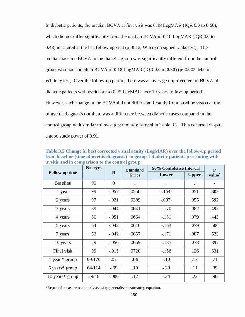
130
In diabetic patients, the median BCVA at first visit was 0.18 LogMAR (IQR 0.0 to 0.60),
which did not differ significantly from the median BCVA of 0.18 LogMAR (IQR 0.0 to
0.48) measured at the last follow up visit (p=0.12, Wilcoxon signed ranks test). The
median baseline BCVA in the diabetic group was significantly different from the control
group who had a median BCVA of 0.18 LogMAR (IQR 0.0 to 0.30) (p<0.002, Mann-
Whitney test). Over the follow-up period, there was an average improvement in BCVA of
diabetic patients with uveitis up to 0.05 LogMAR over 10 years follow-up period.
However, such change in the BCVA did not differ significantly from baseline vision at time
of uveitis diagnosis nor there was a difference between diabetic cases compared to the
control group with similar follow-up period as observed in Table 3.2. This occurred despite
a good study power of 0.91.
Table 3.2 Change in best corrected visual acuity (LogMAR) over the follow-up period
from baseline (time of uveitis diagnosis) in group 1 diabetic patients presenting with
uveitis and in comparison to the control group
Follow up time
No. eyes
B Standard
Error
95% Confidence Interval P
value* Lower Upper
Baseline 99 0 . . . -
1 year 99 -.057 .0550 -.164- .051 .302
2 years 97 -.021 .0389 -.097- .055 .592
3 years 89 -.044 .0641 -.170 .082 .493
4 years 80 -.051 .0664 -.181 .079 .443
5 years 64 -.042 .0618 -.163 .079 .500
7 years 53 -.042 .0657 -.171 .087 .523
10 years 29 -.056 .0659 -.185 .073 .397
Final visit 99 -.015 .0720 -.156 .126 .831
1 year * group 99/170 .02 .06 -.10 .15 .71
5 years* group 64/114 -.09 .10 -.29 .11 .39
10 years* group 29/46 -.006 .12 -.24 .23 .96
*Repeated measurement analysis using generalised estimating equation.
131
Over the follow-up period, the most common ocular complication in this cohort of diabetic
patients with uveitis was cataract (62.6%) followed by macular oedema (45.5%) and
glaucoma (31.3%), all of which were significantly more prevalent in diabetic cases
compared to the control group as presented in Table 3.3. The prevalence of CNVM and
RPE atrophy were also significantly more observed in uveitis eyes of diabetic patients
compared to control group while no significant difference was observed between both
groups in terms of other uveitis complications. By the final visit, vision loss occurred in 28
eyes (28.2%) with equal distribution in the incidence of moderate and severe vision loss
and was found to be significantly more common to occur compared to the control group
(28.2% versus 8.2%, p<0.001). The most common causes for vision loss in diabetic patients
were RPE atrophy, accounting for 35.7% of eyes with vision loss, and macular scar
secondary to CNVM which was observed in 28.5% of cases with vision loss.
Vision loss in diabetic patients occurred at a median interval of 1.16 years (IQR 0.03-5.9
years), ranging from the time of uveitis diagnosis (in six eyes) up to 14 years following the
onset of uveitis. There was a statistically significant difference in the incidence rate of
vision loss between DM cases with uveitis compared to the control group (Log Rank
Mantel-Cox test <0.001) (Figure 3.3).
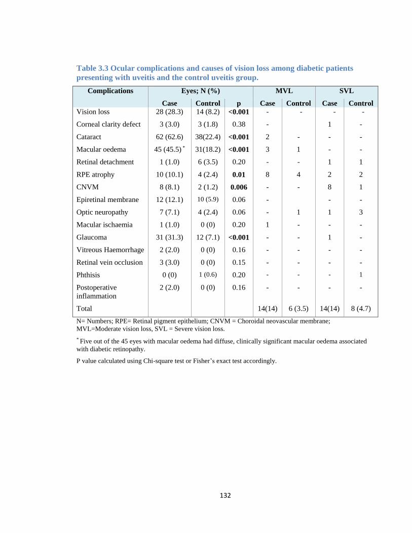
132
Table 3.3 Ocular complications and causes of vision loss among diabetic patients
presenting with uveitis and the control uveitis group.
Complications Eyes; N (%) MVL SVL
Case Control p Case Control Case Control
Vision loss 28 (28.3) 14 (8.2) <0.001 - - - -
Corneal clarity defect 3 (3.0) 3 (1.8) 0.38 - 1 -
Cataract 62 (62.6) 38(22.4) <0.001 2 - - -
Macular oedema 45 (45.5) * 31(18.2) <0.001 3 1 - -
Retinal detachment 1 (1.0) 6 (3.5) 0.20 - - 1 1
RPE atrophy 10 (10.1) 4 (2.4) 0.01 8 4 2 2
CNVM 8 (8.1) 2 (1.2) 0.006 - - 8 1
Epiretinal membrane 12 (12.1) 10 (5.9) 0.06 - - -
Optic neuropathy 7 (7.1) 4 (2.4) 0.06 - 1 1 3
Macular ischaemia 1 (1.0) 0 (0) 0.20 1 - - -
Glaucoma 31 (31.3) 12 (7.1) <0.001 - - 1 -
Vitreous Haemorrhage 2 (2.0) 0 (0) 0.16 - - - -
Retinal vein occlusion 3 (3.0) 0 (0) 0.15 - - - -
Phthisis 0 (0) 1 (0.6) 0.20 - - - 1
Postoperative
inflammation
2 (2.0) 0 (0) 0.16 - - - -
Total 14(14) 6 (3.5) 14(14) 8 (4.7)
N= Numbers; RPE= Retinal pigment epithelium; CNVM = Choroidal neovascular membrane;
MVL=Moderate vision loss, SVL = Severe vision loss.
* Five out of the 45 eyes with macular oedema had diffuse, clinically significant macular oedema associated
with diabetic retinopathy.
P value calculated using Chi-square test or Fisher’s exact test accordingly.
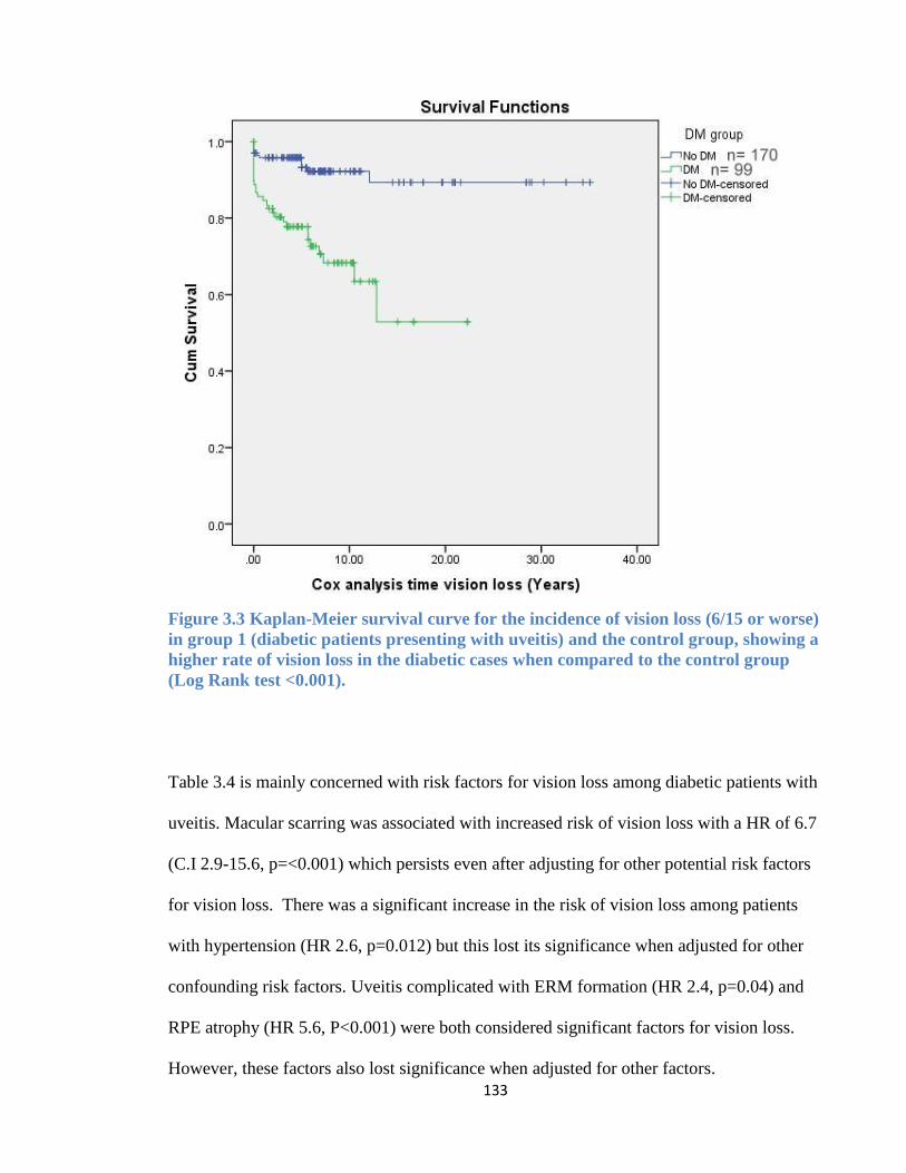
133
Figure 3.3 Kaplan-Meier survival curve for the incidence of vision loss (6/15 or worse)
in group 1 (diabetic patients presenting with uveitis) and the control group, showing a
higher rate of vision loss in the diabetic cases when compared to the control group
(Log Rank test <0.001).
Table 3.4 is mainly concerned with risk factors for vision loss among diabetic patients with
uveitis. Macular scarring was associated with increased risk of vision loss with a HR of 6.7
(C.I 2.9-15.6, p=<0.001) which persists even after adjusting for other potential risk factors
for vision loss. There was a significant increase in the risk of vision loss among patients
with hypertension (HR 2.6, p=0.012) but this lost its significance when adjusted for other
confounding risk factors. Uveitis complicated with ERM formation (HR 2.4, p=0.04) and
RPE atrophy (HR 5.6, P<0.001) were both considered significant factors for vision loss.
However, these factors also lost significance when adjusted for other factors.
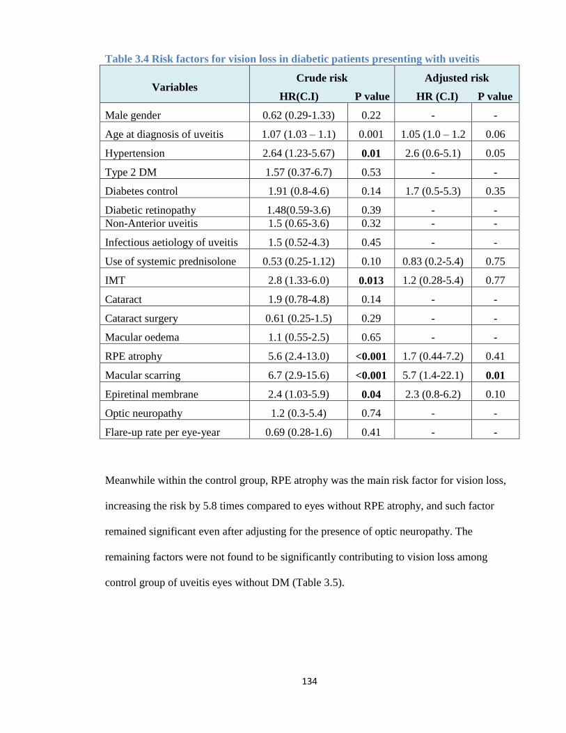
134
Table 3.4 Risk factors for vision loss in diabetic patients presenting with uveitis
Variables Crude risk
HR(C.I) P value
Adjusted risk
HR (C.I) P value
Male gender 0.62 (0.29-1.33) 0.22 - -
Age at diagnosis of uveitis 1.07 (1.03 – 1.1) 0.001 1.05 (1.0 – 1.2 0.06
Hypertension 2.64 (1.23-5.67) 0.01 2.6 (0.6-5.1) 0.05
Type 2 DM 1.57 (0.37-6.7) 0.53 - -
Diabetes control 1.91 (0.8-4.6) 0.14 1.7 (0.5-5.3) 0.35
Diabetic retinopathy 1.48(0.59-3.6) 0.39 - -
Non-Anterior uveitis 1.5 (0.65-3.6) 0.32 - -
Infectious aetiology of uveitis 1.5 (0.52-4.3) 0.45 - -
Use of systemic prednisolone 0.53 (0.25-1.12) 0.10 0.83 (0.2-5.4) 0.75
IMT 2.8 (1.33-6.0) 0.013 1.2 (0.28-5.4) 0.77
Cataract 1.9 (0.78-4.8) 0.14 - -
Cataract surgery 0.61 (0.25-1.5) 0.29 - -
Macular oedema 1.1 (0.55-2.5) 0.65 - -
RPE atrophy 5.6 (2.4-13.0) <0.001 1.7 (0.44-7.2) 0.41
Macular scarring 6.7 (2.9-15.6) <0.001 5.7 (1.4-22.1) 0.01
Epiretinal membrane 2.4 (1.03-5.9) 0.04 2.3 (0.8-6.2) 0.10
Optic neuropathy 1.2 (0.3-5.4) 0.74 - -
Flare-up rate per eye-year 0.69 (0.28-1.6) 0.41 - -
Meanwhile within the control group, RPE atrophy was the main risk factor for vision loss,
increasing the risk by 5.8 times compared to eyes without RPE atrophy, and such factor
remained significant even after adjusting for the presence of optic neuropathy. The
remaining factors were not found to be significantly contributing to vision loss among
control group of uveitis eyes without DM (Table 3.5).
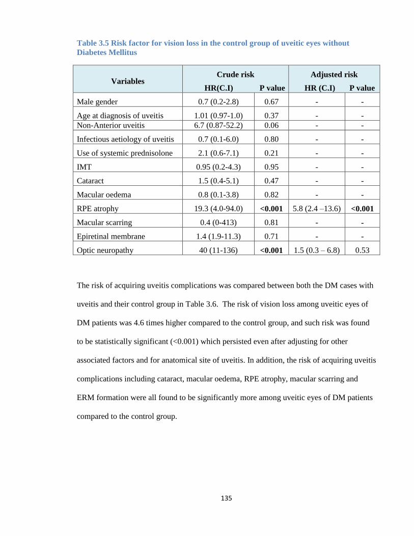
135
Table 3.5 Risk factor for vision loss in the control group of uveitic eyes without
Diabetes Mellitus
Variables Crude risk
HR(C.I) P value
Adjusted risk
HR (C.I) P value
Male gender 0.7 (0.2-2.8) 0.67 - -
Age at diagnosis of uveitis 1.01 (0.97-1.0) 0.37 - -
Non-Anterior uveitis 6.7 (0.87-52.2) 0.06 - -
Infectious aetiology of uveitis 0.7 (0.1-6.0) 0.80 - -
Use of systemic prednisolone 2.1 (0.6-7.1) 0.21 - -
IMT 0.95 (0.2-4.3) 0.95 - -
Cataract 1.5 (0.4-5.1) 0.47 - -
Macular oedema 0.8 (0.1-3.8) 0.82 - -
RPE atrophy 19.3 (4.0-94.0) <0.001 5.8 (2.4 –13.6) <0.001
Macular scarring 0.4 (0-413) 0.81 - -
Epiretinal membrane 1.4 (1.9-11.3) 0.71 - -
Optic neuropathy 40 (11-136) <0.001 1.5 (0.3 – 6.8) 0.53
The risk of acquiring uveitis complications was compared between both the DM cases with
uveitis and their control group in Table 3.6. The risk of vision loss among uveitic eyes of
DM patients was 4.6 times higher compared to the control group, and such risk was found
to be statistically significant (<0.001) which persisted even after adjusting for other
associated factors and for anatomical site of uveitis. In addition, the risk of acquiring uveitis
complications including cataract, macular oedema, RPE atrophy, macular scarring and
ERM formation were all found to be significantly more among uveitic eyes of DM patients
compared to the control group.
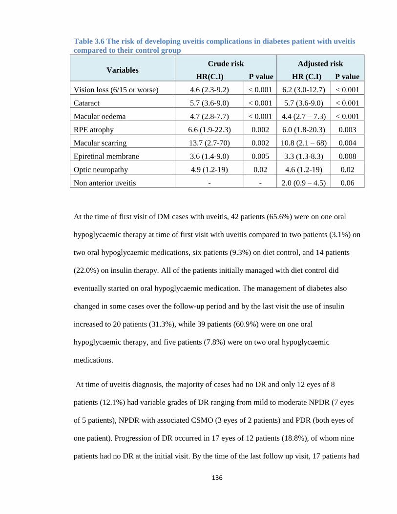
136
Table 3.6 The risk of developing uveitis complications in diabetes patient with uveitis
compared to their control group
Variables Crude risk
HR(C.I) P value
Adjusted risk
HR (C.I) P value
Vision loss (6/15 or worse) 4.6 (2.3-9.2) < 0.001 6.2 (3.0-12.7) < 0.001
Cataract 5.7 (3.6-9.0) < 0.001 5.7 (3.6-9.0) < 0.001
Macular oedema 4.7 (2.8-7.7) < 0.001 4.4 (2.7 – 7.3) < 0.001
RPE atrophy 6.6 (1.9-22.3) 0.002 6.0 (1.8-20.3) 0.003
Macular scarring 13.7 (2.7-70) 0.002 10.8 (2.1 – 68) 0.004
Epiretinal membrane 3.6 (1.4-9.0) 0.005 3.3 (1.3-8.3) 0.008
Optic neuropathy 4.9 (1.2-19) 0.02 4.6 (1.2-19) 0.02
Non anterior uveitis - - 2.0 (0.9 – 4.5) 0.06
At the time of first visit of DM cases with uveitis, 42 patients (65.6%) were on one oral
hypoglycaemic therapy at time of first visit with uveitis compared to two patients (3.1%) on
two oral hypoglycaemic medications, six patients (9.3%) on diet control, and 14 patients
(22.0%) on insulin therapy. All of the patients initially managed with diet control did
eventually started on oral hypoglycaemic medication. The management of diabetes also
changed in some cases over the follow-up period and by the last visit the use of insulin
increased to 20 patients (31.3%), while 39 patients (60.9%) were on one oral
hypoglycaemic therapy, and five patients (7.8%) were on two oral hypoglycaemic
medications.
At time of uveitis diagnosis, the majority of cases had no DR and only 12 eyes of 8
patients (12.1%) had variable grades of DR ranging from mild to moderate NPDR (7 eyes
of 5 patients), NPDR with associated CSMO (3 eyes of 2 patients) and PDR (both eyes of
one patient). Progression of DR occurred in 17 eyes of 12 patients (18.8%), of whom nine
patients had no DR at the initial visit. By the time of the last follow up visit, 17 patients had
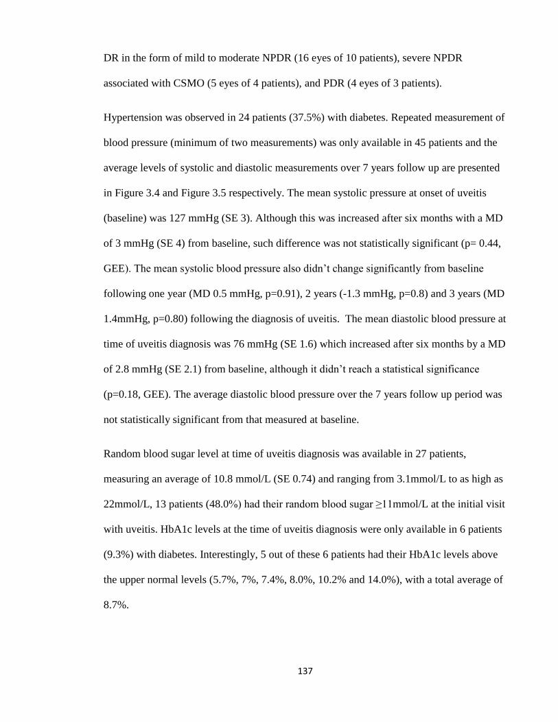
137
DR in the form of mild to moderate NPDR (16 eyes of 10 patients), severe NPDR
associated with CSMO (5 eyes of 4 patients), and PDR (4 eyes of 3 patients).
Hypertension was observed in 24 patients (37.5%) with diabetes. Repeated measurement of
blood pressure (minimum of two measurements) was only available in 45 patients and the
average levels of systolic and diastolic measurements over 7 years follow up are presented
in Figure 3.4 and Figure 3.5 respectively. The mean systolic pressure at onset of uveitis
(baseline) was 127 mmHg (SE 3). Although this was increased after six months with a MD
of 3 mmHg (SE 4) from baseline, such difference was not statistically significant (p= 0.44,
GEE). The mean systolic blood pressure also didn’t change significantly from baseline
following one year (MD 0.5 mmHg, p=0.91), 2 years (-1.3 mmHg, p=0.8) and 3 years (MD
1.4mmHg, p=0.80) following the diagnosis of uveitis. The mean diastolic blood pressure at
time of uveitis diagnosis was 76 mmHg (SE 1.6) which increased after six months by a MD
of 2.8 mmHg (SE 2.1) from baseline, although it didn’t reach a statistical significance
(p=0.18, GEE). The average diastolic blood pressure over the 7 years follow up period was
not statistically significant from that measured at baseline.
Random blood sugar level at time of uveitis diagnosis was available in 27 patients,
measuring an average of 10.8 mmol/L (SE 0.74) and ranging from 3.1mmol/L to as high as
22mmol/L, 13 patients (48.0%) had their random blood sugar ≥11mmol/L at the initial visit
with uveitis. HbA1c levels at the time of uveitis diagnosis were only available in 6 patients
(9.3%) with diabetes. Interestingly, 5 out of these 6 patients had their HbA1c levels above
the upper normal levels (5.7%, 7%, 7.4%, 8.0%, 10.2% and 14.0%), with a total average of
8.7%.
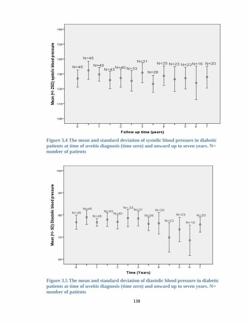
138
Figure 3.4 The mean and standard deviation of systolic blood pressure in diabetic
patients at time of uveitis diagnosis (time zero) and onward up to seven years. N=
number of patients
Figure 3.5 The mean and standard deviation of diastolic blood pressure in diabetic
patients at time of uveitis diagnosis (time zero) and onward up to seven years. N=
number of patients
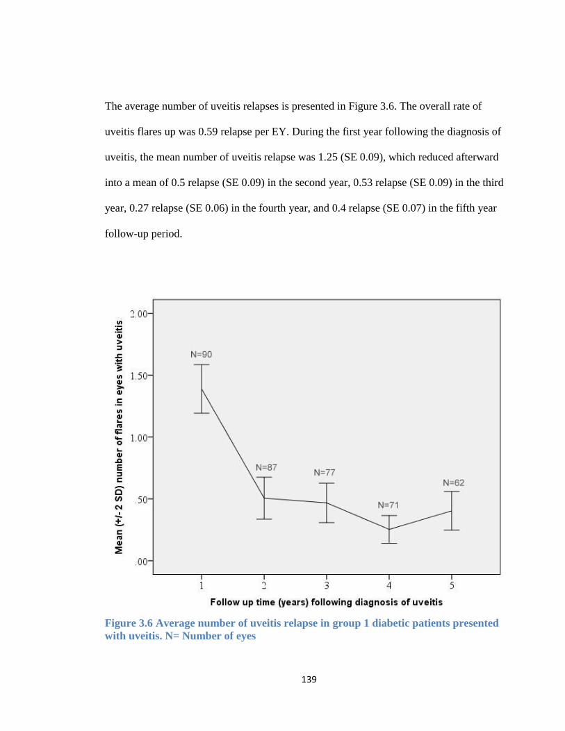
139
The average number of uveitis relapses is presented in Figure 3.6. The overall rate of
uveitis flares up was 0.59 relapse per EY. During the first year following the diagnosis of
uveitis, the mean number of uveitis relapse was 1.25 (SE 0.09), which reduced afterward
into a mean of 0.5 relapse (SE 0.09) in the second year, 0.53 relapse (SE 0.09) in the third
year, 0.27 relapse (SE 0.06) in the fourth year, and 0.4 relapse (SE 0.07) in the fifth year
follow-up period.
Figure 3.6 Average number of uveitis relapse in group 1 diabetic patients presented
with uveitis. N= Number of eyes
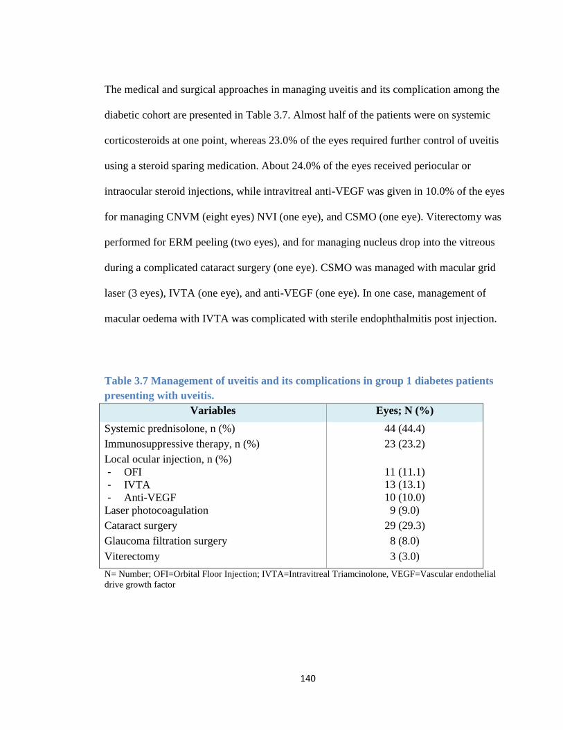
140
The medical and surgical approaches in managing uveitis and its complication among the
diabetic cohort are presented in Table 3.7. Almost half of the patients were on systemic
corticosteroids at one point, whereas 23.0% of the eyes required further control of uveitis
using a steroid sparing medication. About 24.0% of the eyes received periocular or
intraocular steroid injections, while intravitreal anti-VEGF was given in 10.0% of the eyes
for managing CNVM (eight eyes) NVI (one eye), and CSMO (one eye). Viterectomy was
performed for ERM peeling (two eyes), and for managing nucleus drop into the vitreous
during a complicated cataract surgery (one eye). CSMO was managed with macular grid
laser (3 eyes), IVTA (one eye), and anti-VEGF (one eye). In one case, management of
macular oedema with IVTA was complicated with sterile endophthalmitis post injection.
Table 3.7 Management of uveitis and its complications in group 1 diabetes patients
presenting with uveitis.
Variables Eyes; N (%)
Systemic prednisolone, n (%) 44 (44.4)
Immunosuppressive therapy, n (%) 23 (23.2)
Local ocular injection, n (%)
- OFI
- IVTA
- Anti-VEGF
11 (11.1)
13 (13.1)
10 (10.0)
Laser photocoagulation 9 (9.0)
Cataract surgery 29 (29.3)
Glaucoma filtration surgery 8 (8.0)
Viterectomy 3 (3.0)
N= Number; OFI=Orbital Floor Injection; IVTA=Intravitreal Triamcinolone, VEGF=Vascular endothelial
drive growth factor
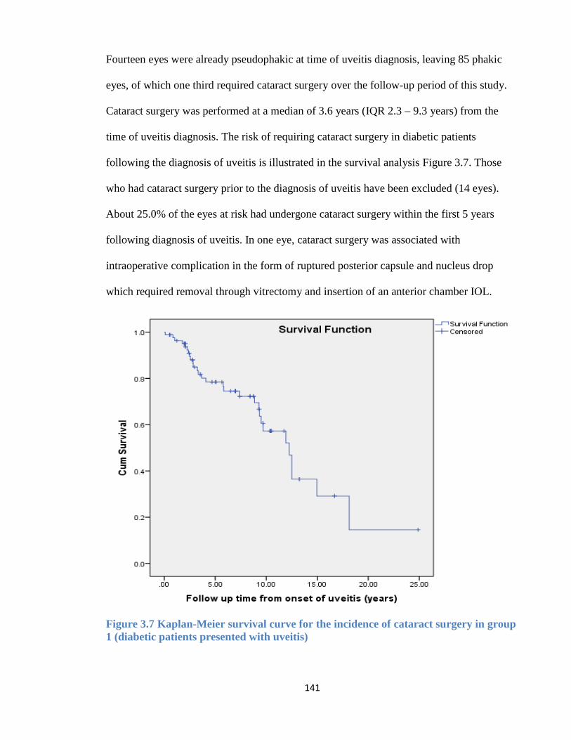
141
Fourteen eyes were already pseudophakic at time of uveitis diagnosis, leaving 85 phakic
eyes, of which one third required cataract surgery over the follow-up period of this study.
Cataract surgery was performed at a median of 3.6 years (IQR 2.3 – 9.3 years) from the
time of uveitis diagnosis. The risk of requiring cataract surgery in diabetic patients
following the diagnosis of uveitis is illustrated in the survival analysis Figure 3.7. Those
who had cataract surgery prior to the diagnosis of uveitis have been excluded (14 eyes).
About 25.0% of the eyes at risk had undergone cataract surgery within the first 5 years
following diagnosis of uveitis. In one eye, cataract surgery was associated with
intraoperative complication in the form of ruptured posterior capsule and nucleus drop
which required removal through vitrectomy and insertion of an anterior chamber IOL.
Figure 3.7 Kaplan-Meier survival curve for the incidence of cataract surgery in group
1 (diabetic patients presented with uveitis)
142
3.4.2 Group 2 (Uveitis patients diagnosed with diabetes)
In this group, 96 eyes of 52 uveitis patients were subsequently diagnosed with DM,
accounting for 4.4% of the 1169 uveitis patient cohort. The diagnosis of DM occurred
within a mean of 8.4 years (SE 0.68) from time of diagnosis of uveitis. Thirty-eight patients
(73%) were considered steroid-induced DM, accounting for 3.2% of the uveitis cohort
versus 1.2% of the uveitis cases who were diagnosed with DM despite the lack of prior
corticosteroid use.
Following the diagnosis of DM, patients were subsequently monitored for an average
follow-up period of 10.3 (0.2) years until last visit. The main demographic and clinical
characteristics of the studied cohort are presented in Table 3.8. Half of the uveitis eyes
were idiopathic while sarcoidosis was the most common disease associated with uveitis in
this group. Hypertension was observed in 17 patients (32.7%) with a median of 120mmHg
(IQR 110-140) systolic blood pressure and 80mmHg (IQR 70-90) diastolic blood pressure
at time of DM diagnosis.
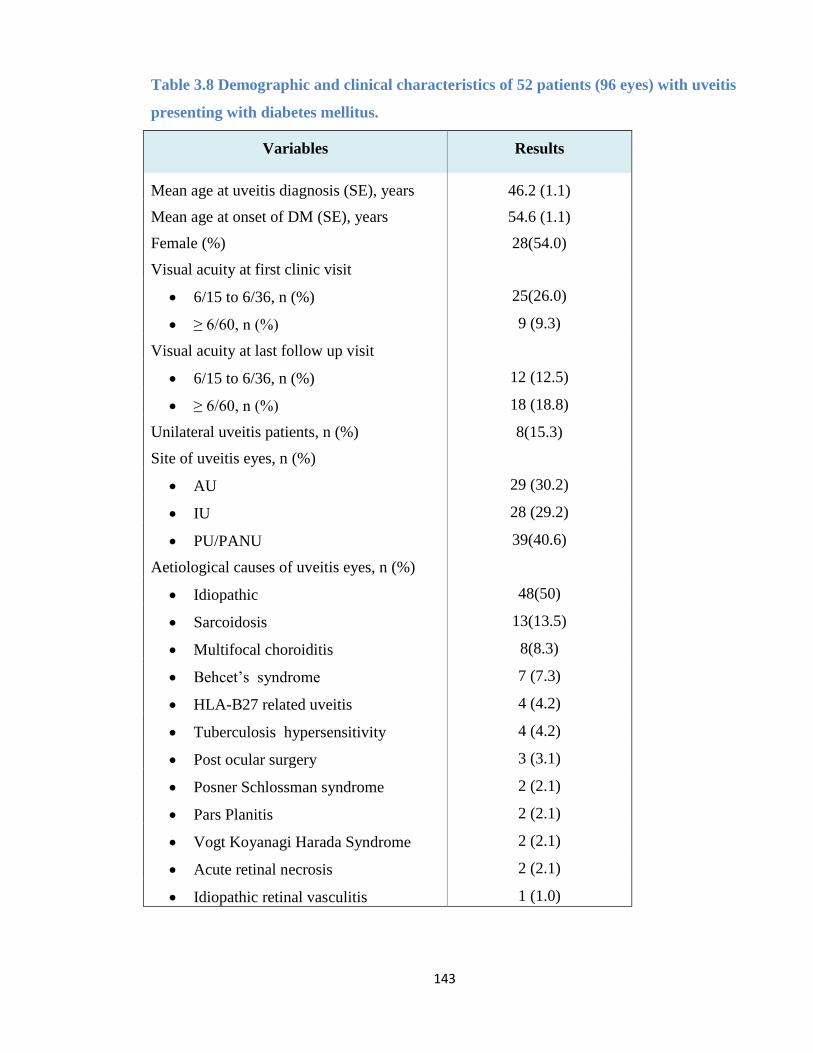
143
Table 3.8 Demographic and clinical characteristics of 52 patients (96 eyes) with uveitis
presenting with diabetes mellitus.
Variables Results
Mean age at uveitis diagnosis (SE), years 46.2 (1.1)
Mean age at onset of DM (SE), years 54.6 (1.1)
Female (%) 28(54.0)
Visual acuity at first clinic visit
6/15 to 6/36, n (%) 25(26.0)
≥ 6/60, n (%) 9 (9.3)
Visual acuity at last follow up visit
6/15 to 6/36, n (%) 12 (12.5)
≥ 6/60, n (%) 18 (18.8)
Unilateral uveitis patients, n (%) 8(15.3)
Site of uveitis eyes, n (%)
AU 29 (30.2)
IU 28 (29.2)
PU/PANU 39(40.6)
Aetiological causes of uveitis eyes, n (%)
Idiopathic 48(50)
Sarcoidosis 13(13.5)
Multifocal choroiditis 8(8.3)
Behcet’s syndrome 7 (7.3)
HLA-B27 related uveitis 4 (4.2)
Tuberculosis hypersensitivity 4 (4.2)
Post ocular surgery 3 (3.1)
Posner Schlossman syndrome 2 (2.1)
Pars Planitis 2 (2.1)
Vogt Koyanagi Harada Syndrome 2 (2.1)
Acute retinal necrosis 2 (2.1)
Idiopathic retinal vasculitis 1 (1.0)
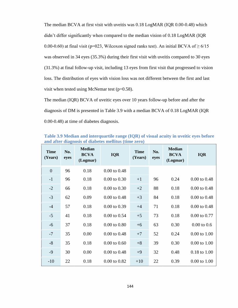
144
The median BCVA at first visit with uveitis was 0.18 LogMAR (IQR 0.00-0.48) which
didn’t differ significantly when compared to the median vision of 0.18 LogMAR (IQR
0.00-0.60) at final visit (p=023, Wilcoxon signed ranks test). An initial BCVA of ≥ 6/15
was observed in 34 eyes (35.3%) during their first visit with uveitis compared to 30 eyes
(31.3%) at final follow-up visit, including 13 eyes from first visit that progressed to vision
loss. The distribution of eyes with vision loss was not different between the first and last
visit when tested using McNemar test (p=0.58).
The median (IQR) BCVA of uveitic eyes over 10 years follow-up before and after the
diagnosis of DM is presented in Table 3.9 with a median BCVA of 0.18 LogMAR (IQR
0.00-0.48) at time of diabetes diagnosis.
Table 3.9 Median and interquartile range (IQR) of visual acuity in uveitic eyes before
and after diagnosis of diabetes mellitus (time zero)
Time
(Years)
No.
eyes
Median
BCVA
(Logmar)
IQR Time
(Years)
No.
eyes
Median
BCVA
(Logmar)
IQR
0 96 0.18 0.00 to 0.48
-1 96 0.18 0.00 to 0.30 +1 96 0.24 0.00 to 0.48
-2 66 0.18 0.00 to 0.30 +2 88 0.18 0.00 to 0.48
-3 62 0.09 0.00 to 0.48 +3 84 0.18 0.00 to 0.48
-4 57 0.18 0.00 to 0.39 +4 71 0.18 0.00 to 0.48
-5 41 0.18 0.00 to 0.54 +5 73 0.18 0.00 to 0.77
-6 37 0.18 0.00 to 0.80 +6 63 0.30 0.00 to 0.6
-7 35 0.00 0.00 to 0.48 +7 52 0.24 0.00 to 1.00
-8 35 0.18 0.00 to 0.60 +8 39 0.30 0.00 to 1.00
-9 30 0.00 0.00 to 0.48 +9 32 0.48 0.18 to 1.00
-10 22 0.18 0.00 to 0.82 +10 22 0.39 0.00 to 1.00
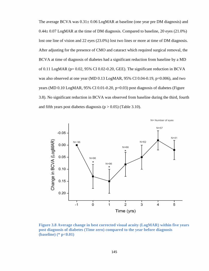
145
The average BCVA was 0.31± 0.06 LogMAR at baseline (one year pre DM diagnosis) and
0.44± 0.07 LogMAR at the time of DM diagnosis. Compared to baseline, 20 eyes (21.0%)
lost one line of vision and 22 eyes (23.0%) lost two lines or more at time of DM diagnosis.
After adjusting for the presence of CMO and cataract which required surgical removal, the
BCVA at time of diagnosis of diabetes had a significant reduction from baseline by a MD
of 0.11 LogMAR (p= 0.02, 95% CI 0.02-0.20, GEE). The significant reduction in BCVA
was also observed at one year (MD 0.13 LogMAR, 95% CI 0.04-0.19, p=0.006), and two
years (MD 0.10 LogMAR, 95% CI 0.01-0.20, p=0.03) post diagnosis of diabetes (Figure
3.8). No significant reduction in BCVA was observed from baseline during the third, fourth
and fifth years post diabetes diagnosis (p > 0.05) (Table 3.10).
Figure 3.8 Average change in best corrected visual acuity (LogMAR) within five years
post diagnosis of diabetes (Time zero) compared to the year before diagnosis
(baseline) (* p<0.01)
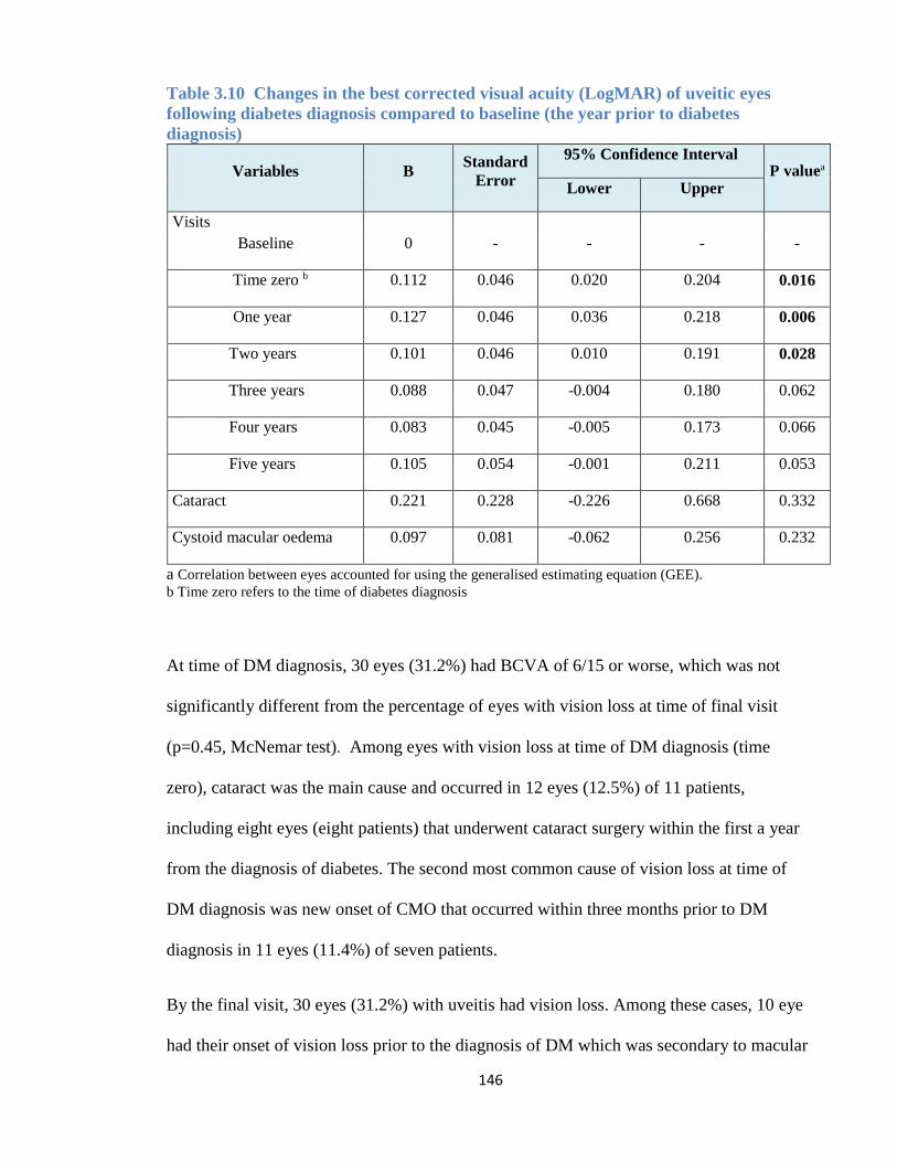
146
Table 3.10 Changes in the best corrected visual acuity (LogMAR) of uveitic eyes
following diabetes diagnosis compared to baseline (the year prior to diabetes
diagnosis)
Variables B Standard
Error
95% Confidence Interval P valuea
Lower Upper
Visits
Baseline 0 - - - -
Time zero b 0.112 0.046 0.020 0.204 0.016
One year 0.127 0.046 0.036 0.218 0.006
Two years 0.101 0.046 0.010 0.191 0.028
Three years 0.088 0.047 -0.004 0.180 0.062
Four years 0.083 0.045 -0.005 0.173 0.066
Five years 0.105 0.054 -0.001 0.211 0.053
Cataract 0.221 0.228 -0.226 0.668 0.332
Cystoid macular oedema 0.097 0.081 -0.062 0.256 0.232
a Correlation between eyes accounted for using the generalised estimating equation (GEE).
b Time zero refers to the time of diabetes diagnosis
At time of DM diagnosis, 30 eyes (31.2%) had BCVA of 6/15 or worse, which was not
significantly different from the percentage of eyes with vision loss at time of final visit
(p=0.45, McNemar test). Among eyes with vision loss at time of DM diagnosis (time
zero), cataract was the main cause and occurred in 12 eyes (12.5%) of 11 patients,
including eight eyes (eight patients) that underwent cataract surgery within the first a year
from the diagnosis of diabetes. The second most common cause of vision loss at time of
DM diagnosis was new onset of CMO that occurred within three months prior to DM
diagnosis in 11 eyes (11.4%) of seven patients.
By the final visit, 30 eyes (31.2%) with uveitis had vision loss. Among these cases, 10 eye
had their onset of vision loss prior to the diagnosis of DM which was secondary to macular
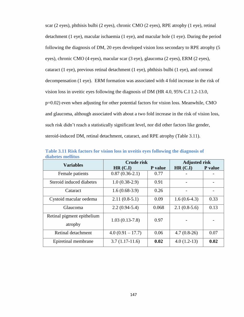
147
scar (2 eyes), phthisis bulbi (2 eyes), chronic CMO (2 eyes), RPE atrophy (1 eye), retinal
detachment (1 eye), macular ischaemia (1 eye), and macular hole (1 eye). During the period
following the diagnosis of DM, 20 eyes developed vision loss secondary to RPE atrophy (5
eyes), chronic CMO (4 eyes), macular scar (3 eye), glaucoma (2 eyes), ERM (2 eyes),
cataract (1 eye), previous retinal detachment (1 eye), phthisis bulbi (1 eye), and corneal
decompensation (1 eye). ERM formation was associated with 4 fold increase in the risk of
vision loss in uveitic eyes following the diagnosis of DM (HR 4.0, 95% C.I 1.2-13.0,
p=0.02) even when adjusting for other potential factors for vision loss. Meanwhile, CMO
and glaucoma, although associated with about a two fold increase in the risk of vision loss,
such risk didn’t reach a statistically significant level, nor did other factors like gender,
steroid-induced DM, retinal detachment, cataract, and RPE atrophy (Table 3.11).
Table 3.11 Risk factors for vision loss in uveitis eyes following the diagnosis of
diabetes mellitus
Variables Crude risk
HR (C.I) P value
Adjusted risk
HR (C.I) P value
Female patients 0.87 (0.36-2.1) 0.77 - -
Steroid induced diabetes 1.0 (0.38-2.9) 0.91 - -
Cataract 1.6 (0.68-3.9) 0.26 - -
Cystoid macular oedema 2.11 (0.8-5.1) 0.09 1.6 (0.6-4.3) 0.33
Glaucoma 2.2 (0.94-5.4) 0.068 2.1 (0.8-5.6) 0.13
Retinal pigment epithelium
atrophy 1.03 (0.13-7.8) 0.97 - -
Retinal detachment 4.0 (0.91 – 17.7) 0.06 4.7 (0.8-26) 0.07
Epiretinal membrane 3.7 (1.17-11.6) 0.02 4.0 (1.2-13) 0.02
148
During the year post DM diagnosis, the average daily dose of prednisolone was
significantly lower compared to the year prior to diagnosis (14.7 mg /day versus 10.2 mg
/day, p=0.03, Wilcoxon signed rank test). Twenty two patients (42.0%) were using
systemic corticosteroids over the year prior to DM diagnosis which didn’t differ
significantly when compared to 24 patients (46.0%) on corticosteroids over the year post
DM diagnosed (p = 0.7, Mc Neemar test). Similarly, no statistically significant difference
was observed in the proportion of patients using IMT therapy over the year pre DM
diagnosis versus the year post DM diagnosis (17.0% versus 19.0% respectively, p = 0.95)
nor in the proportion of eyes treated with OFI/IVTA (10.5% versus 10.5% respectively,
p=0.95).
The treatments used for uveitis relapses are presented in Table 2.10. There was a reduction
in the proportion of relapses treated with systemic steroids from 97/138 (70.2%) pre
diabetes to 50/90 (55.6%) post diabetes diagnosis. On the other hand, treatment of relapses
with local steroid injection in combination with systemic steroid increased from 5 (3.6%)
pre diabetes to 14 (15.5%) post diabetes. The difference in the use of systemic
corticosteroids alone or combined with local corticosteroid injections pre and post DM was
found to be statistically significant (p= 0.003, Chi square test). On three occasions,
treatment had to be modified due to the presence of hyperglycaemia; one case had bilateral
CMO treated using OFIs instead of increasing systemic prednisolone because of the
patient’s poor glycaemic control. The other two cases were started on 40 mg rather than 60
mg systemic prednisolone to treat uveitis flare up. There was no statistically significant
difference in the success rate of relapse treatment using systemic steroid, OFI/IVTA, or
combination of both when compared before and after the diagnosis of diabetes.
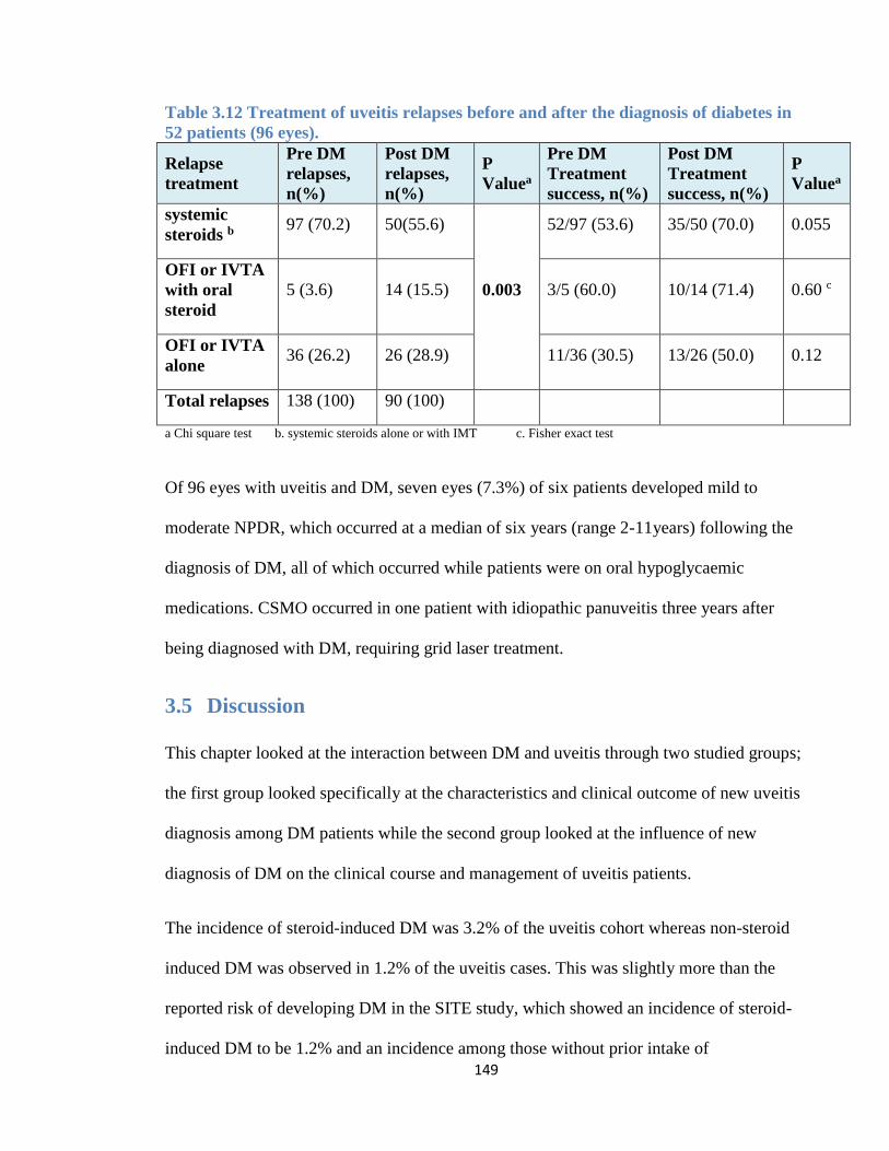
149
Table 3.12 Treatment of uveitis relapses before and after the diagnosis of diabetes in
52 patients (96 eyes).
Relapse
treatment
Pre DM
relapses,
n(%)
Post DM
relapses,
n(%)
P
Valuea
Pre DM
Treatment
success, n(%)
Post DM
Treatment
success, n(%)
P
Valuea
systemic
steroids b 97 (70.2) 50(55.6)
0.003
52/97 (53.6) 35/50 (70.0) 0.055
OFI or IVTA
with oral
steroid
5 (3.6) 14 (15.5) 3/5 (60.0) 10/14 (71.4) 0.60 c
OFI or IVTA
alone 36 (26.2) 26 (28.9) 11/36 (30.5) 13/26 (50.0) 0.12
Total relapses 138 (100) 90 (100)
a Chi square test b. systemic steroids alone or with IMT c. Fisher exact test
Of 96 eyes with uveitis and DM, seven eyes (7.3%) of six patients developed mild to
moderate NPDR, which occurred at a median of six years (range 2-11years) following the
diagnosis of DM, all of which occurred while patients were on oral hypoglycaemic
medications. CSMO occurred in one patient with idiopathic panuveitis three years after
being diagnosed with DM, requiring grid laser treatment.
3.5 Discussion
This chapter looked at the interaction between DM and uveitis through two studied groups;
the first group looked specifically at the characteristics and clinical outcome of new uveitis
diagnosis among DM patients while the second group looked at the influence of new
diagnosis of DM on the clinical course and management of uveitis patients.
The incidence of steroid-induced DM was 3.2% of the uveitis cohort whereas non-steroid
induced DM was observed in 1.2% of the uveitis cases. This was slightly more than the
reported risk of developing DM in the SITE study, which showed an incidence of steroid-
induced DM to be 1.2% and an incidence among those without prior intake of
150
corticosteroids to be 0.19% (413). This can be attributed to the method of DM diagnosis in
the SITE study which depended on history of initiating hypoglycaemic medications in the
uveitis patients whereas the diagnosis of DM in our cohort were additionally based on
abnormal blood glucose levels observed during regular follow up visits at the uveitis clinic.
The onset of uveitis was significantly delayed in patients with type 1 DM (median 14.4
years) when compared to type 2 DM (median 4.2 years). This could be attributed to the
time when type 1 DM is typically diagnosed at a younger age (mean 6.5 years of age) when
compared to type 2 DM (419).
In both groups of diabetic patients, idiopathic uveitis and sarcoidosis were the most
common aetiological classifications of uveitis types. While most of the studies on diabetic
patients presenting with idiopathic uveitis are AU (396–398), only 40.0% of our of diabetic
cohort with idiopathic uveitis were AU. Within the first group of diabetic patients, 10.0%
of the uveitic eyes had an infectious aetiology, which was similar to another study on
diabetic patients among which 12.5% were presented with an infectious aetiology (403).
We reported the time from diabetes diagnosis to onset of uveitis as median of 4.2 years,
using median as the data was not normally distributed. However, for the purpose of
comparison, the mean duration between time of DM diagnosis and the first presentation
with uveitis in this study was 6.9 years (SD 6.4) which was almost identical to the mean
duration of 6.8 years (SD 8.3) reported by Oswal et al (403).
The first group did not have a significant difference in the repeated measurement of their
BCVA when compared to their vision at time of uveitis onset. This is unlike the second
group of uveitis cases which, when compared to the year preceding the diagnosis of uveitis,
showed a significant reduction in BCVA at time of diabetes diagnosis and for up to two
years afterwards. The visual acuity, however, gradually returned to its prediabetic level
151
over the 3rd, 4th and 5th years post diabetes diagnosis. This reduction in BCVA soon after
the onset of DM was most likely attributable to new onset CMO or accelerated progression
of cataract, both associated with the diagnosis of DM. Both DM and uveitis have been
associated with breakdown of BRB leading to increased vascular permeability with
subsequent fluid retention and formation of CMO (256). Uveitis and DM combined would
have a synergistic effect on the development of CMO and cataract, increasing the chance of
visual loss from these complications. Alternatively, the drop in vision at the time of DM
diagnosis occurred secondary to uveitis relapse or CMO formation requiring the use of high
dose of systemic corticosteroids, which in turn triggered the onset of DM.
In this study, vision loss occurred in 28.2% of group 1 diabetic patients who subsequently
developed uveitis, which was similar to the results of a study by Oswal et al who reported a
final BCVA of 6/18 or worse in one third of the eyes from 36 diabetic patients presented
with uveitis (407). The risk of vision loss among uveitic eyes of DM patients was 4.6 times
higher when compared to the control group, mainly because the former group had also a
significantly higher risk of uveitis complications which included cataract, macular oedema,
RPE atrophy, all of which are also known ocular complications of DM as well. The cause
of vision loss in our group 1 cohort was mainly secondary to RPE atrophy and CNVM
associated macular scarring. This was different from the Oswal et al study which mainly
attributed the vision loss to be secondary to new and chronic CMO, cataract and glaucoma.
The mechanism of CNVM and RPE atrophy in our diabetic patients is not clear. While this
finding could be coincidental, other explanations could be related to the increased
expression of COX-2 and VEGF observed in diabetic patients leading to an increased risk
of CNVM formation within uveitis associated retinal atrophy or scars. The pathogenesis of
CNVM is multifactorial, but there is increasing evidence to suggest that COX enzymes can
152
contribute to its formation. COX-2 can be found in the RPE cells and is mainly upregulated
by proinflammatory cytokines. Studies on diabetic rats found COX-2 to play an important
role in the production of PGE2 and VEGF in the retina (204,205) which could eventually
lead to BRB discruption, mediate leukocyte migration and promote CNVM formation
(420). The role of COX-2 in the formation of CNVM has also been supported by the
positive expression of COX-2 within CNVM of human eyes (421).
Management of CMO due to uveitis in diabetic patients can be challenging. Bilateral CMO
in patients with non-infectious uveitis would ideally be treated with high dose systemic
corticosteroid. However, in the presence of uncontrolled hyperglycaemia this treatment
option carries an increased risk of inducing diabetic ketoacidosis and thus may be replaced
with local administration of corticosteroid or using a lower starting dose of systemic
corticosteroid and more rapid tapering. In our second group cohort, uveitis relapses that
occurred during the period post DM diagnosis were significantly more likely to be managed
by adding OFI/IVTA rather than increasing the dose of systemic corticosteroid. In addition,
the mean dose of systemic prednisolone was significantly reduced in the year post DM
diagnosis when compared to the previous year.
Among DM patients who had blood tests at time of presenting with uveitis, most cases
showed poor hypoglycaemic control with an average blood glucose level of 10.8mmol/L
and 48% had a random blood sugar ≥11mmol/L together with an average HbA1c level of
8.7%. These results were comparable to the diabetic group in the study of Oswal et al who
measured an average blood glucose level of 13.6 mmols/L, and mean HbA1c level of 8.7%
at time of the first presentation with uveitis (403). It is difficult in this study to address
whether there is an association between poor glycaemic control and the onset of uveitis
based on the limited number of patients who had blood test results at time of uveitis
153
presentation. However, the Oswal et al group who as well lacked blood test results for most
of their patients did not find a significant association between uveitis activity at first
presentation and glycaemic control (403). It is recommended to check the blood glucose
level in patients with DM presenting with their first onset of uveitis, regardless of the
aetiology of uveitis or severity of inflammation to detect cases with poor glycaemic control
and to allow for its management in collaboration with their health care provider.
Within the first group of diabetic patients, new onset or progression of DR occurred in 18%
of the eyes during the follow-up period following the diagnosis of uveitis, including two
eyes (2.0%) that progressed to PDR, while in the second group, 7.3% of the eyes
progressed from no DR to develop mild NPDR. A recent study on the progression of DR
among diabetic patients in UK found that within 10 years, 16.4% developed
preproliferative DR, 1.2% had sight threatening maculopathy, and 1.5% developed
proliferative DR(422). We do acknowledge that this is a descriptive analysis obtained from
our study cohort and that no conclusions can be drawn regarding the impact of uveitis on
the progression of DR due to the lack of comparison with a parallel control group of
diabetic patients without uveitis. The study of Oswal et al reported more cases who had
progression of DR into PDR following onset of uveitis when compared to our studied
group [7 eyes (14.5%) versus 2 eyes (2%); respectively]. However, this might be attributed
to the fact that Oswal et al studied group had larger proportion of patients who had DR at
time of uveitis diagnosis when compared to our group (65.5% versus 12%, respectively)
(407). Whether uveitis can accelerate the development of DR is still unclear but the
possibility has been suggested by previous published papers who observed an increase in
the progression of DR following endophthalmitis (423), ARN, and toxoplasmosis
chorioretinitis (404). On the other hand, one author proposed a protective effect of uveitis
154
on the development of DR after reporting a case with progression to proliferative DR only
in the eye not affected by FHC (424). Progression of DR in patients with uveitis might be
related to the effects of inflammatory mediators on the retinal vasculature. There is more
evidence now supporting the role of inflammatory cells, cytokines and other inflammatory
products in the development of DR (199). The aim of treating uveitis in diabetic patients is
to preserve vision and prevent or treat ocular complications while maintaining good
glycaemic control. For this purpose, close collaboration with the primary care physician is
important to achieve glycaemic control, especially in uveitis patients requiring high doses
of systemic corticosteroids with or without IMT. It is also recommended that some IMT
used in the treatment of uveitis, such as cyclosporine, should be either avoided or used with
caution due to their hyperglycaemic effect (425).
In conclusion, our study is of interest as it attempts to complete our understanding of the
synergistic effect of uveitis and diabetes by examining the effect of newly diagnosed
diabetes on eyes with pre existing uveitis and the effect of diabetes on the uveitis treatment
strategies used. This was done using, to the best of our knowledge, the largest cohort of
diabetic patients with uveitis as a whole, and within each of the two studied groups. We
have concluded that incidence of DM in uveitis patients is associated with a significant
reduction in visual acuity within the first two years post diagnosis that soon returns to the
pre diabetic visual acuity by five years. Diabetic patients diagnosed with uveitis have a
higher risk of vision loss compare to eyes with uveitis alone, and thus a good control of the
DM and uveitis complications is required to maintain a stable vision in these patients.
Reduction in systemic corticosteroid therapy and increased usage of local therapy implies
that clinicians are adjusting their treatment protocols following DM diagnosis, and this has
not change the ability to control the uveitis relapses. Given the association between DM,
155
corticosteroids and some second-line IMT used in treating uveitis, it is important to
optimise treatment protocols for these conditions when they occur concurrently.
3.6 Publications and poster presentations
Poster presentation at the UCL graduate poster event 2012
Talat L, Tompkin-Netzer O, Taylor SRJ, MD Din S, Barr A, Isa H, and Lightman S.
"The influence of diabetes mellitus on the management and visual outcome of
patients with uveitis". Acta Ophthalmol. 2014 Jun;92(4):329-30
Poster presentation "The influence of diabetes mellitus on the management and
visual outcome of patients with uveitis" at RCOphth annual meeting 2013
Poster presentation "The influence of diabetes mellitus on the management and
visual outcome of patients with uveitis" at WOC Tokyo 2014.
156
4 CHAPTER FOUR: COMPARISON OF VISUAL
OUTCOME IN UVEITIS WITH AND WITHOUT
RETINAL VASCULITIS AND PREDICTORS FOR
PROGNOSIS OF ISCHAEMIC RETINAL VASCULITIS
4.1 Introduction
Retinal vasculitis is a clinical finding in 6-15% of eyes with uveitis (263). Its incidence has
been reported in the United States as 1 to 2 cases per 100,000 population, including isolated
retinal vasculitis or in association with systemic diseases (426). It can present as
intermediate or posterior uveitis in association with an underlying systemic disease such as
BD, sarcoidosis, MS, and collagen-vascular diseases (264,265), or as an isolated ocular
disease. Rosenbaum et al after reviewing 1390 patients with uveitis identified 15% with
retinal vasculitis (263). Retinal vasculitis presents clinically as a spectrum varying from
mild venous sheathing to severe obstructive vascular occlusion. Vascular damage can result
in loss of vessel wall integrity, leakage of blood constituents into the retinal extracellular
space and CMO, a significant factor contributing to vision loss (427). In the presence of
occlusive retinal vasculitis, vision loss can occur as a consequence to CMO, or secondary to
NVD or NVE which can bleed resulting in VH or cause fibrovascular traction and
subsequently tractional retinal detachment and vision loss. NVI can also occur leading to
neovascular glaucoma and permanent vision loss (427). Irreversible vision loss can be
inevitable when the area of ischaemia is involving the posterior pole resulting in macular
ischaemia (16).
In a retrospective study of 113 eyes with retinal vasculitis in eastern India, capillary
nonperfusion was the most common FFA finding seen in retinal vasculitis, found in 40% of
the cases, followed by collateral vessels, seen in 19.5% of eyes with vasculitis (428).
157
Different causes of retinal vasculitis carry variable risks of developing retinal ischaemia
ranging from being common in presumed TB related retinal vasculitis and BD to a more
rare association in sarcoidosis and MS (265,269).
Graham et al identified 150 patients with retinal vasculitis from a single uveitis clinic in
UK after excluding ischaemia and neoplastic cases. Out of these cases, 67 patients had
idiopathic vasculitis without associated systemic disease, among which peripheral vascular
sheathing was observed in 64%, periphlebitis in 15%, RVO in 9% and CMO in 60% of
cases. Retinal NV was observed in 16% of the patients with isolated retinal vasculitis, and
less than half of patients with NV had associated retinal ischaemia. Capillary closure on
FFA was more commonly observed in patients with retinal vasculitis associated with BD
(14 patients, 48%) compared to idiopathic vasculitis (14 patients, 23%) and sarcoidosis
with retinal vasculitis (two patients, 14%) (429).
Ku et al (430) examined the visual outcome and prognostic factors in 114 patients (203
eyes) with vasculitis obtained from a cohort of 1390 patients with uveitis as a continuation
of the work initially reported by Rosenbaum et al (263). They found the mean change in
BCVA from first visit was 0.01 LogMAR per year of follow-up. Improvement in BCVA
from first visit by at least two Snellen lines during the follow up occurred in 33.6%.
Improvement in visual acuity from first visit was more likely in non-white subjects, and in
eyes with infectious uveitis or worse vision at first presentation (430).
Ocular involvement in BD is associated with a high risk of visual loss, mainly secondary to
complications associated with retinal vasculitis (277,431). The contribution of BD to the
overall incidence of retinal vasculitis can vary based on the population at risk. A review of
1390 uveitis cases on the west coast of the United States found 207 patients with evidence
158
of retinal vasculitis; of these cases, only 14 patients had BD (263). On the other hand,
retinal vasculitis is common among patients with ocular BD. In one multicentre study, 22%
of eyes with ocular BD had retinal vasculitis (277). In a retrospective study of 107 patients
with ocular BD, the 10 year risk of developing severe visual loss of 6/60 or worse was 13%
and ischaemic maculopathy secondary to BRVO was attributed to half the cases of
irreversible severe visual loss (275). Eyes with ocular sarcoidosis mainly present with non-
ischaemic retinal vasculitis. In a study including 75 eyes of patients with sarcoid related
uveitis, 37% had retinal vasculitis, three of which had ischaemic vasculitis associated with
NV (432). In another study involving 68 patients with posterior uveitis related to
sarcoidosis, NVD and VH were reported in 4% of cases, with an increased incidence of VH
up to 16% in the young age group (433). BRVO, although very rare, has been previously
reported in sarcoidosis associated uveitis especially among young age group in the presence
(434) or absence (435) of iridocyclitis.
Despite the potential risks of retinal vasculitis in causing vision loss, only limited numbers
of studies related to the outcome of this condition have been published, especially for eyes
with ischaemic retinal vasculitis. So far, only two papers addressed some aspects of the
clinical features and visual outcome of ischaemic vasculitis in comparison to non-ischaemic
vasculitis. One paper by Palmer et al in 1996 examined 53 patients with idiopathic retinal
vasculitis (20 ischaemic and 33 non ischaemic) and found no significant difference in the
median number of relapses per year between both groups (0.29 relapse per year for
ischaemic versus 0.36 per year in non ischaemic retinal vasculitis) and found visual loss to
be more common in eyes with ischaemic vasculitis (34%) compared to the non-ischaemic
group (6%) (436). In the second more recent retrospective study by Ali et al, 56 patients
with 101 eyes affected with non-infectious vasculitis with a minimum one year follow up
159
period were selected. Vasculitis was defined based on documented clinical ocular
examination for the presence of different clinical findings of vasculitis, such as perivascular
sheathing, intraretinal haemorrhage, or cotton wool spots while only 39 eyes had FFA
available to document additional findings of vasculitis such as vascular leakage or
occlusion. Only six eyes (5.9%) with occlusive vasculitis were available to compare with
non-occlusive retinal vasculitis and found no change in BCVA from time of diagnosis until
the last follow up period (258). Both studies have not included all aetiological forms of
ischaemic vasculitis and the numbers of eyes with ischaemic vasculitis were considerably
small. No study so far had addressed the outcome of NV management in the setting of
ischaemic vasculitis and the rate of its recurrence following initial laser treatment.
Treatment of retinal vasculitis depends on the underlying cause, extent of vessel
involvement, effect on visual acuity and presence of retinal complications, such as retinal
ischaemia and NV. In cases when treatment is warranted this may include use of
corticosteroids with the addition of immunosuppressant drugs when required, together with
anti-microbial agents in cases of suspected infectious aetiology. When NV has occurred
laser photocoagulation is the mainstay in management. However, the role of retinal laser
and anti-inflammatory medications in preventing further NV formation and ischaemia
progression has not been fully addressed.
160
4.2 Aims and objectives
1) Describe the demographic and clinical characteristics of eyes with retinal vasculitis
2) Describe the demographic and clinical characteristics of eyes with ischaemic retinal
vasculitis
3) Assess visual outcome and risk factors for vision loss in eyes with retinal vasculitis
in comparison to eyes with no vasculitis.
4) Assess visual outcome and risk factors of vision loss in eyes with ischaemic retinal
vasculitis and in comparison to eyes with non-ischaemic vasculitis.
5) Examine the treatment strategies used in the management of ischaemic retinal
vasculitis and the rate of uveitis relapse and ischaemia progression following
initiation of anti-inflammatory medications and retinal laser photocoagulation,
respectively.
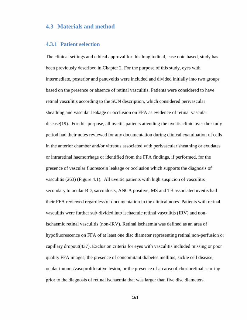
161
4.3 Materials and method
4.3.1 Patient selection
The clinical settings and ethical approval for this longitudinal, case note based, study has
been previously described in Chapter 2. For the purpose of this study, eyes with
intermediate, posterior and panuveitis were included and divided initially into two groups
based on the presence or absence of retinal vasculitis. Patients were considered to have
retinal vasculitis according to the SUN description, which considered perivascular
sheathing and vascular leakage or occlusion on FFA as evidence of retinal vascular
disease(19). For this purpose, all uveitis patients attending the uveitis clinic over the study
period had their notes reviewed for any documentation during clinical examination of cells
in the anterior chamber and/or vitreous associated with perivascular sheathing or exudates
or intraretinal haemorrhage or identified from the FFA findings, if performed, for the
presence of vascular fluorescein leakage or occlusion which supports the diagnosis of
vasculitis (263) (Figure 4.1). All uveitic patients with high suspicion of vasculitis
secondary to ocular BD, sarcoidosis, ANCA positive, MS and TB associated uveitis had
their FFA reviewed regardless of documentation in the clinical notes. Patients with retinal
vasculitis were further sub-divided into ischaemic retinal vasculitis (IRV) and non-
ischaemic retinal vasculitis (non-IRV). Retinal ischaemia was defined as an area of
hypofluorescence on FFA of at least one disc diameter representing retinal non-perfusion or
capillary dropout(437). Exclusion criteria for eyes with vasculitis included missing or poor
quality FFA images, the presence of concomitant diabetes mellitus, sickle cell disease,
ocular tumour/vasoproliferative lesion, or the presence of an area of chorioretinal scarring
prior to the diagnosis of retinal ischaemia that was larger than five disc diameters.
162
Figure 4.1 Fundus Fluoresceine angiography of the right eye with vasculitis showing
perivascular fluorescein leakage (white arrow) suggestive of active inflammation
associated with parafoveal fluorescein staining secondary to macular oedema (black
arrow).
4.3.2 Data collection
Data was gathered at the time of patient inclusion as presented in Appendix 1 and included
demographic information, anatomical and aetiological type of uveitis, and BCVA at
baseline and final visit. Baseline was defined as the time of the first visit with a diagnosis
of uveitis or, for the IRV group, time of diagnosis of retinal ischaemia. Further information
was gathered regarding treatment of vasculitis including the use of systemic or local
corticosteroids and steroid sparing drugs. In eyes with IRV, the incidence of NV formation
requiring retinal laser photocoagulation was documented. In addition, we measured the rate
of progression of ischaemia defined as evidence of NV formation in new areas requiring
further laser photocoagulation or the presence of new areas of capillary non-perfusion.
163
4.3.3 Fundus fluorescein angiography image analysis
FFA images were either taken using a digital retinal camera system (Topcon TRC 50IX;
Topcon Medical Systems Inc, Paramus, NJ) or the ultra-widefield imaging (Optos PLC,
Dunfermline, Scotland). For images taken through the Topcon camera system, an
automated mosaic software program [i2k Retina (DualAlign LLC, Clifton Park, NY)] was
used to combine together overlapping retinal fundus photographs and construct a single,
easy to review, composite image which helps to evaluate the presence of NVD, NVE, NVI,
macular ischaemia, and angiographic CMO (438) (Figure 4.2). Macular ischaemia was
defined as a foveal avascular zone (FAZ) ≥ 1,000 µm at its widest diameter, or broken
perifoveal capillary rings at the border of the FAZ (186). In addition to reporting CMO
based on the OCT findings defined in Chapter 2, angiographic CMO was also described,
and was defined as macular leakage at 5-10 minutes post fluorescein sodium injection.
For the purpose of quantifying the area of retinal nonperfusion, only eyes with ultra-
widefield FFA were included to calculate the ischaemic index using previously described
methodology (439–441). In this method, the total area of capillary nonperfusion seen in
arteriovenous phase image was measured and expressed as a percentage of the total image
area in pixels. This was done using the manual area measurement function in ImageJ
software (ImageJ 1.44p, National Institutes of Health, Bethesda, MD, USA) as in the
example shown in Error! Reference source not found.. The central FFA images were
used to measure the total image area for calculating the ischaemic index, thus avoiding
areas of peripheral distortion of the wide-field image owing to the spherical curvature of the
eye. However, peripherally steered FFA images were used to determine the extent and
boundaries of the peripheral nonperfusion. Disc areas (DA) of nonperfusion were also
quantified. One disc area was defined for each eye as the number of pixels composing the
164
optic disc (442) and hence the outlined area of capillary nonperfusion was divided over one
DA to give the area of retinal ischaemia measured in DA.
4.3.4 Statistical analysis
Detailed description of the statistical method used was presented earlier in Chapter 2.
Repeated measurements analysis of the BCVA and its difference from baseline was done
using a multivariate linear regression method obtained from GEE test. Kaplan-Meier
survival analysis with Log-Rank (Mantel-Cox) test were applied together with survival
curve graphs to compare the survival rates for the incidence of vision loss, ischaemia
following diagnosis of uveitis, as well as incidence of NV or initiation of first laser and its
relapse. The HR and 95% C.I for vision loss, CMO and NV relapse were measured using
Cox proportional hazards regression analysis.
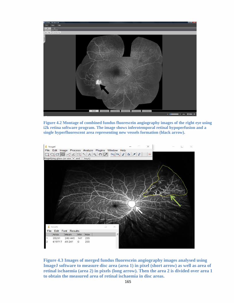
165
Figure 4.2 Montage of combined fundus fluorescein angiography images of the right eye using
i2k retina software program. The image shows inferotemporal retinal hypoperfusion and a
single hyperfluorescent area representing new vessels formation (black arrow).
Figure 4.3 Images of merged fundus fluorescein angiography images analysed using
ImageJ software to measure disc area (area 1) in pixel (short arrow) as well as area of
retinal ischaemia (area 2) in pixels (long arrow). Then the area 2 is divided over area 1
to obtain the measured area of retinal ischaemia in disc areas.
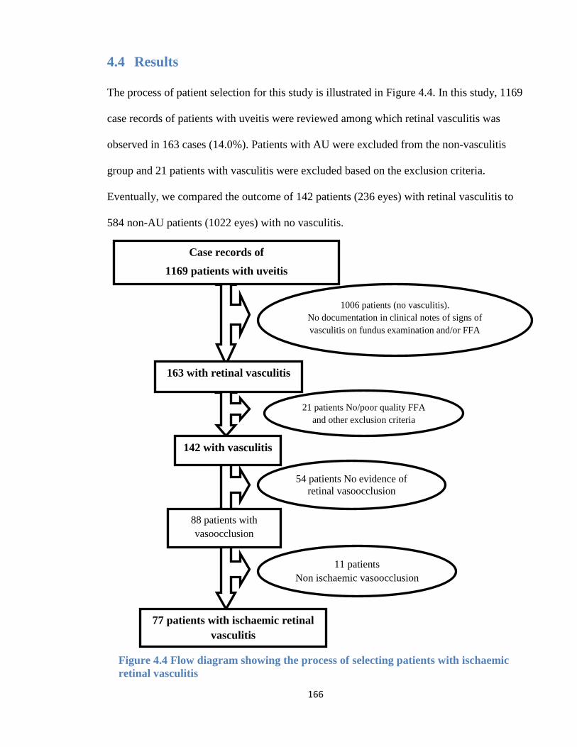
166
4.4 Results
The process of patient selection for this study is illustrated in Figure 4.4. In this study, 1169
case records of patients with uveitis were reviewed among which retinal vasculitis was
observed in 163 cases (14.0%). Patients with AU were excluded from the non-vasculitis
group and 21 patients with vasculitis were excluded based on the exclusion criteria.
Eventually, we compared the outcome of 142 patients (236 eyes) with retinal vasculitis to
584 non-AU patients (1022 eyes) with no vasculitis.
Case records of
1169 patients with uveitis
163 with retinal vasculitis
21 patients No/poor quality FFA
and other exclusion criteria
142 with vasculitis
54 patients No evidence of
retinal vasoocclusion
1006 patients (no vasculitis).
No documentation in clinical notes of signs of
vasculitis on fundus examination and/or FFA
88 patients with
vasoocclusion
11 patients
Non ischaemic vasoocclusion
77 patients with ischaemic retinal
vasculitis
Figure 4.4 Flow diagram showing the process of selecting patients with ischaemic
retinal vasculitis
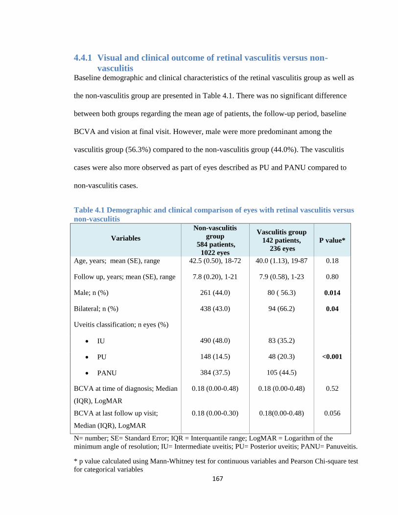
167
4.4.1 Visual and clinical outcome of retinal vasculitis versus non-
vasculitis Baseline demographic and clinical characteristics of the retinal vasculitis group as well as
the non-vasculitis group are presented in Table 4.1. There was no significant difference
between both groups regarding the mean age of patients, the follow-up period, baseline
BCVA and vision at final visit. However, male were more predominant among the
vasculitis group (56.3%) compared to the non-vasculitis group (44.0%). The vasculitis
cases were also more observed as part of eyes described as PU and PANU compared to
non-vasculitis cases.
Table 4.1 Demographic and clinical comparison of eyes with retinal vasculitis versus
non-vasculitis
Variables
Non-vasculitis
group
584 patients,
1022 eyes
Vasculitis group
142 patients,
236 eyes
P value*
Age, years; mean (SE), range 42.5 (0.50), 18-72 40.0 (1.13), 19-87 0.18
Follow up, years; mean (SE), range 7.8 (0.20), 1-21 7.9 (0.58), 1-23 0.80
Male; n (%) 261 (44.0) 80 ( 56.3) 0.014
Bilateral; n (%) 438 (43.0) 94 (66.2) 0.04
Uveitis classification; n eyes (%)
IU 490 (48.0) 83 (35.2)
PU 148 (14.5) 48 (20.3) <0.001
PANU 384 (37.5) 105 (44.5)
BCVA at time of diagnosis; Median
(IQR), LogMAR
0.18 (0.00-0.48) 0.18 (0.00-0.48) 0.52
BCVA at last follow up visit;
Median (IQR), LogMAR
0.18 (0.00-0.30) 0.18(0.00-0.48) 0.056
N= number; SE= Standard Error; IQR = Interquantile range; LogMAR = Logarithm of the
minimum angle of resolution; IU= Intermediate uveitis; PU= Posterior uveitis; PANU= Panuveitis.
* p value calculated using Mann-Whitney test for continuous variables and Pearson Chi-square test
for categorical variables
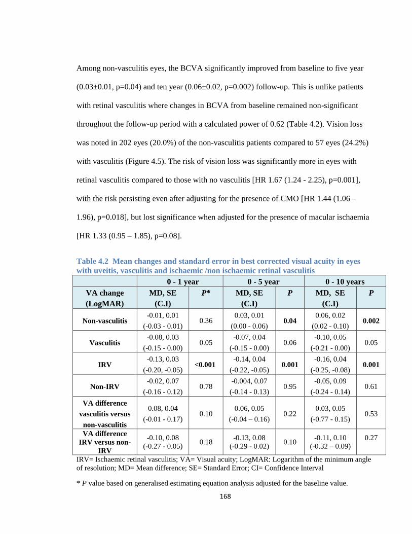
168
Among non-vasculitis eyes, the BCVA significantly improved from baseline to five year
(0.03±0.01, p=0.04) and ten year (0.06±0.02, p=0.002) follow-up. This is unlike patients
with retinal vasculitis where changes in BCVA from baseline remained non-significant
throughout the follow-up period with a calculated power of 0.62 (Table 4.2). Vision loss
was noted in 202 eyes (20.0%) of the non-vasculitis patients compared to 57 eyes (24.2%)
with vasculitis (Figure 4.5). The risk of vision loss was significantly more in eyes with
retinal vasculitis compared to those with no vasculitis [HR 1.67 (1.24 - 2.25), p=0.001],
with the risk persisting even after adjusting for the presence of CMO [HR 1.44 (1.06 –
1.96), p=0.018], but lost significance when adjusted for the presence of macular ischaemia
[HR 1.33 (0.95 – 1.85), p=0.08].
Table 4.2 Mean changes and standard error in best corrected visual acuity in eyes
with uveitis, vasculitis and ischaemic /non ischaemic retinal vasculitis
0 - 1 year 0 - 5 year 0 - 10 years
VA change
(LogMAR)
MD, SE
(C.I)
P* MD, SE
(C.I)
P MD, SE
(C.I)
P
Non-vasculitis -0.01, 0.01
(-0.03 - 0.01) 0.36
0.03, 0.01
(0.00 - 0.06) 0.04
0.06, 0.02
(0.02 - 0.10) 0.002
Vasculitis -0.08, 0.03
(-0.15 - 0.00) 0.05
-0.07, 0.04
(-0.15 - 0.00) 0.06
-0.10, 0.05
(-0.21 - 0.00) 0.05
IRV -0.13, 0.03
(-0.20, -0.05) <0.001
-0.14, 0.04
(-0.22, -0.05) 0.001
-0.16, 0.04
(-0.25, -0.08) 0.001
Non-IRV -0.02, 0.07
(-0.16 - 0.12) 0.78
-0.004, 0.07
(-0.14 - 0.13) 0.95
-0.05, 0.09
(-0.24 - 0.14) 0.61
VA difference
vasculitis versus
non-vasculitis
0.08, 0.04
(-0.01 - 0.17) 0.10
0.06, 0.05
(-0.04 – 0.16) 0.22
0.03, 0.05
(-0.77 - 0.15) 0.53
VA difference
IRV versus non-
IRV
-0.10, 0.08
(-0.27 - 0.05) 0.18
-0.13, 0.08
(-0.29 - 0.02) 0.10
-0.11, 0.10
(-0.32 – 0.09)
0.27
IRV= Ischaemic retinal vasculitis; VA= Visual acuity; LogMAR: Logarithm of the minimum angle
of resolution; MD= Mean difference; SE= Standard Error; CI= Confidence Interval
* P value based on generalised estimating equation analysis adjusted for the baseline value.
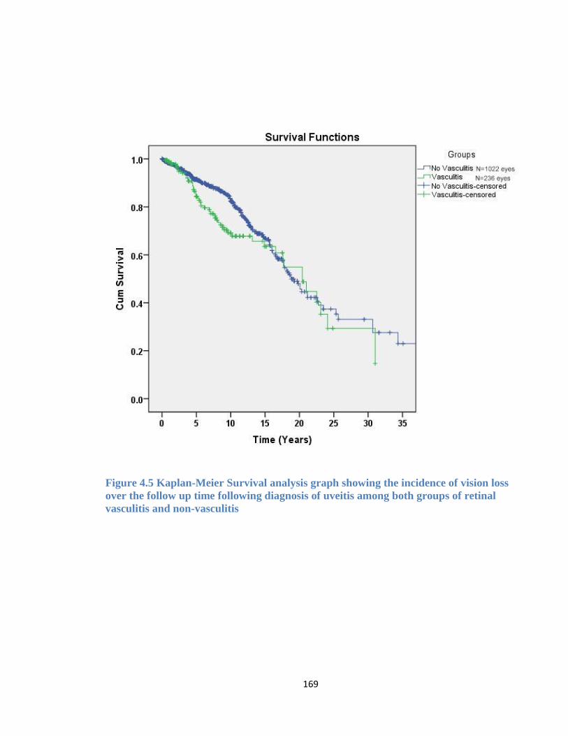
169
Figure 4.5 Kaplan-Meier Survival analysis graph showing the incidence of vision loss
over the follow up time following diagnosis of uveitis among both groups of retinal
vasculitis and non-vasculitis
170
Risk factors contributing to vision loss among both non-vasculitis and vasculitis groups are
listed in Table 4.3. In non-vasculitis eyes, the risk of vision loss was three times more
likely to occur in eyes with macular scarring (HR 3.7, C.I 2.3-6.0, p=<0.001), optic
neuropathy (HR 3.4, C.I. 2.0-5.7, p=<0.001) and retinal detachment (HR 3.6, C.I. 2.1-6.2,
p=<0.001). In eyes with retinal vasculitis, macular ischaemia remained a significant factor
that increased the risk of vision loss by more than four times even after adjusting for other
associated complications (HR 4.4, C.I 2.0-9.6, p<0.001) (Table 4.3).
CMO was reported in 103 eyes (43.6%) with retinal vasculitis compared to 197 eyes
(19.3%) among the non-vasculitis group. Eyes with retinal vasculitis had more than twice
the risk of developing CMO compared to non-vasculitis eyes (HR 2.2, C.I 1.6-2.5,
p=<0.001). Cataract was more prevalent in non-vasculitis eyes (21.4%) compared to
vasculitis eyes (11.5%) with a significantly increased risk of developing cataract in the non-
vasculitis group (HR 1.9, C.I. 1.3-2.8, p=0.006). No significant difference was observed
between vasculitis cases compared to non-vasculitis when looking at other complications
such as retinal detachment (2.1% versus 2.3%, respectively, p=0.72), retinal pigment
epithelial atrophy (1.7% versus 3.9%, p= 0.18), ERM formation (11.4% versus 10.0%, p=
0.38) and optic neuropathy (2.5% versus 3.5%, p=0.53).
Systemic prednisolone was used in the management of 168 eyes (71.2%) with vasculitis
compared to 640 eyes (63.0%) without vasculitis with no significant difference between the
two groups (HR 1.06, C.I. 0.8-1.2, p=0.49). IMTs were used in the management of 69 eyes
(29.2%) with retinal vasculitis compared to 144 eyes (14.2%) with no vasculitis. Eyes with
retinal vasculitis were twice more likely to require IMT as part of their management when
compared to non-vasculitis eyes (HR 2.0, C.I. 1.5-2.7, p=<0.001).
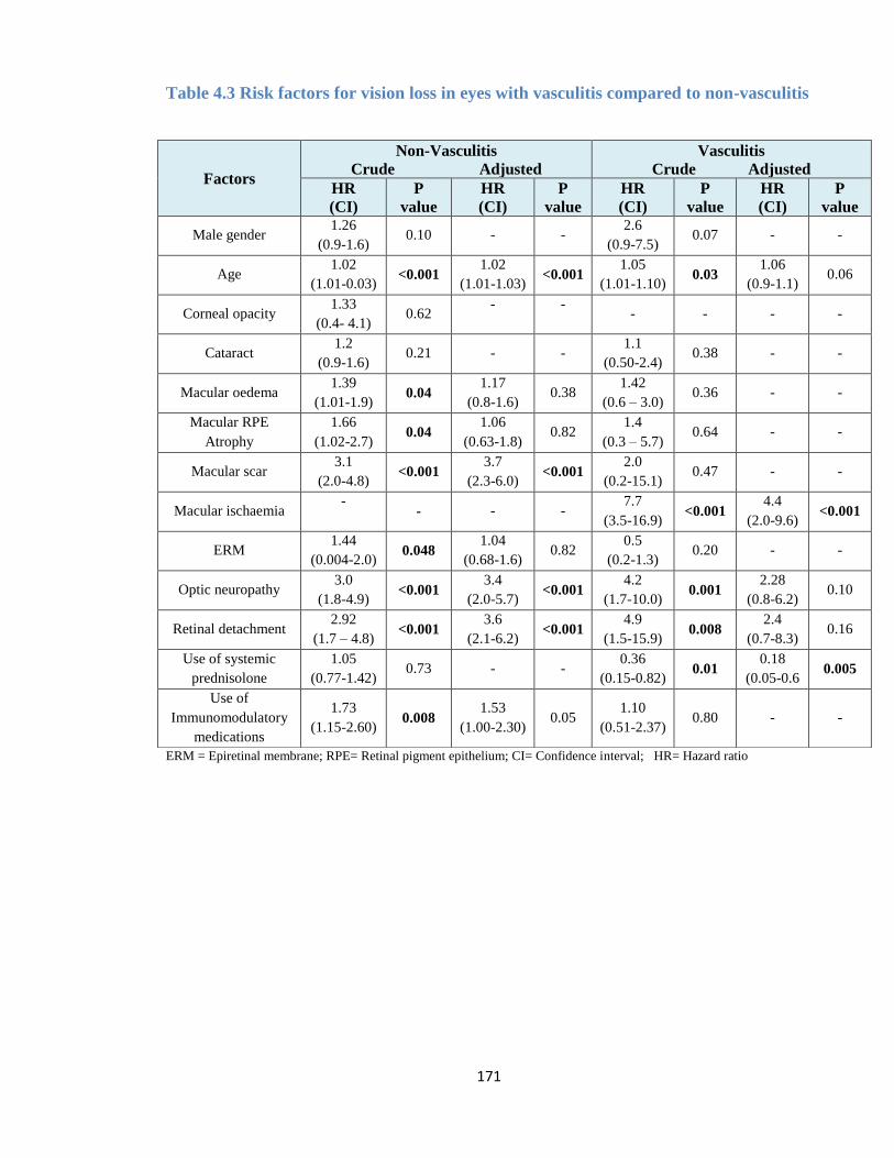
171
Table 4.3 Risk factors for vision loss in eyes with vasculitis compared to non-vasculitis
ERM = Epiretinal membrane; RPE= Retinal pigment epithelium; CI= Confidence interval; HR= Hazard ratio
Factors
Non-Vasculitis
Crude Adjusted
Vasculitis
Crude Adjusted
HR
(CI)
P
value
HR
(CI)
P
value
HR
(CI)
P
value
HR
(CI)
P
value
Male gender 1.26
(0.9-1.6) 0.10 - -
2.6
(0.9-7.5) 0.07 - -
Age 1.02
(1.01-0.03) <0.001
1.02
(1.01-1.03) <0.001
1.05
(1.01-1.10) 0.03
1.06
(0.9-1.1) 0.06
Corneal opacity 1.33
(0.4- 4.1) 0.62
-
-
- - - -
Cataract 1.2
(0.9-1.6) 0.21 - -
1.1
(0.50-2.4) 0.38 - -
Macular oedema 1.39
(1.01-1.9) 0.04
1.17
(0.8-1.6) 0.38
1.42
(0.6 – 3.0) 0.36 - -
Macular RPE
Atrophy
1.66
(1.02-2.7) 0.04
1.06
(0.63-1.8) 0.82
1.4
(0.3 – 5.7) 0.64 - -
Macular scar 3.1
(2.0-4.8) <0.001
3.7
(2.3-6.0) <0.001
2.0
(0.2-15.1) 0.47 - -
Macular ischaemia -
- - -
7.7
(3.5-16.9) <0.001
4.4
(2.0-9.6) <0.001
ERM 1.44
(0.004-2.0) 0.048
1.04
(0.68-1.6) 0.82
0.5
(0.2-1.3) 0.20 - -
Optic neuropathy 3.0
(1.8-4.9) <0.001
3.4
(2.0-5.7) <0.001
4.2
(1.7-10.0) 0.001
2.28
(0.8-6.2) 0.10
Retinal detachment 2.92
(1.7 – 4.8) <0.001
3.6
(2.1-6.2) <0.001
4.9
(1.5-15.9) 0.008
2.4
(0.7-8.3) 0.16
Use of systemic
prednisolone
1.05
(0.77-1.42) 0.73 - -
0.36
(0.15-0.82) 0.01
0.18
(0.05-0.6 0.005
Use of
Immunomodulatory
medications
1.73
(1.15-2.60) 0.008
1.53
(1.00-2.30) 0.05
1.10
(0.51-2.37) 0.80 - -
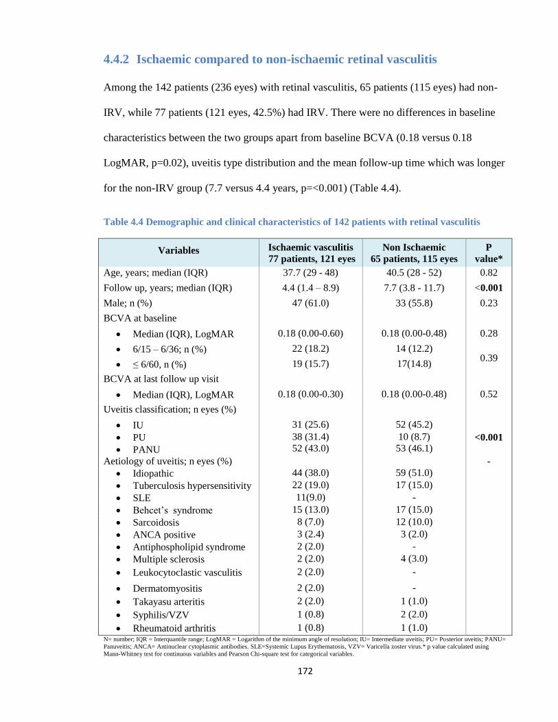
172
4.4.2 Ischaemic compared to non-ischaemic retinal vasculitis
Among the 142 patients (236 eyes) with retinal vasculitis, 65 patients (115 eyes) had non-
IRV, while 77 patients (121 eyes, 42.5%) had IRV. There were no differences in baseline
characteristics between the two groups apart from baseline BCVA (0.18 versus 0.18
LogMAR, p=0.02), uveitis type distribution and the mean follow-up time which was longer
for the non-IRV group (7.7 versus 4.4 years, p=<0.001) (Table 4.4).
Table 4.4 Demographic and clinical characteristics of 142 patients with retinal vasculitis
Variables Ischaemic vasculitis
77 patients, 121 eyes
Non Ischaemic
65 patients, 115 eyes
P
value*
Age, years; median (IQR) 37.7 (29 - 48) 40.5 (28 - 52) 0.82
Follow up, years; median (IQR) 4.4 (1.4 – 8.9) 7.7 (3.8 - 11.7) <0.001
Male; n (%) 47 (61.0) 33 (55.8) 0.23
BCVA at baseline
Median (IQR), LogMAR 0.18 (0.00-0.60) 0.18 (0.00-0.48) 0.28
6/15 – 6/36; n (%) 22 (18.2) 14 (12.2) 0.39
≤ 6/60, n (%) 19 (15.7) 17(14.8)
BCVA at last follow up visit
Median (IQR), LogMAR 0.18 (0.00-0.30) 0.18 (0.00-0.48) 0.52
Uveitis classification; n eyes (%)
IU 31 (25.6) 52 (45.2)
<0.001 PU
PANU
38 (31.4)
52 (43.0)
10 (8.7)
53 (46.1)
Aetiology of uveitis; n eyes (%) -
Idiopathic 44 (38.0) 59 (51.0)
Tuberculosis hypersensitivity 22 (19.0) 17 (15.0)
SLE 11(9.0) -
Behcet’s syndrome 15 (13.0) 17 (15.0)
Sarcoidosis 8 (7.0) 12 (10.0)
ANCA positive 3 (2.4) 3 (2.0)
Antiphospholipid syndrome 2 (2.0) -
Multiple sclerosis 2 (2.0) 4 (3.0)
Leukocytoclastic vasculitis 2 (2.0) -
Dermatomyositis 2 (2.0) -
Takayasu arteritis 2 (2.0) 1 (1.0)
Syphilis/VZV 1 (0.8) 2 (2.0)
Rheumatoid arthritis 1 (0.8) 1 (1.0) N= number; IQR = Interquantile range; LogMAR = Logarithm of the minimum angle of resolution; IU= Intermediate uveitis; PU= Posterior uveitis; PANU=
Panuveitis; ANCA= Antinuclear cytoplasmic antibodies. SLE=Systemic Lupus Erythematosis, VZV= Varicella zoster virus.* p value calculated using
Mann-Whitney test for continuous variables and Pearson Chi-square test for categorical variables.
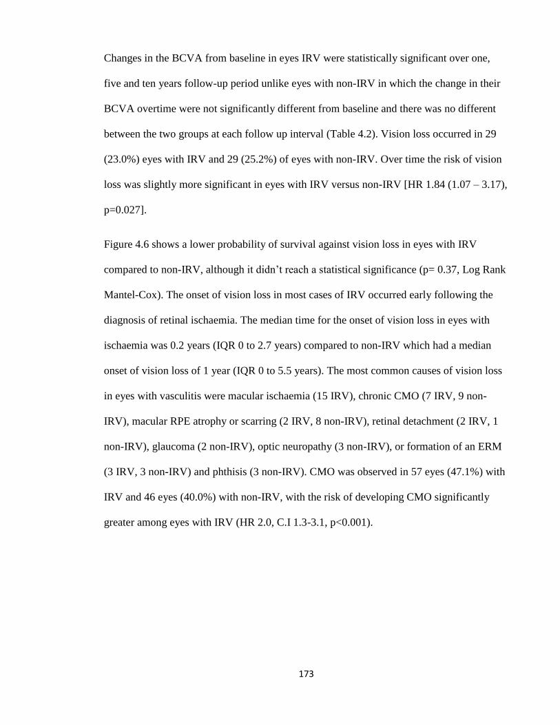
173
Changes in the BCVA from baseline in eyes IRV were statistically significant over one,
five and ten years follow-up period unlike eyes with non-IRV in which the change in their
BCVA overtime were not significantly different from baseline and there was no different
between the two groups at each follow up interval (Table 4.2). Vision loss occurred in 29
(23.0%) eyes with IRV and 29 (25.2%) of eyes with non-IRV. Over time the risk of vision
loss was slightly more significant in eyes with IRV versus non-IRV [HR 1.84 (1.07 – 3.17),
p=0.027].
Figure 4.6 shows a lower probability of survival against vision loss in eyes with IRV
compared to non-IRV, although it didn’t reach a statistical significance (p= 0.37, Log Rank
Mantel-Cox). The onset of vision loss in most cases of IRV occurred early following the
diagnosis of retinal ischaemia. The median time for the onset of vision loss in eyes with
ischaemia was 0.2 years (IQR 0 to 2.7 years) compared to non-IRV which had a median
onset of vision loss of 1 year (IQR 0 to 5.5 years). The most common causes of vision loss
in eyes with vasculitis were macular ischaemia (15 IRV), chronic CMO (7 IRV, 9 non-
IRV), macular RPE atrophy or scarring (2 IRV, 8 non-IRV), retinal detachment (2 IRV, 1
non-IRV), glaucoma (2 non-IRV), optic neuropathy (3 non-IRV), or formation of an ERM
(3 IRV, 3 non-IRV) and phthisis (3 non-IRV). CMO was observed in 57 eyes (47.1%) with
IRV and 46 eyes (40.0%) with non-IRV, with the risk of developing CMO significantly
greater among eyes with IRV (HR 2.0, C.I 1.3-3.1, p<0.001).
174
Figure 4.6 Kaplan-Meier survival analysis curve showing the incidence of vision loss
over the follow-up time since the diagnosis of ischaemia in eyes with ischaemic retinal
vasculitis (IRV) and since the diagnosis of uveitis in eyes with non-ischaemic retinal
vasculitis (non-IRV).
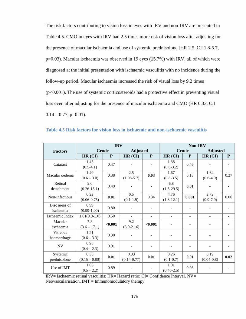
175
The risk factors contributing to vision loss in eyes with IRV and non-IRV are presented in
Table 4.5. CMO in eyes with IRV had 2.5 times more risk of vision loss after adjusting for
the presence of macular ischaemia and use of systemic prednisolone [HR 2.5, C.I 1.8-5.7,
p=0.03). Macular ischaemia was observed in 19 eyes (15.7%) with IRV, all of which were
diagnosed at the initial presentation with ischaemic vasculitis with no incidence during the
follow-up period. Macular ischaemia increased the risk of visual loss by 9.2 times
(p<0.001). The use of systemic corticosteroids had a protective effect in preventing visual
loss even after adjusting for the presence of macular ischaemia and CMO (HR 0.33, C.I
0.14 – 0.77, p=0.01).
Table 4.5 Risk factors for vision loss in ischaemic and non-ischaemic vasculitis
IRV= Ischaemic retinal vasculitis; HR= Hazard ratio; CI= Confidence Interval. NV=
Neovascularisation. IMT = Immunomodulatory therapy
Factors
IRV
Crude Adjusted
Non-IRV
Crude Adjusted
HR (CI) P HR (CI) P HR (CI) P HR (CI) P
Cataract 1.45
(0.5-4.1) 0.47 - -
1.38
(0.6-3.2) 0.46 - -
Macular oedema 1.40
(0.6 – 3.0) 0.38
2.5
(1.08-5.7) 0.03
1.67
(0.8-3.5) 0.18
1.64
(0.6-4.0) 0.27
Retinal
detachment
2.0
(0.26-15.1) 0.49 - -
6.8
(1.5-29.5) 0.01 - -
Non-infectious 0.22
(0.06-0.75) 0.01
0.5
(0.1-1.9) 0.34
4.76
(1.8-12.1) 0.001
2.72
(0.9-7.9) 0.06
Disc areas of
ischaemia
0.99
(0.99-1.00) 0.80 - - - - - -
Ischaemic Index 1.01(0.9-1.0) 0.50 - - - - - -
Macular
ischaemia
7.8
(3.6 – 17.1) <0.001
9.2
(3.9-21.6) <0.001 - - - -
Vitreous
haemorrhage
1.51
(0.6 – 3.3) 0.30 - - - - - -
NV 0.95
(0.4 – 2.3) 0.91 - - - - - -
Systemic
prednisolone
0.35
(0.15 – 0.80) 0.01
0.33
(0.14-0.77) 0.01
0.26
(0.1-0.7) 0.01
0.19
(0.04-0.8) 0.02
Use of IMT 1.05
(0.5 – 2.2) 0.89 - -
1.01
(0.40-2.5) 0.98 - -
176
4.4.3 Characteristics of retinal ischaemia
Retinal ischaemia was already established during first visit with uveitis in 70 eyes (57.8%).
In the remaining 51 eyes, ischaemia occurred at a median of 7 years (IQR 2.0 – 9.3 years).
The area of ischaemia was localised to the peripheral area in 45 eyes (37.2%), extended to
the midperipheral area in 45 eyes (37.2%), and was either confined to the posterior pole or
was extended to involve this area in 31 eyes (25.6%). The area of ischaemia involved one
quadrant in 41 eyes (33.9%), two in 42 eyes (34.7%), three in 13 eyes (10.7%), and all four
quadrants in 25 eyes (20.7%).
The area of retinal ischaemia was measured in 63 eyes with ultra-widefield FFA. The
median area of ischemia was 99 DA (IQR 48 – 169 DA) with 52 eyes (82.5%) had an area
of ischaemia at diagnosis ≥30 DA. When measuring the ischaemic index, the median area
of ischaemia was 25.8% (IQR 10.2 – 46%) with 17 eyes (27%) had an ischaemic index >
50%.
Established complications of retinal ischaemia included VH in 26 eyes (21.5%), NVD in
18 (14.9%), NVE in 75 (62.0%), and NVI in 4 eyes (3.3%). Retinal laser photocoagulation
was administered in 75 eyes (62.0%), out of which 29 eyes (38.1%) had NV relapse and
required an additional laser treatment (14 for treating NV at a new location and 15 for NV
that failed to respond to the initial laser therapy). The first retinal laser photocoagulation
was given at a median of 5 months (IQR 1-12) from baseline, ranging from 0 to 179
months post diagnosis of ischaemia. The additional laser given for NV relapse was given at
a median of 10.5 months (IQR 7.75 – 23.5) from the initial laser, ranging from 5 to 76
months.
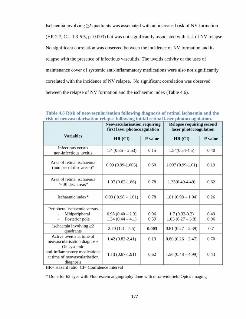
177
Ischaemia involving ≥2 quadrants was associated with an increased risk of NV formation
(HR 2.7, C.I. 1.3-5.5, p=0.003) but was not significantly associated with risk of NV relapse.
No significant correlation was observed between the incidence of NV formation and its
relapse with the presence of infectious vasculitis. The uveitis activity or the uses of
maintenance cover of systemic anti-inflammatory medications were also not significantly
correlated with the incidence of NV relapse. No significant correlation was observed
between the relapse of NV formation and the ischaemic index (Table 4.6).
Table 4.6 Risk of neovascularisation following diagnosis of retinal ischaemia and the
risk of neovascularisation relapse following initial retinal laser photocoagulation.
Variables
Neovascularisation requiring
first laser photocoagulation
Relapse requiring second
laser photocoagulation
HR (CI) P value HR (CI) P value
Infectious versus
non-infectious uveitis 1.4 (0.86 – 2.53) 0.15 1.54(0.54-4.5) 0.40
Area of retinal ischaemia
(number of disc areas)* 0.99 (0.99-1.003) 0.60 1.007 (0.99-1.01) 0.19
Area of retinal ischaemia
≥ 30 disc areas* 1.07 (0.62-1.86) 0.78 1.35(0.40-4.49) 0.62
Ischaemic index* 0.99 ( 0.98 – 1.01) 0.78 1.01 (0.98 – 1.04) 0.26
Peripheral ischaemia versus
- Midperipheral
- Posterior pole
0.98 (0.40 – 2.3)
1.34 (0.44 – 4.1)
0.96
0.59
1.7 (0.33-9.2)
1.03 (0.27 – 3.8)
0.49
0.96
Ischaemia involving ≥2
quadrants 2.70 (1.3 – 5.5) 0.003 0.81 (0.27 – 2.39) 0.7
Active uveitis at time of
neovascularisation diagnosis 1.42 (0.83-2.41) 0.19 0.80 (0.26 – 2.47) 0.70
On systemic
anti-inflammatory medications
at time of neovascularisation
diagnosis
1.13 (0.67-1.91) 0.62 1.56 (0.48 – 4.99) 0.43
HR= Hazard ratio; CI= Confidence Interval
* Done for 63 eyes with Fluorescein angiography done with ultra-widefield Optos imaging
178
4.5 Discussion
In this study, we examined the impact of retinal vasculitis on vision loss, as well as the
progression of IRV and the effect of treatment. We found that (1) vasculitis eyes were more
likely to develop vision loss than eyes without vasculitis, mainly secondary to macular
ischemia. (2) Eyes with retinal vasculitis had more than twice the risk of developing CMO
compared to non-vasculitis. 3) Eyes with IRV carried more risk of vision loss than eyes
with non-IRV. (3) The greatest risk factors related to vision loss in eyes with IRV were
macular ischaemia and chronic CMO. (4) Systemic corticosteroids were an important
protective factor, reducing the risk of vision loss in both IRV and non-IRV eyes. (5) Retinal
ischaemia involving two or more quadrants of the retina is significantly correlated with the
initial NV formation.
The prevalence of retinal vasculitis among our uveitis cohort was 14.0%. This is
comparable to the study by Rosenbaum et al on1390 patients with uveitis out of which 15%
had retinal vasculitis (263). Meanwhile, the prevalence of retinal ischaemia in our vasculitis
cohort was 51.2%, which was much higher than that reported by Ali et al, which described
occlusive vasculitis in only six eyes (5.9%) out of 101 eyes with vasculitis (258). The low
prevalence of ischaemic vasculitis in their study could be attributed to the lack of FFA
images in most of their cases, as it was only available in 39 eyes making it difficult to
identify areas of capillary non-perfusion and thus underestimating the prevalence of
occlusive vasculitis in their study. Furthermore, ultra-wide field FFA images were not
employed unlike our cohort group in which about half of the eyes had their FFA undertaken
using Ultra-widefield imaging. Previous studies had shown that utilisation of ultra-
179
widefield angiography increases the ability to detect peripheral retinal ischaemia and
NV(443).
Unlike the non-vasculitis eyes which showed an average improvement in vision from
baseline to five and ten year follow-up, the average visual acuity was not significantly
different for eyes with vasculitis. While our cohort of eyes with vasculitis had significantly
worse visual outcome compared to the non-vasculitis eyes, this difference was less
significant once both groups were adjusted for the presence of macular ischaemia,
emphasizing the role of macular ischaemia in determining the visual outcome of eyes with
vasculitis. A study examining 53 patients with idiopathic retinal vasculitis observed severe
visual loss more often in eyes with ischaemic vasculitis (34%) compared to the non-
ischaemic group (6%) (436). Ku et al (430) examined the visual outcome and predictors for
its prognosis in 114 patients (203 eyes) with vasculitis and found the mean change in
BCVA from first visit was 0.01 LogMAR per year of follow up, with 33.6% of the eyes
having an improvement in BCVA by ≥ 2 lines in 33.6%. Improvement in vision over the
follow-up time was more predominant non-white patients, infectious uveitis, and those with
worse vision at first presentation (430).
In this studied cohort, macular ischaemia and CMO were related to vision loss, suggesting
a direct involvement in ocular morbidity among eyes with vasculitis. This finding was
consistent with the results observed in a recent study on 82 eyes with vasculitis in which
poor VA was independently associated with central macular thickness and the size of FAZ
(444). The increased risk of CMO in the IRV compared to the non-IRV group in our study
was also observed in previous study by Ali et al (258) which suggests that additional
factors apart from inflammation are involved in producing CMO in eyes with IRV and that
180
areas of nonperfusion may also promote the release of elevated levels of VEGF leading to
increased vascular permeability and development of CMO, as is found in cases of DR and
retinal vein occlusion (439). In a study of DR it was noted that retinal ischaemia increased
the risk of developing DMO by 3.75 times when compared with non-ischaemic DR (437).
Thus, tight control of the underlying disease with the aim of preserving the macula region
should be the paramount objective and guide treatment decisions. We should also consider
the additional risk of PRP itself in inducing CMO and thus increasing its prevalence among
eyes with IRV compared to non-IRV (445).
Most of the eyes with retinal ischaemia received their initial laser treatment within the first
year following the diagnosis of ischaemia, suggesting the need to closely observe the
ischaemic retina during this period for the development of NV. Among eyes with IRV that
underwent initial laser treatment, 38.1% required more than one session of laser
photocoagulation. The rate of receiving additional laser therapy in our cohort was similar
to that observed in a study on Eales’ disease in which 47% of eyes that underwent laser
therapy required additional retinal laser photocoagulation(446). The need for additional
laser therapy in our study did not reflect an increased risk of vision loss.
Systemic corticosteroids were used in the management of 71.2% of the eyes with vasculitis,
which was not different from the non-vasculitis eyes group of which 63.0% were treated
with systemic corticosteroids at one point. A study on eyes with retinal vasculitis also
reported the use of systemic prednisolone in the management of about two third of their
patients (258). While some patients with retinal vasculitis might experience sight
threatening complications that require the use of high dose corticosteroids with or without
IMT, a considerable number of cases are presented with low grade uveitis and vascular
181
leakage confined to the fundus periphery with no direct risk to vision, requiring only
observation or treatment with local corticosteroids.
In our cohort, the use of corticosteroids was associated with a reduced risk of vision loss,
including eyes with IRV and non-IRV. The use of corticosteroids and other anti-
inflammatory medications in managing vasculitis and some of its associated complications,
such as CMO, contributes to their role in preventing vision loss (447). Interestingly,
macular ischaemia developed early in the diagnosis of IRV, with no incidence observed
over the rest of the follow up period; thus the role of anti-inflammatory medications in
preventing macular ischaemia is something to be addressed in future studies. On the other
hand, the study did not find a significant role for anti-inflammatory medications in
preventing ischaemic relapse and NV formation.
While the ischaemic index and area of ischaemia measured in DA in relation to NV
formation did not reach a statistically significant level, the study found that retinal
ischaemia involving ≥2 quadrants can significantly increase the risk of NV formation. This
is similar to a study on IRVAN cases which suggested using laser photocoagulation in eyes
with retinal ischaemia even before NV is formed when there is more than two quadrants
affected with capillary non-perfusion (361).
The study has a number of limitations related to its retrospective nature and the selection of
patients from heterogeneous pathologies. However, the common clinical presentation of
retinal vasculitis and its functional consequences suggest that the outcome of these
conditions can be collated and examined together. Furthermore, retinal ischaemia is an
uncommon occurrence in conditions associated with retinal vasculitis and this study
represents a unique opportunity to examine the long-term visual outcome of eyes with
182
ischaemic vasculitis and the rate of progression of NV formation following laser therapy
and immunosuppressive treatment. Another limitation was associated with the use of
Optos FFA images in measuring the peripheral area of ischaemia. Due to the spherical
curvature of the eye, ultra-widefield images commonly associated with blurred areas within
the far periphery when the central portion of the image is in focus. When we excluded these
blurred far peripheral areas, the total area measured is slightly smaller than the 200 degree
achieved by the ellipsoidal mirror within the Optos imaging system.
In conclusion, the long term visual outcome in eyes with retinal vasculitis is worse when
compared to eyes without vasculitis, mainly due to the risk of macular ischaemia. In
addition to the management algorithm of retinal vasculitis presented in Figure 1.10, the
control of vasculitis with the use of systemic immunosuppressant and specifically
corticosteroids is essential part in vasculitis management as it provides long-term protection
by preventing further deterioration in visual function, although in this study the anti-
inflammatory medication by itself did not prevent further NV formation. It is also
recommended to apply laser photocoagulation when ≥ 2 quadrants of retina are affected by
capillary non-perfusion as this group carries a higher risk of NV formation.
4.6 Publications and Poster presentations
Talat L, Lightman S, Tomkins-Netzer O. Ischemic retinal vasculitis and its
management. J Ophthalmol. 2014;2014:197675
Poster presentation “Visual outcome and treatment strategies among patients with
ischaemic retinal vasculitis.” RCOphth Annual Meeting 2014.
Poster presentation “Ischaemic retinal vasculitis management and visual outcome”
Submitted for participation at the coming Faculty of Brain Sciences Annual Post-
Graduate Research Poster and Presentation Symposium 2013-2014.
183
5 CHAPTER FIVE: THE INFLUENCE OF CATARACT
SURGERY IN UVEITIS ON THE VISUAL OUTCOME
AND RATE OF INFLAMMATION RELAPSE AND
MANAGEMENT
5.1 Introduction
Cataract is one of the leading causes of vision loss worldwide, accounting for 33% of cases
of blindness and 18% of cases with moderate to server vision loss during 2010 (448).
Cataract is one of the most common complications associated with uveitis and the leading
cause of vision loss in uveitis as general (147,449). Cataract may be seen in up to 50-78%
of patients with uveitis depending on the type of uveitis, being more common in chronic
anterior uveitis such as FHC and JIA as well as IU (156,450).
The incidence of cataract in uveitis can vary according to the anatomical and aetiological
type of uveitis, severity of inflammation, use of topical or local steroid therapy, and
duration of uveitis with reported average of 7.5 years from time of uveitis diagnosis until
the eye require cataract surgery (147). One large cohort study involving 3000 patients with
uveitis, 35% complicated with cataract formation among which 80% required cataract
surgery (38). Within the same study, FHC was the most common aetiology, accounting for
23% of cataract cases among the uveitis cohort and was associated with the highest
incidence of cataract, reported in 70 - 78% of FHC cases (38,450). In children, cataract
formation is the most frequently observed complication in JIA related uveitis, with an
estimated incidence rate of 0.04 per EY and a prevalence rate that varies from 9% to 80%
of uveitis cases (451).
184
Cataract in uveitis may develop as a result of the intraocular inflammation itself or due to
the chronic corticosteroid treatment with the risk of cataract formation varying based on the
duration, dose, frequency and application mode of corticosteroid therapy (452,453).
Cataract in uveitis may develop as a result of the intraocular inflammation itself due to the
effect of multiple factors such as free oxygen radicals, lysosomal enzymes, immune
complex deposition on lens capsule, hypoxia, and altered composition or flow of the
aqueous humor. Chronic corticosteroid usage, whether topical, local or systemic
administration can also contribute to the loss of lens transparency in uveitic eyes and this
risk varies based on the duration, dose, frequency and application mode of corticosteroid
treatment (84,454). In JIA associated uveitis, several retrospective series have described
risk factors for cataract, which include presence of posterior synechiae at initial
presentation with uveitis, the use of systemic corticosteroids, the use of topical
corticosteroids more than 3 times per day, and persistent active inflammation. Thorne and
his co-workers studied the incidence of cataract and its risk factors in 75 children with JIA
associated uveitis. Cataract was already present at initial presentation in 25% of the eyes,
and cataract surgery was needed in 23% of the eyes with JIA uveitis. In this study, the
presence of posterior synechiae at initial presentation with uveitis, the use of systemic
corticosteroids, the use of topical corticosteroids more than 3 times per day, and persistent
active inflammation were all considered risk factors for the development of cataract in
these patients. Meanwhile, the use of topical corticosteroids less than 3 times per day, was
associated with 68% reduced risk of cataract formation, although such association was not
independent from associated risk factors for cataract development (84,454). The risk of
cataract development in JIA uveitis can also extend into adulthood, especially in the
absence of early and prompt control of uveitis in these cases. A case series on 30 eyes of
185
adults with active JIA uveitis that extended into adulthood period found 53% who had a
new incidence of cataract requiring surgery. These patients had a mean age of uveitis onset
at 11.5 years and never been started on IMT agents during childhood. Systemic
corticosteroid can also be cataractogenic but its role in cataract formation is of less
significance when compared to topical or local routes of steroid administration (38).
While most uveitis complications can be addressed with medical therapies as an option,
surgical removal of the cataract is the only treatment option available for this specific
uveitis complication. In a study involving 1799 eyes with uveitis, 474 (26%) required
cataract surgery within an average of 7.5 years from the time of uveitis diagnosis (147).
Whether cataract removal in uveitis eyes can alter the long term visual outcome and uveitis
complications has been the aim of variable published studies which has been recently
addressed in a systematic review and meta-analysis study (376). The review’s aggregate
outcome of 24 studies on uveitic cataract with heterogeneous aetiology found 70% of cases
achieved a postoperative vision of 6/12 or better. According to the type of surgery, the
review included 13 articles that studied phacoemulsification with IOL implantation among
uveitic eyes with variable aetiologies and sample numbers ranging from 32 eyes to a
maximum of 140 eyes (369,455–466). Phacoemulsification with IOL implantation achieved
good visual outcome in 68% while 73% did so following extracapsular cataract extraction.
According to the insertion of IOL post cataract removal, those with IOL implantation
achieved a postoperative vision of 6/12 or better in 70% of cases compared to only 54% of
uveitic eyes had cataract removal without IOL implantation, suggesting that on average, the
uveitic eyes which undergoing cataract removal with IOL implantation can have a
significantly better visual outcome when compared to eyes with cataract removal without
IOL implantation (376).
186
FHC has been one of the most widely studied cases of uveitis and its outcome following
cataract surgery. It also has one of the best visual outcomes following cataract surgery
compared to other causes of uveitis. In the meta-analysis research, 22 studies evaluating
cataract surgery outcome in eyes with FHC found a cumulative 85% of the eyes to have a
postoperative BCVA of 6/12 or more (376). Meanwhile, cataract surgery in JIA associated
uveitis have in general worse visual outcome and more chance of developing sight
threatening complications compared to cataract surgery outcome in children with idiopathic
or non-JIA related uveitis (467). The prognosis in cataract surgery for JIA associated
uveitis is strongly based on the degree of uveitis activity control prior to cataract surgery. In
the meta-analysis study, JIA associated uveitis was reported to achieve good visual
outcome in 65% of cases following cataract surgery, this can increase to 71% among eyes
with controlled uveitis (376) compared to only 22% of the eyes undergoing cataract surgery
with preoperative active uveitis (371).
In most eyes with chorioretinal inflammation, the visual outcome post cataract surgery is
less favourable compared to those with mixed uveitis aetiology and in eyes with AU and IU
possibly due to pre-existing maculopathy in eyes with PU and PANU that guard the vision
prognosis post surgery. Eyes with IU undergoing cataract surgery can achieve a
postoperative vision of 6/12 or better in 70% of cases, making it very similar to that of a
mixed uveitis cohort, whereas ocular BD did worse, achieving a good postoperative vision
in only 36% of the eyes. The same goes for sympathetic ophthalmitis in which the
postoperative vision of 6/12 or better is only achieved in 40% of cases VKH associated
uveitis can have a postoperative visual outcome of 6/12 or better in only half of the cases
following cataract surgery (376).
187
A study reviewed the long-term follow-up of cataract surgery in 61 uveitic patients (72
eyes) and found that after a minimum of 5 years follow-up, 82% of eyes maintained a
visual improvement of at least 2 Snellen lines from preoperative vision, with 74% having a
visual acuity of 6/9 or better. Postoperative CMO was observed in one third of the cases
and 15% required subsequent glaucoma drainage surgery (468). It was noted though that
more than half of the cataract surgery (41 eyes) were performed using extracapsular
cataract extraction. The majority of eyes included in the study were chronic anterior uveitis
(55 eyes including 33 FHC) and only two eyes with IU and 13 with PU; this may explain
the relatively good vision maintained during the follow up period. Elgohary et al.
retrospectively analysed the outcomes of 101 eyes undergoing cataract surgery in the
setting of uveitis, but excluding patients with JIA. This group found that approximately
71% of patients had visual improvement of more than 2 Snellen lines by final follow-up
and the probability of losing one line of vision after 6 years of follow-up was 52%.
However, reactivation of uveitis and CMO was only documented within the first 3 months
post cataract surgery during which 14% had reactivation of their uveitis, 21% developed
postoperative CMO, and 39% developed PCO (463).
The vision loss in uveitic eyes following cataract surgery can be attributed to many
preoperative factors as well as complications that occur during the surgery or afterwards.
Some of the uveitis complications may already exist prior to cataract surgery, leading to
poor vision gain post surgery such as pre-existing amblyopia, or maculopathy, including
RPE atrophy or macular ischaemia, or secondary to optic neuropathy due to neuritis,
ischaemia or advanced glaucoma. Eyes with chronic may also present with variable pre-
existing complications such as anterior and posterior synechiae which requires further
manipulation of the iris and prolongs the duration of surgery. This would increase the risk
188
of postoperative complications or exaggerate postoperative inflammation leading to a poor
visual outcome.
Recurrence of uveitis had been previously reported in one study to occur in 51.3% out of 38
eyes with uveitis following extra capsular cataract extraction with IOL implantation (378).
Sight threatening postoperative complications that can occur in uveitic eyes following
cataract surgery with variable risk based on anatomical classification of the uveitis as well
as the degree of inflammation control prior to the surgery and the use of prophylactic
therapy. CMO in uveitic eyes is reported to occur in 33% to 56% of cases post
extracapsular cataract extraction (378,379) compared to 10% to 59% (with an incidence of
0.02 per EY) following cataract removal using phacoemulsification (380,469). PCO is also
one of the common complications, reported in a study among 27% to 48% of eyes post
cataract surgery with an incidence of 0.07 per EY with FHC and chronic AU have a high
risk of developing PCO following phacoemulsification surgery.(380,449) Other reported
complications include ERM (378,379), increased IOP and hypotony/phthisis. A reported
study on the outcome of phacoemulsification following a minimum of three years follow up
in adult patients with uveitis reported hypotony to occur in 5% of cases with an incidence
of 0.02 per EY associated with postoperative chronic inflammation. The same study
reported elevated IOP to occur in 10.2% of cases with an incidence of 0.04 per EY, mainly
in eyes with anterior uveitis and regardless of the degree of postoperative
inflammation(380).
While there have been variable studies looking at the visual outcome of uveitic eyes
following cataract surgery, limited studies have looked at the influence of cataract surgery
on the uveitis activity parameters including rate of uveitis relapse and the use of topical and
systemic prednisolone compared to the period prior to surgery.
189
5.2 Aims and objectives
1) Assess changes in the best corrected visual acuity over the follow up period post
cataract surgery in uveitic eyes compared to the baseline vision during the first
postoperative week.
2) Examine risk factors for vision loss following cataract surgery in uveitic eyes.
3) Examine the incidence of intraoperative and postoperative complications of cataract
surgery as well as the incidence of uveitis complications over the follow-up period.
4) Study the influence of cataract surgery on the uveitis activity through comparing the
rate of uveitis relapse following cataract surgery compared to the rate prior to the
surgery. In addition, the rate of using topical and systemic steroids and the use of
ocular steroid injections and steroid-sparing treatment following cataract surgery
were compared to the preoperative period.
190
5.3 Materials and method
The study settings and its ethical approval as well as the definition of the general data
related to uveitis characteristics and measurement of visual acuity were previously
described in Chapter 2. Clinical and demographic data of interest were collected using the
designed data collection form in Appendix 2.
In this study, uveitis patients with pseudophakia were compared to a randomly selected
control group of uveitis patients with phakic eyes at time of uveitis database collection.
Pseudophakic eyes were included if they had a minimum follow up period of one year prior
to and after undergoing cataract surgery in the form of phacoemulsification with IOL
implantation. Exclusion criteria include infectious retinitis or choroiditis, aphakia, and
history of vitrectomy and retinal detachment prior to cataract surgery. The date of diagnosis
of uveitis was recorded together with the date of diagnosis of cataract defined as the first
ophthalmologist visit confirming the diagnosis of lens opacity on clinical examination
associated with at least 2 Snellen line drop in vision. The type of cataract was classified as
either nuclear, cortical or posterior subcapsular cataract (389). Time of cataract surgery was
documented together with the use of prophylactic steroid cover in eyes with previous
history of CMO or PANU. When indicated, prophylaxis treatment was given in the form of
oral prednisolone at a dose of 40mg per day given two weeks prior to surgery and then
taper down postoperatively according to the inflammation status postoperatively. In cases
where there was a high risk associated with high dose systemic prednisolone, an alternative
prophylaxis method was used in the form of IVTA, mainly for eyes not known to be steroid
responders or at risk of increased IOP.
191
The BCVA was recorded preoperatively (within 2 months prior to surgery) and then
postoperatively at 1 week. The change in BCVA from the baseline (1st week postoperative
vision) was measured during 1 month, 3 months, 6 months, and 12 months postoperatively
and then annually afterward until last visit together with the incidence of vision loss (6/15
or worse).
OCT images of the macular area were obtained from the Topcon OCT system (3D OCT-
1000; Topcon Corp, Tokyo) and Spectralis HRA OCT (Heidelberg Engineering,
Heidelberg, Germany) subject to the availability of the images taken during the follow-up
visits. A preoperative OCT image within three months prior to surgery was required to be
available together with a minimum of one OCT image postoperatively in order to measure
change in the retinal thickness over the postoperative period compared to the preoperative
image. The central foveal thickness (CFT) in the central circle with 1mm diameter were
measured automatically as micrometer (μm) through the program software and presented in
the output report (470). Uveitis relapses were evaluated during the preoperative and
postoperative period through counting the number of the relapses per eye per year (EY)
divided, respectively, for the preoperative and postoperative follow-up time (380). The use
of topical steroid drops, local and systemic steroids and IMT was also compared over the
period prior and post surgery. The rate of using topical steroid drops (>3 times/day versus
≤3 times/day) and the use of systemic prednisolone (>7.5mg/day versus ≤ 7.5mg/day) was
measured as number of months being on the medication per EY. The use of the cut-off of
≤3 times /day for the topical drops and ≤ 7.5mg/day for systemic prednisolone as they had
shown to be associated with the minimum risk of side effects (83,84). Intraoperative
complications such as posterior capsule rupture and the need for anterior vitrectomy were
documented if present. Postoperative complications and the time of onset were also noted
192
including CMO, PCO and the date of capsulotomy if done, hypotony, elevated IOP and
date of glaucoma filtration surgery if done, and any other sight threatening complications.
5.3.1 Statistical analysis
Detailed description of the statistical method used was presented earlier in Chapter 2.
Repeated measurements analysis of the BCVA and its difference from preoperative vision
(baseline) was done using a multivariate linear regression method obtained from GEE test.
Kaplan-Meier survival analysis with Log-Rank (Mantel-Cox) test was applied together with
survival curve graphs to compare the survival rates for the incidence of vision loss and
postoperative CMO. The HR and 95% C.I for vision loss and postoperative CMO was
measured using Cox proportional hazards regression analysis.
The incidence rates (EY) and 95% C.I for uveitis relapse and use of topical and systemic
corticosteroids over the period before and after cataract surgery were calculated using
negative binominal regression with log link model.
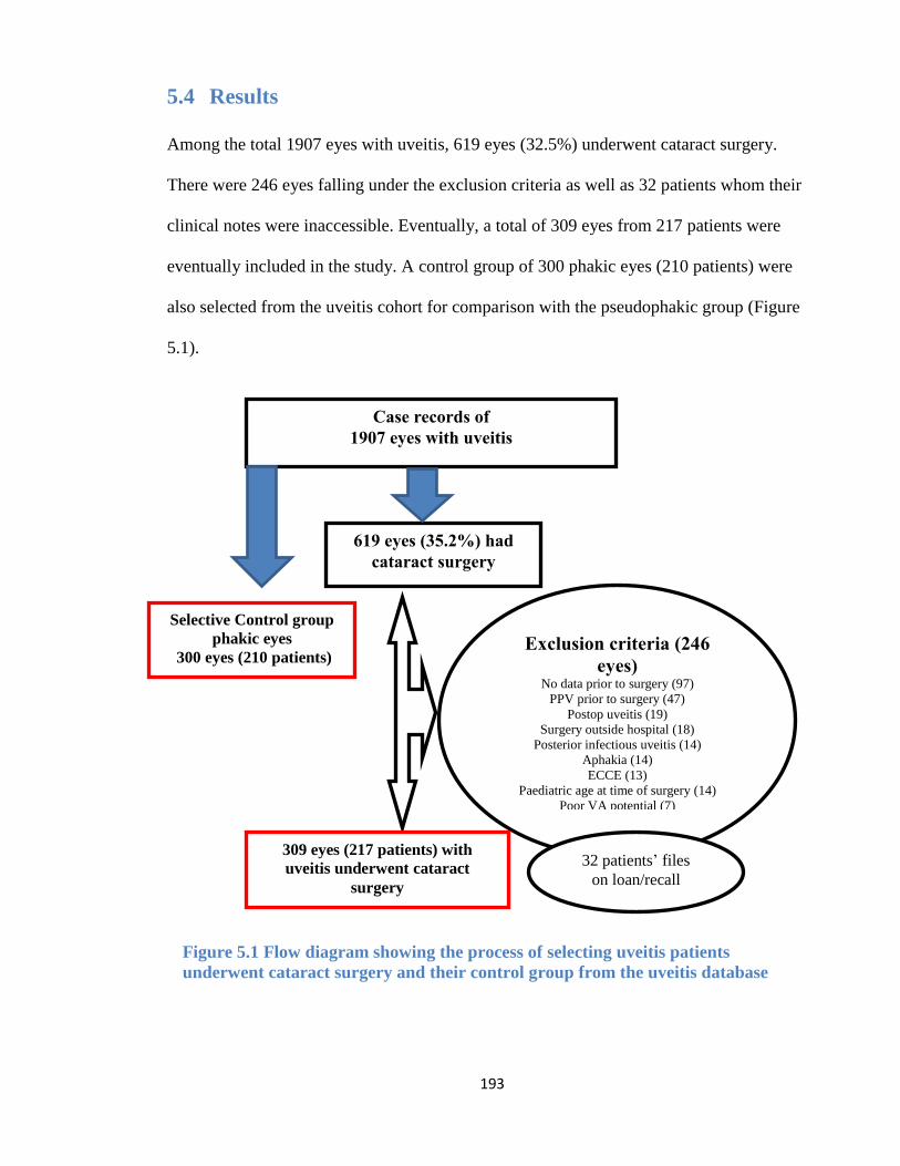
193
5.4 Results
Among the total 1907 eyes with uveitis, 619 eyes (32.5%) underwent cataract surgery.
There were 246 eyes falling under the exclusion criteria as well as 32 patients whom their
clinical notes were inaccessible. Eventually, a total of 309 eyes from 217 patients were
eventually included in the study. A control group of 300 phakic eyes (210 patients) were
also selected from the uveitis cohort for comparison with the pseudophakic group (Figure
5.1).
Case records of
1907 eyes with uveitis
619 eyes (35.2%) had
cataract surgery
Exclusion criteria (246
eyes)
No data prior to surgery (97)
PPV prior to surgery (47)
Postop uveitis (19)
Surgery outside hospital (18)
Posterior infectious uveitis (14)
Aphakia (14)
ECCE (13)
Paediatric age at time of surgery (14)
Poor VA potential (7)
309 eyes (217 patients) with
uveitis underwent cataract
surgery
Selective Control group
phakic eyes
300 eyes (210 patients)
32 patients’ files
on loan/recall
Figure 5.1 Flow diagram showing the process of selecting uveitis patients
underwent cataract surgery and their control group from the uveitis database
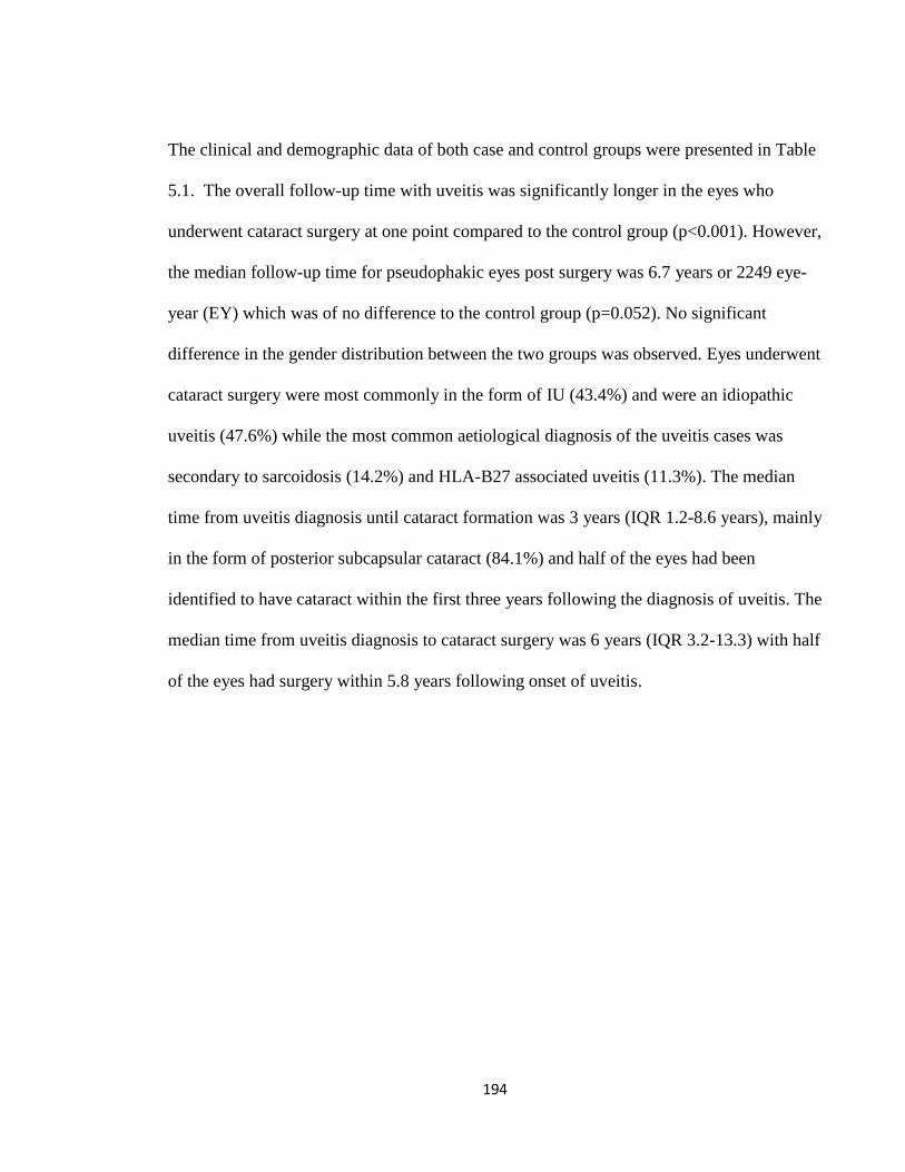
194
The clinical and demographic data of both case and control groups were presented in Table
5.1. The overall follow-up time with uveitis was significantly longer in the eyes who
underwent cataract surgery at one point compared to the control group (p<0.001). However,
the median follow-up time for pseudophakic eyes post surgery was 6.7 years or 2249 eye-
year (EY) which was of no difference to the control group (p=0.052). No significant
difference in the gender distribution between the two groups was observed. Eyes underwent
cataract surgery were most commonly in the form of IU (43.4%) and were an idiopathic
uveitis (47.6%) while the most common aetiological diagnosis of the uveitis cases was
secondary to sarcoidosis (14.2%) and HLA-B27 associated uveitis (11.3%). The median
time from uveitis diagnosis until cataract formation was 3 years (IQR 1.2-8.6 years), mainly
in the form of posterior subcapsular cataract (84.1%) and half of the eyes had been
identified to have cataract within the first three years following the diagnosis of uveitis. The
median time from uveitis diagnosis to cataract surgery was 6 years (IQR 3.2-13.3) with half
of the eyes had surgery within 5.8 years following onset of uveitis.
195
Table 5.1 Demographic and clinical characteristics of pseudophakic uveitic eyes and
their phakic control group
Variables Cases group
309 eyes, 217patients
Control group
300eyes, 210patients
P value*
Follow-up time with uveitis,
Median (IQR), Years 12.4 (8.6-17.9) 5.8 (3.0-10.0)
<0.001
Follow-up time post surgery,
Median (IQR), Years 6.7 (3.9-10.6) 5.8 (3.0- 10.0)
0.052
Female; n patients (%) 136 (62.7) 127 (60.5) 0.35
BCVA LogMAR first visit,
Median (IQR) 0.18 (0.00-0.48) 0.00 (0.00-0.30)
<0.001
BCVA LogMAR last visit,
Median (IQR) 0.18 (0.00-0.30) 0.00 (0.00-0.18)
<0.001
Classification of uveitis; n
eyes (%)
Anterior uveitis 96 (31) 114 (38)
Intermediate uveitis 134(43.4) 92 (30.6) 0.23
Panuveitis 79 (25.6) 94 (31.4)
Aetiological causes of uveitis;
n eyes (%)
Idiopathic 147 (47.6) 220 (73.3)
Sarcoidosis 44 (14.2) 25 (8.3)
HLA-B27 35 (11.3) 15 (5)
Tuberculosis
hypersensitivity 14 (4.5) 14 (4.7)
Behcet’s disease 11 (3.6) 7 (2.3)
Fuchs’ heterochromic
iridocylitis 13 (4.2) 0 (0)
Multifocal choroiditis 12 (3.9) 0(0)
Herpes virus 9(2.9) 6 (2)
ANCA positive 6 (1.9) 2 (0.7)
Multiple sclerosis 8 (2.6) 0(0)
Rheumatoid arthritis 5 (1.6) 2 (0.7)
Vogt Koyanagi Harada
syndrome 5 (1.6) 9 (3)
N= number; SE= Standard error; IQR= Interquantile range; ANCA= Antinuclear cytoplasmic antibodies.
*p value tested using Mann-Whitney U test for continuous data, and Chi-square test for categorical data
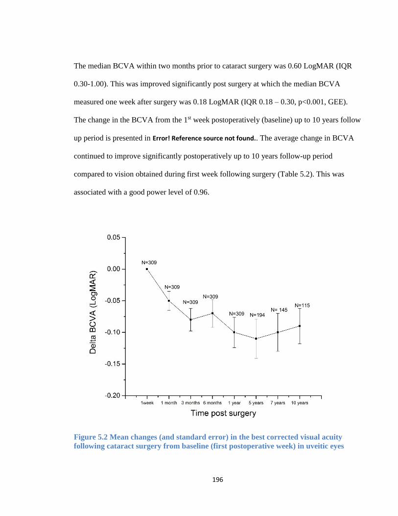
196
The median BCVA within two months prior to cataract surgery was 0.60 LogMAR (IQR
0.30-1.00). This was improved significantly post surgery at which the median BCVA
measured one week after surgery was 0.18 LogMAR (IQR 0.18 – 0.30, p<0.001, GEE).
The change in the BCVA from the 1st week postoperatively (baseline) up to 10 years follow
up period is presented in Error! Reference source not found.. The average change in BCVA
continued to improve significantly postoperatively up to 10 years follow-up period
compared to vision obtained during first week following surgery (Table 5.2). This was
associated with a good power level of 0.96.
Figure 5.2 Mean changes (and standard error) in the best corrected visual acuity
following cataract surgery from baseline (first postoperative week) in uveitic eyes
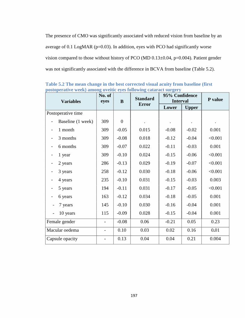
197
The presence of CMO was significantly associated with reduced vision from baseline by an
average of 0.1 LogMAR (p=0.03). In addition, eyes with PCO had significantly worse
vision compared to those without history of PCO (MD 0.13±0.04, p=0.004). Patient gender
was not significantly associated with the difference in BCVA from baseline (Table 5.2).
Table 5.2 The mean change in the best corrected visual acuity from baseline (first
postoperative week) among uveitic eyes following cataract surgery
Variables
No. of
eyes B Standard
Error
95% Confidence
Interval P value
Lower Upper
Postoperative time
- Baseline (1 week) 309 0 . . . .
- 1 month 309 -0.05 0.015 -0.08 -0.02 0.001
- 3 months 309 -0.08 0.018 -0.12 -0.04 <0.001
- 6 months 309 -0.07 0.022 -0.11 -0.03 0.001
- 1 year 309 -0.10 0.024 -0.15 -0.06 <0.001
- 2 years 286 -0.13 0.029 -0.19 -0.07 <0.001
- 3 years 258 -0.12 0.030 -0.18 -0.06 <0.001
- 4 years 235 -0.10 0.031 -0.15 -0.03 0.003
- 5 years 194 -0.11 0.031 -0.17 -0.05 <0.001
- 6 years 163 -0.12 0.034 -0.18 -0.05 0.001
- 7 years 145 -0.10 0.030 -0.16 -0.04 0.001
- 10 years 115 -0.09 0.028 -0.15 -0.04 0.001
Female gender - -0.08 0.06 -0.21 0.05 0.23
Macular oedema - 0.10 0.03 0.02 0.16 0.01
Capsule opacity - 0.13 0.04 0.04 0.21 0.004
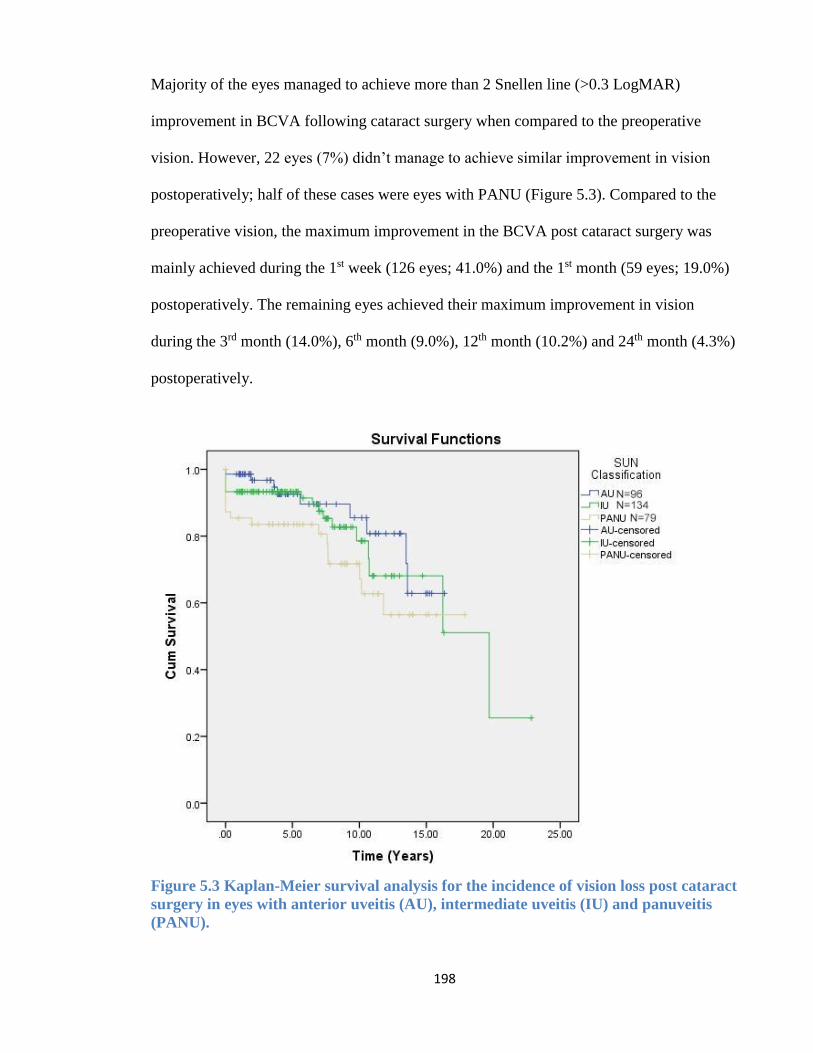
198
Majority of the eyes managed to achieve more than 2 Snellen line (>0.3 LogMAR)
improvement in BCVA following cataract surgery when compared to the preoperative
vision. However, 22 eyes (7%) didn’t manage to achieve similar improvement in vision
postoperatively; half of these cases were eyes with PANU (Figure 5.3). Compared to the
preoperative vision, the maximum improvement in the BCVA post cataract surgery was
mainly achieved during the 1st week (126 eyes; 41.0%) and the 1st month (59 eyes; 19.0%)
postoperatively. The remaining eyes achieved their maximum improvement in vision
during the 3rd month (14.0%), 6th month (9.0%), 12th month (10.2%) and 24th month (4.3%)
postoperatively.
Figure 5.3 Kaplan-Meier survival analysis for the incidence of vision loss post cataract
surgery in eyes with anterior uveitis (AU), intermediate uveitis (IU) and panuveitis
(PANU).
199
By the end of the follow-up period, the median BCVA in pseudophakic eyes was 0.18
LogMAR (IQR 0.00 – 0.30) which was significantly worse than the final median BCVA of
0.00 LogMAR (IQR 0.00-0.18) in the control group (Table 5.1). The incidence rate of
vision loss post cataract surgery was 0.02/EY and occurred in 58 pseudophakic eyes
(18.8%) compared to 22 phakic eyes (7.4%) from the control group. The difference
between both groups was found to be statistically significant (p<0.001, Chi-Square test).
Vision loss in pseudophakic group was mainly secondary to chronic CMO (23 eyes), RPE
atrophy (22 eyes), optic neuropathy (9 eyes), glaucomatous optic neuropathy (5 eyes),
ERM (7 eyes), retinal detachment (5 eyes), corneal scar (3eyes), CRVO (1 eye), macular
hole (1 eye), macular scar (1 eye) and hypotony (1 eye). Uveitic eyes that experienced
CMO post cataract surgery demonstrate a significantly more chance of vision loss
compared to eyes that haven’t exhibit similar complication (p=0.002, Log Rank Mentel-
Cox) as presented in Figure 5.4. Similarly, eyes that had an incidence of increased IOP
>30mmHg had more chance of vision loss compared to the other group with no similar
increase in the IOP (p=0.009, Log Rank Mantel-Cox) (Figure 5.5).
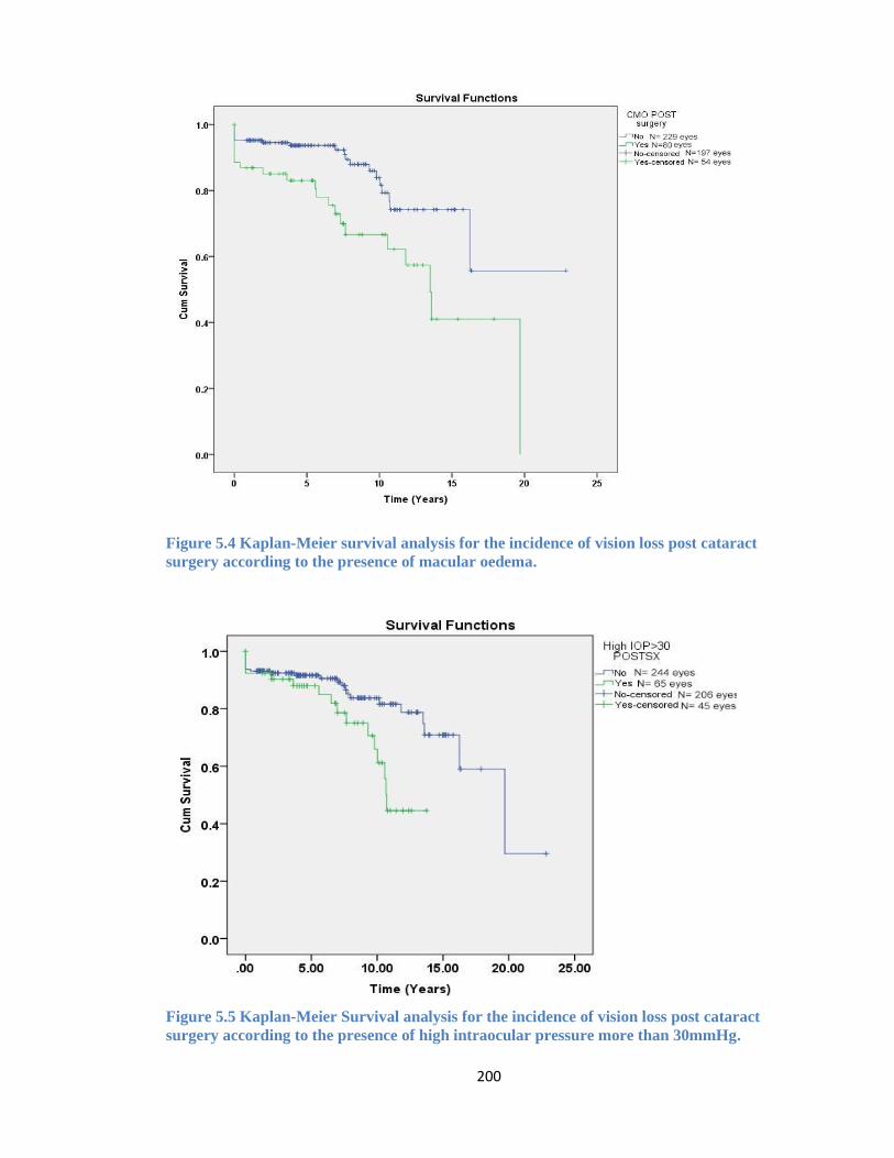
200
Figure 5.4 Kaplan-Meier survival analysis for the incidence of vision loss post cataract
surgery according to the presence of macular oedema.
Figure 5.5 Kaplan-Meier Survival analysis for the incidence of vision loss post cataract
surgery according to the presence of high intraocular pressure more than 30mmHg.
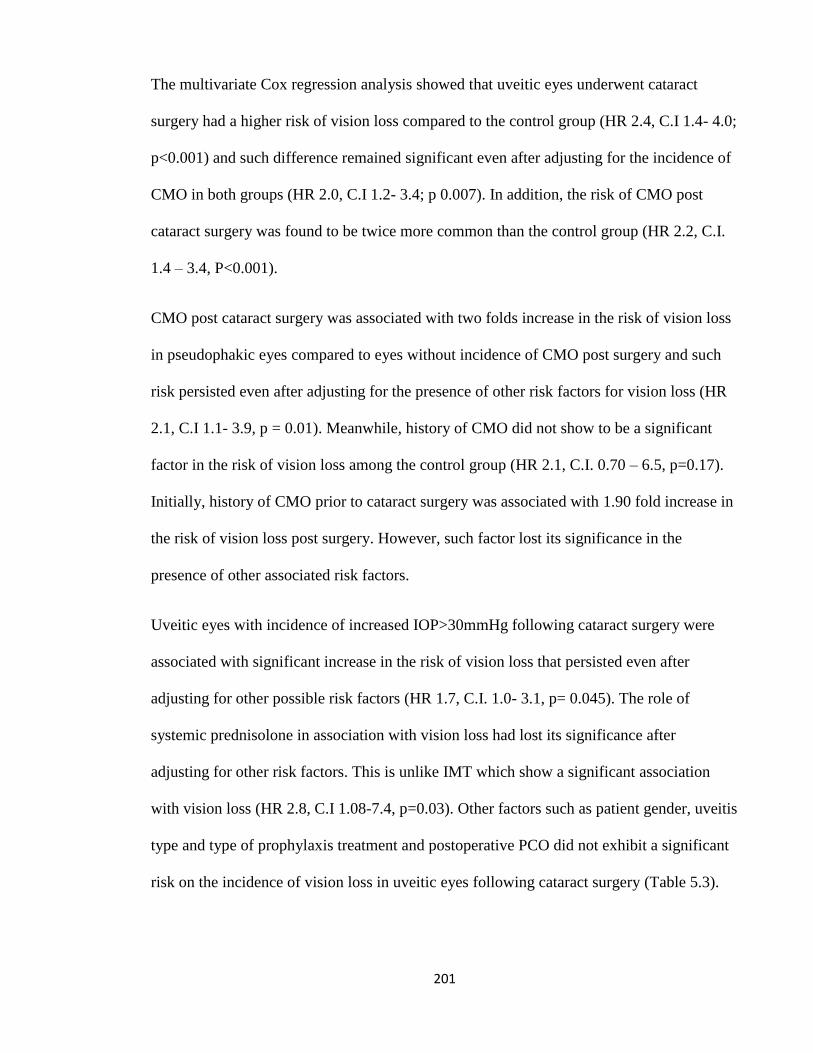
201
The multivariate Cox regression analysis showed that uveitic eyes underwent cataract
surgery had a higher risk of vision loss compared to the control group (HR 2.4, C.I 1.4- 4.0;
p<0.001) and such difference remained significant even after adjusting for the incidence of
CMO in both groups (HR 2.0, C.I 1.2- 3.4; p 0.007). In addition, the risk of CMO post
cataract surgery was found to be twice more common than the control group (HR 2.2, C.I.
1.4 – 3.4, P<0.001).
CMO post cataract surgery was associated with two folds increase in the risk of vision loss
in pseudophakic eyes compared to eyes without incidence of CMO post surgery and such
risk persisted even after adjusting for the presence of other risk factors for vision loss (HR
2.1, C.I 1.1- 3.9, p = 0.01). Meanwhile, history of CMO did not show to be a significant
factor in the risk of vision loss among the control group (HR 2.1, C.I. 0.70 – 6.5, p=0.17).
Initially, history of CMO prior to cataract surgery was associated with 1.90 fold increase in
the risk of vision loss post surgery. However, such factor lost its significance in the
presence of other associated risk factors.
Uveitic eyes with incidence of increased IOP>30mmHg following cataract surgery were
associated with significant increase in the risk of vision loss that persisted even after
adjusting for other possible risk factors (HR 1.7, C.I. 1.0- 3.1, p= 0.045). The role of
systemic prednisolone in association with vision loss had lost its significance after
adjusting for other risk factors. This is unlike IMT which show a significant association
with vision loss (HR 2.8, C.I 1.08-7.4, p=0.03). Other factors such as patient gender, uveitis
type and type of prophylaxis treatment and postoperative PCO did not exhibit a significant
risk on the incidence of vision loss in uveitic eyes following cataract surgery (Table 5.3).
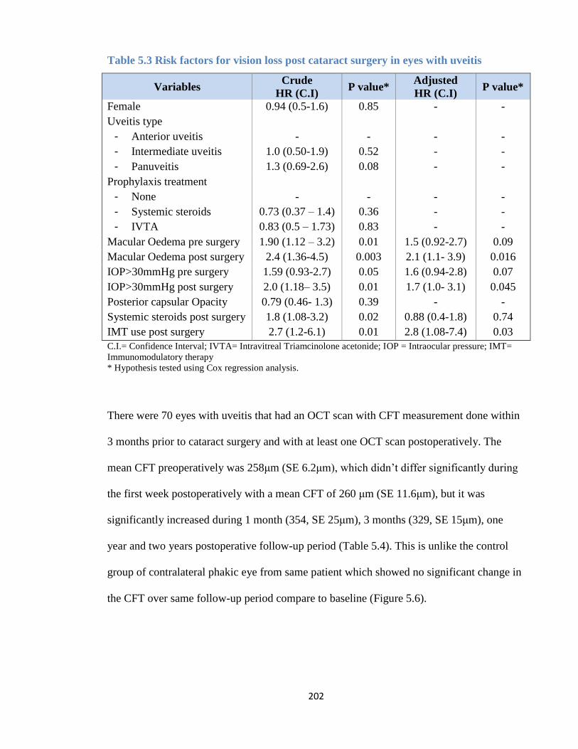
202
Table 5.3 Risk factors for vision loss post cataract surgery in eyes with uveitis
Variables Crude
HR (C.I) P value*
Adjusted
HR (C.I) P value*
Female 0.94 (0.5-1.6) 0.85 - -
Uveitis type
- Anterior uveitis
- Intermediate uveitis
- Panuveitis
-
1.0 (0.50-1.9)
1.3 (0.69-2.6)
-
0.52
0.08
-
-
-
-
-
-
Prophylaxis treatment
- None
- Systemic steroids
- IVTA
-
0.73 (0.37 – 1.4)
0.83 (0.5 – 1.73)
-
0.36
0.83
-
-
-
-
-
-
Macular Oedema pre surgery 1.90 (1.12 – 3.2) 0.01 1.5 (0.92-2.7) 0.09
Macular Oedema post surgery 2.4 (1.36-4.5) 0.003 2.1 (1.1- 3.9) 0.016
IOP>30mmHg pre surgery 1.59 (0.93-2.7) 0.05 1.6 (0.94-2.8) 0.07
IOP>30mmHg post surgery 2.0 (1.18– 3.5) 0.01 1.7 (1.0- 3.1) 0.045
Posterior capsular Opacity 0.79 (0.46- 1.3) 0.39 - -
Systemic steroids post surgery 1.8 (1.08-3.2) 0.02 0.88 (0.4-1.8) 0.74
IMT use post surgery 2.7 (1.2-6.1) 0.01 2.8 (1.08-7.4) 0.03
C.I.= Confidence Interval; IVTA= Intravitreal Triamcinolone acetonide; IOP = Intraocular pressure; IMT=
Immunomodulatory therapy
* Hypothesis tested using Cox regression analysis.
There were 70 eyes with uveitis that had an OCT scan with CFT measurement done within
3 months prior to cataract surgery and with at least one OCT scan postoperatively. The
mean CFT preoperatively was 258μm (SE 6.2μm), which didn’t differ significantly during
the first week postoperatively with a mean CFT of 260 μm (SE 11.6μm), but it was
significantly increased during 1 month (354, SE 25μm), 3 months (329, SE 15μm), one
year and two years postoperative follow-up period (Table 5.4). This is unlike the control
group of contralateral phakic eye from same patient which showed no significant change in
the CFT over same follow-up period compare to baseline (Figure 5.6).
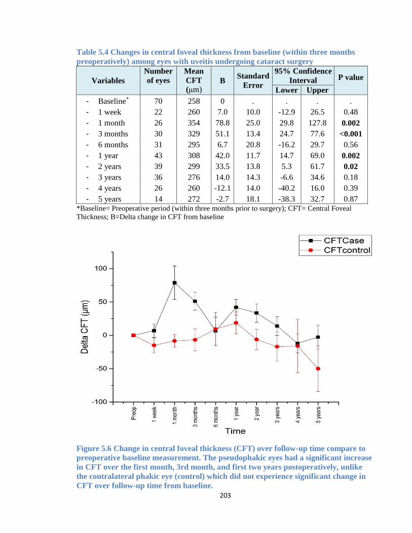
203
Table 5.4 Changes in central foveal thickness from baseline (within three months
preoperatively) among eyes with uveitis undergoing cataract surgery
Variables
Number
of eyes
Mean
CFT
(μm) B
Standard
Error
95% Confidence
Interval P value
Lower Upper
- Baseline* 70 258 0 . . . .
- 1 week 22 260 7.0 10.0 -12.9 26.5 0.48
- 1 month 26 354 78.8 25.0 29.8 127.8 0.002
- 3 months 30 329 51.1 13.4 24.7 77.6 <0.001
- 6 months 31 295 6.7 20.8 -16.2 29.7 0.56
- 1 year 43 308 42.0 11.7 14.7 69.0 0.002
- 2 years 39 299 33.5 13.8 5.3 61.7 0.02
- 3 years 36 276 14.0 14.3 -6.6 34.6 0.18
- 4 years 26 260 -12.1 14.0 -40.2 16.0 0.39
- 5 years 14 272 -2.7 18.1 -38.3 32.7 0.87 *Baseline= Preoperative period (within three months prior to surgery); CFT= Central Foveal
Thickness; B=Delta change in CFT from baseline
Figure 5.6 Change in central foveal thickness (CFT) over follow-up time compare to
preoperative baseline measurement. The pseudophakic eyes had a significant increase
in CFT over the first month, 3rd month, and first two years postoperatively, unlike
the contralateral phakic eye (control) which did not experience significant change in
CFT over follow-up time from baseline.
204
Figure 5.7 shows a decreasing trend in the average number of uveitis relapse up to eight
years following cataract surgery when compared to the period preceding the surgery. The
median number of relapses within the first year following cataract surgery was 0 relapse
(IQR 0.0 – 1.0) compared to a median of 0.0 relapse (IQR 0.0 – 1.0) prior to the surgery,
with no significant difference in the rate of uveitis relapse between both time periods (-
0.01, 95% C.I -0.3 – 1.2, p= 0.50, Poisson loglinear model). This changed when looking at
the annual rate of relapses over 5 years follow up period [median 0.4 relapse/EY (IQR 0.0 –
1.0) pre surgery versus 0.2 relapse/EY (0.0 – 0.6) post surgery] with a significant reduction
in the annual number of relapses/EY following surgery (-1.2, 95% C.I -2.0 to -0.2,
p=0.012).
Figure 5.7 The mean and standard error bars of uveitis relapse over 10 years before
and after cataract surgery.
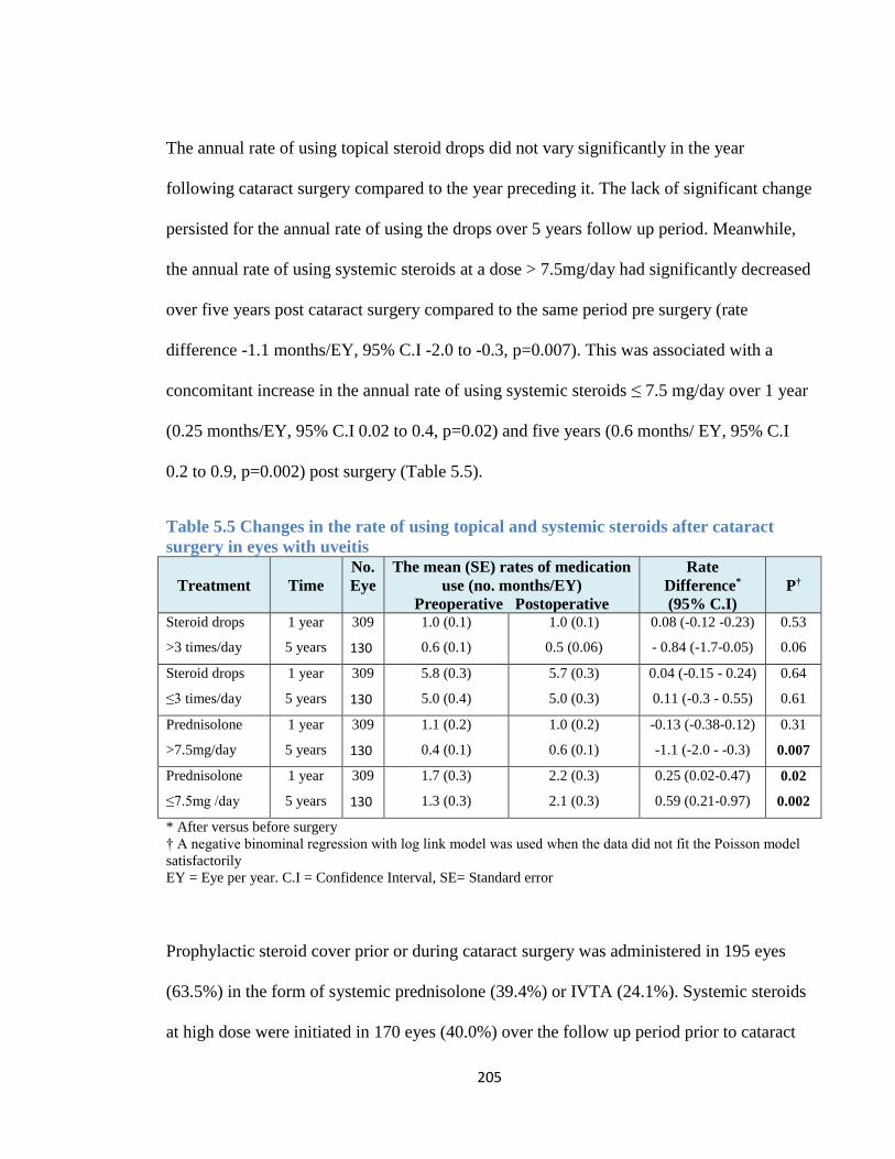
205
The annual rate of using topical steroid drops did not vary significantly in the year
following cataract surgery compared to the year preceding it. The lack of significant change
persisted for the annual rate of using the drops over 5 years follow up period. Meanwhile,
the annual rate of using systemic steroids at a dose > 7.5mg/day had significantly decreased
over five years post cataract surgery compared to the same period pre surgery (rate
difference -1.1 months/EY, 95% C.I -2.0 to -0.3, p=0.007). This was associated with a
concomitant increase in the annual rate of using systemic steroids ≤ 7.5 mg/day over 1 year
(0.25 months/EY, 95% C.I 0.02 to 0.4, p=0.02) and five years (0.6 months/ EY, 95% C.I
0.2 to 0.9, p=0.002) post surgery (Table 5.5).
Table 5.5 Changes in the rate of using topical and systemic steroids after cataract
surgery in eyes with uveitis
Treatment Time
No.
Eye
The mean (SE) rates of medication
use (no. months/EY)
Preoperative Postoperative
Rate
Difference*
(95% C.I)
P†
Steroid drops
>3 times/day
1 year
5 years
309
130
1.0 (0.1)
0.6 (0.1)
1.0 (0.1)
0.5 (0.06)
0.08 (-0.12 -0.23)
- 0.84 (-1.7-0.05)
0.53
0.06
Steroid drops
≤3 times/day
1 year
5 years
309
130
5.8 (0.3)
5.0 (0.4)
5.7 (0.3)
5.0 (0.3)
0.04 (-0.15 - 0.24)
0.11 (-0.3 - 0.55)
0.64
0.61
Prednisolone
>7.5mg/day
1 year
5 years
309
130
1.1 (0.2)
0.4 (0.1)
1.0 (0.2)
0.6 (0.1)
-0.13 (-0.38-0.12)
-1.1 (-2.0 - -0.3)
0.31
0.007
Prednisolone
≤7.5mg /day
1 year
5 years
309
130
1.7 (0.3)
1.3 (0.3)
2.2 (0.3)
2.1 (0.3)
0.25 (0.02-0.47)
0.59 (0.21-0.97)
0.02
0.002
* After versus before surgery
† A negative binominal regression with log link model was used when the data did not fit the Poisson model
satisfactorily
EY = Eye per year. C.I = Confidence Interval, SE= Standard error
Prophylactic steroid cover prior or during cataract surgery was administered in 195 eyes
(63.5%) in the form of systemic prednisolone (39.4%) or IVTA (24.1%). Systemic steroids
at high dose were initiated in 170 eyes (40.0%) over the follow up period prior to cataract
206
surgery. This was significantly decreased into 107 eyes (21.7%) following cataract surgery
(p=0.007, McNemar test). Likewise, immunomodulatory agents were initiated in 46 eyes
(14.9%) prior to cataract surgery compared to only 18 eyes (5.8%) who initiated the
medication following surgery (p<0.001, McNemar test). Periocular/intraocular steroids
were given into 97 eyes (31.3%) for managing uveitis activity prior to cataract surgery (34
eyes had one injection, 22 eyes had two injections, and 41 eyes had three injections or
more). This was reduced into 50 eyes (17.0%) who had ocular steroids injection after
cataract surgery (23 eyes had one injection, 10 eyes had two injections, and 19 eyes had
three injections or more).
The prevalence of increased IOP >30mmHg was significantly less within the period
following cataract surgery (23.0% versus 36.4%, p= 0.002, McNemar test), with 29 eyes
had their first onset of increased IOP >30mmHg after cataract surgery without prior
incidence before that. The incidence rate of high IOP>30mmHg was 0.01/EY. PCO
occurred in 104 eyes (33.7%) at a median of 8 months (IQR 4.8 – 38 months) post cataract
surgery and required capsulotomy within a median of 15 months (IQR 5.5 to 54 months)
post cataract surgery. Other intraoperative and postoperative complications include IOL
sublaxation/dislocation in four eyes (1.3%), hypotony in two eyes (0.6%), sterile
endophthalmitis with intraoperative IVTA use in two eyes (0.6%), corneal decompensation
in one eye (0.3%), intraoperative rupture of posterior capsule with nucleus drop in one eye
(0.3%), and retinal detachment in one eye (0.4%).
The prevalence of CMO formation following cataract surgery was significantly more than
the control group [80 eyes (26.9%) versus 31 eyes (10.3%), p<0.001) and occurred at a
median of 5.3 months (IQR 1.4 – 21.7 months) postoperatively with 10.4% had CMO
within the first three months postoperatively. The incidence rate of CMO postoperatively
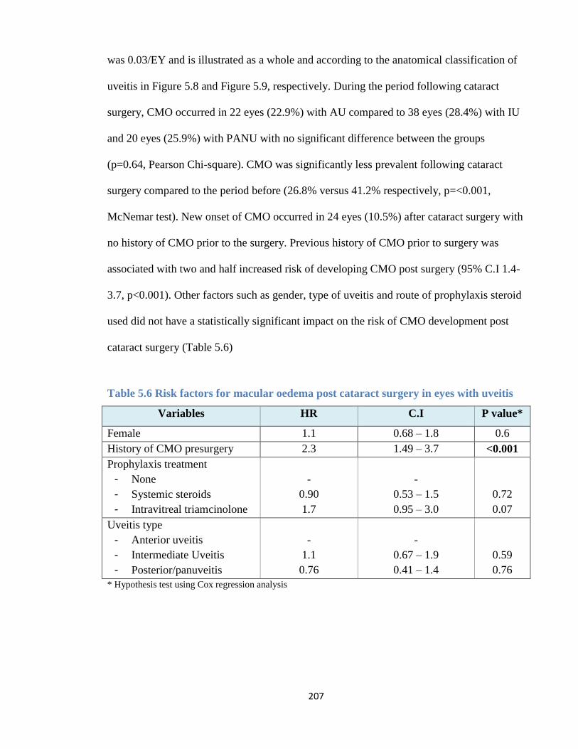
207
was 0.03/EY and is illustrated as a whole and according to the anatomical classification of
uveitis in Figure 5.8 and Figure 5.9, respectively. During the period following cataract
surgery, CMO occurred in 22 eyes (22.9%) with AU compared to 38 eyes (28.4%) with IU
and 20 eyes (25.9%) with PANU with no significant difference between the groups
(p=0.64, Pearson Chi-square). CMO was significantly less prevalent following cataract
surgery compared to the period before (26.8% versus 41.2% respectively, p=<0.001,
McNemar test). New onset of CMO occurred in 24 eyes (10.5%) after cataract surgery with
no history of CMO prior to the surgery. Previous history of CMO prior to surgery was
associated with two and half increased risk of developing CMO post surgery (95% C.I 1.4-
3.7, p<0.001). Other factors such as gender, type of uveitis and route of prophylaxis steroid
used did not have a statistically significant impact on the risk of CMO development post
cataract surgery (Table 5.6)
Table 5.6 Risk factors for macular oedema post cataract surgery in eyes with uveitis
Variables HR C.I P value*
Female 1.1 0.68 – 1.8 0.6
History of CMO presurgery 2.3 1.49 – 3.7 <0.001
Prophylaxis treatment
- None
- Systemic steroids
- Intravitreal triamcinolone
-
0.90
1.7
-
0.53 – 1.5
0.95 – 3.0
0.72
0.07
Uveitis type
- Anterior uveitis
- Intermediate Uveitis
- Posterior/panuveitis
-
1.1
0.76
-
0.67 – 1.9
0.41 – 1.4
0.59
0.76
* Hypothesis test using Cox regression analysis
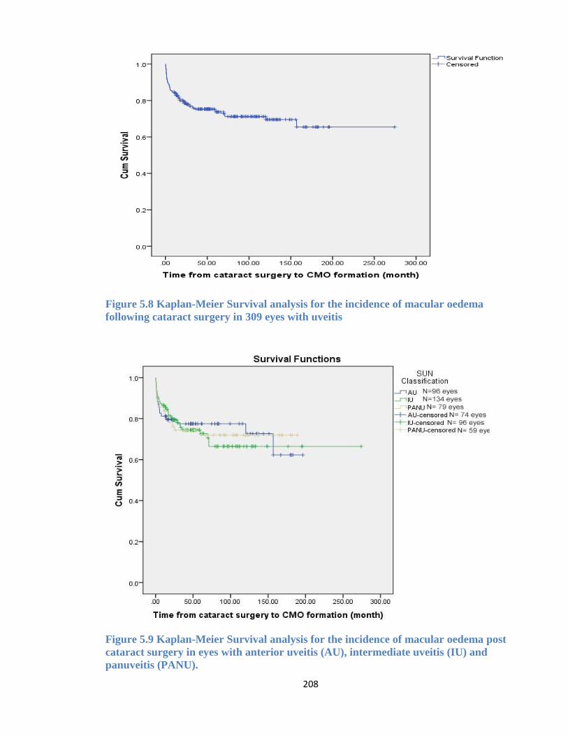
208
Figure 5.8 Kaplan-Meier Survival analysis for the incidence of macular oedema
following cataract surgery in 309 eyes with uveitis
Figure 5.9 Kaplan-Meier Survival analysis for the incidence of macular oedema post
cataract surgery in eyes with anterior uveitis (AU), intermediate uveitis (IU) and
panuveitis (PANU).
209
5.5 Discussion
This study examined the influence of cataract surgery on the visual outcome and incidence
of sight threatening complications, as well as the uveitis activity and treatment required
post surgery. The results showed that 1) the best visual outcome following cataract surgery
is achieved during the first postoperative week and continues to improve postoperatively. 2)
Uveitic eyes underwent phacoemulsification had a higher risk of vision loss and CMO
formation compared to the control phakic group. 3) Eyes with CMO and PCO had worse
vision over the follow-up time compared to the vision achieved during the first week
postoperatively. 4) Postoperative CMO and high IOP >30mmHg increase the risk of vision
loss in uveitic eyes following cataract surgery. 5) Cataract surgery was associated with a
lower uveitis relapse rate over the five years following cataract surgery when compared to
the rate over the same period prior to surgery. 6) A previous history of CMO prior to
cataract surgery was associated with increased risk of developing CMO post surgery. 7)
And finally, while there was no significant difference in the rate of using topical steroid
drops following surgery compared to the preoperative period, there was a significant
reduction in the rate of using systemic steroids at a dose >7.5 mg/day over the period of
five years post surgery when compared to the same period pre surgery.
The majority of uveitic eyes achieved a postoperative vision improvement of > 0.3
LogMAR from the preoperative vision in 93% of cases. This is better than the results
reported in a study by Ram et al on 108 eyes with uveitis which had a postoperative
improvement of ≥ 0.3 LogMAR in 71.3% of the patients (464). The high rate of significant
postoperative improvement in BCVA in our studied cohort could have partly be influence
210
by the study exclusion criteria which excluded cases with preoperative macular scar which
could have affected the postoperative vision improvement anticipated early following
cataract surgery. However, there were still cases who failed to achieve an improvement in
vision by a minimum of two Snellen line postoperatively. This could be attributed to a few
cases where the cataract was too dense making it difficult to have a good view of the
macula by fundus examination as well as using OCT imaging. Therefore, there might be
macula oedema or atrophy that pre-existed preoperatively and its influence on the
postoperative vision has only become apparent after the removal of the dense cataract.
Our study showed that majority of the eyes did achieve a final visual acuity of 6/12 or
better in 81.2% following cataract surgery. This was slightly better than the outcome of a
recent systematic review and meta-analysis of 24 studies on uveitic eyes with 70% of cases
following cataract surgery (68% following phacoemulsification) which achieved a
postoperative vision of 6/12 or better by the end of the follow up period (376). In another
study by Abbouda et al , 74.5 % of uveitic eyes had a final BCVA better than 6/18
following a minimum of three years follow up period post cataract surgery (380). SVL
occurred in 8.3% of our uveitis cohort following cataract surgery, which is slightly more
than what is observed in a study by Ram et al which reported severe vision loss of 5.5% at
the final follow up visit of uveitic eyes underwent cataract removal. This can possibly be
attributed to the longer postoperative follow-up in our study (median 78 months compared
to a mean of 22 months follow up) and the difference in the uveitis aetiology of the cohorts
and preoperative complications (464).
The risk of vision loss post cataract surgery in our uveitis cohort was strongly associated
with the presence of CMO as well as high IOP >30mmHg over the postoperative period. In
this study, the rate of CMO post cataract surgery was 26.9%, including 28.4% of eyes with
211
IU had CMO postoperatively. This falls within previous reports on the rate of CMO
following cataract surgery in eyes with parsplanitis which has been reported to range from
12 to 59% (469). The incidence of CMO postoperatively among AU patients in our studied
cohort (22.9%) was noted to be more when compared to a recent study on the outcome of
phacoemulsification in 55 eyes with cataract secondary to AU which had a CMO incidence
of 12.7% postoperatively.(471) The higher rate of postoperative CMO among our AU cases
might be attributed to the higher portion of HLA-B27 associated AU in our cohort which
has the tendency to develop more severe postoperative inflammation and CMO (93).
Our study found the incidence of CMO post cataract surgery in eyes with no prior history
of CMO preoperatively was 10.5%, which is almost identical to another study looking at
long term outcome of at least three years post cataract surgery which detected new onset
CMO in 10.2% of the uveitic eyes without a prior history of CMO before surgery (380).
CMO following cataract surgery or Irvine-Gass syndrome, usually occurs in the 4th to 6th
week postoperatively and has been reported to occur in 5% of nonuveitic eyes following
uncomplicated phacoemulsification (470). In our study, the incidence of CMO within the
first three months postoperatively was 10.3%, slightly more than that observed in
nonuveitic eyes from the above study. Furthermore, the incidence of CMO in our
pseudophakic group was significantly more than the control phakic group. Postoperative
CMO has inflammatory mediators as key players in the pathogenesis but the risk is still
more in eyes with history of uveitis and that cataract surgery may be associated with other
factors that lead to increase inflammation and capillary permeability in the retinal layer
which has already been compromised by inflammatory episodes prior to surgery. The role
of inflammation and breakdown of the BRB in the pathogenesis of postoperative CMO was
also supported by previous studies which showed patients with clinically significant CMO
212
postoperatively have higher aqueous flare values than pseudophakic and phakic eyes
without CMO, especially in the first postoperative day before it declines thereafter (472). A
recent study did show eyes with chronic postoperative inflammation were significantly
associated with higher risk of developing postoperative CMO and that eyes with more than
one postoperative uveitis relapse per year were associated with a seven fold increase in the
risk of developing CMO following cataract surgery (380). We also found that a previous
history of CMO preoperatively in eyes with uveitis can be associated with a 2.5 fold
increase in the risk of CMO postoperatively. This conclusion has been similarly drawn by
Agrawal et al. who suggested an increased risk of developing CMO postoperatively in eyes
with previous episodes prior to surgery (372).
The incidence of endophthalmitis in this study was 0.6% which were two cases of
suspected to be sterile endophthalmitis that occurred following intraoperative prophylactic
injection of IVTA, given to reduce the risk of postoperative CMO, and with no growth of
microorganisms observed from the intravitreal sample. Nevertheless, it was still treated as
possible bacterial endophthalmitis. Sterile endophthalmitis can occur post IVTA injection
with an incidence report ranging from none(97) to 0.21% (105), while the incidence of
endophthalmitis among eyes in general following cataract surgery had been reported to
occur in 0.12% (383). It was difficult to compare the incidence of endophthalmitis among
our uveitis cohort to other studies due to limited reports available on the actual incidence of
endophthalmitis among uveitic eyes following cataract surgery. This is possibly due to the
relatively small sample size included in the published reports making it difficult to draw a
close estimate for the incidence of a rare complication as endophthalmitis in these cases.
In this study, cataract surgery on uveitic eyes was not associated with an increased uveitis
relapse rate, as there was a significantly lower relapse rate over the follow-up period post
213
cataract surgery when compared to the preoperative period. This was also reflected in the
rate of using systemic prednisolone at a dose >7.5mg/day which was significantly less in
the postoperative period compared to preoperative period and there was no increase in the
rate of using topical steroid drops postoperatively compared to preoperative period. To our
knowledge there have not been previous studies looking at the rate of using systemic and
topical steroids over the time period prior versus post surgery to compare our results with.
However, the reduced relapse rate following cataract surgery was observed in a study in
Taiwan on 62 eyes with recurrent uveitis after excluding eyes with chronic uveitis in their
analysis. They found the relapse rate of 0.48 relapses per year postoperatively to be
significantly less when compared to the preoperative relapse rate of 1.32 relapses per year
(473). The lower relapse rate post cataract surgery as compared to the period prior to
surgery might reflect the natural trend of reduced uveitis activity over time. Our results
however suggest that cataract surgery with IOL implantation in uveitic eyes, with good
preoperative inflammation control and under prophylactic steroid cover when indicated,
does not on a long term basis increase the overall rate of uveitis relapse or the rate of using
topical or systemic steroid therapy. Postoperative relapse rate can vary widely according to
the aetiological and anatomical type of uveitis and the studied group, leading to a wide
range of postoperative uveitis relapse reported in the literature which can vary from as low
as 5% of eyes post cataract surgery with intraoperative IVTA reported by Okravi et al (474)
to 41% in another study by Estafanous et al (455).
The adequate control of inflammation prior to cataract surgery and slow tapering of topical
and oral prednisolone, if used, is crucial in preventing recurrence of uveitis postoperatively
and the associated risk of poor visual outcome in uveitic eyes following cataract surgery.
The use of either systemic prednisolone or intraoperative steroid injection as a prophylactic
214
measure against postoperative inflammation in our studied group had a similar effect on the
visual outcome and the incidence of CMO postoperatively. This finding complements the
study findings of Roesel et al which found a similar effect of a single intraoperative OFI of
steroids to that of postoperative oral prednisolone in terms of the visual outcome
postoperatively and the ability to control postoperative inflammation and CMO(475). A
conclusion that was similarly drawn in another study looking at intraoperative IVTA
injection in comparison to postoperative oral prednisolone (476).
In conclusion, our study demonstrates an overall good visual outcome following
phacoemulsification with IOL implantation in our uveitis cohort, with the vision continuing
to improve from the 1st week postoperative vision onwards over the first three years
postoperatively. Vision loss occurred in 18.2% of the eyes postoperatively, with CMO and
high IOP >30mmHg as major risk factors. The lower relapse rate over the follow-up period
post cataract surgery compared to the preoperative period together with a lower rate of
using systemic prednisolone at high dose >7.5mg/day postoperatively compared to
preoperative period suggests that cataract surgery on uveitic eyes is not associated with
worsening of uveitis control nor does it increase the need for using topical or systemic
steroids over the long term.
215
6 CHAPTER SIX: SUMMARY OF THE RESEARCH
CONCLUSIONS AND RECOMMENDATIONS
In this thesis, the main aim was to address the influence of a group of systemic and ocular
factors on the outcome of uveitic eyes and its management. The increasing incidence of
DM worldwidely has had a tremendous impact on the health and wellbeing of all
population groups. The first study focused on the interaction between DM and uveitis
through two studied groups; the first group addressed the characteristics and clinical
outcome of new onset of uveitis among known diabetic patients while the second group
looked at the influence of new onset of diabetes on the clinical course and management of
uveitis patients. The overall incidence of DM among our uveitis cohort was 4.4%. In both
groups of diabetic patients, idiopathic uveitis and sarcoidosis were the most common
aetiological classification of uveitis. While the general view is that AU is the main form of
uveitis seen in diabetes (396–398), only 40% of our cohort of diabetic patients with
idiopathic uveitis had AU. Within the first group of diabetic patients, 10% of the uveitic
eyes had an infectious aetiology. The onset of uveitis was significantly delayed in patients
with type 1 DM compared to type 2 DM, reflecting the time when type 1 DM is typically
diagnosed at a younger age (mean 6.5 years of age) compared to type 2 DM (419).
In diabetic patients presenting with uveitis, the vision remained mostly stable over the
follow-up time when compared to the first visit with uveitis. The prevalence of vision loss
was significantly more in the diabetic group presented with uveitis compared to non-
diabetic control group, mainly secondary to RPE atrophy and CNVM associated macular
scarring. The prevalence of RPE atrophy and CNVM formation in these patients, while it
216
could be coincidental finding, it can also be explained by the presence of proinflammatory
cytokines which mediates the expression of COX-2 enzyme as the latter has been detected
in the RPE cells of diabetic rats. There is increasing evidence that the COX-2 enzyme,
which mediates the formation of PGs and VEGF, plays a role in the formation of CNVM
formation and has been expressed within the CNVM of human eyes with age related
macular degeneration. Whether similar mechanism is contributing to the CNVM formation
in uveitic eyes of diabetic patients is a question of interest to be addressed in future studies.
Within group 2, the incidence of DM in uveitis patients was associated with a significant
reduction in visual acuity within the first two years post diagnosis but returned to the
average pre diabetic vision afterward. Reductions in systemic corticosteroid therapy and
increased usage of local therapy imply that clinicians are adjusting their treatment protocols
following DM diagnosis, without changing the ability to control uveitis relapse. Given the
association between DM, corticosteroids and some second-line IMT used in treating
uveitis, it is important to optimise treatment protocols for these conditions when they occur
concurrently. The management of uveitis and its complications should take into
consideration keeing the patients within the accepted diabetes control level agreed by his
physician. When possible, it is more preferred to provide the anti-inflammatory
medications locally rather than systemic medications which can compromise the diabetes
control in these patients.
Management of CMO due to uveitis in diabetic patients can be challenging. Bilateral CMO
in patients with non-infectious uveitis would ideally be treated with high dose systemic
corticosteroid. However, in the presence of uncontrolled hyperglycaemia this treatment
option carries an increased risk of side effects and thus can be replaced with local
administration of corticosteroid or using a lower starting dose of systemic corticosteroid
217
and more rapid tapering. In our second group cohort, uveitis relapses that occurred
following the diagnosis of DM tend to be managed by adding OFI/IVTA rather than
increasing the dose of systemic corticosteroid. In addition, the mean dose of systemic
prednisolone was significantly reduced in the year post DM when compared to the previous
year. While these changes in the use of systemic corticosteroid may reduce the risk of
hyperglycaemia, they carry the risk of uveitis relapses and increase the chance of visual
loss.
Most of the diabetic patients presented with uveitis within the first group and had an
available blood test showed poor hypoglycaemic control at time of uveitis diagnosis. It was
difficult in this study to address whether there is an association between poor glycaemic
control and the onset of uveitis due to the limited number of patients with blood test results
at the onset of uveitis. It is recommended to check the blood glucose level of patients with
DM presenting with their first onset of uveitis, regardless of the aetiology of uveitis or
severity of inflammation to detect cases with poor glycaemic control and allow for its
management in collaboration with their health care provider.
Within the first group, new onset or progression of DR occurred in 18% of the eyes of
diabetic patients during the follow-up period following the diagnosis of uveitis, including
2% that progressed to PDR, while in the second group, 7.3% of the eyes progressed from
no DR to develop mild, non-proliferative DR. We do acknowledge that this is a descriptive
analysis of our study cohort and that no conclusions can be drawn regarding the impact of
uveitis on the progression of DR due to the lack of comparison with a control group.
Whether uveitis can accelerate the development of DR is still unclear and it would require
prospective controlled clinical trials to establish an answer to this question. In theory,
progression of DR in patients with uveitis might be related to the synergistic effect of
218
inflammatory mediators on the retinal vasculature which has already been compromised in
diabetic patients. In conclusion, this study expands our understanding of the cumulative
effect of uveitis and diabetes using, to the best of our knowledge, the largest cohort of
diabetic patients with uveitis.
The aim of treating uveitis in diabetic patients is to preserve vision and prevent or treat
ocular complications while maintaining good glycaemic control in diabetic patients. In
managing diabetic patients with uveitis, it is recommended to have a close collaboration
with the primary care physician to achieve glycaemic control, especially in uveitis patients
requiring high doses of systemic corticosteroids with or without IMT.
In the second study, the prevalence of retinal vasculitis among our uveitis cohort was
14.0% which was similar to the prevalence observed in other large cohort studies. The long
term visual outcome was worse in eyes with retinal vasculitis compared to eyes without
vasculitis, mainly due to the risk of macular ischaemia. However, the risk of vision loss did
not differ between eyes with IRV and non-IRV cases when adjusting for the presence of
macular ischaemia.
In both subgroups of eyes with no-vasculitis and vasculitis, older age groups at time of
uveitis diagnosis were significantly associated with increase in the risk of vision loss per
year, respectively. Capillary non-perfusion involving ≥ quadrants of the retina is a
significant risk factor for NV formation and thus laser photocoagulation may be considered
at an early stage in these cases. Most of the eyes with retinal ischaemia received their initial
laser treatment within the first year following the diagnosis of ischaemia, suggesting the
need to closely observe eyes with retinal ischaemia during this period for the development
of NV.
219
Control of vasculitis with systemic anti-inflammatory medications, as well as the
application of retinal laser photocoagulation for managing NV formation can result in long-
term stabilisation of the disease and prevent further deterioration in visual function,
although in this study the use of anti-inflammatory medications did not result in preventing
further NV formation as 38.1% had a relapse with further NV formation requiring more
retinal laser photocoagulation.
In eyes with vasculitis, macular ischaemia and CMO were related to vision loss, suggesting
a direct involvement in ocular morbidity. CMO in eyes with IRV had 2.5 times more risk of
vision loss while macular ischaemia, which occurred in 15.7% of eyes with IRV, increased
the risk of visual loss by 9.2 times. This finding was consistent with the results observed in
a recent study on eyes with ischaemic retinal vasculitis in which a significant correlation
was observed between poor visual acuity and increased central macular thickness and
foveal avascular zone even when adjusted for neovascularisation and peripheral ischaemia
indexes. Thus, tight control of the underlying disease with the aim of preserving the
macular region should be paramount objectives and guide treatment decisions. In this
study, the use of corticosteroids was associated with a reduced risk of vision loss in both
IRV and non-IRV. The use of corticosteroids and other anti-inflammatory medications in
managing vasculitis and some of its associated complications, such as CMO, contributes to
their role in preventing vision loss. Interestingly, macular ischaemia occurred early in the
diagnosis of IRV, with no incidence observed over the rest of the follow up period; thus the
role of anti-inflammatory medications in preventing macular ischaemia is something to be
addressed in future studies. On the other hand, the study did not find a significant role for
anti-inflammatory medications in preventing ischaemic relapse and NV formation.
220
In the third study, cataract surgery removal using phacoemulsification with IOL
implantation was associated with improving vision from the 1st week postoperative onward
over the first 3 years postoperatively. The incidence rate of vision loss was 0.02/EY and
occurred in 18.8% of cases which was significantly worse than control group of phakic
eyes, mainly related to the incidence of CMO and high IOP >30mmHg during the
postoperative period. The adequate control of uveitis activity prior to undergoing cataract
surgery together with the improving techniques and technology in cataract surgery have
been the main factor contributing to the visual outcome observed in this uveitis cohort and
in preventing significant worsening of uveitis activity post surgery. This was also supported
by the finding of a lower relapse rate over the follow-up period postoperatively compared
to the preoperative period together with a lower rate of using systemic prednisolone at a
high dose >7.5mg per day postoperatively compared to preoperative period suggested that
cataract surgery on uveitic eyes did not cause worsening of uveitis control nor did it
increase the need for topical or systemic steroids over a long period of time following
cataract surgery. These findings may also suggest that cataract formation in this uveitis
cohort was not essentially associated with more severe disease as the cataract surgery was
not associated with more severe postoperative inflammation. The administration of
prophylactic cortiocsteroids together with proper inflammation control for a considerable
period prior to surgery and with slow tapering of topical and systemic corticosteroids
postoperatively are all key elements in maintaining a good postoperative outcome in uveitis
patients.
As a conclusion, this thesis was able to address the influence of three factors on the visual
outcome and inflammation activity and management. Newly diagnosed DM in uveitic eyes
require careful management plans that can address the significant increase risk of reversible
221
vision loss mainly secondary to CMO and cataract and afterward take the risk of
hyperglycaemia into consideration when planning for long term uveitis control. We have
also shown that the adequate management of retinal vasculitis in this cohort has been able
to maintain good visual outcome and prevent the occurrence of irreversible vision loss in
most of the cases although the relapse of NV formation in ischaemic area remains an issue
that can occur independently from the uveitis activity. And finally, with the use of current
standards of good inflammation control around the time of cataract surgery, uveitic eyes are
able to maintain a good visual outcome postoperatively with no increase in the rate of
uveitis activity and treatment over a long period of time.
222
7 REFERENCES
1. Gritz DC, Wong IG. Incidence and prevalence of uveitis in Northern California; the Northern California Epidemiology of Uveitis Study. Ophthalmology. 2004 Mar;111(3):491–500; discussion 500.
2. Wakefield D, Chang JH. Epidemiology of uveitis. Int Ophthalmol Clin. 2005;45(2):1–13.
3. Kump LI, Cervantes-Castañeda RA, Androudi SN, Foster CS. Analysis of pediatric uveitis cases at a tertiary referral center. Ophthalmology. 2005 Jul;112(7):1287–92.
4. Nagpal A, Leigh JF, Acharya NR. Epidemiology of uveitis in children. Int Ophthalmol Clin. 2008;48(3):1–7.
5. de Smet MD, Taylor SRJ, Bodaghi B, Miserocchi E, Murray PI, Pleyer U, et al. Understanding uveitis: the impact of research on visual outcomes. Prog Retin Eye Res. 2011 Nov;30(6):452–70.
6. Rothova A, Suttorp-van Schulten MS, Frits Treffers W, Kijlstra A. Causes and frequency of blindness in patients with intraocular inflammatory disease. Br J Ophthalmol. 1996 Apr;80(4):332–6.
7. Bodaghi B, Cassoux N, Wechsler B, Hannouche D, Fardeau C, Papo T, et al. Chronic severe uveitis: etiology and visual outcome in 927 patients from a single center. Medicine (Baltimore). 2001 Jul;80(4):263–70.
8. Durrani OM, Meads CA, Murray PI. Uveitis: a potentially blinding disease. Ophthalmol J Int Ophtalmol Int J Ophthalmol Z Für Augenheilkd. 2004 Aug;218(4):223–36.
9. Suttorp-Schulten MS, Rothova A. The possible impact of uveitis in blindness: a literature survey. Br J Ophthalmol. 1996 Sep;80(9):844–8.
10. Durrani OM, Tehrani NN, Marr JE, Moradi P, Stavrou P, Murray PI. Degree, duration, and causes of visual loss in uveitis. Br J Ophthalmol. 2004 Sep;88(9):1159–62.
11. Gurwitz JH, Bohn RL, Glynn RJ, Monane M, Mogun H, Avorn J. Glucocorticoids and the risk for initiation of hypoglycemic therapy. Arch Intern Med. 1994 Jan 10;154(1):97–101.
12. Gulliford MC, Charlton J, Latinovic R. Risk of diabetes associated with prescribed glucocorticoids in a large population. Diabetes Care. 2006 Dec;29(12):2728–9.
13. Clore JN, Thurby-Hay L. Glucocorticoid-induced hyperglycemia. Endocr Pract Off J Am Coll Endocrinol Am Assoc Clin Endocrinol. 2009 Aug;15(5):469–74.
14. The Information Centre for Health and Social Care. Quality and Outcomes Framework (QOF) 2009 [Internet]. 2009 [cited 2012 Nov 15]. Available from: http://www.ic.nhs.uk/statistics-and-data-collections/supporting-information/audits-and-performance/the-quality-and-outcomes-framework/qof-2008/09/data-tables/prevalence-data-tables
223
15. Palmer HE, Stanford MR, McCartney AC, Graham EM. Non-caseating granulomas as a cause of ischaemic retinal vasculitis. Br J Ophthalmol. 1997 Nov;81(11):1018–9.
16. Bentley CR, Stanford MR, Shilling JS, Sanders MD, Graham EM. Macular ischaemia in posterior uveitis. Eye Lond Engl. 1993;7 ( Pt 3):411–4.
17. El-Asrar AMA, Al-Kharashi SA. Full panretinal photocoagulation and early vitrectomy improve prognosis of retinal vasculitis associated with tuberculoprotein hypersensitivity (Eales’ disease). Br J Ophthalmol. 2002 Nov;86(11):1248–51.
18. Ross M., Pawlina W. Histology a text and atlas with correlated cell and molecular biology. 5th ed. USA: Lippincott: Williams & Wilkins; 2006.
19. Jabs DA, Nussenblatt RB, Rosenbaum JT. Standardization of uveitis nomenclature for reporting clinical data. Results of the First International Workshop. Am J Ophthalmol. 2005 Sep;140(3):509–16.
20. Bloch-Michel E, Nussenblatt RB. International Uveitis Study Group recommendations for the evaluation of intraocular inflammatory disease. Am J Ophthalmol. 1987 Feb 15;103(2):234–5.
21. Deschenes J, Murray PI, Rao NA, Nussenblatt RB. International Uveitis Study Group (IUSG): clinical classification of uveitis. Ocul Immunol Inflamm. 2008 Feb;16(1):1–2.
22. Rothova A, Suttorp-van Schulten MS, Frits Treffers W, Kijlstra A. Causes and frequency of blindness in patients with intraocular inflammatory disease. Br J Ophthalmol. 1996 Apr;80(4):332–6.
23. The World Health Report, WHO. Global data on visual impairments. [Internet]. 2010 [cited 2015 Mar 31]. Available from: http://www.who.int/blindness/GLOBALDATAFINALforweb.pdf
24. Bodaghi B, Cassoux N, Wechsler B, Hannouche D, Fardeau C, Papo T, et al. Chronic severe uveitis: etiology and visual outcome in 927 patients from a single center. Medicine (Baltimore). 2001 Jul;80(4):263–70.
25. Rothova A, Buitenhuis HJ, Meenken C, Brinkman CJ, Linssen A, Alberts C, et al. Uveitis and systemic disease. Br J Ophthalmol. 1992 Mar;76(3):137–41.
26. Chang JH, McCluskey PJ, Wakefield D. Acute anterior uveitis and HLA-B27. Surv Ophthalmol. 2005 Aug;50(4):364–88.
27. Chang JH-M, Wakefield D. Uveitis: a global perspective. Ocul Immunol Inflamm. 2002 Dec;10(4):263–79.
28. Ohguro N, Sonoda K-H, Takeuchi M, Matsumura M, Mochizuki M. The 2009 prospective multi-center epidemiologic survey of uveitis in Japan. Jpn J Ophthalmol. 2012 Sep;56(5):432–5.
29. Ronday MJ, Stilma JS, Barbe RF, Kijlstra A, Rothova A. Blindness from uveitis in a hospital population in Sierra Leone. Br J Ophthalmol. 1994 Sep;78(9):690–3.
224
30. Taylor MJ, Hoerauf A, Bockarie M. Lymphatic filariasis and onchocerciasis. Lancet. 2010 Oct 2;376(9747):1175–85.
31. Thuruthumaly C, Yee DC, Rao PK. Presumed ocular histoplasmosis. Curr Opin Ophthalmol. 2014 Nov;25(6):508–12.
32. Zucchiatti I, Miserocchi E, Sacconi R, Bandello F, Modorati G. HLA-A29-Positive Uveitis: Birdshot Chorioretinopathy, What Else? Case Rep Ophthalmol. 2013 Sep;4(3):287–93.
33. Yang P, Zhang Z, Zhou H, Li B, Huang X, Gao Y, et al. Clinical patterns and characteristics of uveitis in a tertiary center for uveitis in China. Curr Eye Res. 2005 Nov;30(11):943–8.
34. Tugal-Tutkun I. Pediatric uveitis. J Ophthalmic Vis Res. 2011 Oct;6(4):259–69.
35. Angeles-Han ST, Pelajo CF, Vogler LB, Rouster-Stevens K, Kennedy C, Ponder L, et al. Risk markers of juvenile idiopathic arthritis-associated uveitis in the Childhood Arthritis and Rheumatology Research Alliance (CARRA) Registry. J Rheumatol. 2013 Dec;40(12):2088–96.
36. Moradi A, Amin RM, Thorne JE. The role of gender in juvenile idiopathic arthritis-associated uveitis. J Ophthalmol. 2014;2014:461078.
37. Khairallah M, Yahia SB, Ladjimi A, Messaoud R, Zaouali S, Attia S, et al. Pattern of uveitis in a referral centre in Tunisia, North Africa. Eye Lond Engl. 2007 Jan;21(1):33–9.
38. Jones NP. The Manchester Uveitis Clinic: The First 3000 Patients-Epidemiology and Casemix. Ocul Immunol Inflamm. 2013 Dec 2;
39. Tang WM, Pulido JS, Eckels DD, Han DP, Mieler WF, Pierce K. The association of HLA-DR15 and intermediate uveitis. Am J Ophthalmol. 1997 Jan;123(1):70–5.
40. Maldini C, Lavalley MP, Cheminant M, de Menthon M, Mahr A. Relationships of HLA-B51 or B5 genotype with Behcet’s disease clinical characteristics: systematic review and meta-analyses of observational studies. Rheumatol Oxf Engl. 2012 May;51(5):887–900.
41. Shi T, Lv W, Zhang L, Chen J, Chen H. Association of HLA-DR4/HLA-DRB1*04 with Vogt-Koyanagi-Harada disease: a systematic review and meta-analysis. Sci Rep. 2014;4:6887.
42. Levinson RD, Rajalingam R, Park MS, Reed EF, Gjertson DW, Kappel PJ, et al. Human leukocyte antigen A29 subtypes associated with birdshot retinochoroidopathy. Am J Ophthalmol. 2004 Oct;138(4):631–4.
43. Nussenblatt RB. Proctor Lecture. Experimental autoimmune uveitis: mechanisms of disease and clinical therapeutic indications. Invest Ophthalmol Vis Sci. 1991 Dec;32(13):3131–41.
44. Romagnani S. Human TH1 and TH2 subsets: doubt no more. Immunol Today. 1991 Aug;12(8):256–7.
45. Boyd SR, Young S, Lightman S. Immunopathology of the noninfectious posterior and intermediate uveitides. Surv Ophthalmol. 2001 Dec;46(3):209–33.
225
46. Imamura Y, Kurokawa MS, Yoshikawa H, Nara K, Takada E, Masuda C, et al. Involvement of Th1 cells and heat shock protein 60 in the pathogenesis of intestinal Behcet’s disease. Clin Exp Immunol. 2005 Feb;139(2):371–8.
47. Yu HG, Lee DS, Seo JM, Ahn JK, Yu YS, Lee WJ, et al. The number of CD8+ T cells and NKT cells increases in the aqueous humor of patients with Behçet’s uveitis. Clin Exp Immunol. 2004 Aug;137(2):437–43.
48. Bardak Y, Aridoğan BC. The demonstration of serum interleukin 6-8, tumor necrosis factor-alpha, complement, and immunoglobulin levels in Behçet’s disease with ocular involvement. Ocul Immunol Inflamm. 2004 Mar;12(1):53–8.
49. Ozdamar Y, Berker N, Bahar G, Soykan E, Bicer T, Ozkan SS, et al. Inflammatory mediators and posterior segment involvement in ocular Behcet disease. Eur J Ophthalmol. 2009 Dec;19(6):998–1003.
50. Chan CC, Benezra D, Rodrigues MM, Palestine AG, Hsu SM, Murphree AL, et al. Immunohistochemistry and electron microscopy of choroidal infiltrates and Dalen-Fuchs nodules in sympathetic ophthalmia. Ophthalmology. 1985 Apr;92(4):580–90.
51. Chan CC, Nussenblatt RB, Fujikawa LS, Palestine AG, Stevens G, Parver LM, et al. Sympathetic ophthalmia. Immunopathological findings. Ophthalmology. 1986 May;93(5):690–5.
52. Kahn M, Pepose JS, Green WR, Miller J, Foos RY. Immunocytologic findings in a case of Vogt-Koyanagi-Harada syndrome. Ophthalmology. 1993 Aug;100(8):1191–8.
53. Wang C, Tian Y, Lei B, Xiao X, Ye Z, Li F, et al. Decreased IL-27 expression in association with an increased Th17 response in Vogt-Koyanagi-Harada disease. Invest Ophthalmol Vis Sci. 2012 Jul;53(8):4668–75.
54. Amadi-Obi A, Yu C-R, Liu X, Mahdi RM, Clarke GL, Nussenblatt RB, et al. TH17 cells contribute to uveitis and scleritis and are expanded by IL-2 and inhibited by IL-27/STAT1. Nat Med. 2007 Jun;13(6):711–8.
55. Kuiper JJW, Mutis T, de Jager W, de Groot-Mijnes JDF, Rothova A. Intraocular interleukin-17 and proinflammatory cytokines in HLA-A29-associated birdshot chorioretinopathy. Am J Ophthalmol. 2011 Aug;152(2):177–182.e1.
56. Sugita S, Takase H, Kawaguchi T, Taguchi C, Mochizuki M. Cross-reaction between tyrosinase peptides and cytomegalovirus antigen by T cells from patients with Vogt-Koyanagi-Harada disease. Int Ophthalmol. 2007 Jun;27(2–3):87–95.
57. Lightman S. Immune mechanisms in autoimmune ocular disease. Eye Lond Engl. 1988;2 ( Pt 3):260–6.
58. Herbort CP. Appraisal, work-up and diagnosis of anterior uveitis: a practical approach. Middle East Afr J Ophthalmol. 2009 Oct;16(4):159–67.
59. D’Alessandro LP, Forster DJ, Rao NA. Anterior uveitis and hypopyon. Am J Ophthalmol. 1991 Sep 15;112(3):317–21.
226
60. Henderly DE, Haymond RS, Rao NA, Smith RE. The significance of the pars plana exudate in pars planitis. Am J Ophthalmol. 1987 May 15;103(5):669–71.
61. Zein G, Berta A, Foster CS. Multiple sclerosis-associated uveitis. Ocul Immunol Inflamm. 2004 Jun;12(2):137–42.
62. Gerstenblith AT, Thorne JE, Sobrin L, Do DV, Shah SM, Foster CS, et al. Punctate inner choroidopathy: a survey analysis of 77 persons. Ophthalmology. 2007 Jun;114(6):1201–4.
63. Febvay C, Kodjikian L, Maucort-Boulch D, Perard L, Iwaz J, Jamilloux Y, et al. Clinical features and diagnostic evaluation of 83 biopsy-proven sarcoid uveitis cases. Br J Ophthalmol. 2015 Apr 2;
64. Becker ML, Rose CD. Blau syndrome and related genetic disorders causing childhood arthritis. Curr Rheumatol Rep. 2005 Dec;7(6):427–33.
65. Ang M, Wong WL, Li X, Chee S-P. Interferon γ release assay for the diagnosis of uveitis associated with tuberculosis: a Bayesian evaluation in the absence of a gold standard. Br J Ophthalmol. 2013 Aug;97(8):1062–7.
66. Francis JM, Joynson DH. Duration of specific immunoglobulin A antibody following acute toxoplasmosis as determined by enzyme immunoassay and immunosorbent agglutination assay. Eur J Clin Microbiol Infect Dis Off Publ Eur Soc Clin Microbiol. 1993 Jul;12(7):556–9.
67. Sherar MD, Foster FS. The design and fabrication of high frequency poly(vinylidene fluoride) transducers. Ultrason Imaging. 1989 Apr;11(2):75–94.
68. Peizeng Y, Qianli M, Xiangkun H, Hongyan Z, Li W, Kijlstra A. Longitudinal study of anterior segment inflammation by ultrasound biomicroscopy in patients with acute anterior uveitis. Acta Ophthalmol (Copenh). 2009 Mar;87(2):211–5.
69. Häring G, Nölle B, Wiechens B. Ultrasound biomicroscopic imaging in intermediate uveitis. Br J Ophthalmol. 1998 Jun;82(6):625–9.
70. Adl MA, LeHoang P, Bodaghi B. Use of fluorescein angiography in the diagnosis and management of uveitis. Int Ophthalmol Clin. 2012;52(4):1–12.
71. Stanga PE, Lim JI, Hamilton P. Indocyanine green angiography in chorioretinal diseases: indications and interpretation: an evidence-based update. Ophthalmology. 2003 Jan;110(1):15-21-23.
72. Nicholson BP, Nigam D, Miller D, Agrón E, Dalal M, Jacobs-El N, et al. Comparison of wide-field fluorescein angiography and 9-field montage angiography in uveitis. Am J Ophthalmol. 2014 Mar;157(3):673–7.
73. Campbell JP, Leder HA, Sepah YJ, Gan T, Dunn JP, Hatef E, et al. Wide-field retinal imaging in the management of noninfectious posterior uveitis. Am J Ophthalmol. 2012 Nov;154(5):908–911.e2.
74. Hee MR, Izatt JA, Swanson EA, Huang D, Schuman JS, Lin CP, et al. Optical coherence tomography of the human retina. Arch Ophthalmol Chic Ill 1960. 1995 Mar;113(3):325–32.
227
75. Markomichelakis NN, Halkiadakis I, Pantelia E, Peponis V, Patelis A, Theodossiadis P, et al. Patterns of macular edema in patients with uveitis: qualitative and quantitative assessment using optical coherence tomography. Ophthalmology. 2004 May;111(5):946–53.
76. Kempen JH, Sugar EA, Jaffe GJ, Acharya NR, Dunn JP, Elner SG, et al. Fluorescein angiography versus optical coherence tomography for diagnosis of uveitic macular edema. Ophthalmology. 2013 Sep;120(9):1852–9.
77. Durrani K, Zakka FR, Ahmed M, Memon M, Siddique SS, Foster CS. Systemic therapy with conventional and novel immunomodulatory agents for ocular inflammatory disease. Surv Ophthalmol. 2011 Dec;56(6):474–510.
78. Barnes PJ. Glucocorticosteroids: current and future directions. Br J Pharmacol. 2011 May;163(1):29–43.
79. Antonetti DA, Wolpert EB, DeMaio L, Harhaj NS, Scaduto RC Jr. Hydrocortisone decreases retinal endothelial cell water and solute flux coincident with increased content and decreased phosphorylation of occludin. J Neurochem. 2002 Feb;80(4):667–77.
80. Fischer S, Renz D, Schaper W, Karliczek GF. In vitro effects of dexamethasone on hypoxia-induced hyperpermeability and expression of vascular endothelial growth factor. Eur J Pharmacol. 2001 Jan 12;411(3):231–43.
81. Hedayatfar A, Hashemi H, Asgari S, Chee S-P. Comparison of efficacy and ocular surface toxicity of topical preservative-free methylprednisolone and preserved prednisolone in the treatment of acute anterior uveitis. Cornea. 2014 Apr;33(4):366–72.
82. Biswas J, Ganeshbabu TM, Raghavendran SR, Raizada S, Mondkar SV, Madhavan HN. Efficacy and safety of 1% rimexolone versus 1% prednisolone acetate in the treatment of anterior uveitis--a randomized triple masked study. Int Ophthalmol. 2004 May;25(3):147–53.
83. Thorne JE, Woreta F, Kedhar SR, Dunn JP, Jabs DA. Juvenile idiopathic arthritis-associated uveitis: incidence of ocular complications and visual acuity loss. Am J Ophthalmol. 2007 May;143(5):840–6.
84. Thorne JE, Woreta FA, Dunn JP, Jabs DA. Risk of cataract development among children with juvenile idiopathic arthritis-related uveitis treated with topical corticosteroids. Ophthalmology. 2010 Jul;117(7):1436–41.
85. Tawara A, Tou N, Kubota T, Harada Y, Yokota K. Immunohistochemical evaluation of the extracellular matrix in trabecular meshwork in steroid-induced glaucoma. Graefes Arch Clin Exp Ophthalmol. 2008 Apr 2;246(7):1021–8.
86. Jabs DA, Rosenbaum JT, Foster CS, Holland GN, Jaffe GJ, Louie JS, et al. Guidelines for the use of immunosuppressive drugs in patients with ocular inflammatory disorders: recommendations of an expert panel. Am J Ophthalmol. 2000 Oct;130(4):492–513.
87. Charkoudian LD, Ying G, Pujari SS, Gangaputra S, Thorne JE, Foster CS, et al. High-dose intravenous corticosteroids for ocular inflammatory diseases. Ocul Immunol Inflamm. 2012 Apr;20(2):91–9.
228
88. Read RW, Yu F, Accorinti M, Bodaghi B, Chee S-P, Fardeau C, et al. Evaluation of the effect on outcomes of the route of administration of corticosteroids in acute Vogt-Koyanagi-Harada disease. Am J Ophthalmol. 2006 Jul;142(1):119–24.
89. Buchman AL. Side effects of corticosteroid therapy. J Clin Gastroenterol. 2001 Oct;33(4):289–94.
90. Kanis JA, Johansson H, Oden A, McCloskey EV. Guidance for the adjustment of FRAX according to the dose of glucocorticoids. Osteoporos Int J Establ Result Coop Eur Found Osteoporos Natl Osteoporos Found USA. 2011 Mar;22(3):809–16.
91. Compston J, Bowring C, Cooper A, Cooper C, Davies C, Francis R, et al. Diagnosis and management of osteoporosis in postmenopausal women and older men in the UK: National Osteoporosis Guideline Group (NOGG) update 2013. Maturitas. 2013 Aug;75(4):392–6.
92. Gaudio PA. A review of evidence guiding the use of corticosteroids in the treatment of intraocular inflammation. Ocul Immunol Inflamm. 2004 Sep;12(3):169–92.
93. Leder HA, Jabs DA, Galor A, Dunn JP, Thorne JE. Periocular triamcinolone acetonide injections for cystoid macular edema complicating noninfectious uveitis. Am J Ophthalmol. 2011 Sep;152(3):441–448.e2.
94. Sen HN, Vitale S, Gangaputra SS, Nussenblatt RB, Liesegang TL, Levy-Clarke GA, et al. Periocular corticosteroid injections in uveitis: effects and complications. Ophthalmology. 2014 Nov;121(11):2275–86.
95. Beer PM, Bakri SJ, Singh RJ, Liu W, Peters GB 3rd, Miller M. Intraocular concentration and pharmacokinetics of triamcinolone acetonide after a single intravitreal injection. Ophthalmology. 2003 Apr;110(4):681–6.
96. Kok H, Lau C, Maycock N, McCluskey P, Lightman S. Outcome of intravitreal triamcinolone in uveitis. Ophthalmology. 2005 Nov;112(11):1916.e1-7.
97. Park UC, Park JH, Yu HG. Long-term outcome of intravitreal triamcinolone acetonide injection for the treatment of uveitis attacks in Behçet disease. Ocul Immunol Inflamm. 2014 Feb;22(1):27–33.
98. Moreker MR, Lodhi SA, Pathengay A. Role of intravitreal triamcinolone as an adjuvant in the management of Vogt-Koyanagi-Harada disease. Indian J Ophthalmol. 2007 Dec;55(6):479–80.
99. Wadhwa N, Garg SP, Mehrotra A. Prospective evaluation of intravitreal triamcinolone acetonide in serpiginous choroiditis. Ophthalmol J Int Ophtalmol Int J Ophthalmol Z Für Augenheilkd. 2010;224(3):183–7.
100. Moschos MM, Gatzioufas Z, Margetis I. Intravitreal Triamcinolone Acetonide for Macular Edema in HLA-B27 Negative Ankylosing Spondylitis. Case Rep Ophthalmol. 2010;1(2):105–9.
101. Bae JH, Lee CS, Lee SC. Efficacy and safety of intravitreal bevacizumab compared with intravitreal and posterior sub-tenon triamcinolone acetonide for treatment of uveitic cystoid macular edema. Retina Phila Pa. 2011 Jan;31(1):111–8.
229
102. Sallam A, Comer RM, Chang JH, Grigg JR, Andrews R, McCluskey PJ, et al. Short-term safety and efficacy of intravitreal triamcinolone acetonide for uveitic macular edema in children. Arch Ophthalmol. 2008 Feb;126(2):200–5.
103. Gillies MC, Simpson JM, Billson FA, Luo W, Penfold P, Chua W, et al. Safety of an intravitreal injection of triamcinolone: results from a randomized clinical trial. Arch Ophthalmol. 2004 Mar;122(3):336–40.
104. Jager RD, Aiello LP, Patel SC, Cunningham ET Jr. Risks of intravitreous injection: a comprehensive review. Retina Phila Pa. 2004 Oct;24(5):676–98.
105. Cheung CSY, Wong AWT, Lui A, Kertes PJ, Devenyi RG, Lam W-C. Incidence of endophthalmitis and use of antibiotic prophylaxis after intravitreal injections. Ophthalmology. 2012 Aug;119(8):1609–14.
106. Rush R, Sheth S. Fulminant toxoplasmic retinochoroiditis following intravitreal triamcinolone administration. Indian J Ophthalmol. 2012 Apr;60(2):141–3.
107. London NJS, Chiang A, Haller JA. The dexamethasone drug delivery system: indications and evidence. Adv Ther. 2011 May;28(5):351–66.
108. Williams GA, Haller JA, Kuppermann BD, Blumenkranz MS, Weinberg DV, Chou C, et al. Dexamethasone posterior-segment drug delivery system in the treatment of macular edema resulting from uveitis or Irvine-Gass syndrome. Am J Ophthalmol. 2009 Jun;147(6):1048–54, 1054-2.
109. Tomkins-Netzer O, Taylor SRJ, Bar A, Lula A, Yaganti S, Talat L, et al. Treatment with Repeat Dexamethasone Implants Results in Long-Term Disease Control in Eyes with Noninfectious Uveitis. Ophthalmology. 2014 Mar 17;
110. Taylor SRJ, Tomkins-Netzer O, Joshi L, Morarji J, McLoone E, Lightman S. Dexamethasone implant in pediatric uveitis. Ophthalmology. 2012 Nov;119(11):2412–2412.e2.
111. Lowder C, Belfort R, Lightman S, Foster CS, Robinson MR, Schiffman RM, et al. Dexamethasone intravitreal implant for noninfectious intermediate or posterior uveitis. Arch Ophthalmol. 2011;129(5):545–553.
112. Bansal R, Bansal P, Kulkarni P, Gupta V, Sharma A, Gupta A. Wandering Ozurdex® implant. J Ophthalmic Inflamm Infect. 2011 Sep 30;2(1):1–5.
113. Multicenter Uveitis Steroid Treatment (MUST) Trial Research Group, Kempen JH, Altaweel MM, Holbrook JT, Jabs DA, Louis TA, et al. Randomized comparison of systemic anti-inflammatory therapy versus fluocinolone acetonide implant for intermediate, posterior, and panuveitis: the multicenter uveitis steroid treatment trial. Ophthalmology. 2011 Oct;118(10):1916–26.
114. Arcinue CA, Cerón OM, Foster CS. A comparison between the fluocinolone acetonide (Retisert) and dexamethasone (Ozurdex) intravitreal implants in uveitis. J Ocul Pharmacol Ther. 2013 Jun;29(5):501–7.
230
115. Barry RJ, Nguyen QD, Lee RW, Murray PI, Denniston AK. Pharmacotherapy for uveitis: current management and emerging therapy. Clin Ophthalmol Auckl NZ. 2014;8:1891–911.
116. Gangaputra S, Newcomb CW, Liesegang TL, Kaçmaz RO, Jabs DA, Levy-Clarke GA, et al. Methotrexate for ocular inflammatory diseases. Ophthalmology. 2009 Nov;116(11):2188–2198.e1.
117. Okada AA. Immunomodulatory therapy for ocular inflammatory disease: a basic manual and review of the literature. Ocul Immunol Inflamm. 2005 Oct;13(5):335–51.
118. Pasadhika S, Kempen JH, Newcomb CW, Liesegang TL, Pujari SS, Rosenbaum JT, et al. Azathioprine for ocular inflammatory diseases. Am J Ophthalmol. 2009 Oct;148(4):500–509.e2.
119. Uchiyama E, Papaliodis GN, Lobo A-M, Sobrin L. Side-effects of anti-inflammatory therapy in uveitis. Semin Ophthalmol. 2014 Nov;29(5–6):456–67.
120. Daniel E, Thorne JE, Newcomb CW, Pujari SS, Kaçmaz RO, Levy-Clarke GA, et al. Mycophenolate mofetil for ocular inflammation. Am J Ophthalmol. 2010 Mar;149(3):423-432-2.
121. Pujari SS, Kempen JH, Newcomb CW, Gangaputra S, Daniel E, Suhler EB, et al. Cyclophosphamide for ocular inflammatory diseases. Ophthalmology. 2010 Feb;117(2):356–65.
122. Mudun BA, Ergen A, Ipcioglu SU, Burumcek EY, Durlu Y, Arslan MO. Short-term chlorambucil for refractory uveitis in Behcet’s disease. Ocul Immunol Inflamm. 2001 Dec;9(4):219–29.
123. Miserocchi E, Baltatzis S, Ekong A, Roque M, Foster CS. Efficacy and safety of chlorambucil in intractable noninfectious uveitis: the Massachusetts Eye and Ear Infirmary experience. Ophthalmology. 2002 Jan;109(1):137–42.
124. Kapturczak MH, Meier-Kriesche HU, Kaplan B. Pharmacology of calcineurin antagonists. Transplant Proc. 2004 Mar;36(2 Suppl):25S–32S.
125. Ozdal PC, Ortaç S, Taskintuna I, Firat E. Long-term therapy with low dose cyclosporin A in ocular Behçet’s disease. Doc Ophthalmol Adv Ophthalmol. 2002 Nov;105(3):301–12.
126. Kaçmaz RO, Kempen JH, Newcomb C, Daniel E, Gangaputra S, Nussenblatt RB, et al. Cyclosporine for ocular inflammatory diseases. Ophthalmology. 2010 Mar;117(3):576–84.
127. Murphy CC, Greiner K, Plskova J, Duncan L, Frost NA, Forrester JV, et al. Cyclosporine vs tacrolimus therapy for posterior and intermediate uveitis. Arch Ophthalmol Chic Ill 1960. 2005 May;123(5):634–41.
128. Hogan AC, McAvoy CE, Dick AD, Lee RWJ. Long-term efficacy and tolerance of tacrolimus for the treatment of uveitis. Ophthalmology. 2007 May;114(5):1000–6.
129. Pasadhika S, Rosenbaum JT. Update on the use of systemic biologic agents in the treatment of noninfectious uveitis. Biol Targets Ther. 2014;8:67–81.
231
130. Lin J, Ziring D, Desai S, Kim S, Wong M, Korin Y, et al. TNFalpha blockade in human diseases: an overview of efficacy and safety. Clin Immunol Orlando Fla. 2008 Jan;126(1):13–30.
131. Kaltsonoudis E, Voulgari PV, Konitsiotis S, Drosos AA. Demyelination and other neurological adverse events after anti-TNF therapy. Autoimmun Rev. 2014 Jan;13(1):54–8.
132. Suhler EB, Smith JR, Giles TR, Lauer AK, Wertheim MS, Kurz DE, et al. Infliximab therapy for refractory uveitis: 2-year results of a prospective trial. Arch Ophthalmol Chic Ill 1960. 2009 Jun;127(6):819–22.
133. Levy-Clarke G, Jabs DA, Read RW, Rosenbaum JT, Vitale A, Van Gelder RN. Expert Panel Recommendations for the Use of Anti-Tumor Necrosis Factor Biologic Agents in Patients with Ocular Inflammatory Disorders. Ophthalmology. 2013 Dec 17;
134. Sfikakis PP, Theodossiadis PG, Katsiari CG, Kaklamanis P, Markomichelakis NN. Effect of infliximab on sight-threatening panuveitis in Behçet’s disease. Lancet. 2001 Jul 28;358(9278):295–6.
135. Rajaraman RT, Kimura Y, Li S, Haines K, Chu DS. Retrospective case review of pediatric patients with uveitis treated with infliximab. Ophthalmology. 2006 Feb;113(2):308–14.
136. Artornsombudh P, Gevorgyan O, Payal A, Siddique SS, Foster CS. Infliximab treatment of patients with birdshot retinochoroidopathy. Ophthalmology. 2013 Mar;120(3):588–92.
137. Baughman RP, Bradley DA, Lower EE. Infliximab in chronic ocular inflammation. Int J Clin Pharmacol Ther. 2005 Jan;43(1):7–11.
138. Rudwaleit M, Rødevand E, Holck P, Vanhoof J, Kron M, Kary S, et al. Adalimumab effectively reduces the rate of anterior uveitis flares in patients with active ankylosing spondylitis: results of a prospective open-label study. Ann Rheum Dis. 2009 May;68(5):696–701.
139. Smith JA, Thompson DJS, Whitcup SM, Suhler E, Clarke G, Smith S, et al. A randomized, placebo-controlled, double-masked clinical trial of etanercept for the treatment of uveitis associated with juvenile idiopathic arthritis. Arthritis Rheum. 2005 Feb 15;53(1):18–23.
140. Heiligenhaus A, Miserocchi E, Heinz C, Gerloni V, Kotaniemi K. Treatment of severe uveitis associated with juvenile idiopathic arthritis with anti-CD20 monoclonal antibody (rituximab). Rheumatol Oxf Engl. 2011 Aug;50(8):1390–4.
141. Miserocchi E, Pontikaki I, Modorati G, Gattinara M, Meroni PL, Gerloni V. Anti-CD 20 monoclonal antibody (rituximab) treatment for inflammatory ocular diseases. Autoimmun Rev. 2011 Nov;11(1):35–9.
142. Edelsten C, Lee V, Bentley CR, Kanski JJ, Graham EM. An evaluation of baseline risk factors predicting severity in juvenile idiopathic arthritis associated uveitis and other chronic anterior uveitis in early childhood. Br J Ophthalmol. 2002 Jan;86(1):51–6.
143. Ozdal PC, Vianna RNG, Deschênes J. Visual outcome of juvenile rheumatoid arthritis-associated uveitis in adults. Ocul Immunol Inflamm. 2005 Feb;13(1):33–8.
232
144. Sabri K, Saurenmann RK, Silverman ED, Levin AV. Course, complications, and outcome of juvenile arthritis-related uveitis. J AAPOS Off Publ Am Assoc Pediatr Ophthalmol Strabismus Am Assoc Pediatr Ophthalmol Strabismus. 2008 Dec;12(6):539–45.
145. Ayuso VK, Ten Cate HAT, van der Does P, Rothova A, de Boer JH. Male gender and poor visual outcome in uveitis associated with juvenile idiopathic arthritis. Am J Ophthalmol. 2010 Jun;149(6):987–93.
146. Woreta F, Thorne JE, Jabs DA, Kedhar SR, Dunn JP. Risk factors for ocular complications and poor visual acuity at presentation among patients with uveitis associated with juvenile idiopathic arthritis. Am J Ophthalmol. 2007 Apr;143(4):647–55.
147. Tomkins-Netzer O, Talat L, Bar A, Lula A, Taylor SRJ, Joshi L, et al. Long-term clinical outcome and causes of vision loss in patients with uveitis. Ophthalmology. 2014 Dec;121(12):2387–92.
148. Javadi M-A, Jafarinasab M-R, Araghi A-AS, Mohammadpour M, Yazdani S. Outcomes of phacoemulsification and in-the-bag intraocular lens implantation in Fuchs’ heterochromic iridocyclitis. J Cataract Refract Surg. 2005 May;31(5):997–1001.
149. Tejwani S, Murthy S, Sangwan VS. Cataract extraction outcomes in patients with Fuchs’ heterochromic cyclitis. J Cataract Refract Surg. 2006 Oct;32(10):1678–82.
150. Quinn CJ. Cystoid macular edema. Optom Clin Off Publ Prentice Soc. 1996;5(1):111–30.
151. Ray S, D’Amico DJ. Pseudophakic cystoid macular edema. Semin Ophthalmol. 2002 Dec;17(3–4):167–80.
152. Mentes J, Erakgun T, Afrashi F, Kerci G. Incidence of cystoid macular edema after uncomplicated phacoemulsification. Ophthalmol J Int Ophtalmol Int J Ophthalmol Z Für Augenheilkd. 2003 Dec;217(6):408–12.
153. Ursell PG, Spalton DJ, Whitcup SM, Nussenblatt RB. Cystoid macular edema after phacoemulsification: relationship to blood-aqueous barrier damage and visual acuity. J Cataract Refract Surg. 1999 Nov;25(11):1492–7.
154. Lardenoye CWTA, van Kooij B, Rothova A. Impact of macular edema on visual acuity in uveitis. Ophthalmology. 2006 Aug;113(8):1446–9.
155. Pivetti-Pezzi P, Accorinti M, La Cava M, Colabelli Gisoldi RA, Abdulaziz MA. Endogenous uveitis: an analysis of 1,417 cases. Ophthalmol J Int Ophtalmol Int J Ophthalmol Z Für Augenheilkd. 1996;210(4):234–8.
156. Malinowski SM, Pulido JS, Folk JC. Long-term visual outcome and complications associated with pars planitis. Ophthalmology. 1993 Jun;100(6):818–824; discussion 825.
157. Tugal-Tutkun I, Onal S, Altan-Yaycioglu R, Huseyin Altunbas H, Urgancioglu M. Uveitis in Behçet disease: an analysis of 880 patients. Am J Ophthalmol. 2004 Sep;138(3):373–80.
158. Schubert HD. Postsurgical hypotony: relationship to fistulization, inflammation, chorioretinal lesions, and the vitreous. Surv Ophthalmol. 1996 Oct;41(2):97–125.
233
159. Expert Committee on the Diagnosis and Classification of Diabetes Mellitus. Report of the expert committee on the diagnosis and classification of diabetes mellitus. Diabetes Care. 2003;26 Suppl 1:S5-20.
160. Kuzuya T, Nakagawa S, Satoh J, Kanazawa Y, Iwamoto Y, Kobayashi M, et al. Report of the Committee on the classification and diagnostic criteria of diabetes mellitus. Diabetes Res Clin Pract. 2002 Jan;55(1):65–85.
161. Smith SC Jr. Multiple risk factors for cardiovascular disease and diabetes mellitus. Am J Med. 2007 Mar;120(3 Suppl 1):S3–11.
162. Prasad DS, Kabir Z, Dash AK, Das BC. Prevalence and risk factors for diabetes and impaired glucose tolerance in Asian Indians: A community survey from urban Eastern India. Diabetes Metab Syndr. 2012 Apr;6(2):96–101.
163. Carey VJ, Walters EE, Colditz GA, Solomon CG, Willett WC, Rosner BA, et al. Body fat distribution and risk of non-insulin-dependent diabetes mellitus in women. The Nurses’ Health Study. Am J Epidemiol. 1997 Apr 1;145(7):614–9.
164. Stratton IM, Kohner EM, Aldington SJ, Turner RC, Holman RR, Manley SE, et al. UKPDS 50: risk factors for incidence and progression of retinopathy in Type II diabetes over 6 years from diagnosis. Diabetologia. 2001 Feb;44(2):156–63.
165. Adler AI, Stevens RJ, Manley SE, Bilous RW, Cull CA, Holman RR. Development and progression of nephropathy in type 2 diabetes: the United Kingdom Prospective Diabetes Study (UKPDS 64). Kidney Int. 2003 Jan;63(1):225–32.
166. Diabetic retinopathy. Diabetes Care. 2000;23 Suppl 1:S73-76.
167. Nathan DM. Long-term complications of diabetes mellitus. N Engl J Med. 1993 Jun 10;328(23):1676–85.
168. Min TZ, Stephens MW, Kumar P, Chudleigh RA. Renal complications of diabetes. Br Med Bull. 2012 Oct 31;
169. Bader MS. Diabetic foot infection. Am Fam Physician. 2008 Jul 1;78(1):71–9.
170. Brown JS, Wessells H, Chancellor MB, Howards SS, Stamm WE, Stapleton AE, et al. Urologic complications of diabetes. Diabetes Care. 2005 Jan;28(1):177–85.
171. Hakim LS, Goldstein I. Diabetic sexual dysfunction. Endocrinol Metab Clin North Am. 1996 Jun;25(2):379–400.
172. Genuth S, Alberti KGMM, Bennett P, Buse J, Defronzo R, Kahn R, et al. Follow-up report on the diagnosis of diabetes mellitus. Diabetes Care. 2003 Nov;26(11):3160–7.
173. Gillett MJ. International Expert Committee Report on the Role of the A1C Assay in the Diagnosis of Diabetes. Clin Biochem Rev. 2009 Nov;30(4):197–200.
234
174. Alberti KG, Zimmet PZ. Definition, diagnosis and classification of diabetes mellitus and its complications. Part 1: diagnosis and classification of diabetes mellitus provisional report of a WHO consultation. Diabet Med J Br Diabet Assoc. 1998 Jul;15(7):539–53.
175. National Institute for Health and Care Excellence. Diabetes in adults quality standard (QS6); Quality statement 4: Glycaemic control [Internet]. 2011 [cited 2013 Apr 1]. Available from: http://www.nice.org.uk/guidance/qs6/chapter/Quality-statement-4-Glycaemic-control
176. Gary TL, Genkinger JM, Guallar E, Peyrot M, Brancati FL. Meta-analysis of randomized educational and behavioral interventions in type 2 diabetes. Diabetes Educ. 2003 Jun;29(3):488–501.
177. Sigal RJ, Kenny GP, Wasserman DH, Castaneda-Sceppa C, White RD. Physical activity/exercise and type 2 diabetes: a consensus statement from the American Diabetes Association. Diabetes Care. 2006 Jun;29(6):1433–8.
178. Rydén L, Standl E, Bartnik M, Van den Berghe G, Betteridge J, de Boer M-J, et al. Guidelines on diabetes, pre-diabetes, and cardiovascular diseases: executive summary. The Task Force on Diabetes and Cardiovascular Diseases of the European Society of Cardiology (ESC) and of the European Association for the Study of Diabetes (EASD). Eur Heart J. 2007 Jan;28(1):88–136.
179. Nathan DM, Buse JB, Davidson MB, Heine RJ, Holman RR, Sherwin R, et al. Management of hyperglycaemia in type 2 diabetes: a consensus algorithm for the initiation and adjustment of therapy. A consensus statement from the American Diabetes Association and the European Association for the Study of Diabetes. Diabetologia. 2006 Aug;49(8):1711–21.
180. Alemzadeh R, Palma-Sisto P, Holzum M, Parton E, Kicher J. Continuous subcutaneous insulin infusion attenuated glycemic instability in preschool children with type 1 diabetes mellitus. Diabetes Technol Ther. 2007 Aug;9(4):339–47.
181. Alemzadeh R, Palma-Sisto P, Parton EA, Holzum MK. Continuous subcutaneous insulin infusion and multiple dose of insulin regimen display similar patterns of blood glucose excursions in pediatric type 1 diabetes. Diabetes Technol Ther. 2005 Aug;7(4):587–96.
182. Hirsch IB, Farkas-Hirsch R, Skyler JS. Intensive insulin therapy for treatment of type I diabetes. Diabetes Care. 1990 Dec;13(12):1265–83.
183. Knowler WC, Barrett-Connor E, Fowler SE, Hamman RF, Lachin JM, Walker EA, et al. Reduction in the incidence of type 2 diabetes with lifestyle intervention or metformin. N Engl J Med. 2002 Feb 7;346(6):393–403.
184. Gangji AS, Cukierman T, Gerstein HC, Goldsmith CH, Clase CM. A systematic review and meta-analysis of hypoglycemia and cardiovascular events: a comparison of glyburide with other secretagogues and with insulin. Diabetes Care. 2007 Feb;30(2):389–94.
185. Malaisse WJ. Pharmacology of the meglitinide analogs: new treatment options for type 2 diabetes mellitus. Treat Endocrinol. 2003;2(6):401–14.
186. Early Treatment Diabetic Retinopathy Study Research Group. Classification of diabetic retinopathy from fluorescein angiograms. ETDRS report number 11. Early Treatment
235
Diabetic Retinopathy Study Research Group. Ophthalmology. 1991 May;98(5 Suppl):807–22.
187. ETDRS Research Group. Grading diabetic retinopathy from stereoscopic color fundus photographs--an extension of the modified Airlie House classification. ETDRS report number 10. Early Treatment Diabetic Retinopathy Study Research Group. Ophthalmology. 1991 May;98(5 Suppl):786–806.
188. Wilkinson CP, Ferris FL 3rd, Klein RE, Lee PP, Agardh CD, Davis M, et al. Proposed international clinical diabetic retinopathy and diabetic macular edema disease severity scales. Ophthalmology. 2003 Sep;110(9):1677–82.
189. Harding S, Greenwood R, Aldington S, Gibson J, Owens D, Taylor R, et al. Grading and disease management in national screening for diabetic retinopathy in England and Wales. Diabet Med J Br Diabet Assoc. 2003 Dec;20(12):965–71.
190. Heng LZ, Comyn O, Peto T, Tadros C, Ng E, Sivaprasad S, et al. Diabetic retinopathy: pathogenesis, clinical grading, management and future developments. Diabet Med J Br Diabet Assoc. 2013 Jun;30(6):640–50.
191. Yau JWY, Rogers SL, Kawasaki R, Lamoureux EL, Kowalski JW, Bek T, et al. Global prevalence and major risk factors of diabetic retinopathy. Diabetes Care. 2012 Mar;35(3):556–64.
192. Klein R, Klein BE, Moss SE, Davis MD, DeMets DL. The Wisconsin epidemiologic study of diabetic retinopathy. IV. Diabetic macular edema. Ophthalmology. 1984;91:1464–74.
193. Schram MT, Chaturvedi N, Schalkwijk C, Giorgino F, Ebeling P, Fuller JH, et al. Vascular risk factors and markers of endothelial function as determinants of inflammatory markers in type 1 diabetes: the EURODIAB Prospective Complications Study. Diabetes Care. 2003;26:2165–73.
194. Martin PM, Roon P, Van Ells TK, Ganapathy V, Smith SB. Death of retinal neurons in streptozotocin-induced diabetic mice. Invest Ophthalmol Vis Sci. 2004;45:3330–6.
195. Ning X, Baoyu Q, Yuzhen L, Shuli S, Reed E, Li QQ. Neuro-optic cell apoptosis and microangiopathy in KKAY mouse retina. Int J Mol Med. 2004;13:87–92.
196. Barber AJ, Lieth E, Khin SA, Antonetti DA, Buchanan AG, Gardner TW. Neural apoptosis in the retina during experimental and human diabetes. Early onset and effect of insulin. J Clin Invest. 1998;102:783–91.
197. Johnsen-Soriano S, Sancho-Tello M, Arnal E, Navea A, Cervera E, Bosch-Morell F, et al. IL-2 and IFN-gamma in the retina of diabetic rats. Graefes Arch Clin Exp Ophthalmol Albrecht Von Graefes Arch Für Klin Exp Ophthalmol. 2010;248:985–90.
198. Gverović Antunica A, Karaman K, Znaor L, Sapunar A, Buško V, Puzović V. IL-12 concentrations in the aqueous humor and serum of diabetic retinopathy patients. Graefes Arch Clin Exp Ophthalmol Albrecht Von Graefes Arch Für Klin Exp Ophthalmol. 2012;250:815–21.
236
199. Kern TS. Contributions of inflammatory processes to the development of the early stages of diabetic retinopathy. Exp Diabetes Res. 2007;2007:95103.
200. Schröder S, Palinski W, Schmid-Schönbein GW. Activated monocytes and granulocytes, capillary nonperfusion, and neovascularization in diabetic retinopathy. Am J Pathol. 1991 Jul;139(1):81–100.
201. Kowluru RA, Koppolu P, Chakrabarti S, Chen S. Diabetes-induced activation of nuclear transcriptional factor in the retina, and its inhibition by antioxidants. Free Radic Res. 2003;37:1169–80.
202. Zheng L, Szabó C, Kern TS. Poly(ADP-ribose) polymerase is involved in the development of diabetic retinopathy via regulation of nuclear factor-kappaB. Diabetes. 2004;53:2960–7.
203. Miyamoto K, Khosrof S, Bursell SE, Rohan R, Murata T, Clermont AC, et al. Prevention of leukostasis and vascular leakage in streptozotocin-induced diabetic retinopathy via intercellular adhesion molecule-1 inhibition. Proc Natl Acad Sci U S A. 1999;96:10836–41.
204. Ayalasomayajula SP, Amrite AC, Kompella UB. Inhibition of cyclooxygenase-2, but not cyclooxygenase-1, reduces prostaglandin E2 secretion from diabetic rat retinas. Eur J Pharmacol. 2004;498:275–8.
205. Ayalasomayajula SP, Kompella UB. Celecoxib, a selective cyclooxygenase-2 inhibitor, inhibits retinal vascular endothelial growth factor expression and vascular leakage in a streptozotocin-induced diabetic rat model. Eur J Pharmacol. 2003;458:283–9.
206. Gerhardinger C, Brown LF, Roy S, Mizutani M, Zucker CL, Lorenzi M. Expression of vascular endothelial growth factor in the human retina and in nonproliferative diabetic retinopathy. Am J Pathol. 1998;152:1453–62.
207. Chen H-S, Wu T-E, Hsiao L-C, Lin S-H. Interaction between glycaemic control and serum insulin-like growth factor 1 on the risk of retinopathy in type 2 diabetes. Eur J Clin Invest. 2012 Apr;42(4):447–54.
208. Beránek M, Kanková K, Benes P, Izakovicová-Hollá L, Znojil V, Hájek D, et al. Polymorphism R25P in the gene encoding transforming growth factor-beta (TGF-beta1) is a newly identified risk factor for proliferative diabetic retinopathy. Am J Med Genet. 2002 May 15;109(4):278–83.
209. Zheng Z, Chen H, Ke G, Fan Y, Zou H, Sun X, et al. Protective effect of perindopril on diabetic retinopathy is associated with decreased vascular endothelial growth factor-to-pigment epithelium-derived factor ratio: involvement of a mitochondria-reactive oxygen species pathway. Diabetes. 2009 Apr;58(4):954–64.
210. Zhu X, Zou H. PEDF in diabetic retinopathy: a protective effect of oxidative stress. J Biomed Biotechnol. 2012;2012:580687.
211. Johnsen-Soriano S, Sancho-Tello M, Arnal E, Navea A, Cervera E, Bosch-Morell F, et al. IL-2 and IFN-gamma in the retina of diabetic rats. Graefes Arch Clin Exp Ophthalmol Albrecht Von Graefes Arch Für Klin Exp Ophthalmol. 2010 Jul;248(7):985–90.
237
212. Meleth AD, Agrón E, Chan C-C, Reed GF, Arora K, Byrnes G, et al. Serum inflammatory markers in diabetic retinopathy. Invest Ophthalmol Vis Sci. 2005 Nov;46(11):4295–301.
213. Wakabayashi Y, Usui Y, Okunuki Y, Kezuka T, Takeuchi M, Iwasaki T, et al. Increases of vitreous monocyte chemotactic protein 1 and interleukin 8 levels in patients with concurrent hypertension and diabetic retinopathy. Retina Phila Pa. 2011 Oct;31(9):1951–7.
214. Gverović Antunica A, Karaman K, Znaor L, Sapunar A, Buško V, Puzović V. IL-12 concentrations in the aqueous humor and serum of diabetic retinopathy patients. Graefes Arch Clin Exp Ophthalmol Albrecht Von Graefes Arch Für Klin Exp Ophthalmol. 2012 Jun;250(6):815–21.
215. Mohamed Q, Gillies MC, Wong TY. Management of diabetic retinopathy: a systematic review. JAMA. 2007 Aug 22;298(8):902–16.
216. Reichard P, Nilsson BY, Rosenqvist U. The effect of long-term intensified insulin treatment on the development of microvascular complications of diabetes mellitus. N Engl J Med. 1993 Jul 29;329(5):304–9.
217. UKPDS Group. Intensive blood-glucose control with sulphonylureas or insulin compared with conventional treatment and risk of complications in patients with type 2 diabetes (UKPDS 33). UK Prospective Diabetes Study (UKPDS) Group. Lancet. 1998 Sep 12;352(9131):837–53.
218. UKPDS Research Group. Effect of intensive blood-glucose control with metformin on complications in overweight patients with type 2 diabetes (UKPDS 34). UK Prospective Diabetes Study (UKPDS) Group. Lancet. 1998 Sep 12;352(9131):854–65.
219. Colayco DC, Niu F, McCombs JS, Cheetham TC. A1C and cardiovascular outcomes in type 2 diabetes: a nested case-control study. Diabetes Care. 2011 Jan;34(1):77–83.
220. King P, Peacock I, Donnelly R. The UK prospective diabetes study (UKPDS): clinical and therapeutic implications for type 2 diabetes. Br J Clin Pharmacol. 1999 Nov;48(5):643–8.
221. Ohkubo Y, Kishikawa H, Araki E, Miyata T, Isami S, Motoyoshi S, et al. Intensive insulin therapy prevents the progression of diabetic microvascular complications in Japanese patients with non-insulin-dependent diabetes mellitus: a randomized prospective 6-year study. Diabetes Res Clin Pract. 1995 May;28(2):103–17.
222. DCCT Research Group. The absence of a glycemic threshold for the development of long-term complications: the perspective of the Diabetes Control and Complications Trial. Diabetes. 1996 Oct;45(10):1289–98.
223. Stratton IM, Adler AI, Neil HA, Matthews DR, Manley SE, Cull CA, et al. Association of glycaemia with macrovascular and microvascular complications of type 2 diabetes (UKPDS 35): prospective observational study. BMJ. 2000 Aug 12;321(7258):405–12.
224. Matthews DR, Stratton IM, Aldington SJ, Holman RR, Kohner EM, UK Prospective Diabetes Study Group. Risks of progression of retinopathy and vision loss related to tight blood pressure control in type 2 diabetes mellitus: UKPDS 69. Arch Ophthalmol Chic Ill 1960. 2004 Nov;122(11):1631–40.
238
225. ACCORD Study Group, ACCORD Eye Study Group, Chew EY, Ambrosius WT, Davis MD, Danis RP, et al. Effects of medical therapies on retinopathy progression in type 2 diabetes. N Engl J Med. 2010 Jul 15;363(3):233–44.
226. Keech A, Simes RJ, Barter P, Best J, Scott R, Taskinen MR, et al. Effects of long-term fenofibrate therapy on cardiovascular events in 9795 people with type 2 diabetes mellitus (the FIELD study): randomised controlled trial. Lancet Lond Engl. 2005 Nov 26;366(9500):1849–61.
227. Stefánsson E. The therapeutic effects of retinal laser treatment and vitrectomy. A theory based on oxygen and vascular physiology. Acta Ophthalmol Scand. 2001 Oct;79(5):435–40.
228. Bressler SB, Almukhtar T, Aiello LP, Bressler NM, Ferris FL, Glassman AR, et al. Green or yellow laser treatment for diabetic macular edema: exploratory assessment within the Diabetic Retinopathy Clinical Research Network. Retina Phila Pa. 2013 Dec;33(10):2080–8.
229. Lavinsky D, Cardillo JA, Melo LAS, Dare A, Farah ME, Belfort R. Randomized clinical trial evaluating mETDRS versus normal or high-density micropulse photocoagulation for diabetic macular edema. Invest Ophthalmol Vis Sci. 2011 Jun;52(7):4314–23.
230. ETDRS Research Group. Photocoagulation for diabetic macular edema. Early Treatment Diabetic Retinopathy Study report number 1. Early Treatment Diabetic Retinopathy Study research group. Arch Ophthalmol. 1985 Dec;103(12):1796–806.
231. Beck RW, Edwards AR, Aiello LP, Bressler NM, Ferris F, Glassman AR, et al. Three-year follow-up of a randomized trial comparing focal/grid photocoagulation and intravitreal triamcinolone for diabetic macular edema. Arch Ophthalmol. 2009 Mar;127(3):245–51.
232. Bandello F, Pognuz R, Polito A, Pirracchio A, Menchini F, Ambesi M. Diabetic macular edema: classification, medical and laser therapy. Semin Ophthalmol. 2003 Dec;18(4):251–8.
233. Virgili G, Parravano M, Menchini F, Evans JR. Anti-vascular endothelial growth factor for diabetic macular oedema. Cochrane Database Syst Rev. 2014;10:CD007419.
234. National Institute for Health and Care Excellence. Ranibizumab for treating diabetic macular oedema (rapid review of technology appraisal guidance 237) [Internet]. 2013 [cited 2015 Jul 1]. Available from: http://www.nice.org.uk/guidance/TA274/chapter/1-Guidance
235. Massin P, Bandello F, Garweg JG, Hansen LL, Harding SP, Larsen M, et al. Safety and efficacy of ranibizumab in diabetic macular edema (RESOLVE Study): a 12-month, randomized, controlled, double-masked, multicenter phase II study. Diabetes Care. 2010 Nov;33(11):2399–405.
236. Mitchell P, Bandello F, Schmidt-Erfurth U, Lang GE, Massin P, Schlingemann RO, et al. The RESTORE study: ranibizumab monotherapy or combined with laser versus laser monotherapy for diabetic macular edema. Ophthalmology. 2011 Apr;118(4):615–25.
237. Schmidt-Erfurth U, Lang GE, Holz FG, Schlingemann RO, Lanzetta P, Massin P, et al. Three-year outcomes of individualized ranibizumab treatment in patients with diabetic macular edema: the RESTORE extension study. Ophthalmology. 2014 May;121(5):1045–53.
239
238. Brown DM, Nguyen QD, Marcus DM, Boyer DS, Patel S, Feiner L, et al. Long-term outcomes of ranibizumab therapy for diabetic macular edema: the 36-month results from two phase III trials: RISE and RIDE. Ophthalmology. 2013 Oct;120(10):2013–22.
239. Diabetic Retinopathy Clinical Research Network, Elman MJ, Qin H, Aiello LP, Beck RW, Bressler NM, et al. Intravitreal ranibizumab for diabetic macular edema with prompt versus deferred laser treatment: three-year randomized trial results. Ophthalmology. 2012 Nov;119(11):2312–8.
240. Michaelides M, Kaines A, Hamilton RD, Fraser-Bell S, Rajendram R, Quhill F, et al. A prospective randomized trial of intravitreal bevacizumab or laser therapy in the management of diabetic macular edema (BOLT study) 12-month data: report 2. Ophthalmology. 2010 Jun;117(6):1078–1086.e2.
241. Rajendram R, Fraser-Bell S, Kaines A, Michaelides M, Hamilton RD, Esposti SD, et al. A 2-year prospective randomized controlled trial of intravitreal bevacizumab or laser therapy (BOLT) in the management of diabetic macular edema: 24-month data: report 3. Arch Ophthalmol Chic Ill 1960. 2012 Aug;130(8):972–9.
242. Sultan MB, Zhou D, Loftus J, Dombi T, Ice KS, Macugen 1013 Study Group. A phase 2/3, multicenter, randomized, double-masked, 2-year trial of pegaptanib sodium for the treatment of diabetic macular edema. Ophthalmology. 2011 Jun;118(6):1107–18.
243. National Institute for Health and Care Excellence. Macular oedema (diabetic) - aflibercept: final appraisal determination document [Internet]. 2015 [cited 2015 Jul 1]. Available from: http://www.nice.org.uk/guidance/GID-TAG472/documents/macular-oedema-diabetic-aflibercept-final-appraisal-determination-document2
244. Do DV, Nguyen QD, Boyer D, Schmidt-Erfurth U, Brown DM, Vitti R, et al. One-year outcomes of the da Vinci Study of VEGF Trap-Eye in eyes with diabetic macular edema. Ophthalmology. 2012 Aug;119(8):1658–65.
245. Korobelnik J-F, Do DV, Schmidt-Erfurth U, Boyer DS, Holz FG, Heier JS, et al. Intravitreal aflibercept for diabetic macular edema. Ophthalmology. 2014 Nov;121(11):2247–54.
246. Diabetic Retinopathy Clinical Research Network, Wells JA, Glassman AR, Ayala AR, Jampol LM, Aiello LP, et al. Aflibercept, bevacizumab, or ranibizumab for diabetic macular edema. N Engl J Med. 2015 Mar 26;372(13):1193–203.
247. Diabetic Retinopathy Clinical Research Network. A randomized trial comparing intravitreal triamcinolone acetonide and focal/grid photocoagulation for diabetic macular edema. Ophthalmology. 2008 Sep;115(9):1447–9, 1449-10.
248. Haller JA, Kuppermann BD, Blumenkranz MS, Williams GA, Weinberg DV, Chou C, et al. Randomized controlled trial of an intravitreous dexamethasone drug delivery system in patients with diabetic macular edema. Arch Ophthalmol Chic Ill 1960. 2010 Mar;128(3):289–96.
249. Callanan DG, Gupta S, Boyer DS, Ciulla TA, Singer MA, Kuppermann BD, et al. Dexamethasone intravitreal implant in combination with laser photocoagulation for the treatment of diffuse diabetic macular edema. Ophthalmology. 2013 Sep;120(9):1843–51.
240
250. Boyer DS, Yoon YH, Belfort R, Bandello F, Maturi RK, Augustin AJ, et al. Three-year, randomized, sham-controlled trial of dexamethasone intravitreal implant in patients with diabetic macular edema. Ophthalmology. 2014 Oct;121(10):1904–14.
251. National Institute for Health and Care Excellence. Macular oedema (diabetic) - dexamethasone intravitreal implant: final appraisal determination document [Internet]. 2015 [cited 2015 Jul 1]. Available from: http://www.nice.org.uk/guidance/GID-TAG459/documents/macular-oedema-diabetic-dexamethasone-intravitreal-implant-final-appraisal-determination-document2
252. Campochiaro PA, Brown DM, Pearson A, Chen S, Boyer D, Ruiz-Moreno J, et al. Sustained delivery fluocinolone acetonide vitreous inserts provide benefit for at least 3 years in patients with diabetic macular edema. Ophthalmology. 2012 Oct;119(10):2125–32.
253. Cunha-Vaz J, Ashton P, Iezzi R, Campochiaro P, Dugel PU, Holz FG, et al. Sustained delivery fluocinolone acetonide vitreous implants: long-term benefit in patients with chronic diabetic macular edema. Ophthalmology. 2014 Oct;121(10):1892–903.
254. National Institute for Health and Care Excellence. Fluocinolone acetonide intravitreal implant for treating chronic diabetic macular oedema after an inadequate response to prior therapy (rapid review of technology appraisal guidance 271) [Internet]. 2013 [cited 2015 Jun 6]. Available from: http://www.nice.org.uk/guidance/ta301/resources/diabetic-macular-oedema-fluocinolone-acetonide-intravitreal-implant-rapid-review-of-ta271-final-appraisal-determination-document2
255. Yang Y, Bailey C, Holz FG, Eter N, Weber M, Baker C, et al. Long-term outcomes of phakic patients with diabetic macular oedema treated with intravitreal fluocinolone acetonide (FAc) implants. Eye Lond Engl. 2015 Jun 26;
256. Scholl S, Kirchhof J, Augustin AJ. Pathophysiology of macular edema. Ophthalmologica. 2010;224 Suppl 1:8–15.
257. Snell RS, Lemp MA. Clinical anatomy of the eye. Boston: Blackwell Science; 1989.
258. Ali A, Ku JH, Suhler EB, Choi D, Rosenbaum JT. The course of retinal vasculitis. Br J Ophthalmol. 2014 Jun 1;98(6):785–9.
259. Gass JD, Olson CL. Sarcoidosis with optic nerve and retinal involvement. Arch Ophthalmol. 1976 Jun;94(6):945–50.
260. Eichenbaum JW, Friedman AH, Mamelok AE. A clinical and histopathological review of intermediate uveitis (‘pars planitis’). Bull N Y Acad Med. 1988 Mar;64(2):164–74.
261. Hughes EH, Dick AD. The pathology and pathogenesis of retinal vasculitis. Neuropathol Appl Neurobiol. 2003 Aug;29(4):325–40.
262. Rosenbaum DM, Rosenbaum PS, Gupta A, Michaelson MD, Hall DH, Kessler JA. Retinal ischemia leads to apoptosis which is ameliorated by aurintricarboxylic acid. Vision Res. 1997 Dec;37(24):3445–51.
241
263. Rosenbaum JT, Ku J, Ali A, Choi D, Suhler EB. Patients with retinal vasculitis rarely suffer from systemic vasculitis. Semin Arthritis Rheum. 2012 Jun;41(6):859–65.
264. Walton RC, Ashmore ED. Retinal vasculitis. Curr Opin Ophthalmol. 2003 Dec;14(6):413–9.
265. Bhaleeya SD, Davis J. Imaging retinal vascular changes in uveitis. Int Ophthalmol Clin. 2012;52(4):83–96.
266. Paović J, Paović P, Vukosavljević M. Clinical and immunological features of retinal vasculitis in systemic diseases. Vojnosanit Pregl Mil-Med Pharm Rev. 2009 Dec;66(12):961–5.
267. Cunningham ET, Koehler JE. Ocular bartonellosis. Am J Ophthalmol. 2000 Sep;130(3):340–9.
268. Read RW, Zhang JA, Ishimoto SI, Rao NA. Evaluation of the role of human retinal vascular endothelial cells in the pathogenesis of CMV retinitis. Ocul Immunol Inflamm. 1999 Dec;7(3–4):139–46.
269. Abu El-Asrar AM, Herbort CP, Tabbara KF. Differential Diagnosis of Retinal Vasculitis. Middle East Afr J Ophthalmol. 2009;16(4):202–18.
270. Yazici H, Yazici Y. Criteria for Behçet’s disease with reflections on all disease criteria. J Autoimmun. 2014 Mar;48–49:104–7.
271. O’Neill TW, Rigby AS, Silman AJ, Barnes C. Validation of the International Study Group criteria for Behçet’s disease. Br J Rheumatol. 1994 Feb;33(2):115–7.
272. Criteria for diagnosis of Behçet’s disease. International Study Group for Behçet’s Disease. Lancet. 1990 May 5;335(8697):1078–80.
273. International Team for the Revision of the International Criteria for Behçet’s Disease (ITR-ICBD). The International Criteria for Behçet’s Disease (ICBD): a collaborative study of 27 countries on the sensitivity and specificity of the new criteria. J Eur Acad Dermatol Venereol JEADV. 2014 Mar;28(3):338–47.
274. Atmaca LS. Fundus changes associated with Behçet’s disease. Graefes Arch Clin Exp Ophthalmol Albrecht Von Graefes Arch Für Klin Exp Ophthalmol. 1989;227(4):340–4.
275. Taylor SRJ, Singh J, Menezo V, Wakefield D, McCluskey P, Lightman S. Behçet disease: visual prognosis and factors influencing the development of visual loss. Am J Ophthalmol. 2011 Dec;152(6):1059–66.
276. Kahloun R, Ben Yahia S, Mbarek S, Attia S, Zaouali S, Khairallah M. Macular involvement in patients with Behçet’s uveitis. J Ophthalmic Inflamm Infect. 2012 May 2;2(3):121–4.
277. Kaçmaz RO, Kempen JH, Newcomb C, Gangaputra S, Daniel E, Levy-Clarke GA, et al. Ocular inflammation in Behçet disease: incidence of ocular complications and of loss of visual acuity. Am J Ophthalmol. 2008 Dec;146(6):828–36.
278. Tugal-Tutkun I, Onal S, Altan-Yaycioglu R, Kir N, Urgancioglu M. Neovascularization of the optic disc in Behçet’s disease. Jpn J Ophthalmol. 2006 Jun;50(3):256–65.
242
279. de Chadarévian JP, Raphael SA, Murphy GF. Histologic, ultrastructural, and immunocytochemical features of the granulomas seen in a child with the syndrome of familial granulomatous arthritis, uveitis, and rash. Arch Pathol Lab Med. 1993 Oct;117(10):1050–2.
280. Rothova A. Ocular involvement in sarcoidosis. Br J Ophthalmol. 2000 Jan;84(1):110–6.
281. Pan J, Kapur M, McCallum R. Noninfectious immune-mediated uveitis and ocular inflammation. Curr Allergy Asthma Rep. 2014 Jan;14(1):409.
282. Herbort CP, Rao NA, Mochizuki M, members of Scientific Committee of First International Workshop on Ocular Sarcoidosis. International criteria for the diagnosis of ocular sarcoidosis: results of the first International Workshop On Ocular Sarcoidosis (IWOS). Ocul Immunol Inflamm. 2009 Jun;17(3):160–9.
283. Wakefield D, Chang JH, Amjadi S, Maconochie Z, Abu El-Asrar A, McCluskey P. What is new HLA-B27 acute anterior uveitis? Ocul Immunol Inflamm. 2011 Apr;19(2):139–44.
284. Manousaridis K, Ong E, Stenton C, Gupta R, Browning AC, Pandit R. Clinical presentation, treatment, and outcomes in presumed intraocular tuberculosis: experience from Newcastle upon Tyne, UK. Eye Lond Engl. 2013 Apr;27(4):480–6.
285. Al-Mezaine HS, Al-Muammar A, Kangave D, Abu El-Asrar AM. Clinical and optical coherence tomographic findings and outcome of treatment in patients with presumed tuberculous uveitis. Int Ophthalmol. 2008 Dec;28(6):413–23.
286. Fullerton DG, Shrivastava A, Munavvar M, Jain S, Howells J, Macdowall P. Pulmonary tuberculosis presenting with central retinal vein occlusion. Br J Ophthalmol. 2007 Dec;91(12):1714–5.
287. Yuksel E, Ozdek S. Unusual presentation of ocular tuberculosis: multiple chorioretinitis, retinal vasculitis and ischaemic central retinal vein occlusion. Clin Exp Optom J Aust Optom Assoc. 2013 Jul;96(4):428–9.
288. Gupta V, Gupta A, Rao NA. Intraocular Tuberculosis—An Update. Surv Ophthalmol. 2007 Nov;52(6):561–87.
289. Asherson RA, Merry P, Acheson JF, Harris EN, Hughes GR. Antiphospholipid antibodies: a risk factor for occlusive ocular vascular disease in systemic lupus erythematosus and the ‘primary’ antiphospholipid syndrome. Ann Rheum Dis. 1989 May;48(5):358–61.
290. Au A, O’Day J. Review of severe vaso-occlusive retinopathy in systemic lupus erythematosus and the antiphospholipid syndrome: associations, visual outcomes, complications and treatment. Clin Experiment Ophthalmol. 2004 Feb;32(1):87–100.
291. Klinkhoff AV, Beattie CW, Chalmers A. Retinopathy in systemic lupus erythematosus: relationship to disease activity. Arthritis Rheum. 1986 Sep;29(9):1152–6.
292. Ushiyama O, Ushiyama K, Koarada S, Tada Y, Suzuki N, Ohta A, et al. Retinal disease in patients with systemic lupus erythematosus. Ann Rheum Dis. 2000 Sep;59(9):705–8.
243
293. Jabs DA, Fine SL, Hochberg MC, Newman SA, Heiner GG, Stevens MB. Severe retinal vaso-occlusive disease in systemic lupus erythematous. Arch Ophthalmol. 1986 Apr;104(4):558–63.
294. Yen Y-C, Weng S-F, Chen H-A, Lin Y-S. Risk of retinal vein occlusion in patients with systemic lupus erythematosus: a population-based cohort study. Br J Ophthalmol. 2013 Sep;97(9):1192–6.
295. Ho T-Y, Chung Y-M, Lee A-F, Tsai C-Y. Severe vaso-occlusive retinopathy as the primary manifestation in a patient with systemic lupus erythematosus. J Chin Med Assoc JCMA. 2008 Jul;71(7):377–80.
296. Aronson AJ, Ordoñez NG, Diddie KR, Ernest JT. Immune-complex deposition in the eye in systemic lupus erythematosus. Arch Intern Med. 1979 Nov;139(11):1312–3.
297. Belmont HM, Abramson SB, Lie JT. Pathology and pathogenesis of vascular injury in systemic lupus erythematosus. Interactions of inflammatory cells and activated endothelium. Arthritis Rheum. 1996 Jan;39(1):9–22.
298. Xu H, Forrester JV, Liversidge J, Crane IJ. Leukocyte trafficking in experimental autoimmune uveitis: breakdown of blood-retinal barrier and upregulation of cellular adhesion molecules. Invest Ophthalmol Vis Sci. 2003 Jan;44(1):226–34.
299. Dougal MA, Evans LS, McClellan KR, Robinson J. Central retinal artery occlusion in systemic lupus erythematosus. Ann Ophthalmol. 1983 Jan;15(1):38–40.
300. el-Asrar AM, Naddaf HO, al-Momen AK, al-Balla SR. Systemic lupus erythematosus flare-up manifesting as a cilioretinal artery occlusion. Lupus. 1995 Apr;4(2):158–60.
301. Silverman M, Lubeck MJ, Briney WG. Central retinal vein occlusion complicating systemic lupus erythematosus. Arthritis Rheum. 1978 Oct;21(7):839–43.
302. Miyakis S, Lockshin MD, Atsumi T, Branch DW, Brey RL, Cervera R, et al. International consensus statement on an update of the classification criteria for definite antiphospholipid syndrome (APS). J Thromb Haemost JTH. 2006 Feb;4(2):295–306.
303. Kalogeropoulos CD, Spyrou P, Stefaniotou MI, Tsironi EE, Drosos AA, Psilas KG. Anticardiolipin antibodies and occlusive vascular disease of the eye: prospective study. Doc Ophthalmol Adv Ophthalmol. 1998;95(2):109–20.
304. Cobo-Soriano R, Sánchez-Ramón S, Aparicio MJ, Teijeiro MA, Vidal P, Suárez-Leoz M, et al. Antiphospholipid antibodies and retinal thrombosis in patients without risk factors: a prospective case-control study. Am J Ophthalmol. 1999 Dec;128(6):725–32.
305. Yehudai D, Shoenfeld Y, Toubi E. Looking into the eyes of patients with antiphospholipid syndrome. Clin Rev Allergy Immunol. 2007 Apr;32(2):192–7.
306. Levy J, Baumgarten A, Rosenthal G, Rabinowitz R, Lifshitz T. Consecutive central retinal artery and vein occlusions in primary antiphospholipid syndrome. Retina Phila Pa. 2002 Dec;22(6):784–6.
244
307. Carrero JL, Sanjurjo FJN. Bilateral cilioretinal artery occlusion in antiphospholipid syndrome. Retina Phila Pa. 2006 Jan;26(1):104–6.
308. Turaka K, Bryan JS, Kwong HM Jr, Ziemianski MC. Bilateral occlusive retinal vasculitis in a patient with primary antiphospholipid antibody syndrome. Can J Ophthalmol J Can Ophtalmol. 2012 Dec;47(6):e60-61.
309. Tugcu B, Acar N, Coskun CT, Celik S, Yigit FU. Nonarteritic anterior ischemic optic neuropathy as the presenting manifestation of primary antiphospholipid syndrome. Indian J Ophthalmol. 2013 Apr 10;
310. Srinivasan S, Fern A, Watson WH, McColl MD. Reversal of nonarteritic anterior ischemic optic neuropathy associated with coexisting primary antiphospholipid syndrome and Factor V Leiden mutation. Am J Ophthalmol. 2001 May;131(5):671–3.
311. Utz VM, Tang J. Ocular manifestations of the antiphospholipid syndrome. Br J Ophthalmol. 2011 Apr;95(4):454–9.
312. Biousse V, Trichet C, Bloch-Michel E, Roullet E. Multiple sclerosis associated with uveitis in two large clinic-based series. Neurology. 1999 Jan 1;52(1):179–81.
313. Rucker CW. Sheathing of the retinal veins in multiple sclerosis. Review of pertinent literature. Mayo Clin Proc Mayo Clin. 1972 May;47(5):335–40.
314. Rucker CW. Sheathing of the retinal veins in multiple sclerosis. Res Publ - Assoc Res Nerv Ment Dis. 1950;28:396–402.
315. Lightman S, McDonald WI, Bird AC, Francis DA, Hoskins A, Batchelor JR, et al. Retinal venous sheathing in optic neuritis. Its significance for the pathogenesis of multiple sclerosis. Brain J Neurol. 1987 Apr;110 ( Pt 2):405–14.
316. Burkholder BM, Dunn JP. Multiple sclerosis-associated uveitis. Expert Rev Ophthalmol. 2012;7(6):587–94.
317. Arnold AC, Pepose JS, Hepler RS, Foos RY. Retinal periphlebitis and retinitis in multiple sclerosis. I. Pathologic characteristics. Ophthalmology. 1984 Mar;91(3):255–62.
318. Engell T, Jensen OA, Klinken L. Periphlebitis retinae in multiple sclerosis. A histopathological study of two cases. Acta Ophthalmol (Copenh). 1985 Feb;63(1):83–8.
319. Graham EM, Francis DA, Sanders MD, Rudge P. Ocular inflammatory changes in established multiple sclerosis. J Neurol Neurosurg Psychiatry. 1989 Dec;52(12):1360–3.
320. Valentincic NV, Kraut A, Rothova A. Vitreous Hemorrhage in Multiple Sclerosis-Associated Uveitis. Ocul Immunol Inflamm. 2007 Jan;15(1):19–25.
321. Vine AK. Severe periphlebitis, peripheral retinal ischemia, and preretinal neovascularization in patients with multiple sclerosis. Am J Ophthalmol. 1992 Jan 15;113(1):28–32.
245
322. Katsimpris JM, Petropoulos JK, Pharmakakis NM. [Bilateral peripheral retinal neovascularization in a patient with multiple sclerosis]. J Fr Ophtalmol. 2002 Oct;25(8):813–6.
323. Towler HM, Lightman S. Symptomatic intraocular inflammation in multiple sclerosis. Clin Experiment Ophthalmol. 2000 Apr;28(2):97–102.
324. Patte M, Rouher FN, Vernay D, Delaire J-C, Bacin F. [Proliferative retinal vasculitis and multiple sclerosis: a case report]. J Fr Ophtalmol. 2003 Apr;26(4):381–5.
325. Turner SJ, Dharmasena A, Deane J. Bilateral rubeosis iridis and rubeotic glaucoma due to peripheral occlusive vasculitis associated with multiple sclerosis. Ocul Immunol Inflamm. 2011 Oct;19(5):373–5.
326. Chang TS, Aylward GW, Davis JL, Mieler WF, Oliver GL, Maberley AL, et al. Idiopathic retinal vasculitis, aneurysms, and neuro-retinitis. Retinal Vasculitis Study. Ophthalmology. 1995 Jul;102(7):1089–97.
327. Kaiser PK, Lee MS, Martin DA. Occlusive vasculitis in a patient with concomitant West Nile virus infection. Am J Ophthalmol. 2003 Nov;136(5):928–30.
328. Teitelbaum BA, Newman TL, Tresley DJ. Occlusive retinal vasculitis in a patient with West Nile virus. Clin Exp Optom J Aust Optom Assoc. 2007 Nov;90(6):463–7.
329. Tseng C-C, Chen S-N, Hwang J-F, Lin C-J, Chen H-S. Progressive outer retinal necrosis associated with occlusive vasculitis in acquired immunodeficiency syndrome. J Formos Med Assoc Taiwan Yi Zhi. 2015 May;114(5):469–72.
330. Grizzard WS. Aminoglycoside toxicity in the treatment of endophthalmitis. Arch Ophthalmol Chic Ill 1960. 1995 Mar;113(3):262–3.
331. Brown GC, Eagle RC, Shakin EP, Gruber M, Arbizio VV. Retinal toxicity of intravitreal gentamicin. Arch Ophthalmol Chic Ill 1960. 1990 Dec;108(12):1740–4.
332. Witkin AJ, Shah AR, Engstrom RE, Kron-Gray MM, Baumal CR, Johnson MW, et al. Postoperative Hemorrhagic Occlusive Retinal Vasculitis: Expanding the Clinical Spectrum and Possible Association with Vancomycin. Ophthalmology. 2015 Jul;122(7):1438–51.
333. Saatci OA, Koçak N, Durak I, Ergin MH. Unilateral retinal vasculitis, branch retinal artery occlusion and subsequent retinal neovascularization in Crohn’s disease. Int Ophthalmol. 2001;24(2):89–92.
334. Salmon JF, Ursell PG, Frith P. Neovascular glaucoma as a complication of retinal vasculitis in Crohn disease. Am J Ophthalmol. 2000 Oct;130(4):528–30.
335. Falavarjani KG, Parvaresh MM, Shahraki K, Nekoozadeh S, Amirfarhangi A. Central retinal artery occlusion in Crohn disease. J AAPOS Off Publ Am Assoc Pediatr Ophthalmol Strabismus Am Assoc Pediatr Ophthalmol Strabismus. 2012 Aug;16(4):392–3.
336. Zierhut M, Abu El-Asrar AM, Bodaghi B, Tugal-Tutkun I. Therapy of ocular behçet disease. Ocul Immunol Inflamm. 2014 Feb;22(1):64–76.
246
337. Masuda K, Nakajima A, Urayama A, Nakae K, Kogure M, Inaba G. Double-masked trial of cyclosporin versus colchicine and long-term open study of cyclosporin in Behçet’s disease. Lancet. 1989 May 20;1(8647):1093–6.
338. Saadoun D, Wechsler B, Terrada C, Hajage D, Le Thi Huong D, Resche-Rigon M, et al. Azathioprine in severe uveitis of Behçet’s disease. Arthritis Care Res. 2010 Dec;62(12):1733–8.
339. Palejwala NV, Walia HS, Yeh S. Ocular Manifestations of Systemic Lupus Erythematosus: A Review of the Literature. Autoimmune Dis [Internet]. 2012 [cited 2013 Oct 20];2012. Available from: http://www.ncbi.nlm.nih.gov/pmc/articles/PMC3395333/
340. Al Rashidi S, Al Fawaz A, Kangave D, Abu El-Asrar AM. Long-term Clinical Outcomes in Patients with Refractory Uveitis Associated with Behçet Disease Treated with Infliximab. Ocul Immunol Inflamm. 2013 Jun 4;
341. Bawazeer A, Raffa LH, Nizamuddin SHM. Clinical experience with adalimumab in the treatment of ocular Behçet disease. Ocul Immunol Inflamm. 2010 Jun;18(3):226–32.
342. Giansanti F, Barbera ML, Virgili G, Pieri B, Emmi L, Menchini U. Infliximab for the treatment of posterior uveitis with retinal neovascularization in Behçet disease. Eur J Ophthalmol. 2004 Oct;14(5):445–8.
343. Kawaguchi T, Sugita S, Yamada Y, Miyanaga M, Mochizuki M. Regression of optic disc neovascularization in patients with Behçet’s uveoretinitis after infliximab therapy. J Ocul Pharmacol Ther Off J Assoc Ocul Pharmacol Ther. 2010 Dec;26(6):627–30.
344. Petropoulos IK, Vaudaux JD, Guex-Crosier Y. Anti-TNF-alpha therapy in patients with chronic non-infectious uveitis: the experience of Jules Gonin Eye Hospital. Klin Monatsblätter Für Augenheilkd. 2008 May;225(5):457–61.
345. Erckens RJ, Mostard RLM, Wijnen PAHM, Schouten JS, Drent M. Adalimumab successful in sarcoidosis patients with refractory chronic non-infectious uveitis. Graefes Arch Clin Exp Ophthalmol Albrecht Von Graefes Arch Für Klin Exp Ophthalmol. 2012 May;250(5):713–20.
346. Diaz-Llopis M, García-Delpech S, Salom D, Udaondo P, Hernández-Garfella M, Bosch-Morell F, et al. Adalimumab therapy for refractory uveitis: a pilot study. J Ocul Pharmacol Ther Off J Assoc Ocul Pharmacol Ther. 2008 Jun;24(3):351–61.
347. Cruz BA, Reis DD, Araujo CAA, Minas Gerais Vasculitis Study Group. Refractory retinal vasculitis due to sarcoidosis successfully treated with infliximab. Rheumatol Int. 2007 Oct;27(12):1181–3.
348. Dragnev D, Barr D, Kulshrestha M, Shanmugalingam S. Sarcoid panuveitis associated with etanercept treatment, resolving with adalimumab. BMJ Case Rep. 2013;2013.
349. Fonollosa A, Artaraz J, Les I, Martinez-Berriotxoa A, Izquierdo JP, Lopez AS, et al. Sarcoid intermediate uveitis following etanercept treatment: a case report and review of the literature. Ocul Immunol Inflamm. 2012 Feb;20(1):44–8.
247
350. Mohan N, Edwards ET, Cupps TR, Oliverio PJ, Sandberg G, Crayton H, et al. Demyelination occurring during anti-tumor necrosis factor alpha therapy for inflammatory arthritides. Arthritis Rheum. 2001 Dec;44(12):2862–9.
351. Cheema RA, Al-Askar E, Cheema HR. Infliximab therapy for idiopathic retinal vasculitis, aneurysm, and neuroretinitis syndrome. J Ocul Pharmacol Ther Off J Assoc Ocul Pharmacol Ther. 2011 Aug;27(4):407–10.
352. Reddy V, Jayne D, Close D, Isenberg D. B-cell depletion in SLE: clinical and trial experience with rituximab and ocrelizumab and implications for study design. Arthritis Res Ther. 2013;15(Suppl 1):S2.
353. Donnithorne KJ, Read RW, Lowe R, Weiser P, Cron RQ, Beukelman T. Retinal vasculitis in two pediatric patients with systemic lupus erythematosus: a case report. Pediatr Rheumatol Online J. 2013;11(1):25.
354. Kötter I, Günaydin I, Zierhut M, Stübiger N. The use of interferon alpha in Behçet disease: review of the literature. Semin Arthritis Rheum. 2004 Apr;33(5):320–35.
355. Bodaghi B, Gendron G, Wechsler B, Terrada C, Cassoux N, Huong DLT, et al. Efficacy of interferon alpha in the treatment of refractory and sight threatening uveitis: a retrospective monocentric study of 45 patients. Br J Ophthalmol. 2007 Mar;91(3):335–9.
356. Kötter I, Eckstein AK, Stübiger N, Zierhut M. Treatment of ocular symptoms of Behçet’s disease with interferon alpha 2a: a pilot study. Br J Ophthalmol. 1998 May;82(5):488–94.
357. Stuebiger N, Koetter I, Zierhut M. Complete regression of retinal neovascularization after therapy with interferon alfa in Behçet’s disease. Br J Ophthalmol. 2000 Dec;84(12):1437–8.
358. Becker MD, Heiligenhaus A, Hudde T, Storch-Hagenlocher B, Wildemann B, Barisani-Asenbauer T, et al. Interferon as a treatment for uveitis associated with multiple sclerosis. Br J Ophthalmol. 2005 Oct;89(10):1254–7.
359. Atmaca LS, Batioğlu F, Idil A. Retinal and disc neovascularization in Behçet’s disease and efficacy of laser photocoagulation. Graefes Arch Clin Exp Ophthalmol Albrecht Von Graefes Arch Für Klin Exp Ophthalmol. 1996 Feb;234(2):94–9.
360. Samuel MA, Equi RA, Chang TS, Mieler W, Jampol LM, Hay D, et al. Idiopathic retinitis, vasculitis, aneurysms, and neuroretinitis (IRVAN): new observations and a proposed staging system. Ophthalmology. 2007 Aug;114(8):1526–1529.e1.
361. Rouvas A, Nikita E, Markomichelakis N, Theodossiadis P, Pharmakakis N. Idiopathic retinal vasculitis, arteriolar macroaneurysms and neuroretinitis: clinical course and treatment. J Ophthalmic Inflamm Infect. 2013;3(1):21.
362. Karagiannis D, Soumplis V, Georgalas I, Kandarakis A. Ranibizumab for idiopathic retinal vasculitis, aneurysms, and neuroretinitis: favorable results. Eur J Ophthalmol. 2010 Aug;20(4):792–4.
248
363. Kurup S, Lew J, Byrnes G, Yeh S, Nussenblatt R, Levy-Clarke G. Therapeutic efficacy of intravitreal bevacizumab on posterior uveitis complicated by neovascularization. Acta Ophthalmol (Copenh). 2009 May;87(3):349–52.
364. Lee WJ, Cho HY, Lee YJ, Lee BR, Shin JP. Intravitreal bevacizumab for severe vaso-occlusive retinopathy in systemic lupus erythematosus. Rheumatol Int. 2013 Jan;33(1):247–51.
365. Jeon S, Lee WK. Aggravated capillary non-perfusion after intravitreal bevacizumab for macular edema secondary to systemic lupus erythematosus and anti-phospholipid syndrome. Lupus. 2012 Mar;21(3):335–7.
366. Foster CS, Rashid S. Management of coincident cataract and uveitis. Curr Opin Ophthalmol. 2003 Feb;14(1):1–6.
367. Van Gelder RN, Leveque TK. Cataract surgery in the setting of uveitis. Curr Opin Ophthalmol. 2009 Jan;20(1):42–5.
368. Cordero-Coma M, Garzo I, Calleja S, Galán E, Franco M, Ruíz de Morales JG. Preoperative cataract surgery use of an intravitreal dexamethasone implant (Ozurdex) in a patient with juvenile idiopathic arthritis and chronic anterior uveitis. J AAPOS Off Publ Am Assoc Pediatr Ophthalmol Strabismus Am Assoc Pediatr Ophthalmol Strabismus. 2013 Dec;17(6):632–4.
369. Alió JL, Chipont E, BenEzra D, Fakhry MA, International Ocular Inflammation Society, Study Group of Uveitic Cataract Surgery. Comparative performance of intraocular lenses in eyes with cataract and uveitis. J Cataract Refract Surg. 2002 Dec;28(12):2096–108.
370. Androudi S, Ahmed M, Fiore T, Brazitikos P, Foster CS. Combined pars plana vitrectomy and phacoemulsification to restore visual acuity in patients with chronic uveitis. J Cataract Refract Surg. 2005 Mar;31(3):472–8.
371. BenEzra D, Cohen E. Cataract surgery in children with chronic uveitis. Ophthalmology. 2000 Jul;107(7):1255–60.
372. Agrawal R, Murthy S, Ganesh SK, Phaik CS, Sangwan V, Biswas J. Cataract surgery in uveitis. Int J Inflamm. 2012;2012:548453.
373. Leung TG, Lindsley K, Kuo IC. Types of intraocular lenses for cataract surgery in eyes with uveitis. Cochrane Database Syst Rev. 2014;3:CD007284.
374. Suelves AM, Siddique SS, Schurko B, Foster CS. Anterior chamber intraocular lens implantation in patients with a history of chronic uveitis: five-year follow-up. J Cataract Refract Surg. 2014 Jan;40(1):77–81.
375. Suelves AM, Kruh JN, Aznar-Peña I, Siddique SS, Foster CS. Long-term safety and visual outcomes of anterior chamber intraocular lens implantation in patients with a history of chronic uveitis. J Cataract Refract Surg. 2012 Oct;38(10):1777–82.
376. Mehta S, Linton M, Kempen J. Outcomes of Cataract Surgery in Patients With Uveitis: A Systematic Review and Meta-analysis. Am J Ophthalmol. 2014 Oct;158(4):676–92.
249
377. Sijssens KM, Los LI, Rothova A, Schellekens P a. WJF, van de Does P, Stilma JS, et al. Long-term ocular complications in aphakic versus pseudophakic eyes of children with juvenile idiopathic arthritis-associated uveitis. Br J Ophthalmol. 2010 Sep;94(9):1145–9.
378. Foster RE, Lowder CY, Meisler DM, Zakov ZN. Extracapsular cataract extraction and posterior chamber intraocular lens implantation in uveitis patients. Ophthalmology. 1992 Aug;99(8):1234–41.
379. Krishna R, Meisler DM, Lowder CY, Estafanous M, Foster RE. Long-term follow-up of extracapsular cataract extraction and posterior chamber intraocular lens implantation in patients with uveitis. Ophthalmology. 1998 Sep;105(9):1765–9.
380. Abbouda A, Tortorella P, Restivo L, Santoro E, De Marco F, La Cava M. Follow-Up Study of Over Three Years of Patients with Uveitis after Cataract Phacoemulsification: Outcomes and Complications. Semin Ophthalmol. 2015 Feb 20;1–10.
381. Daniel E, Pistilli M, Pujari SS, Kaçmaz RO, Nussenblatt RB, Rosenbaum JT, et al. Risk of hypotony in noninfectious uveitis. Ophthalmology. 2012 Nov;119(11):2377–85.
382. Harper SL, Foster CS. Intraocular lens explantation in uveitis. Int Ophthalmol Clin. 2000;40(1):107–16.
383. Taban M, Behrens A, Newcomb RL, Nobe MY, Saedi G, Sweet PM, et al. Acute endophthalmitis following cataract surgery: a systematic review of the literature. Arch Ophthalmol Chic Ill 1960. 2005 May;123(5):613–20.
384. Endophthalmitis Study Group, European Society of Cataract & Refractive Surgeons. Prophylaxis of postoperative endophthalmitis following cataract surgery: results of the ESCRS multicenter study and identification of risk factors. J Cataract Refract Surg. 2007 Jun;33(6):978–88.
385. Holladay JT. Visual acuity measurements. J Cataract Refract Surg. 2004 Feb;30(2):287–90.
386. Lange C, Feltgen N, Junker B, Schulze-Bonsel K, Bach M. Resolving the clinical acuity categories ‘hand motion’ and ‘counting fingers’ using the Freiburg Visual Acuity Test (FrACT). Graefes Arch Clin Exp Ophthalmol Albrecht Von Graefes Arch Für Klin Exp Ophthalmol. 2009 Jan;247(1):137–42.
387. Kwon YH, Kim CS, Zimmerman MB, Alward WL, Hayreh SS. Rate of visual field loss and long-term visual outcome in primary open-angle glaucoma. Am J Ophthalmol. 2001 Jul;132(1):47–56.
388. Lattanzio R, Torres Gimeno A, Battaglia Parodi M, Bandello F. Retinal vein occlusion: current treatment. Ophthalmol J Int Ophtalmol Int J Ophthalmol Z Für Augenheilkd. 2011;225(3):135–43.
389. Chylack LT, Wolfe JK, Singer DM, Leske MC, Bullimore MA, Bailey IL, et al. The Lens Opacities Classification System III. The Longitudinal Study of Cataract Study Group. Arch Ophthalmol Chic Ill 1960. 1993 Jun;111(6):831–6.
250
390. Iannetti L, Spinucci G, Abbouda A, De Geronimo D, Tortorella P, Accorinti M. Spectral-domain optical coherence tomography in uveitic macular edema: morphological features and prognostic factors. Ophthalmol J Int Ophtalmol Int J Ophthalmol Z Für Augenheilkd. 2012;228(1):13–8.
391. Fan Q, Teo Y-Y, Saw S-M. Application of advanced statistics in ophthalmology. Invest Ophthalmol Vis Sci. 2011 Aug;52(9):6059–65.
392. Brechner RJ, Cowie CC, Howie LJ, Herman WH, Will JC, Harris MI. Ophthalmic examination among adults with diagnosed diabetes mellitus. JAMA J Am Med Assoc. 1993;270:1714–8.
393. Scanlon PH. The English national screening programme for sight-threatening diabetic retinopathy. J Med Screen. 2008;15(1):1–4.
394. Bunce C, Xing W, Wormald R. Causes of blind and partial sight certifications in England and Wales: April 2007-March 2008. Eye Lond Engl. 2010 Nov;24(11):1692–9.
395. Kostev K, Rathmann W. Diabetic retinopathy at diagnosis of type 2 diabetes in the UK: a database analysis. Diabetologia. 2012 Oct 6;
396. Noyes HD. Retinitis in Glycosuria. Trans Am Ophthalmol Soc. 1868;1(4–5):71–5.
397. Guy RJ, Richards F, Edmonds ME, Watkins PJ. Diabetic autonomic neuropathy and iritis: an association suggesting an immunological cause. Br Med J Clin Res Ed. 1984 Aug 11;289(6441):343–5.
398. Rothova A, Buitenhuis HJ, Meenken C, Brinkman CJ, Linssen A, Alberts C, et al. Uveitis and systemic disease. Br J Ophthalmol. 1992 Mar;76(3):137–41.
399. Spalding FM, Curtis WS. Retinitis and Other Changes in the Eyes of Diabetics. Boston Med Surg J. 1927;197:165–76.
400. Waite JH, Beetham WP. The visual mechanism in diabetes mellitus. N Engl J Med. 1935;212:429–443.
401. Guy RJ, Richards F, Edmonds ME, Watkins PJ. Diabetic autonomic neuropathy and iritis: an association suggesting an immunological cause. Br Med J Clin Res Ed. 1984;289:343–5.
402. Rothova A, Meenken C, Michels RP, Kijlstra A. Uveitis and diabetes mellitus. Am J Ophthalmol. 1988 Jul 15;106(1):17–20.
403. Oswal KS, Sivaraj RR, Stavrou P, Murray PI. Clinical features of patients with diabetes mellitus presenting with their first episode of uveitis. Ocul Immunol Inflamm. 2009 Dec;17(6):390–3.
404. Knol JA, van Kooij B, de Valk HW, Rothova A. Rapid progression of diabetic retinopathy in eyes with posterior uveitis. Am J Ophthalmol. 2006 Feb;141(2):409–12.
405. Rothova A, Meenken C, Michels RP, Kijlstra A. Uveitis and diabetes mellitus. Am J Ophthalmol. 1988 Jul 15;106(1):17–20.
251
406. Oshika T, Kato S, Funatsu H. Quantitative assessment of aqueous flare intensity in diabetes. Graefes Arch Clin Exp Ophthalmol. 1989 Nov;227(6):518–20.
407. Oswal KS, Sivaraj RR, Murray PI, Stavrou P. Clinical course and visual outcome in patients with diabetes mellitus and uveitis. BMC Res Notes. 2013 Apr 29;6(1):167.
408. Asensio C, Muzzin P, Rohner-Jeanrenaud F. Role of glucocorticoids in the physiopathology of excessive fat deposition and insulin resistance. Int J Obes Relat Metab Disord J Int Assoc Study Obes. 2004 Dec;28 Suppl 4:S45-52.
409. Davani B, Portwood N, Bryzgalova G, Reimer MK, Heiden T, Ostenson C-G, et al. Aged transgenic mice with increased glucocorticoid sensitivity in pancreatic beta-cells develop diabetes. Diabetes. 2004 Feb;53 Suppl 1:S51-59.
410. Kappe C, Fransson L, Wolbert P, Ortsäter H. Glucocorticoids suppress GLP-1 secretion: possible contribution to their diabetogenic effects. Clin Sci Lond Engl 1979. 2015 Apr 8;
411. Øzbay LA, Smidt K, Mortensen DM, Carstens J, Jørgensen KA, Rungby J. Cyclosporin and tacrolimus impair insulin secretion and transcriptional regulation in INS-1E beta-cells. Br J Pharmacol. 2011 Jan;162(1):136–46.
412. Penfornis A, Kury-Paulin S. Immunosuppressive drug-induced diabetes. Diabetes Metab. 2006 Dec;32(5 Pt 2):539–46.
413. Udoetuk JD, Dai Y, Ying G-S, Daniel E, Gangaputra S, Rosenbaum JT, et al. Risk of corticosteroid-induced hyperglycemia requiring medical therapy among patients with inflammatory eye diseases. Ophthalmology. 2012 Aug;119(8):1569–74.
414. Durrani OM, Tehrani NN, Marr JE, Moradi P, Stavrou P, Murray PI. Degree, duration, and causes of visual loss in uveitis. Br J Ophthalmol. 2004 Sep;88(9):1159–62.
415. Delcourt C, Villatte-Cathelineau B, Vauzelle-Kervroedan F, Cathelineau G, Papoz L. Visual impairment in type 2 diabetic patients. A multicentre study in France. CODIAB-INSERM-ZENECA Pharma Study Group. Acta Ophthalmol Scand. 1995 Aug;73(4):293–8.
416. Sundling V, Platou CGP, Jansson RW, Bertelsen G, Wøllo E, Gulbrandsen P. Retinopathy and visual impairment in diabetes, impaired glucose tolerance and normal glucose tolerance: the Nord-Trøndelag Health Study (the HUNT study). Acta Ophthalmol (Copenh). 2012 May;90(3):237–43.
417. Callanan DG, Jaffe GJ, Martin DF, Pearson PA, Comstock TL. Treatment of posterior uveitis with a fluocinolone acetonide implant: three-year clinical trial results. Arch Ophthalmol. 2008 Sep;126(9):1191–201.
418. Okada AA, Wakabayashi T, Morimura Y, Kawahara S, Kojima E, Asano Y, et al. Trans-Tenon’s retrobulbar triamcinolone infusion for the treatment of uveitis. Br J Ophthalmol. 2003 Aug;87(8):968–71.
419. Lundgren M, Sahlin Å, Svensson C, Carlsson A, Cedervall E, Jönsson B, et al. Reduced morbidity at diagnosis and improved glycemic control in children previously enrolled in DiPiS follow-up. Pediatr Diabetes. 2014 Nov;15(7):494–501.
252
420. Kim SJ, Flach AJ, Jampol LM. Nonsteroidal anti-inflammatory drugs in ophthalmology. Surv Ophthalmol. 2010 Apr;55(2):108–33.
421. Maloney SC, Fernandes BF, Castiglione E, Antecka E, Martins C, Marshall J-C, et al. Expression of cyclooxygenase-2 in choroidal neovascular membranes from age-related macular degeneration patients. Retina Phila Pa. 2009 Feb;29(2):176–80.
422. Jones CD, Greenwood RH, Misra A, Bachmann MO. Incidence and progression of diabetic retinopathy during 17 years of a population-based screening program in England. Diabetes Care. 2012 Mar;35(3):592–6.
423. Dev S, Pulido JS, Tessler HH, Mittra RA, Han DP, Mieler WF, et al. Progression of diabetic retinopathy after endophthalmitis. Ophthalmology. 1999 Apr;106(4):774–81.
424. Murray DC, Sung VC, Headon MP. Asymmetric diabetic retinopathy associated with Fuch’s heterochromic cyclitis. Br J Ophthalmol. 1999 Aug;83(8):988–9.
425. Pollock SH, Reichbaum MI, Collier BH, D’Souza MJ. Inhibitory effect of cyclosporine A on the activity of oral hypoglycemic agents in rats. J Pharmacol Exp Ther. 1991;258:8–12.
426. Hughes EH, Dick AD. The pathology and pathogenesis of retinal vasculitis. Neuropathol Appl Neurobiol. 2003 Aug;29(4):325–40.
427. El-Asrar AMA, Herbort CP, Tabbara KF. A clinical approach to the diagnosis of retinal vasculitis. Int Ophthalmol. 2010 Apr;30(2):149–73.
428. Saurabh K, Das RR, Biswas J, Kumar A. Profile of retinal vasculitis in a tertiary eye care center in Eastern India. Indian J Ophthalmol. 2011;59(4):297–301.
429. Graham EM, Stanford MR, Sanders MD, Kasp E, Dumonde DC. A point prevalence study of 150 patients with idiopathic retinal vasculitis: 1. Diagnostic value of ophthalmological features. Br J Ophthalmol. 1989 Sep;73(9):714–21.
430. Ku JH, Ali A, Suhler EB, Choi D, Rosenbaum JT. Characteristics and visual outcome of patients with retinal vasculitis. Arch Ophthalmol Chic Ill 1960. 2012 Oct;130(10):1261–6.
431. Verity DH, Wallace GR, Vaughan RW, Stanford MR. Behçet’s disease: from Hippocrates to the third millennium. Br J Ophthalmol. 2003 Sep;87(9):1175–83.
432. Lobo A, Barton K, Minassian D, Du Bois RM, Lightman S. Visual loss in sarcoid-related uveitis. Clin Experiment Ophthalmol. 2003;31(4):310–316.
433. Khalatbari D, Stinnett S, McCallum RM, Jaffe GJ. Demographic-related variations in posterior segment ocular sarcoidosis. Ophthalmology. 2004;111(2):357–62.
434. Ohara K, Okubo A, Sasaki H, Kamata K. Branch retinal vein occlusion in a child with ocular sarcoidosis. Am J Ophthalmol. 1995 Jun;119(6):806–7.
435. Momtchilova M, Pelosse B, Ngoma E, Laroche L. [Branch retinal vein occlusion and sarcoidosis in a child: a case report]. J Fr Ophtalmol. 2011 Apr;34(4):243–7.
253
436. Palmer HE, Stanford MR, Sanders MD, Graham EM. Visual outcome of patients with idiopathic ischaemic and non-ischaemic retinal vasculitis. Eye Lond Engl. 1996;10 ( Pt 3):343–8.
437. Wessel MM, Nair N, Aaker GD, Ehrlich JR, D’Amico DJ, Kiss S. Peripheral retinal ischaemia, as evaluated by ultra-widefield fluorescein angiography, is associated with diabetic macular oedema. Br J Ophthalmol. 2012 May;96(5):694–8.
438. Chen J, Ausayakhun S, Ausayakhun S, Jirawison C, Khouri CM, Porco TC, et al. Comparison of autophotomontage software programs in eyes with CMV retinitis. Invest Ophthalmol Vis Sci. 2011;52(13):9339–44.
439. Prasad PS, Oliver SCN, Coffee RE, Hubschman J-P, Schwartz SD. Ultra wide-field angiographic characteristics of branch retinal and hemicentral retinal vein occlusion. Ophthalmology. 2010 Apr;117(4):780–4.
440. Tsui I, Kaines A, Havunjian MA, Hubschman S, Heilweil G, Prasad PS, et al. Ischemic index and neovascularization in central retinal vein occlusion. Retina Phila Pa. 2011 Jan;31(1):105–10.
441. Sim DA, Keane PA, Rajendram R, Karampelas M, Selvam S, Powner MB, et al. Patterns of peripheral retinal and central macula ischemia in diabetic retinopathy as evaluated by ultra-widefield fluorescein angiography. Am J Ophthalmol. 2014 Jul;158(1):144–153.e1.
442. Wykoff CC, Brown DM, Croft DE, Major JC, Wong TP. Progressive retinal nonperfusion in ischemic central retinal vein occlusion. Retina Phila Pa. 2015 Jan;35(1):43–7.
443. Leder HA, Campbell JP, Sepah YJ, Gan T, Dunn JP, Hatef E, et al. Ultra-wide-field retinal imaging in the management of non-infectious retinal vasculitis. J Ophthalmic Inflamm Infect. 2013;3(1):30.
444. Karampelas M, Sim DA, Chu C, Carreno E, Keane PA, Zarranz-Ventura J, et al. Quantitative Analysis of Peripheral Vasculitis, Ischemia, and Vascular Leakage in Uveitis Using Ultra-Widefield Fluorescein Angiography. Am J Ophthalmol. 2015 Feb 21;
445. McDonald HR, Schatz H. Macular edema following panretinal photocoagulation. Retina Phila Pa. 1985 Winter-Spring;5(1):5–10.
446. Dehghan MH, Ahmadieh H, Soheilian M, Azarmina M, Mashayekhi A, Naghibozakerin J. Therapeutic effects of laser photocoagulation and/or vitrectomy in Eales’ disease. Eur J Ophthalmol. 2004 Dec;15(3):379–83.
447. Ermakova NA. Efficacy of corticosteroids and cyclosporin in the treatment of retinal vasculitis in patients with Behçet’s disease. Adv Exp Med Biol. 2003;528:563–5.
448. Bourne RRA, Stevens GA, White RA, Smith JL, Flaxman SR, Price H, et al. Causes of vision loss worldwide, 1990-2010: a systematic analysis. Lancet Glob Health. 2013 Dec;1(6):e339-349.
449. Okhravi N, Lightman SL, Towler HM. Assessment of visual outcome after cataract surgery in patients with uveitis. Ophthalmology. 1999 Apr;106(4):710–22.
254
450. Velilla S, Dios E, Herreras JM, Calonge M. Fuchs’ heterochromic iridocyclitis: a review of 26 cases. Ocul Immunol Inflamm. 2001 Sep;9(3):169–75.
451. Angeles-Han S, Yeh S. Prevention and management of cataracts in children with juvenile idiopathic arthritis-associated uveitis. Curr Rheumatol Rep. 2012 Apr;14(2):142–9.
452. McGhee CNJ, Dean S, Danesh-Meyer H. Locally administered ocular corticosteroids: benefits and risks. Drug Saf Int J Med Toxicol Drug Exp. 2002;25(1):33–55.
453. Carnahan MC, Goldstein DA. Ocular complications of topical, peri-ocular, and systemic corticosteroids. Curr Opin Ophthalmol. 2000 Dec;11(6):478–83.
454. Kump LI, Castañeda RAC, Androudi SN, Reed GF, Foster CS. Visual outcomes in children with juvenile idiopathic arthritis-associated uveitis. Ophthalmology. 2006 Oct;113(10):1874–7.
455. Estafanous MF, Lowder CY, Meisler DM, Chauhan R. Phacoemulsification cataract extraction and posterior chamber lens implantation in patients with uveitis. Am J Ophthalmol. 2001 May;131(5):620–5.
456. Lam DS, Law RW, Wong AK. Phacoemulsification, primary posterior capsulorhexis, and capsular intraocular lens implantation for uveitic cataract. J Cataract Refract Surg. 1998 Aug;24(8):1111–8.
457. Monden Y. Intraocular lenses in patients with uveitis. Kurume Med J. 2002;49(3):91–7.
458. Papaliodis GN, Nguyen QD, Samson CM, Foster CS. Intraocular lens tolerance in surgery for cataracta complicata: assessment of four implant materials. Semin Ophthalmol. 2002 Dec;17(3–4):120–3.
459. Moschos MM, Bui M-A, Guex-Crosier Y. Phacoemulsification with intraocular lens implantation in patients with uveitis. Klin Monatsblätter Für Augenheilkd. 2004 May;221(5):324–7.
460. Meacock WR, Spalton DJ, Bender L, Antcliff R, Heatley C, Stanford MR, et al. Steroid prophylaxis in eyes with uveitis undergoing phacoemulsification. Br J Ophthalmol. 2004 Sep;88(9):1122–4.
461. Mandić Z, Petric I, Bencić G, Vatavuk Z, Bojić L. Postoperative outcomes after implantation of intraocular lenses in eyes with cataract and uveitis. Coll Antropol. 2005;29 Suppl 1:9–12.
462. Akova YA, Küçükerdönmez C, Gedik S. Clinical results of phacoemulsification in patients with uveitis. Ophthalmic Surg Lasers Imaging Off J Int Soc Imaging Eye. 2006 Jun;37(3):204–11.
463. Elgohary MA, McCluskey PJ, Towler HMA, Okhravi N, Singh RP, Obikpo R, et al. Outcome of phacoemulsification in patients with uveitis. Br J Ophthalmol. 2007 Jul;91(7):916–21.
464. Ram J, Gupta A, Kumar S, Kaushik S, Gupta N, Severia S. Phacoemulsification with intraocular lens implantation in patients with uveitis. J Cataract Refract Surg. 2010 Aug;36(8):1283–8.
255
465. Kawaguchi T, Mochizuki M, Miyata K, Miyata N. Phacoemulsification cataract extraction and intraocular lens implantation in patients with uveitis. J Cataract Refract Surg. 2007 Feb;33(2):305–9.
466. Kang YH, Lee JH. Phacoemulsification and posterior chamber intraocular lens implantation in uveitis. Korean J Ophthalmol KJO. 1997 Dec;11(2):94–7.
467. Quiñones K, Cervantes-Castañeda RA, Hynes AY, Daoud YJ, Foster CS. Outcomes of cataract surgery in children with chronic uveitis. J Cataract Refract Surg. 2009 Apr;35(4):725–31.
468. Rahman I, Jones NP. Long-term results of cataract extraction with intraocular lens implantation in patients with uveitis. Eye Lond Engl. 2005 Feb;19(2):191–7.
469. Ganesh SK, Babu K, Biswas J. Phacoemulsification with intraocular lens implantation in cases of pars planitis. J Cataract Refract Surg. 2004 Oct;30(10):2072–6.
470. Vukicevic M, Gin T, Al-Qureshi S. Prevalence of optical coherence tomography-diagnosed postoperative cystoid macular oedema in patients following uncomplicated phaco-emulsification cataract surgery. Clin Experiment Ophthalmol. 2012 Apr;40(3):282–7.
471. Kosker M, Sungur G, Celik T, Unlu N, Simsek S. Phacoemulsification with intraocular lens implantation in patients with anterior uveitis. J Cataract Refract Surg. 2013 Jul;39(7):1002–7.
472. Ersoy L, Caramoy A, Ristau T, Kirchhof B, Fauser S. Aqueous flare is increased in patients with clinically significant cystoid macular oedema after cataract surgery. Br J Ophthalmol. 2013 Jul;97(7):862–5.
473. Lin C-P, Yeh P-T, Chen P-F, Yang C-M, Hu F-R. Cataract extraction surgery in patients with uveitis in Taiwan: risk factors and outcomes. J Formos Med Assoc Taiwan Yi Zhi. 2014 Jun;113(6):377–84.
474. Okhravi N, Morris A, Kok HS, Menezo V, Dowler JGF, Hykin PG, et al. Intraoperative use of intravitreal triamcinolone in uveitic eyes having cataract surgery: pilot study. J Cataract Refract Surg. 2007 Jul;33(7):1278–83.
475. Roesel M, Heinz C, Koch JM, Heiligenhaus A. Comparison of orbital floor triamcinolone acetonide and oral prednisolone for cataract surgery management in patients with non-infectious uveitis. Graefes Arch Clin Exp Ophthalmol Albrecht Von Graefes Arch Für Klin Exp Ophthalmol. 2010 May;248(5):715–20.
476. Dada T, Dhawan M, Garg S, Nair S, Mandal S. Safety and efficacy of intraoperative intravitreal injection of triamcinolone acetonide injection after phacoemulsification in cases of uveitic cataract. J Cataract Refract Surg. 2007 Sep;33(9):1613–8.
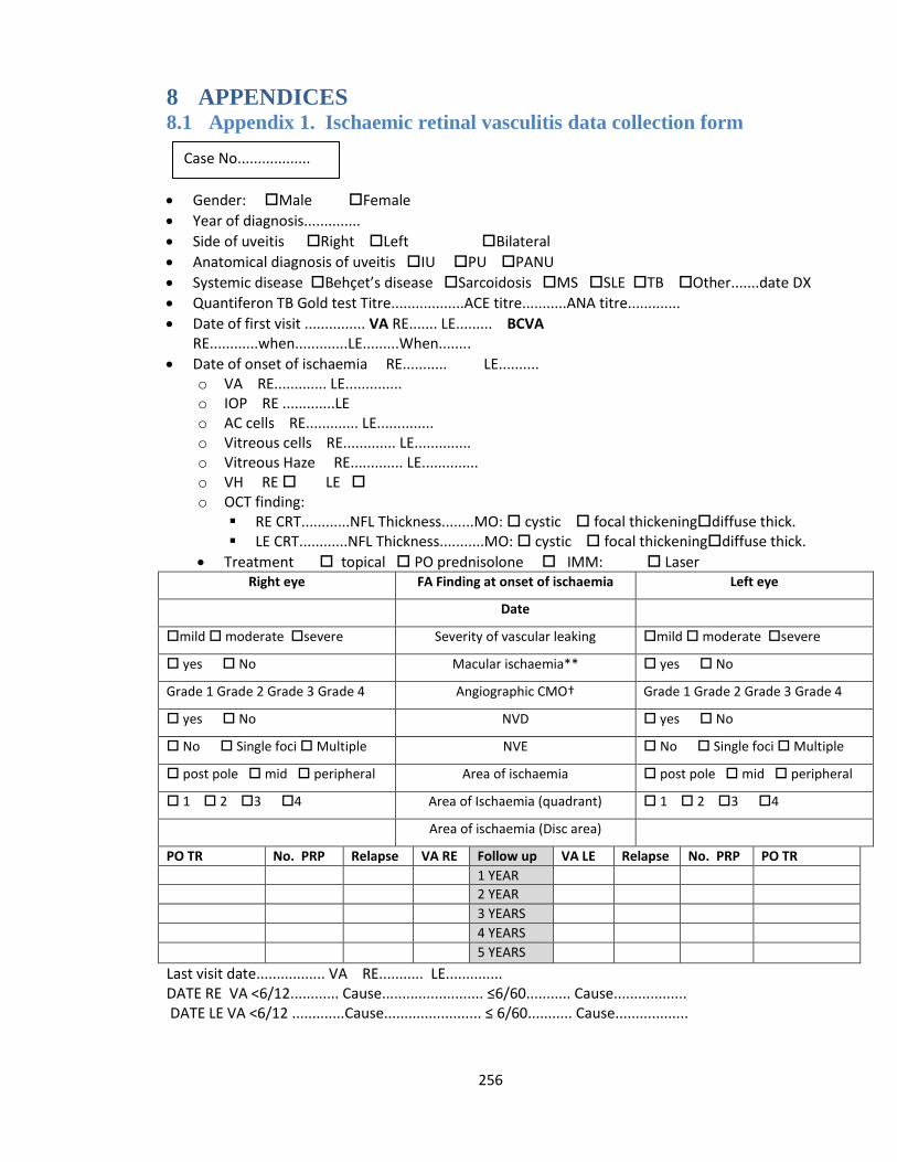
256
8 APPENDICES 8.1 Appendix 1. Ischaemic retinal vasculitis data collection form
Gender: Male Female
Year of diagnosis..............
Side of uveitis Right Left Bilateral
Anatomical diagnosis of uveitis IU PU PANU
Systemic disease Behçet’s disease Sarcoidosis MS SLE TB Other.......date DX
Quantiferon TB Gold test Titre..................ACE titre...........ANA titre.............
Date of first visit ............... VA RE....... LE......... BCVA RE............when.............LE.........When........
Date of onset of ischaemia RE........... LE.......... o VA RE............. LE.............. o IOP RE .............LE o AC cells RE............. LE.............. o Vitreous cells RE............. LE.............. o Vitreous Haze RE............. LE.............. o VH RE LE o OCT finding:
RE CRT............NFL Thickness........MO: cystic focal thickeningdiffuse thick. LE CRT............NFL Thickness...........MO: cystic focal thickeningdiffuse thick.
Treatment topical PO prednisolone IMM: Laser Right eye FA Finding at onset of ischaemia Left eye
Date
mild moderate severe Severity of vascular leaking mild moderate severe
yes No Macular ischaemia** yes No
Grade 1 Grade 2 Grade 3 Grade 4 Angiographic CMO† Grade 1 Grade 2 Grade 3 Grade 4
yes No NVD yes No
No Single foci Multiple NVE No Single foci Multiple
post pole mid peripheral Area of ischaemia post pole mid peripheral
1 2 3 4 Area of Ischaemia (quadrant) 1 2 3 4
Area of ischaemia (Disc area)
PO TR No. PRP Relapse VA RE Follow up VA LE Relapse No. PRP PO TR
1 YEAR
2 YEAR
3 YEARS
4 YEARS
5 YEARS
Last visit date................. VA RE........... LE.............. DATE RE VA <6/12............ Cause......................... ≤6/60........... Cause.................. DATE LE VA <6/12 .............Cause........................ ≤ 6/60........... Cause..................
Case No..................
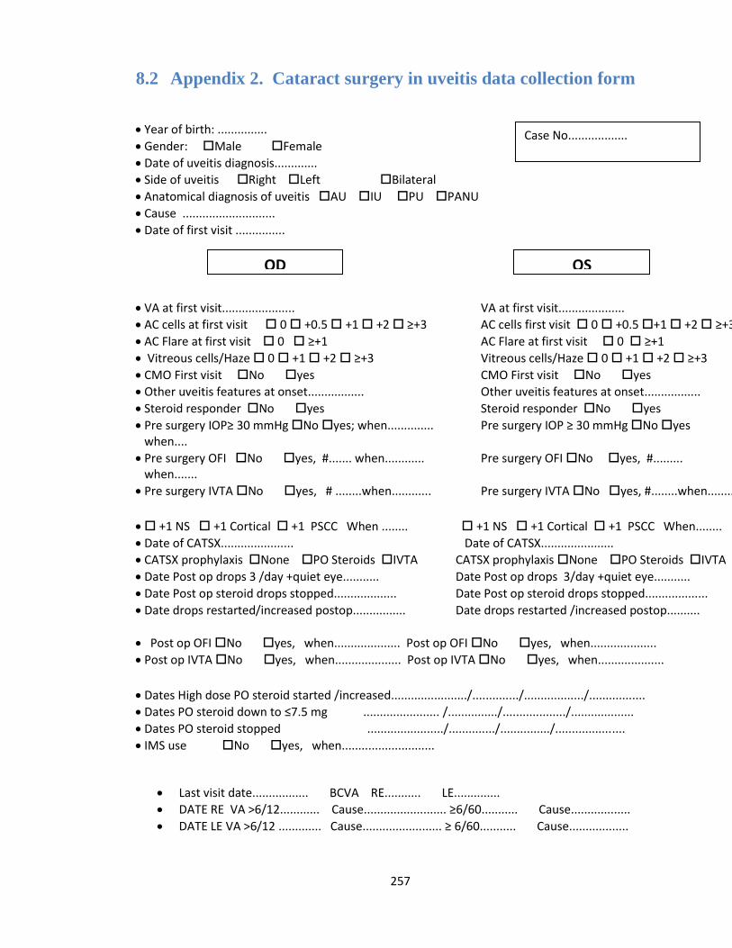
257
8.2 Appendix 2. Cataract surgery in uveitis data collection form
Year of birth: ...............
Gender: Male Female
Date of uveitis diagnosis.............
Side of uveitis Right Left Bilateral
Anatomical diagnosis of uveitis AU IU PU PANU
Cause ............................
Date of first visit ...............
VA at first visit...................... VA at first visit....................
AC cells at first visit 0 +0.5 +1 +2 ≥+3 AC cells first visit 0 +0.5 +1 +2 ≥+3
AC Flare at first visit 0 ≥+1 AC Flare at first visit 0 ≥+1
Vitreous cells/Haze 0 +1 +2 ≥+3 Vitreous cells/Haze 0 +1 +2 ≥+3
CMO First visit No yes CMO First visit No yes
Other uveitis features at onset................. Other uveitis features at onset.................
Steroid responder No yes Steroid responder No yes
Pre surgery IOP≥ 30 mmHg No yes; when.............. Pre surgery IOP ≥ 30 mmHg No yes when....
Pre surgery OFI No yes, #....... when............ Pre surgery OFI No yes, #......... when.......
Pre surgery IVTA No yes, # ........when............ Pre surgery IVTA No yes, #........when........
+1 NS +1 Cortical +1 PSCC When ........ +1 NS +1 Cortical +1 PSCC When........
Date of CATSX...................... Date of CATSX......................
CATSX prophylaxis None PO Steroids IVTA CATSX prophylaxis None PO Steroids IVTA
Date Post op drops 3 /day +quiet eye........... Date Post op drops 3/day +quiet eye...........
Date Post op steroid drops stopped................... Date Post op steroid drops stopped...................
Date drops restarted/increased postop................ Date drops restarted /increased postop..........
Post op OFI No yes, when.................... Post op OFI No yes, when....................
Post op IVTA No yes, when.................... Post op IVTA No yes, when....................
Dates High dose PO steroid started /increased......................./............../................../.................
Dates PO steroid down to ≤7.5 mg ....................... /.............../.................../...................
Dates PO steroid stopped ......................./............../.............../.....................
IMS use No yes, when............................
Last visit date................. BCVA RE........... LE..............
DATE RE VA >6/12............ Cause......................... ≥6/60........... Cause..................
DATE LE VA >6/12 ............. Cause........................ ≥ 6/60........... Cause..................
Case No..................
OD OS
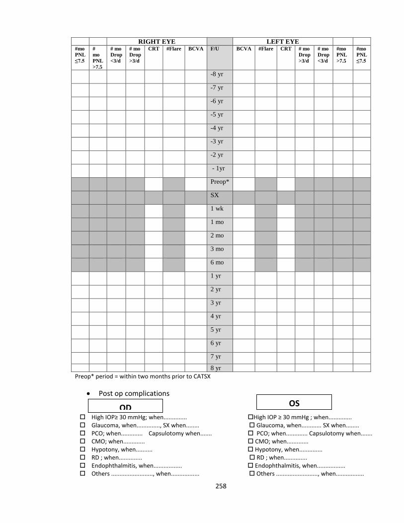
258
RIGHT EYE LEFT EYE #mo
PNL
≤7.5
#
mo
PNL
>7.5
# mo
Drop
<3/d
# mo
Drop
>3/d
CRT #Flare BCVA F/U BCVA #Flare CRT # mo
Drop
>3/d
# mo
Drop
<3/d
#mo
PNL
>7.5
#mo
PNL
≤7.5
-8 yr
-7 yr
-6 yr
-5 yr
-4 yr
-3 yr
-2 yr
- 1yr
Preop*
SX
1 wk
1 mo
2 mo
3 mo
6 mo
1 yr
2 yr
3 yr
4 yr
5 yr
6 yr
7 yr
8 yr
Preop* period = within two months prior to CATSX
Post op complications
High IOP≥ 30 mmHg; when.............. High IOP ≥ 30 mmHg ; when.............. Glaucoma, when.............., SX when........ Glaucoma, when............ SX when........ PCO; when............. Capsulotomy when....... PCO; when............. Capsulotomy when....... CMO; when............. CMO; when............. Hypotony, when.......... Hypotony, when.............. RD ; when.............. RD ; when.............. Endophthalmitis, when................. Endophthalmitis, when................. Others ........................., when................. Others ........................., when.................
OD OS