Embed Size (px)
Citation preview

Loading the Multilayer Dextran Sulfate/Protamine MicrosizedCapsules with Peroxidase
Nadezda G. Balabushevich,†,‡ Olga P. Tiourina,† Dmitry V. Volodkin,†,‡
Natalia I. Larionova,‡ and Gleb B. Sukhorukov*,†
Max-Planck Institute of Colloids and Interfaces, Golm/Potsdam, 14476, Germany, andFaculty of Chemistry, Lomonosov Moscow State University, Moscow, 119992, Russia
Received January 29, 2003; Revised Manuscript Received June 25, 2003
Stable polyelectrolyte capsules were produced by the layer-by-layer (LbL) assembling of biodegradablepolyelectrolytes, dextran sulfate and protamine, on melamine formaldehyde (MF) microcores followed bythe cores decomposition at low pH. The mean diameter of the capsules at pH 3-5 was 8.0( 0.2µm, whichis more than that diameter of the templates (5.12( 0.15 µm). With pH growing up to 7-8, the capsulesenlarged, swelling up to the diameter 9-10µm. The microcapsules were loaded with horseradish peroxidase.Seemingly, peroxidase is embedded in the gellike structure in the microcapsule interior formed by MFresidues in the complex with polymers used for LbL coating as proved by Raman confocal spectroscopy.The amount of finally incorporated peroxidase increased from 0.2× 10⁸ to 2.2× 10⁸ peroxidase moleculesper capsule with pH growing from 5 to 8. The pH shifts causing changes in capsule swelling and thereplacement of solutions without pH shifts lead to the protein loss. The encapsulated peroxidase showed ahigh activity (57%), which remained stable for 12 months.
Introduction
Design and biofunctionalization of microparticles are thechallenging research fields in biotechnology, especially whenthe enzyme properties are to be imparted in these particles.Fabrication of micron- and submicron-sized polymer spheres,capsules or liposomes containing proteins is a substantialtask for applications in biomedicine, cosmetology, ecology,and pharmaceutical and food industries.1
A novel concept of microencapsulation of different typesof materials was recently developed.2 It is based on the layer-by-layer (LbL) assembly of oppositely charged macromol-ecules onto the surface of colloid particles. The LbL methodexplores the electrostatic interaction at each step of adsorptionand can involve many substances as layer constituents, suchas synthetic polyelectrolytes, proteins, nucleic acids, lipids,inorganic nanoparticles, and multivalent dyes.3-5 Subse-quently, the LbL technology was transferred from flatmicroscopic substrates to surfaces of submicron-and few-micron-sized colloidal particles.6 Up to now, differentcolloidal cores were used to template the LbL polyelectrolyteassembly on their surfaces, for instance, organic latexparticles, inorganic particles, dye and drug nanocrystals,compact form of DNA, protein aggregates, gel beads, andbiological cells.6-12 The colloidal core can be decomposedin the conditions where the polyelectrolyte multilayers arestable leading to formation of hollow polyelectrolyte cap-sules.13,14 The most important features of polyelectrolytemultilayer shells, which make them promising for encapsula-
tion of different materials, are the possibilities for a widerange of controlling the capsule wall properties, such as shellthickness tuneable in the nanometer range, compatibility,affinity, and degradation.7 Polyelectrolyte multilayers aresensitive to pH value or to solvent mixture. Therefore, thepermeability of polyelectrolyte multilayer shells undergoesreversible changes when the pH value changes over pK valueof polyelectrolyte used for shell assembly.15 The reversiblechanges in permeability make possible the encapsulation ofenzymes. This was shown by chymotrypsin16 and urease17
encapsulation in polyelectrolyte capsules composed of poly-(styrene sulfonate) (PSS) and poly(allylamine) (PAH). Theproteins and enzymes might be also embedded into themelamine formaldehyde (MF)/PSS complex formed in thecapsule interior by spontaneous deposition.18
For many applications, the capsules must be composedof biocompatible polymers. In this paper, the encapsulationof peroxidase was performed in capsules made of dextransulfate and protamine. The encapsulation payload, enzymeactivity, and release were studied.
Experimental Section
Materials. Dextran sulfate,Mw 500 000, protamine,horseradish peroxidase, peroxidase fluorescein isothiocynatelabeled (FITC-peroxidase), 2,2′-azino-bis(3-ethylbenz-thia-zoline-6-sulfonic acid) (ABTS), rhodamine 6G, and 6-car-boxyfluorescein were purchased from Sigma. Amplex Redreagent was purchased from Molecular Probes, U.S.A. MFparticles with a diameter of 5.12( 0.15µm were purchasedfrom Microparticles GmbH, Germany.
Preparation of Polyelectrolyte Microcapsules.The poly-electrolyte multilayer assembly was fabricated on MF
* To whom correspondence should be addressed. Fax:+49 331 5679202. E-mail: [email protected].
† Max-Planck Institute of Colloids and Interfaces.‡ Lomonosov Moscow State University.
1191Biomacromolecules 2003,4, 1191-1197
10.1021/bm0340321 CCC: $25.00 © 2003 American Chemical SocietyPublished on Web 08/16/2003

particles at pH 5.0 and 0.2 M NaCl by alternate adsorptionof dextran sulfate and protamine. The polymer concentrationwas 5 mg/mL. Each adsorption cycle was completed withthree centrifugation steps followed by re-suspension in purewater before the next polymer was added to the particles.Such a washing procedure was used to avoid polyelectrolytecomplex formation outside the particle surface.13 The coreswere hydrolyzed at pH 1.7 in HCl solution. Then microcap-sules were washed with water until pH 3.0. The concentrationof the microcapsule suspension was calculated directly fromnumerous confocal images by counting the amount ofmicrocapsules in the defined volume and equalled (7( 3)× 1010 particles/L. The suspension of microcapsules at pH3.0 could be stored at 4°C over a month long period.
Encapsulation of Peroxidase into the Dextran Sulfate/Protamine Microcapsules.A 0.05-0.20 mL microcapsulesuspension in water was centrifuged (2000 g, 2 min), andthe supernatant was removed. Then the microcapsules weremixed with 0.95-0.80 mL of peroxidase solution (1-3 mg/mL) in the universal buffer (0.02 M H3PO4, 0.02 M CH3-COOH, 0.02 M H3BO3 + 0.1 M NaOH, pH 4-8). Afterincubation for 1 h at room temperature, this mixture wascentrifuged (2000 g, 2 min) and washed 1-3 times with thebuffer. The supernatants, washings, and microcapsulessuspension were used to determine the protein content andthe enzyme activity.
Determination of Protein Concentration. The proteinconcentration in solution and in suspensions of microcapsuleswas determined according to the Lowry method.19 Todetermine the concentration of the encapsulated protein,microcapsules were ultrasonicated and centrifuged (5000 g,3 min) before optical measurements.
The loading capacity (L) was evaluated by the followingequation:
where [c] is the concentration of the encapsulated protein,mg/mL; NA is the Avogadro constant;Mw is the molecularweight of peroxidase, g/mol; [N] is microcapsule concentra-tion, number of microcapsules per L.
Assay of the Peroxidase Activity.0.905-0.925 mL of0.1 M acetate buffer, pH 5.0, 0.005-0.025 mL of peroxidasesolution (0.05-0.04 mg/mL) or microcapsule suspension, and0.020 mL of ABTS solution (8 mg/mL) were placed in aquartz cell. Then 0.050 mL of a H2O2 solution (0.5%) wasadded.20 The absorbance was measured at 403 nm using aUV spectrometer (Cary UV-visible). The activity of per-oxidase was 550 U/mg. 1 U oxidizes 1µmole ABTS permin at 25 °C, pH 5.0. The activity of the encapsulatedperoxidase vs the activity of an equal amount of the nativeprotein was expressed in present.
Effect of pH on Peroxidase Release from Microcap-sules.Peroxidase was encapsulated at pH 8.0 as mentionedabove, the suspension was centrifuged, and the supernatantwas removed. The protein content in microcapsules wasaccepted as 100%. 1 mL of universal buffer solution (pH4.0, 5.0 or 8.0) was added to the capsules, and afterincubation for 10 min, the mixture was centrifuged and theprotein content was determined in the supernatant. Precipi-
tated microcapsules were resuspended in a new portion (1mL) of the same buffer solution and the procedure wasrepeated. The cumulative release of protein was calculatedas a ratio of total protein released, determined in supernatantsafter a certain time of incubation, to initial protein inmicrocapsules prepared at pH 8.0.
Effect of a Number of Polyelectrolyte Layers onEnzyme Release from Microcapsules.Peroxidase wasencapsulated at pH 8.0, and a part of the capsules loadedwith protein was covered by 2 bilayers of dextran sulfate/protamine to produce 6 bilayer microcapsules. Then themicrocapsules were resuspended in the buffer (pH 7.0) toobtain the final concentration 0.2 mg protein/mL. Themicrocapsules suspension was incubated at 20°C withconstant agitation at 40 rpm. After 10, 30, 60, 120, 180, 240,and 420 min, the aliquots (0.05 mL) of the suspension werewithdrawn and centrifuged. The supernatants were used todetermine the enzyme activity.
Confocal Laser Scanning Microscopy (CLSM).TheCLSM was carried out on a LEICA TCS system (Aristoplan(Germany), 100× oil immersion) using commercial soft-ware.
Scanning Electron Microscopy (SEM).SEM was con-ducted using a Zeiss DSM 40 instrument operated at anacceleration voltage of 3 keV. Samples were prepared byapplying a drop of the suspension onto glass wafers. Thewater was evaporated before the samples were covered witha thin gold film.
Raman Spectroscopy.Raman spectra and images weremade in water using a Confocal Raman Microscope (CRM200,Witec) with a piezo scanner (P-500, Physik Instrumente) andobjectives (× 60, NA ) 0.80 or ×100 oil, NA ) 1.25,Nikon). In a typical experiment, a circularly polarized laser(CrystaLaser,λ ) 532 nm) was focused on the material withdiffraction limited spot size (∼λ/2). An avalanche photodiodedetector (APD) was used to record high resolution Ramanimages. To have a Raman spectroscopic analysis intomicroparticles, we focused the beam spot exactly inside thespherical particle, but for measuring onto the surface, ofparticles it was focused on the edge. Concentrated solutionsof protamine (50 mg/mL) and dextran sulfate (100 mg/mL)were used for spectroscopic analysis of polyelectrolytes.
Visualization of Active Peroxidase in Microcapsules.0.01 mL of a microcapsule suspension was mixed with 0.08mL of 0.25 M phosphate buffer (pH 7.4), 0.05 mL of 10mM Amplex Red reagent in DMSO, and 0.05 mL of 20 mMH2O2. Resorufin production was followed by fluorescenceCLSM at 587 nm.
Results and Discussion
To elaborate biocompatible capsules containing enzymes,the pair of oppositely charged polyelectrolytes should to beproperly chosen according to the following prerequisites.First, these polymers should not affect the enzyme activity.Second, the capsules composed of these polymers shouldbe fairly stable, which is not always the case because somecore dissolution procedure might lead either to the shellrupture or even to disassembling of multilayer films.
L ) [c]NA/Mw[N]
1192 Biomacromolecules, Vol. 4, No. 5, 2003 Balabushevich et al.

Dextran sulfate with a molecular weight of (Mw) 500 000was chosen as the polyanion. As the polycation, we selectedthe strong basic protein protamine (salmin) with theMw about5000 and a high content of arginine, which is up to 70% ofthe total amount of amino acids.21 Such a polyelectrolytecombination has not been used so far for multilayer buildup. Both polyelectrolytes revealed no influence on peroxidaseactivity in 0.1 M acetate buffer at pH 5.0 known as theoptimal condition for enzyme function. The weight ratio ofpolyelectrolytes to enzyme in these experiments varied from1:1 to 100:1. Table 1 shows that the chosen polyelectrolytesonly slightly influenced the activity of the enzyme for 7 days.According to our results (Table 1), the addition of thesolution obtained by dissolution of MF particles at acidifyingup to pH 1.7 to peroxidase does not suppress the enzymeactivity. It is worth mentioning that protamine and melamineformaldehyde hydrolysate gave some activation of peroxidase(24%) after a short incubation. This effect disappeared afterincubation for a few days. So some quantity of MF andpolyelectrolytes did not negatively influence peroxidase.
The capsules were fabricated according to the conventionalmethod14 by the sequential adsorption of 4 bilayers of dextransulfate/protamine on MF cores in 0.2 M NaCl (pH 5.0). Asit is stressed by Gao et al.,22 the process of core dissolutioncalled for a very slow adjusting of pH to the required valueand had to be tuned for MF cores from different lots. Atfast pH adjustment to low pH values, the capsule walldissolves in a minute time scale after core dissolution. Inthis study then, MF cores were decomposed at pH 1.7, andthe capsules were washed thoroughly in these conditions. Itshould take into account that native MF cores from differentlots were dissolved at the same pH at which a degradationof this cores coated by the polyelectrolyte shell was observed.Dextran sulfate/protamine microcapsules prepared were keptat pH 3.0 for prevention of microflora growth.
The morphology of the capsules was studied by means ofconfocalmicroscopy (Figure 1a). The capsules became swol-len compared to initial templates (5.12µm). The averagesize of the microcapsules at pH 3-5 was about 8.0( 0.2µm. With a pH increase up to 7-8, the microcapsulesenlarged (swelled) up to a diameter of 9-10 µm. In basicsolutions (pH 9-11), the microcapsules changed in form(Figure 1d), but this pH range is not used for working withenzymes.
The results of SEM (Figure 2) showed that the capsuleswere porous visually as if they had a loose shell. Thepresence of a salient middle region testified that the capsuleswere not hollow. We can assume that there was a weakly
cross-linked gellike matrix inside the shell, which preventedthe loss of the round form by microcapsules on drying. So,microcapsules prepared in this work differed from the hollowcapsules fabricated using PSS and PAA.14,18,23
Table 1. Retention of the Enzymatic Activity of Free Peroxidase (in the Presence of Components for Microcapsule Preparation) andEncapsulated Enzyme in 0.1 M Acetate Buffer, pH 5.0, 20 °C
relative activity of peroxidase, %
free enzyme
duration of storage buffer dextran sulfatea protaminea
melamin formaldehydehydrolysatea encapsulated enzyme
1 h 100 ( 4 101 ( 5 124 ( 6 124 ( 6 57 ( 34 days 85 ( 4 95 ( 4 102 ( 5 104 ( 5 55 ( 27 days 83 ( 4 91 ( 4 98 ( 5 89 ( 3 53 ( 2
a Ratio in solution component: peroxidase ) 100:1 (w/w).
Figure 1. CLSM image of dextran sulfate/protamine microcapsules:a. at pH 5; b. loaded with 6-carboxyfluorescein at pH 5; c. loadedwith rhodamine 6G at pH 5; d. loaded with rhodamine 6G at pH 9; e.(transmission), f. (fluorescence), loaded with FITC-peroxidase at pH5.
Figure 2. SEM image of dextran sulfate/protaminemicrocapsules.
Dextran Sulfate/Protamine Microsized Capsules Biomacromolecules, Vol. 4, No. 5, 2003 1193

To elucidate the capsule interior composition, we appliedvarious dyes which permeated through the microcapsule walland bound exclusively either to the positively or negativelycharged groups.24 The low molecular weight, negativelycharged 6-carboxyfluorescein (Mw 376) and the positivelycharged rhodamine 6G (Mw 479) penetrated and werehomogeneously distributed inside the microcapsules in thepH range 5-8 (Figure 1b-d). Impregnation of differentlycharged dyes inside the capsule revealed the presence of ahomogeneous matrix with both positively and negativelycharged groups inside the microcapsules.
Positively charged FITC-peroxidase (Mw 44000, isoelectricpoint pI 8.8) also concentrated inside the microcapsules atpH 3-8 compared to the solution surrounding the micro-spheres (Figure 1 e, f).
To confirm the localization of the active enzyme intomicrocapsules, we used the reagent Amplex Red (10-acetyl-3,7-dihydroxyphenoxazine). In the presence of peroxidase,the Amplex Red reagent reacts with H2O2 in a 1:1 stoichi-ometry to produce the red-fluorescent oxidation product,resorufin. Resorufin has absorption and fluorescence emissionmaxima of approximately 563 and 587 nm. Figure 3 showsthat an increase of the fluorescence emission of resorufinwas observed inside microcapsules but not in a surroundingsolution.
Our recently results (ref 25) show that, in the range of pH5-8, various proteins such as insulin (Mw 6500, pI 5.5),aprotinin (Mw 6500, pI 10.5), trypsin (Mw 24 000, pI 10.5),chymotrypsin (Mw 25 000, pI 8.8), glucose oxidase (Mw
160 000, pI 5.5), and catalase (Mw 250 000, pI 5.4) canpenetrate into microcapsules such as peroxidase.
In contrast, alginate/protamine microcapsules, prepared onMF cores dissolved at pH 1.7, adsorbed only positivelycharged substances.26 Therefore, the presence of the alginategel in the interior of the microcapsules was suggested butnot proved.26 Meanwhile, an existing negatively chargedcomplex, formed by PSS and MF degradation products, waspostulated for PSS/PAH or poly(diallyldimethylammoniumchloride) (PDADMAC) microcapsules.18 The positivelycharged water-soluble substances (including proteins) werespontaneously deposited inside these microcapsules.18
The microcapsules prepared in this study regardless of theirage adsorbed in the interior both positively and negativelycharged species of various molecular masses.
Apparently, the existence of other components can besuggested besides melamine formaldehyde resins inside thecapsules. We used confocal Raman spectroscopy to detect
the presence of polyelectrolytes (protamine and dextransulfate) inside as well as on the surface of microparticlesprepared in this study (coated MF cores and microcapsules).
Raman spectra of polyelectrolytes used for multilayercoating, native, and coated MF cores and also of fabricatedhollow microcapsules are given in Figure 4. One can seethe Raman spectrum of protamine (Figure 4A) as a line withno exact peaks for detection of this polyelectrolyte, wereasdextran sulfate showed a spectrum with an intense peak atabout 1100 cm-1 (Figure 4B, peak 1). This peak, which canbe assigned to the vibration of sulfonate groups in thepolymer, was used as a fingerprint for dextran sulfate. TheRaman spectrum of melamine formaldehyde microcores isshown in Figure 4C, peak2. The pronounced peak with anexperimental frequency 977 cm-1 is typical for melamineand melamine formaldehyde resins.27,28
We can see clearly the appearance of the Raman peakassigned to dextran sulfate on the spectrum obtained fromsurface signal of the multilayer polyelectrolyte coated MFcore (Figure 4D). However, upon focusing inside the coatedMF particle, a single peak corresponding to melamineformaldehyde resins (Figure 4E) was found, indicating adense structure of the MF core because of which thepolyelectrolytes cannot penetrate inside the particle. How-ever, when the coated core was exposed to conditions atwhich it dissolves, the polyelectrolytes penetrated into thespace contributed by the dissolving core, which was con-firmed by spectra corresponding to the signal from thecapsule shell (Figure 4F) and from the interior of the capsule(Figure 4G). The appearance of the peak of dextran sulfatewas evident from the spectra in addition to the peak of theMF resin.
Thus, we can conclude that dextran sulfate from the shellinteracted with some of the positively charged MF degrada-tion products. It could cause a special redistribution of thepolymers, which resulted in the formation of a porous pH-sensitive gellike matrix on the basis of MF-oligomerscomplexes with both polyelectrolytes used for the LbLassembly. It can be concluded that the properties of thematrix, which is crucial for the spontaneous adsorption ofsubstances in the interior of the microcapsules prepared on
Figure 3. Imaging of peroxidase activity inside dextran sulfate/protamine microcapsules: A. transmission; B-E. fluorescence imageof resorufin after 1, 11, 12, and 13 min, respectively.
Figure 4. Raman spectroscopy analysis: Raman spectra of prota-mine, (A); dextran sulfate, (B); melamine formaldehyde cores (C); ashell (D) and interior (E) of coated MF cores; and a shell (F) andinterior (G) of hollow microcapsules.
1194 Biomacromolecules, Vol. 4, No. 5, 2003 Balabushevich et al.

MF cores, seemed to be mainly governed by the polyelec-trolytes used for the shell fabrication. The process of gelationwas rather quick when dextran sulfate and protamine, as wellas alginate and protamine, were used for LbL assembly.However, it is well-known29 that equilibration in processeswith PSS participation establishes rather slowly. It seems tobe a cause of the finding that freshly prepared PSS/PDADMAC microcapsules were not favorable for thespontaneous deposition of the enzyme.30
Further, we studied the principles of peroxidase encapsula-tion in multilayer dextran sulfate/protamine microcapsulessuch as pH and the number of microcapsules. The influenceof the pH values on the process of peroxidase encapsulationand its keeping inside is shown in Figure 5. With the pHincreased from 5 (according to conditions of capsule fabrica-tion) to 8, the loading capacity increased from 0.2× 10⁸ to2.2 × 108 peroxidase molecules per microcapsule, respec-tively. The microcapsule swelling at higher pH seemed tobe a reason for an increase in the peroxidase encapsulationpayload. Figure 5 demonstrates that the enzyme was partiallyreleased from the microcapsule after 3-fold washings of themicrocapsules with the buffer used for protein encapsulation.On the other hand, when peroxidase-loaded microcapsuleswere incubated in buffer solution without its change andstirring, release of the enzyme for 24 h was negligible. Figure5 shows that the pH of washing solutions in the range 5-8did not influence on the relative activity of encapsulatedperoxidase, which was found about 50% using ABTS.
Table 2 shows that at pH 5.0 the amount of proteinencapsulated at pH 5.0 and its activity increased proportion-ally with an increase in the number of microcapsules. Thespecific activity of encapsulated peroxidase was 57% ofinitial enzyme activity. At the storage of protein-loadedmicrocapsules, the specific activity of encapsulated peroxi-dase after 7 days decreased down to 4%, whereas the lossof activity of native peroxidase was 17% (Table 1).
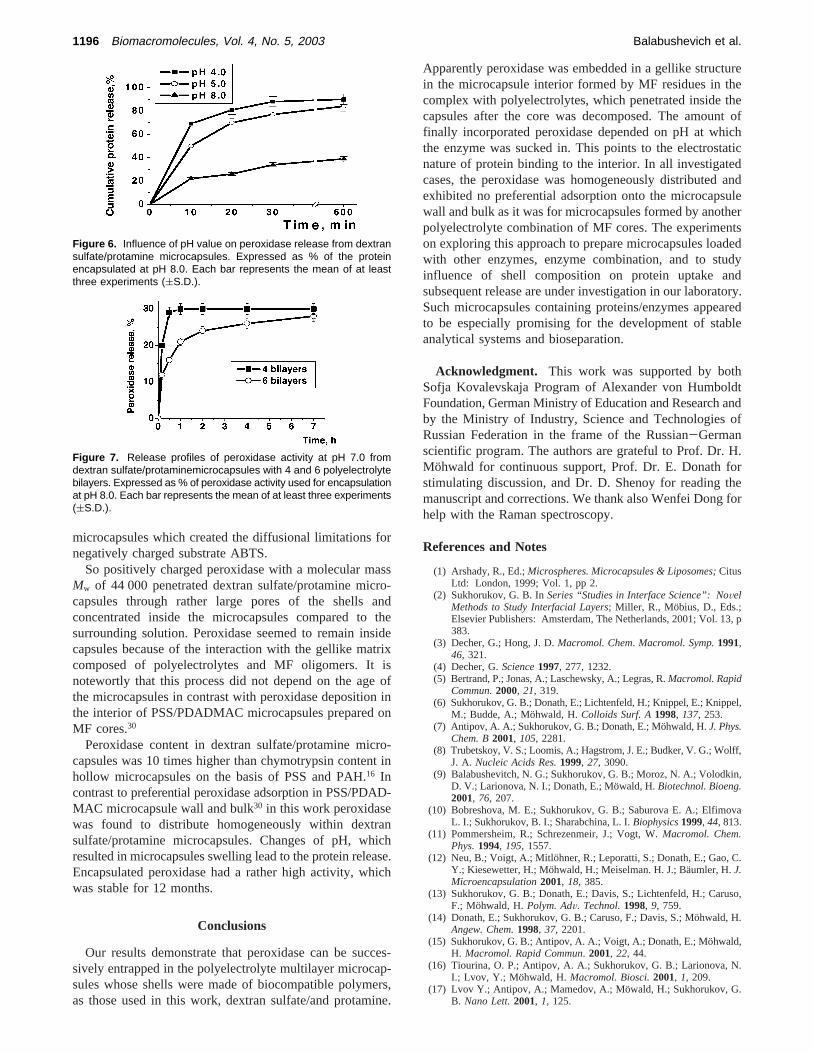
The effect of the pH of the medium on the release of theperoxidase encapsulated at pH 8.0 is shown in Figure 6.Microcapsules were not washed before the release test. Onecan see at pH 4.0 and 5.0 the protein was released very fastfor 10 min (70-80%) but then the leakage was noticeablyslow. We suppose that the reason of this change in releaserate is the decreasing size of the microcapsules at low a pHvalue. However, when the pH at which the microparticleswere fabricated and the pH at which the protein was releasedare the same (pH 8.0), the protein release for 10 min wascalculated about 20%, which can be explained by the releaseof protein molecules that interacted weakly with the micro-capsule surface followed by a very slow release of theperoxidase encapsulated inside.
The microcapsules containing peroxidase can be thencoated with additional layers of polyelectrolytes. But thisprocess caused considerable (up to 70%) loss of enzymebecause of multiple washing steps between adsorption steps.Nevertheless, the leakage of peroxidase from microcapsuleswith 6 polyelectrolyte bilayers at the first 4 h was slowerthan from to initial 4 bilayer microcapsules (Figure 7). Notethat peroxidase released from the capsules recovered itsactivity to 90-95% of the native enzyme activity. So we
presume that the decrease in the activity of the encapsulatedperoxidase (57% to control values) could be the consequenceof extensive interpolyelectrolyte gel formation inside the
Figure 5. Influence of pH value on peroxidase encapsulation intodextran sulfate/protamine microcapsules and specific activity of theencapsulated enzyme. Number of microcapsules 1.4 × 10⁷. Eachbar represents the mean of at least three experiments ((S.D.).
Table 2. Properties of Peroxidase Loaded Dextran Sulfate/Protamine Microcapsules at pH 5.0, 20 °C
number ofmicrocapsules,
×107
protein intomicrocapsules,
mg
peroxidaseactivity,
U
relative activityof peroxidase,
%
0.35 0.016 ( 0.001 5.0 ( 0.2 57 ( 30.70 0.035 ( 0.002 11.0 ( 0.5 57 ( 21.40 0.070 ( 0.003 22.0 ( 0.6 57 ( 3
Dextran Sulfate/Protamine Microsized Capsules Biomacromolecules, Vol. 4, No. 5, 2003 1195
microcapsules which created the diffusional limitations fornegatively charged substrate ABTS.
So positively charged peroxidase with a molecular massMw of 44 000 penetrated dextran sulfate/protamine micro-capsules through rather large pores of the shells andconcentrated inside the microcapsules compared to thesurrounding solution. Peroxidase seemed to remain insidecapsules because of the interaction with the gellike matrixcomposed of polyelectrolytes and MF oligomers. It isnotewortly that this process did not depend on the age ofthe microcapsules in contrast with peroxidase deposition inthe interior of PSS/PDADMAC microcapsules prepared onMF cores.30
Peroxidase content in dextran sulfate/protamine micro-capsules was 10 times higher than chymotrypsin content inhollow microcapsules on the basis of PSS and PAH.16 Incontrast to preferential peroxidase adsorption in PSS/PDAD-MAC microcapsule wall and bulk30 in this work peroxidasewas found to distribute homogeneously within dextransulfate/protamine microcapsules. Changes of pH, whichresulted in microcapsules swelling lead to the protein release.Encapsulated peroxidase had a rather high activity, whichwas stable for 12 months.
Conclusions
Our results demonstrate that peroxidase can be succes-sively entrapped in the polyelectrolyte multilayer microcap-sules whose shells were made of biocompatible polymers,as those used in this work, dextran sulfate/and protamine.
Apparently peroxidase was embedded in a gellike structurein the microcapsule interior formed by MF residues in thecomplex with polyelectrolytes, which penetrated inside thecapsules after the core was decomposed. The amount offinally incorporated peroxidase depended on pH at whichthe enzyme was sucked in. This points to the electrostaticnature of protein binding to the interior. In all investigatedcases, the peroxidase was homogeneously distributed andexhibited no preferential adsorption onto the microcapsulewall and bulk as it was for microcapsules formed by anotherpolyelectrolyte combination of MF cores. The experimentson exploring this approach to prepare microcapsules loadedwith other enzymes, enzyme combination, and to studyinfluence of shell composition on protein uptake andsubsequent release are under investigation in our laboratory.Such microcapsules containing proteins/enzymes appearedto be especially promising for the development of stableanalytical systems and bioseparation.
Acknowledgment. This work was supported by bothSofja Kovalevskaja Program of Alexander von HumboldtFoundation, German Ministry of Education and Research andby the Ministry of Industry, Science and Technologies ofRussian Federation in the frame of the Russian-Germanscientific program. The authors are grateful to Prof. Dr. H.Mohwald for continuous support, Prof. Dr. E. Donath forstimulating discussion, and Dr. D. Shenoy for reading themanuscript and corrections. We thank also Wenfei Dong forhelp with the Raman spectroscopy.
References and Notes
(1) Arshady, R., Ed.;Microspheres. Microcapsules & Liposomes;CitusLtd: London, 1999; Vol. 1, pp 2.
(2) Sukhorukov, G. B. InSeries “Studies in Interface Science”: NoVelMethods to Study Interfacial Layers; Miller, R., Mobius, D., Eds.;Elsevier Publishers: Amsterdam, The Netherlands, 2001; Vol. 13, p383.
(3) Decher, G.; Hong, J. D.Macromol. Chem. Macromol. Symp.1991,46, 321.
(4) Decher, G.Science1997, 277, 1232.(5) Bertrand, P.; Jonas, A.; Laschewsky, A.; Legras, R.Macromol. Rapid
Commun.2000, 21, 319.(6) Sukhorukov, G. B.; Donath, E.; Lichtenfeld, H.; Knippel, E.; Knippel,
M.; Budde, A.; Mohwald, H.Colloids Surf. A1998, 137, 253.(7) Antipov, A. A.; Sukhorukov, G. B.; Donath, E.; Mo¨hwald, H.J. Phys.
Chem. B2001, 105, 2281.(8) Trubetskoy, V. S.; Loomis, A.; Hagstrom, J. E.; Budker, V. G.; Wolff,
J. A. Nucleic Acids Res.1999, 27, 3090.(9) Balabushevitch, N. G.; Sukhorukov, G. B.; Moroz, N. A.; Volodkin,
D. V.; Larionova, N. I.; Donath, E.; Mo¨wald, H.Biotechnol. Bioeng.2001, 76, 207.
(10) Bobreshova, M. E.; Sukhorukov, G. B.; Saburova E. A.; ElfimovaL. I.; Sukhorukov, B. I.; Sharabchina, L. I.Biophysics1999, 44, 813.
(11) Pommersheim, R.; Schrezenmeir, J.; Vogt, W.Macromol. Chem.Phys.1994, 195, 1557.
(12) Neu, B.; Voigt, A.; Mitlohner, R.; Leporatti, S.; Donath, E.; Gao, C.Y.; Kiesewetter, H.; Mo¨hwald, H.; Meiselman. H. J.; Ba¨umler, H.J.Microencapsulation2001, 18, 385.
(13) Sukhorukov, G. B.; Donath, E.; Davis, S.; Lichtenfeld, H.; Caruso,F.; Mohwald, H.Polym. AdV. Technol.1998, 9, 759.
(14) Donath, E.; Sukhorukov, G. B.; Caruso, F.; Davis, S.; Mo¨hwald, H.Angew. Chem.1998, 37, 2201.
(15) Sukhorukov, G. B.; Antipov, A. A.; Voigt, A.; Donath, E.; Mo¨hwald,H. Macromol. Rapid Commun. 2001, 22, 44.
(16) Tiourina, O. P.; Antipov, A. A.; Sukhorukov, G. B.; Larionova, N.I.; Lvov, Y.; Mohwald, H.Macromol. Biosci.2001, 1, 209.
(17) Lvov Y.; Antipov, A.; Mamedov, A.; Mo¨wald, H.; Sukhorukov, G.B. Nano Lett.2001, 1, 125.
Figure 6. Influence of pH value on peroxidase release from dextransulfate/protamine microcapsules. Expressed as % of the proteinencapsulated at pH 8.0. Each bar represents the mean of at leastthree experiments ((S.D.).
Figure 7. Release profiles of peroxidase activity at pH 7.0 fromdextran sulfate/protaminemicrocapsules with 4 and 6 polyelectrolytebilayers. Expressed as % of peroxidase activity used for encapsulationat pH 8.0. Each bar represents the mean of at least three experiments((S.D.).
1196 Biomacromolecules, Vol. 4, No. 5, 2003 Balabushevich et al.

(18) Gao, C. Y.; Donath, E.; Mo¨hwald, H.; Shen,J. Angew. Chem., Int.Ed. 2002, 41 (20), 3789.
(19) Lowry, O. H.; Rosebrough, N. J.; Farr, A. L.; Randall, R. J.J. Biol.Chem.1951, 193, 265.
(20) Childs, R.; Bardsley, W.Biochem. J. 1975, 145, 93.(21) Callanan, M. J.; Carroll, W. R.; Mitchell, E. R. J. Biol. Chem. 1957,
229, 279.(22) Gao, C.; Moya, S.; Lichtenfeld, H.; Casoli, A.; Fielder, H.; Donath,
E.; Mohwald, H.Macromol. Mater. Eng. 2001, 286, 355.(23) Sukhorukov, G. B.; Donath, E.; Moya, S.; Susha, A. S.; Voigt, A.;
Hartmann, J.; Mo¨hwald. H.J. Microecapsulation2000, 17, 177.(24) Caruso, F.; Lichtenfeld, H.; Donath, E.; Mo¨hwald. H.Macromolecules
1999, 32, 2317.
(25) Larionova, N. I.; Balabushevich, N. G.; Sukhorukov, G. B.; Tiourina,O. P.; Zimina, E. P.XI Intern. BRG Workshop on Bioencapsulation;Strasbourg, 2003, May 25-27, # 20.
(26) Tiourina, O. P.; Sukhorukov, G. B.Int. J. Pharm. 2002, 242, 155.(27) Koglin, E.; Kip, B.; Meier, R.J. Phys. Chem.1996, 100, 5078.(28) Scheepers, M. L.; Gelan, J. M.; Carleer, R. A.; Adriaensens, P. J.;
Vanderzande, D. J.; Kip, B. J.; Brandts P. M.Vibr. Spectrosc.1993,6, 55.
(29) Michaels, A.Encyclopedia of Polymer Science and Technology;Interscience Publ.: New York, 1969; Vol. 10, pp 765.
(30) Gao, C. Y.; Liu, X. Y.; Shen, J. C.; Mo¨hwald, H.Chem. Commun.2002, 17, 1928.
BM0340321
Dextran Sulfate/Protamine Microsized Capsules Biomacromolecules, Vol. 4, No. 5, 2003 1197