Embed Size (px)
Citation preview
HAL Id: hal-01912305https://hal.archives-ouvertes.fr/hal-01912305
Submitted on 11 Jun 2021
HAL is a multi-disciplinary open accessarchive for the deposit and dissemination of sci-entific research documents, whether they are pub-lished or not. The documents may come fromteaching and research institutions in France orabroad, or from public or private research centers.
L’archive ouverte pluridisciplinaire HAL, estdestinée au dépôt et à la diffusion de documentsscientifiques de niveau recherche, publiés ou non,émanant des établissements d’enseignement et derecherche français ou étrangers, des laboratoirespublics ou privés.
Distributed under a Creative Commons Attribution| 4.0 International License
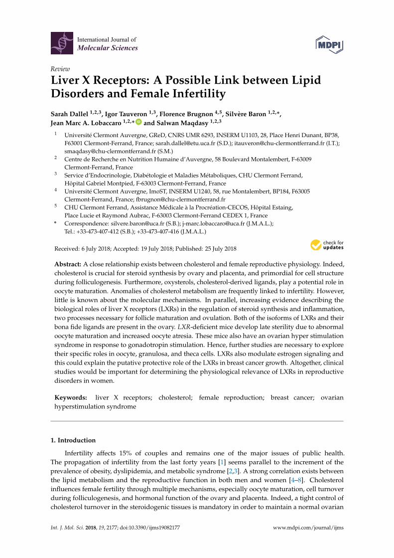
Liver X Receptors: A Possible Link between LipidDisorders and Female Infertility
Sarah Dallel, Igor Tauveron, Florence Brugnon, Silvère Baron, JeanLobaccaro, Salwan Maqdasy
To cite this version:Sarah Dallel, Igor Tauveron, Florence Brugnon, Silvère Baron, Jean Lobaccaro, et al.. Liver X Re-ceptors: A Possible Link between Lipid Disorders and Female Infertility. International Journal ofMolecular Sciences, MDPI, 2018, 19 (8), pp.2177. �10.3390/ijms19082177�. �hal-01912305�
International Journal of
Molecular Sciences
Review
Liver X Receptors: A Possible Link between LipidDisorders and Female Infertility
Sarah Dallel 1,2,3, Igor Tauveron 1,3, Florence Brugnon 4,5, Silvère Baron 1,2,*,Jean Marc A. Lobaccaro 1,2,* ID and Salwan Maqdasy 1,2,3
1 Université Clermont Auvergne, GReD, CNRS UMR 6293, INSERM U1103, 28, Place Henri Dunant, BP38,F63001 Clermont-Ferrand, France; [email protected] (S.D.); [email protected] (I.T.);[email protected] (S.M.)
2 Centre de Recherche en Nutrition Humaine d’Auvergne, 58 Boulevard Montalembert, F-63009Clermont-Ferrand, France
3 Service d’Endocrinologie, Diabétologie et Maladies Métaboliques, CHU Clermont Ferrand,Hôpital Gabriel Montpied, F-63003 Clermont-Ferrand, France
4 Université Clermont Auvergne, ImoST, INSERM U1240, 58, rue Montalembert, BP184, F63005Clermont-Ferrand, France; [email protected]
5 CHU Clermont Ferrand, Assistance Médicale à la Procréation-CECOS, Hôpital Estaing,Place Lucie et Raymond Aubrac, F-63003 Clermont-Ferrand CEDEX 1, France
* Correspondence: [email protected] (S.B.); [email protected] (J.M.A.L.);Tel.: +33-473-407-412 (S.B.); +33-473-407-416 (J.M.A.L.)
Received: 6 July 2018; Accepted: 19 July 2018; Published: 25 July 2018�����������������
Abstract: A close relationship exists between cholesterol and female reproductive physiology. Indeed,cholesterol is crucial for steroid synthesis by ovary and placenta, and primordial for cell structureduring folliculogenesis. Furthermore, oxysterols, cholesterol-derived ligands, play a potential role inoocyte maturation. Anomalies of cholesterol metabolism are frequently linked to infertility. However,little is known about the molecular mechanisms. In parallel, increasing evidence describing thebiological roles of liver X receptors (LXRs) in the regulation of steroid synthesis and inflammation,two processes necessary for follicle maturation and ovulation. Both of the isoforms of LXRs and theirbona fide ligands are present in the ovary. LXR-deficient mice develop late sterility due to abnormaloocyte maturation and increased oocyte atresia. These mice also have an ovarian hyper stimulationsyndrome in response to gonadotropin stimulation. Hence, further studies are necessary to exploretheir specific roles in oocyte, granulosa, and theca cells. LXRs also modulate estrogen signaling andthis could explain the putative protective role of the LXRs in breast cancer growth. Altogether, clinicalstudies would be important for determining the physiological relevance of LXRs in reproductivedisorders in women.
Keywords: liver X receptors; cholesterol; female reproduction; breast cancer; ovarianhyperstimulation syndrome
1. Introduction
Infertility affects 15% of couples and remains one of the major issues of public health.The propagation of infertility from the last forty years [1] seems parallel to the increment of theprevalence of obesity, dyslipidemia, and metabolic syndrome [2,3]. A strong correlation exists betweenthe lipid metabolism and the reproductive function in both men and women [4–8]. Cholesterolinfluences female fertility through multiple mechanisms, especially oocyte maturation, cell turnoverduring folliculogenesis, and hormonal function of the ovary and placenta. Indeed, a tight control ofcholesterol turnover in the steroidogenic tissues is mandatory in order to maintain a normal ovarian
Int. J. Mol. Sci. 2018, 19, 2177; doi:10.3390/ijms19082177 www.mdpi.com/journal/ijms
Int. J. Mol. Sci. 2018, 19, 2177 2 of 15
function. Hence, an excess or insufficiency of cholesterol is deleterious for cell function. The turnoverstarts from the cholesterol uptake through low density lipoprotein (LDL) and high density lipoprotein(HDL) receptors (LDLR and SR-BI, respectively), cholesterol esterification and storage, cholesterolester hydrolase activity, cholesterol de novo synthesis pathway, and cholesterol efflux promoted byATP-binding cassette (ABC) proteins. The latter are target genes of liver X receptors.
Liver X receptors (LXRα/NR1H3 and LXRβ/NR1H2) are nuclear receptors (NRs) for oxysterols,which are largely implicated in cholesterol homeostasis [9]. Because of their roles in the regulation ofnumerous metabolic functions, LXRs could represent part of the molecular link between lipid disordersand infertility. Thus, many arguments led to deeply investigating LXRs in female reproductivefunction, namely: (1) the close relationship between cholesterol and ovarian physiology [4,10,11]; (2) theincreasing body of literature describing the novel biological roles of LXRs in the regulation of steroidsynthesis and inflammation, two processes necessary for follicle maturation and ovulation [12–17];(3) the fact that oxysterols, which are the physiological ligands [18], play a potential role in thematuration of the oocytes [19,20]; (4) and the role of LXRs in spermatogenesis suggests common germcell pathways regulated by LXRs [7,14,17].
2. Fertility Disorders and Abnormal Lipid Homeostasis
Ovarian dysfunction is frequently observed in patients with metabolic syndrome and obesity,two frequent pathological situations linked to polycystic ovary syndrome (PCOS) [21,22], which is themain cause of amenorrhea and infertility in women [23–25]. These women share the main metabolicanomalies related to overweight and insulin resistance [26]. Indeed, 50–70% of women who sufferfrom PCOS are obese and 43% of them suffer from metabolic syndrome characterized by low HDLlevels [6]. These patients also present inefficient folliculogenesis [27]. Furthermore, glycation endproducts in obese patients affect granulosa cell physiology counteracting a LH effect on such cells,and inducing the inflammatory cytokine secretion responsible for anovulation [28]. Furthermore, theoocytes of women with PCOS are smaller.
The Danish register of 47,000 couples identified a link between body mass index and infertility [29].Moreover, the chances of success of assisted reproduction techniques to obtain a pregnancy are reducedin these women. They are resistant to gonadotrophin stimulation. The ovarian dysfunction in obesewomen is reflected by lower anti-Mullerian hormone (AMH) and inhibin B levels, two markers ofgranulosa cells reflecting the ovarian follicular reserve and the endocrine activity of the ovary [30,31].The association between obesity and infertility seems to be correlated, at least in part, to anomalies inlipid metabolism [21]. Indeed, free cholesterol levels are positively correlated with the mean durationnecessary to become pregnant [4].
Actually, the molecular evidences linking cholesterol anomalies and infertility came from mousemodels. Cholesterol-rich diet alters the oocyte quality and reduces the ovulation rate in mice [32].The follicles of these mice are apoptotic [33]. Furthermore, the oxidative stress seems to be higher inthe oocytes of obese mice [34]. At the molecular level, the female mice deficient for Abca1 encodingthe ATP-binding cassette, A1, involved in cholesterol efflux, have low HDL levels and suffer fromsteroidogenesis defects, a reduced number of pups per litter, and placenta anomalies [10]. Likewise,the mice deficient for Srb1 encoding the scavenger receptor class B type 1 (SRB1/SCARB1), also knownas HDL-receptor, are infertile, with lower cholesteryl ester levels in the ovary and with defects inembryogenesis and implantation [35]. Conversely, excessive free cholesterol could also affect themeiosis in Srb1−/− or in wild type mice fed a cholesterol-rich diet [11]. The oocyte of Srb1−/− miceskips meiosis arrest and expulses spontaneously its second germinal vesicle, explaining the sterility insuch mice.
Apolipoprotein E (Apoe)-deficient mice have a lower expression of Cyp19a1, encodingaromatase, and Hsd3b, encoding 3 β-hydroxysteroid dehydrogenase/∆5-4-isomerase, whichcatalyzes the conversion of pregnenolone to progesterone, and the oxidative conversion of other
Int. J. Mol. Sci. 2018, 19, 2177 3 of 15
∆5-ene-3-beta-hydroxy steroid. The folliculogenesis is enhanced, however, it is counteracted byexcessive follicular atresia. Paradoxically, no modification in their fertility rates has been identified [36].
The analysis of infertile women revealed polymorphisms on SCARB1, consolidating the linkbetween cholesterol, its receptor, and fertility. In another study, the HDL and apolipoprotein (APO) A-1levels in the follicular fluid obtained during in vitro fertilization were negatively correlated with theembryonic development in the early stages [37]. Likewise, the APOE polymorphisms alter the plasmacholesterol and apolipoprotein levels [38], and are associated with reduced reproductive efficiency inwomen with APOE2 protein subtype [39].
In summary, cholesterol metabolism anomalies are directly linked to oocyte maturation and tochances of fertility. Because various nuclear receptors (NRs) are involved in the control of cholesterolhomeostasis, many research groups have looked for a putative implication of these transcriptionfactors in the control of the female fertility. If the ‘classical’ steroid NRs, such as those of progesterone(PR/NR3C3), estrogens (ERα/NR3A1 and ERβ/NR3A2), and androgens (AR/NR3C4), have beenextensively studied, the ‘lipid’ NRs, such as LXRs, FXR (bile acid receptor; NR1H4), SHP (smallheterodimeric partner; NR0B1), and LRH1 (liver receptor homolog 1; NR5A2), have been the topic ofinvestigations, mainly because of the phenotypes observed in the mice lacking the genes encodingthese NRs. We will focus this review on LXRs.
3. LXRs and Their Ligands in the Ovary
LXRα and LXRβ are two NRs whose natural ligands and activators are derived from specificoxidized forms of cholesterol [18,40], or dendrogenin A, the product of a stereo-selective condensationof 5,6α-epoxycholesterol with histamine [41]. The discovery of this ligand identified the existence of anew metabolic branch at the crossroad between cholesterol and histamine metabolism [42].
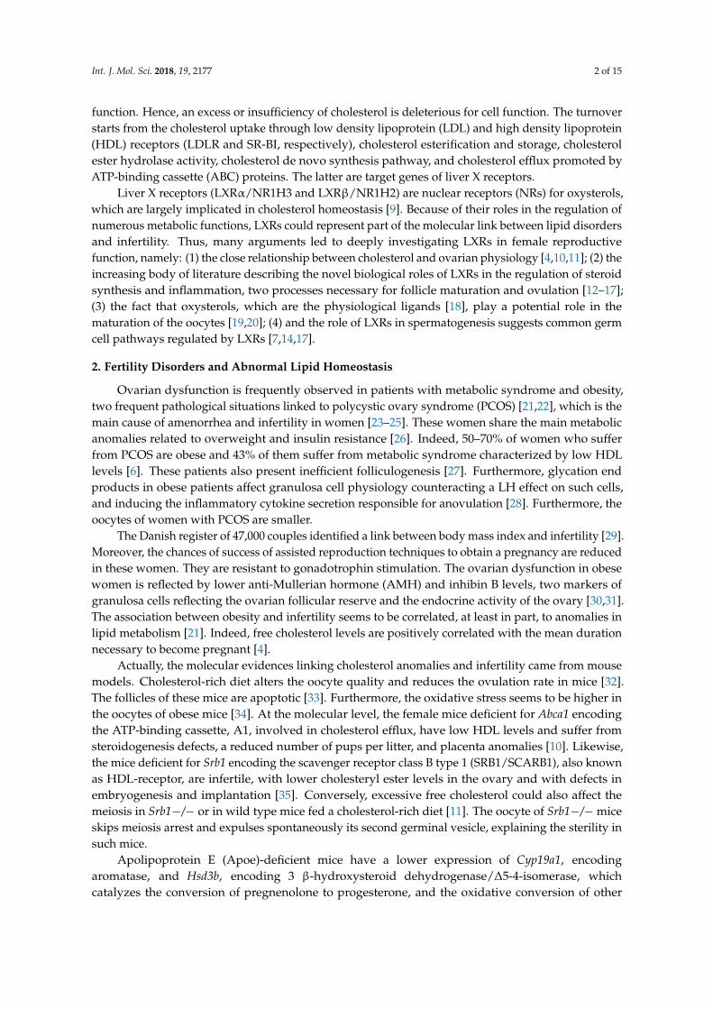
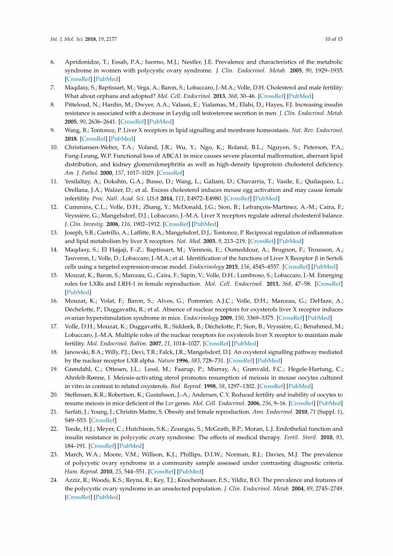
Both of the isoforms are found in the oocyte with a predominance of LXRβ [16,20]. This expressionis induced by the human chorionic gonadotropin hormone (hCG), and plays an important role insteroidogenesis in humans [43] as well as mice [16]. Follicular fluid meiosis-activating sterol (FFMAS),which can activate LXRs, increases after stimulation by gonadotropins [44,45] (Figure 1). This incrementis necessary for the oocyte to resume meiosis just before ovulation. Indeed, the luteinizing hormone(LH) surge during folliculogenesis, necessary for ovulation, induces meiosis resumption of the oocyte.This indirect effect is mediated by the FFMAS produced by granulosa cells. Thus, FFMAS stimulatesits receptor on the oocytes and LXRα was suggested as a candidate [18,40]. Furthermore, FFMASpromotes embryo implantation [46].
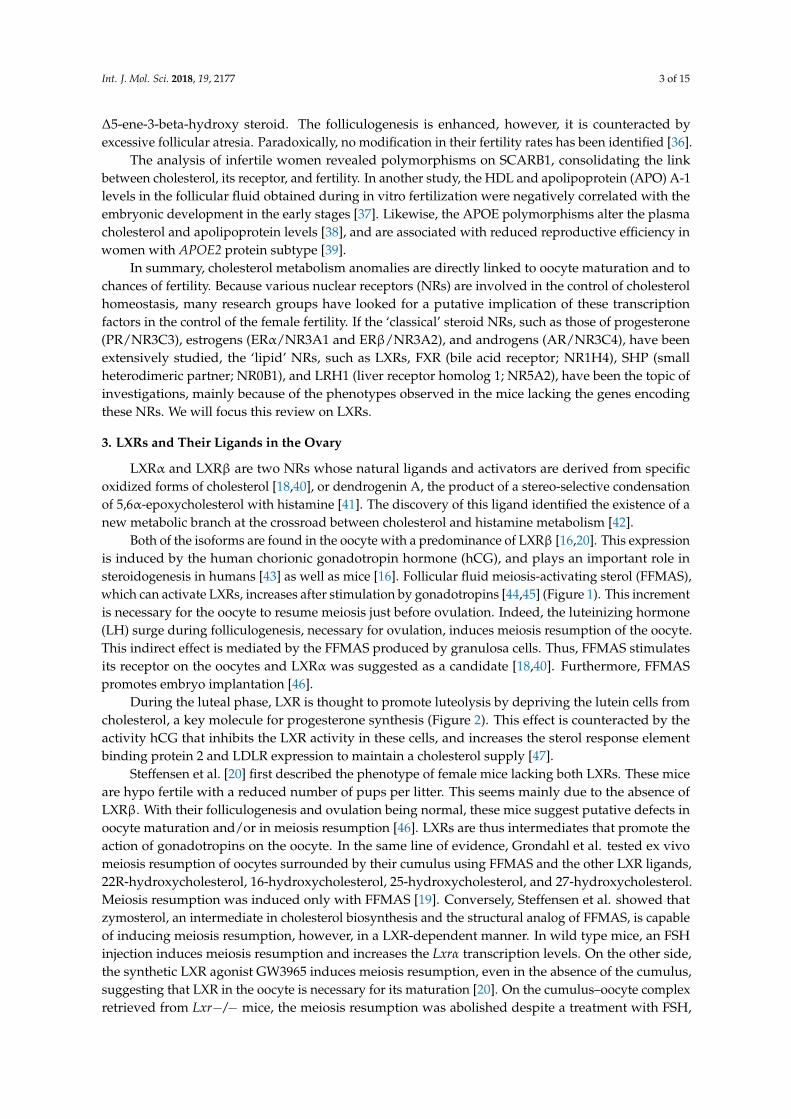
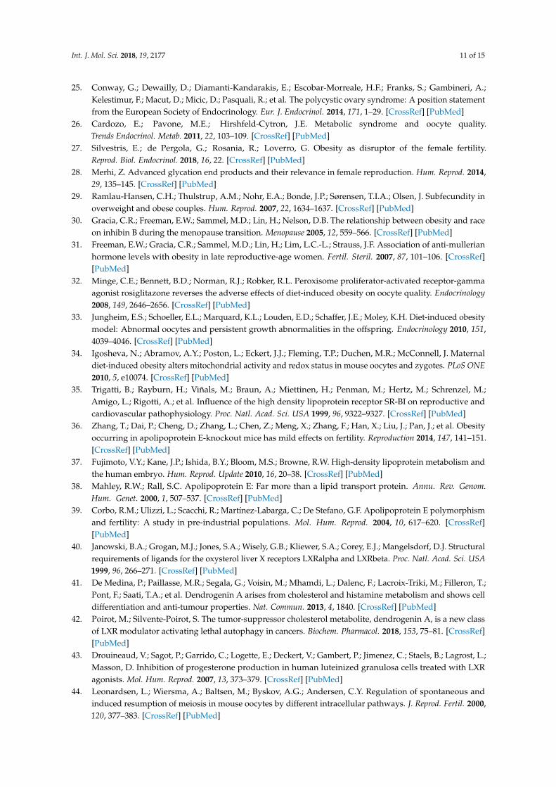
During the luteal phase, LXR is thought to promote luteolysis by depriving the lutein cells fromcholesterol, a key molecule for progesterone synthesis (Figure 2). This effect is counteracted by theactivity hCG that inhibits the LXR activity in these cells, and increases the sterol response elementbinding protein 2 and LDLR expression to maintain a cholesterol supply [47].
Steffensen et al. [20] first described the phenotype of female mice lacking both LXRs. These miceare hypo fertile with a reduced number of pups per litter. This seems mainly due to the absence ofLXRβ. With their folliculogenesis and ovulation being normal, these mice suggest putative defects inoocyte maturation and/or in meiosis resumption [46]. LXRs are thus intermediates that promote theaction of gonadotropins on the oocyte. In the same line of evidence, Grondahl et al. tested ex vivomeiosis resumption of oocytes surrounded by their cumulus using FFMAS and the other LXR ligands,22R-hydroxycholesterol, 16-hydroxycholesterol, 25-hydroxycholesterol, and 27-hydroxycholesterol.Meiosis resumption was induced only with FFMAS [19]. Conversely, Steffensen et al. showed thatzymosterol, an intermediate in cholesterol biosynthesis and the structural analog of FFMAS, is capableof inducing meiosis resumption, however, in a LXR-dependent manner. In wild type mice, an FSHinjection induces meiosis resumption and increases the Lxrα transcription levels. On the other side,the synthetic LXR agonist GW3965 induces meiosis resumption, even in the absence of the cumulus,suggesting that LXR in the oocyte is necessary for its maturation [20]. On the cumulus–oocyte complexretrieved from Lxr−/− mice, the meiosis resumption was abolished despite a treatment with FSH,
Int. J. Mol. Sci. 2018, 19, 2177 4 of 15
zymosterol, or GW3965 [20]. These findings suggest that FSH activates the FFMAS production from thecumulus, which then activates the LXR in the oocyte. Likewise, our team pointed out that deficient-LXRmice have a delayed sterility and an inefficient folliculogenesis under stimulation, with an importantnumber of atretic oocytes on retrieval, after stimulation by gonadotropins [16]. Altogether, LXRs areimportant for oocyte maturation and survival.Int. J. Mol. Sci. 2018, 19, x FOR PEER REVIEW 4 of 16
Figure 1. Role of liver X receptors (LXRs) in oocyte meiosis and in estradiol synthesis. When follicle-stimulating hormone (FSH )reaches its receptor on the granulosa cells, it increases the concentration of follicular fluid meiosis-activating sterol (FFMAS) by increasing its synthesis, a ligand of LXRα/β. This in turn induces the final steps of the oocyte meiosis. In addition, when the LXRα/β is activated by a ligand (in this figure T0901317, a synthetic ligand, purple square), they increase the production of estradiol. α/β—LXRα, or LXRβ; E2—estradiol; FFMAS—follicular fluid meiosis-activating sterol (pink square).
During the luteal phase, LXR is thought to promote luteolysis by depriving the lutein cells from cholesterol, a key molecule for progesterone synthesis (Figure 2). This effect is counteracted by the activity hCG that inhibits the LXR activity in these cells, and increases the sterol response element binding protein 2 and LDLR expression to maintain a cholesterol supply [47].
Figure 1. Role of liver X receptors (LXRs) in oocyte meiosis and in estradiol synthesis. Whenfollicle-stimulating hormone (FSH )reaches its receptor on the granulosa cells, it increases theconcentration of follicular fluid meiosis-activating sterol (FFMAS) by increasing its synthesis, a ligandof LXRα/β. This in turn induces the final steps of the oocyte meiosis. In addition, when theLXRα/β is activated by a ligand (in this figure T0901317, a synthetic ligand, purple square), theyincrease the production of estradiol. α/β—LXRα, or LXRβ; E2—estradiol; FFMAS—follicular fluidmeiosis-activating sterol (pink square).
As in the testes [14,17] and adrenal glands [12], LXRs are also involved in the regulation of steroidsynthesis in the ovary [16]. Indeed, the synthetic LXR ligand T0901317 induces the estradiol synthesisin wild type mice by activating the transcription of the steroidogenic acute regulatory protein (StAR), atransport protein that regulates cholesterol transfer within the mitochondria, which is the rate-limitingstep in the production of steroid hormones. In the ovary, LXRs also control the transcription ofcytochrome P450 side-chain cleavage (Cyp11A1), a mitochondrial enzyme that catalyzes the conversionof cholesterol to pregnenolone, the first reaction in the process of steroidogenesis.
Interestingly, LXRs seem to control any excessive estradiol synthesis during gonadotropinstimulation. Indeed, the gonadotropin stimulation of the Lxr−/− mice leads to an exaggeratedhormonal response, with an excessive estradiol secretion [16]. This effect contributes to the phenotypeof ovarian hyper stimulation syndrome (OHSS), observed in LXR-deficient mice. After a gonadotropinstimulation, the ovaries of the Lxr−/− mice harvest large hemorrhagic follicles with an exaggeratedinflammatory and ovulatory response. This signals a diagnosis of OHSS, which is characterized bythe excessive accumulation of vasoactive and angiogenic substances activating the vascular epithelialgrowth factor (VEGF) and interleukin (IL) 6 signaling, leading to a systemic inflammatory response,and variable clinical manifestations of cardiovascular collapse, septic shock, and thromboembolism inthe most severe cases [48–53].
Int. J. Mol. Sci. 2018, 19, 2177 5 of 15
Int. J. Mol. Sci. 2018, 19, x FOR PEER REVIEW 5 of 16
Figure 2. Role of LXRs in progesterone production and luteolysis. When the human chorionic gonadotropin hormone (hCG) reaches its receptor, it increases (green arrow) the concentration of cholesterol, by acting on low density lipoprotein receptor (LDLR) (uptake) and sterol response binding element (SREBP2) (de novo synthesis), and favors the production of progesterone (P4). Activation of LXRα/β by one of their bona fide ligands, produced from the cholesterol oxidation, stimulates the production of ATP-binding cassette transporter (ABC) proteins, inducing a cholesterol depletion within the cell, a decrease in progesterone synthesis, and finally, the luteolysis. hCG also inhibits LXR transcriptional activity. α/β—LXRα or LXRβ; ABCs—ATP-binding cassette transporters; hCG—human chorionic gonadotropin; P4—progesterone. LXR ligands are represented by the purple square.
Steffensen et al. [20] first described the phenotype of female mice lacking both LXRs. These mice are hypo fertile with a reduced number of pups per litter. This seems mainly due to the absence of LXRβ. With their folliculogenesis and ovulation being normal, these mice suggest putative defects in oocyte maturation and/or in meiosis resumption [46]. LXRs are thus intermediates that promote the action of gonadotropins on the oocyte. In the same line of evidence, Grondahl et al. tested ex vivo meiosis resumption of oocytes surrounded by their cumulus using FFMAS and the other LXR ligands, 22R-hydroxycholesterol, 16-hydroxycholesterol, 25-hydroxycholesterol, and 27-hydroxycholesterol. Meiosis resumption was induced only with FFMAS [19]. Conversely, Steffensen et al. showed that zymosterol, an intermediate in cholesterol biosynthesis and the structural analog of FFMAS, is capable of inducing meiosis resumption, however, in a LXR-dependent manner. In wild type mice, an FSH injection induces meiosis resumption and increases the Lxrα transcription levels. On the other side, the synthetic LXR agonist GW3965 induces meiosis resumption, even in the absence of the cumulus, suggesting that LXR in the oocyte is necessary for its maturation [20]. On the cumulus–oocyte complex retrieved from Lxr−/− mice, the meiosis resumption was abolished despite a treatment with FSH, zymosterol, or GW3965 [20]. These findings suggest that FSH activates the FFMAS production from the cumulus, which then activates the LXR in the oocyte. Likewise, our team pointed out that deficient-LXR mice have a delayed sterility and an inefficient folliculogenesis under stimulation, with an important number of atretic oocytes on retrieval, after stimulation by gonadotropins [16]. Altogether, LXRs are important for oocyte maturation and survival.
Figure 2. Role of LXRs in progesterone production and luteolysis. When the human chorionicgonadotropin hormone (hCG) reaches its receptor, it increases (green arrow) the concentration ofcholesterol, by acting on low density lipoprotein receptor (LDLR) (uptake) and sterol responsebinding element (SREBP2) (de novo synthesis), and favors the production of progesterone (P4).Activation of LXRα/β by one of their bona fide ligands, produced from the cholesterol oxidation,stimulates the production of ATP-binding cassette transporter (ABC) proteins, inducing a cholesteroldepletion within the cell, a decrease in progesterone synthesis, and finally, the luteolysis. hCG alsoinhibits LXR transcriptional activity. α/β—LXRα or LXRβ; ABCs—ATP-binding cassette transporters;hCG—human chorionic gonadotropin; P4—progesterone. LXR ligands are represented by thepurple square.
Although VEGF levels and increased IL6 signaling (through its receptor sIL-6Rα) are linkedto OHSS, no molecular driver has been identified yet. Furthermore, the literature informationare contradictory in linking these two cytokines to OHSS [54]. As LXRs have anti-inflammatoryproperties from inhibiting IL6, COX2, tumor necrosis factor TNFα [55], and the downstream of VEGFsignaling [56], they could potentially be implicated in the prevention of OHSS [13,57–59]. Altogether,the genetic models enlighten the LXRs as key factors for the endocrine and exocrine functions of theovary, and suggest that these NRs could be considered gatekeepers against an exaggerated ovarianresponse to gonadotropins. Even though clinical investigations should be performed, the exact roles ofLXRs in femal ovarian physiology is thus questioned.
4. LXRs, Uterus, and Placenta
The endometrium trophicity is influenced by estrogens and progesterone in each menstrualcycle, in to be prepared for eventual pregnancy. When implantation takes place, the placentadevelops in the endometrium permitting maternofetal exchange during pregnancy. Myometriumis necessary during labor. Obesity is related to many complications associated with pregnancy(e.g., gestational diabetes mellitus, tromboembolic problems, and hypertensive disorders such aspreeclampsia or eclampsia) [60]. Hence, obese patients and/or those with a metabolic syndromeusually suffer from dystocia, contractility defects during labor that could affect the perinatal morbidity,and mortality [61,62]. Likewise, before pregnancy, the body mass index [63,64] and its increase [65]
Int. J. Mol. Sci. 2018, 19, 2177 6 of 15
during pregnancy have been associated with a higher risk for caesarean delivery at the term ofpregnancy, for failure to progress in the labor.
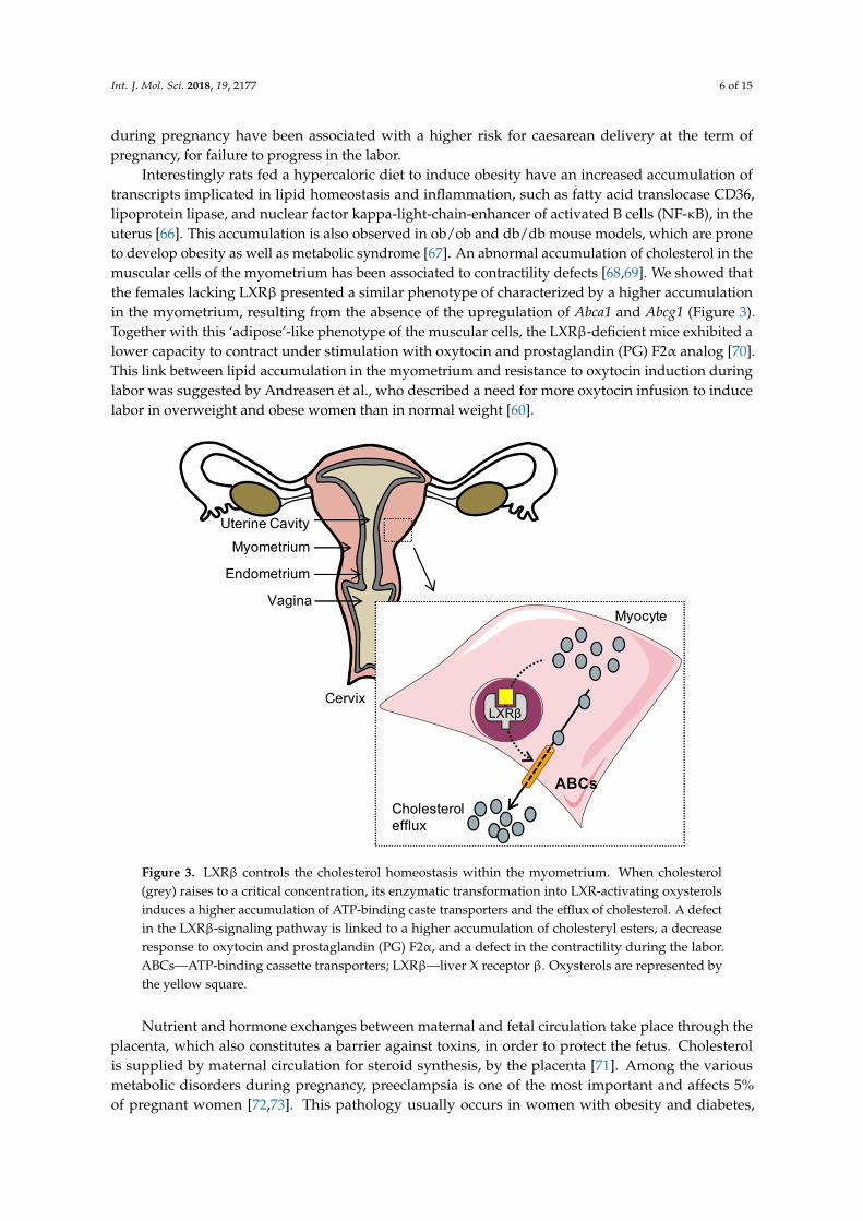
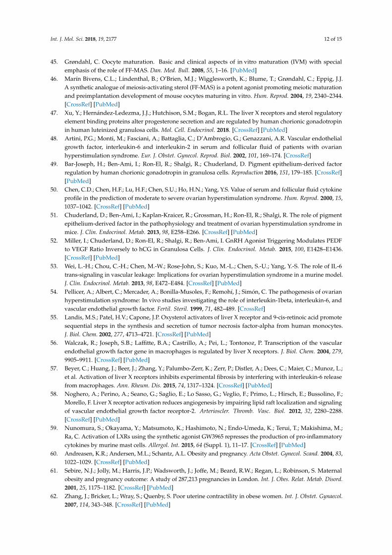
Interestingly rats fed a hypercaloric diet to induce obesity have an increased accumulation oftranscripts implicated in lipid homeostasis and inflammation, such as fatty acid translocase CD36,lipoprotein lipase, and nuclear factor kappa-light-chain-enhancer of activated B cells (NF-κB), in theuterus [66]. This accumulation is also observed in ob/ob and db/db mouse models, which are proneto develop obesity as well as metabolic syndrome [67]. An abnormal accumulation of cholesterol in themuscular cells of the myometrium has been associated to contractility defects [68,69]. We showed thatthe females lacking LXRβ presented a similar phenotype of characterized by a higher accumulationin the myometrium, resulting from the absence of the upregulation of Abca1 and Abcg1 (Figure 3).Together with this ‘adipose’-like phenotype of the muscular cells, the LXRβ-deficient mice exhibited alower capacity to contract under stimulation with oxytocin and prostaglandin (PG) F2α analog [70].This link between lipid accumulation in the myometrium and resistance to oxytocin induction duringlabor was suggested by Andreasen et al., who described a need for more oxytocin infusion to inducelabor in overweight and obese women than in normal weight [60].
Int. J. Mol. Sci. 2018, 19, x FOR PEER REVIEW 7 of 16
Figure 3. LXRβ controls the cholesterol homeostasis within the myometrium. When cholesterol (grey) raises to a critical concentration, its enzymatic transformation into LXR-activating oxysterols induces a higher accumulation of ATP-binding caste transporters and the efflux of cholesterol. A defect in the LXRβ-signaling pathway is linked to a higher accumulation of cholesteryl esters, a decrease response to oxytocin and prostaglandin (PG) F2α, and a defect in the contractility during the labor. ABCs—ATP-binding cassette transporters; LXRβ—liver X receptor β. Oxysterols are represented by the yellow square.
Nutrient and hormone exchanges between maternal and fetal circulation take place through the placenta, which also constitutes a barrier against toxins, in order to protect the fetus. Cholesterol is supplied by maternal circulation for steroid synthesis, by the placenta [71]. Among the various metabolic disorders during pregnancy, preeclampsia is one of the most important and affects 5% of pregnant women [72,73]. This pathology usually occurs in women with obesity and diabetes, and is characterized by hypertension and proteinuria [74,75]. The hallmark of this syndrome is an insufficient trophoblast invasion with contractile spiral uterine arteries, leading to acute atherosis (atherosclerosis-like), vasoconstriction, and hypertension [75,76]. Elevated oxidized LDL (rich in sterols) levels reduce vessel invasion and produce preeclampsia [77]. Early and progressive LXRα and LXRβ expression in the placenta (seven days post-coitum in mice, six weeks of pregnancy in women) participate to maintain the cholesterol available for trophoblast cells, and modulates arterial invasion [78,79]. Many oxysterols, especially 25-hydroxycholesterol, increase in the placenta during pregnancy [80]. Furthermore, LXRs activate the ABC protein expression in trophoblast cells for eliminating excessive cholesterol and toxic sterols [78,81]. LXRβ also controls trophoblast invasion [77,82,83]. Interestingly, the LXRα and ABCA1 genes are overexpressed in the placenta tissue of the women that suffered from preeclampsia [79], while another study demonstrated a reduction in LXRβ in these patients [84]. Endoglin/CD105 is membrane receptor that induces endothelial relaxation [78,85,86]. The production of soluble Endoglin/CD105 by the membrane metalloproteinase-14 induces the squelching of TGF-β1, endothelial dysfunction, and impaired relaxation, altogether, preeclampsia [78,85,86]. We pointed out that Endoglin/CD105 is an atypical LXR target gene, which could explain how LXR could reduce the trophoblast invasion and the risk of preeclampsia [78,85,86]. Interestingly, a single
Figure 3. LXRβ controls the cholesterol homeostasis within the myometrium. When cholesterol(grey) raises to a critical concentration, its enzymatic transformation into LXR-activating oxysterolsinduces a higher accumulation of ATP-binding caste transporters and the efflux of cholesterol. A defectin the LXRβ-signaling pathway is linked to a higher accumulation of cholesteryl esters, a decreaseresponse to oxytocin and prostaglandin (PG) F2α, and a defect in the contractility during the labor.ABCs—ATP-binding cassette transporters; LXRβ—liver X receptor β. Oxysterols are represented bythe yellow square.
Nutrient and hormone exchanges between maternal and fetal circulation take place through theplacenta, which also constitutes a barrier against toxins, in order to protect the fetus. Cholesterolis supplied by maternal circulation for steroid synthesis, by the placenta [71]. Among the variousmetabolic disorders during pregnancy, preeclampsia is one of the most important and affects 5%of pregnant women [72,73]. This pathology usually occurs in women with obesity and diabetes,
Int. J. Mol. Sci. 2018, 19, 2177 7 of 15
and is characterized by hypertension and proteinuria [74,75]. The hallmark of this syndrome is aninsufficient trophoblast invasion with contractile spiral uterine arteries, leading to acute atherosis(atherosclerosis-like), vasoconstriction, and hypertension [75,76]. Elevated oxidized LDL (rich insterols) levels reduce vessel invasion and produce preeclampsia [77]. Early and progressive LXRαand LXRβ expression in the placenta (seven days post-coitum in mice, six weeks of pregnancyin women) participate to maintain the cholesterol available for trophoblast cells, and modulatesarterial invasion [78,79]. Many oxysterols, especially 25-hydroxycholesterol, increase in the placentaduring pregnancy [80]. Furthermore, LXRs activate the ABC protein expression in trophoblastcells for eliminating excessive cholesterol and toxic sterols [78,81]. LXRβ also controls trophoblastinvasion [77,82,83]. Interestingly, the LXRα and ABCA1 genes are overexpressed in the placentatissue of the women that suffered from preeclampsia [79], while another study demonstrated areduction in LXRβ in these patients [84]. Endoglin/CD105 is membrane receptor that inducesendothelial relaxation [78,85,86]. The production of soluble Endoglin/CD105 by the membranemetalloproteinase-14 induces the squelching of TGF-β1, endothelial dysfunction, and impairedrelaxation, altogether, preeclampsia [78,85,86]. We pointed out that Endoglin/CD105 is an atypicalLXR target gene, which could explain how LXR could reduce the trophoblast invasion and the riskof preeclampsia [78,85,86]. Interestingly, a single nucleotide polymorphism within the sequencesencoding LXRβ has been significantly associated with the risk of preeclampsia in a study of over 155women that presented this disorder [87].
5. LXRs, Modulation of Estrogen Activity, and Breast Cancer
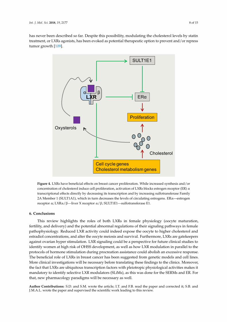
Even though breast cancer cannot be defined as a woman reproductive disease per se, it has beendirectly associated to the circulating levels of estrogens and the levels and/or mutations of ERα by theirroles in the growth and proliferation of epithelial cells. Hence, pharmacological management partlytargets the estrogen pathway by using selective estrogen receptor modulators (SERM; e.g., tamoxifenor raloxifen) or degraders (SERD; e.g., fulvestran), LHRH analogs, and/or aromatase inhibitors [88,89].Nevertheless, these therapies induce menopausal symptoms and some breast cancers are negative forERα [90]. A significant correlation between ERα positive breast cancer/obesity/metabolic syndromefrom one side, and statin, an inhibitor of 3-hydroxy-3-methylglutaryl CoA reductase and cholesterolde novo synthesis, from the other side, have been revealed by various studies [91–94]. Beside theexcessive aromatization of estrogen by the adiposity, and the excessive production of insulin-likegrowth factors and inflammatory cytokines [95], LXRs could represent a molecular link. Indeed, LXRsand ERα exert reciprocal effects on each other. As mentioned above, LXRs are gatekeepers againstexcessive E2 production after hormonal stimulation. LXR activation reduces ERα expression [96].Furthermore, LXRs decrease the free estrogen level by increased sulfotransferase activity (EST orSULT1E1) [97]. On the other side, E2 decreases the LXR mRNA expression [98–101]. The ERα in theliver is recruited to the SREBP1c promoter, through direct binding to LXR, and prevents coactivatorrecruitment to LXR in an estrogen dependent manner [100].
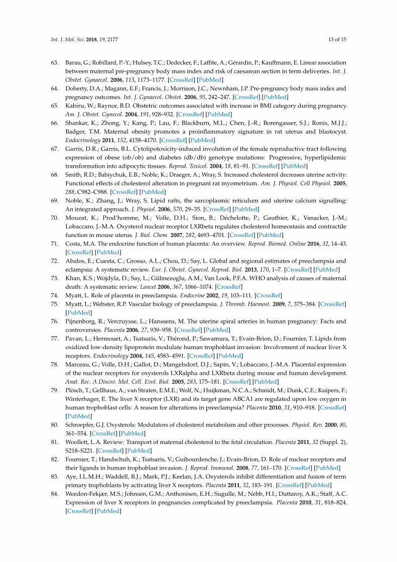
LXRs were also identified to have an anti-proliferative effect in both ER positive and ERnegative cells lines [96,102]. They could block cell proliferation-invasion by downregulating thecell cycle and cholesterol metabolism genes. The hypothesis of the LXR protective effect, through itsanti-proliferative, pro-apoptotic role is based on cholesterol deprivation, as cholesterol is indispensablefor cell proliferation [103–105]. Furthermore, the LXR activation reduced different breast cancer celllines in vitro, by the suppression of cyclin proteins, ERα, and increased P53 protein levels [96]. LXRmanipulation could help in estrogen deprivation, necessary for breast cancer treatment [106]. Themolecular mechanism seems to be linked to LXRβ, which inhibits the proliferation of human breastcancer cells through the PI3K–Akt pathway [107] or an E2F-mediated mechanism [102,107]. Altogether,this thus indicates a positive impact of LXRs for protection against the development of breast cancer(Figure 4), even though 27-hydroxycholesterol, which is a LXR-ligand, acts as ERα ligand SERM [108]in breast cancer, cannot exclude a negative role of LXRs when 27-hydroxycholesterol increases, which
Int. J. Mol. Sci. 2018, 19, 2177 8 of 15
has never been described so far. Despite this possibility, modulating the cholesterol levels by statintreatment, or LXRs agonists, has been evoked as potential therapeutic option to prevent and/or represstumor growth [109].Int. J. Mol. Sci. 2018, 19, x FOR PEER REVIEW 9 of 16
Figure 4. LXRs have beneficial effects on breast cancer proliferation. While increased synthesis and/or concentration of cholesterol induce cell proliferation, activation of LXRs blocks estrogen receptor (ER) α transcriptional effects directly by decreasing its transcription and by increasing sulfotransferase Family 2A Member 1 (SULT1A1), which in turn decreases the levels of circulating estrogens. ERα—estrogen receptor α; LXRα/β—liver X receptor α/β; SULT1E1—sulfotransferase E1.
6. Conclusions
This review highlights the roles of both LXRs in female physiology (oocyte maturation, fertility, and delivery) and the potential abnormal regulations of their signaling pathways in female pathophysiology. Reduced LXR activity could indeed expose the oocyte to higher cholesterol and estradiol concentrations, and alter the oocyte meiosis and survival. Furthermore, LXRs are gatekeepers against ovarian hyper stimulation. LXR signaling could be a perspective for future clinical studies to identify women at high risk of OHSS development, as well as how LXR modulation in parallel to the protocols of hormone stimulation during procreation assistance could abolish an excessive response. The beneficial role of LXRs in breast cancer has been suggested from genetic models and cell lines. More clinical investigations will be necessary before translating these findings to the clinics. Moreover, the fact that LXRs are ubiquitous transcription factors with pleiotropic physiological activities makes it mandatory to identify selective LXR modulators (SLiMs), as this was done for the SERMs and ER. For that, new pharmacology paradigms will be necessary as well.
Author Contributions: S.D. and S.M. wrote the article; I.T. and F.B. read the paper and corrected it; S.B. and J.M.A.L. wrote the paper and supervised the scientific work leading to this review.
Funding: Région Auvergne-Rhône-Alpes, Fond Européen de Développement Régional (FEDER), Centre Hospitalier Universitaire Clermont-Ferrand, for SB-JMAL lab. The funders had no role in data collection and analysis, decision to publish, or preparation of the manuscript.
Acknowledgments: We would like to thank the Baron & Lobaccaro’s lab for helpful discussion.
Conflicts of Interest: The authors declare no conflict of interest.
Figure 4. LXRs have beneficial effects on breast cancer proliferation. While increased synthesis and/orconcentration of cholesterol induce cell proliferation, activation of LXRs blocks estrogen receptor (ER) αtranscriptional effects directly by decreasing its transcription and by increasing sulfotransferase Family2A Member 1 (SULT1A1), which in turn decreases the levels of circulating estrogens. ERα—estrogenreceptor α; LXRα/β—liver X receptor α/β; SULT1E1—sulfotransferase E1.
6. Conclusions
This review highlights the roles of both LXRs in female physiology (oocyte maturation,fertility, and delivery) and the potential abnormal regulations of their signaling pathways in femalepathophysiology. Reduced LXR activity could indeed expose the oocyte to higher cholesterol andestradiol concentrations, and alter the oocyte meiosis and survival. Furthermore, LXRs are gatekeepersagainst ovarian hyper stimulation. LXR signaling could be a perspective for future clinical studies toidentify women at high risk of OHSS development, as well as how LXR modulation in parallel to theprotocols of hormone stimulation during procreation assistance could abolish an excessive response.The beneficial role of LXRs in breast cancer has been suggested from genetic models and cell lines.More clinical investigations will be necessary before translating these findings to the clinics. Moreover,the fact that LXRs are ubiquitous transcription factors with pleiotropic physiological activities makes itmandatory to identify selective LXR modulators (SLiMs), as this was done for the SERMs and ER. Forthat, new pharmacology paradigms will be necessary as well.
Author Contributions: S.D. and S.M. wrote the article; I.T. and F.B. read the paper and corrected it; S.B. andJ.M.A.L. wrote the paper and supervised the scientific work leading to this review.
Int. J. Mol. Sci. 2018, 19, 2177 9 of 15
Funding: Région Auvergne-Rhône-Alpes, Fond Européen de Développement Régional (FEDER), CentreHospitalier Universitaire Clermont-Ferrand, for SB-JMAL lab. The funders had no role in data collection andanalysis, decision to publish, or preparation of the manuscript.
Acknowledgments: We would like to thank the Baron & Lobaccaro’s lab for helpful discussion.
Conflicts of Interest: The authors declare no conflict of interest.
Abbreviations
ABCA1/G1 ATP-binding cassetteA1/G1AMH anti-Mullerian hormoneAPO apolipoproteinCOX cyclooxygenaseCyp11A1 cytochrome P450 side-chain cleavageCyp19a1 cytochrome P450 aromataseER estrogen receptorEST estrogen sulfotransferaseFFMAS follicular fluid meiosis-activating sterolhCG human chorionic gonadotropinHDL high density lipoproteinHsd3b 3 β-hydroxysteroid dehydrogenase/∆5-4-isomeraseIL6 interleukin 6iNOS inducible nitric oxide synthaseJAK/STAT janus kinase/signal transducer and activator of transcription proteinLDL low density lipoproteinLDLR LDL receptor; LXR, liver X receptorLxr−/− mice LXR-deficient miceNR nuclear receptorOHSS ovarian hyper stimulation syndromePCOS polycystic ovary syndromePI3K phosphoinositide 3-kinaseSCARB1 scavenger receptor class B typeSREBP sterol response element binding proteinSLiMs selective liver X receptor modulatorsStAR steroidogenic acute regulatory proteinSULT2A1 Sulfotransferase Family 2A Member 1VEGF vascular endothelial growth factor27OHC 27-hydroxycholesterol
References
1. Petraglia, F.; Serour, G.I.; Chapron, C. The changing prevalence of infertility. Int. J. Gynaecol. Obstet. 2013,123, S4–S8. [CrossRef] [PubMed]
2. Kasturi, S.S.; Tannir, J.; Brannigan, R.E. The metabolic syndrome and male infertility. J. Androl. 2008, 29,251–259. [CrossRef] [PubMed]
3. Norman, R.J.; Noakes, M.; Wu, R.; Davies, M.J.; Moran, L.; Wang, J.X. Improving reproductive performancein overweight/obese women with effective weight management. Hum. Reprod. Update 2004, 10, 267–280.[CrossRef] [PubMed]
4. Schisterman, E.F.; Mumford, S.L.; Browne, R.W.; Barr, D.B.; Chen, Z.; Louis, G.M.B. Lipid concentrations andcouple fecundity: The LIFE study. J. Clin. Endocrinol. Metab. 2014, 99, 2786–2794. [CrossRef] [PubMed]
5. Eisenberg, M.L.; Kim, S.; Chen, Z.; Sundaram, R.; Schisterman, E.F.; Buck Louis, G.M. The relationshipbetween male BMI and waist circumference on semen quality: Data from the LIFE study. Hum. Reprod. 2014,29, 193–200. [CrossRef] [PubMed]
Int. J. Mol. Sci. 2018, 19, 2177 10 of 15
6. Apridonidze, T.; Essah, P.A.; Iuorno, M.J.; Nestler, J.E. Prevalence and characteristics of the metabolicsyndrome in women with polycystic ovary syndrome. J. Clin. Endocrinol. Metab. 2005, 90, 1929–1935.[CrossRef] [PubMed]
7. Maqdasy, S.; Baptissart, M.; Vega, A.; Baron, S.; Lobaccaro, J.-M.A.; Volle, D.H. Cholesterol and male fertility:What about orphans and adopted? Mol. Cell. Endocrinol. 2013, 368, 30–46. [CrossRef] [PubMed]
8. Pitteloud, N.; Hardin, M.; Dwyer, A.A.; Valassi, E.; Yialamas, M.; Elahi, D.; Hayes, F.J. Increasing insulinresistance is associated with a decrease in Leydig cell testosterone secretion in men. J. Clin. Endocrinol. Metab.2005, 90, 2636–2641. [CrossRef] [PubMed]
9. Wang, B.; Tontonoz, P. Liver X receptors in lipid signalling and membrane homeostasis. Nat. Rev. Endocrinol.2018. [CrossRef] [PubMed]
10. Christiansen-Weber, T.A.; Voland, J.R.; Wu, Y.; Ngo, K.; Roland, B.L.; Nguyen, S.; Peterson, P.A.;Fung-Leung, W.P. Functional loss of ABCA1 in mice causes severe placental malformation, aberrant lipiddistribution, and kidney glomerulonephritis as well as high-density lipoprotein cholesterol deficiency.Am. J. Pathol. 2000, 157, 1017–1029. [CrossRef]
11. Yesilaltay, A.; Dokshin, G.A.; Busso, D.; Wang, L.; Galiani, D.; Chavarria, T.; Vasile, E.; Quilaqueo, L.;Orellana, J.A.; Walzer, D.; et al. Excess cholesterol induces mouse egg activation and may cause femaleinfertility. Proc. Natl. Acad. Sci. USA 2014, 111, E4972–E4980. [CrossRef] [PubMed]
12. Cummins, C.L.; Volle, D.H.; Zhang, Y.; McDonald, J.G.; Sion, B.; Lefrançois-Martinez, A.-M.; Caira, F.;Veyssière, G.; Mangelsdorf, D.J.; Lobaccaro, J.-M.A. Liver X receptors regulate adrenal cholesterol balance.J. Clin. Investig. 2006, 116, 1902–1912. [CrossRef] [PubMed]
13. Joseph, S.B.; Castrillo, A.; Laffitte, B.A.; Mangelsdorf, D.J.; Tontonoz, P. Reciprocal regulation of inflammationand lipid metabolism by liver X receptors. Nat. Med. 2003, 9, 213–219. [CrossRef] [PubMed]
14. Maqdasy, S.; El Hajjaji, F.-Z.; Baptissart, M.; Viennois, E.; Oumeddour, A.; Brugnon, F.; Trousson, A.;Tauveron, I.; Volle, D.; Lobaccaro, J.-M.A.; et al. Identification of the functions of Liver X Receptor β in Sertolicells using a targeted expression-rescue model. Endocrinology 2015, 156, 4545–4557. [CrossRef] [PubMed]
15. Mouzat, K.; Baron, S.; Marceau, G.; Caira, F.; Sapin, V.; Volle, D.H.; Lumbroso, S.; Lobaccaro, J.-M. Emergingroles for LXRs and LRH-1 in female reproduction. Mol. Cell. Endocrinol. 2013, 368, 47–58. [CrossRef][PubMed]
16. Mouzat, K.; Volat, F.; Baron, S.; Alves, G.; Pommier, A.J.C.; Volle, D.H.; Marceau, G.; DeHaze, A.;Déchelotte, P.; Duggavathi, R.; et al. Absence of nuclear receptors for oxysterols liver X receptor inducesovarian hyperstimulation syndrome in mice. Endocrinology 2009, 150, 3369–3375. [CrossRef] [PubMed]
17. Volle, D.H.; Mouzat, K.; Duggavathi, R.; Siddeek, B.; Déchelotte, P.; Sion, B.; Veyssière, G.; Benahmed, M.;Lobaccaro, J.-M.A. Multiple roles of the nuclear receptors for oxysterols liver X receptor to maintain malefertility. Mol. Endocrinol. Baltim. 2007, 21, 1014–1027. [CrossRef] [PubMed]
18. Janowski, B.A.; Willy, P.J.; Devi, T.R.; Falck, J.R.; Mangelsdorf, D.J. An oxysterol signalling pathway mediatedby the nuclear receptor LXR alpha. Nature 1996, 383, 728–731. [CrossRef] [PubMed]
19. Grøndahl, C.; Ottesen, J.L.; Lessl, M.; Faarup, P.; Murray, A.; Grønvald, F.C.; Hegele-Hartung, C.;Ahnfelt-Rønne, I. Meiosis-activating sterol promotes resumption of meiosis in mouse oocytes culturedin vitro in contrast to related oxysterols. Biol. Reprod. 1998, 58, 1297–1302. [CrossRef] [PubMed]
20. Steffensen, K.R.; Robertson, K.; Gustafsson, J.-A.; Andersen, C.Y. Reduced fertility and inability of oocytes toresume meiosis in mice deficient of the Lxr genes. Mol. Cell. Endocrinol. 2006, 256, 9–16. [CrossRef] [PubMed]
21. Sarfati, J.; Young, J.; Christin-Maitre, S. Obesity and female reproduction. Ann. Endocrinol. 2010, 71 (Suppl. 1),S49–S53. [CrossRef]
22. Teede, H.J.; Meyer, C.; Hutchison, S.K.; Zoungas, S.; McGrath, B.P.; Moran, L.J. Endothelial function andinsulin resistance in polycystic ovary syndrome: The effects of medical therapy. Fertil. Steril. 2010, 93,184–191. [CrossRef] [PubMed]
23. March, W.A.; Moore, V.M.; Willson, K.J.; Phillips, D.I.W.; Norman, R.J.; Davies, M.J. The prevalenceof polycystic ovary syndrome in a community sample assessed under contrasting diagnostic criteria.Hum. Reprod. 2010, 25, 544–551. [CrossRef] [PubMed]
24. Azziz, R.; Woods, K.S.; Reyna, R.; Key, T.J.; Knochenhauer, E.S.; Yildiz, B.O. The prevalence and features ofthe polycystic ovary syndrome in an unselected population. J. Clin. Endocrinol. Metab. 2004, 89, 2745–2749.[CrossRef] [PubMed]
Int. J. Mol. Sci. 2018, 19, 2177 11 of 15
25. Conway, G.; Dewailly, D.; Diamanti-Kandarakis, E.; Escobar-Morreale, H.F.; Franks, S.; Gambineri, A.;Kelestimur, F.; Macut, D.; Micic, D.; Pasquali, R.; et al. The polycystic ovary syndrome: A position statementfrom the European Society of Endocrinology. Eur. J. Endocrinol. 2014, 171, 1–29. [CrossRef] [PubMed]
26. Cardozo, E.; Pavone, M.E.; Hirshfeld-Cytron, J.E. Metabolic syndrome and oocyte quality.Trends Endocrinol. Metab. 2011, 22, 103–109. [CrossRef] [PubMed]
27. Silvestris, E.; de Pergola, G.; Rosania, R.; Loverro, G. Obesity as disruptor of the female fertility.Reprod. Biol. Endocrinol. 2018, 16, 22. [CrossRef] [PubMed]
28. Merhi, Z. Advanced glycation end products and their relevance in female reproduction. Hum. Reprod. 2014,29, 135–145. [CrossRef] [PubMed]
29. Ramlau-Hansen, C.H.; Thulstrup, A.M.; Nohr, E.A.; Bonde, J.P.; Sørensen, T.I.A.; Olsen, J. Subfecundity inoverweight and obese couples. Hum. Reprod. 2007, 22, 1634–1637. [CrossRef] [PubMed]
30. Gracia, C.R.; Freeman, E.W.; Sammel, M.D.; Lin, H.; Nelson, D.B. The relationship between obesity and raceon inhibin B during the menopause transition. Menopause 2005, 12, 559–566. [CrossRef] [PubMed]
31. Freeman, E.W.; Gracia, C.R.; Sammel, M.D.; Lin, H.; Lim, L.C.-L.; Strauss, J.F. Association of anti-mullerianhormone levels with obesity in late reproductive-age women. Fertil. Steril. 2007, 87, 101–106. [CrossRef][PubMed]
32. Minge, C.E.; Bennett, B.D.; Norman, R.J.; Robker, R.L. Peroxisome proliferator-activated receptor-gammaagonist rosiglitazone reverses the adverse effects of diet-induced obesity on oocyte quality. Endocrinology2008, 149, 2646–2656. [CrossRef] [PubMed]
33. Jungheim, E.S.; Schoeller, E.L.; Marquard, K.L.; Louden, E.D.; Schaffer, J.E.; Moley, K.H. Diet-induced obesitymodel: Abnormal oocytes and persistent growth abnormalities in the offspring. Endocrinology 2010, 151,4039–4046. [CrossRef] [PubMed]
34. Igosheva, N.; Abramov, A.Y.; Poston, L.; Eckert, J.J.; Fleming, T.P.; Duchen, M.R.; McConnell, J. Maternaldiet-induced obesity alters mitochondrial activity and redox status in mouse oocytes and zygotes. PLoS ONE2010, 5, e10074. [CrossRef] [PubMed]
35. Trigatti, B.; Rayburn, H.; Viñals, M.; Braun, A.; Miettinen, H.; Penman, M.; Hertz, M.; Schrenzel, M.;Amigo, L.; Rigotti, A.; et al. Influence of the high density lipoprotein receptor SR-BI on reproductive andcardiovascular pathophysiology. Proc. Natl. Acad. Sci. USA 1999, 96, 9322–9327. [CrossRef] [PubMed]
36. Zhang, T.; Dai, P.; Cheng, D.; Zhang, L.; Chen, Z.; Meng, X.; Zhang, F.; Han, X.; Liu, J.; Pan, J.; et al. Obesityoccurring in apolipoprotein E-knockout mice has mild effects on fertility. Reproduction 2014, 147, 141–151.[CrossRef] [PubMed]
37. Fujimoto, V.Y.; Kane, J.P.; Ishida, B.Y.; Bloom, M.S.; Browne, R.W. High-density lipoprotein metabolism andthe human embryo. Hum. Reprod. Update 2010, 16, 20–38. [CrossRef] [PubMed]
38. Mahley, R.W.; Rall, S.C. Apolipoprotein E: Far more than a lipid transport protein. Annu. Rev. Genom.Hum. Genet. 2000, 1, 507–537. [CrossRef] [PubMed]
39. Corbo, R.M.; Ulizzi, L.; Scacchi, R.; Martínez-Labarga, C.; De Stefano, G.F. Apolipoprotein E polymorphismand fertility: A study in pre-industrial populations. Mol. Hum. Reprod. 2004, 10, 617–620. [CrossRef][PubMed]
40. Janowski, B.A.; Grogan, M.J.; Jones, S.A.; Wisely, G.B.; Kliewer, S.A.; Corey, E.J.; Mangelsdorf, D.J. Structuralrequirements of ligands for the oxysterol liver X receptors LXRalpha and LXRbeta. Proc. Natl. Acad. Sci. USA1999, 96, 266–271. [CrossRef] [PubMed]
41. De Medina, P.; Paillasse, M.R.; Segala, G.; Voisin, M.; Mhamdi, L.; Dalenc, F.; Lacroix-Triki, M.; Filleron, T.;Pont, F.; Saati, T.A.; et al. Dendrogenin A arises from cholesterol and histamine metabolism and shows celldifferentiation and anti-tumour properties. Nat. Commun. 2013, 4, 1840. [CrossRef] [PubMed]
42. Poirot, M.; Silvente-Poirot, S. The tumor-suppressor cholesterol metabolite, dendrogenin A, is a new classof LXR modulator activating lethal autophagy in cancers. Biochem. Pharmacol. 2018, 153, 75–81. [CrossRef][PubMed]
43. Drouineaud, V.; Sagot, P.; Garrido, C.; Logette, E.; Deckert, V.; Gambert, P.; Jimenez, C.; Staels, B.; Lagrost, L.;Masson, D. Inhibition of progesterone production in human luteinized granulosa cells treated with LXRagonists. Mol. Hum. Reprod. 2007, 13, 373–379. [CrossRef] [PubMed]
44. Leonardsen, L.; Wiersma, A.; Baltsen, M.; Byskov, A.G.; Andersen, C.Y. Regulation of spontaneous andinduced resumption of meiosis in mouse oocytes by different intracellular pathways. J. Reprod. Fertil. 2000,120, 377–383. [CrossRef] [PubMed]
Int. J. Mol. Sci. 2018, 19, 2177 12 of 15
45. Grøndahl, C. Oocyte maturation. Basic and clinical aspects of in vitro maturation (IVM) with specialemphasis of the role of FF-MAS. Dan. Med. Bull. 2008, 55, 1–16. [PubMed]
46. Marín Bivens, C.L.; Lindenthal, B.; O’Brien, M.J.; Wigglesworth, K.; Blume, T.; Grøndahl, C.; Eppig, J.J.A synthetic analogue of meiosis-activating sterol (FF-MAS) is a potent agonist promoting meiotic maturationand preimplantation development of mouse oocytes maturing in vitro. Hum. Reprod. 2004, 19, 2340–2344.[CrossRef] [PubMed]
47. Xu, Y.; Hernández-Ledezma, J.J.; Hutchison, S.M.; Bogan, R.L. The liver X receptors and sterol regulatoryelement binding proteins alter progesterone secretion and are regulated by human chorionic gonadotropinin human luteinized granulosa cells. Mol. Cell. Endocrinol. 2018. [CrossRef] [PubMed]
48. Artini, P.G.; Monti, M.; Fasciani, A.; Battaglia, C.; D’Ambrogio, G.; Genazzani, A.R. Vascular endothelialgrowth factor, interleukin-6 and interleukin-2 in serum and follicular fluid of patients with ovarianhyperstimulation syndrome. Eur. J. Obstet. Gynecol. Reprod. Biol. 2002, 101, 169–174. [CrossRef]
49. Bar-Joseph, H.; Ben-Ami, I.; Ron-El, R.; Shalgi, R.; Chuderland, D. Pigment epithelium-derived factorregulation by human chorionic gonadotropin in granulosa cells. Reproduction 2016, 151, 179–185. [CrossRef][PubMed]
50. Chen, C.D.; Chen, H.F.; Lu, H.F.; Chen, S.U.; Ho, H.N.; Yang, Y.S. Value of serum and follicular fluid cytokineprofile in the prediction of moderate to severe ovarian hyperstimulation syndrome. Hum. Reprod. 2000, 15,1037–1042. [CrossRef] [PubMed]
51. Chuderland, D.; Ben-Ami, I.; Kaplan-Kraicer, R.; Grossman, H.; Ron-El, R.; Shalgi, R. The role of pigmentepithelium-derived factor in the pathophysiology and treatment of ovarian hyperstimulation syndrome inmice. J. Clin. Endocrinol. Metab. 2013, 98, E258–E266. [CrossRef] [PubMed]
52. Miller, I.; Chuderland, D.; Ron-El, R.; Shalgi, R.; Ben-Ami, I. GnRH Agonist Triggering Modulates PEDFto VEGF Ratio Inversely to hCG in Granulosa Cells. J. Clin. Endocrinol. Metab. 2015, 100, E1428–E1436.[CrossRef] [PubMed]
53. Wei, L.-H.; Chou, C.-H.; Chen, M.-W.; Rose-John, S.; Kuo, M.-L.; Chen, S.-U.; Yang, Y.-S. The role of IL-6trans-signaling in vascular leakage: Implications for ovarian hyperstimulation syndrome in a murine model.J. Clin. Endocrinol. Metab. 2013, 98, E472–E484. [CrossRef] [PubMed]
54. Pellicer, A.; Albert, C.; Mercader, A.; Bonilla-Musoles, F.; Remohí, J.; Simón, C. The pathogenesis of ovarianhyperstimulation syndrome: In vivo studies investigating the role of interleukin-1beta, interleukin-6, andvascular endothelial growth factor. Fertil. Steril. 1999, 71, 482–489. [CrossRef]
55. Landis, M.S.; Patel, H.V.; Capone, J.P. Oxysterol activators of liver X receptor and 9-cis-retinoic acid promotesequential steps in the synthesis and secretion of tumor necrosis factor-alpha from human monocytes.J. Biol. Chem. 2002, 277, 4713–4721. [CrossRef] [PubMed]
56. Walczak, R.; Joseph, S.B.; Laffitte, B.A.; Castrillo, A.; Pei, L.; Tontonoz, P. Transcription of the vascularendothelial growth factor gene in macrophages is regulated by liver X receptors. J. Biol. Chem. 2004, 279,9905–9911. [CrossRef] [PubMed]
57. Beyer, C.; Huang, J.; Beer, J.; Zhang, Y.; Palumbo-Zerr, K.; Zerr, P.; Distler, A.; Dees, C.; Maier, C.; Munoz, L.;et al. Activation of liver X receptors inhibits experimental fibrosis by interfering with interleukin-6 releasefrom macrophages. Ann. Rheum. Dis. 2015, 74, 1317–1324. [CrossRef] [PubMed]
58. Noghero, A.; Perino, A.; Seano, G.; Saglio, E.; Lo Sasso, G.; Veglio, F.; Primo, L.; Hirsch, E.; Bussolino, F.;Morello, F. Liver X receptor activation reduces angiogenesis by impairing lipid raft localization and signalingof vascular endothelial growth factor receptor-2. Arterioscler. Thromb. Vasc. Biol. 2012, 32, 2280–2288.[CrossRef] [PubMed]
59. Nunomura, S.; Okayama, Y.; Matsumoto, K.; Hashimoto, N.; Endo-Umeda, K.; Terui, T.; Makishima, M.;Ra, C. Activation of LXRs using the synthetic agonist GW3965 represses the production of pro-inflammatorycytokines by murine mast cells. Allergol. Int. 2015, 64 (Suppl. 1), 11–17. [CrossRef] [PubMed]
60. Andreasen, K.R.; Andersen, M.L.; Schantz, A.L. Obesity and pregnancy. Acta Obstet. Gynecol. Scand. 2004, 83,1022–1029. [CrossRef] [PubMed]
61. Sebire, N.J.; Jolly, M.; Harris, J.P.; Wadsworth, J.; Joffe, M.; Beard, R.W.; Regan, L.; Robinson, S. Maternalobesity and pregnancy outcome: A study of 287,213 pregnancies in London. Int. J. Obes. Relat. Metab. Disord.2001, 25, 1175–1182. [CrossRef] [PubMed]
62. Zhang, J.; Bricker, L.; Wray, S.; Quenby, S. Poor uterine contractility in obese women. Int. J. Obstet. Gynaecol.2007, 114, 343–348. [CrossRef] [PubMed]
Int. J. Mol. Sci. 2018, 19, 2177 13 of 15
63. Barau, G.; Robillard, P.-Y.; Hulsey, T.C.; Dedecker, F.; Laffite, A.; Gérardin, P.; Kauffmann, E. Linear associationbetween maternal pre-pregnancy body mass index and risk of caesarean section in term deliveries. Int. J.Obstet. Gynaecol. 2006, 113, 1173–1177. [CrossRef] [PubMed]
64. Doherty, D.A.; Magann, E.F.; Francis, J.; Morrison, J.C.; Newnham, J.P. Pre-pregnancy body mass index andpregnancy outcomes. Int. J. Gynaecol. Obstet. 2006, 95, 242–247. [CrossRef] [PubMed]
65. Kabiru, W.; Raynor, B.D. Obstetric outcomes associated with increase in BMI category during pregnancy.Am. J. Obstet. Gynecol. 2004, 191, 928–932. [CrossRef] [PubMed]
66. Shankar, K.; Zhong, Y.; Kang, P.; Lau, F.; Blackburn, M.L.; Chen, J.-R.; Borengasser, S.J.; Ronis, M.J.J.;Badger, T.M. Maternal obesity promotes a proinflammatory signature in rat uterus and blastocyst.Endocrinology 2011, 152, 4158–4170. [CrossRef] [PubMed]
67. Garris, D.R.; Garris, B.L. Cytolipotoxicity-induced involution of the female reproductive tract followingexpression of obese (ob/ob) and diabetes (db/db) genotype mutations: Progressive, hyperlipidemictransformation into adipocytic tissues. Reprod. Toxicol. 2004, 18, 81–91. [CrossRef] [PubMed]
68. Smith, R.D.; Babiychuk, E.B.; Noble, K.; Draeger, A.; Wray, S. Increased cholesterol decreases uterine activity:Functional effects of cholesterol alteration in pregnant rat myometrium. Am. J. Physiol. Cell Physiol. 2005,288, C982–C988. [CrossRef] [PubMed]
69. Noble, K.; Zhang, J.; Wray, S. Lipid rafts, the sarcoplasmic reticulum and uterine calcium signalling:An integrated approach. J. Physiol. 2006, 570, 29–35. [CrossRef] [PubMed]
70. Mouzat, K.; Prod’homme, M.; Volle, D.H.; Sion, B.; Déchelotte, P.; Gauthier, K.; Vanacker, J.-M.;Lobaccaro, J.-M.A. Oxysterol nuclear receptor LXRbeta regulates cholesterol homeostasis and contractilefunction in mouse uterus. J. Biol. Chem. 2007, 282, 4693–4701. [CrossRef] [PubMed]
71. Costa, M.A. The endocrine function of human placenta: An overview. Reprod. Biomed. Online 2016, 32, 14–43.[CrossRef] [PubMed]
72. Abalos, E.; Cuesta, C.; Grosso, A.L.; Chou, D.; Say, L. Global and regional estimates of preeclampsia andeclampsia: A systematic review. Eur. J. Obstet. Gynecol. Reprod. Biol. 2013, 170, 1–7. [CrossRef] [PubMed]
73. Khan, K.S.; Wojdyla, D.; Say, L.; Gülmezoglu, A.M.; Van Look, P.F.A. WHO analysis of causes of maternaldeath: A systematic review. Lancet 2006, 367, 1066–1074. [CrossRef]
74. Myatt, L. Role of placenta in preeclampsia. Endocrine 2002, 19, 103–111. [CrossRef]75. Myatt, L.; Webster, R.P. Vascular biology of preeclampsia. J. Thromb. Haemost. 2009, 7, 375–384. [CrossRef]
[PubMed]76. Pijnenborg, R.; Vercruysse, L.; Hanssens, M. The uterine spiral arteries in human pregnancy: Facts and
controversies. Placenta 2006, 27, 939–958. [CrossRef] [PubMed]77. Pavan, L.; Hermouet, A.; Tsatsaris, V.; Thérond, P.; Sawamura, T.; Evain-Brion, D.; Fournier, T. Lipids from
oxidized low-density lipoprotein modulate human trophoblast invasion: Involvement of nuclear liver Xreceptors. Endocrinology 2004, 145, 4583–4591. [CrossRef] [PubMed]
78. Marceau, G.; Volle, D.H.; Gallot, D.; Mangelsdorf, D.J.; Sapin, V.; Lobaccaro, J.-M.A. Placental expressionof the nuclear receptors for oxysterols LXRalpha and LXRbeta during mouse and human development.Anat. Rec. A Discov. Mol. Cell. Evol. Biol. 2005, 283, 175–181. [CrossRef] [PubMed]
79. Plösch, T.; Gellhaus, A.; van Straten, E.M.E.; Wolf, N.; Huijkman, N.C.A.; Schmidt, M.; Dunk, C.E.; Kuipers, F.;Winterhager, E. The liver X receptor (LXR) and its target gene ABCA1 are regulated upon low oxygen inhuman trophoblast cells: A reason for alterations in preeclampsia? Placenta 2010, 31, 910–918. [CrossRef][PubMed]
80. Schroepfer, G.J. Oxysterols: Modulators of cholesterol metabolism and other processes. Physiol. Rev. 2000, 80,361–554. [CrossRef] [PubMed]
81. Woollett, L.A. Review: Transport of maternal cholesterol to the fetal circulation. Placenta 2011, 32 (Suppl. 2),S218–S221. [CrossRef] [PubMed]
82. Fournier, T.; Handschuh, K.; Tsatsaris, V.; Guibourdenche, J.; Evain-Brion, D. Role of nuclear receptors andtheir ligands in human trophoblast invasion. J. Reprod. Immunol. 2008, 77, 161–170. [CrossRef] [PubMed]
83. Aye, I.L.M.H.; Waddell, B.J.; Mark, P.J.; Keelan, J.A. Oxysterols inhibit differentiation and fusion of termprimary trophoblasts by activating liver X receptors. Placenta 2011, 32, 183–191. [CrossRef] [PubMed]
84. Weedon-Fekjær, M.S.; Johnsen, G.M.; Anthonisen, E.H.; Sugulle, M.; Nebb, H.I.; Duttaroy, A.K.; Staff, A.C.Expression of liver X receptors in pregnancies complicated by preeclampsia. Placenta 2010, 31, 818–824.[CrossRef] [PubMed]
Int. J. Mol. Sci. 2018, 19, 2177 14 of 15
85. Henry-Berger, J.; Mouzat, K.; Baron, S.; Bernabeu, C.; Marceau, G.; Saru, J.-P.; Sapin, V.; Lobaccaro, J.-M.A.;Caira, F. Endoglin (CD105) expression is regulated by the liver X receptor alpha (NR1H3) in humantrophoblast cell line JAR. Biol. Reprod. 2008, 78, 968–975. [CrossRef] [PubMed]
86. Venkatesha, S.; Toporsian, M.; Lam, C.; Hanai, J.; Mammoto, T.; Kim, Y.M.; Bdolah, Y.; Lim, K.-H.; Yuan, H.-T.;Libermann, T.A.; et al. Soluble endoglin contributes to the pathogenesis of preeclampsia. Nat. Med. 2006, 12,642–649. [CrossRef] [PubMed]
87. Mouzat, K.; Mercier, E.; Polge, A.; Evrard, A.; Baron, S.; Balducchi, J.-P.; Brouillet, J.-P.; Lumbroso, S.; Gris, J.-C.A common polymorphism in NR1H2 (LXRbeta) is associated with preeclampsia. BMC Med. Genet. 2011, 12,145. [CrossRef] [PubMed]
88. Burstein, H.J.; Mangu, P.B.; Somerfield, M.R.; Schrag, D.; Samson, D.; Holt, L.; Zelman, D.; Ajani, J.A.American Society of Clinical Oncology American Society of Clinical Oncology clinical practice guidelineupdate on the use of chemotherapy sensitivity and resistance assays. J. Clin. Oncol. 2011, 29, 3328–3330.[CrossRef] [PubMed]
89. Obiorah, I.; Jordan, V.C. Progress in endocrine approaches to the treatment and prevention of breast cancer.Maturitas 2011, 70, 315–321. [CrossRef] [PubMed]
90. Foulkes, W.D.; Smith, I.E.; Reis-Filho, J.S. Triple-negative breast cancer. N. Engl. J. Med. 2010, 363, 1938–1948.[CrossRef] [PubMed]
91. Bianchini, F.; Kaaks, R.; Vainio, H. Overweight, obesity, and cancer risk. Lancet Oncol. 2002, 3, 565–574.[CrossRef]
92. Kitahara, C.M.; Berrington de González, A.; Freedman, N.D.; Huxley, R.; Mok, Y.; Jee, S.H.; Samet, J.M.Total cholesterol and cancer risk in a large prospective study in Korea. J. Clin. Oncol. 2011, 29, 1592–1598.[CrossRef] [PubMed]
93. Ferraroni, M.; Gerber, M.; Decarli, A.; Richardson, S.; Marubini, E.; Crastes de Paulet, P.; Crastes dePaulet, A.; Pujol, H. HDL-cholesterol and breast cancer: A joint study in northern Italy and southern France.Int. J. Epidemiol. 1993, 22, 772–780. [CrossRef] [PubMed]
94. Nielsen, S.F.; Nordestgaard, B.G.; Bojesen, S.E. Statin use and reduced cancer-related mortality. N. Engl.J. Med. 2012, 367, 1792–1802. [CrossRef] [PubMed]
95. Renehan, A.G.; Roberts, D.L.; Dive, C. Obesity and cancer: Pathophysiological and biological mechanisms.Arch. Physiol. Biochem. 2008, 114, 71–83. [CrossRef] [PubMed]
96. Vedin, L.-L.; Lewandowski, S.A.; Parini, P.; Gustafsson, J.-A.; Steffensen, K.R. The oxysterol receptor LXRinhibits proliferation of human breast cancer cells. Carcinogenesis 2009, 30, 575–579. [CrossRef] [PubMed]
97. Song, W.C. Biochemistry and reproductive endocrinology of estrogen sulfotransferase. Ann. N. Y. Acad. Sci.2001, 948, 43–50. [CrossRef] [PubMed]
98. Kramer, P.R.; Wray, S. 17-Beta-estradiol regulates expression of genes that function in macrophage activationand cholesterol homeostasis. J. Steroid Biochem. Mol. Biol. 2002, 81, 203–216. [CrossRef]
99. Lundholm, L.; Movérare, S.; Steffensen, K.R.; Nilsson, M.; Otsuki, M.; Ohlsson, C.; Gustafsson, J.-A.;Dahlman-Wright, K. Gene expression profiling identifies liver X receptor alpha as an estrogen-regulatedgene in mouse adipose tissue. J. Mol. Endocrinol. 2004, 32, 879–892. [CrossRef] [PubMed]
100. Han, S.-I.; Komatsu, Y.; Murayama, A.; Steffensen, K.R.; Nakagawa, Y.; Nakajima, Y.; Suzuki, M.; Oie, S.;Parini, P.; Vedin, L.-L.; et al. Estrogen receptor ligands ameliorate fatty liver through a nonclassical estrogenreceptor/Liver X receptor pathway in mice. Hepatol. Baltim. 2014, 59, 1791–1802. [CrossRef] [PubMed]
101. D’Eon, T.M.; Souza, S.C.; Aronovitz, M.; Obin, M.S.; Fried, S.K.; Greenberg, A.S. Estrogen regulation ofadiposity and fuel partitioning. Evidence of genomic and non-genomic regulation of lipogenic and oxidativepathways. J. Biol. Chem. 2005, 280, 35983–35991. [CrossRef] [PubMed]
102. Nguyen-Vu, T.; Vedin, L.-L.; Liu, K.; Jonsson, P.; Lin, J.Z.; Candelaria, N.R.; Candelaria, L.P.; Addanki, S.;Williams, C.; Gustafsson, J.-Å.; et al. Liver × receptor ligands disrupt breast cancer cell proliferation throughan E2F-mediated mechanism. Breast Cancer Res. 2013, 15, R51. [CrossRef] [PubMed]
103. El Roz, A.; Bard, J.-M.; Valin, S.; Huvelin, J.-M.; Nazih, H. Macrophage apolipoprotein E and proliferation ofMCF-7 breast cancer cells: Role of LXR. Anticancer Res. 2013, 33, 3783–3789. [PubMed]
104. El Roz, A.; Bard, J.-M.; Huvelin, J.-M.; Nazih, H. LXR agonists and ABCG1-dependent cholesterol effluxin MCF-7 breast cancer cells: Relation to proliferation and apoptosis. Anticancer Res. 2012, 32, 3007–3013.[PubMed]
Int. J. Mol. Sci. 2018, 19, 2177 15 of 15
105. El Roz, A.; Bard, J.M.; Huvelin, J.M.; Nazih, H. The anti-proliferative and pro-apoptotic effects of the trans9,trans11 conjugated linoleic acid isomer on MCF-7 breast cancer cells are associated with LXR activation.Prostaglandins Leukot. Essent. Fatty Acids 2013, 88, 265–272. [CrossRef] [PubMed]
106. Gong, H.; Guo, P.; Zhai, Y.; Zhou, J.; Uppal, H.; Jarzynka, M.J.; Song, W.-C.; Cheng, S.-Y.; Xie, W. Estrogendeprivation and inhibition of breast cancer growth in vivo through activation of the orphan nuclear receptorliver X receptor. Mol. Endocrinol. Baltim. 2007, 21, 1781–1790. [CrossRef] [PubMed]
107. Hassan, T.S.; Paniccia, A.; Russo, V.; Steffensen, K.R. LXR Inhibits Proliferation of Human Breast CancerCells through the PI3K-Akt Pathway. Nucl. Recept. Res. 2015, 2, 101154. [CrossRef]
108. Nelson, E.R.; Wardell, S.E.; Jasper, J.S.; Park, S.; Suchindran, S.; Howe, M.K.; Carver, N.J.; Pillai, R.V.;Sullivan, P.M.; Sondhi, V.; et al. 27-Hydroxycholesterol links hypercholesterolemia and breast cancerpathophysiology. Science 2013, 342, 1094–1098. [CrossRef] [PubMed]
109. Warner, M.; Gustafsson, J.-A. On estrogen, cholesterol metabolism, and breast cancer. N. Engl. J. Med. 2014,370, 572–573. [CrossRef] [PubMed]
© 2018 by the authors. Licensee MDPI, Basel, Switzerland. This article is an open accessarticle distributed under the terms and conditions of the Creative Commons Attribution(CC BY) license (http://creativecommons.org/licenses/by/4.0/).


![[PPT]Lipid Transport & Storage - Welcome to qums - qumseprints.qums.ac.ir/1313/1/Lipid Transport & Storage.pptx · Web viewBIOMEDICAL IMPORTANCE Fat Diet Synthesized (liver & adipose](https://img.pdfslide.us/doc/110x75/5aa076f27f8b9a67178e435c/pptlipid-transport-storage-welcome-to-qums-transport-storagepptxweb-viewbiomedical.jpg)


![Ishac CV Lipid-Lowering Agents CV Lipid... · Lipid-Lowering Agents Edward JN Ishac, ... CHF, Arrhythmia and Angina ... [required for LP binding to receptors] [HDL]](https://img.pdfslide.us/doc/110x75/5cdb32c388c9934e688c8b12/ishac-cv-lipid-lowering-agents-cv-lipid-lipid-lowering-agents-edward-jn-ishac.jpg)