Embed Size (px)
Citation preview
CHAPTER SIX
Solubilization of G Protein-Coupled Receptors: A ConvenientStrategy to Explore Lipid–Receptor InteractionAmitabha Chattopadhyay*,{,1, Bhagyashree D. Rao†,{, Md. Jafurulla**CSIR-Centre for Cellular and Molecular Biology, Hyderabad, India†CSIR-Indian Institute of Chemical Technology, Hyderabad, India{Academy of Scientific and Innovative Research, New Delhi, India1Corresponding author: e-mail address: [email protected]
Contents
1. G Protein-Coupled Receptors 1182. Membrane Lipids in GPCR Organization and Function 1183. Cholesterol: An Important Modulator of GPCR Function 1194. Membrane Protein Solubilization: An Essential Step Toward Purification 120
4.1 Choice of an appropriate detergent 1214.2 CMC of detergents 1244.3 Detergent–lipid–protein ratio 125
5. Solubilization as a Strategy to Monitor Lipid–Protein Interactions 1256. Conclusions and Future Perspectives 129Acknowledgments 130References 130
Abstract
G protein-coupled receptors (GPCRs) are the largest class of molecules involved in signaltransduction across cell membranes and are major drug targets. Since GPCRs are inte-gral membrane proteins, their structure and function are modulated by membranelipids. In particular, membrane cholesterol is an important lipid in the context of GPCRfunction. Solubilization of integral membrane proteins is a process in which the proteinsand lipids in native membranes are dissociated in the presence of a suitable amphiphilicdetergent. Interestingly, solubilization offers a convenient approach to monitor lipid–receptor interaction as it results in differential extents of lipid solubilization, therebyallowing to assess the role of specific lipids on receptor function. In this review, wehighlight how this solubilization strategy is utilized to decipher novel information aboutthe structural stringency of cholesterol necessary for supporting the function of theserotonin1A receptor. We envision that insight in GPCR–lipid interaction would resultin better understanding of GPCR function in health and disease.
Methods in Enzymology, Volume 557 # 2015 Elsevier Inc.ISSN 0076-6879 All rights reserved.http://dx.doi.org/10.1016/bs.mie.2015.01.001
117
1. G PROTEIN-COUPLED RECEPTORS
The G protein-coupled receptors (GPCRs) represent the largest
and most diverse group of proteins in mammals, involved in information
transfer (signal transduction) from outside the cell to the cellular interior
(Chattopadhyay, 2014; Perez, 2003; Pierce, Premont, & Lefkowitz, 2002;
Rosenbaum, Rasmussen, & Kobilka, 2009). GPCRs are seven transmem-
brane domain proteins and include more than 800 members which are
encoded by approximately 5% of human genes (Zhang, DeVries, &
Skolnick, 2006). They transmit extracellular signals to the cellular interior
by concerted changes in the transmembrane domain structure (Deupi &
Kobilka, 2010; Nygaard et al., 2013). GPCRs respond to a variety of phys-
iological stimuli that include endogenous ligands (such as biogenic amines)
and exogenous ligands (e.g., odorants, pheromones, and photons) for
sensory perception. As a consequence, GPCRs regulate a large number
of physiological processes such as neurotransmission, secretion, cellular dif-
ferentiation, growth, entry of pathogens into host cells, and inflammatory
and immune responses. For this reason, GPCRs represent major drug targets
in all clinical areas (Ellis & The Nature Reviews Drug Discovery GPCR
Questionnaire Participants, 2004; Heilker, Wolff, Tautermann, & Bieler,
2009; Insel, Tang, Hahntow, & Michel, 2007; Jacoby, Bouhelal,
Gerspacher, & Seuwen, 2006). It is estimated that approximately 50% of
clinically prescribed drugs and 25 of the 100 top-selling drugs target GPCRs
(Schlyer & Horuk, 2006; Thomsen, Frazer, & Unett, 2005).
2. MEMBRANE LIPIDS IN GPCR ORGANIZATIONAND FUNCTION
GPCRs are integral membrane proteins with seven passes across the
membrane, and as a result, a considerable portion of GPCRs remains in con-
tact with the membrane lipid environment. Membrane lipids therefore act as
important modulators of GPCR structure and function. Cells possess the
ability to vary their membrane lipid composition in response to a variety
of stress and stimuli, thereby changing the environment and the activity
of the membrane receptors. Such interplay between the function of a given
GPCR and its immediate lipid environment in the membrane is physiolog-
ically relevant. Results from our laboratory and others have shown that the
interaction of GPCRs with membrane lipids is crucial for their structure and
118 Amitabha Chattopadhyay et al.

function (Burger, Gimpl, & Fahrenholz, 2000; Jafurulla & Chattopadhyay,
2013a; Oates & Watts, 2011; Paila & Chattopadhyay, 2010; Pucadyil &
Chattopadhyay, 2006; Soubias & Gawrisch, 2012). It has recently been
reported that even the interaction between GPCRs and G-proteins could
be modulated by membrane lipids (Inagaki et al., 2012). Interestingly, the
membrane lipid environment of GPCRs has been implicated in disease pro-
gression during aging (Alemany et al., 2007). The most studied lipid in the
context of GPCR–lipid interaction is cholesterol.
3. CHOLESTEROL: AN IMPORTANT MODULATOROF GPCR FUNCTION
Cholesterol is a crucial membrane lipid in higher eukaryotes. It plays
an important role in membrane organization, dynamics, function, and
sorting (Mouritsen & Zuckermann, 2004; Simons & Ikonen, 2000). Typi-
cally, membrane cholesterol is distributed in a nonrandom fashion in
domains in biological and model membranes (Chaudhuri &
Chattopadhyay, 2011; Lingwood & Simons, 2010; Mukherjee &
Maxfield, 2004; Xu & London, 2000). These membrane domains are
believed to play a key role in membrane sorting and trafficking
(Simons & van Meer, 1988), signal transduction (Simons & Toomre,
2000), and the entry of pathogens into host cells (Chattopadhyay &
Jafurulla, 2012; Pucadyil & Chattopadhyay, 2007; Roy, Kumar, Jafurulla,
Mandal, & Chattopadhyay, 2014).
The role of membrane cholesterol in the organization and function of
membrane proteins in general, and GPCRs in particular is an exciting
and contemporary area of research (Burger et al., 2000; Jafurulla &
Chattopadhyay, 2013a; Oates & Watts, 2011; Paila & Chattopadhyay,
2010; Pucadyil & Chattopadhyay, 2006; Soubias & Gawrisch, 2012). The
mechanism underlying the effect of membrane cholesterol on the structure
and function of membrane receptors is not straightforward and still emerging
(Lee, 2011; Paila & Chattopadhyay, 2009, 2010). Membrane cholesterol
could modulate the function of membrane proteins by direct (specific) inter-
action, which could induce local conformational change(s) in the receptor.
Another mechanism proposes an indirect effect by altering the physical
properties of the membrane in which the protein is embedded. A third
possibility could be a combination of both types of effects.
As stated above, membrane cholesterol has been reported to influence
the function of a number of GPCRs. A representative GPCR in the context
119Solubilization and Lipid–Receptor Interaction

of cholesterol sensitivity of receptor organization, dynamics, and function is
the serotonin1A receptor ( Jafurulla & Chattopadhyay, 2013a; Paila &
Chattopadhyay, 2010; Pucadyil & Chattopadhyay, 2006). The serotonin1Areceptor is an important neurotransmitter receptor, which acts as a drug
target for neuropsychiatric disorders (Celada, Bortolozzi, & Artigas, 2013;
Kalipatnapu & Chattopadhyay, 2007; M€uller, Carey, Huston, & De
Souza Silva, 2007; Pucadyil, Kalipatnapu, & Chattopadhyay, 2005; Savitz,
Lucki, & Drevets, 2009). The receptor is implicated in the generation
and modulation of various cognitive, behavioral, and developmental func-
tions. Previous work from our laboratory has shown that the organization,
dynamics, and function of the serotonin1A receptor are critically dependent
on membrane cholesterol (reviewed in Jafurulla & Chattopadhyay, 2013a,
2013b; Paila & Chattopadhyay, 2010; Pucadyil & Chattopadhyay, 2006).
Utilizing a number of approaches, we showed that membrane cholesterol
plays an important role in the ligand-binding activity and G-protein cou-
pling of the receptor. These approaches include: (i) physical depletion of
membrane cholesterol using MβCD; (ii) treatment with agents such as nys-
tatin and digitonin, which complex cholesterol and modulate the availability
of membrane cholesterol without physically depleting it; (iii) oxidation of
cholesterol to cholestenone (chemical modification) using cholesterol oxi-
dase; and (iv) metabolic inhibition of cholesterol biosynthesis using inhibi-
tors such as statin and AY 9944. Another important approach used by us to
monitor the effect of specific lipids on receptor function was solubilization of
the receptor using suitable detergents. Solubilization offers a convenient way
to explore lipid–receptor interaction as it results in differential extents of
lipid solubilization, thereby allowing to assess the role of specific lipids on
receptor function (see below).
4. MEMBRANE PROTEIN SOLUBILIZATION:AN ESSENTIAL STEP TOWARD PURIFICATION
Biological membranes represent a complex milieu of a large variety of
lipids and proteins, the organization of which allows the membrane to carry
out its function. A commonly used approach to study membranes is to dis-
sociate the membrane into its components. An important step in this direc-
tion is purification of membrane proteins, an area of considerable
experimental challenge (Anson, 2009). Experiments performed using puri-
fied and reconstituted membrane receptors have helped significantly in our
current understanding of the function of membrane receptors. An essential
120 Amitabha Chattopadhyay et al.
criterion for purification of a transmembrane protein is that the protein must
be carefully removed from the native membrane environment and dispersed
in solution. This is carried out using suitable amphiphilic detergents and the
process is known as solubilization (Duquesne & Sturgis, 2010; Helenius &
Simons, 1975; Hjelmeland & Chrambach, 1984; Jones, Earnest, &
McNamee, 1987; Kalipatnapu & Chattopadhyay, 2005; Kubicek, Block,
Maertens, Spriestersbach, & Labahn, 2014; Madden, 1986; Prive, 2007;
Seddon, Curnow, & Booth, 2004).
Solubilization of membrane proteins could be defined as a process in
which proteins and lipids, held together in native membranes, are suitably
dissociated in a buffered solution containing an appropriate detergent.
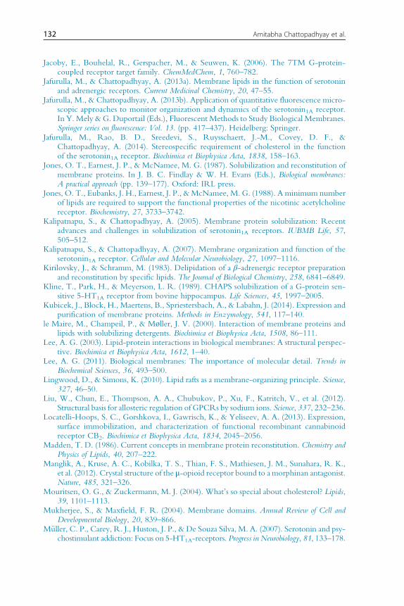
The dissociation of the native membrane leads to the formation of small clus-
ters of protein, lipid, and detergent that remain dissolved in the aqueous
solution (see Fig. 1). An important criterion for effective solubilization
and purification of membrane proteins is that the function of the protein
should be retained to the maximum possible extent. This poses a consider-
able challenge since many detergents irreversibly denature membrane pro-
teins (Garavito & Ferguson-Miller, 2001), which is responsible for the
modest list of membrane proteins solubilized with retention of function.
In case of GPCRs, solubilization and purification from natural sources is still
rare due to low amounts of the receptor present in the native tissue. Since
solubilization constitutes the crucial first step toward purification of any
transmembrane receptor, it is important to identify factors responsible for
achieving successful solubilization. We outline below some crucial aspects
of membrane receptor solubilization.
4.1 Choice of an appropriate detergentEfficient solubilization of functional GPCRs utilizing a suitable detergent
constitutes the first step in their molecular characterization. Detergents
are soluble amphiphiles with critical micelle concentrations (CMCs) typi-
cally in the range of millimolar. The ability of a detergent to solubilize mem-
branes is related to its hydrophile–lipophile balance (HLB), especially for
solubilization by nonionic detergents (Helenius & Simons, 1975;
Neugebauer, 1990). This principle has been utilized earlier in order to
achieve optimum solubilization of membrane proteins (Slinde &
Flatmark, 1976). HLB is an empirical parameter and is a measure of the
hydrophilic character of a detergent. It is calculated as the weight percentage
of hydrophilic versus lipophilic groups present in a detergent. Detergents
121Solubilization and Lipid–Receptor Interaction

with a relatively high HLB value of 12–20 are recommended for efficient
solubilization of membrane proteins without denaturation (Bhairi &
Mohan, 2001).
Detergents that belong to the class of nonionic and zwitterionic deter-
gents are particularly popular for their ability to solubilize membrane pro-
teins with retention of function. An important member of this class of
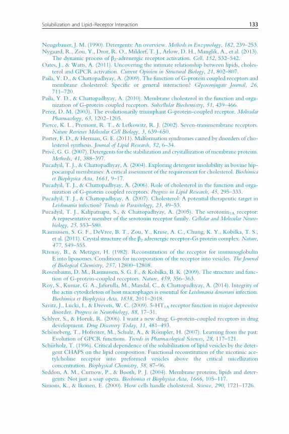
detergents is CHAPS (3-[(3-cholamidopropyl)dimethylammonio]-1-
propanesulfonate; see Fig. 2A), which is a mild, nondenaturing, and zwit-
terionic detergent (Hjelmeland, 1980). CHAPS is a synthetic detergent that
combines useful features of both the bile salt hydrophobic group and the
Detergent
A B
D CLipid–receptor–detergent complexLipid–detergent
mixed micelles
Detergentmicelles
Receptor–detergentcomplex
Figure 1 A schematic representation of different stages of solubilization of biologicalmembranes by detergents. When detergents are added to biological membranes(shown in (A)), the detergent monomers (shown in maroon (dark gray in the print ver-sion) with single tails) bind to the membrane and cause minimum perturbation at lowconcentrations (B). With increasing detergent concentration, themembrane bilayer getsfurther perturbed (C). At even higher detergent concentrations, complexes of deter-gent, lipid, and receptor of varying compositions are formed. These complexes includelipid–detergent mixed micelles, lipid–receptor–detergent complex, receptor–detergentcomplex, and detergent micelles (D).
122 Amitabha Chattopadhyay et al.
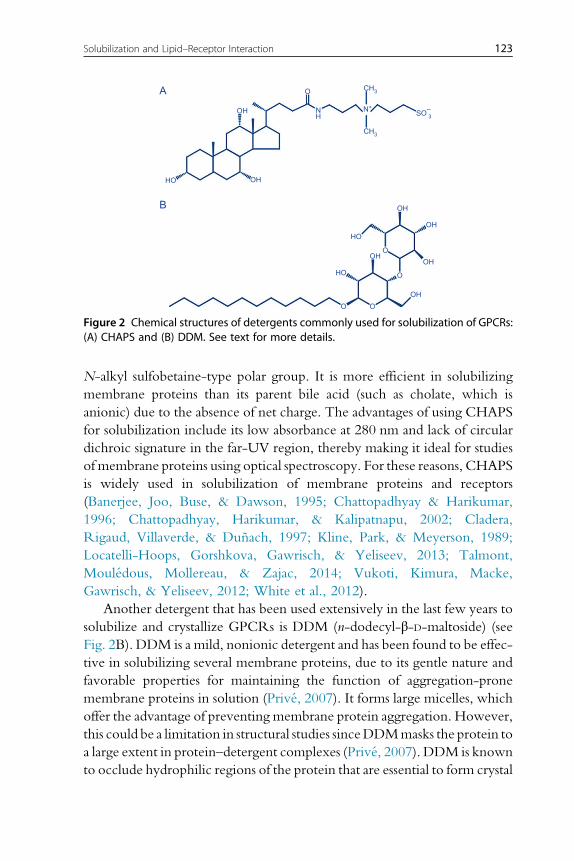
N-alkyl sulfobetaine-type polar group. It is more efficient in solubilizing
membrane proteins than its parent bile acid (such as cholate, which is
anionic) due to the absence of net charge. The advantages of using CHAPS
for solubilization include its low absorbance at 280 nm and lack of circular
dichroic signature in the far-UV region, thereby making it ideal for studies
of membrane proteins using optical spectroscopy. For these reasons, CHAPS
is widely used in solubilization of membrane proteins and receptors
(Banerjee, Joo, Buse, & Dawson, 1995; Chattopadhyay & Harikumar,
1996; Chattopadhyay, Harikumar, & Kalipatnapu, 2002; Cladera,
Rigaud, Villaverde, & Dunach, 1997; Kline, Park, & Meyerson, 1989;
Locatelli-Hoops, Gorshkova, Gawrisch, & Yeliseev, 2013; Talmont,
Mouledous, Mollereau, & Zajac, 2014; Vukoti, Kimura, Macke,
Gawrisch, & Yeliseev, 2012; White et al., 2012).
Another detergent that has been used extensively in the last few years to
solubilize and crystallize GPCRs is DDM (n-dodecyl-β-D-maltoside) (see
Fig. 2B). DDM is a mild, nonionic detergent and has been found to be effec-
tive in solubilizing several membrane proteins, due to its gentle nature and
favorable properties for maintaining the function of aggregation-prone
membrane proteins in solution (Prive, 2007). It forms large micelles, which
offer the advantage of preventing membrane protein aggregation. However,
this could be a limitation in structural studies sinceDDMmasks the protein to
a large extent in protein–detergent complexes (Prive, 2007). DDM is known
to occlude hydrophilic regions of the protein that are essential to form crystal
A
B
OH
HO
O
SO−3
N+
CH3
CH3
OH
OH
OH
OH
OH
HO
HO
OHO
O
OO
NH
Figure 2 Chemical structures of detergents commonly used for solubilization of GPCRs:(A) CHAPS and (B) DDM. See text for more details.
123Solubilization and Lipid–Receptor Interaction
contacts, which is not conducive for crystallization of GPCRs (Tate, 2012).
This was avoided in later studies by increasing the hydrophilic regions of the
GPCRs using antibodies or fusion proteins. DDM has been used to effec-
tively solubilize GPCRs such as β2-adrenergic receptor (Cherezov et al.,
2007; Rasmussen et al., 2011), A2A adenosine receptor (Liu et al., 2012),
μ-opioid receptor (Manglik et al., 2012), κ-opioid receptor (Wu et al.,
2012), β1-adrenergic receptor (Huang, Chen, Zhang, & Huang, 2013),
serotonin1B receptor (Wang et al., 2013), serotonin2B receptor (Wacker
et al., 2013), and metabotropic glutamate type 1 receptor (Wu et al.,
2014). Some GPCRs such as neurotensin receptor (White et al., 2012)
and CB2 cannabinoid receptor (Locatelli-Hoops et al., 2013; Vukoti et al.,
2012) have been solubilized utilizing a combination of DDM and CHAPS.
It should be noted that the choice of a suitable detergent for optimal sol-
ubilization of a given membrane protein has to be worked out on an indi-
vidual basis (Prive, 2007). For example, efficient solubilization of the IgE
receptor has been shown to occur with the anionic detergent cholate but
not with the nonionic detergent octyl glucoside (Rivnay & Metzger,
1982). Compatibility of the detergent in biochemical assays is another
important factor to be considered.
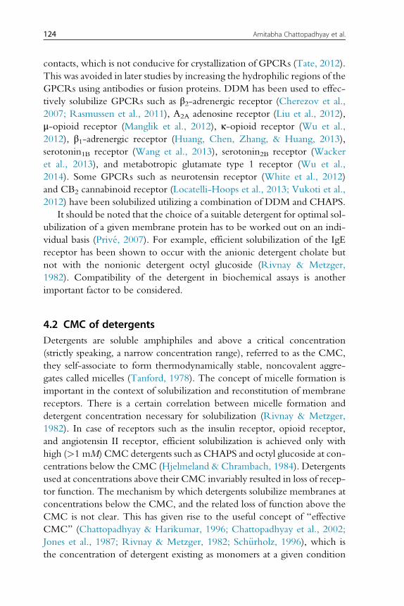
4.2 CMC of detergentsDetergents are soluble amphiphiles and above a critical concentration
(strictly speaking, a narrow concentration range), referred to as the CMC,
they self-associate to form thermodynamically stable, noncovalent aggre-
gates called micelles (Tanford, 1978). The concept of micelle formation is
important in the context of solubilization and reconstitution of membrane
receptors. There is a certain correlation between micelle formation and
detergent concentration necessary for solubilization (Rivnay & Metzger,
1982). In case of receptors such as the insulin receptor, opioid receptor,
and angiotensin II receptor, efficient solubilization is achieved only with
high (>1 mM) CMC detergents such as CHAPS and octyl glucoside at con-
centrations below the CMC (Hjelmeland & Chrambach, 1984). Detergents
used at concentrations above their CMC invariably resulted in loss of recep-
tor function. The mechanism by which detergents solubilize membranes at
concentrations below the CMC, and the related loss of function above the
CMC is not clear. This has given rise to the useful concept of “effective
CMC” (Chattopadhyay & Harikumar, 1996; Chattopadhyay et al., 2002;
Jones et al., 1987; Rivnay & Metzger, 1982; Sch€urholz, 1996), which is
the concentration of detergent existing as monomers at a given condition
124 Amitabha Chattopadhyay et al.

(such as lipids, proteins, ionic strength, pH, and temperature). Solubilization
could therefore be carried out below the CMC if the effective CMC is lower
than literature CMC. Another key parameter is the critical solubilization
concentration (CSC), which is the minimal detergent concentration
required to disrupt a given membrane into micellar dispersion (Prive,
2007). Selective solubilization of membrane proteins at detergent concen-
trations below CSC could be an effective purification strategy.
4.3 Detergent–lipid–protein ratioMembrane solubilization by detergents is a multistep process (Helenius &
Simons, 1975; Hjelmeland & Chrambach, 1984; Jones et al., 1987; le
Maire, Champeil, & Møller, 2000; see Fig. 1). The relative detergent–
lipid–protein ratio is an important factor for optimal solubilization of mem-
brane proteins. At a given protein or lipid concentration, with increasing
detergent concentration, an increase in solubilized lipid (Pucadyil &
Chattopadhyay, 2004) or protein (Demoliou-Mason & Barnard, 1984) is
observed until saturation is reached. However, it is not advisable to use high
detergent concentrations since membrane protein function is often com-
promised under such conditions. To overcome this, a mild concentration
of detergent could be used which may balance these two aspects, that is,
maximize solubilization yet preserve protein function. Arriving at an opti-
mal detergent, lipid, and protein ratio involves trial and error by carrying out
solubilization over a wide range of detergent–lipid ratios.
An empirical relationship between these experimental parameters was
developed in which the parameter (ρ) was defined as the molar ratio of
detergent to lipid optimal for functional solubilization (Rivnay &
Metzger, 1982).
ρ¼ Detergent½ ��CMCeff
Phospholipid½ �where CMCeff represents the effective CMC determined under specific
experimental conditions (as mentioned above). An increase in solubilization
is expectedwith increase in the value of the ρ parameter (generally up to�2).
5. SOLUBILIZATION AS A STRATEGY TO MONITORLIPID–PROTEIN INTERACTIONS
As mentioned earlier, solubilization provides a convenient approach
to explore lipid–receptor interaction since it results in differential extents
125Solubilization and Lipid–Receptor Interaction

of lipid solubilization, thereby allowing to assess the role of specific lipids on
receptor function. A common feature often associated with membrane sol-
ubilization is delipidation (loss of lipids). This results in loss of protein func-
tion since lipid–protein interactions play a crucial role in maintaining the
structure and function of integral membrane proteins and receptors (Lee,
2003). For example, displacement of annular lipids from the receptor was
shown to be an integral feature of detergent-induced inactivation in case
of the nicotinic acetylcholine receptor ( Jones, Eubanks, Earnest, &
McNamee, 1988). Interestingly, the phenomenon of delipidation caused
by solubilization and the subsequent loss of membrane protein function
has been effectively utilized to gain molecular insight into the specific lipid
requirements of membrane proteins ( Jones et al., 1988; Kirilovsky &
Schramm, 1983).
This strategy has been successfully utilized for exploring lipid–GPCR
interaction. It was previously reported that solubilization of the native hip-
pocampal serotonin1A receptors using CHAPS results in loss of receptor
activity and membrane cholesterol (Banerjee, Buse, & Dawson, 1990;
Banerjee et al., 1995; Chattopadhyay, Jafurulla, Kalipatnapu, Pucadyil, &
Harikumar, 2005). We previously demonstrated that specific ligand binding
of the serotonin1A receptor could be restored upon replenishment of cho-
lesterol into solubilized membranes (Chattopadhyay et al., 2005). Utilizing
this experimental strategy, we were able to examine the degree of stringency
required by closely related analogs of cholesterol, necessary for restoring
receptor activity. In order to explore the structural stringency of cholesterol
necessary for supporting receptor function, we replaced cholesterol with its
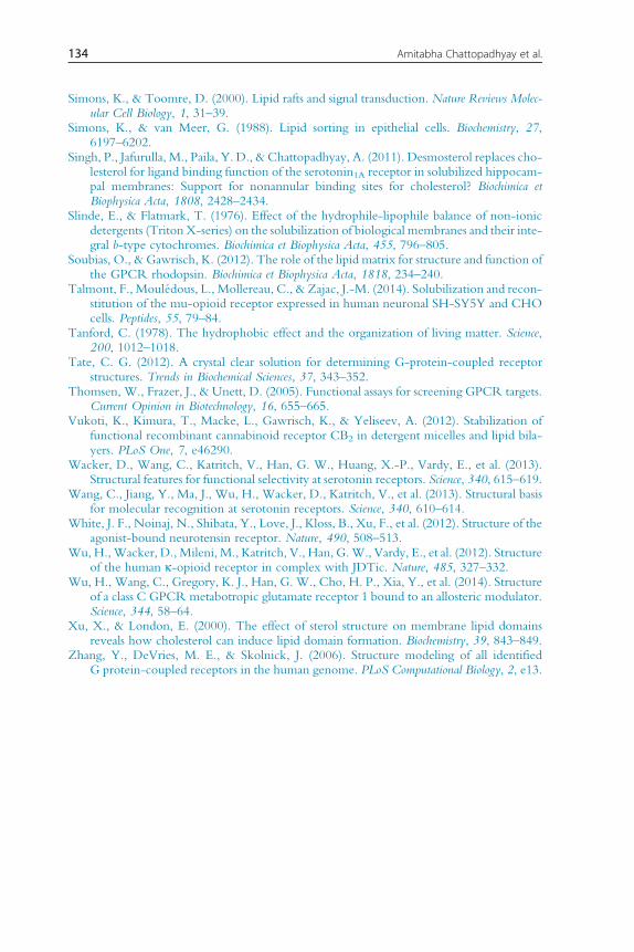
close structural analogs with minor differences (see Fig. 3). In one set of
experiments, solubilized membranes were replenished with
7-dehydrocholesterol (7-DHC) and desmosterol, which are immediate bio-
synthetic precursors of cholesterol in the Kandutsch–Russell and Bloch
pathways, respectively, both of which differ with cholesterol merely in an
additional double bond. While 7-DHC differs with cholesterol only in a
double bond at the seventh position in the sterol ring, desmosterol differs
with cholesterol only in a double bond at the 24th position in its flexible
alkyl side chain (see Fig. 3). Accumulation of either 7-DHC or desmosterol
due to defective sterol biosynthesis has been shown to result in fatal neuro-
logical disorders (Porter & Herman, 2011). Figure 4A shows that while des-
mosterol could support receptor function, 7-DHC could not restore
receptor activity (Chattopadhyay et al., 2007; Singh et al., 2011). This brings
out the fine stringency of cholesterol requirement for receptor function
126 Amitabha Chattopadhyay et al.

since the presence of an additional double bond in the sterol ring (7-DHC)
appears more detrimental to receptor function than the presence of an extra
double bond in the alkyl side chain (desmosterol).
The degree of structural stringency was explored further by examining
whether stereoisomers of cholesterol (ent-cholesterol and epi-cholesterol)
could support receptor function. While ent-cholesterol is the enantiomer
of cholesterol and is a nonsuperimposable mirror image of cholesterol,
epi-cholesterol is a diastereomer (not a mirror image of cholesterol) (see
HO
H3C
H3CCH3
CH3H3C
H H
H
H
HO
A
B C
ED
H3C
H3CCH3
CH3H3C
H H
H
H
HO
H3C
H3C
CH3
CH3H3C
H H
H
H
HO
H3C
H3C
CH3
CH3H3C
H H
H
H
HO
H3C
H3CCH3
CH3H3C
H H
H
H
Figure 3 Chemical structures of (A) cholesterol, (B) 7-dehydrocholesterol,(C) desmosterol, (D) ent-cholesterol, and (E) epi-cholesterol. Both 7-dehydrocholesterol(7-DHC) and desmosterol are immediate biosynthetic precursors of cholesterol inKandutsch–Russell and Bloch pathways, respectively, differing with cholesterol only ina double bond. While 7-dehydrocholesterol differs with cholesterol only in a double bondat the 7th position in the sterol ring, desmosterol differs with cholesterol only in a doublebond at the 24th position of the flexible alkyl side chain. Patients with mutations inenzymes that catalyze the final step in these pathways exhibit low levels of serum cho-lesterol and accumulation (high levels) of the respective immediate precursor (7-DHC ordesmosterol) leading to diseases such as the Smith–Lemli–Opitz syndrome (SLOS) anddesmosterolosis. Both ent-cholesterol and epi-cholesterol are stereoisomers of choles-terol. ent-Cholesterol is the enantiomer of cholesterol and is a nonsuperimposable mirrorimage of cholesterol, whereas epi-cholesterol is a diastereomer of cholesterol whichdiffers with cholesterol in the orientation of hydroxyl group at carbon-3 position.ent-Cholesterol shares similar physicochemical properties with cholesterol butepi-cholesterol does not.
127Solubilization and Lipid–Receptor Interaction
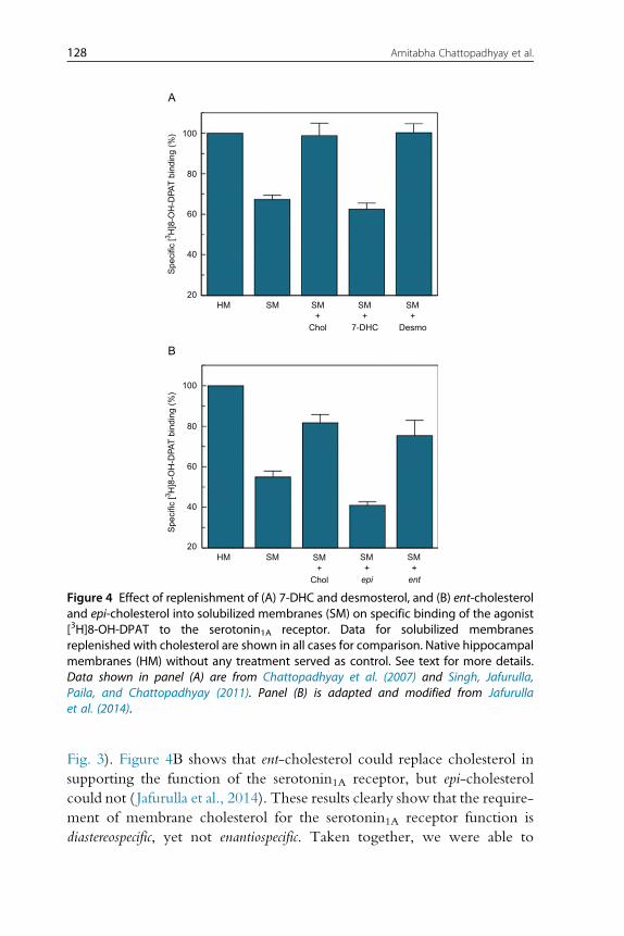
Fig. 3). Figure 4B shows that ent-cholesterol could replace cholesterol in
supporting the function of the serotonin1A receptor, but epi-cholesterol
could not ( Jafurulla et al., 2014). These results clearly show that the require-
ment of membrane cholesterol for the serotonin1A receptor function is
diastereospecific, yet not enantiospecific. Taken together, we were able to
20HM SM SM
+Chol
+7-DHC
+Desmo
SM SM
40
60
Spe
cific
[3 H]8
-OH
-DPA
T b
indi
ng (
%)
Spe
cific
[3 H]8
-OH
-DPA
T b
indi
ng (
%)
80
100
A
B
20HM SM SM
+Chol
+epi
+ent
SM SM
40
60
80
100
Figure 4 Effect of replenishment of (A) 7-DHC and desmosterol, and (B) ent-cholesteroland epi-cholesterol into solubilized membranes (SM) on specific binding of the agonist[3H]8-OH-DPAT to the serotonin1A receptor. Data for solubilized membranesreplenished with cholesterol are shown in all cases for comparison. Native hippocampalmembranes (HM) without any treatment served as control. See text for more details.Data shown in panel (A) are from Chattopadhyay et al. (2007) and Singh, Jafurulla,Paila, and Chattopadhyay (2011). Panel (B) is adapted and modified from Jafurullaet al. (2014).
128 Amitabha Chattopadhyay et al.
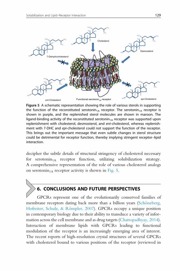
decipher the subtle details of structural stringency of cholesterol necessary
for serotonin1A receptor function, utilizing solubilization strategy.
A comprehensive representation of the role of various cholesterol analogs
on serotonin1A receptor activity is shown in Fig. 5.
6. CONCLUSIONS AND FUTURE PERSPECTIVES
GPCRs represent one of the evolutionarily conserved families of
membrane receptors dating back more than a billion years (Sch€oneberg,Hofreiter, Schulz, & R€ompler, 2007). GPCRs occupy a unique position
in contemporary biology due to their ability to transduce a variety of infor-
mation across the cell membrane and as drug targets (Chattopadhyay, 2014).
Interaction of membrane lipids with GPCRs leading to functional
modulation of the receptor is an increasingly emerging area of interest.
The recent reports of high-resolution crystal structures of several GPCRs
with cholesterol bound to various positions of the receptor (reviewed in
Functional serotonin1A receptorent-Cholesterol epi-Cholesterol
7-Dehydrocholesterol
Cholesterol
Desmosterol
CH3
CH3
H3C
H3C
H3C
H
H
HO
H H
CH3
CH3
H3C
H3C
H3C
H
H
HO
H H
CH3
CH3
H3C
H3C
H3C
H
H
HO
H H
CH3
CH3
H3C
H3C
H3C
H
H
HO
H H
CH3
CH3
H3C
H3C
H3C
H
H
HO
H H
Figure 5 A schematic representation showing the role of various sterols in supportingthe function of the reconstituted serotonin1A receptor. The serotonin1A receptor isshown in purple, and the replenished sterol molecules are shown in maroon. Theligand-binding activity of the reconstituted serotonin1A receptor was supported uponreplenishment with cholesterol, desmosterol, and ent-cholesterol, whereas replenish-ment with 7-DHC and epi-cholesterol could not support the function of the receptor.This brings out the important message that even subtle changes in sterol structurecould be detrimental for receptor function, thereby implying stringent receptor–lipidinteraction.
129Solubilization and Lipid–Receptor Interaction
Chattopadhyay, 2014; Jafurulla & Chattopadhyay, 2013a) have provided
more impact to this field. Unfortunately, a majority of GPCRs are not avail-
able in purified form from native sources. This is a severe limitation in
attempts to reconstitute the purified receptor into a defined lipid environ-
ment, thereby preventing lipid–receptor studies using established
approaches. In this overall scenario, solubilization using suitable detergents
which allow selective depletion of membrane lipids offers a window of
opportunity to assess the lipid specificity of GPCRs. The knowledge gained
from these studies will provide a better understanding of specific lipid
dependence of receptor function. Such advances in deciphering molecular
details of receptor–lipid interaction would lead to better understanding of
GPCR function in health and disease.
ACKNOWLEDGMENTSWork in A.C.’s laboratory was supported by the Council of Scientific and Industrial
Research, Govt. of India. B.D.R. thanks the University Grants Commission for the
award of a Junior Research Fellowship. A.C. gratefully acknowledges support from J.C.
Bose Fellowship (Department of Science and Technology, Govt. of India). A.C. is an
Adjunct Professor of Jawaharlal Nehru University (New Delhi), Indian Institute of
Science Education and Research (Mohali), Indian Institute of Technology (Kanpur), and
Honorary Professor of the Jawaharlal Nehru Centre for Advanced Scientific Research
(Bangalore). We thank G. Aditya Kumar for help in making figures, and members of the
Chattopadhyay laboratory for their comments and discussions.
REFERENCESAlemany, R., Perona, J. S., Sanchez-Dominguez, J. M., Montero, E., Canizares, J.,
Bressani, R., et al. (2007). G protein-coupled receptor systems and their lipid environ-ment in health disorders during aging. Biochimica et Biophysica Acta, 1768, 964–975.
Anson, L. (2009). Membrane protein biophysics. Nature, 459, 343.Banerjee, P., Buse, J. T., & Dawson, G. (1990). Asymmetric extraction of membrane lipids
by CHAPS. Biochimica et Biophysica Acta, 1044, 305–314.Banerjee, P., Joo, J. B., Buse, J. T., & Dawson, G. (1995). Differential solubilization of lipids
along with membrane proteins by different classes of detergents. Chemistry and Physics ofLipids, 77, 65–78.
Bhairi, S. M., & Mohan, C. (2001). Detergents—A guide to the properties and uses of detergents inbiological systems. San Diego, CA: Calbiochem-Novabiochem.
Burger, K., Gimpl, G., & Fahrenholz, F. (2000). Regulation of receptor function by choles-terol. Cellular and Molecular Life Sciences, 57, 1577–1592.
Celada, P., Bortolozzi, A., & Artigas, F. (2013). Serotonin 5-HT1A receptors as targets foragents to treat psychiatric disorders: Rationale and current status of research.CNSDrugs,27, 703–716.
Chattopadhyay, A. (2014). GPCRs: Lipid-dependent membrane receptors that act as drugtargets. Advances in Biology, 2014, 143023.
Chattopadhyay, A., & Harikumar, K. G. (1996). Dependence of critical micelle concentra-tion of a zwitterionic detergent on ionic strength: Implications in receptor solubilization.FEBS Letters, 391, 199–202.
130 Amitabha Chattopadhyay et al.

Chattopadhyay, A., Harikumar, K. G., & Kalipatnapu, S. (2002). Solubilization of highaffinity G-protein-coupled serotonin1A receptors from bovine hippocampus usingpre-micellar CHAPS at low concentration. Molecular Membrane Biology, 19, 211–220.
Chattopadhyay, A., & Jafurulla, M. (2012). Role of membrane cholesterol in leishmanialinfection. Advances in Experimental Medicine and Biology, 749, 201–213.
Chattopadhyay, A., Jafurulla, M., Kalipatnapu, S., Pucadyil, T. J., & Harikumar, K. G.(2005). Role of cholesterol in ligand binding and G-protein coupling of serotonin1Areceptors solubilized from bovine hippocampus. Biochemical and Biophysical Research Com-munications, 327, 1036–1041.
Chattopadhyay, A., Paila, Y. D., Jafurulla, M., Chaudhuri, A., Singh, P., Murty, M. R. V. S.,et al. (2007). Differential effects of cholesterol and 7-dehydrocholesterol on ligand bind-ing of solubilized hippocampal serotonin1A receptors: Implications in SLOS. Biochemicaland Biophysical Research Communications, 363, 800–805.
Chaudhuri, A., & Chattopadhyay, A. (2011). Transbilayer organization of membrane cho-lesterol at low concentrations: Implications in health and disease. Biochimica et BiophysicaActa, 1808, 19–25.
Cherezov, V., Rosenbaum, D. M., Hanson, M. A., Rasmussen, S. G. F., Thian, F. S.,Kobilka, T. S., et al. (2007). High-resolution crystal structure of an engineered humanβ2-adrenergic G protein-coupled receptor. Science, 318, 1258–1265.
Cladera, J., Rigaud, J.-L., Villaverde, J., & Dunach, M. (1997). Liposome solubilization andmembrane protein reconstitution using Chaps and Chapso. European Journal of Biochem-istry, 243, 798–804.
Demoliou-Mason, C. D., & Barnard, E. A. (1984). Solubilization in high yield of opioidreceptors retaining high-affinity delta, mu and kappa binding sites. FEBS Letters, 170,378–382.
Deupi, X., & Kobilka, B. K. (2010). Energy landscapes as a tool to integrate GPCR structure,dynamics, and function. Physiology (Bethesda), 25, 293–303.
Duquesne, K., & Sturgis, J. N. (2010). Membrane protein solubilization.Methods in MolecularBiology, 601, 205–217.
Ellis, C., & The Nature Reviews Drug Discovery GPCRQuestionnaire Participants (2004).The state of GPCR research in 2004. Nature Reviews Drug Discovery, 3, 577–626.
Garavito, R. M., & Ferguson-Miller, S. (2001). Detergents as tools in membrane biochem-istry. The Journal of Biological Chemistry, 276, 32403–32406.
Heilker, R., Wolff, M., Tautermann, C. S., & Bieler, M. (2009). G-protein-coupledreceptor-focused drug discovery using a target class platform approach. Drug DiscoveryToday, 14, 231–240.
Helenius, A., & Simons, K. (1975). Solubilization of membranes by detergents. Biochimica etBiophysica Acta, 415, 29–79.
Hjelmeland, L. M. (1980). A nondenaturing zwitterionic detergent for membrane biochem-istry: Design and synthesis. Proceedings of the National Academy of Sciences of the United Statesof America, 77, 6368–6370.
Hjelmeland, L. M., & Chrambach, A. (1984). Solubilization of functional membrane-boundreceptors. In J. C. Venter & L. C. Harrison (Eds.), Membranes, detergents, and receptorsolubilization (pp. 35–46). New York: Alan R. Liss.
Huang, J., Chen, S., Zhang, J. J., & Huang, X.-Y. (2013). Crystal structure of oligomeric β1-adrenergic G protein-coupled receptors in ligand-free basal state. Nature Structural &Molecular Biology, 20, 419–425.
Inagaki, S., Ghirlando, R., White, J. F., Gvozdenovic-Jeremic, J., Northup, J. K., &Grisshammer, R. (2012). Modulation of the interaction between neurotensin receptorNTS1 and Gq protein by lipid. Journal of Molecular Biology, 417, 95–111.
Insel, P. A., Tang, C.-M., Hahntow, I., & Michel, M. C. (2007). Impact of GPCRs in clin-ical medicine: Monogenic diseases, genetic variants and drug targets. Biochimica etBiophysica Acta, 1768, 994–1005.
131Solubilization and Lipid–Receptor Interaction
Jacoby, E., Bouhelal, R., Gerspacher, M., & Seuwen, K. (2006). The 7TM G-protein-coupled receptor target family. ChemMedChem, 1, 760–782.
Jafurulla, M., & Chattopadhyay, A. (2013a). Membrane lipids in the function of serotoninand adrenergic receptors. Current Medicinal Chemistry, 20, 47–55.
Jafurulla, M., & Chattopadhyay, A. (2013b). Application of quantitative fluorescence micro-scopic approaches to monitor organization and dynamics of the serotonin1A receptor.In Y. Mely & G. Duportail (Eds.), Fluorescent Methods to Study Biological Membranes.Springer series on fluorescence: Vol. 13. (pp. 417–437). Heidelberg: Springer.
Jafurulla, M., Rao, B. D., Sreedevi, S., Ruysschaert, J.-M., Covey, D. F., &Chattopadhyay, A. (2014). Stereospecific requirement of cholesterol in the functionof the serotonin1A receptor. Biochimica et Biophysica Acta, 1838, 158–163.
Jones, O. T., Earnest, J. P., & McNamee, M. G. (1987). Solubilization and reconstitution ofmembrane proteins. In J. B. C. Findlay & W. H. Evans (Eds.), Biological membranes:A practical approach (pp. 139–177). Oxford: IRL press.
Jones, O. T., Eubanks, J. H., Earnest, J. P., &McNamee, M. G. (1988). A minimum numberof lipids are required to support the functional properties of the nicotinic acetylcholinereceptor. Biochemistry, 27, 3733–3742.
Kalipatnapu, S., & Chattopadhyay, A. (2005). Membrane protein solubilization: Recentadvances and challenges in solubilization of serotonin1A receptors. IUBMB Life, 57,505–512.
Kalipatnapu, S., & Chattopadhyay, A. (2007). Membrane organization and function of theserotonin1A receptor. Cellular and Molecular Neurobiology, 27, 1097–1116.
Kirilovsky, J., & Schramm, M. (1983). Delipidation of a β-adrenergic receptor preparationand reconstitution by specific lipids. The Journal of Biological Chemistry, 258, 6841–6849.
Kline, T., Park, H., & Meyerson, L. R. (1989). CHAPS solubilization of a G-protein sen-sitive 5-HT1A receptor from bovine hippocampus. Life Sciences, 45, 1997–2005.
Kubicek, J., Block, H., Maertens, B., Spriestersbach, A., & Labahn, J. (2014). Expression andpurification of membrane proteins. Methods in Enzymology, 541, 117–140.
le Maire, M., Champeil, P., & Møller, J. V. (2000). Interaction of membrane proteins andlipids with solubilizing detergents. Biochimica et Biophysica Acta, 1508, 86–111.
Lee, A. G. (2003). Lipid-protein interactions in biological membranes: A structural perspec-tive. Biochimica et Biophysica Acta, 1612, 1–40.
Lee, A. G. (2011). Biological membranes: The importance of molecular detail. Trends inBiochemical Sciences, 36, 493–500.
Lingwood, D., & Simons, K. (2010). Lipid rafts as a membrane-organizing principle. Science,327, 46–50.
Liu, W., Chun, E., Thompson, A. A., Chubukov, P., Xu, F., Katritch, V., et al. (2012).Structural basis for allosteric regulation of GPCRs by sodium ions. Science, 337, 232–236.
Locatelli-Hoops, S. C., Gorshkova, I., Gawrisch, K., & Yeliseev, A. A. (2013). Expression,surface immobilization, and characterization of functional recombinant cannabinoidreceptor CB2. Biochimica et Biophysica Acta, 1834, 2045–2056.
Madden, T. D. (1986). Current concepts in membrane protein reconstitution. Chemistry andPhysics of Lipids, 40, 207–222.
Manglik, A., Kruse, A. C., Kobilka, T. S., Thian, F. S., Mathiesen, J. M., Sunahara, R. K.,et al. (2012). Crystal structure of the μ-opioid receptor bound to a morphinan antagonist.Nature, 485, 321–326.
Mouritsen, O. G., & Zuckermann, M. J. (2004). What’s so special about cholesterol? Lipids,39, 1101–1113.
Mukherjee, S., & Maxfield, F. R. (2004). Membrane domains. Annual Review of Cell andDevelopmental Biology, 20, 839–866.
M€uller, C. P., Carey, R. J., Huston, J. P., & De Souza Silva, M. A. (2007). Serotonin and psy-chostimulant addiction: Focus on 5-HT1A-receptors. Progress in Neurobiology, 81, 133–178.
132 Amitabha Chattopadhyay et al.
Neugebauer, J. M. (1990). Detergents: An overview.Methods in Enzymology, 182, 239–253.Nygaard, R., Zou, Y., Dror, R. O., Mildorf, T. J., Arlow, D. H., Manglik, A., et al. (2013).
The dynamic process of β2-adrenergic receptor activation. Cell, 152, 532–542.Oates, J., & Watts, A. (2011). Uncovering the intimate relationship between lipids, choles-
terol and GPCR activation. Current Opinion in Structural Biology, 21, 802–807.Paila, Y. D., & Chattopadhyay, A. (2009). The function of G-protein coupled receptors and
membrane cholesterol: Specific or general interaction? Glycoconjugate Journal, 26,711–720.
Paila, Y. D., & Chattopadhyay, A. (2010). Membrane cholesterol in the function and orga-nization of G-protein coupled receptors. Subcellular Biochemistry, 51, 439–466.
Perez, D. M. (2003). The evolutionarily triumphant G-protein-coupled receptor. MolecularPharmacology, 63, 1202–1205.
Pierce, K. L., Premont, R. T., & Lefkowitz, R. J. (2002). Seven-transmembrane receptors.Nature Reviews Molecular Cell Biology, 3, 639–650.
Porter, F. D., & Herman, G. E. (2011). Malformation syndromes caused by disorders of cho-lesterol synthesis. Journal of Lipid Research, 52, 6–34.
Prive, G. G. (2007). Detergents for the stabilization and crystallization of membrane proteins.Methods, 41, 388–397.
Pucadyil, T. J., & Chattopadhyay, A. (2004). Exploring detergent insolubility in bovine hip-pocampal membranes: A critical assessment of the requirement for cholesterol. Biochimicaet Biophysica Acta, 1661, 9–17.
Pucadyil, T. J., & Chattopadhyay, A. (2006). Role of cholesterol in the function and orga-nization of G-protein coupled receptors. Progress in Lipid Research, 45, 295–333.
Pucadyil, T. J., & Chattopadhyay, A. (2007). Cholesterol: A potential therapeutic target inLeishmania infection? Trends in Parasitology, 23, 49–53.
Pucadyil, T. J., Kalipatnapu, S., & Chattopadhyay, A. (2005). The serotonin1A receptor:A representative member of the serotonin receptor family. Cellular and Molecular Neuro-biology, 25, 553–580.
Rasmussen, S. G. F., DeVree, B. T., Zou, Y., Kruse, A. C., Chung, K. Y., Kobilka, T. S.,et al. (2011). Crystal structure of the β2 adrenergic receptor-Gs protein complex.Nature,477, 549–555.
Rivnay, B., & Metzger, H. (1982). Reconstitution of the receptor for immunoglobulinE into liposomes. Conditions for incorporation of the receptor into vesicles. The Journalof Biological Chemistry, 257, 12800–12808.
Rosenbaum, D. M., Rasmussen, S. G. F., & Kobilka, B. K. (2009). The structure and func-tion of G-protein-coupled receptors. Nature, 459, 356–363.
Roy, S., Kumar, G. A., Jafurulla, M., Mandal, C., & Chattopadhyay, A. (2014). Integrity ofthe actin cytoskeleton of host macrophages is essential for Leishmania donovani infection.Biochimica et Biophysica Acta, 1838, 2011–2018.
Savitz, J., Lucki, I., & Drevets, W. C. (2009). 5-HT1A receptor function in major depressivedisorder. Progress in Neurobiology, 88, 17–31.
Schlyer, S., & Horuk, R. (2006). I want a new drug: G-protein-coupled receptors in drugdevelopment. Drug Discovery Today, 11, 481–493.
Sch€oneberg, T., Hofreiter, M., Schulz, A., & R€ompler, H. (2007). Learning from the past:Evolution of GPCR functions. Trends in Pharmacological Sciences, 28, 117–121.
Sch€urholz, T. (1996). Critical dependence of the solubilization of lipid vesicles by the deter-gent CHAPS on the lipid composition. Functional reconstitution of the nicotinic ace-tylcholine receptor into preformed vesicles above the critical micellizationconcentration. Biophysical Chemistry, 58, 87–96.
Seddon, A. M., Curnow, P., & Booth, P. J. (2004). Membrane proteins, lipids and deter-gents: Not just a soap opera. Biochimica et Biophysica Acta, 1666, 105–117.
Simons, K., & Ikonen, E. (2000). How cells handle cholesterol. Science, 290, 1721–1726.
133Solubilization and Lipid–Receptor Interaction
Simons, K., & Toomre, D. (2000). Lipid rafts and signal transduction. Nature Reviews Molec-ular Cell Biology, 1, 31–39.
Simons, K., & van Meer, G. (1988). Lipid sorting in epithelial cells. Biochemistry, 27,6197–6202.
Singh, P., Jafurulla, M., Paila, Y. D., & Chattopadhyay, A. (2011). Desmosterol replaces cho-lesterol for ligand binding function of the serotonin1A receptor in solubilized hippocam-pal membranes: Support for nonannular binding sites for cholesterol? Biochimica etBiophysica Acta, 1808, 2428–2434.
Slinde, E., & Flatmark, T. (1976). Effect of the hydrophile-lipophile balance of non-ionicdetergents (Triton X-series) on the solubilization of biological membranes and their inte-gral b-type cytochromes. Biochimica et Biophysica Acta, 455, 796–805.
Soubias, O., & Gawrisch, K. (2012). The role of the lipid matrix for structure and function ofthe GPCR rhodopsin. Biochimica et Biophysica Acta, 1818, 234–240.
Talmont, F., Mouledous, L., Mollereau, C., & Zajac, J.-M. (2014). Solubilization and recon-stitution of the mu-opioid receptor expressed in human neuronal SH-SY5Y and CHOcells. Peptides, 55, 79–84.
Tanford, C. (1978). The hydrophobic effect and the organization of living matter. Science,200, 1012–1018.
Tate, C. G. (2012). A crystal clear solution for determining G-protein-coupled receptorstructures. Trends in Biochemical Sciences, 37, 343–352.
Thomsen, W., Frazer, J., & Unett, D. (2005). Functional assays for screening GPCR targets.Current Opinion in Biotechnology, 16, 655–665.
Vukoti, K., Kimura, T., Macke, L., Gawrisch, K., & Yeliseev, A. (2012). Stabilization offunctional recombinant cannabinoid receptor CB2 in detergent micelles and lipid bila-yers. PLoS One, 7, e46290.
Wacker, D., Wang, C., Katritch, V., Han, G. W., Huang, X.-P., Vardy, E., et al. (2013).Structural features for functional selectivity at serotonin receptors. Science, 340, 615–619.
Wang, C., Jiang, Y., Ma, J., Wu, H., Wacker, D., Katritch, V., et al. (2013). Structural basisfor molecular recognition at serotonin receptors. Science, 340, 610–614.
White, J. F., Noinaj, N., Shibata, Y., Love, J., Kloss, B., Xu, F., et al. (2012). Structure of theagonist-bound neurotensin receptor. Nature, 490, 508–513.
Wu, H.,Wacker, D.,Mileni,M., Katritch, V., Han, G.W., Vardy, E., et al. (2012). Structureof the human κ-opioid receptor in complex with JDTic. Nature, 485, 327–332.
Wu, H., Wang, C., Gregory, K. J., Han, G. W., Cho, H. P., Xia, Y., et al. (2014). Structureof a class C GPCRmetabotropic glutamate receptor 1 bound to an allosteric modulator.Science, 344, 58–64.
Xu, X., & London, E. (2000). The effect of sterol structure on membrane lipid domainsreveals how cholesterol can induce lipid domain formation. Biochemistry, 39, 843–849.
Zhang, Y., DeVries, M. E., & Skolnick, J. (2006). Structure modeling of all identifiedG protein-coupled receptors in the human genome. PLoS Computational Biology, 2, e13.
134 Amitabha Chattopadhyay et al.