Embed Size (px)
Citation preview
Gut, 1974, 15, 165-172
Liver structure and function in cholelithiasis: Effectof chenodeoxycholic acid'G. D. BELL, H. Y. I. MOK, M. THWE, G. M. MURPHY, K. HENRY, ANDR. H. DOWLING2
From the Departments of Medicine and Histopathology, Royal Postgraduate Medical School,Hammersmith Hospital, London, and The Institute of Child Health, University ofBirmingham
SUMMARY Although, in suitable patients, oral chenodeoxycholic acid (CDCA) dissolves gallstones,the results of recent animal studies suggest that it might be hepatotoxic. Liver function was thereforestudied in patients with gallstones before and during treatment with CDCA and liver biopsies werecarried out both in patients with cholelithiasis given bile acid therapy and in those who had beengiven no medical treatment. In 25 patients treated with 0.5-1 5 g CDCA/day (7-20 mg kg bodyweight-' day-') there was no significant change in serum bilirubin, albumin, globulin, transaminase,isocitric dehydrogenase, alkaline phosphatase, and gamma glutamyl transpeptidase levels beforeand at monthly intervals during six months' treatment. The kinetics of bromsulphthalein (BSP)clearance and its apparent transport maximum were not significantly changed during CDCAtherapy. The mean fasting serum bile acid concentrations of 18.0 ± SEM 1.2 ,umoles/litre beforeand 20.0 ± 3.5 j&moles/litre during treatment were both significantly greater than control values.Liver histology was not appreciably different in 11 patients treated with CDCA from that in eightpatients with untreated cholelithiasis and in three patients who had received CDCA three to fourmonths before biopsy. These results suggest that in doses of 0.5 to 1.5 g/day CDCA is not hepato-toxic in man.
Oral chenodeoxycholic acid (CDCA) improvescholesterol solubility in bile (Thistle and Schoen-field, 1971) and in suitable patients it dissolvesgallstones (Danzinger, Hofmann, Thistle, andSchoenfield, 1972; Bell, Whitney, and Dowling, 1972;Thistle and Hofmann, 1973), but, as with any newdrug, the benefits of treatment with CDCA must beweighed against possible complications. One suchpotential complication is hepatic dysfunction andindeed Small (1971) predicted that this might happeneither as a direct effect of CDCA itself on the liver,or indirectly through the formation of lithocholicacid, the bacterial metabolite of CDCA, which isknown to be hepatotoxic.Although the initial reports of CDCA treatment
1Presented in part at the Annual Meeting of the Association ofPhysicians, Glasgow,in April 1973 (Bell et al, Quart.J. Med., 42,824-825 (Abstr.), 1973.'Request for reprints to R.H.D., Gastroenterology Unit, Departmentof Medicine, Guy's Hospital Medical School, London SEI 9RT.
Received for publication 30 January 1974.
in man described no significant hepatic dysfunction(Danzinger et al, 1972; Bell et al, 1972) morerecent animal studies have again raised the questionof hepatotoxicity. In Rhesus monkeys given 40,80, or 120 mg CDCA kg body weight-' day-' bymouth for six months, there were raised levels ofserum alkaline phosphatase and oxaloacetic trans-aminase while liver histology showed bile ducthyperplasia and mononuclear infiltration of theportal tracts (Lee, 1973, personal communication).
It is not yet known whether these changes inliver structure and function are confined to theRhesus monkey and therefore a study of the effectsof CDCA on the liver in man is of fundamentalimportance if further patients with cholesterol gall-stones are to be treated with this bile acid. Thispaper presents our own results for liver structure inpatients with gallstones during treatment withCDCA and in those who had been given no medicaltherapy, and liver function in patients with gall-stones before and during treatment with cheno-deoxycholic acid for periods up to 15 months.
165
on October 25, 2021 by guest. P
rotected by copyright.http://gut.bm
j.com/
Gut: first published as 10.1136/gut.15.3.165 on 1 M
arch 1974. Dow
nloaded from
G. D. Bell, H. Y. L Mok, M. Thwe, G. M. Murphy, K. Henry, and R. H. Dowling
The Patients
Twenty-eight patients with radiologically provengallstones (six men and 22 women) were treated withCDCA for periods ranging from six weeks to 15months. Their mean age was 50.2 years (range 24-75). The criteria for selecting these patients fortreatment|have already been described (Bell et al,1972).
DOSE OF CDCAIn 25 patients the dose of CDCA ranged from 0.5 to1*5 g per day (equivalent to approximately 7-20mg/kg body weight) while three patients receivedonly 0.25 g per day (3.5 mg/kg body weight).
Methods
STUDIES OF LIVER STRUCTURE AND
FUNCTIONLiver function was studied by measuring the levels ofcirculating liver enzymes and serum bile acids, byfollowing the kinetics of BSP clearance from theplasma after an intravenous bolus injection of BSP(5 mg/kg), and by measuring the apparent transportmaximum (Tm) of BSP (Wheeler, Meltzer, andBradley, 1960). Liver structure was studied by lightmicroscopic examination of percutaneous Menghinineedle and intraoperative wedge and needle biopsiesof the liver.
Serum bilirubin, albumin, and globulin levelsThese were measured by standard laboratorymethods before and at monthly intervals duringtreatment.
Circulating liver enzymesThe levels of two hepatocellular enzymes, serumglutamic oxaloacetic transaminase (SGOT) andisocitrate dehydrogenase (ICD), and two 'biliarytract' enzymes, alkaline phosphatase and gammaglutamyl transpeptidase (GGT), were also measuredbefore, and in most cases at monthly intervals during,bile acid treatment using standard laboratorymethods.
Bromsulphthalein studiesThe kinetics of BSP clearance were measured ineight patients with gallstones receiving CDCA andin five patients with gallstones who had not beentreated. Following a bolus injection of BSP into oneantecubital vein (5 mg/kg body weight), blood sam-ples were taken from the opposite arm at five, 10,15, 20, 30, 35, 40, 45, 50, and 60 minutes. The fallin plasma concentration of BSP was then plottedagainst time.
The apparent transport maximum (Tm) andstorage capacity (S) of BSP (Wheeler et al, 1960)were measured in six patients during CDCA treat-ment and in four patients with gallstones who hadreceived no treatment. In one patient, the apparentBSP Tm and S were measured before and againafter three months' treatment with 1.0 g CDCA perday.
Serum bile acidsThe total fasting serum bile acid concentrationswere measured at least 12 hr after the last dose ofCDCA by spectrofluorimetry combined with thehydroxy steroid dehydrogenase enzyme assay(Murphy, Billing, and Baron, 1970). Six estimationswere made in five untreated patients with gallstonesand in 12 patients receiving 0.25-1.0 g/CDCA perday. The results in both groups were then comparedwith the findings in a control group of 24 subjects(12 male and 12 female) previously studied by oneof us (G.M.M.).
LIVER STRUCTUREBefore the recent results, showing that CDCA wastoxic to the liver of Rhesus monkeys (Lee, 1973),we had not felt justified in performing liver biopsieson relatively asymptomatic patients with gallstoneswho had normal conventional liver function tests.However, after it became known that structuralchanges might occur in the liver of primates, liverbiopsies were performed in a series of patients withgallstones. Clinical details of the patients studiedtogether with the results of their liver histology aregiven in table I.
Eight patients with gallstones who had not beentreated with CDCA had liver biopsies (threepercutaneous needle biopsies and five operativeneedle biopsies obtained during elective cholecystec-tomy) and 11 patients who had been treated with0.25-1 g CDCA/day for periods ranging from sixweeks to 15 months (eight had Menghini needlebiopsies, two had wedge biopsies, and one anintraoperative needle biopsy). In addition, liverbiopsies were obtained in three patients, three tofour months after CDCA treatment had beenwithdrawn (two percutaneous and one operativeneedle biopsy) one of whom had also been biopsiedduring therapy. Informed consent was obtained inall cases.
Results
SERUM BILIRUBIN, ALBUMIN, ANDGLOBULIN LEVELSThese levels were essentially normal throughout the
166
on October 25, 2021 by guest. P
rotected by copyright.http://gut.bm
j.com/
Gut: first published as 10.1136/gut.15.3.165 on 1 M
arch 1974. Dow
nloaded from
Liver structure andfunction in cholelithiasis: Effect of chenodeoxycholic acid 167
Group Patient Sex Age CDCA Treatment Biopsy1 Positive Histological Features'
Duration Average Parenchyma Portal Tracts(months) Dose
(glday) Fatty Lipo- Other Features Bile Duct Round CellChange fuscin Proliferation Infiltrate
1 1 F 60 - - ON 0 0 0 0 0(patients with 2 M 59 - - ON ++ i 0 0 +gallstones not 3 F 47 - - ON 0 ± 0 0 0treated with CDCA.) 4 F 48 - - ON 0 + + Occasional poly- 0 0
morphs insinusoids
5 F 50 - - ON 0 + 0 0 +6 F 73 - - PC N 0 ± Occasional poly- Oedema
morphs insinusoids
7 F 47 - - PC N 0 + 0 0 +8 -F 25 - - PCN 0 + 0 0 0
11 9 F 70 IS 1-0 PC N +++ + 0 0 0(patients with 10 F 70 14 1.0 PC N 0 0 Occasional poly- 0 0gallstones biopsied morphs induring treatment sinusoidswith CDCA) 11' F 44 12 1.0 PC N 0 + 0 + 0
12 F 56 10 025 PCN 0 + 0 0 +13 M 65 12 1.0 PC N 0 + 0 0 +14 F 56 12 0.75 PC N 0 + Occasional poly- 0 +
morphs insinusoids
IS F 62 11 025 PCN + i 0 0 016 M 56 6 0.75 PC N 0 + 0 Pus cells in bile duct lumina17 F 33 15 0.75 SW 0 ± Polymorphs, 0 +
especially roundcentral vein
18 F 24 2 075 SW 0 + Polymorphs ++ i +Hepatic cellnecrosis
19 F 66 2 0.25 PC N 0 ± 0 0 0
III 11' F 44 12/12 till 1-0 PC N 0 + 0 + 0(patients with 3/12 beforegallstones previously biopsytreated with CDCA) 20 F 52 14/12 till 1.0 ON + + + 0 0 +
4/12 beforebiopsy
21 F 33 3/12 till 1.0 PC N 0 + 0 0 03/12 beforebiopsy
Table I Liver histology in patients with gallstones before and after treatment with CDCA'Biopsy PCN = percutaneous Menghini needle, ON = operative needle, SW = surgical wedge'Histological features, 0 = absent or normal, ± = trace or very slight, + = minimal, + + = moderate, +++ = moderate to marked'Patient no. 11 biopsied both during treatment and after chenodeoxycholic acid had been withdrawn
Serum Enzyme Normal Range Mean Serum Level ± SEM
Before Treatment After Treatment with Chenodeoxycholic Acid(N = no. ofpatients) 1/12 3/12 6/12
Alkaline phosphatase 3-13 6-3 ± 04 6.2' ± 0-3 6.51 0-4 6 21 ± 0-4(KAU) (27) (27) (22) (21)GGT 2-25 16-3 ± 26 19221 3-8 16771 3.5 19.01 ± 57(IU) (12) (12) (1 1) (10)SGOT 2-20 10-3 + 05 12.21 + 1-0 11.11 + 08 10.11 ± 1-0(IU) (23) (23) (21) (20)ICD 3-8 9-2 ± 08 1001 ± 1-0 9.31 ± 08 821 ± 07(IU) (21) (21) (22) (19)
Table II Serum alkaline phosphatase, gamma glutamyl transpeptidase, SGOT, and ICD before and aftertreatment with chenodeoxycholic acid in a dose of05 to 15 g per day in patients with gallstones'Not significant
on October 25, 2021 by guest. P
rotected by copyright.http://gut.bm
j.com/
Gut: first published as 10.1136/gut.15.3.165 on 1 M
arch 1974. Dow
nloaded from
G. D. Bell, H. Y. I. Mok, M. Thwe, G. M. Murphy, K. Henry, and R. H. Dowling
period of treatment. There was no change in any ofthe mean values before and after CDCA.
CIRCULATING LIVER ENZYMESRepresentative results of the four serum liverenzymes studied are shown by the findings beforeand during therapy with 0 5-1 5 g CDCA/day atone, three, and six months (table II). There was nosignificant change in the mean levels of any of thefour enzymes studied.
Alkaline phosphataseThe range of readings before treatment was 4-10 KAunits and 3-11 units while on treatment. Therewere no values outside our normal laboratoryrange (3-13 KAU).
Gamma glutamyl transpeptidase (GGT)The range before therapy was 6-30 IU and duringtreatment 4-65 IU (N 2-25 IU). Four patientsshowed fluctuating levels of GGT, greater than thenormal range, on at least one occasion. Of thesefour patients, one took night sedation with bar-biturates while a second admitted to a moderatelyheavy alcoholic intake.
Serum glutamic oxaloacetic transaminase (SGOT)(N2-20 IU)The range before treatment was 7-16 IU and during
16
12
3
(21)
4
treatment 5-22 IU. Four patients showed transientelevations of SGOT to levels just above the normallaboratory range but all returned to normal spon-taneously without reduction in the dose of bile acid.
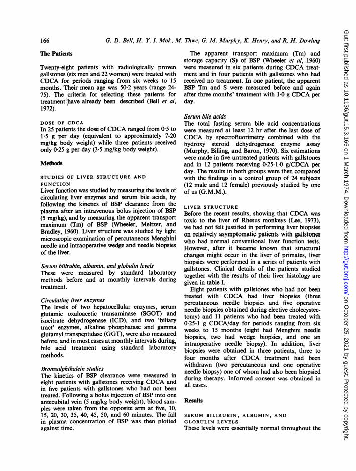
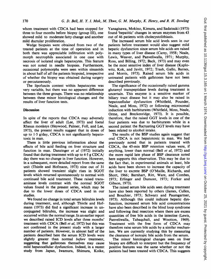
Isocitrate dehydrogenase (ICD) (N 3-8 IU)The mean level in 21 patients with gallstones beforestarting treatment of 9.2 ± 0.8 (range 3-16) wasabove the upper limit of normal-nine of the 21patients having mildly elevated ICD levels beforestarting CDCA (fig 1). Although occasional patientsshowed increased ICD values during treatment, therewas no consistent trend to raised levels (rangeafter treatment 2-20).
BROMSULPHTHALEIN STUDIES
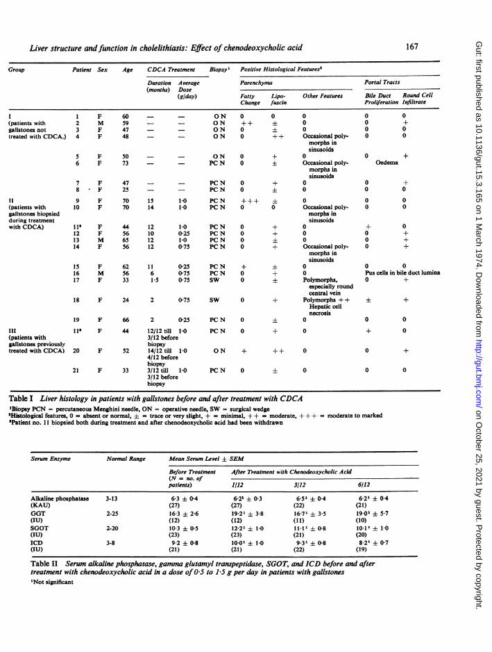
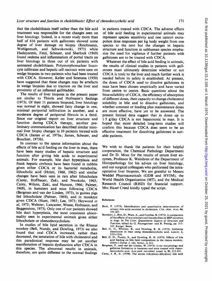
Clearance of a single bolus injection (5 mg/kg)There was no difference in the mean plasma dis-appearance curves over the first 25 minutes (fig 2).However, during the next 35 minutes, the eightpatients with cholelithiasis treated with CDCAcleared the dye more rapidly than the controls,although the difference between the two groups at45, 50, and 60 minutes did not reach statisticalsignificance.
Apparent transport maximum (Tm)The mean BSP Tm in the six treated patients of
(22) (19)
4
Bo NS m
Before 1 month
r 3mNS B 6NS
Before, 3 months Before 6 months
Fig 1 Serum isocitric dehydrogenase levels before and after one, three, and six months' treatment with 075 to1.0 g chenodeoxycholic acid/day. The normal laboratory range is shown as the crosshatched zones between brokenlines. The E-shaped symbols indicate mean values ± SEMs.
168
on October 25, 2021 by guest. P
rotected by copyright.http://gut.bm
j.com/
Gut: first published as 10.1136/gut.15.3.165 on 1 M
arch 1974. Dow
nloaded from
Liver structure andfunction in cholelithiasis: Effect of chenodeoxycholic acid
Before 0 MEAN
On CDCA Rx * C SEM
s (5)i (8
0 10 25 40 60TIME
Clearance following bolusinjection (5mg/Kg)
Fig 2 The apparent BSP transport maximum excretory capacity (Wheeler et al, 1960) measured in patientswith gallstones who had received no treatment (open histogram) or 1 g CDCAiday for three months (closedhistogram; mean values ± SEMs). The semilogarithmic plot shows the clearance ofa single bolus injection ofBSP (5 mg/kg body weight) in patients given CDCA (closed circles) and in patients who had received no bileacids (open circles).
6.86 mg/min (range 40-8.1) was essentially the sameas in the four untreated patients with gallstones(mean 6-38; range 46-7.7 mg/min). In one patient,studied before and after treatment, the Tm wasagain unchanged, being 7-1 mg/min before and 7.3mg/min three months after 0.75 g CDCA/day. Therapid late phase of clearance of the single bolusinjection of BSP in patients treated with CDCA wastherefore not associated with an increased maximumexcretory capacity (Tm).
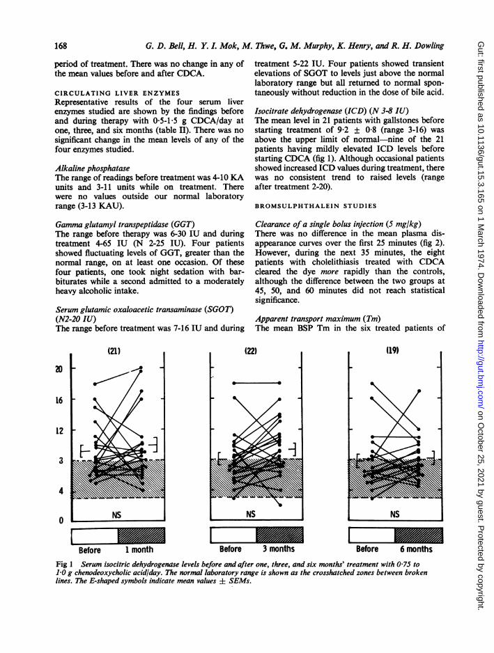
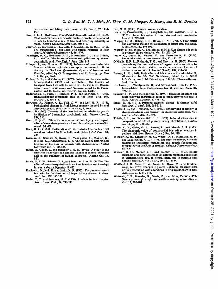
FASTING SERUM BILE ACIDSThe results of total serum bile acid levels are shownin figure 3. Based on six estimations in five patientswith gallstones who had not been treated, the meanvalue of 18.0 ± 1.2 (range 14.0-23.5) umoles/litrewas not significantly different from the mean valueof 20.0 (± 3.5; range 3.2-56.0 ,umoles/litre) in 12patients treated with 0 25-1.0 g CDCA/day. How-ever, the results in both groups of patients weresignificantly greater than those in the controlpopulation. These modest rises in fasting serum bileacid concentrations were never associated withpruritus.
LIVER STRUCTUREThe positive histological features on liver histologyare summarized in table I. Of the 11 patientstreated with CDCA who had liver biopsies (table I,
group II) only two showed significant changes onhistology. In one, there was mild proliferation ofbile ducts and in another there was moderate to
60
cl1.!Y
30 1-
20
10
0
H 3
* 0
A
E-*NORMALS UNTREATED TREATED
GALLSTORE GALLSTONEPATIENTS PATIENTS
(CDCA 0.25-l.Og
Fig 3 Fasting serum bile acid concentrations (jmoles/litre) in control subjects, in untreated patients withgallstones, and in patients given 0.25-1.0 g CDCA/day.The 'E' symbols show mean values ± SEMs.
marked fatty change in the absence of diabetes,obesity and a history of alcohol intake. In otherpatients in this group there was a minimal increasein mononuclear cells in the portal tracts but this wasnot thought to be significant. In the control group,all of whom had cholelithiasis (table I, group I),four of the eight patients showed this latter non-specific change and one further patient showedmoderate fatty change. In the three patients in
109
8
BSPTm 7mg/min 6
5
4
3
21
10
5
SerumBSPmg/lOO ml
1
0.5
0.2
Apparent Tm
169
on October 25, 2021 by guest. P
rotected by copyright.http://gut.bm
j.com/
Gut: first published as 10.1136/gut.15.3.165 on 1 M
arch 1974. Dow
nloaded from
G. D. Bell, H. Y. L Mok, M. Thwe, G. M. Murphy, K. Henry, and R. H. Dowling
whom treatment with CDCA had been stopped forthree to four months before biopsy (group III), oneshowed mild to moderate fatty change and anothermild ductular proliferation.Wedge biopsies were obtained from two of the
treated patients at the time of operation and inboth there was appreciable infiltration with poly-morph neutrophils associated in one case withnecrosis of isolated single hepatocytes. This featurewas not noted in needle biopsies. Furthermore,occasional polymorphs were seen within sinusoidsin about half of all the patients biopsied, irrespectiveof whether the biopsy was obtained during surgeryor percutaneously.The lipofuscin content within hepatocytes was
very variable, but there was no apparent differencebetween the three groups. There was no relationshipbetween these minor histological changes and theresults of liver function tests.
Discussion
In spite of the reports that CDCA may adverselyaffect the liver of adult (Lee, 1973) and foetalRhesus monkeys (Heywood, Palmer, Foll, and Lee,1973), the present results suggest that in doses ofup to 1.5 g/day, CDCA is not significantly hepato-toxic in man.There is little previous information about the
effects of bile acid feeding on liver structure andfunction in man. Danzinger et al (1972) initiallyfound that during treatment with up to 4.5 g CDCA/day there was no change in liver function. However,in a subsequent, more detailed report from the sameunit (Thistle and Hofmann, 1973) nine out of 31patients showed transient slight rises in SGOTlevels which returned spontaneously to normal withcontinued bile acid treatment. These raised trans-aminase levels contrast with the normal SGOTvalues found in the present series, which may bedue to the lower doses of CDCA used in ourstudies.We found no change in total serum bilirubin levels
during treatment, and, although Thistle and Hof-mann (1973) did find a significant increase in un-conjugated bilirubin, the increase was slight andoccurred within the normal range. In an earlier reportwe described raised ICD levels after three months'treatment with CDCA (Bell et al, 1972) but this wasnot confirmed in the present study with a largernumber of patients. However, in almost half of thepatients described here the serum ICD levels wereslightly greater than normal before treatment,suggesting that gallstones themselves may causemild hepatocellular dysfunction. Indeed, in a recentstudy from Japan, Iwamura, Shimura, Koike,
Yanagisawa, Mokino, Kimura, and Sadatsuki (1973)found 'hepatitic' changes in serum enzymes from 43out of 46 patients with cholecystolithiasis.The increased serum bile acid levels seen in our
patients before treatment would also suggest mildhepatic dysfunction since serum bile acids are raisedin many types of liver disease (Carey, 1958; Neale,Lewis, Weaver, and Panveliwalla, 1971; Murphy,Ross, and Billing, 1972; Back, 1973) and may evenbe the most sensitive index of liver disease (Kaplo-witz, Kok, and Javitt, 1972; Trash, Gallo, Barnes,and Morris, 1973). Raised serum bile acids inuntreated patients with gallstones have not beendescribed previously.The significance of the occasionally raised gamma
glutamyl transpeptidase levels during treatment isuncertain. This enzyme is a sensitive marker ofbiliary tract disease but it may also be raised inhepatocellular dysfunction (Whitfield, Pounder,Neale, and Moss, 1972) or following microsomalinduction with barbiturates (Whitfield, Moss, Neale,Orme, and Breckenridge, 1973). It is possible,therefore, that the raised GGT levels in one of thefour patients was due to barbiturates while in asecond patient the fluctuating GGT levels may havebeen related to alcohol intake.The results of the BSP studies again suggest that
oral CDCA is not hepatotoxic in man. We hadpreviously noted that in patients treated withCDCA, the 45-min BSP retention values were, ifanything, lower than normal (Bell et al, 1972) andthe more rapid late phase of BSP clearance notedhere supports this observation. This may be due tothe fact that, in experimental animals at least, bileacids have been shown to increase the capacity ofthe liver to excrete BSP (O'Maille, Richards, andShort, 1966; Barnhart, Ritt, Ware, and Combes,1973; Erlinger and Dumont, 1973; Forker andGibson, 1973).The raised serum bile acids seen during treatment
have also been reported by others (James, Cullen,and Bouchier, 1973; Schwarz and Paumgartner,1973). Although this could indicate hepatic dys-function, increased serum bile acid concentrationshave also been described in the blind loop syndromeand following ileal resection where there are excessquantities of free bile acids in the intestine (Lewis,Panveliwalla, Tabaqchali, and Wootton, 1969).Treatment with the free form of CDCA maytherefore raise serum bile acids by a similar mechan-ism. We are currently studying this by measuringthe clearance of isotopic bile acids from the serum.The minor histological changes seen on liver
biopsy are difficult to interpret but the frequency ofpositive features was the same whether or not thepatients had been treated with CDCA. This suggests
170
on October 25, 2021 by guest. P
rotected by copyright.http://gut.bm
j.com/
Gut: first published as 10.1136/gut.15.3.165 on 1 M
arch 1974. Dow
nloaded from
Liver structure andfunction in cholelithiasis: Effect of chenodeoxycholic acid 171
that the cholelithiasis itself rather than the bile acidtreatment was responsible for the changes seen onliver histology. Indeed, in a recent study more thanhalf of 616 patients with gallstones showed somedegree of liver damage on biopsy (Reichmann,Wohlgemuth, and Schwokowski, 1973) whileHadziyannis, Feizi, Scheuer, and Sherlock (1969)found oedema and inflammation of portal tracts onliver histology in three out of six patients withuntreated cholelithiasis. Polymorphonuclear leuco-cyte infiltrates and hepatic cell necrosis were seen onwedge biopsies in two patients who had been treatedwith CDCA. However, Keller and Smetana (1950)have suggested that these changes may be artefactsin wedge biopsies due to traction on the liver andproximity of an inflamed gallbladder.The results of liver biopsies in the present paper
are similar to Thistle and Hofmann's findings(1973). Of their 11 patients biopsied, liver histologywas normal in eight, showed fatty change in one,minimal periportal infiltration in another, and amoderate degree of periportal fibrosis in a third.Since our original report on liver structure andfunction during CDCA therapy, another pre-liminary communication has described similar mini-mal liver biopsy changes in 10 patients treated withCDCA (James et al, 1973a; James, Scheuer, andBouchier, 1973b).
In contrast to the sparse information about theeffects of bile acid feeding on the liver in man, therehave been many studies of hepatic structure andfunction after giving bile acids to experimentalanimals. For example, bile duct hyperplasia andfrank hepatic cirrhosis have been found in rabbitsgiven either CDCA or its bacterial metabolite,lithocholic acid (Holsti, 1960, 1962) and similarchanges have been seen in rats after lithocholate(Carey, Hoffbauer, Zaki, and Nwokolo, 1965;Carey, Wilson, Zaki, and Hanson, 1966; Palmer,1969), in hamsters and mice following CDCA(Bergman and van der Linden, 1973), in guinea pigsfed lithocholate (Palmer, 1969), and in monkeysgiven CDCA (Hunt, 1965; Lee, 1973; Heywood etal, 1973; Webster, Lancaster, Wease, Hofmann, andBaggenstoss, 1973). Only one of our patients showedbile duct hyperplasia, the most consistent abnor-mality seen in experimental animals given eitherlithocholate or chenodeoxycholate.
In studies of bile lipid secretion in the Rhesusmonkey (Bell, Nundy, and Dowling, 1973) we alsofound that oral CDCA increased, rather thandecreased, the saturation of bile with cholesterol andthis paradoxical response may be yet anothermanifestation of hepatic dysfunction after CDCA inthis species. The abnormal results in animals,therefore, are quite different to the normal findings
in patients treated with CDCA. The adverse effectsof bile acid feeding in experimental animals mayrepresent species sensitivity and one cannot extra-polate dose responses per kg body weight from onespecies to the next but the changes in hepaticstructure and function in subhuman species empha-size the need for vigilance if further patients withgallstones are to be treated with CDCA.
Whatever the effect of bile acid feeding in animals,the results of clinical studies in patients with gall-stones must ultimately determine whether or notCDCA is toxic to the liver and much further work isneeded before its safety is established. At present,the doses of CDCA used to dissolve gallstones inman have been chosen empirically and have variedfrom centre to centre. Basic questions about thebioavailability of CDCA, the efficiency of absorptionof different doses, their capacity to change cholesterolsolubility in bile and to dissolve gallstones, andwhether constant or loading plus maintenance dosesare more effective, have yet to be answered. Thepresent limited data suggest that in doses up to15 g/day CDCA is not hepatotoxic in man. It ishoped that more detailed long-term studies willconfirm this because CDCA does seem to be aneffective treatment for dissolving gallstones in suit-able patients.
We wish to thank the patients for their helpfulcooperation, the Chemical Pathology Departmentand Dr D. Moss for the results of serum liver en-zymes, Professor K. Weinbren of the Department ofHistopathology for his advice on liver histology,and our surgical colleagues who performed the intra-operative liver biopsies. We are grateful to MessrsWeddel Pharmaceuticals (GDB and HYIM), theWorld Health Organization (MT), and the MedicalResearch Council (RHD) for financial support.Mrs Hazel Creed kindly typed the script.
References
Back, P. (1973). Identification and quantitative determination ofurinary bile acids excreted in cholestasis. Clin. chim. Acta, 44,199-207.
Barnhart, J., Ritt, D., Ware, A., and Combes, B. (1973). A comparisonof the effects of taurocholate and theophylline on BSP excretionin dogs. In The Liver. Quantitative Aspects of Structure andFunction, edited by G. Paumgartner and R. Preisig, pp. 315-325. Karger, Basle.
Bell, G. D., Whitney, B., and Dowling, R. H. (1972). Gallstonedissolution in man using chenodeoxycholic acid. Lancet, 2,1213-1216.
Bell, G. D., Nundy, S., and Dowling, R. H. (1973). Effects of bileacid feeding on bile lipid composition in the rhesus monkey.(Abstr.) Europ. J. clin. Invest., 3, 211.
Bergman, F., and van der Linden, W. (1973). Liver morphology andgallstone formation in hamsters and mice treated with cheno-deoxycholic acid. Acta path. microbiol. scand. A, 81, 213-221.
Carey, J. B., Jr. (1958). The serum trihydroxy-dihydroxy bile acid
on October 25, 2021 by guest. P
rotected by copyright.http://gut.bm
j.com/
Gut: first published as 10.1136/gut.15.3.165 on 1 M
arch 1974. Dow
nloaded from
172 G. D. Bell, H. Y. L. Mok, M. Thwe, G. M. Murphy, K. Henry, and R. H. Dowling
ratio in liver and biliary tract disease. J. clin. Invest., 37, 1494-1503.
Carey, J. B.,Jr., Hoffbauer,F.W.,Zaki,F.G.,andNwokolo,C.(1965).Choledocholithiasis and hepatic ductular proliferation inducedin rats by lithocholic acid (a bile acid occurring naturally inman). (Abstr.) Gastroenterology, 48, 809-810.
Carey, J. B., Jr., Wilson, I. D., Zaki, F. G., and Hanson, R. F. (1966).The metabolism of bile acids with special reference to liverinjury. Medicine (Baltimore), 45, 461-470.
Danzinger, R. G., Hofmann, A. F., Schoenfield, L. J., and Thistle,J. L. (1972). Dissolution of cholesterol gallstones by cheno-deoxycholic acid. New Engl. J. Med., 286, 1-8.
Erlinger, S., and Dumont, M. (1973). Influence of canalicular bileflow on sulfobromophthalein transport maximum in bile inthe dog. In The Liver. Quantitative Aspects of Structure andFunction, edited by G. Paumgartner and R. Preisig, pp. 306-314. Karger, Basle.
Forker, E. L., and Gibson, G. (1973). Interaction between sulfo-bromophthalein (BSP) and taurocholate. The kinetics oftransport from liver cells to bile in rats. In The Liver. Quanti-tative Aspects of Structure and Function, edited by G. Paum-gartner and R. Preisig, pp. 326-336. Karger, Basle.
Hadziyannis, S., Feizi, T., Scheuer, P. J., and Sherlock, S. (1969).Immunoglobulin-containing cells in the liver. Clin. exp.Immunol., 5, 499-514.
Heywood, R., Palmer, A. K., Foll, C. V., and Lee, M. R. (1973).Pathological changes in fetal Rhesus monkey induced by oralchenodeoxycholic acid. (Letter.) Lancet, 2, 1021.
Holsti, P. (1960). Cirrhosis of the liver induced in rabbits by gastricinstillation of 3-monohydroxycholanic acid. Nature (Lond.),186, 250.
Holsti, P. (1962). Bile acids as a cause of liver injury: cirrhogeniceffect ofchenodesoxycholic acid in rabbits. Actapath. microbiol.scand., 54, 479.
Hunt, R. D. (1965). Proliferation of bile ductules (the ductular cellreaction) induced by lithocholic acid. (Abstr.) Fed. Proc., 24,431.
Iwamura, K., Shimura, S., Koike, H., Yanagisawa, F., Mokino, K.,Kimura, K., and Sadatsuki, T. (1972). Clinical and pathologicalfindings of the liver in patients with cholelithiasis. (Abstr.)Gastroent. jap., 7, 184-185.
James, O., Cullen, J., and Bouchier, I. A. D. (1973a). A study of theeffectiveness, toxicity and bile salt kinetics of chenodeoxycholicacid in the treatment of human gallstones. (Abstr.) Gut, 14,827.
James, 0. F. W., Scheuer, P. J., and Bouchier, I. A. D. (1973b). Theeffect of chenodeoxycholic acid on liver function and histologyin man. (Abstr.) Digestion, 8, 432.
Kaplowitz, N., Kok, E., and Javitt, N. B. (1973). Postprandial serumbile acid for the detection of hepatobiiary disease. J. Amer.med. Ass., 225, 292-293.
Keller, T. C., and Smetana, H. F. (1950). Artefacts in liver biopsies.Amer. J. clin. Path., 20, 738-741.
Lee, M. R. (1973). Personal communication.Lewis, B., Panveliwalla, D., Tabaqchali, S., and Wootton, I. D. P.
(1969). Serum-bile-acids in the stagnant-loop syndrome.Lancet, 1, 219-220.
Murphy, G. M., Billing, B. H., Baron, D. N. (1970). A fluorimetricand enzymatic method for estimation of serum total bile acids.J. clin. Path., 23, 594-598.
Murphy, G. M., Ross, A., and Billing, B. H. (1972). Serum bile acidsin primary biliary cirrhosis. Gut, 13, 201-206.
Neale, G., Lewis, B., Weaver, V., and Panveliwalla, D. (1971).Serum bile acids in liver disease. Gut, 12, 145-152.
O'Maille, E. R. L., Richards, T. G., and Short, A. H. (1966). Factorsdetermining the maximal rate of organic anion secretion bythe liver and further evidence on the hepatic site of action ofthe hormone secretin. J. Physiol. (Lond.), 186, 424-438.
Palmer, R. H. (1969). Toxic effects of lithocholic acid and related 5S,-H steroids. In Bile Salt Metabolism, edited by L. SchiffJ. B. Carey, and J. M. Dietschy, pp. 184-204. Thomas, Spring-field, Illinois.
Reichmann, J., Wohlgemuth, B., and Schwokowski, Ch. F. (1973).Lebersch8iden beim Gallensteinleiden. Z. ges. inn. Med., 28,327-330.
Schwarz, H. P., and Paumgartner, G. (1973). Elevation of serum bileacids following therapeutic doses of chenodeoxycholic acid inman. (Abstr.) Digestion, 8, 431-432.
Small, D. M. (1971). Prestone gallstone disease-is therapy safe?New Engl. J. Med., 284, 214-216.
Thistle, J. L., and Hofmann, A. F. (1973). Efficacy and specificity ofchenodeoxycholic acid therapy for dissolving gallstones. NewEngl. J. Med., 289, 655-659.
Thistle, J. L., and Schoenfield, L. J. (1971). Induced alterations incomposition of bile of persons having cholelithiasis. Gastro-enterology, 61, 488-496.
Trash, D. B., Gallo, G. A., Barnes, S., and Morris. J. S. (1973).The diagnostic value of postprandial bile salt estimations inpatients with liver disease. (Abstr.) Gut, 14, 819.
Webster, K. H., Lancaster, M. C., Wease, D. F., Hofmann, A. F.,and Baggenstoss, A. H. (1973). The effect of primary bile acidfeeding on cholesterol metabolism and hepatic function andmorphology in the Rhesus monkey. (Abstr.) Gastroenterology,65, 576.
Wheeler, H. O., Meltzer, J. I., and Bradley, S. E. (1960). Biliarytransport and hepatic storage of sulfobromophthalein sodiumin unanesthetised dog, in normal man, and in patients withhepatic disease. J. clin. Invest., 39, 1131-1144.
Whitfield, J. B., Moss, D. W., Neale, G., Orme, M., and Brecken-ridge, A. (1973). Changes in plasma y-glutamyl transpeptidaseactivity associated with alterations in drug metabolism in man.Brit. med. J., 1, 316-318.
Whitfield, J. B., Pounder, R., Neale, G., and Moss, D. W. (1972).Serum gamma glutamyl transpeptidase activity in liver disease.Gut, 13, 702-708.
on October 25, 2021 by guest. P
rotected by copyright.http://gut.bm
j.com/
Gut: first published as 10.1136/gut.15.3.165 on 1 M
arch 1974. Dow
nloaded from


![CHOLELITHIASIS [Autosaved]](https://img.pdfslide.us/doc/110x75/577ce5051a28abf1038fa5b3/cholelithiasis-autosaved.jpg)