Embed Size (px)
Citation preview
Lipid domains control myelin basic protein adsorptionand membrane interactions between model myelinlipid bilayersDong Woog Leea, Xavier Banquya,b, Kai Kristiansena, Yair Kaufmana, Joan M. Boggsc,d, and Jacob N. Israelachvilia,e,1
aDepartment of Chemical Engineering, University of California, Santa Barbara, CA 93106; bFaculty of Pharmacy, Université de Montréal, Montréal, QC, CanadaH3C 3J7; cDepartment of Molecular Structure and Function, The Hospital for Sick Children, Toronto, ON, Canada M5G 1X8; dDepartment of LaboratoryMedicine and Pathobiology, University of Toronto, Toronto, ON, Canada M5G 1L5; and eMaterials Department, University of California, Santa Barbara,CA 93106
Contributed by Jacob N. Israelachvili, January 21, 2014 (sent for review October 4, 2013)
The surface forces apparatus and atomic force microscope wereused to study the effects of lipid composition and concentrationsof myelin basic protein (MBP) on the structure of model lipidbilayers, as well as the interaction forces and adhesion betweenthem. The lipid bilayers had a lipid composition characteristic ofthe cytoplasmic leaflets of myelin from “normal” (healthy) and“disease-like” [experimental allergic encephalomyelitis (EAE)]animals. They showed significant differences in the adsorptionmechanism of MBP. MBP adsorbs on normal bilayers to forma compact film (3–4 nm) with strong intermembrane adhesion(∼0.36 mJ/m2), in contrast to its formation of thicker (7–8 nm)swelled films with weaker intermembrane adhesion (∼0.13 mJ/m2)on EAE bilayers. MBP preferentially adsorbs to liquid-disorderedsubmicron domains within the lipid membranes, attributed to hy-drophobic attractions. These results show a direct connection be-tween the lipid composition of membranes and membrane–proteinadsorption mechanisms that affects intermembrane spacing andadhesion and has direct implications for demyelinating diseases.
lipid raft | biomembrane adhesion | myelin structure | multiple sclerosis |intrinsically unstructured proteins
Myelin is an asymmetric multilamellar membrane wrappedaround the axons of the central nervous system (CNS) and
consists of alternating extracellular and cytoplasmic leaflets(1–3). The bilayer-associated proteins, mainly myelin basic pro-tein (MBP) and proteolipid protein, play an essential role instabilizing and maintaining the myelin structure. The bilayers arein close contact (∼3 nm separation between lipid headgroup–water interfaces), providing a low dielectric constant through thecompact bilayers, which is essential for efficient and fast saltatorypropagation of nerve impulses. Any structural changes of themyelin sheath in the CNS, including lesion formation, loss ofadhesion, swelling of the water gaps, vacuolization, vesiculation,and complete delamination (demyelination) of the myelin sheath(4–6), are signatures of several inflammatory neurological dis-orders. These types of disorders are characterized by a broadspectrum of neurological symptoms, such as physical and cog-nitive disabilities, with multiple sclerosis (MS) being one of themost common demyelinating diseases (2).The primary cause of structural changes in the myelin is still
under debate; however, morphological changes of the myelinstructure due to diseases such as MS are well known. A well-studied and accepted animal model for MS is the experimentalallergic encephalomyelitis (EAE) of the marmoset (2, 6). Usingthis model, recent studies conducted with the surface forcesapparatus (SFA) have shown that a loss of adhesion force (7)and structural changes of model membranes with lipid compo-sition characteristic of myelin accompanied compositional alter-ations of the lipid species (8), as well as an electric chargeimbalance between lipid molecules and MBP (9). These alterations
also change the lateral distribution and stability of phase-separatedlipid domains (or rafts) within model myelin membranes (8, 10).MBP is one of the most abundant proteins in the CNS and is
an intrinsically unstructured (disordered) protein (11). MBP actsas an intermembrane adhesion protein between the cytoplasmicleaflets of the myelin sheath. The predominant size and chargeisoform of MBP in healthy and mature myelin has a molecularweight of 18.5 kDa and a net positive charge of 19 (12, 13).Several studies conducted with model and extracted myelinbilayers (9, 14–19) showed that because of its high content ofpositively charged residues, MBP binds to the negatively chargedlipids of the cytoplasmic leaflets of the bilayer via electrostaticinteraction in addition to hydrophobic interactions. However,studies have shown that the MBP charge component composi-tion, as well as the balance between charged lipids and MBPcharge components, changes in EAE (as well as in MS) tissues(20, 21). Other studies (9, 15) demonstrated the importance ofthe hydrophobic interaction between MBP and lipid membranesby showing that MBP specifically binds to lipid domain bound-aries (defects), altering the lateral organization of the modelmyelin bilayers. A recent study also showed that MBP’s associ-ation with the cytoplasmic leaflet of the myelin membraneinduces a phase transition into a cohesive mesh-like proteinnetwork (22, 23).
Significance
The proper functioning of multilayer membrane systems, suchas myelin, requires the multilamellar membranes to be tightlywrapped around the axon fibers, thereby allowing efficientelectric signal transmission. Slight changes in lipid compositionin myelin membranes will alter their domain sizes and dis-tributions, and the intermembrane adhesive properties. Usingthe surface forces apparatus and atomic force microscope, westudied the adsorption of myelin basic protein (MBP) to modelmyelin lipid bilayer membranes of varying compositions, andtheir effects on the structure, equilibrium spacing (swelling),and adhesion force between them. We find that MBP prefer-entially adsorbs to “disordered” submicron domains, affectingregular spacing and adhesion. These findings provide insightsinto lipid–protein interactions and membrane-associated (e.g.,demyelinating) diseases.
Author contributions: D.W.L. designed research; D.W.L., X.B., K.K., and Y.K. performedresearch; D.W.L. analyzed data; and D.W.L., X.B., K.K., Y.K., J.M.B., and J.N.I. wrotethe paper.
The authors declare no conflict of interest.
Freely available online through the PNAS open access option.1To whom correspondence should be addressed. E-mail: [email protected].
This article contains supporting information online at www.pnas.org/lookup/suppl/doi:10.1073/pnas.1401165111/-/DCSupplemental.
E768–E775 | PNAS | Published online February 10, 2014 www.pnas.org/cgi/doi/10.1073/pnas.1401165111
Dow
nloa
ded
by g
uest
on
Feb
ruar
y 7,
202
2

One open question regarding the stability of myelin mem-branes is how the concentration of MBP affects the interactionforces between myelin bilayers. We reconstructed and used twotypes of supported model monolayers with a lipid compositioncharacteristic of “normal” or “healthy” and “disease-like” EAEmyelin deposited on a dipalmitoylphosphatidylethanolamine(DPPE) monolayer to examine the effect of lipid composition,domains, and fluidity on the interaction forces, film viscosity, andMBP adsorption mechanism between these model myelin bilayers.The lipid composition used is based on data from Inouye andKirschner (1, 24) and Ohler et al. (6) (Table 1). For convenience,these reconstituted bilayers are referred to as “model normalbilayers,” “model EAE bilayers,” and “model myelin bilayers”throughout this article. The bilayers were prepared using theLangmuir–Blodgett technique (25), and the interaction forceswere measured using surface forces apparatus (SFA) model 2000(26), with the capacity of measuring interaction forces and sep-aration distances between macroscopic surfaces with a resolutionof 10 nN and 0.1 nm, respectively. The MBP adsorption mech-anism onto the bilayers also was examined using an atomic forcemicroscope (AFM). This study aims to establish the relationshipbetween the structure of the model lipid myelin bilayers andprotein adsorption and also to quantify the effect of MBP con-centration on the intermembrane interaction forces.
ResultsInteraction Forces Between Supported Model Cytoplasmic MyelinBilayers. MBP is a flexible, elongated biomolecule (11) thatcontains both charged and hydrophobic/hydrophilic groups andinteracts readily with lipid bilayers. The adhesive strength anddensity of an ensemble of MBPs between myelin lipid bilayers,obtained through force–distance (F–D) and thin-film viscositymeasurements (see Fig. 1 for a schematic of the experimentalsetup), provided qualitative and quantitative information aboutthe interfacial MBP conformation and adsorption mechanism atthe molecular level.Force–distance profiles show there is a critical adsorption
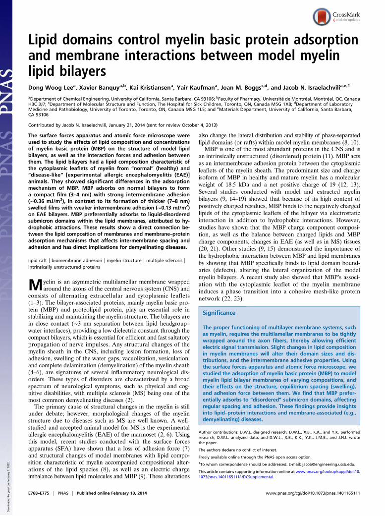
concentration, Ccrit, of MBP above which MBP adsorbs (Fig. 2).This adsorption threshold is a characteristic feature of domainformations of molecules (2D micelles, clusters, or rafts) (27). Atbulk concentrations C of MBP that are less than 1.2 ng/mL, theMBP adsorbs to neither normal (Fig. 2 A–C) nor EAE (Fig. 2 D–F) model myelin bilayers and the bilayer’s interactions are sim-ilar to two bare bilayers. When the MBP concentration exceeds2.9 ng/mL, rapid and cooperative adsorption of MBP occurs, asmay be ascertained from the following observations (Fig. 2): (i)Over a slight increase in the MBP concentration, a sharp out-ward shift of the electric double-layer interaction and the steric“hard wall” (Dsw = 2DB + DMBP at F/R ∼4 mN/m) from Dsw ∼10nm (corresponding to two bilayers in contact, DB = 5 nm andDMBP = 0 nm) to Dsw ∼13 nm (two normal bilayers plus onelayer of MBP, DB = 5 nm and DMBP = 3 nm) or Dsw ∼17 nm(two EAE bilayers plus a swollen layer of MBP, DB = 5 nm andDMBP = 7 nm) is observed. (ii) The adhesion (pull-off) force
increases concomitantly with the adsorption of MBP, as mea-sured on separation.Intermembrane distances and adhesion are important param-
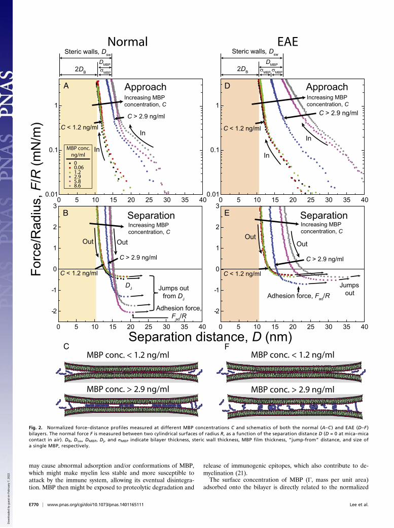
eters for how well a myelin membrane functions. A low inter-membrane distance within the multilayer myelin membrane givesa lower dielectric constant between the core axon and the sur-rounding medium and, according to cable theory, a faster tran-sition time of nervous signal through the axon, compared with aswelled membrane (9). Fig. 3 shows the normalized adhesionforce Fad/R (= min[F/R]), adhesion energy per unit area Ead[= –2Fad/3πR, the Johnson, Kendall, and Roberts (JKR) model(28, 29)], steric hard-wall thickness Dsw, and jump-from distanceDj measured by the SFA, as a function of bulk MBP concen-tration C. As shown in Fig. 3A, the increase of Fad/R with C isabrupt and large (Fad/R ∼–1.7 mN/m, Ead ∼0.36 mJ/m2) in thenormal bilayer, whereas in the EAE bilayer, the Fad/R increase israther gradual and smaller (Fad/R ∼–0.6 mN/m, Ead ∼0.13 mJ/m2).As shown in Fig. 3B, the comparison of Dsw clearly shows thatmodel EAE bilayers exhibit a swollen layer of MBP (DMBP ∼7–8nm) at C > 2.9 ng/mL, whereas DMBP in the normal bilayer iscompact, with a thickness equal to that of a single MBP mole-cule, σMBP (DMBP ∼3–4 nm) as measured by electron microscopy,X-ray, and computational techniques (14, 30, 31). Following thesame trend, Dj is larger in the EAE bilayer (Fig. 3C), which isa possible indication of the formation of a thicker gel-like layeron EAE bilayers at C > 5.8 ng/mL. Structural changes (e.g.,swelling, lesion formations) in autoimmune diseases such asEAE and MS have been observed in the extracellular leaflet ofmyelin membranes in the CNS only, by using electron micros-copy (4, 6). Other studies have reported an alteration in theMBP composition and concentration in the cytoplasmic leaflet (12,20). Fig. 3 clearly shows that both the adhesion energy between(model) cytoplasmic leaflets and the intermembrane distance aresensitive to MBP concentration. Any out-of-balance concentra-tion of MBP likely will result in some instability in myelin structureand greater access of anti-MBP T cells and/or antibodies to MBPat the cytoplasmic leaflet, possibly after some degree of break-down of myelin due to other factors.The differences in adhesion and MBP layer thickness between
normal and EAE bilayers demonstrate the sensitivity of theseproperties to subtle changes in lipid composition (Table 1),which is related to fluidity and lipid domain distributions (8, 10).These changes in lipid membrane fluidity and domain structures
Table 1. Lipid compositions used for the reconstitutedcytoplasmic myelin lipid monolayers
Mole % lipid
Lipid class Normal EAE
Cholesterol 31.6 37.4PS− 7.3 7.4SM+/− 6.2 2.2PC+/− 25.9 20.1PE+/− 29.0 32.9
glue layermica substrate
mica substrate
Rglue
Force, F
DMBP
DB
Silver layer
MBP layer
DPPEmonolayer
DPPE
DiffuseBuffersolution
MBP layer
monolayerMyelin lipidmonolayer
Myelin lipidmonolayer
Fig. 1. Schematic representation of the model myelin bilayer system.Two apposing model myelin bilayers deposited on solid mica substrateswere immersed in an aqueous buffer solution (saturated with lipids) witha bulk MBP concentration of C. By using the Langmuir–Blodgett tech-nique, a DPPE monolayer was deposited as the supporting layer onatomically smooth mica. The myelin (cytoplasmic leaflet) lipid monolayerwas deposited above the predeposited DPPE monolayer. To adsorb MBPto the bilayer surfaces, the MBP solution (in sodium nitrate buffer) wasinjected into the medium with a syringe after separating two bilayers farfrom each other (D > 1 mm). An equilibration time of 30 min was usedafter every injection.
Lee et al. PNAS | Published online February 10, 2014 | E769
BIOPH
YSICSAND
COMPU
TATIONALBIOLO
GY
PNASPL
US
Dow
nloa
ded
by g
uest
on
Feb
ruar
y 7,
202
2
may cause abnormal adsorption and/or conformations of MBP,which might make myelin less stable and more susceptible toattack by the immune system, allowing its eventual disintegra-tion. MBP then might be exposed to proteolytic degradation and
release of immunogenic epitopes, which also contribute to de-myelination (21).The surface concentration of MBP (Γ, mass per unit area)
adsorbed onto the bilayer is directly related to the normalized
Forc
e/R
adiu
s, F
/R (m
N/m
)
ApproachA
In
InC < 1.2 ng/ml
Increasing MBPconcentration, C
C > 2.9 ng/ml
SeparationIncreasing MBPconcentration, C
Out Out
Jumps outfrom DJ
Adhesion force,Fad/R
Separation distance, D (nm)
Increasing MBPconcentration, C
Approach
C > 2.9 ng/ml
C < 1.2 ng/ml
In
In
Separation
Out
Increasing MBPconcentration, C
Out
Jumps outAdhesion force, Fad/R
MBP conc. ng/ml
00.061.22.95.88.6
2DB
Steric walls, Dsw Steric walls, Dsw
σMBP
Normal EAE
2DB
DJ
0 5 10 15 20 25 30 35 40 0 5 10 15 20 25 30 35 400.01
0.1
1
0.01
0.1
1
-2
-1
0
1
2
3
-2
-1
0
1
2
3
C < 1.2 ng/ml
C > 2.9 ng/ml
C < 1.2 ng/ml
C > 2.9 ng/ml
σMBPσMBP
MBP conc. > 2.9 ng/ml
MBP conc. < 1.2 ng/ml
MBP conc. > 2.9 ng/ml
MBP conc. < 1.2 ng/ml
DMBP DMBP
B
C
D
E
F
0 5 10 15 20 25 30 35 40 0 5 10 15 20 25 30 35 40
Fig. 2. Normalized force–distance profiles measured at different MBP concentrations C and schematics of both the normal (A–C ) and EAE (D–F )bilayers. The normal force F is measured between two cylindrical surfaces of radius R, as a function of the separation distance D (D = 0 at mica–micacontact in air). DB, Dsw, DMBP, Dj, and σMBP indicate bilayer thickness, steric wall thickness, MBP film thickness, “jump-from” distance, and size ofa single MBP, respectively.
E770 | www.pnas.org/cgi/doi/10.1073/pnas.1401165111 Lee et al.
Dow
nloa
ded
by g
uest
on
Feb
ruar
y 7,
202
2
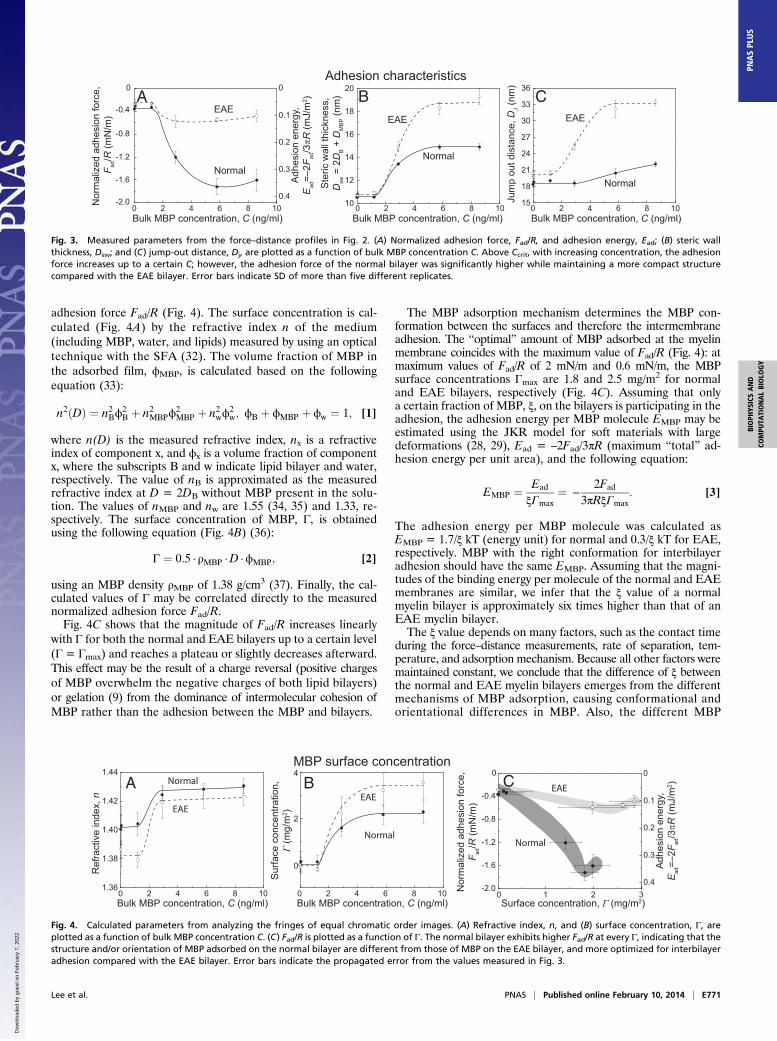
adhesion force Fad/R (Fig. 4). The surface concentration is cal-culated (Fig. 4A) by the refractive index n of the medium(including MBP, water, and lipids) measured by using an opticaltechnique with the SFA (32). The volume fraction of MBP inthe adsorbed film, ϕMBP, is calculated based on the followingequation (33):
n2ðDÞ ¼ n2Bϕ2B þ n2MBPϕ
2MBP þ n2wϕ
2w; ϕB þ ϕMBP þ ϕw ¼ 1; [1]
where n(D) is the measured refractive index, nx is a refractiveindex of component x, and ϕx is a volume fraction of componentx, where the subscripts B and w indicate lipid bilayer and water,respectively. The value of nB is approximated as the measuredrefractive index at D = 2DB without MBP present in the solu-tion. The values of nMBP and nw are 1.55 (34, 35) and 1.33, re-spectively. The surface concentration of MBP, Γ, is obtainedusing the following equation (Fig. 4B) (36):
Γ ¼ 0:5 · ρMBP ·D ·ϕMBP; [2]
using an MBP density ρMBP of 1.38 g/cm3 (37). Finally, the cal-culated values of Γ may be correlated directly to the measurednormalized adhesion force Fad/R.Fig. 4C shows that the magnitude of Fad/R increases linearly
with Γ for both the normal and EAE bilayers up to a certain level(Γ = Γmax) and reaches a plateau or slightly decreases afterward.This effect may be the result of a charge reversal (positive chargesof MBP overwhelm the negative charges of both lipid bilayers)or gelation (9) from the dominance of intermolecular cohesion ofMBP rather than the adhesion between the MBP and bilayers.
The MBP adsorption mechanism determines the MBP con-formation between the surfaces and therefore the intermembraneadhesion. The “optimal” amount of MBP adsorbed at the myelinmembrane coincides with the maximum value of Fad/R (Fig. 4): atmaximum values of Fad/R of 2 mN/m and 0.6 mN/m, the MBPsurface concentrations Γmax are 1.8 and 2.5 mg/m2 for normaland EAE bilayers, respectively (Fig. 4C). Assuming that onlya certain fraction of MBP, ξ, on the bilayers is participating in theadhesion, the adhesion energy per MBP molecule EMBP may beestimated using the JKR model for soft materials with largedeformations (28, 29), Ead = –2Fad/3πR (maximum “total” ad-hesion energy per unit area), and the following equation:
EMBP ¼ Ead
ξΓmax¼ −
2Fad
3πRξΓmax: [3]
The adhesion energy per MBP molecule was calculated asEMBP = 1.7/ξ kT (energy unit) for normal and 0.3/ξ kT for EAE,respectively. MBP with the right conformation for interbilayeradhesion should have the same EMBP. Assuming that the magni-tudes of the binding energy per molecule of the normal and EAEmembranes are similar, we infer that the ξ value of a normalmyelin bilayer is approximately six times higher than that of anEAE myelin bilayer.The ξ value depends on many factors, such as the contact time
during the force–distance measurements, rate of separation, tem-perature, and adsorption mechanism. Because all other factors weremaintained constant, we conclude that the difference of ξ betweenthe normal and EAE myelin bilayers emerges from the differentmechanisms of MBP adsorption, causing conformational andorientational differences in MBP. Also, the different MBP
0
0 0
2 4 6 8 10 0 2 4 6 8 10
-0.40.1
0.2
0.3
0.4
-0.8
-1.2
-1.6
-2.0Nor
mal
ized
adh
esio
n fo
rce,
Fad
/R (m
N/m
)
Adh
esio
n en
ergy
,E
ad=–
2Fad
/3πR
(mJ/
m2 )A B C
EAE
Normal
15
18
21
24
27
30
33
36
Jum
p ou
t dis
tanc
e, D
J (nm
)
Normal
EAE
10
12
14
16
18
20
Ste
ric w
all t
hick
ness
,D
sw =
2D
B +
DM
BP (n
m)
Normal
EAE
Adhesion characteristics
Bulk MBP concentration, C (ng/ml) Bulk MBP concentration, C (ng/ml)0 2 4 6 8 10Bulk MBP concentration, C (ng/ml)
Fig. 3. Measured parameters from the force–distance profiles in Fig. 2. (A) Normalized adhesion force, Fad/R, and adhesion energy, Ead; (B) steric wallthickness, Dsw; and (C) jump-out distance, Dj, are plotted as a function of bulk MBP concentration C. Above Ccrit, with increasing concentration, the adhesionforce increases up to a certain C; however, the adhesion force of the normal bilayer was significantly higher while maintaining a more compact structurecompared with the EAE bilayer. Error bars indicate SD of more than five different replicates.
Ref
ract
ive
inde
x, n
Sur
face
con
cent
ratio
n,Γ
(mg/
m2 )
A B CNormal
Normal
EAEEAE
Normal
EAE
1.36
1.38
1.40
1.42
1.44
0 2 4 6 8 10 0 2 4 6 8 10Bulk MBP concentration, C (ng/ml) Bulk MBP concentration, C (ng/ml)
0
2
4
Surface concentration, Γ (mg/m2)0 1 2 3
-2.0
-1.6
-1.2
-0.8
-0.4
0
Nor
mal
ized
adh
esio
n fo
rce,
F ad/R
(mN
/m)
MBP surface concentration0
0.1
0.2
0.3
0.4
Adh
esio
n en
ergy
,E
ad=–
2Fad
/3πR
(mJ/
m2 )
Fig. 4. Calculated parameters from analyzing the fringes of equal chromatic order images. (A) Refractive index, n, and (B) surface concentration, Γ, areplotted as a function of bulk MBP concentration C. (C) Fad/R is plotted as a function of Γ. The normal bilayer exhibits higher Fad/R at every Γ, indicating that thestructure and/or orientation of MBP adsorbed on the normal bilayer are different from those of MBP on the EAE bilayer, and more optimized for interbilayeradhesion compared with the EAE bilayer. Error bars indicate the propagated error from the values measured in Fig. 3.
Lee et al. PNAS | Published online February 10, 2014 | E771
BIOPH
YSICSAND
COMPU
TATIONALBIOLO
GY
PNASPL
US
Dow
nloa
ded
by g
uest
on
Feb
ruar
y 7,
202
2
adsorption mechanisms mainly are a result of the difference inthe lipid compositions and fluidity.
MBP Film Viscosity Between Supported Cytoplasmic Model MyelinBilayers. The thin film rheology measurement between the modelmyelin bilayers provides information about the thin film viscosity, η;hydrodynamic thickness, ΔH; and diffuse layer thickness (38, 39)of MBP. The thin film rheology study measures the couplingbetween oscillation (vibration) of the upper cylindrical surface(amplitude Ao and frequency v) in the z-direction with a lowersurface that is suspended on a cantilever spring with spring
stiffness K (38) (seeMaterials and Methods for details). When theseparation distance D between the two apposing surfaces is large,the lower surface does not move because of weak viscous cou-pling between the two surfaces. As D is reduced gradually whilethe upper surface is oscillated, the lower surface starts to “feel”the viscous force induced by the oscillation of the upper surfaceand oscillates at a relative amplitude A. When D approaches Dsw,all the energy generated from the oscillation of the upper surfaceis transmitted to the lower surface without any energy loss, causingthe two surfaces to move in phase with A = 0. The relation be-tween viscosity, η; the distance between two surfaces,D; the applied
10 20 30 40
A B C
10 20 30 40
MBP in solution(C < 1.2 ng/ml)
2010 30 40 50
MBP in solution(C > 2.9 ng/ml)
Separation distance, D (nm) Separation distance, D (nm) Separation distance, D (nm)
12π2
R2 v
/(K[(A
0/A)2 -(
1-f/K
)2 ]1/2
(m
3 /N·s
x 1
05 )
0
1
2
3
Depleted
DMBP H
Diffuse MBP
Bare bilayers (no MBP)
(η < ηb)η b
=(1.1 ±
0.1)⋅η w
η b=(1.1 ± 0.1)⋅η w
η b=(1.
2 ± 0.
2)⋅η w
water
water
zone
water
η ≈ (2.5 ± 1.0)⋅η b
layer (η > ηb)
Δ2
Thin film viscosity
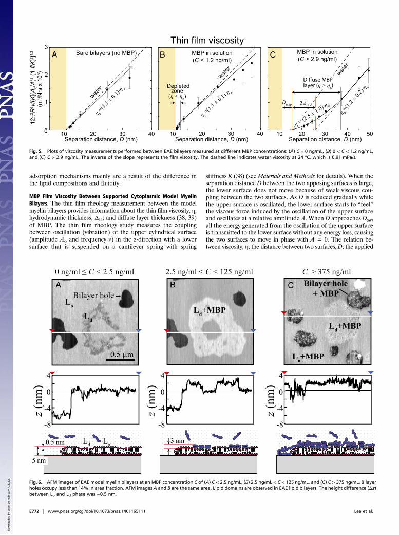
Fig. 5. Plots of viscosity measurements performed between EAE bilayers measured at different MBP concentrations: (A) C = 0 ng/mL, (B) 0 < C < 1.2 ng/mL,and (C) C > 2.9 ng/mL. The inverse of the slope represents the film viscosity. The dashed line indicates water viscosity at 24 °C, which is 0.91 mPa/s.
-8
-4
0
4
-8
-4
0
4
A B C
z (nm
)
z (nm
)
0.5 μm
0 ng/ml ≤ C < 2.5 ng/ml 2.5 ng/ml < C < 125 ng/ml C > 375 ng/ml
0.5 nm 3 nmLo
Lo
Bilayer holeBilayer hole
+ MBP
Ld
Ld
Ld+MBP
Ld+MBP
Lo+MBP
-8
-4
0
4
z (nm
)
5 nm
Fig. 6. AFM images of EAE model myelin bilayers at an MBP concentration C of (A) C < 2.5 ng/mL, (B) 2.5 ng/mL < C < 125 ng/mL, and (C) C > 375 ng/mL. Bilayerholes occupy less than 14% in area fraction. AFM images A and B are the same area. Lipid domains are observed in EAE lipid bilayers. The height difference (Δz)between Lo and Ld phase was ∼0.5 nm.
E772 | www.pnas.org/cgi/doi/10.1073/pnas.1401165111 Lee et al.
Dow
nloa
ded
by g
uest
on
Feb
ruar
y 7,
202
2
vibration frequency, v; the applied vibration amplitude, Ao; and themeasured relative amplitude A is expressed as (38)
ηðDÞ ¼ KD12π2R2v
"�Ao
A
�2
−�1−
fK
�2#1 =
2
; [4]
where f = dF(D)/dD is the gradient of the interfacial force.Fig. 5 shows plots of 12π2R2v/(K[(A0/A)
2–(1–f/K)2]1/2) against
D measured between EAE bilayers, where the inverse slope of theplotted line gives the viscosity. When the bulk concentration, C,of MBP is less than 1.2 ng/mL (Fig. 5B), no signs of MBP ad-sorption onto the bilayers are detected and the results are verysimilar to bare bilayers (Fig. 5A). However, when the separationdistance between two bilayers is small (∼2 nm), a “depletionzone” exists in which η is slightly smaller than the bulk viscosityηb and is similar to water’s viscosity ηw, which is a clear effectfrom MBP in solution that did not adsorb to the bilayer surfaces.The existence of a depletion zone in nonadsorbing polymericsolutions already was shown by Kuhl et al. (40). When C ishigher than 2.9 ng/mL (Fig. 5C), completely different rheologicalproperties related to MBP adsorption are observed: (i) the hy-drodynamic thickness ΔH is shifted outward about ∼5.5 nm foreach surface; (ii) the thickness of the diffusive MBP layer isestimated to be D ∼21 nm; (iii) when the two diffusive MBPlayers overlap, the effective viscosity of the MBP film increasesup to two to threee times of ηb.The trends are similar for normal bilayers (Fig. S1); however,
DMBP, ΔH, and the diffuse MBP layer thickness are ∼2.5, 1.3 and4 nm, respectively, which are smaller compared with those ofEAE bilayers. The smaller values of the above three parametersobtained for normal bilayers compared with EAE bilayers indicatethat MBP is adsorbed in a more compact manner on normalbilayers (without swelling and a thick gel-like layer formation),which is consistent with the higher adhesion of normal bilayers.
Selective Distribution of MBP on the Lipid Membrane. Several pro-teins are known to adhere selectively to lipid domains of biologicalmembranes (41); however, the role of lipid domains in the distri-bution of MBP still is not established. Previous studies (8, 10)conducted with fluorescence microscopy showed the absence ofmicrometer-sized domains at a surface pressure, Π, of ∼30 mN/m.However, because of the resolution limit of the fluorescence mi-croscope, submicron- or nano-sized domains still may exist.Fig. 6 shows the topography of the EAE myelin lipid bilayers
as a function of MBP concentration imaged with an atomic forcemicroscope in aqueous solution. Bilayers without MBP (Fig. 6A)had a thickness of ∼5 nm, which was consistent with the SFAmeasurements. Also, noncircular nano-sized domains (area frac-tion of 8%) were observed and were ∼0.5 nm thinner than thesurrounding bilayer, indicating that these domains were in a liq-uid-disordered phase (Ld) and the surrounding bilayer was in aliquid-ordered phase (Lo).After injecting a low concentration of MBP (Fig. 6A, C ∼2.5
ng/mL), no changes in the bilayer structure were observedcompared with the bare bilayer. However, when C was increasedto 125 ng/mL (Fig. 6B), MBP adsorbed selectively only to Ldlipid domains. MBP-bound lipid domains became ∼2 nm thickerthan the (MBP-free) Lo phase, indicating an MBP thickness of∼3 nm, which is consistent with the SFA measurements andprevious studies (9, 14, 42). Also, the area fraction of bilayerholes (which we used to confirm the bilayer thickness; see Fig. S2for larger AFM images) decreased after the MBP adsorption(from 14% to 12%), which provides evidence of bilayer expan-sion caused by partial penetration of MBP into the upper leafletof the bilayer due to the hydrophobic attraction between thehydrophobic moieties of MBP and hydrocarbon chains of the
myelin bilayer. The selective adsorption of MBP to the Ld phaseshould be a result of the hydrophobic interaction, because thelipids in the Ld phase have a greater molecular area comparedwith those in the Lo phase, i.e., more hydrophobic residues areexposed. The normal model bilayers did not show significantdifferences in terms of selective MBP adsorption; however, theyexhibited a higher area fraction of Ld+MBP phase (8.4%) com-pared with EAE lipid bilayers (5.5%) (Fig. S3).At bulk MBP concentrations up to 375 ng/mL (Fig. 6C), the
MBP adsorbs unselectively to both the Ld and Lo phases, and thethickness difference between the two phases becomes very small(< 0.5 nm). In addition, MBP adsorbs to bilayer holes that causethe depth of the holes in the lipid bilayer to be smaller (2 nm).The nonselective adsorption of MBP to the bilayer surface causesMBP to cover the whole bilayer surface (possibly as a multilayer),forming a gel-like film, as found by viscosity measurements. Thegel-like structure of MBP, which possibly is induced by the co-hesion between β-sheet regimes of MBP (11, 23), may alter thestructure and orientation of MBP. The altered structure of MBPmight not be optimal for the high interbilayer adhesion force inmyelin. This result is consistent with the SFA results showinga decrease in adhesion after an excessive amount of MBP forboth normal and EAE bilayers (Fig. 3).
DiscussionMBP Structure and Adsorption Mechanism on Bilayers. The resultsprovide qualitative and quantitative information regarding notonly the structure of adsorbed MBP but also the mechanism ofhow MBP adsorbs to myelin bilayer surfaces. Based on theseresults and previous studies (14, 42, 43) of normal myelin bilayers,MBP should be forming a thin yet compact structure (Fig. 1)with a thickness of ∼3 nm, holding two opposing bilayers to-gether via electrostatic and hydrophobic interactions. Here wefind that between two EAE bilayers, the compact structure ofMBP is disturbed and forms a swollen gel-like structure (∼6–7 nmthickness) and a thick diffuse MBP layer (∼21 nm) with a hydro-dynamic thickness ∼5.5 nm, as confirmed by normal force (Fig. 2)and film viscosity (Fig. 5) measurements. The thick gel-like MBPlayer causes a significant decrease in the adhesion force (4, 6, 9),which might be one of the causes, at a molecular level, of bilayerdegradation during the progression of demyelinating diseases.The fact that MBP does not adsorb below a certain bulk
concentration Ccrit, whereas rapid adsorption of MBP occursabove Ccrit, is consistent with cooperative adsorption. The co-operative adsorption of MBP is involved closely in protein–protein and protein–lipid/lipid domain interactions, which mightbe an effective way for the MBP to bind to myelin bilayers,resulting in a(n) (optimum) high adhesion force.
Role of Lipid Domains in the Myelin Membrane. The existence andfunctions of lipid domains in myelin membranes already wereshown in many studies (8–10, 44). Normal and EAE myelin lipidmonolayers have different lipid domain sizes and distributions at thesame temperature and surface pressure (8, 10) that are attributed tosmall differences in lipid compositions (Table 1). These differencesin lipid composition also give rise to different membrane fluiditieswhile the lipid charge density remains similar. The measured dif-ferences in adhesion characteristics (Fig. 3) andMBP coverage (Fig.4) between normal and EAE bilayers result from differences inmembrane fluidity and/or lipid domain distribution. Sphingomyelin,known to be critical for the formation of liquid-ordered phases inmulticomponent lipid membranes (41), is up to three times moreabundant in normal compared with EAE myelin bilayers. Choles-terol also is known to contribute to lipid domain formation (41) andaffects the fluidity of membranes (45), and EAE myelin containsmore cholesterol than normal myelin (Table 1).Because the charge density between normal and EAE bilayers is
similar, all the differences between normal and EAE bilayers are
Lee et al. PNAS | Published online February 10, 2014 | E773
BIOPH
YSICSAND
COMPU
TATIONALBIOLO
GY
PNASPL
US
Dow
nloa
ded
by g
uest
on
Feb
ruar
y 7,
202
2
the result of lipid fluidity and/or lipid domains. Without MBP be-tween bilayers, the force profiles show little difference betweennormal and EAE bilayers. Above Ccrit of MBP, the force profilesshow significant discrimination between normal and EAE bilayers:with increasing C, the force profile of normal bilayers shows anincrease in the adhesion force while the bilayers maintain a compactstructure, whereas the EAE bilayers show little increase in the ad-hesion force with significant swelling of the system.AFM results (Fig. 6) show that MBP binds selectively to the Ld
phase of lipid bilayers up to a certain bulk MBP concentration.Thus, it may be concluded that although membrane fluidity andlipid domains have minimal effect on the adhesive propertiesbetween supported lipid bilayers, lipid domains modify the mech-anism of MBP adsorption (e.g., amount, structure, and orientation)onto the model myelin bilayers that directly affect the in-termembrane adhesion mediated by MBP.
Implications for Demyelinating Diseases. The forces betweenbilayers of myelin with phase-separated lipid domains and aninhomogeneous protein distribution are fundamentally differentfrom those between single-phase and homogeneous bilayers.These inhomogeneities can dramatically amplify the effect ofeven small changes in the lipid composition or protein isoform(20), resulting in large changes in bilayer adhesion (7, 15) thatmay cause the myelin sheath to unravel. Lipid domains withinlipid bilayers are known to be correlated with bilayer–bilayerinteractions (46) and may play important roles in adhesion (47),intermembrane spacing (48), and permeability (49). Lipid domainsalso are thought to act as platforms on which proteins can adsorbselectively (50). Accordingly, any difference in lipid domain orga-nization within myelin bilayers most likely will alter the MBPadsorption properties, as well as intermembrane adhesion andspacing, and will have a direct impact on the stability of themyelin structure. When the stability of the membrane structureis disturbed, the myelin might be more susceptible to attack bythe immune system and possibly progress to demyelinating dis-eases such as MS.The primary cause of demyelinating diseases (such as MS)
still is under debate, as is the molecular mechanism of in-termembrane (de-)stabilization. Here, we focused on the effectsof lipid composition and MBP concentration on intermembraneadhesion. With normal model myelin bilayers, we observed acompact protein layer with a thin MBP film, resulting in highintermembrane adhesion, whereas with EAE bilayers, swellingand a decrease in adhesion were observed. Swelling of the myelinmembrane increases the dielectric constant between the neuralcore (e.g., axon) and the medium surrounding the neural cell,which reduces electric conduction through the axon and weakensor deteriorates the signal’s transmission through the nervoussystem. The swelling and loss of adhesion might contribute to thedisruption of myelin structure during the progression of de-myelinating diseases (such as MS), from which one might inferthat the molecular indication of membrane swelling and loss ofadhesion may originate from an alteration of lipid compositionand MBP concentration.
Materials and MethodsNormal and EAE Cytoplasmic Model Myelin. A previous study (6) using NMR andHPLC techniques identified the lipid composition of white matter from normaland diseased marmosets after induction of EAE. With the distribution of lipids inmyelin determined by Inouye and Kirschner (1, 24), lipid compositions of bothnormal and EAE cytoplasmic leaflets were calculated (Table 1).
To mimic the cytoplasmic myelin leaflet, the following porcine brain-extracted lipids were purchased from Avanti Polar Lipids, stored in chloro-form, and kept in a deep freezer (−50 °C) until used: phosphatidylserine(porcine brain PS−), sphingomyelin (porcine brain SM+/−), phosphatidylcho-line (porcine brain PC+/−), phosphatidylethanolamine (porcine brain PE+/−),and cholesterol (ovine wool). The major fatty acid chain lengths of thePC, PE, and PS are 16:0, 18:0, 18:1, and 20:4. Dipalmitoylphosphatidyl-ethanolamine (DPPE), sodium nitrate (purity ≥99.0%), and morpholine-propanesulfonic acid (Mops) sodium salt (purity ≥99.5%) were purchasedfrom Sigma–Aldrich. To disperse the lipids, the following solvents werepurchased from Sigma–Aldrich: chloroform (CHROMASOLV Plus for HPLC,purity ≥99.9%), hexane (RegentPlus, purity ≥99.0%), ethanol (200 proof,HPLC/spectrophotometric grade), and methanol (CHROMASOLV Plus forHPLC, purity ≥99.9%). MBP was isolated from bovine brain white matter (51)and kept in a deep freezer (−50 °C) until use.
Supported Bilayer Preparation. Freshly cleaved and back-silvered atomicallysmoothmica sheets were glued onto two cylindrical glass disks with curvatureradii of 2 cm. Lipid bilayers were deposited onto themica using the Langmuir–Blodgett deposition technique (25). For the first supporting monolayer, 100 μLof 1 mg/mL DPPE solution [3:1 (vol/vol) chloroform/methanol] was spread ontoMilli-Q water (Millipore) and deposited at a surface pressure of 35 mN/m(molecular area of ∼43 Å2). For the second layer, normal and EAE myelinlipid solutions were prepared (see the lipid composition in Table 1) in dis-persed form in the 11:5:4 (vol/vol) hexane/chloroform/ethanol solvent mix-tures. The second layer was deposited onto the DPPE-deposited micasubstrates at a surface pressure of 30 mN/m (molecular area of ∼50–52 Å2)with a subphase of sodium nitrate buffer (150 mM sodium nitrate/10 mMMops sodium salt, pH 7.4). The bilayer-deposited samples were transferredimmediately to the SFA chamber (without being exposed to air) prefilledwith degassed saturated lipid solution (sodium nitrate buffer in contact withlipid crystals for 12 h, and degassed for 2 h before the experiment).
SFA Experiments. The SFA 2000 (26) was used in this study for the forcemeasurements. Bilayer-deposited disks were installed in the SFA chamber ina cross-cylindrical geometry, which at small separation distances, is equiva-lent to a sphere-on-flat geometry. The separation distance between twosurfaces is measured using optical interferometry (52), and the force be-tween two surfaces can be calculated from the deflection of a double-can-tilever spring of known spring constant that holds the lower disk holder. Forthe force–distance measurements, the surfaces approach each other witha speed of 0.5–1.0 nm/s, which is expected to be slow enough to maintaina quasi-equilibrium state at all times. To measure film viscosity (38, 39), apiezoelectric crystal was used to oscillate the upper surface relative to thelower surface at an applied amplitude A0 by applying an ac voltage (15 V,0.5 Hz, square wave for A0 and sine wave for the relative amplitude A) to thepiezoelectric crystal. While oscillating, the surfaces slowly are brought closertogether in a stepwise manner while measuring the relative amplitude Awith varying D.
MBP solution (dissolved in sodium nitrate solution) was prefiltered with anAnotop10 0.1-μm filter (Whatman) and injected into the SFA chamber ina stepwise manner after the two bilayers were separated far from eachother (D > 1 mm). After each injection, the system was equilibrated for 0.5 h,allowing sufficient time for MBP to adsorb to the model myelin bilayers.
AFM Experiments. Images were acquired with an MFP-3D-Bio AFM (AsylumResearch) using an MSNL probe (Bruker) with a spring constant of 0.1 ± 0.05N·m−1. Scanning was performed in tapping mode at room temperature (22 ±1 °C) in lipid-saturated sodium nitrate buffer (150 mM sodium nitrate/10 mMMops sodium salt, pH 7.4). The EAE myelin bilayers were prepared as de-scribed above and covered with 4 mL of sodium nitrate buffer in a reservoir.MBP concentration was increased from 0 to 375 ng/mL by stepwise injectionof MBP solution (prepared as in SFA experiments) into the reservoir. Thesample was equilibrated for 30 min after each injection.
ACKNOWLEDGMENTS. This work was supported by National Institutes ofHealth Grant R01 GM076709. X.B. and Y.K. are grateful for the financial sup-port of the Santa Barbara Foundation through the Otis Williams Fellowship.J.M.B. thanks Canadian Institutes of Health Research Grant MOP 86483.
1. Inouye H, Kirschner DA (1988) Membrane interactions in nerve myelin. I. De-
termination of surface charge from effects of pH and ionic strength on period. Bio-
phys J 53(2):235–245.2. Morell P (1984) Myelin (Plenum, New York), 2nd Ed.3. Martenson R (1992) Myelin: Biology and Chemistry (CRC, Boca Raton, FL).
4. Genain CP, Cannella B, Hauser SL, Raine CS (1999) Identification of autoantibodies
associated with myelin damage in multiple sclerosis. Nat Med 5(2):170–175.5. Hafler DA (2004) Multiple sclerosis. J Clin Invest 113(6):788–794.6. Ohler B, et al. (2004) Role of lipid interactions in autoimmune demyelination. Biochim
Biophys Acta 1688(1):10–17.
E774 | www.pnas.org/cgi/doi/10.1073/pnas.1401165111 Lee et al.
Dow
nloa
ded
by g
uest
on
Feb
ruar
y 7,
202
2
7. Banquy X, Kristiansen K, Lee DW, Israelachvili JN (2012) Adhesion and hemifusion ofcytoplasmic myelin lipid membranes are highly dependent on the lipid composition.Biochim Biophys Acta 1818(3):402–410.
8. Min Y, et al. (2011) Critical and off-critical miscibility transitions in model extracellularand cytoplasmic myelin lipid monolayers. Biophys J 100(6):1490–1498.
9. Min Y, et al. (2009) Interaction forces and adhesion of supported myelin lipid bilayersmodulated by myelin basic protein. Proc Natl Acad Sci USA 106(9):3154–3159.
10. Lee DW, et al. (2011) Relating domain size distribution to line tension and moleculardipole density in model cytoplasmic myelin lipid monolayers. Proc Natl Acad Sci USA108(23):9425–9430.
11. Sedzik J, Kirschner DA (1992) Is myelin basic protein crystallizable? Neurochem Res17(2):157–166.
12. Boggs JM (2006) Myelin basic protein: A multifunctional protein. Cell Mol Life Sci63(17):1945–1961.
13. Harauz G, Ladizhansky V, Boggs JM (2009) Structural polymorphism and multi-functionality of myelin basic protein. Biochemistry 48(34):8094–8104.
14. Beniac DR, et al. (1997) Three-dimensional structure of myelin basic protein. I. Re-construction via angular reconstitution of randomly oriented single particles. J BiolChem 272(7):4261–4268.
15. Hu Y, et al. (2004) Synergistic interactions of lipids and myelin basic protein. Proc NatlAcad Sci USA 101(37):13466–13471.
16. MacNaughtan W, Snook KA, Caspi E, Franks NP (1985) An X-ray diffraction analysis oforiented lipid multilayers containing basic proteins. Biochim Biophys Acta 818(2):132–148.
17. Mueller H, Butt HJ, Bamberg E (1999) Force measurements on myelin basic proteinadsorbed to mica and lipid bilayer surfaces done with the atomic force microscope.Biophys J 76(2):1072–1079.
18. Wheeler D, Bandaru VVR, Calabresi PA, Nath A, Haughey NJ (2008) A defect ofsphingolipid metabolism modifies the properties of normal appearing white matterin multiple sclerosis. Brain 131(Pt 11):3092–3102.
19. Jo EJ, Boggs JM (1995) Aggregation of acidic lipid vesicles by myelin basic protein:Dependence on potassium concentration. Biochemistry 34(41):13705–13716.
20. Husted C (2006) Structural insight into the role of myelin basic protein in multiplesclerosis. Proc Natl Acad Sci USA 103(12):4339–4340.
21. Musse AA, Boggs JM, Harauz G (2006) Deimination of membrane-bound myelin basicprotein in multiple sclerosis exposes an immunodominant epitope. Proc Natl Acad SciUSA 103(12):4422–4427.
22. Aggarwal S, et al. (2013) Myelin membrane assembly is driven by a phase transition ofmyelin basic proteins into a cohesive protein meshwork. PLoS Biol 11(6):e1001577.
23. Kattnig DR, Bund T, Boggs JM, Harauz G, Hinderberger D (2012) Lateral self-assemblyof 18.5-kDa myelin basic protein (MBP) charge component-C1 on membranes. Bio-chim Biophys Acta 1818(11):2636–2647.
24. Inouye H, Kirschner DA (1988) Membrane interactions in nerve myelin: II. De-termination of surface charge from biochemical data. Biophys J 53(2):247–260.
25. Zasadzinski JA, Viswanathan R, Madsen L, Garnaes J, Schwartz DK (1994) Langmuir-Blodgett films. Science 263(5154):1726–1733.
26. Israelachvili J, et al. (2010) Recent advances in the surface forces apparatus (SFA)technique. Rep Prog Phys 73(3):036601.
27. Israelachvili J (1994) Self-assembly in two dimensions—surface micelles and domainformation in monolayers. Langmuir 10(10):3774–3781.
28. Israelachvili JN (2011) Intermolecular and Surface Forces (Academic, London), 3rd Ed.29. Johnson KL, Kendall K, Roberts AD (1971) Surface energy and contact of elastic solids.
Proc R Soc Lond A Math Phys Sci 324(1558):301–313.
30. Haas H, et al. (2004) Small angle x-ray scattering from lipid-bound myelin basic pro-tein in solution. Biophys J 86(1 Pt 1):455–460.
31. Ridsdale RA, Beniac DR, Tompkins TA, Moscarello MA, Harauz G (1997) Three-dimensional structure of myelin basic protein. II. Molecular modeling and consid-erations of predicted structures in multiple sclerosis. J Biol Chem 272(7):4269–4275.
32. Tadmor R, Chen NH, Israelachvili JN (2003) Thickness and refractive index measure-ments using multiple beam interference fringes (FECO). J Colloid Interface Sci 264(2):548–553.
33. Israelachvili JN, Sammut RA, Snyder AW (1976) Birefringence and dichroism of pho-toreceptors. Vision Res 16(1):47–52.
34. Barer R, Tkaczyk S (1954) Refractive index of concentrated protein solutions. Nature173(4409):821–822.
35. Fisk AA (1950) The thicknesses of hemoglobin and bovine serum albumin molecules asunimolecular layers adsorbed onto films of barium stearate. Proc Natl Acad Sci USA36(10):518–523.
36. Raviv U, et al. (2002) Properties and interactions of physigrafted end-functionalizedpoly(ethylene glycol) layers. Langmuir 18(20):7482–7495.
37. Smith R (1982) Self-association of myelin basic protein: Enhancement by detergentsand lipids. Biochemistry 21(11):2697–2701.
38. Israelachvili JN (1986) Measurement of the viscosity of liquids in very thin-films.J Colloid Interface Sci 110(1):263–271.
39. Israelachvili JN (1986) Measurements of the viscosity of thin fluid films between twosurfaces with and without adsorbed polymers. Colloid Polym Sci 264(12):1060–1065.
40. Kuhl TL, Berman AD, Hui SW, Israelachvili JN (1998) Direct measurement of depletionattraction and thin film viscosity between lipid bilayers in aqueous polyethyleneglycol solutions. Macromolecules 31(23):8250–8257.
41. Simons K, Ehehalt R (2002) Cholesterol, lipid rafts, and disease. J Clin Invest 110(5):597–603.
42. Keniry MA, Smith R (1979) Circular dichroic analysis of the secondary structure ofmyelin basic protein and derived peptides bound to detergents and to lipid vesicles.Biochim Biophys Acta 578(2):381–391.
43. Ahmed MAM, Bamm VV, Harauz G, Ladizhansky V (2010) Solid-state NMR spectros-copy of membrane-associated myelin basic protein—conformation and dynamics ofan immunodominant epitope. Biophys J 99(4):1247–1255.
44. Debruin LS, Harauz G (2007) White matter rafting—membrane microdomains inmyelin. Neurochem Res 32(2):213–228.
45. Cooper RA (1978) Influence of increased membrane cholesterol on membrane fluidityand cell function in human red blood cells. J Supramol Struct 8(4):413–430.
46. Tayebi L, et al. (2012) Long-range interlayer alignment of intralayer domains instacked lipid bilayers. Nat Mater 11(12):1074–1080.
47. Nusrat A, et al. (2000) Tight junctions are membrane microdomains. J Cell Sci 113(Pt 10):1771–1781.
48. Kaizuka Y, Groves JT (2004) Structure and dynamics of supported intermembranejunctions. Biophys J 86(2):905–912.
49. Rawicz W, Smith BA, McIntosh TJ, Simon SA, Evans E (2008) Elasticity, strength, andwater permeability of bilayers that contain raft microdomain-forming lipids. BiophysJ 94(12):4725–4736.
50. Simons K, Ikonen E (1997) Functional rafts in cell membranes. Nature 387(6633):569–572.
51. Cheifetz S, Moscarello MA (1985) Effect of bovine basic protein charge micro-heterogeneity on protein-induced aggregation of unilamellar vesicles containinga mixture of acidic and neutral phospholipids. Biochemistry 24(8):1909–1914.
52. Israelachvili J (1973) Thin-film studies using multiple-beam interferometry. J ColloidInterface Sci 44(2):259–272.
Lee et al. PNAS | Published online February 10, 2014 | E775
BIOPH
YSICSAND
COMPU
TATIONALBIOLO
GY
PNASPL
US
Dow
nloa
ded
by g
uest
on
Feb
ruar
y 7,
202
2