Embed Size (px)
Citation preview

PRIMARY DISEASES OF MYELIN

Acquired forms- Characterized by preferential damage to
previously normal myelin with relative preservation of axons and include:
1. Immune-mediated injury, such as multiple sclerosis (MS)
2. Progressive multifocal leukoencephalopathy

3. And injury caused by drugs (chemotherapy)4. Central pontine myelinolysis

- The natural history of demyelinating diseases is determined, by
a. The limited capacity of the CNS to regenerate normal myelin
b. and by the degree of secondary damage to axons that occurs as the disease runs its course.

1.Multiple sclerosis

- Multiple sclerosis (MS) is an autoimmune demyelinating disorder characterized by distinct episodes of neurologic deficits, separated in time, caused by white matter lesions(plaques) that are separated in space.

- Multiple sclerosis is the most common of the demyelinating disorders,
- The disease may become clinically apparent at any age
- Although onset in childhood or after age 50 years is relatively rare.
- Women are affected twice as often as are men.

- In most individuals with MS, the clinical course takes the form of relapsing and remitting episodes of variable duration (weeks to months marked by neurologic deficits, followed by gradual, partial recovery of neurologic function.

- The frequency of relapses tends to decrease during the course of time, but there is a steady neurologic deterioration in most affected individuals.due to accumulated damage to axons

Pathogenesis.
- The lesions of MS are caused by an auto immune response directed against components of the myelin sheath.
- The pathogenesis involves both genetic and environmental factors

- The incidence of MS is 15-fold higher when the disease is present in a first-degree relative
Genetic factors1. There is a strong effect from the DR2
extended haplotype of the major histocompatibility complex;

2. There is association with IL-2 and IL-7 receptor genes
3. and subsequently with a number of genes encoding proteins involved in immune response, including cytokines and their receptors and cytoplasmic signaling proteins

Mechanism of demyelination- The available evidence indicates that the disease
is initiated by TH1 and TH17 T -cells that react against myelin antigens and secrete cytokines.

Immune mechanisms involved in destruction of myelin
a. TH1 cells secrete IFN-γ, which activates macrophages,
b. and TH17 cells promote the recruitment of leukocytes

- The demyelination is caused by these activated leukocytes and their injurious products.
- The infiltrate in plaques and surrounding regions of the brain consists of T cells (mainly CD4+, some CD8+) and macrophages.

- How the autoimmune reaction is initiated is not understood;

• It is postulated that in MS patients the myelin specific T cells escape central tolerance, and have been characterized as low avidity T cells that do not engage their antigen during maturation with sufficient strength to induce deletion.

- These autoreactive T-cells might be activated by environmental factors such as EBV infection and cause their activation.

Morphology
GROSS:1. Is a white matter disease that is best
appreciated in sections of the brain and spinal cord.
2. In the fresh state, the lesions are firmer (sclerosis) than the surrounding white matter
.

MS plaques

Multiple sclerosis; Plaques by H&E and Luxol fast blue


3. Lesions appear well circumscribed, somewhat depressed, gray-tan, irregularly shaped plaques
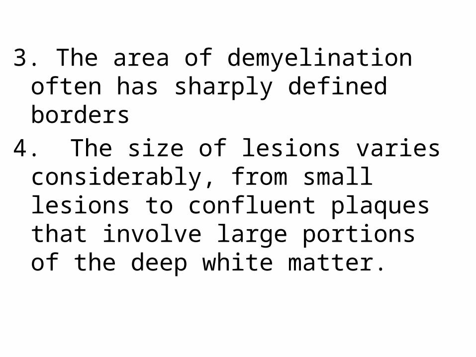
3. The area of demyelination often has sharply defined borders
4. The size of lesions varies considerably, from small lesions to confluent plaques that involve large portions of the deep white matter.

5. Plaques commonly occur a. adjacent to the lateral ventricles, and are also
frequent in theb. optic nerves and chiasm,

b. Brainstem, c. Ascending and descending fiber tracts,d. cerebellum, and spinal cord. Note: Plaques can also extend into gray
matter,

Microscopically- Active plaque a. There is relative preservation of axons and
depletion of oligodendrocytes.b. associated with abundant macrophages
containing lipid-rich, PAS-positive debris.

c. Lymphocytes and monocytes are also present, mostly as perivascular cuffs, especially at the outer edge of the lesion
d. In time, astrocytes undergo reactive changes.

Active MS plaque

• Quiescent: inactive plaques, 1. The inflammatory cells slowly disappear2. Little to no myelin is found, 3. There is a reduction in the number of
oligodendrocyte nuclei;

4. Astrocytic proliferation and gliosis are prominent.
5..Axons show severe demyelination and are also greatly diminished in number.

Quiescent plaque

Quiescent plaque- Luxol fast blue stain


- Abnormally myelinated fibers have also been observed at the edges of typical plaques.
- Although these histologic findings suggest a limited potential for remyelination in the CNS, the remaining axons within most MS plaques remain unmyelinated;

Mechanism of Remyelination- Remyelination Of demyelinated axons is
necessary for restoring their function- But It is incomplete

- It occurs due to migration of oligodendrocyte precursor cells to the lesions;
- Therefore can proliferate and differentiate - but are usually unable to completely
remyelinate remaining axons

Failure of remyelination• Possible mechanisms1. Reexpression on demyelinated axon surface in
MS plaques of neural cell adhesion molecule (NCAM), a negative signal for myelination

2. Astrocytes may form dense glial scar within the plaque and at its edge, so may prevent migration of oligodendocyte precursor cells to the lesions

• Clinical Features.• Although MS lesions can occur anywhere in the
CNS and consequently may induce a wide range of clinical manifestations, certain patterns of neurologic symptoms and signs are more common.

1. Unilateral visual impairment due to involvement of the optic nerve (optic neuritis,) a frequent initial manifestation of MS.

- However, about (10% to 50%, depending on the population studied) with an episode of optic neuritis go on to develop MS (which requires multiple episodes to support the diagnosis).

b. Involvement of the brainstem produces cranial nerve signs, ataxia, nystagmus, and internuclear ophthalmoplegia from interruption of the fibers of the medial longitudinal fasciculus.

c. Spinal cord lesions give rise to motor and sensory impairment of trunk and limbs, spasticity, and difficulties with the voluntary control of bladder function.

• Examination of the CSF in individuals with MS shows
a. a mildly elevated protein level and in one third of cases a moderate pleocytosis.(increase in WBCs)
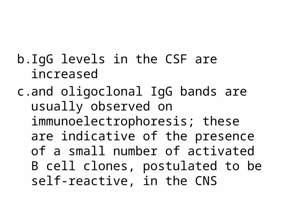
b. IgG levels in the CSF are increased c. and oligoclonal IgG bands are usually
observed on immunoelectrophoresis; these are indicative of the presence of a small number of activated B cell clones, postulated to be self-reactive, in the CNS


Chronic multiple sclerosis patterns1.Relapsing remitting pattern- Most common pattern- Usually symptoms have acute or subacute onset,
with symptoms increasing over days to weeks to months followed by a period of a period of recovery over 1-2 months Remission)

- Remission of symptoms occurs due to redistribution of sodium channels throughout the axolemma of demylinated segment.
- But overtime, over time there is usually a gradual, often stepwise, accumulation of neurologic deficits
- Due to accumulated damage to axons

Mechanism of damage to axons- While MS is characterized by the presence of
de-myelination out of proportion to axonal loss, some injury to axons does occur because
a. chronic and recurrent inflammation release cytotoxic enzymes , matrix metalloproteases and NO from infiltrating lymphocytes cause damage to axons

- Changes in cognitive function can be present, but are often much milder than the other deficits
- In any individual patient, it is hard to predict when the next relapse will occur;

2. Neuromyelitis Optica
- Neuromyelitis optica (NMO) is a syndrome characterized by
1. Synchronous bilateral optic neuritis 2. and spinal cord demyelination.
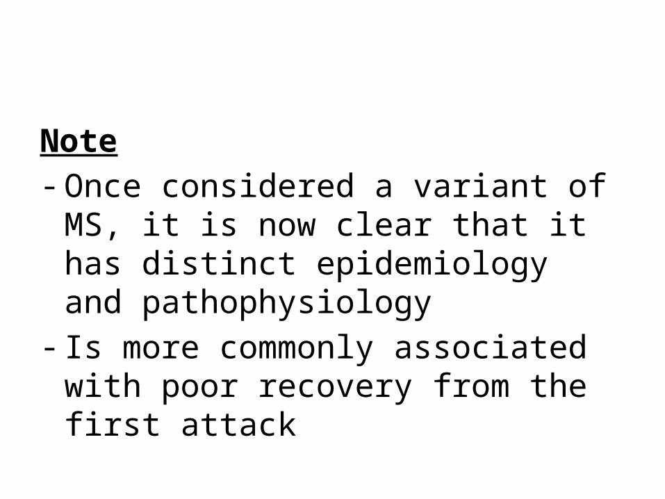
Note- Once considered a variant of MS, it is now
clear that it has distinct epidemiology and pathophysiology
- Is more commonly associated with poor recovery from the first attack

Pathogenesis
- Caused by the by the presence of antibodies against aquaporin-4. channels
- This protein is the major water channel of astrocytes,
- and areas of demyelination in NMO show loss of aquaporin-4. channels

• These antibodies injure astrocytes through complement-dependent mechanisms

Morphology- Within the damaged white matter, there is a. Necrosis, b. an inflammatory infiltrate including neutrophils,c. Vascular deposition of immunoglobulin and
complement.

- CSF- White cells are common often neutrophils

- Therapies include approaches to reduce the antibody burden, either through plasmapheresis or depletion of B cells with anti-CD20 antibody.
- .

3. Central pontine myelinolysis : - Is a nonimmune process- Is an acute process - Characterized by loss of myelin involving the center of the basis pontis and portions of
pontine tegmentum in a symmetric pattern

- It most commonly arises 2 to 6 days after rapid correction of hyponatremia
- The rapid increases in osmolality damage the oligodendrocytes

Mechanism
- A fall in serum sodium (in the absence of a compensatory rise in other osmoles) will cause entry of water into brain cells and consequent brain swelling
- To protect cells from swelling, protective mechanisms will prevent it and include

1. Over the next few hours potassium is lost, and this is maximal after 24 hours.

2.Oorganic osmoles (such as myoinisotol, taurine, and glutamate) will be lost from brain cells over day to a very few days, rendering the cell isotonic to the extracellular fluid and maintaining cell volume.

- Rapid correction of hyponatremia will cause fast rise in the tonicity of extracellular fluid
- And this will be faster than the rate at which organic osmoles can be synthesized and/or transported into the cell, therefore ,the cell will shrink especially the oligodendrocytws.

Morphology
- Loss of myelin- No inflammation- Neurons and axons are preserved - Because of the synchronus onset of the
damage, all lesions are of same stage of myelin loss and reaction

Central pontine myelinolysis

. Progressive multifocal leukoencephalopathy (PML
:Is a demyelinating disease that occurs after reactivation of JC virus in immunosuppressed patients.

PML- inclusions inoligodendrocytes

Subarachnoid Hemorrhage
Causes:A. Saccular (berry) aneurysm- Is the most frequent cause of clinically significant
subarachnoid hemorrhage is rupture of a saccular (berry) aneurysm.
B. Vascular malformation

C. Trauma D. Rupture of an intra-cerebral hemorrhage
into the ventricular system,

A. Saccular (Berry ) aneurysm:- Rupture of saccular aneurysm can occur at any
time- In about one-third of cases it is associated with
acute increases in intracranial pressure, such as with straining at stool or sexual orgasm.

- The main symptom is sudden, excruciating headache (classically described as "the worst headache I've ever had") and rapidly lose consciousness.
- Between 25% and 50% of individuals die with the first rupture, although those who survive typically

improve and recover consciousness in minutes
- Recurring bleeding is common in survivors- about 90% of saccular aneurysms occur in
the anterior circulation - Multiple aneurysms exist in 20% to 30% of
cases.

- Although they are referred to as congenital, they are not present at birth but develop over time because of underlying defects in the vessel media
-

It might be associated with:a. Disorders of extracellular matrix proteins, b. There is an increased risk of aneurysms in
individuals with autosomal dominant polycystic kidney disease
- Overall, aneurysms have a roughly 1.3% per year rate of bleeding.

Complications of subarachnoid hemorrhage1- In the early period there is a risk of additional
ischemic injury from vasospasm involving other vessels.
2- In the healing phase , meningeal fibrosis occurss sometimes leading to obstruction of CSF flow and interruption of pathways of CSF resorption.


A. Atherosclerotic (fusiform) mostly of the basilar artery),
- other types of aneurysms present with cerebral infarction from vascular occlusion and include
B. Mycotic, traumatic, and dissecting aneurysms. , are most often found in the anterior circulation.

B. Vascular Malformations 1. Arteriovenous malformations (AVM)- Is the most common type and the most dangerous- Affect males twice as frequently as females; - The lesion is most often recognized clinically
between the ages of 10 and 30 years

- The lesions presenting as :a. A seizure disorder b. An intra-cerebral hemorrhage c. A subarachnoid hemorrhage

d. Large AVMs occurring in the newborn period can lead to high-output congestive heart failure because of blood shunting directly from arteries to veins.

.
2. Cavernous hemangiomas - Consist of distended, loosely organized vascular
channels with thin collagenized walls. - They are devoid of intervening nervous tissue-

- They occur most often in the cerebellum, pons, and subcortical regions,
- Have a low flow without arterio-venous shunting. - Foci of old hemorrhage, infarction, and calcification
frequently surround the abnormal vessels

3. Capillary telangiectasias - Are microscopic foci of dilated, thin-walled
vascular channels separated by relatively normal brain parenchyma
- Occurring most frequently in the pons.

4. Venous angiomas (varices) - Consist of aggregates of ectatic venous channels. Note: - The latter two types are unlikely to bleed and are
most commonly discovered as incidental lesions.