Embed Size (px)
Citation preview

Lipid composition of lens plasma membrane fractions enriched in fiber junctions
C. R. Fleschner and Richard J. Cenedella
Department of Biochemistry, Kirksville College of Osteopathic Medicine, Kirksville, MO 63501
Abstract Little is known about the lipid environment of lens fiber junctions, the plasma membrane structure proposed to be responsible for passage of low molecular weight metabolites be- tween adjacent lens fiber cells. Plasma membranes of the ocular lens are especially rich in fiber junctions. The resistance ofjunc- tional domains to disruption by detergent or alkali treatment provides the opportunity to isolate a lens plasma membrane fraction enriched in fiber junctions. When examined by electron microscopy, the fiber junction fraction prepared from bovine lenses was enriched with junctional structures by about twofold when compared to total plasma membrane. We compared the protein, phospholipid, and cholesterol concentration of total plasma membrane with fiber junctional membrane from rat and cow lens and from aged normal cataractous human lenses. The principal finding was that junctional membrane contained 20-40% more total lipid than that of the total plasma mem- brane. This was due to a proportionate increase in the relative content (mg/mg protein) of both phospholipid and cholesterol. Exclusive of one exception (nucleus of bovine lens), the cholesterol/phospholipid molar ratios of the two fractions were similar. In the bovine nucleus, the cholesterol/phospholipid molar ratio was substantially higher in the fiber junctional- enriched membrane fraction than in the total plasma mem- brane, suggesting a special association of cholesterol with bovine nuclear fiber junctions. The relative lipid compositions of the plasma membrane and fiber junction-enriched fractions from human normal and cataractous lenses were similar, suggesting that human senile cataractogenesis involves changes in the lens plasma membrane more subtle than would be reflected by gross changes in the membrane lipid composition. "leschner, C. R., and R. J. Cenedella. Lipid composition of lens plasma mem- brane fractions enriched in fiber juncti0ns.J Lipid Res. 1991. 32 45-53.
Supplementary key words membrane lipids membrane choles- terol fiber junctions lens
Gap junctions are plasma membrane structures responsible for the transfer of ions and low molecular weight compounds between adjacent, communicating cells (1, 2). They are found in almost every organ, and are ubiquitous in the animal kingdom (1). Lens fiber junc- tions, commonly accepted to be analogous to gap junc- tions of other tissues, are especially important in the ocular lens, where they comprise a substantial proportion of the fiber cell plasma membrane (3, 4), and where the
extracellular space of the lens is very limited (5). There- fore, fiber junctions are of major importance to maintenance of homeostasis within the fiber cells, which is required for lens transparency. Plasma membrane is es- sentially the only subcellular organelle of the lens since lens fiber cells lose their intracellular organelles soon after being formed (6).
Gap junctions are composed of only protein and lipid (1, 2). The protein component of gap junctions has been extensively examined. Rat liver gap junctions contain homologous proteins of 26 kDa (7) and 21 kDa (8 ) as de- termined by SDS-polyacrylamide gel electrophoresis. The cDNA of the major, 26 kDa protein has been cloned and codes for a polypeptide with a calculated molecular weight of 32 kDa (9). The gap junction protein of rat heart has a predicted molecular weight of 43 kDa based on its cDNA sequence and has regions of high homology to the liver gap junction protein (10).
The identity of the protein components(s) of lens fiber cell junctions is controversial. The 26-28 kDa major in- trinsic polypeptide (MIP) is a candidate fiber junction protein (11-13). MIP is the major protein component of lens fiber junction-enriched preparations (11-15), purified MIP forms channels in reconstituted membranes (16-19), and anti-MIP antibodies label junction structures (20, 21), although nonjunctional membrane is labeled as well
In one instance (22), it was reported that only nonjunc- tional areas of lens plasma membrane were labeled with anti-MIP antibodies. However pentalaminar profiles, which were 4 nm thinner than the described lens fiber junction pentalaminar profiles, were labeled with anti- MIP antibodies (22). The structure of MIP as predicted by its cloned cDNA contains six trans-membrane span- ning regions, one of which is amphipathic, consistent with a role as a junctional protein (23). However, MIP shows no sequence homology with the gap junctional proteins
(21).
Abbreviations: MIP, major intrinsic polypeptide; TPM, total plasma membrane fraction; JF, fiber junctional-enriched membrane fraction.
Journal of Lipid Research Volume 32, 1991 45
by guest, on June 23, 2018w
ww
.jlr.orgD
ownloaded from

from liver (24, 25) or heart (10, 25), and shows no im- munological cross-reactivity with the protein from liver
A second candidate lens fiber junction protein is a 70 kDa protein shown by immunological methods to be asso- ciated with fiber junctions of the outer regions of lens cor- tex (27-29). This protein and its proteolytic products, MP64 and MP38, show some amino terminal sequence homologies with gap junction proteins of heart and liver (25). However, levels of these proteins in fiber junction- enriched membrane preparations are low compared to levels of MIP (25, 27, 28).
In contrast to the junctional protein, little work has been done on the lipid component of these structures. For rat liver (30) and bovine lens (15), the relative lipid com- position of junctional-enriched membrane was reported to be similar to that of total plasma membrane except for the absence of sphingomyelin in rat liver gap junctional membrane (30). However, purified junctional membrane from both chicken lens (12) and mouse liver (31) was reported to be enriched in cholesterol when compared to the total plasma membrane.
The importance of fiber junctions to the ocular lens re- quires further description of the lipid environment of these domains. In order to examine the lipid environment of fiber junctional domains, we isolated total plasma membrane and fiber junctional-enriched membrane from the lenses of rats and cows and from aged normal and cataractous human lenses. These species were selected since they represent the mammals examined most often. The total protein, phospholipid, and cholesterol content of the membrane fractions was determined and the con- tents from the fiber junctional-enriched membranes were compared to those from the total plasma membranes.
(26).
MATERIALS AND METHODS
Lenses
Bovine eyes and chicken eyes were obtained from local abattoirs, and lenses were removed within 4 h after death. Sprague-Dawley rat pups (20-21 days old) of either sex were obtained from Hilltop Lab Animals (Springdale, PA). Normal and cataractous human lenses were obtained from the National Disease Research Interchange (Phila- delphia, PA). Human eyes were removed within 6 h of death, and kept on ice no more than 48 h after death before removal of lenses. All lenses were either used im- mediately or stored frozen ( - 8OOC) until used. The average age of normal human samples was 62 yr (41-78 yr, range) and for cataractous samples was 87 yr (82-90 yr, range). Lenses were decapsulated before homogeniza- tion. For some experiments, decapsulated lenses were
divided into cortical and nuclear regions. Bovine, chick- en, and human lenses were divided into cortical and nuclear regions by gentle stirring in 5 mM Tris, 1 mM EDTA, 5 mM P-mercaptoethanol, pH 8.0 (buffer A), at 0-5OC, under NZ for approximately 2 h. The gentle stir- ring of decapsulated lenses in hypotonic media has proved successful for fractionating bovine lenses into defined cor- tical and nuclear regions (32), and for separating human lenses into cortex and nucleus (33). The cortical and nuclear regions accounted for 57-60% and 40-43%, respectively, of the total dry weight of the lens in agree- ment with previously published values (34). Rat lens cor- tical and nuclear fractions were separated by dissection (35). Cortical and nuclear regions accounted for approx- imately 56% and 44%, respectively, of the total lens dry weight, similar to previously published values (35, 36).
Membrane fractions
Decapsulated lenses or lens regions were homogenized in buffer A using an all-glass Dounce homogenizer. The total plasma membrane fraction was isolated from the homogenates essentially as described by Kistler et al. (37). This method reflects the standard approach for isolation of purified plasma membrane from the lens. All subse- quent steps were done at 4°C. The homogenate was cen- trifuged at 10,000 g for 20 min. The insoluble pellet was extracted twice more with buffer A, followed by sedimen- tation at 10,000 g for 20 min. The water-insoluble pellet was extracted once with 4 M urea (in buffer A) and twice with 8 M urea (in buffer A), each time sedimenting the insoluble material at 20,000 g for 30 min. After the final urea extraction, the urea-insoluble membrane fraction was washed once with buffer A, made to 50% (w/w) sucrose and overlayed with a sucrose density step-gradient consisting of 45%:25%:8% (w/w) sucrose (in buffer A). The gradient was centrifuged at 100,000 g for 60 min. The material migrating at the 45%:25% interface was col- lected, washed once with buffer A, and used as the total plasma membrane fraction (TPM).
Fiber junctional-enriched membrane fraction UF) was prepared from the TPM by extraction with Sarkosyl NL- 97 as described by Takemoto and Hansen (38). The TPM was extracted with 0.1% Sarkosyl NL-97, at room temperature, for 20 min. The JF was sedimented from the mixture by centrifugation at 100,000 g for 45 min, and washed with buffer A. This procedure yields a membrane fraction enriched with junctional membrane as deter- mined by electron microscopic examination (38).
For some experiments, JF and TPM were isolated from bovine lens in the absence of detergent according to Russell, Robison, and Kinoshita (39). Decapsulated bovine lenses were homogenized in 50 mM Tris, 5 mM EDTA, 10 mM 0-mercaptoethanol, pH 7.4 (buffer B).
46 Journal of Lipid Research Volume 32, 1991
by guest, on June 23, 2018w
ww
.jlr.orgD
ownloaded from

The insoluble material was sedimented by centrifugation at 10,000 g for 20 min, and extracted twice more with buffer B. The water-insoluble material was then extracted three times with 7 M urea (in buffer B), each time sedimenting the urea-insoluble material by centrifugation at 20,000 g for 30 min. The urea-insoluble material was assumed to be the TPM. The TPM was subsequently resuspended in 0.1 M NaOH, 1 mM P-mercaptoethanol at O°C for 10 min. The insoluble material was sedimented by centrifugation at 20,000 g for 30 min, and resuspended in 5 mM phosphate, pH 8.0, for 10 min. The insoluble material was sedimented by centrifugation as described above and resuspended in a small volume of buffer A for subsequent analysis. This procedure resulted in a fraction enriched with junctional membrane structures, and which contained essentially 100% MIP (39), without the use of detergents.
Assays for plasma membrane marker enzymatic ac- tivities were not done since these are of questionable value in preparation of membrane fractions from the decap- sulated lens. The plasma membrane of lens fiber cells is essentially the only membrane found in the decapsulated lens. Furthermore, this plasma membrane is specialized to form a very rigid membrane with large areas devoted to fiber junction, and therefore retains only trace enzy- matic activities.
Electron microscopy
Aliquots of membrane preparations were pelleted by centrifugation, and the pellets were fixed with 4% glu- taraldehyde in phosphate-buffered saline. Pellets were suspended in glutaraldehyde + 1% tannic acid, washed, and post-fixed with osmium tetroxide. Samples were washed, stained with uranyl acetate, washed, dehydrated, and embedded in L. R. White resin. Sections were cut, stained with uranyl acetate, and examined.
Electron micrographs of isolated membrane fractions were analzyed by measuring the lengths of fiber junction and unit membrane segments. Only segments that could be clearly identified as fiber junction or unit membrane were measured. All segments containing the typical pen- talaminar profile were counted as fiber junction; thus, our method includes both the so-called thick (16-18 nm) and thin (12-14 nm) fiber junctions (15, 22, 40). The percent- age of total membrane devoted to fiber junction was calculated as 2 x fiber junction length/(2 x fiber junc- tion length + unit membrane length) since fiber junc- tions are composed of two membranes. Two independent analyses of the micrographs were evaluated.
Lipid extraction and analysis
Aliquots of fractions were extracted with 20 volumes of choloroform-methanol 2:l (v/.). The mixtures were
briefly sonicated on ice prior to filtration through sintered glass. Material adhering to the filter was extracted once more with a small volume of chloroform-methanol, and refiltered. The filtrates were combined and washed with 0.2 volumes of 0.88% KCl. The chloroform-methanol ex- tracts were evaporated to dryness under NZ and resuspended to a known volume of chloroform-methanol.
For phospholipid phosphorus analysis, aliquots of-the chloroform-methanol extracts were evaporated to dryness under NZ, ashed at 5OO0C, and the residue was assayed for phosphorus content (41). Analysis of a known amount of phospholipid (dipalmitoylphosphatidylcholine, Sigma, St. Louis, MO) demonstrated that the phospholipids were completely mineralized. The phosphorus content was assumed to be 4% of the total phospholipid content by weight.
For analysis of sterols, aliquots of the chloro- form-methanol extracts were evaporated to dryness under NZ, and the residue was saponified in 0.6 N KOH, 67% ethanol at 90°C for 1 h. Reaction mixtures were diluted 1:l with glass distilled water, and the nonsaponifiable lipids (sterols) were extracted with hexane. The hexane extracts were assayed for sterols by gas-liquid chromato- graphy as previously described (42), using 5-a-cholestane as an internal standard.
Other methods
Total protein was assayed according to Lees and Pax- man (43) using bovine serum albumin as standard. Pro- tein values were calculated by mathematical interpolation from a standard curve fit by least squares linear regres- sion. Proteins were examined by sodium dodecylsulfate polyacrylamide-gel electrophoresis according to Laemmli (44), using 12% resolving gels. Dried gels were scanned with a Hoefer GS 300 scanning densitometer. Lens dry weights were determined by heating 5O-pl samples of lens homogenates overnight at 100°C in pre-dried, pre- weighed tubes.
RESULTS
In order to examine the lipid composition of the lens fiber junctional membrane domain, we isolated the TPM and subsequently purified the JF from the TPM. The protein, phospholipid, and cholesterol contents of these membrane fractions isolated from rat and cow were then compared.
Fig. 1 shows electron micrographs of membrane frac- tions from bovine lens. Both the TPM (Fig. 1A) and the JF (Fig. 1B) contained structures typical of unit mem- brane (small arrows) and fiber junctions (large arrows). Analysis of micrographs equivalent to 82 times the area of Fig. 1 indicated that about 30% of the total membrane
Fleschner Lipid composition of lens fiber junctions 47
by guest, on June 23, 2018w
ww
.jlr.orgD
ownloaded from

polypeptides with molecular masses of 67-70 kDa. These polypeptides may represent MP70 and its proteolysis pro- duct MP64, reported to be components of fiber junctions of the outer cortex (27-29). All the membrane fractions from both cow and rat contained a 19 kDa polypeptide which may be the 18 kDa calmodulin-binding intrinsic protein of lens membrane (45).
The phospholipid and cholesterol contents of the mem- brane fraction are presented graphically in Fig. 3. The JFs from both cortex and nucleus of both the rat and cow tended to have higher relative content of total lipid than the TPM. The trend of increased lipid content of JFs (from both rat and cow) was statistically significant when compared to the corresponding TPMs by two-way analy- sis of variance (F = 5.994, df1 = 10, df, = 1, P < 0.05).
- Differences between the species were not significant (F =
2.062, df, = 10, df, = 10, P > 0.10). Although the JF was enriched with total lipid, we found little evidence that the JF was specifically enriched in cholesterol.
Only in the nuclear region of the bovine lens was the cholesterol/phospholipid molar ratio of the JF substantial- ly greater than that of the TPM (Fig. 4). The apparent increased cholesterol/phospholipid molar ratio of the JF isolated from the nuclear region of the cow lens suggested a special interaction of cholesterol with the fiber junctions of the nuclear bovine lens.
TPM and JF were also prepared from whole bovine lenses according to the method of Russell et al. (39). This
Fig. 1. Electron micrograph of bovine membrane fractions. The total plasma membrane fraction (A) was prepared by sucrose density centri- cow RAT fugation of the urea-insoluble fraction, and an aliquot was prepared for electron microscopy. The fiber junctional enriched membrane (B) was TPM J F TPM JF prepared by Sarkosyl extraction of the total plasma membrane, and an aliquot was prepared for electron microscopy. Large arrows indicate the kDa M C N C N M C N C N M pentalaminar structures characteristic of lens fiber junctions. Small ar- rows indicate typical unit membrane. Bar = 0.1 pm.
" "
" - *". - . - 94"
of the TPM was composed of fiber junction. The JF was composed of about 60% fiber junctions. Thus, although the JF contains substantial unit membrane, it is enriched in fiber junctions by about twofold compared to the TPM.
The protein compositions of the various membrane fractions (as examined by electrophoresis) from rat and cow are shown in Fig. 2. In general, the protein composi- tion of a given JF was quite similar to that of the cor- responding TPM. In all the membrane fractions, MIP (the putative junctional protein) was the major protein band. In the cow (Fig. 2), MIP accounted for 48-5375 of the total protein of the membrane fractions as measured by densitometric scanning of the gels. In the rat, MIP ac- counted for 55-67 % of the total protein of the membrane fractions. The membrane fractions isolated from rat lens cortex contained substantial amounts (11-2876 of total) of
67-
43-
30.
20.1 -
I
Fig. 2. SDS-PAGE of lens membrane fractions. Total plasma mem- brane (TPM) and fiber junctional membrane OF) fractions were prepared from cortical (C) and nuclear (N) samples of lenses from rats and cows as described in the text. Pools of 61 rat lenses and 2 cow lenses were used. The proteins (approximately 10 pg protein per lane) were separated on 12% resolving gels. M, molecular weight markers.
48 Journal of Lipid Research Volume 32, 1991
by guest, on June 23, 2018w
ww
.jlr.orgD
ownloaded from

Phospholipid (mg/mg protein)
0.4 0.2 0.0
0.4 0.2 0.0 C N
Cholesterol Total Lipid (mg/mg protein) (mg/mg protein)
Rat
cow
C N
1.5 r
0.5
0.0 C N
1.5r T
O T o t a l Plasma Membrane
Fiber Junction Membrane Fig. 3. Lipid composition of membrane fractions. Total plasma mem- brane and fiber junctional membrane fractions were prepared from lens cortical (C) and nuclear (N) regions as described in the text. Pools of 30-56 lenses for rat were used per assay. For cow, individual lenses or pools of 2 lenses were used per assay. The values represent the means f the range of two separate experiments.
procedure for preparing junctional-enriched membrane does not involve the use of detergents which have been suspected of nonspecifically extracting phospholipid from the membrane (12, 31). Electron micrographic examina- tion of the membrane fractions prepared in this manner indicated enrichment in fiber junctional membrane con- tent similar to that seen in preparations using detergent (not shown). The relative lipid composition of membranes prepared by this method is shown in Table l . The relative contents of both phospholipid and cholesterol were
Cholesterol /Phospholipid
(mol /mol )
2.5 Rat cow
I .o 0.5
C N C N
0 Total Plasma Membrane
Fiber Junction Membrane
Fig. 4. CholesteroUphospholipid molar ratios of membrane fractions Experimental details and figure labels are as in Fig. 3.
30-40% greater in the JFs than in the TPMs, regardless of the method of preparation. No evidence of a special en- richment of cholesterol was seen in the JF of the whole bovine lens.
The relative lipid composition of TPM and JF isolated from human normal and cataractous lens cortex and nucleus was also examined (Table 2). All of the mem- brane fractions from both cortex and nucleus of the cataractous lenses apparently contained more total lipid relative to membrane protein than did corresponding membrane fractions prepared from normal human lenses. The increased total lipid of the cataractous membranes was not statistically significant. With one exception (nor- mal lens cortex), the JFs had significantly greater relative total lipid contents than the corresponding TPMs (F = 5.1693, dfl = 1, df, = 11, P < 0.05). The cholesterol/phospholipid molar ratios of membrane frac- tions from cataractous lenses were quite similar to those of membrane fractions from normal lenses. The cholesterol/phospholipid molar ratio of the cortical TPM
TABLE 1. Relative lipid composition of bovine lens membranes (cortex + nucleus) prepared in the absence or presence of detergent
Fraction (n) Phospholipid Cholesterol Total Lipid/Protein CholesterollPhospholipid
mg/mg protein mg/mg profein mol/mol
Without detergent Total plasma membrane (2) 0.563 f 0.032 0.236 f 0.048 0.799 f 0.079 0.83 f 0.12 Fiber junction (2) 0.734 * 0.026 0.320 f 0.060 1.053 f 0.085 0.87 f 0.13
With detergent Total plasma membrane (3) 0.377 f 0.101 0.237 * 0.078 0.614 f 0.178 1.21 f 0.06 Fiber junction (3) 0.526 f 0.036 0.294 f 0.040 0.821 f 0.073 1 . 1 1 f 0.09
Total plasma membrane and fiber junctional membrane fractions were prepared from whole bovine lenses either in the absence of detergent accord- ing to the method of Russell et al. (39) or in the presence of detergent as described in the text. The values are the means f range of two separate experiments, or the means * SEM of three experiments.
Fleschner Lipid composition of lens fiber junctions 49
by guest, on June 23, 2018w
ww
.jlr.orgD
ownloaded from
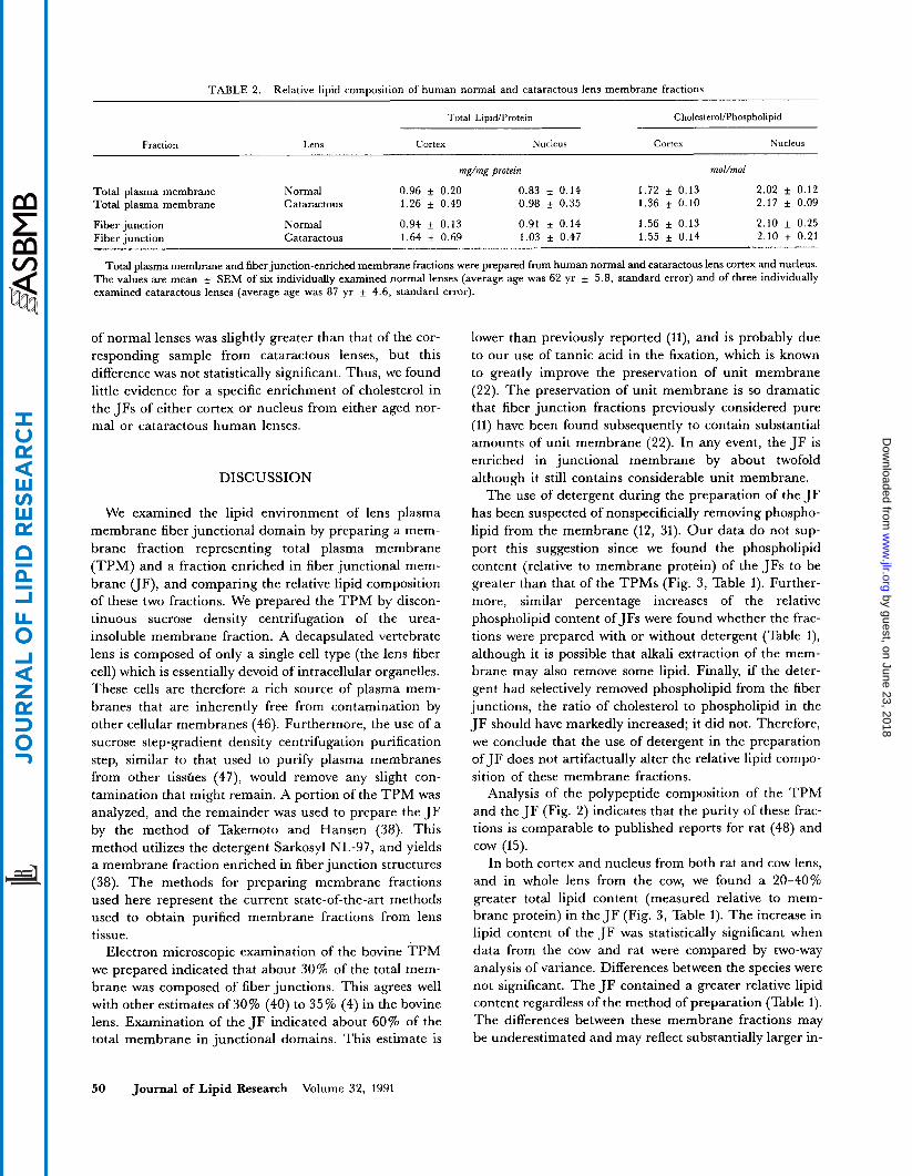
TABLE 2. Relative lipid composition of human normal and cataractous lens membrane fractions
Total LipidIProtein CholesteroUPhospholipid
Fraction Lens Cortex Nucleus Cortex Nucleus
rnE/mE protein mol/mol
Total plasma membrane Normal 0.96 f 0.20 0.83 f 0.14 1.72 f 0.13 2.02 f 0.12 Total plasma membrane Cataractous 1.26 f 0.49 0.98 f 0.35 1.36 f 0.10 2.17 f 0.09
Fiber junction Normal 0.94 f 0.13 0.91 f 0.14 1.56 f 0.13 2.10 f 0.25 Fiber junction Cataractous 1.64 f 0.69 1.03 f 0.47 1.55 * 0.14 2.10 f 0.21
Total plasma membrane and fiber junction-enriched membrane fractions were prepared from human normal and cataractous lens cortex and nucleus. The values are mean f SEM of six individually examined normal lenses (average age was 62 yr f 5.8, standard error) and of three individually examined cataractous lenses (average age was 87 yr f 4.6, standard error).
of normal lenses was slightly greater than that of the cor- responding sample from cataractous lenses, but this difference was not statistically significant. Thus, we found little evidence for a specific enrichment of cholesterol in the JFs of either cortex or nucleus from either aged nor- mal or cataractous human lenses.
DISCUSSION
We examined the lipid environment of lens plasma membrane fiber junctional domain by preparing a mem- brane fraction representing total plasma membrane (TPM) and a fraction enriched in fiber junctional mem- brane UF), and comparing the relative lipid composition of these two fractions. We prepared the TPM by d’ iscon- tinuous sucrose density centrifugation of the urea- insoluble membrane fraction. A decapsulated vertebrate lens is composed of only a single cell type (the lens fiber cell) which is essentially devoid of intracellular organelles. These cells are therefore a rich source of plasma mem- branes that are inherently free from contamination by other cellular membranes (46). Furthermore, the use of a sucrose step-gradient density centrifugation purification step, similar to that used to purify plasma membranes from other tisshes (47), would remove any slight con- tamination that might remain. A portion of the TPM was analyzed, and the remainder was used to prepare the JF by the method of Takemoto and Hansen (38). This method utilizes the detergent Sarkosyl NL-97, and yields a membrane fraction enriched in fiber junction structures (38). The methods for preparing membrane fractions used here represent the current state-of-the-art methods used to obtain purified membrane fractions from lens tissue.
Electron microscopic examination of the bovine TPM we prepared indicated that about 30% of the total mem- brane was composed of fiber junctions. This agrees well with other estimates of 30% (40) to 35% (4) in the bovine lens. Examination of the JF indicated about 60% of the total membrane in junctional domains. This estimate is
50 Journal of Lipid Research Volume 32, 1991
lower than previously reported (H), and is probably due to our use of tannic acid in the fixation, which is known to greatly improve the preservation of unit membrane (22). The preservation of unit membrane is so dramatic that fiber junction fractions previously considered pure (11) have been found subsequently to contain substantial amounts of unit membrane (22). In any event, the JF is enriched in junctional membrane by about twofold although it still contains considerable unit membrane.
The use of detergent during the preparation of the JF has been suspected of nonspecificially removing phospho- lipid from the membrane (12, 31). Our data do not sup- port this suggestion since we found the phospholipid content (relative to membrane protein) of the JFs to be greater than that of the TPMs (Fig. 3, Table 1). Further- more, similar percentage increases of the relative phospholipid content of JFs were found whether the frac- tions were prepared with or without detergent (Table l), although it is possible that alkali extraction of the mem- brane may also remove some lipid. Finally, if the deter- gent had selectively removed phospholipid from the fiber junctions, the ratio of cholesterol to phospholipid in the JF should have markedly increased; it did not. Therefore, we conclude that the use of detergent in the preparation of JF does not artifactually alter the relative lipid compo- sition of these membrane fractions.
Analysis of the polypeptide composition of the TPM and the JF (Fig. 2) indicates that the purity of these frac- tions is comparable to published reports for rat (48) and cow (15).
In both cortex and nucleus from both rat and cow lens, and in whole lens from the cow, we found a 20-40% greater total lipid content (measured relative to mem- brane protein) in the JF (Fig. 3, Table 1). The increase in lipid content of the JF was statistically significant when data from the cow and rat were compared by two-way analysis of variance. Differences between the species were not significant. The JF contained a greater relative lipid content regardless of the method of preparation (Table 1). The differences between these membrane fractions may be underestimated and may reflect substantially larger in-
by guest, on June 23, 2018w
ww
.jlr.orgD
ownloaded from
creases in the fiber junction domain of the plasma mem- brane. Due to current limitations in methodology (there is no procedure to obtain pure unit membrane from lens plasma membranes), the TPM contains fiber junctional domains (about 30%). Furthermore, the best method for obtaining JF yields a preparation which still contains substantial unit membrane (about 40%). Therefore, the observed 20-40% increase in total lipid of the JF com- pared to TPM could actually reflect a much larger in- crease in the junctional domain.
The explanation for the increased lipid/protein of the JF is not obvious. It may suggest that fiber junctional pro- tein increases the lipid order of the membrane in the vicinity of the fiber junction, making this lipid less suscep- tible to detergent (or alkali) extraction. A special interac- tion between membrane intrinsic proteins and membrane lipids that influence the lipid order apparently does exist in lens plasma membrane. Using fluorescence anisotropy, Rintoul, Cundy, and Cenedella (49) have shown that rat lens intrinsic membrane protein orders lens membrane lipids. It has also been shown that human lens plasma membrane lipids are ordered by the membrane proteins (50). This special protein-lipid interaction may influence the lipid extractability, since specific interactions between membrane lipids and protein are thought to influence the detergent extractability of the lipids from membranes (51). A second possible explanation for the increased lipidlprotein of the JF is that it reflects a special structure of lens fiber junction domain such as that proposed by Zampighi et al. (52). In their model, lens fiber junctions are composed of one membrane containing tetragonal crystals of MIP which abuts its partner membrane con- taining no MIP or any other intrinsic membrane protein. Thus, isolated fiber junctions would be composed of one- half of the membrane containing crystals of MIP, but the other half would be composed of membrane lipid only. This might result in a fraction with a greater lipid/protein ratio than that of the bulk, unit membrane. In addition, using freeze-etch electron microscopy, Lo and Reese (53) recently reported that lens fiber junctions were bordered by an intramembrane particle-free area: that is, mem- brane presumably devoid of protein and enriched with lipid. Therefore, a lens membrane preparation enriched in fiber junctions might contain a higher lipid/protein ratio than unit membrane.
The increased total lipid/protein of the JF was due to increases of both phospholipid and cholesterol (Fig. 3). We found little evidence for a specific enrichment of cholesterol in the JF (Fig. 4). Only in the cow lens nucleus was the cholesterol/phospholipid molar ratio of the JF substantially higher than that of the TPM. This was due to a selective enrichment of cholesterol in the JF (Fig. 3). The reason for a specific cholesterol enrichment of the JF of the cow lens nucleus is not clear. It may reflect an aging-associated phenomenon. The cows from which
lenses were obtained for this study were approximately 1 year old. Since lens fiber cells are not shed during the lifetime of an animal, the bovine lens nuclear samples were aged in vivo for a minimum of 1 year. On the other hand, the rats used in this study were only about 3 weeks old, and would not be expected to evidence signs of aging. Furthermore, proteolytic modification of MIP is evident in the nuclear region of the bovine lens (Fig. 2). This alteration of the putative fiber junction protein may affect protein-lipid interactions.
Alcala, Katar, and Maisel (12) reported for chicken lens that the cholesterol/phospholipid molar ratio of fiber junctional-enriched membrane was substantially greater than that of the total plasma membrane. We, on the other hand, found that the cholesterollphospholipid molar ratios of the JF and the TPM of rat lens and cow cortical lens were essentially the same (Fig. 4). In addition, we de- tected no difference in the cholesterol/phospholipid molar ratios between TPM and JF in either the cortex or nucleus of chicken lenses (data not shown). The differences between our findings and theirs are likely due to differences in the methods of preparing the membrane fractions. These include differences in buffer composition and strength, differences in protocols of plasma mem- brane preparation, and differences in detergents used, all of which may influence the properties and composition of the final membrane fractions.
The increased relative content of both phospholipid and cholesterol of JFs from both rat and cow lens cortex and nucleus and from whole cow lens regardless of the method of preparation underscores the importance of lipids to lens fiber junctions. Indeed, other lines of evi- dence from this laboratory have indicated an important role for cholesterol in fiber junctional membrane. Treat- ment of rats with U18666A, a potent cholesterol synthesis inhibitor, resulted in restricted availability of cholesterol to form membranes of the lens cortex (36), which was fur- ther associated with changes in the membrane’s physical properties (49), limited proteolysis of MIP (48), the disap- pearance of identifiable fiber junctions (54), and produc- tion of cataracts (55). These results strongly suggest an important role for cholesterol in the maintenance of fiber junction integrity.
We found little evidence for changes in the relative lipid content of either the TPM or the JF between human nor- mal and cataractous lenses. However, the values reported for human lens samples (Table 2) are quite variable, possibly obscuring small differences that might be impor- tant. The relative amount of cholesterol of cataractous hu- man lens membranes has been extensively studied, but a clear picture has not emerged. Others, like us, found no difference in the relative amount of cholesterol of catarac- tous lenses compared to normal lenses (50, 56, 57). However, some investigators have found the relative cholesterol increased in cataracts (33, 58, 59), while
Fleschner Lipid composition of lens fiber junctions 51
by guest, on June 23, 2018w
ww
.jlr.orgD
ownloaded from

others report a decreased relative cholesterol content of cataracts (60).
Gooden, Takemoto, and Rintoul(50) demonstrated, by measuring fluorescence depolarization of cis and trans parinaric acid probes, that membranes f rom human cataractous lenses are more fluid than membranes from normal human lenses even though the chole- sterol/phospholipid ratios were essentially the same for normal and cataractous lenses. In contrast to the whole membrane (protein plus lipid), they found no significant differences between the fluidity of the total lipids ex- tracted from membranes of normal and cataractous lenses. The authors concluded that significant alterations of protein-lipid interactions were associated with the hu- man cataractous lens membranes (50). Rintoul e t al. (49) observed with rat lenses that membrane proteins rather than lipid contributed more to controll ing membrane fluidity. Changes in the physical state of lens plasma membrane apparently do occur with human and ex- perimental cataracts and appear related to changes in the interation of membrane l ipids and proteins rather than to changes in the relative lipid composition of the membrane (49, 50).
This work was supported by grant EY02568 from the National Eye Institute, National Institutes of Health. We thank Dr. Paul FitzGerald, University of California, Davis, for the electron microscopy and Dr. John T. Matschiner, University of Nebraska Medical Center, Omaha, NE, for scanning the polyacryla- mide gels. We thank Mr. Sidney Osborn for bovine eyes and Mr. W. E. Sedenvall and Ms. Vanessa Miller for chicken eyes. Manuscript received 23 April 1990 and in revisedform 27 A q u s t 1990.
8.
9.
10.
11.
12.
13.
14.
15.
16.
17.
18.
19.
20.
REFERENCES
1. Revel, J-P., B. J. Nicholson, and S. B. Yancey. 1985. Chemistry of gap junctions. Annu. Rev. Physiol. 47: 263-279.
2. Revel, J-P,, B. J. Nicholson, and S. B. Yancey. 1984. Mole- cular organization of gap junctions. Fed. Proc. 43:
3. Kuszak, J., J. Alcala, and H. Maisel. 1980. The surface morphology of embryonic and adult chick lens-fiber cells. Am. J. Anat. 159: 395-410.
4. Kachura, V., S. M. Miller, and R. E. Garfield. 1983. Biochemical and structural characterization of membrane fractions from bovine lens. Invest. Ophthalmol. Visual Sci. 24: 1496-1504.
5. Rae, J. L., and R. T. Mathias. 1985. The physiology of the lens. In The Ocular Lens. H. Maisel, editor. Marcel Dek- ker, Inc., New York. 93-121.
6. Rafferty, N. S. 1985. Lens morphology. In The Ocular Lens. H. Maisel, editor. Marcel Dekker, Inc., New York.
7. Finbow, M., S. B. Yancey, R. Johnson, and J-P. Revel. 1980. Independent lines of evidence suggesting a major gap junctional protein with a molecular weight of 26,000. Proc. Natl. Acad. Sci. USA. 77: 970-974.
2672-2677.
1-60.
21.
22.
23.
24.
25.
26.
Nicholson, B., R. Dermietzel, D. Teplow, 0. Traub, K. Willecke, and J. P. Revel. 1987. Two homologous protein components of hepatic gap junctions. Nature. 329: 732-734. Paul, D. L. 1986. Molecular cloning of cDNA for rat liver gap junction protein. J. Cell Biol. 103: 123-134. Beyer, E. C., D. L. Paul, and D. A. Goodenough. 1987. Connexin 43: a protein from rat heart homologous to a gap junction protein from liver. J Cell Bid. 105: 2621-2629. Goodenough, D. A. 1979. Lens gap junctions: a structural hypothesis for nonregulated low-resistance intercellular pathways. Inuest. Ophthalmol. Visual Sci. 18: 1104-1122. Alcala, J., M. Katar, and H. Maisel. 1983. Lipid composi- tion of chick lens fiber cell gap junctions. Cur,: Eye Res. 2:
Alcala, J., and H. Maisel. 1985. Biochemistry of lens plasma membranes and cytoskeleton. In The Ocular Lens. H. Maisel, editor. Marcel Dekker, Inc., New York.
Kuszak, J, R., J. Alcala, and H. Maisel. 1981. Biochemical and structural features of chick lens gap junctions. Exp. Eye Res. 33: 157-166. Zampighi, G., S. A. Simon, J. D. Robertson, T. J. McIntosh, and M. J. Costello. 1982. On the structural or- ganization of isolated bovine lens fiber junctions. J Cell
Gooden, M. M,, L. J. Takemoto, and D. A. Rintoul. 1985. Reconstitution of MIP26 from single human lenses into ar- tificial membranes. I. Differences in pH sensitivity of cataractous versus normal human fiber cell protein. Cum Eye Res. 4: 1107-1115. Peracchia, C., and S. J. Girsch. 1985. Permeability and gating of lens gap junction channels incorporated into lipo- somes. Cur,: Eye Res. 4: 431-439. Girsch, S. J., and C. Perrachia. 1985. Lens cell-to-cell chan- nel protein, I. Self-assembly into liposomes and permeabili- ty regulation by calmodulin. J. Memb,: Biol. 83: 217-225. Scaglione, B. A., and D. A. Rintoul. 1989. A fluorescence- quenching assay for measuring permeability of reconsti- tuted lens MIP26. Invest. Ophthalmol. Visual Sci. 30:
Bok, D., J. Dockstader, and J. Horwitz. 1982. Immunocy- tochemical localization of the lens main intrinsic polypep- tide (MIP26) in communicating junctions. J. Cell Biol. 92:
FitzGerald, P. G., D. Bok, and J. Horwitz. 1983. Immuno- cytochemical localization of main-intrinsic polypeptide (MIP) in ultrathin frozen sections of rat lens. J. Cell Biol.
Paul, D. L., and D. A. Goodenough. 1983. Preparation, characterization, and localization of antisera against bovine MP26, an integral protein from lens fiber plasma mem- brane. J. Cell Biol. 96: 625-632. Gorin, M. B., S. B. Yancy, J. Cline, J-P. Revel, and J. Horwitz. 1984. The major intrinsic protein (MIP) of bovine lens fiber membrane: characterization and structure based on cDNA cloning. Cell. 39: 49-59. Revel, J-P., S. B. Yancey, B. Nicholson, and J. Hoh. 1986. Sequence diversity of gap junction proteins. In 1986 Junc- tional Complexes of Epithelial Cells (Ciba Foundation Symposium 125). Wiley, Chichester. 108-127. Kistler, J., D. Christie, and S. Bullivant. 1988. Homologies between gap junction proteins in lens, heart and liver. Nature. 331: 721-723. Zigler, J. S., Jr., and J. Horwitz. 1981. Immunochemical studies on the major intrinsic polypeptides from human lens membrane. Inuest. Ophthalmol. Visual Sci. 21: 46-51.
569-578.
169-222.
Biol. 93: 175-189.
961-966.
213-220.
97: 1491-1499.
52 Journal of Lipid Research Volume 32, 1991
by guest, on June 23, 2018w
ww
.jlr.orgD
ownloaded from

27. Kistler, J., B. Kirkland, and S. Bullivant. 1985. Identifica- tion of a 70,000-D protein in lens membrane junctional do- mains. J. Cell Biol. 101: 28-35.
28. Kistler, J., and S. Bullivant. 1987. Protein processing in lens intercellular junctions: cleavange of MP70 to MP38. Invest. Ophthalmol. Visual Sci. 28: 1687-1692.
29. Gruijters, W. T. M., J. Kistler, S. Bullivant, and D. A. Goodenough. 1987. Immunolocalization of MP70 in lens fiber 16-17 nm intercellular junctions. J. Cell Biol. 104:
30. Hertzberg, E. L., and N. B. Gilula. 1979. Isolation and characterization of gap junctions, from rat liver. J. Biol.
31. Henderson, D., H. Eibl, and K. Weber. 1979. Structure and biochemistry of mouse hepatic gap junctions. J. Mol. Biol. 132: 193-218.
32. Li, L-K., and L. So. 1987. Age-dependent lipid and protein changes in individual bovine lenses. Cum Eye Res. 6: 599-605.
33. Cotlier, E., Y. Obara, and B. Toftness. 1978. Cholesterol and phospholipids in protein fractions of human lens and senile cataract. Biochim. Biophys. Acta. 530: 267-278.
34. Broekhuyse, R. M. 1973. Membrane lipid and proteins in ageing lens and cataract. In The Human lens - in Relation to Cataract (Ciba Foundation Symposium 19, new series). Elsevier, Amsterdam. 135-149.
35. Cenedella, R. J. 1984. Regional distribution of sterol and fatty acid synthesis in the ocular lens. Exp. Eye Res. 38: 95-99.
36. Cenedella, R. J. 1985. Regional distribution of lipids and phospholipase a A, activity in normal and cataractous rat lens. Curr. Eye Res. 4: 113-120.
37. Kistler, J., B. Kirkland, K. Gilbert, and S. Bullivant. 1986. Aging of lens fibers. Mapping membrane proteins with monoclonal antibodies. Invest. Ophthalmol. Visual Sci. 27:
38. Takemoto, L. J., and J. S. Hansen. 1981. Gap junctions from the lens: purification and characterization by chemical cross-linking reagent. Biochem. Biophys. Res. Commun. 99:
39. Russell, P., W. G. Robison, Jr., and J. H. Kinoshita. 1981. A new method for rapid isolation of the intrinsic membrane proteins from lens. Exp. Eye Res. 32: 511-516.
40. FitzGerald, P. 1987. Main intrinsic polypeptide proteolysis and fiber cell membrane domains. Invest. Ophthalmol. Visual Sci. 28: 795-805.
41. Pollet, S., S. Ermidou, F. Le Saux, M. Monge, and N. Baumann. 1978. Microanalysis of brain lipids: multiple two-dimensional thin-layer chromatography. J. Lipid Res.
42. Cenedella, R. J. 1982. Digitonide-precipitable sterols: a reevaluation with special attention to lanosterol. Lipid. 17: 443-447.
43. Lees, M. B., and S. Paxman. 1972. Modification of the Lowry procedure for the analysis of proteolipid protein. Anal. Biochem. 47: 184-192.
44. Laemmli, U. K. 1970. Cleavage of structural proteins dur-
565-572.
Chm. 254: 2138-2147.
772-780.
324-331.
19: 916-921.
45.
46.
47.
48.
49.
50.
51.
52.
53.
54.
55.
56.
57.
58.
59.
60.
ing the assembly of the head of bacteriophage T4. Nature.
Muldes, J. W. M,, C. E. M. Voorter, C. Lamers, W. A. de Haard-Hoekman, C. Montecucco, W. J. M. van de Ven, H. Bloemendal, and W. W. de Jong. 1988. MP17, a fiber- specific intrinsic membrane protein from mammalian eye lens. Cum Eye Res. 7: 207-219. Benedetti, E. L., I. Dunia, C. J. Bentzel, A. J. M. Vermorken, M. Kibbelaar, and H. Bloemendal. 1976. A portrait of plasma membrane specializations in eye lens epithelium and fibers. Biochim. Biophys. Acta. 457: 353-384. Neville, D. M. 1968. Isolation of an organ-specific protein antigen from cell-surface membrane of rat liver. Biochim. Biophys. Acta. 154: 540-552. Alcala, J., R. J. Cenedella, and M. Katar. 1985. Limited proteolysis of MP26 in lens fiber plasma membranes of the U18666A-induced cataract in rats. Cum Eye Res. 4: 1001-1005. Rintoul, D. A., K. V. Cundy, and R. J. Cenedella. 1987. Physical properties of membranes and membrane lipids from the fiber cell of the U18666A-cataractous rat. Cum Eye Res. 6: 1343-1348. Gooden, M. M,, L. J. Takemoto, and D. A. Rintoul. 1983. Evidence for reduced lipid order in plasma membranes from cataractous human lenses. Cum Eye Rex 2: 367-375. Helenius, A., and K. Simons. 1975. Solubilization of mem- branes by detergents. Biochim. Biophys. Acta. 415: 29-79. Zampighi, G. A., J. E. Hall, G. R. Ehring, and S. A. Simon. 1989. The structural organization and protein com- position oflens fiberjuncti0ns.J. Cell Biol. 108: 2255-2275. Lo, W-K., and T. S. Reese. 1990. Cell membrane spe- cialization of the ocular lens. P m . Int. Soc. Eye Res. 6: 168 (abstract). Kuszak, J. R., A. R. Khan, and R. J. Cenedella. 1988. An ultra-structural analysis of plasma membrane in the U18666A cataract. Invest. Ophthalmol. Visual Sci. 29: 261-267. Cenedella, R. J., and G. G. Bierkamper. 1979. Mechanism of cataract production by 3-beta(2-diethylamino- ethoxy)androst-17-one hydrochloride, U18666A: an inhibi- tor of cholesterol biosynthesis. Exp. Eye Res. 28: 673-688. Li, L-K., L. So, and A. Spector. 1985. Membrane cholesterol and phospholipid in consecutive concentric sec- tions of human lenses. J. Lipid Res. 26: 600-609. Rosenfeld, L., and A. Spector. 1981. Changes in lipid distri- bution in the human lens with the development of cataract. Exp. Eye Res. 33: 641-650. Roy, D., L. Rosenfeld, and A. Spector. 1982. Lens plasma membrane: isolation and biochemical characterization. Exp. Eye Res. 35: 113-129. Zigman, S., T. Paxhia, G. Marinetti, and S. Girsch. 1984. Lipids of human lens fiber cell membranes. Curr. Eye Res.
Broekhuyse, R. M. 1969. Phospholipids in tissues of the eye. 111. Composition and metabolism of phospholipids in human lens in relation to age and cataract formation. Biochim. Biophys. Acta. 187: 354-365.
227: 680-685.
3: 887-896.
Fleschnes Lipid composition of lens fiber junctions 53
by guest, on June 23, 2018w
ww
.jlr.orgD
ownloaded from