Embed Size (px)
Citation preview
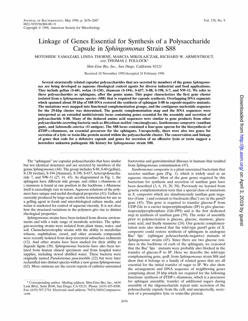
JOURNAL OF BACTERIOLOGY, May 1996, p. 2676–2687 Vol. 178, No. 90021-9193/96/$04.0010Copyright q 1996, American Society for Microbiology
Linkage of Genes Essential for Synthesis of a PolysaccharideCapsule in Sphingomonas Strain S88
MOTOHIDE YAMAZAKI, LINDA THORNE, MARCIA MIKOLAJCZAK, RICHARD W. ARMENTROUT,AND THOMAS J. POLLOCK*
Shin-Etsu Bio, Inc., San Diego, California 92121
Received 10 November 1995/Accepted 24 February 1996
Several structurally related capsular polysaccharides that are secreted by members of the genus Sphingomo-nas are being developed as aqueous rheological control agents for diverse industrial and food applications.They include gellan (S-60), welan (S-130), rhamsan (S-194), S-657, S-88, S-198, S-7, and NW-11. We refer tothese polysaccharides as sphingans, after the genus name. This paper characterizes the first gene clusterisolated from a Sphingomonas species (S88) that is required for capsule synthesis. Overlapping DNA segmentswhich spanned about 50 kbp of S88 DNA restored the synthesis of sphingan S-88 in capsule-negative mutants.The mutations were mapped into functional complementation groups, and the contiguous nucleotide sequencefor the 29-kbp cluster was determined. The genetic complementation map and the DNA sequences wereinterpreted as an extended multicistronic locus containing genes essential for the assembly and secretion ofpolysaccharide S-88. Many of the deduced amino acid sequences were similar to gene products from otherpolysaccharide-secreting bacteria such as Rhizobium meliloti (succinoglycan), Xanthomonas campestris (xanthangum), and Salmonella enterica (O antigen). The S88 locus contained a four-gene operon for the biosynthesis ofdTDP-L-rhamnose, an essential precursor for the sphingans. Unexpectedly, there were also two genes forsecretion of a lytic or toxin-like protein nested within the polysaccharide cluster. The conservation and linkageof genes that code for a defensive capsule and genes for secretion of an offensive lysin or toxin suggest aheretofore unknown pathogenic life history for Sphingomonas strain S88.
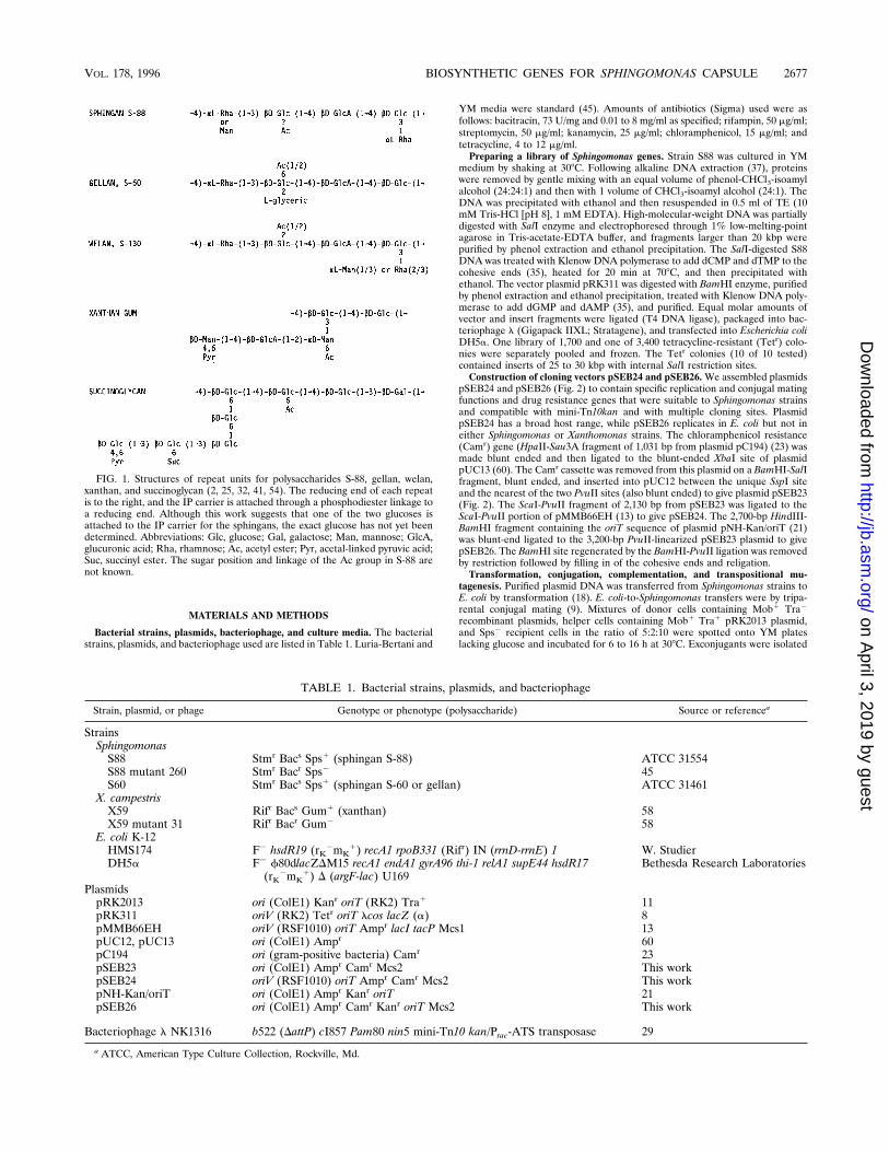
The ‘‘sphingans’’ are capsular polysaccharides that have similarbut not identical structures and are secreted by members of thegenus Sphingomonas (44). The group includes S-88, S-60 (gellan),S-130 (welan), S-194 (rhamsan), S-198, S-657, heteropolysaccha-ride 7, and NW-11 (27, 41, 43). As diagrammed in Fig. 1, thesphingans have different side groups, and either L-rhamnose orL-mannose is found at one position in the backbone. L-Mannoseitself is exceedingly rare in nature. Aqueous solutions of the poly-mers have unique and useful rheological properties (41). Gellan iscurrently produced by large-scale aerobic fermentation for use asa gelling agent in foods and microbiological culture media, andwelan is marketed for control of aqueous viscosity. It is not clearhow the structural variations in the polymers give rise to distinctrheological properties.Sphingomonas strains have been isolated from diverse environ-
ments and with a wide range of metabolic activities. The sphin-gan-secreting strains were isolated from plant tissue, water, andsoil. Chemoheterotrophic strains with the ability to metabolizetoluene, naphthalene, cresol, and other aromatic compoundswere recently isolated from deep terrestrial subsurface sediments(12). And other strains have been studied for their ability todegrade lignin (38). Sphingomonas bacteria have also been iso-lated from human clinical specimens and from hospital watersupplies, including stored distilled water. These bacteria wereoriginally named Pseudomonas paucimobilis (22) but were laterreclassified into distinct species within a new genusSphingomonas(62). More ominous are the recent reports of catheter-associated
bacteremia and gastrointestinal illnesses in humans that resultedfrom Sphingomonas contamination (47).Xanthomonas campestris is a plant-associated bacterium that
secretes xanthan gum (Fig. 1), which is widely used as anaqueous viscosifier. Most of the gum genes required by thisbacterium for synthesis and secretion of xanthan gum havebeen described (3, 6, 19, 20, 58). Previously we learned fromgenetic complementation tests that a special class of mutationsin X. campestris which are simultaneously xanthan gum nega-tive (Gum2) and resistant to bacitracin (Bacr) are in the gumDgene (45). This gene is required to transfer glucose-P fromUDP-Glc to a carrier isoprenylphosphate (IP) to give glucose-isoprenylpyrophosphate (Glc-PPI) and is the first dedicatedstep in synthesis of xanthan gum (59). The order of assemblyprior to polymerization is glucose, glucose, mannose, glucu-ronic acid, and finally mannose (24). Our genetic complemen-tation tests also showed that the wild-type gumD gene of X.campestris could restore synthesis of sphingans in analogousBacr Sps2 (sphingan polysaccharide-negative) mutants ofSphingomonas strains (45). Since there are two glucose resi-dues in the backbone of each of the sphingans, we reasonedthat the Bacr Sps2 mutants were probably also blocked in thetransfer of glucose-P to IP. Here we describe the wild-typecomplementing gene, spsB, from Sphingomonas strain S88 andshow that it belongs to a family of related genes that are allessential for the initial transfer of sugar to IP. We also showthe arrangement and DNA sequence of neighboring genescomprising about 29 kbp which are required for the followingfunctions: synthesis of dTDP-L-rhamnose, which is a precursorfor sphingan assembly; transfer of additional sugars duringassembly of the oligosaccharide repeat unit; secretion of thepolysaccharide capsule from the cell; and unexpectedly, secre-tion of a presumptive lytic or toxin-like protein.
* Corresponding author. Mailing address: Shin-Etsu Bio, Inc., 6650Lusk Blvd., Suite B106, San Diego, CA 92121. Phone: (619) 455-8500.Fax: (619) 587-2716. Electronic mail address: 76554,3460.Compuserve.com.
2676
on April 3, 2019 by guest
http://jb.asm.org/
Dow
nloaded from
MATERIALS AND METHODS
Bacterial strains, plasmids, bacteriophage, and culture media. The bacterialstrains, plasmids, and bacteriophage used are listed in Table 1. Luria-Bertani and
YM media were standard (45). Amounts of antibiotics (Sigma) used were asfollows: bacitracin, 73 U/mg and 0.01 to 8 mg/ml as specified; rifampin, 50 mg/ml;streptomycin, 50 mg/ml; kanamycin, 25 mg/ml; chloramphenicol, 15 mg/ml; andtetracycline, 4 to 12 mg/ml.Preparing a library of Sphingomonas genes. Strain S88 was cultured in YM
medium by shaking at 308C. Following alkaline DNA extraction (37), proteinswere removed by gentle mixing with an equal volume of phenol-CHCl3-isoamylalcohol (24:24:1) and then with 1 volume of CHCl3-isoamyl alcohol (24:1). TheDNA was precipitated with ethanol and then resuspended in 0.5 ml of TE (10mM Tris-HCl [pH 8], 1 mM EDTA). High-molecular-weight DNA was partiallydigested with SalI enzyme and electrophoresed through 1% low-melting-pointagarose in Tris-acetate-EDTA buffer, and fragments larger than 20 kbp werepurified by phenol extraction and ethanol precipitation. The SalI-digested S88DNA was treated with Klenow DNA polymerase to add dCMP and dTMP to thecohesive ends (35), heated for 20 min at 708C, and then precipitated withethanol. The vector plasmid pRK311 was digested with BamHI enzyme, purifiedby phenol extraction and ethanol precipitation, treated with Klenow DNA poly-merase to add dGMP and dAMP (35), and purified. Equal molar amounts ofvector and insert fragments were ligated (T4 DNA ligase), packaged into bac-teriophage l (Gigapack IIXL; Stratagene), and transfected into Escherichia coliDH5a. One library of 1,700 and one of 3,400 tetracycline-resistant (Tetr) colo-nies were separately pooled and frozen. The Tetr colonies (10 of 10 tested)contained inserts of 25 to 30 kbp with internal SalI restriction sites.Construction of cloning vectors pSEB24 and pSEB26.We assembled plasmids
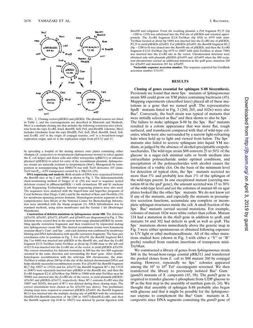
pSEB24 and pSEB26 (Fig. 2) to contain specific replication and conjugal matingfunctions and drug resistance genes that were suitable to Sphingomonas strainsand compatible with mini-Tn10kan and with multiple cloning sites. PlasmidpSEB24 has a broad host range, while pSEB26 replicates in E. coli but not ineither Sphingomonas or Xanthomonas strains. The chloramphenicol resistance(Camr) gene (HpaII-Sau3A fragment of 1,031 bp from plasmid pC194) (23) wasmade blunt ended and then ligated to the blunt-ended XbaI site of plasmidpUC13 (60). The Camr cassette was removed from this plasmid on a BamHI-SalIfragment, blunt ended, and inserted into pUC12 between the unique SspI siteand the nearest of the two PvuII sites (also blunt ended) to give plasmid pSEB23(Fig. 2). The ScaI-PvuII fragment of 2,130 bp from pSEB23 was ligated to theScaI-PvuII portion of pMMB66EH (13) to give pSEB24. The 2,700-bp HindIII-BamHI fragment containing the oriT sequence of plasmid pNH-Kan/oriT (21)was blunt-end ligated to the 3,200-bp PvuII-linearized pSEB23 plasmid to givepSEB26. The BamHI site regenerated by the BamHI-PvuII ligation was removedby restriction followed by filling in of the cohesive ends and religation.Transformation, conjugation, complementation, and transpositional mu-
tagenesis. Purified plasmid DNA was transferred from Sphingomonas strains toE. coli by transformation (18). E. coli-to-Sphingomonas transfers were by tripa-rental conjugal mating (9). Mixtures of donor cells containing Mob1 Tra2
recombinant plasmids, helper cells containing Mob1 Tra1 pRK2013 plasmid,and Sps2 recipient cells in the ratio of 5:2:10 were spotted onto YM plateslacking glucose and incubated for 6 to 16 h at 308C. Exconjugants were isolated
FIG. 1. Structures of repeat units for polysaccharides S-88, gellan, welan,xanthan, and succinoglycan (2, 25, 32, 41, 54). The reducing end of each repeatis to the right, and the IP carrier is attached through a phosphodiester linkage toa reducing end. Although this work suggests that one of the two glucoses isattached to the IP carrier for the sphingans, the exact glucose has not yet beendetermined. Abbreviations: Glc, glucose; Gal, galactose; Man, mannose; GlcA,glucuronic acid; Rha, rhamnose; Ac, acetyl ester; Pyr, acetal-linked pyruvic acid;Suc, succinyl ester. The sugar position and linkage of the Ac group in S-88 arenot known.
TABLE 1. Bacterial strains, plasmids, and bacteriophage
Strain, plasmid, or phage Genotype or phenotype (polysaccharide) Source or referencea
StrainsSphingomonasS88 Stmr Bacs Sps1 (sphingan S-88) ATCC 31554S88 mutant 260 Stmr Bacr Sps2 45S60 Stmr Bacs Sps1 (sphingan S-60 or gellan) ATCC 31461
X. campestrisX59 Rifr Bacs Gum1 (xanthan) 58X59 mutant 31 Rifr Bacr Gum2 58
E. coli K-12HMS174 F2 hsdR19 (rK
2mK1) recA1 rpoB331 (Rifr) IN (rrnD-rrnE) 1 W. Studier
DH5a F2 f80dlacZDM15 recA1 endA1 gyrA96 thi-1 relA1 supE44 hsdR17(rK
2mK1) D (argF-lac) U169
Bethesda Research Laboratories
PlasmidspRK2013 ori (ColE1) Kanr oriT (RK2) Tra1 11pRK311 oriV (RK2) Tetr oriT lcos lacZ (a) 8pMMB66EH oriV (RSF1010) oriT Ampr lacI tacP Mcs1 13pUC12, pUC13 ori (ColE1) Ampr 60pC194 ori (gram-positive bacteria) Camr 23pSEB23 ori (ColE1) Ampr Camr Mcs2 This workpSEB24 oriV (RSF1010) oriT Ampr Camr Mcs2 This workpNH-Kan/oriT ori (ColE1) Ampr Kanr oriT 21pSEB26 ori (ColE1) Ampr Camr Kanr oriT Mcs2 This work
Bacteriophage l NK1316 b522 (DattP) cI857 Pam80 nin5 mini-Tn10 kan/Ptac-ATS transposase 29
a ATCC, American Type Culture Collection, Rockville, Md.
VOL. 178, 1996 BIOSYNTHETIC GENES FOR SPHINGOMONAS CAPSULE 2677
on April 3, 2019 by guest
http://jb.asm.org/
Dow
nloaded from
by spreading a loopful of the mating mixture onto plates containing eitherrifampin (X. campestris) or streptomycin (Sphingomonas strains) to select againstthe E. coli helper and donor cells and either tetracycline (pRK311) or chloram-phenicol (pSEB24) to select for entry of the recombinant plasmids. Sphingomo-nas strains are naturally resistant to streptomycin (Stmr). Mutagenesis by trans-position in nonsuppressing host HMS174 was with Tn10 derivative 103 (mini-Tn10 kan/Ptac-ATS transposase) carried by l NK1316 (29).DNA sequencing and analysis. Both strands of DNA were sequenced between
the BamHI sites at bp 2 and 28800 as shown in Fig. 3. The dideoxynucleotidechain-terminating method of Sanger et al. (50) was used to sequence nesteddeletions created in pBluescriptIIKS(1) with exonuclease III and S1 nuclease(Lark Sequencing Technologies). Internal sequencing primers were also used.The sequences were analyzed with the SuperClone and SuperSee programs ofCoral Software (San Diego, Calif.) and by the method of Kyte and Doolittle (33)for membrane-spanning protein domains. Homologous protein segments in thecomprehensive data library at the National Center for Biotechnology Informa-tion were identified with the blastp program (1). DNA hybridization was bystandard methods, using nylon membranes and the Genius 1 kit (BoehringerMannheim).Construction of deletion mutations in Sphingomonas strain S88. The deletions
(DTn358, DTn365, DTn372, DTn495, and DTn493) are diagrammed in Fig. 3. Thedeletions were created first in the narrow-host-range plasmid pSEB26 by assem-bling specific restriction fragments and then transferred by triparental matinginto Sphingomonas strain S88. The desired recombinant strains were kanamycinresistant (Kanr), Cams, and Sps2, and each deletion was confirmed by membraneblotting and DNA hybridization with specific restriction fragments. The base paircoordinates refer to positions in Fig. 3. For DTn358, the BamHI fragment B4.5(bp 2 to 4480) was inserted into pSEB26 at the BamHI site, and then the EcoRIfragment E5.9::Tn10kan (mini-Tn10kan at about bp 25,000 close to the left endof E5.9) was inserted into the EcoRI site of the vector, to yield pSEB26-DTn358.The correct orientation for deletion formation in S88 has the two S88 segmentsoriented in the same direction and surrounding the Kanr gene. After double-homologous recombination with the wild-type S88 chromosome, the mini-Tn10kan is within about 200 bp of the site of the deleted chromosomal DNA andhelps identify successful recombination events. For DTn365 the BamHI fragmentB2.7 (bp 10927 to 13524) and for DTn372 the BamHI fragment B7.1 (bp 13524to 20407) were separately inserted into pSEB26 at the BamHI site, and then theEcoRI fragment E2.4::DTn10kan (bp 30600 to 33000 with mini-Tn10kan near bp30800) was inserted into the EcoRI site of the vector, to yield plasmids pSEB26-DTn365 and pSEB26-DTn372, respectively. Since B7.1 contains EcoRI sites (bp18807 and 20249), this part of B7.1 was deleted during these cloning steps. Thecorrect orientations were chosen as for DTn358 (see above). Two preliminarycloning steps were required to construct pSEB26-DTn493: the BamHI fragmentB12.6 was inserted into the multiple cloning site of pSEB26 with the orientationHindIII-PstI-BamHI-(insertion of bp-1200 to 10927)-BamHI-EcoRI, and thenthe BamHI segment (bp 6140 to 10927) was deleted by partial digestion with
BamHI and religation. From the resulting plasmid, a PstI fragment P2.35 (bp21200 to 1150) was subcloned into the PstI site of pSEB26 and oriented appro-priately. The EcoRI fragment E2.0::Tn10kan (bp 4926 to 6979 with mini-Tn10kan located at about bp 5400) was inserted into the EcoRI site of pSEB26-P2.35 to yield pSEB26-DTn493. For pSEB26-DTn495, the BamHI fragment B1.2(bp 21200 to 0) was cloned into the BamHI site of pSEB26, and then the EcoRIfragment E12.8::Tn10kan (bp 6979 to 18807 with mini-Tn10kan at about 7300)was inserted into the EcoRI site in the vector. Chromosomal deletions wereobtained only with plasmids pSEB26-DTn493 and -DTn495 when the S88 recip-ient chromosome carried an additional mutation in the spsB gene: mutation 260for DTn493 and mutation 265 for DTn495.Nucleotide sequence accession number. The sequence reported has GenBank
accession number U51197.
RESULTS
Cloning of genes essential for sphingan S-88 biosynthesis.Previously we found that most Sps2 mutants of Sphingomonasstrain S88 could grow on YM plates containing bacitracin (45).Mapping experiments (described later) placed all of these mu-tations in a gene that we named spsB. The representativeSpsB2 mutants listed in Fig. 3 (260, 265, and 102w) were alsoBacr. Conversely, the bac8 strain was typical of mutants thatwere initially selected as Bacr and then shown to also be Sps2.The failure to make sphingan S-88 by the Sps2 Bacr mutantsresulted in a colony appearance that was more flat, roughsurfaced, and translucent compared with that of wild-type col-onies, which were also surrounded by a narrow light-refractinghalo when held up to light and viewed from below. The Sps2
mutants also failed to secrete sphingans into liquid YM me-dium, as judged by the absence of alcohol-precipitable exopoly-saccharides. The wild-type strain S88 converts 25 to 50% of theglucose in a sugar-rich minimal salts or broth medium intoextracellular polysaccharide under optimal conditions, andprecipitation of the polysaccharides with alcohol causes theformation of a visible clot. On the basis of the minimum levelfor detection of typical clots, the Sps2 mutants secreted nomore than 5% and probably less than 1% of the sphingan ofthe wild-type parent. In one exceptional mutant (carrying mu-tation 68 in the spsF gene), the amount secreted was 15 to 30%of the wild-type level and yet the colonies of mutant 68 on agarplates looked like the other Sps2mutants. We do not know yetif any of the mutants, and especially the mutants in presump-tive secretion functions, accumulate any complete or incom-plete sphingan structures inside the cell. A small fraction of theSps2 Bacr mutants carried second mutations. For example,colonies of mutant 102w were white rather than yellow. Mutant134 had a mutation in the rhsD gene in addition to spsB, andmutants 54 and 302 had defects in spsK as well as spsB. TheSps2 mutations shown immediately above the genetic map inFig. 3 were either spontaneous or obtained following exposureto UV light or ethyl methanesulfonate. All of the other muta-tions studied here (shown in Fig. 3 with either a ‘‘Y’’ or ‘‘B’’prefix) resulted from random insertions of transposon mini-Tn10kan.We constructed a library of genes from Sphingomonas strain
S88 in the broad-host-range cosmid pRK311 and transferredthe pooled clones from E. coli to S88 mutant 260 by conjugalmating. However, repeatedly no Sps1 colonies appearedamong the 103 to 104 Tetr exconjugants screened. We thentransferred the library to previously isolated Bacr Gum2
(gumD) mutants of X. campestris (45, 58). The gumD gene isrequired to transfer glucose-1-phosphate from UDP-glucose toIP as the first step in the assembly of xanthan gum (6, 24). Wethought that assembly of sphingan S-88 probably also beganwith glucose and that it might be possible for the Sphingomo-nas enzyme to complement the Bacr Gum2 mutants in X.campestris since DNA segments containing the gumD gene of
FIG. 2. Cloning vectors pSEB24 and pSEB26. The plasmid sources are listedin Table 1, and the rearrangements are described in Materials and Methods.Mcs1 is a multiple cloning site that includes the following restriction sites (clock-wise from the top): EcoRI, SmaI, BamHI, SalI, PstI, andHindIII. Likewise, Mcs2includes (clockwise from the top) HindIII, PstI, SalI, XbaI, BamHI, SmaI, SstI,and EcoRI. oriT is the origin of conjugal transfer, oriV is a broad-host-rangereplication origin, and ori is the replication origin from pUC12 and 13.
2678 YAMAZAKI ET AL. J. BACTERIOL.
on April 3, 2019 by guest
http://jb.asm.org/
Dow
nloaded from
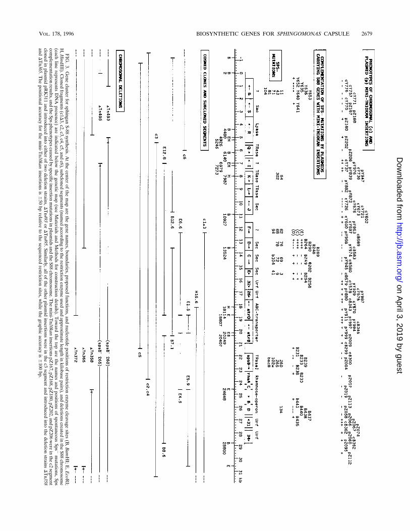
FIG.3.Gene
clusterforsphingan
S-88synthesis.A
tthe
centerofthe
mapare
thegene
names,boundaries,proposed
functions,andnucleotide
positionsofrestriction
enzymecleavage
sites(B,BamHI;E,EcoR
I;H,HindIII).C
lonedfragm
ents(c1D
3,c2,c3,c4,c5,andc6),subcloned
segments
(namedaccording
totherestriction
enzymeused
andapproxim
atelength
inkilobase
pairs),anddeletions
createdintheS88
chromosom
e(each
linerepresents
DNApresence)
areindicated
belowthe
geneticmap(see
Materials
andMethods
forconstruction
details).Toward
thetop
arethe
namesand
positionsofspontaneous
Sps2mutations,
Spscom
plementation
results,andtheSpsphenotypescaused
byspecific
insertionmutationsin
plasmidsand
theS88
chromosom
e.Themini-T
n10kaninsertionspZ
167,pZ168,pZ
180,pZ202,and
pZ206
wereinthec2segm
entcloned
inplasm
idpRK311
andintroduced
intoeither
oftwodeletion
strains,DTn493
orDTn495.Sim
ilarly,allofthe
otherplasm
idinsertions
were
inthe
c3segm
entand
introducedinto
thedeletion
strainsDTn358
andDTn365.T
hepositionalaccuracy
forthe
mini-T
n10kaninsertions
is650bprelative
tothe
sequencedrestriction
sites,while
thegraphic
accuracyis
6100
bp.
VOL. 178, 1996 BIOSYNTHETIC GENES FOR SPHINGOMONAS CAPSULE 2679
on April 3, 2019 by guest
http://jb.asm.org/
Dow
nloaded from
X. campestris could restore sphingan S-88 synthesis in Sphin-gomonas Bacr Sps2 mutants (45). From the intergeneric mat-ings, we found Gum1 colonies of X. campestris on YM platesat a frequency of about 1/103 to 104 Tetr exconjugants. Thedifference in complementation efficiency between Sphingomo-nas strain S88 and X. campestris is partly due to the moreobvious visual distinction between mucoid and nonmucoid col-onies of X. campestris but mainly reflects increased degradationof incoming plasmids in Sphingomonas strain S88.Plasmids were purified from the complemented X. campes-
tris mutants by transformation of E. coli and then mated intoSphingomonas strain S88 mutant 260. About 5 to 25% of theexconjugants became Sps1 in appearance on agar plates; forcomplementation of mutant 260 and other Sps2 mutants, thiscorresponded to restoration of at least 75% of the wild-typelevel of sphingan secreted into liquid medium. The frequencyof conjugal transfer for the vector alone (pRK311) was 100 to1,000 times higher than for the larger recombinant plasmids.Although most of the recombinant plasmids suffered extensivedeletions when mated into Sphingomonas strains, one, pRK311-S88c1, was recovered intact and used for subsequent work.The leftmost 21 kbp of S88 clone 1 (c1) is shown in Fig. 3 assubclone c1D3. Plasmid pRK311-S88c1 also restored polysaccha-ride synthesis to several other independently isolated Bacr
Sps2 mutants of strain S88 and Bacr Gum2 mutants of X.campestris. We isolated the polysaccharides that were secretedinto the culture medium by the exconjugants for each interge-neric mating and verified by thin-layer chromatography thatacid hydrolysates contained the neutral sugars expected for thepolysaccharide of the recipient cell (data not shown). Exopoly-saccharide from Sphingomonas mutant 260 bearing plasmidpRK311-S88c1 contained glucose, mannose, and rhamnose,while X. campestris mutant 31 with pRK311-S88c1 containedonly glucose and mannose. Each polysaccharide also containedglucuronic acid, although the recovery of the acidic sugar wassystematically low because of the hydrolysis conditions.To verify that the cloned DNA in plasmid pRK311-S88c1D3
derived from contiguous sequences in the S88 cellular DNA,we labeled plasmid pRK311-S88c1D3 DNA and hybridized itto separated restriction fragments of cellular DNA from Sphin-gomonas strain S88. The positive hybridization to EcoRI frag-ments (left to right on Fig. 3) of about 12, 1.5, 4.5, 5.9, and 2.4kbp was consistent with continuity for the cloned DNA in boththe wild type and mutant (data not shown).We extended the cloned region toward the left in Fig. 3 by
screening the library for segments that complemented theSps2 Bacs mutants 76 and 78. We screened 104 to 106 excon-jugants and obtained four more clones (S88 c2, c3, c4, and c5)that partially overlapped the S88 c1 segment. Similarly, c6 wasisolated by complementing Sps2 mutants 43, 71, and 104. Thefive cloned segments were each about 22 to 28 kb in length, andat least one end of each segment is shown in Fig. 3. Weidentified a set of about 15 Sps2 mutations that were notcomplemented by either c2 or c1D3. None of these mutationswere complemented by the full-length c1, which extends fur-ther to the right than c1D3 by about 8 kbp, and none werecomplemented by c6, which extends to the left of c2 in Fig. 3 byabout 18 kbp. Most of these Sps2 mutations were restored toSps1 by complementation with three overlapping cloned seg-ments (c10, c11, and c12) which do not overlap with the c6, c2,or c1 cloned segments. The set of unlinked mutations suggestsadditional genes that are essential for sphingan synthesis butwhich are not immediately adjacent to the cluster of genesshown in Fig. 3.Mapping of the sps genes by functional complementation.
The boundaries of the spsG, spsK, spsF, spsD, spgC, spsE, spsB,
and rhsD genes were determined by complementation testsusing the Sps2 point mutants as recipients in conjugation. Theresults are summarized in Fig. 3 above the map. Recombinantplasmids bearing subcloned DNA segments were exposed toinsertional mutagenesis with mini-Tn10kan in E. coli and thentransferred by mating into Sps2 mutants of strain S88. Twomatable broad-host-range plasmid vectors, pRK311 andpSEB24, were used (Fig. 2). The exconjugants that receivedthe drug resistance marker of the entering plasmid were thenscored as Sps1 or Sps2 on the basis of colonial appearance.The Bacr Sps2 S88 mutants were mapped initially to the E4.5subclone, and then the E4.5 segment was exposed to randominsertional mutagenesis with mini-Tn10kan. Insertions into po-sitions B231 and B230 (Fig. 3) did not affect the restoration ofsphingan synthesis for mutant 260; however, insertions B233,B239, and B238 blocked complementation. Mutant 134 wascomplemented by segment B8.6 but not by either E4.5 or E5.9.Mutant 134 has a defect in the spsB gene and also in the nearbyrhsD gene. The more precise location of the rhsDmutation wasdetermined by exposing the B8.6 segment to mini-Tn10kanmutagenesis and analyzing the complementation pattern forthe B441, B440, B438, B437, and B435 insertions. Mutants 54and 302 also appeared to be double mutants with defects in thespsK and spsB genes.The spsF mutants (62, 68, and 94) were localized and sepa-
rated from the nearby spsDCE cluster as a result of the lack ofcomplementation by segments B12.6 and c1D3 and by restora-tion of sphingan S-88 synthesis by clones c3 and c5. Comple-mentation results for the contiguous spsD, spsC, and spsEgenes following insertional mutagenesis of the E6.6 fragmentare also shown in Fig. 3. The complementation results sug-gested two groups, the first including mutations 76 and 78 andthe second comprising mutations 69, 72, b104, 3, 9, and 41.Later analysis of the DNA sequences resolved the latter groupinto two distinct contiguous genes: spsC and spsE. The spsGmutations (11, 43, 71, 81, and 104) were complemented by thec6 clone and the B12.6 segment. Insertional mutagenesis of theB12.6 segment yielded plasmids Y652, Y635, Y636, Y653,Y640, and Y641. Of these, only Y652 and Y641 were able tocomplement the spsG mutants.Phenotypes of mini-Tn10kan chromosomal and plasmid in-
sertions. Segments of cloned S88 DNA were ligated to thematable narrow-host-range Camr plasmid pSEB26 (Fig. 2),exposed to mutagenesis by mini-Tn10kan in E. coli, and thenconjugally transferred into wild-type (Sps1) Sphingomonasstrain S88. The plasmids were not able to replicate in Sphin-gomonas cells, and so maintenance of the Kanr gene requiredrecombination with the bacterial chromosome. We selectedonly those recombinants that were Kanr and Cams, expectingthat this group did not retain plasmid sequences. Although wedid not verify the physical structures of these DNA substitu-tions, we have routinely used the same plasmids, strains, andselection schemes to create site-specific chromosomal dele-tions (bottom of Fig. 3) and confirmed those double-recombi-nation events by restriction mapping and DNA hybridization.Colonies of the Kanr Cams chromosomal recombinants werejudged as Sps1 or Sps2 (shown at the top of Fig. 3 for inser-tions labeled with a prefix ‘‘c’’). For the mutants showing anSps2 phenotype (cY776, cY757, cY771, cY770, cY726, cY725,cY676, cY673, cY721, cY602, cB589, cB583, cB580, cB579, andcB300), it was reasonable to believe that double recombinationoccurred. However, for the Sps1 recombinants, it was possiblethat the entire plasmid integrated into the chromosome at oneof the homologous regions, resulting in one defective and onenormal gene in the chromosome.To avoid the double-recombination uncertainty, we created
2680 YAMAZAKI ET AL. J. BACTERIOL.
on April 3, 2019 by guest
http://jb.asm.org/
Dow
nloaded from

large site-specific deletions in the chromosome and then intro-duced replicating plasmids that carried either the S88 c2 or theS88 c3 DNA segment with single mini-Tn10kan insertions toinactivate certain genes. The positions and Sps1 or Sps2 phe-notypes of the insertions are shown near the top of Fig. 3 withthe prefix ‘‘p.’’ The results from the chromosomal and plasmidmutation strategies conform to one another, and both essentialand nonessential regions were observed.DNA sequence: G1C content, use of rare codons, and trans-
lational start sequences. The DNA sequence of 28,804 bp wasdetermined for both strands (Fig. 3). An average profile (andstandard deviation) for a typical Sphingomonas gene in thiscluster was determined on the basis of the skewed G1C con-tents, rare codon frequencies, and Shine-Dalgarno or transla-tion initiation sequences (Table 2). A high frequency of G or Cin the third codon position was typical for each of the genes. Aset of rarely used codons for Sphingomonas strains was iden-tified early in the work by analyzing 2,500 codons from therhsACBD operon and the spsB, -D, -C, and -E genes. Each rarecodon in the set was present at less than 0.2% of the total andincluded AGA, AGG, CGA, TGT, GGA, ATA, CTA, TTA,TTG, AAA, TTT, CCA, CCT, AGT, TCA, TCT, ACA, andACT. Translation usually initiates in E. coli adjacent anddownstream from a sequence that is complementary to the 39terminus of 16S rRNA (51). The analogous Shine-Dalgarnosequence complementary to 16S rRNA in Sphingomonas pauci-mobilis DSM1098 is TAAGGAGGTG (40).If a gene in this cluster matched the average gene profile and
could be mutated to give an Sps2 phenotype, then it was givenan sps designation. However, our search for protein similaritiesprovided no significant hint as to the possible functions of thespsG, -I, and -F genes. In addition, there were four other openreading frames that satisfied the typical gene profile and failedto show any significant similarity to protein sequences in com-puter databanks. However, since mutations in these putativegenes did not visibly alter polysaccharide synthesis, they were
labeled Urf, for unidentified reading frame. There were fourUrf sequences (Urf32, Urf26, Urf31, and Urf34), which werenamed according to the size of the deduced protein in kilodal-tons.spsB: glucosyl-IP transferase. Most of the Sps2 mutations
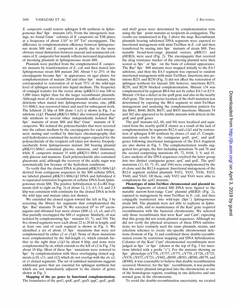
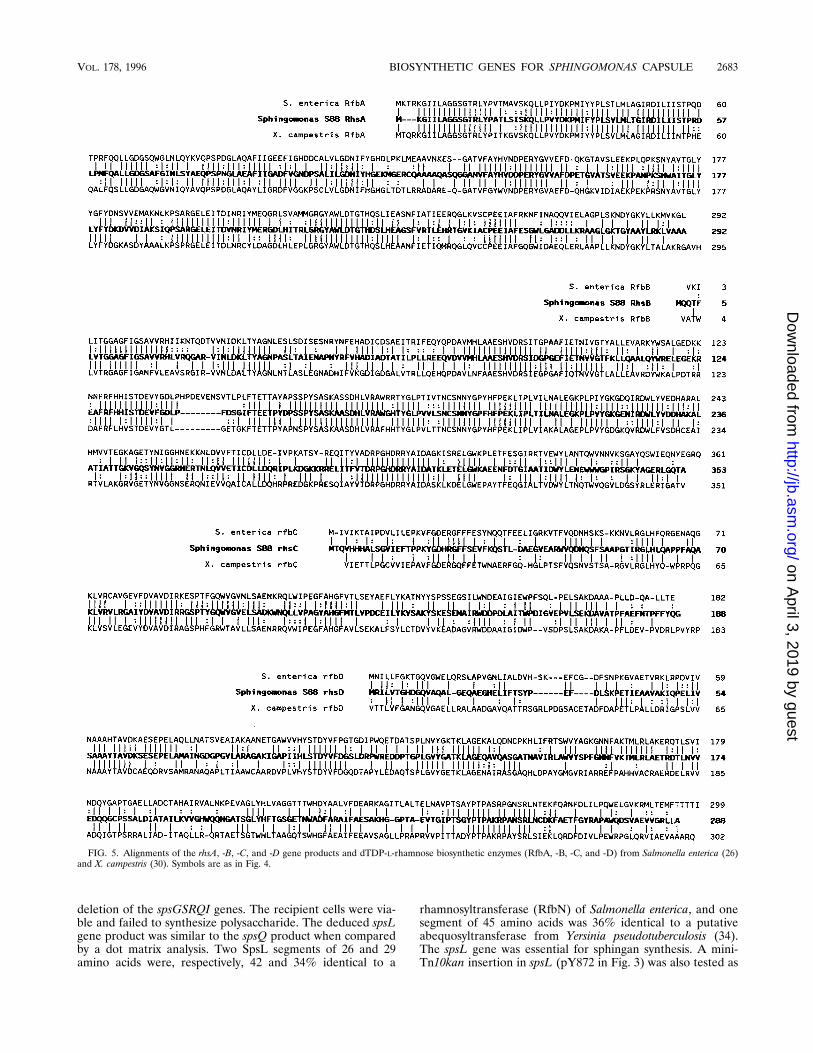
that were isolated following UV or chemical mutagenesis werein the spsB gene. The SpsB protein is believed to catalyze thefirst step in assembly of sphingan S-88 because of the strikingsimilarity of the deduced amino acid sequence of SpsB to othergene products believed to code for glycosyl-IP transferases.Figure 4 shows an alignment of amino acid sequences of sus-pected glucosyl- and galactosyl-IP transferases. There is con-siderable homology for the C-terminal halves of these proteins.Although the N-terminal regions lack this extensive homology,the SpsB protein is similar to the RfbP protein of Salmonellaenterica (26) by having several hydrophobic regions which sug-gest membrane-spanning domains (underlined in Fig. 4). Thehydrophobic domains of SpsB include amino acids 35 to 59(12.2 average hydropathy), 68 to 86 (11.7), 105 to 123 (12.3),and 282 to 303 (12.9). The position of the latter hydrophobicsegment was common to these related gene products and waslocated in mid-protein adjacent to the region of greatest ho-mology.The spsB coding domain deduced from the DNA sequence
was confirmed by complementation studies. We observedwhether different insertions of mini-Tn10kan in the E4.5 seg-ment interfered with complementation of Bacr Sps2 mutants.The sites of insertion of mini-Tn10kan and complementationresults are shown above the spsB gene in Fig. 3. Three mini-Tn10kan insertions, (B233, B239, and B238) failed to restoresphingan synthesis to the S88 Sps2 mutant 260, while severalflanking insertions, including B231 and B230, retained the abil-ity to supply the missing function.Rhamnose biosynthetic operon. The deduced amino acid
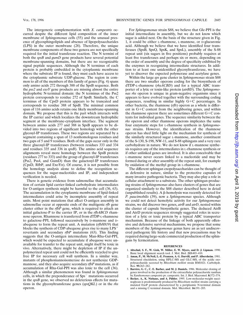
sequences of the proteins coded by the rhsACBD genes werevery similar to enzymes from Salmonella enterica group B and
TABLE 2. Profiles of sps genes
Gene% G1C by codon position Codons
Putative translational start sequencesTotal 1st 2nd 3rd Total % Rare
spsB 66 65 43 90 470 2 TTGAGGGAGCCCGACGAGGCAATGAACspsC 68 70 48 87 447 1 GACAGCGGACGAGGCCCACCAGTGAATspsD 68 71 50 83 301 4 TGACAAGGGCCGTATTCATGCATGCATspsE 68 65 47 91 235 0 GCACGGAGCTTCAGTAAACTGATGGACspsF 64 61 51 79 432 6 TTGTACTGGAGGCCATTGATAATGAAGspsG 65 61 45 89 539 4 CGATTATCTAAGGGGTTGGTCATGGCGspsI 67 69 45 88 300 3 CGTGCCGGCTGGGAGGTCTTCATGAAGspsJ 68 73 44 85 462 3 CCGAAATTAAGAGGTGTTCGAGTGGCTspsK 68 72 46 85 352 3 GGCGGGAGGCAGGCGGGATCAATGGGGspsL 68 72 52 81 288 5 GGCACAGTGGAGTGCCAAGCGATGAGCspsQ 68 70 51 84 315 3 CAGCACGGGTAAGAACGAGGCATGGAAspsR 61 54 46 85 670 3 CGCGTAACGAGGGTAGAGTACATGCCGspsS 65 65 47 84 452 5 GCAGGACTTCTATCACGTCTGATGACGrhsA 65 64 45 87 292 0.3 CCCGCGCCATGGGGATTTTGAATGAAGrhsC 65 65 43 88 188 2 GCGCAAGCTGGTAGCCGCGGCATGACCrhsB 65 63 42 89 353 0.6 TTCTTCTATCAGGGCTGATCCATGCAGrhsD 69 66 49 91 288 0.3 GCGGTTGGGGCAGACCGCCTGATGCGCatrB 69 70 44 91 728 1 CCATGGAGGCAGAGTACCGGAATGACAatrD 70 73 49 88 464 2 CGGATATGGGGAGATTGCCGCATGAACAvg 67 67 47 87 2.5SD 2 5 3 3 1.8
Urf32 66 65 46 89 293 3 ACGGCTATTGAATTGGATTCCATGACCUrf26 67 69 44 89 232 3 TCACACGGCGCCGGAGGCCCCATGTTCUrf31 65 61 41 91 270 2 AGACCGGGGCTGATCGAACCGATGCTTUrf34 68 64 50 91 318 3 GCGCAATGACACGCGGCCGGAATGACA
VOL. 178, 1996 BIOSYNTHETIC GENES FOR SPHINGOMONAS CAPSULE 2681
on April 3, 2019 by guest
http://jb.asm.org/
Dow
nloaded from
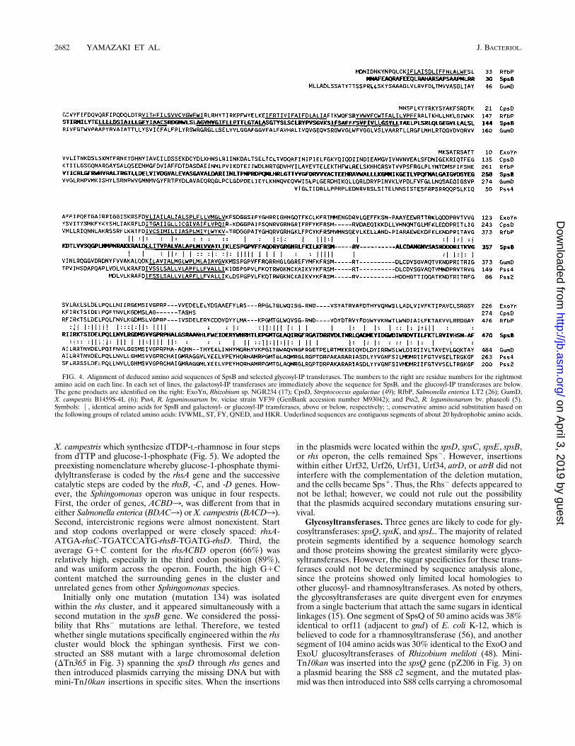
X. campestris which synthesize dTDP-L-rhamnose in four stepsfrom dTTP and glucose-1-phosphate (Fig. 5). We adopted thepreexisting nomenclature whereby glucose-1-phosphate thymi-dylyltransferase is coded by the rhsA gene and the successivecatalytic steps are coded by the rhsB, -C, and -D genes. How-ever, the Sphingomonas operon was unique in four respects.First, the order of genes, ACBD3, was different from that ineither Salmonella enterica (BDAC3) or X. campestris (BACD3).Second, intercistronic regions were almost nonexistent. Startand stop codons overlapped or were closely spaced: rhsA-ATGA-rhsC-TGATCCATG-rhsB-TGATG-rhsD. Third, theaverage G1C content for the rhsACBD operon (66%) wasrelatively high, especially in the third codon position (89%),and was uniform across the operon. Fourth, the high G1Ccontent matched the surrounding genes in the cluster andunrelated genes from other Sphingomonas species.Initially only one mutation (mutation 134) was isolated
within the rhs cluster, and it appeared simultaneously with asecond mutation in the spsB gene. We considered the possi-bility that Rhs2 mutations are lethal. Therefore, we testedwhether single mutations specifically engineered within the rhscluster would block the sphingan synthesis. First we con-structed an S88 mutant with a large chromosomal deletion(DTn365 in Fig. 3) spanning the spsD through rhs genes andthen introduced plasmids carrying the missing DNA but withmini-Tn10kan insertions in specific sites. When the insertions
in the plasmids were located within the spsD, spsC, spsE, spsB,or rhs operon, the cells remained Sps2. However, insertionswithin either Urf32, Urf26, Urf31, Urf34, atrD, or atrB did notinterfere with the complementation of the deletion mutation,and the cells became Sps1. Thus, the Rhs2 defects appeared tonot be lethal; however, we could not rule out the possibilitythat the plasmids acquired secondary mutations ensuring sur-vival.Glycosyltransferases. Three genes are likely to code for gly-
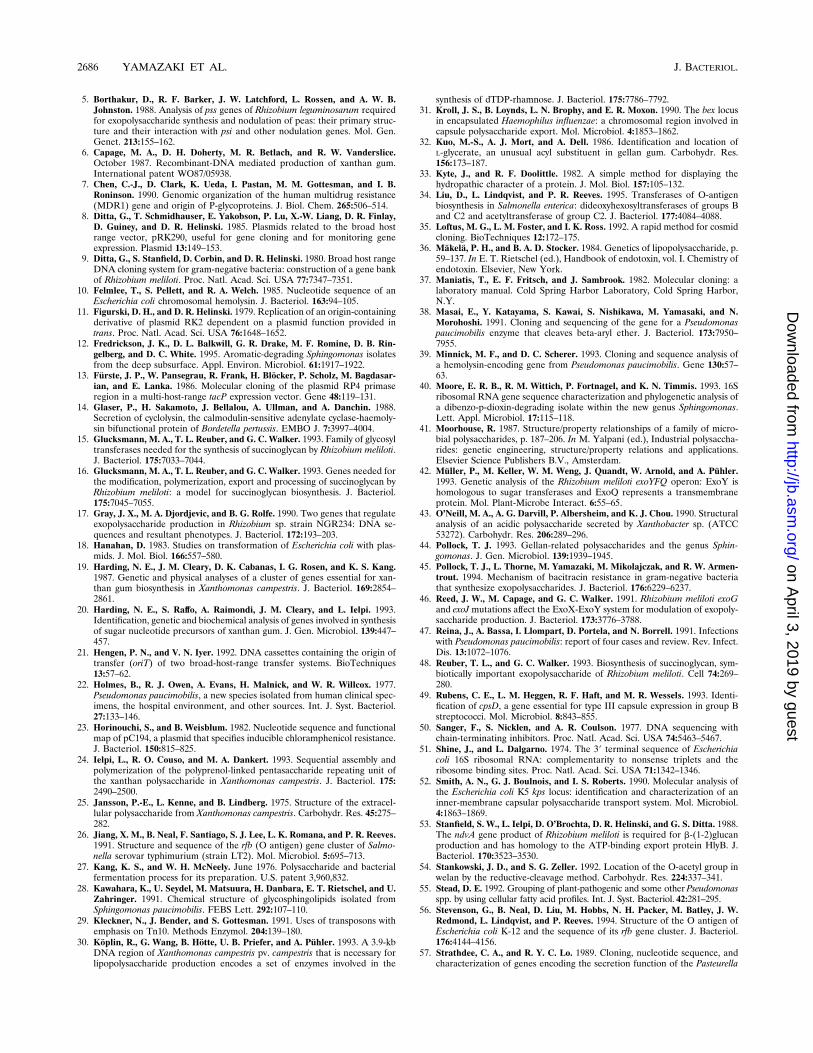
cosyltransferases: spsQ, spsK, and spsL. The majority of relatedprotein segments identified by a sequence homology searchand those proteins showing the greatest similarity were glyco-syltransferases. However, the sugar specificities for these trans-ferases could not be determined by sequence analysis alone,since the proteins showed only limited local homologies toother glucosyl- and rhamnosyltransferases. As noted by others,the glycosyltransferases are quite divergent even for enzymesfrom a single bacterium that attach the same sugars in identicallinkages (15). One segment of SpsQ of 50 amino acids was 38%identical to orf11 (adjacent to gnd) of E. coli K-12, which isbelieved to code for a rhamnosyltransferase (56), and anothersegment of 104 amino acids was 30% identical to the ExoO andExoU glucosyltransferases of Rhizobium meliloti (48). Mini-Tn10kan was inserted into the spsQ gene (pZ206 in Fig. 3) ona plasmid bearing the S88 c2 segment, and the mutated plas-mid was then introduced into S88 cells carrying a chromosomal
FIG. 4. Alignment of deduced amino acid sequences of SpsB and selected glycosyl-IP transferases. The numbers to the right are residue numbers for the rightmostamino acid on each line. In each set of lines, the galactosyl-IP transferases are immediately above the sequence for SpsB, and the glucosyl-IP transferases are below.The gene products are identified on the right: ExoYn, Rhizobium sp. NGR234 (17); CpsD, Streptococcus agalactiae (49); RfbP, Salmonella enterica LT2 (26); GumD,X. campestris B1459S-4L (6); Pss4, R. leguminosarum bv. viciae strain VF39 (GenBank accession number M93042); and Pss2, R. leguminosarum bv. phaseoli (5).Symbols: P, identical amino acids for SpsB and galactosyl- or glucosyl-IP transferases, above or below, respectively; :, conservative amino acid substitution based onthe following groups of related amino acids: IVWML, ST, FY, QNED, and HKR. Underlined sequences are contiguous segments of about 20 hydrophobic amino acids.
2682 YAMAZAKI ET AL. J. BACTERIOL.
on April 3, 2019 by guest
http://jb.asm.org/
Dow
nloaded from
deletion of the spsGSRQI genes. The recipient cells were via-ble and failed to synthesize polysaccharide. The deduced spsLgene product was similar to the spsQ product when comparedby a dot matrix analysis. Two SpsL segments of 26 and 29amino acids were, respectively, 42 and 34% identical to a
rhamnosyltransferase (RfbN) of Salmonella enterica, and onesegment of 45 amino acids was 36% identical to a putativeabequosyltransferase from Yersinia pseudotuberculosis (34).The spsL gene was essential for sphingan synthesis. A mini-Tn10kan insertion in spsL (pY872 in Fig. 3) was also tested as
FIG. 5. Alignments of the rhsA, -B, -C, and -D gene products and dTDP-L-rhamnose biosynthetic enzymes (RfbA, -B, -C, and -D) from Salmonella enterica (26)and X. campestris (30). Symbols are as in Fig. 4.
VOL. 178, 1996 BIOSYNTHETIC GENES FOR SPHINGOMONAS CAPSULE 2683
on April 3, 2019 by guest
http://jb.asm.org/
Dow
nloaded from
described above for pZ206, and the mutant failed to synthesizesphingan. Searches for proteins similar to SpsK produced onlymarginal similarities, of which the predominant members wereglycosyltransferases containing a common putative binding sitefor UDP. The possible involvement of UDP suggests a glu-cosyl- or glucuronosyltransferase. A mutant with a specificinsertion in the spsK gene (pY882 in Fig. 3) also failed to makepolysaccharide when tested as described above for spsL.Secretion. Common strategies for secretion of polysaccha-
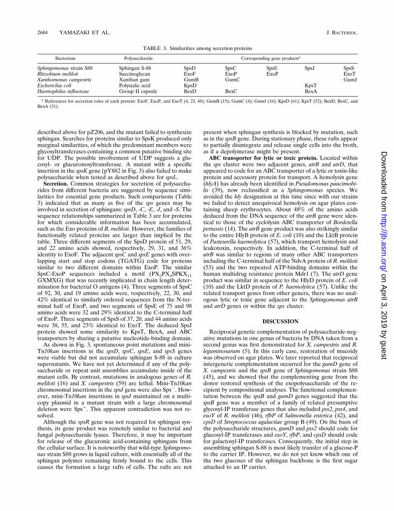
rides from different bacteria are suggested by sequence simi-larities for essential gene products. Such comparisons (Table3) indicated that as many as five of the sps genes may beinvolved in secretion of sphingans: spsD, -C, -E, -J, and -S. Thesequence relationships summarized in Table 3 are for proteinsfor which considerable information has been accumulated,such as the Exo proteins of R. meliloti. However, the families offunctionally related proteins are larger than implied by thetable. Three different segments of the SpsD protein of 51, 29,and 22 amino acids showed, respectively, 29, 31, and 36%identity to ExoF. The adjacent spsC and spsE genes with over-lapping start and stop codons (TGATG) code for proteinssimilar to two different domains within ExoP. The similarSpsC-ExoP sequences included a motif (PX2PX4SPKX11GXMXG) that was recently implicated in chain length deter-mination for bacterial O antigens (4). Three segments of SpsCof 92, 30, and 19 amino acids were, respectively, 22, 30, and42% identical to similarly ordered sequences from the N-ter-minal half of ExoP, and two segments of SpsE of 75 and 98amino acids were 32 and 29% identical to the C-terminal halfof ExoP. Three segments of SpsS of 37, 20, and 44 amino acidswere 38, 55, and 23% identical to ExoT. The deduced SpsJprotein showed some similarity to KpsT, BexA, and ABCtransporters by sharing a putative nucleotide-binding domain.As shown in Fig. 3, spontaneous point mutations and mini-
Tn10kan insertions in the spsD, spsC, spsE, and spsS geneswere viable but did not accumulate sphingan S-88 in culturesupernatants. We have not yet determined if any of the poly-saccharide or repeat unit assemblies accumulate inside of themutant cells. By contrast, mutations in analogous genes of R.meliloti (16) and X. campestris (59) are lethal. Mini-Tn10kanchromosomal insertions in the spsJ gene were also Sps2. How-ever, mini-Tn10kan insertions in spsJ maintained on a multi-copy plasmid in a mutant strain with a large chromosomaldeletion were Sps1. This apparent contradiction was not re-solved.Although the spsR gene was not required for sphingan syn-
thesis, its gene product was remotely similar to bacterial andfungal polysaccharide lyases. Therefore, it may be importantfor release of the glucuronic acid-containing sphingans fromthe cellular surface. It is noteworthy that wild-type Sphingomo-nas strain S88 grows in liquid culture, with essentially all of thesphingan polymer remaining firmly bound to the cells. Thiscauses the formation a large rafts of cells. The rafts are not
present when sphingan synthesis is blocked by mutation, suchas in the spsB gene. During stationary phase, these rafts appearto partially disintegrate and release single cells into the broth,as if a depolymerase might be present.ABC transporter for lytic or toxic protein. Located within
the sps cluster were two adjacent genes, atrB and atrD, thatappeared to code for an ABC transporter of a lytic or toxin-likeprotein and accessory protein for transport. A hemolysin gene(hlyA) has already been identified in Pseudomonas paucimobi-lis (39), now reclassified as a Sphingomonas species. Weavoided the hly designation at this time since with our strainswe failed to detect unequivocal hemolysis on agar plates con-taining sheep erythrocytes. About 48% of the amino acidsdeduced from the DNA sequence of the atrB gene were iden-tical to those of the cyclolysin ABC transporter of Bordetellapertussis (14). The atrB gene product was also strikingly similarto the entire HlyB protein of E. coli (10) and the LktB proteinof Pasteurella haemolytica (57), which transport hemolysin andleukotoxin, respectively. In addition, the C-terminal half ofatrB was similar to regions of many other ABC transportersincluding the C-terminal half of the NdvA protein of R. meliloti(53) and the two repeated ATP-binding domains within thehuman multidrug resistance protein Mdr1 (7). The atrD geneproduct was similar in sequence to the HlyD protein of E. coli(10) and the LktD protein of P. haemolytica (57). Unlike therelated transport genes from other genera, there was no anal-ogous lytic or toxic gene adjacent to the Sphingomonas atrBand atrD genes or within the sps cluster.
DISCUSSION
Reciprocal genetic complementation of polysaccharide-neg-ative mutations in one genus of bacteria by DNA taken from asecond genus was first demonstrated for X. campestris and R.leguminosarum (5). In this early case, restoration of mucoidywas observed on agar plates. We later reported that reciprocalintergeneric complementation occurred for the gumD gene ofX. campestris and the spsB gene of Sphingomonas strain S88(45), and we showed that the complementing gene from thedonor restored synthesis of the exopolysaccharide of the re-cipient by compositional analyses. The functional complemen-tation between the spsB and gumD genes suggested that thespsB gene was a member of a family of related presumptiveglycosyl-IP transferase genes that also included pss2, pss4, andexoY of R. meliloti (46), rfbP of Salmonella enterica (42), andcpsD of Streptococcus agalactiae group B (49). On the basis ofthe polysaccharide structures, gumD and pss2 should code forglucosyl-IP transferases and exoY, rfbP, and cpsD should codefor galactosyl-IP transferases. Consequently, the initial step inassembling sphingan S-88 is most likely transfer of a glucose-Pto the carrier IP. However, we do not yet know which one ofthe two glucoses of the sphingan backbone is the first sugarattached to an IP carrier.
TABLE 3. Similarities among secretion proteins
Bacterium Polysaccharide Corresponding gene productsa
Sphingomonas strain S88 Sphingan S-88 SpsD SpsC SpsE SpsJ SpsSRhizobium meliloti Succinoglycan ExoF ExoP ExoP ExoTXanthomonas campestris Xanthan gum GumB GumC GumJEscherichia coli Polysialic acid KpsD KpsTHaemophilus influenzae Group II capsule BexD BexC BexA
a References for secretion roles of each protein: ExoF, ExoP, and ExoT (4, 23, 48); GumB (15); GumC (4); GumJ (16); KpsD (61); KpsT (52); BexD, BexC, andBexA (31).
2684 YAMAZAKI ET AL. J. BACTERIOL.
on April 3, 2019 by guest
http://jb.asm.org/
Dow
nloaded from
The intergeneric complementation with X. campestris oc-curred despite the different lipid composition of the innermembrane of Sphingomonas cells (55) and the unusual pres-ence of glycosphingolipids and absence of lipopolysaccharides(LPS) in the outer membrane (28). Therefore, the uniquemembrane components of these two genera are not specificallyrequired for the initial transferase reaction. The N termini ofboth the spsB and gumD gene products have several potentialmembrane-spanning domains, but there are no recognizablesignal peptide sequences. Although the N terminus of eachprotein is probably embedded in the cytoplasmic membranewhere the substrate IP is found, they must each have access tothe cytoplasmic substrate UDP-glucose. The region in com-mon to all of the members of this family of genes (Fig. 4) spansonly amino acids 272 through 388 of the SpsB sequence. Boththe pss2 and exoY gene products are missing almost the entirehydrophobic N-terminal domain: the N terminus of the Pss2protein corresponds to residue 272 of SpsB. Similarly, the Cterminus of the CpsD protein appears to be truncated andcorresponds to residue 388 of SpsB. The minimal commonspan of 116 amino acids retains one likely membrane-spanningdomain. We propose that this is the part which interacts withthe IP carrier and which localizes the downstream hydrophilicsegment at the membrane-cytoplasm interface. The segmentbetween amino acids 277 and 388 in SpsB appears to be di-vided into two regions of significant homology with the otherglycosyl-IP transferases. These two regions are separated by asegment containing a span of 13 nonhomologous residues andtwo gaps of 5 and 9 residues. Both of the gaps occur only in thethree glucosyl-IP transferases (between residues 333 and 334and residues 335 and 336 in spsB). The amino acid sequencealignments reveal more homology between the SpsB protein(residues 277 to 333) and the group of glucosyl-IP transferases(Pss2, Pss4, and GumD) than the galactosyl-IP transferases(CpsD, RfbP, and ExoYn). Of course, it is not possible fromthe homology analysis alone to define the recognition se-quences for the sugar-nucleotides and IP, and independentverification is needed.There is genetic evidence from salmonellae that accumula-
tion of certain lipid carrier-linked carbohydrate intermediatesfor O-antigen synthesis might be harmful to the cell (36, 63).The accumulation in the cytoplasmic membrane of lipid-linkedintermediates could arise by incomplete assembly of repeatunits. Most point mutations that affect O-antigen assembly insalmonellae occur at opposite ends of the multigenic rfb genecluster either in the rfbP gene, which is required to attach aninitial galactose-P to the carrier IP, or in the rfbABCD rham-nose operon. Rhamnose is transferred from dTDP-L-rhamnoseto galactose-PPI, followed successively by D-mannose and D-abequose to form the repeat unit. A mutation (rfbH819) thatblocks the synthesis of CDP-abequose gives rise to many LPS1
revertants and secondary rfbP mutations (63). This findingsuggests that the O-antigen intermediate Man-Rha-Gal-PPI,which would be expected to accumulate if abequose were un-available for transfer to the repeat unit, might itself be toxic invivo. Alternatively, there might be depletion of IP if the un-polymerized repeat unit could not be efficiently recycled to givefree IP for necessary cell wall synthesis. In a similar way,mutants of phosphomannoisomerase do not synthesize GDP-mannose, and they also acquire secondary rfbP mutations, as ifaccumulation of Rha-Gal-PPI was also toxic to the cell (36).Although a similar phenomenon was found in Sphingomonascells, in which the preponderance of Sps2 mutations occurredin the spsB gene, we observed no deleterious effects for muta-tions in the glycosyltransferase genes (spsQKL) or in the rhsoperon.
For Sphingomonas strain S88, we believe that Glc-PPI is theinitial intermediate in assembly, but we do not know whichsugar is added next. On the basis of the structure given in Fig.1, it could be either L-rhamnose, L-mannose, or D-glucuronicacid. Although we believe that we have identified four trans-ferases (SpsB, SpsQ, SpsK, and SpsL), assembly of the S-88repeat unit (six sugars in five positions) probably requires atleast five transferases and perhaps six or more, depending onthe order of assembly and the degree of specificity exhibited bythe enzymes in recognizing intermediate structures. In addi-tion to at least one unidentified glycosyltransferase, we haveyet to discover the expected polymerase and acetylase genes.Within the large sps gene cluster in Sphingomonas strain S88
there are two smaller operons coding for the biosynthesis ofdTDP-L-rhamnose (rhsACBD) and for a typical ABC trans-porter of a lytic or toxin-like protein (atrBD). The Sphingomo-nas rhs operon is unique in gram-negative organisms since itappears to have evolved together with the surrounding codingsequences, resulting in similar highly G1C percentages. Inother bacteria, the rhamnose (rfb) operon as a whole is differ-ent in G1C content from the neighboring genes, and withinthe rhamnose operon there are sometimes different G1C con-tents for individual genes. The sequence similarity between therhs operon and other rhamnose operons implicates the samefour enzymatic steps for L-rhamnose synthesis in Sphingomo-nas strains. However, the identification of the rhamnoseoperon has shed little light on the mechanism for synthesis ofthe similarly structured sugar L-mannose, another constituentof the S-130, S-198, NW-11, and S-88 sphingans and a very rarecarbohydrate in nature. We do not know if L-mannose synthe-sis requires any of the intermediates in L-rhamnose synthesis orif other unlinked genes are involved. It is also conceivable thatL-mannose never occurs linked to a nucleotide and may beformed during or after assembly of the repeat unit, for exampleby oxidation of the methyl group in L-rhamnose.The different sphingan exopolysaccharides can be thought of
as defensive in nature, similar to the protective capsules ofmany invasive pathogenic bacteria. They may also play a role incellular attachment to a substrate. The other sphingan-produc-ing strains of Sphingomonas also have clusters of genes that areorganized similarly to the S88 cluster described here in detail(unpublished results). A b-hemolysin was recently detected forP. paucimobilis (39), now a Sphingomonas species. Althoughwe could not detect hemolytic activity for our Sphingomonasstrains, we did discover two genes, atrB and atrD, nested withinthe cluster of capsule biosynthetic genes. The deduced AtrBand AtrD protein sequences strongly suggested roles in secre-tion of a lytic or toxic protein by a typical ABC transportermechanism. Because of the linkage of both apparently offen-sive and defensive survival mechanisms, we propose that somemembers of the Sphingomonas genus have an as yet undiscov-ered pathogenic life history and that new precautions may berequired during large-scale commercial production of the sphin-gans by fermentation.
REFERENCES1. Altschul, S. F., W. Gish, W. Miller, E. W. Myers, and D. J. Lipman. 1990.Basic local alignment search tool. J. Mol. Biol. 215:403–410.
2. Aman, P., M. McNeil, L.-E. Franzen, A. G. Darvill, and P. Albersheim. 1981.Structural elucidation, using HPLC-MS and GLC-MS, of the acidic exo-polysaccharide secreted by Rhizobium meliloti strain RM1021. Carbohydr.Res. 95:263–282.
3. Barrere, G. C., C. E. Barber, and M. J. Daniels. 1986. Molecular cloning ofgenes involved in the production of the extracellular polysaccharide xanthanby Xanthomonas campestris pv. campestris. Int. J. Biol. Macromol. 8:372–374.
4. Becker, A., K. Niehaus, and A. Puhler. 1995. Low-molecular-weight succi-noglycan is predominantly produced by Rhizobium meliloti strains carrying amutated ExoP protein characterized by a periplasmic N-terminal domainand a missing C-terminal domain. Mol. Microbiol. 16:191–203.
VOL. 178, 1996 BIOSYNTHETIC GENES FOR SPHINGOMONAS CAPSULE 2685
on April 3, 2019 by guest
http://jb.asm.org/
Dow
nloaded from
5. Borthakur, D., R. F. Barker, J. W. Latchford, L. Rossen, and A. W. B.Johnston. 1988. Analysis of pss genes of Rhizobium leguminosarum requiredfor exopolysaccharide synthesis and nodulation of peas: their primary struc-ture and their interaction with psi and other nodulation genes. Mol. Gen.Genet. 213:155–162.
6. Capage, M. A., D. H. Doherty, M. R. Betlach, and R. W. Vanderslice.October 1987. Recombinant-DNA mediated production of xanthan gum.International patent WO87/05938.
7. Chen, C.-J., D. Clark, K. Ueda, I. Pastan, M. M. Gottesman, and I. B.Roninson. 1990. Genomic organization of the human multidrug resistance(MDR1) gene and origin of P-glycoproteins. J. Biol. Chem. 265:506–514.
8. Ditta, G., T. Schmidhauser, E. Yakobson, P. Lu, X.-W. Liang, D. R. Finlay,D. Guiney, and D. R. Helinski. 1985. Plasmids related to the broad hostrange vector, pRK290, useful for gene cloning and for monitoring geneexpression. Plasmid 13:149–153.
9. Ditta, G., S. Stanfield, D. Corbin, and D. R. Helinski. 1980. Broad host rangeDNA cloning system for gram-negative bacteria: construction of a gene bankof Rhizobium meliloti. Proc. Natl. Acad. Sci. USA 77:7347–7351.
10. Felmlee, T., S. Pellett, and R. A. Welch. 1985. Nucleotide sequence of anEscherichia coli chromosomal hemolysin. J. Bacteriol. 163:94–105.
11. Figurski, D. H., and D. R. Helinski. 1979. Replication of an origin-containingderivative of plasmid RK2 dependent on a plasmid function provided intrans. Proc. Natl. Acad. Sci. USA 76:1648–1652.
12. Fredrickson, J. K., D. L. Balkwill, G. R. Drake, M. F. Romine, D. B. Rin-gelberg, and D. C. White. 1995. Aromatic-degrading Sphingomonas isolatesfrom the deep subsurface. Appl. Environ. Microbiol. 61:1917–1922.
13. Furste, J. P., W. Pansegrau, R. Frank, H. Blocker, P. Scholz, M. Bagdasar-ian, and E. Lanka. 1986. Molecular cloning of the plasmid RP4 primaseregion in a multi-host-range tacP expression vector. Gene 48:119–131.
14. Glaser, P., H. Sakamoto, J. Bellalou, A. Ullman, and A. Danchin. 1988.Secretion of cyclolysin, the calmodulin-sensitive adenylate cyclase-haemoly-sin bifunctional protein of Bordetella pertussis. EMBO J. 7:3997–4004.
15. Glucksmann, M. A., T. L. Reuber, and G. C. Walker. 1993. Family of glycosyltransferases needed for the synthesis of succinoglycan by Rhizobium meliloti.J. Bacteriol. 175:7033–7044.
16. Glucksmann, M. A., T. L. Reuber, and G. C. Walker. 1993. Genes needed forthe modification, polymerization, export and processing of succinoglycan byRhizobium meliloti: a model for succinoglycan biosynthesis. J. Bacteriol.175:7045–7055.
17. Gray, J. X., M. A. Djordjevic, and B. G. Rolfe. 1990. Two genes that regulateexopolysaccharide production in Rhizobium sp. strain NGR234: DNA se-quences and resultant phenotypes. J. Bacteriol. 172:193–203.
18. Hanahan, D. 1983. Studies on transformation of Escherichia coli with plas-mids. J. Mol. Biol. 166:557–580.
19. Harding, N. E., J. M. Cleary, D. K. Cabanas, I. G. Rosen, and K. S. Kang.1987. Genetic and physical analyses of a cluster of genes essential for xan-than gum biosynthesis in Xanthomonas campestris. J. Bacteriol. 169:2854–2861.
20. Harding, N. E., S. Raffo, A. Raimondi, J. M. Cleary, and L. Ielpi. 1993.Identification, genetic and biochemical analysis of genes involved in synthesisof sugar nucleotide precursors of xanthan gum. J. Gen. Microbiol. 139:447–457.
21. Hengen, P. N., and V. N. Iyer. 1992. DNA cassettes containing the origin oftransfer (oriT) of two broad-host-range transfer systems. BioTechniques13:57–62.
22. Holmes, B., R. J. Owen, A. Evans, H. Malnick, and W. R. Willcox. 1977.Pseudomonas paucimobilis, a new species isolated from human clinical spec-imens, the hospital environment, and other sources. Int. J. Syst. Bacteriol.27:133–146.
23. Horinouchi, S., and B. Weisblum. 1982. Nucleotide sequence and functionalmap of pC194, a plasmid that specifies inducible chloramphenicol resistance.J. Bacteriol. 150:815–825.
24. Ielpi, L., R. O. Couso, and M. A. Dankert. 1993. Sequential assembly andpolymerization of the polyprenol-linked pentasaccharide repeating unit ofthe xanthan polysaccharide in Xanthomonas campestris. J. Bacteriol. 175:2490–2500.
25. Jansson, P.-E., L. Kenne, and B. Lindberg. 1975. Structure of the extracel-lular polysaccharide from Xanthomonas campestris. Carbohydr. Res. 45:275–282.
26. Jiang, X. M., B. Neal, F. Santiago, S. J. Lee, L. K. Romana, and P. R. Reeves.1991. Structure and sequence of the rfb (O antigen) gene cluster of Salmo-nella serovar typhimurium (strain LT2). Mol. Microbiol. 5:695–713.
27. Kang, K. S., and W. H. McNeely. June 1976. Polysaccharide and bacterialfermentation process for its preparation. U.S. patent 3,960,832.
28. Kawahara, K., U. Seydel, M. Matsuura, H. Danbara, E. T. Rietschel, and U.Zahringer. 1991. Chemical structure of glycosphingolipids isolated fromSphingomonas paucimobilis. FEBS Lett. 292:107–110.
29. Kleckner, N., J. Bender, and S. Gottesman. 1991. Uses of transposons withemphasis on Tn10. Methods Enzymol. 204:139–180.
30. Koplin, R., G. Wang, B. Hotte, U. B. Priefer, and A. Puhler. 1993. A 3.9-kbDNA region of Xanthomonas campestris pv. campestris that is necessary forlipopolysaccharide production encodes a set of enzymes involved in the
synthesis of dTDP-rhamnose. J. Bacteriol. 175:7786–7792.31. Kroll, J. S., B. Loynds, L. N. Brophy, and E. R. Moxon. 1990. The bex locus
in encapsulated Haemophilus influenzae: a chromosomal region involved incapsule polysaccharide export. Mol. Microbiol. 4:1853–1862.
32. Kuo, M.-S., A. J. Mort, and A. Dell. 1986. Identification and location ofL-glycerate, an unusual acyl substituent in gellan gum. Carbohydr. Res.156:173–187.
33. Kyte, J., and R. F. Doolittle. 1982. A simple method for displaying thehydropathic character of a protein. J. Mol. Biol. 157:105–132.
34. Liu, D., L. Lindqvist, and P. R. Reeves. 1995. Transferases of O-antigenbiosynthesis in Salmonella enterica: dideoxyhexosyltransferases of groups Band C2 and acetyltransferase of group C2. J. Bacteriol. 177:4084–4088.
35. Loftus, M. G., L. M. Foster, and I. K. Ross. 1992. A rapid method for cosmidcloning. BioTechniques 12:172–175.
36. Makela, P. H., and B. A. D. Stocker. 1984. Genetics of lipopolysaccharide, p.59–137. In E. T. Rietschel (ed.), Handbook of endotoxin, vol. I. Chemistry ofendotoxin. Elsevier, New York.
37. Maniatis, T., E. F. Fritsch, and J. Sambrook. 1982. Molecular cloning: alaboratory manual. Cold Spring Harbor Laboratory, Cold Spring Harbor,N.Y.
38. Masai, E., Y. Katayama, S. Kawai, S. Nishikawa, M. Yamasaki, and N.Morohoshi. 1991. Cloning and sequencing of the gene for a Pseudomonaspaucimobilis enzyme that cleaves beta-aryl ether. J. Bacteriol. 173:7950–7955.
39. Minnick, M. F., and D. C. Scherer. 1993. Cloning and sequence analysis ofa hemolysin-encoding gene from Pseudomonas paucimobilis. Gene 130:57–63.
40. Moore, E. R. B., R. M. Wittich, P. Fortnagel, and K. N. Timmis. 1993. 16Sribosomal RNA gene sequence characterization and phylogenetic analysis ofa dibenzo-p-dioxin-degrading isolate within the new genus Sphingomonas.Lett. Appl. Microbiol. 17:115–118.
41. Moorhouse, R. 1987. Structure/property relationships of a family of micro-bial polysaccharides, p. 187–206. In M. Yalpani (ed.), Industrial polysaccha-rides: genetic engineering, structure/property relations and applications.Elsevier Science Publishers B.V., Amsterdam.
42. Muller, P., M. Keller, W. M. Weng, J. Quandt, W. Arnold, and A. Puhler.1993. Genetic analysis of the Rhizobium meliloti exoYFQ operon: ExoY ishomologous to sugar transferases and ExoQ represents a transmembraneprotein. Mol. Plant-Microbe Interact. 6:55–65.
43. O’Neill, M. A., A. G. Darvill, P. Albersheim, and K. J. Chou. 1990. Structuralanalysis of an acidic polysaccharide secreted by Xanthobacter sp. (ATCC53272). Carbohydr. Res. 206:289–296.
44. Pollock, T. J. 1993. Gellan-related polysaccharides and the genus Sphin-gomonas. J. Gen. Microbiol. 139:1939–1945.
45. Pollock, T. J., L. Thorne, M. Yamazaki, M. Mikolajczak, and R. W. Armen-trout. 1994. Mechanism of bacitracin resistance in gram-negative bacteriathat synthesize exopolysaccharides. J. Bacteriol. 176:6229–6237.
46. Reed, J. W., M. Capage, and G. C. Walker. 1991. Rhizobium meliloti exoGand exoJ mutations affect the ExoX-ExoY system for modulation of exopoly-saccharide production. J. Bacteriol. 173:3776–3788.
47. Reina, J., A. Bassa, I. Llompart, D. Portela, and N. Borrell. 1991. Infectionswith Pseudomonas paucimobilis: report of four cases and review. Rev. Infect.Dis. 13:1072–1076.
48. Reuber, T. L., and G. C. Walker. 1993. Biosynthesis of succinoglycan, sym-biotically important exopolysaccharide of Rhizobium meliloti. Cell 74:269–280.
49. Rubens, C. E., L. M. Heggen, R. F. Haft, and M. R. Wessels. 1993. Identi-fication of cpsD, a gene essential for type III capsule expression in group Bstreptococci. Mol. Microbiol. 8:843–855.
50. Sanger, F., S. Nicklen, and A. R. Coulson. 1977. DNA sequencing withchain-terminating inhibitors. Proc. Natl. Acad. Sci. USA 74:5463–5467.
51. Shine, J., and L. Dalgarno. 1974. The 39 terminal sequence of Escherichiacoli 16S ribosomal RNA: complementarity to nonsense triplets and theribosome binding sites. Proc. Natl. Acad. Sci. USA 71:1342–1346.
52. Smith, A. N., G. J. Boulnois, and I. S. Roberts. 1990. Molecular analysis ofthe Escherichia coli K5 kps locus: identification and characterization of aninner-membrane capsular polysaccharide transport system. Mol. Microbiol.4:1863–1869.
53. Stanfield, S. W., L. Ielpi, D. O’Brochta, D. R. Helinski, and G. S. Ditta. 1988.The ndvA gene product of Rhizobium meliloti is required for b-(1-2)glucanproduction and has homology to the ATP-binding export protein HlyB. J.Bacteriol. 170:3523–3530.
54. Stankowski, J. D., and S. G. Zeller. 1992. Location of the O-acetyl group inwelan by the reductive-cleavage method. Carbohydr. Res. 224:337–341.
55. Stead, D. E. 1992. Grouping of plant-pathogenic and some otherPseudomonasspp. by using cellular fatty acid profiles. Int. J. Syst. Bacteriol. 42:281–295.
56. Stevenson, G., B. Neal, D. Liu, M. Hobbs, N. H. Packer, M. Batley, J. W.Redmond, L. Lindqvist, and P. Reeves. 1994. Structure of the O antigen ofEscherichia coli K-12 and the sequence of its rfb gene cluster. J. Bacteriol.176:4144–4156.
57. Strathdee, C. A., and R. Y. C. Lo. 1989. Cloning, nucleotide sequence, andcharacterization of genes encoding the secretion function of the Pasteurella
2686 YAMAZAKI ET AL. J. BACTERIOL.
on April 3, 2019 by guest
http://jb.asm.org/
Dow
nloaded from

haemolytica leukotoxin determinant. J. Bacteriol. 171:916–928.58. Thorne, L., L. Tansey, and T. J. Pollock. 1987. Clustering of mutations
blocking synthesis of xanthan gum by Xanthomonas campestris. J. Bacteriol.169:3593–3600.
59. Vanderslice, R. W., D. H. Doherty, M. A. Capage, M. R. Betlach, R. A.Hassler, N. M. Henderson, J. Ryan-Graniero, and M. Tecklenburg. 1989.Genetic engineering of polysaccharide structure in Xanthomonas campestris,p. 145–156. In V. Crescenzi, I. C. M. Dea, S. Paoletti, S. S. Stivala, and I. W.Sutherland (ed.), Biomedical and biotechnological advances in industrialpolysaccharides. Gordon and Breach Science Publishers, New York.
60. Vieira, J., and J. Messing. 1982. The pUC plasmids, an M13mp7-derivedsystem for insertion mutagenesis and sequencing with synthetic universalprimers. Gene 19:259–268.
61. Wunder, D. E., W. Aaronson, S. F. Hayes, J. M. Bliss, and R. P. Silver. 1994.Nucleotide sequence and mutational analysis of the gene encoding KpsD, aperiplasmic protein involved in transport of polysialic acid in Escherichia coliK1. J. Bacteriol. 176:4025–4033.
62. Yabuuchi, E., I. Yano, H. Oyaizu, Y. Hashimoto, T. Ezaki, and H.Yamamoto. 1990. Proposals of Sphingomonas paucimobilis gen. nov. andcomb. nov., Sphingomonas parapaucimobilis sp. nov., Sphingomonas yanoikuyaesp. nov., Sphingomonas adhaesiva sp. nov., Sphingomonas capsulata comb.nov., and two genospecies of the genus Sphingomonas. Microbiol. Immunol.34:99–119.
63. Yuasa, R., M. Levinthal, and H. Nikaido. 1969. Biosynthesis of cell walllipopolysaccharide in mutants of Salmonella. J. Bacteriol. 100:433–444.
VOL. 178, 1996 BIOSYNTHETIC GENES FOR SPHINGOMONAS CAPSULE 2687
on April 3, 2019 by guest
http://jb.asm.org/
Dow
nloaded from