Embed Size (px)
Citation preview
Supporting information
Journal of Thermal Analysis and Calorimetry
Two-stage interaction of the tumor nursing galectin-1 with the anti angiogenic peptide
anginex
Zsófia Hegedüsa, Edit Wébera, Lea Véghb, Balázs Váczib, Vilmos Tubakb, Éva Kriston-Pálb, Zoltán Kelec, Éva
Monostorib, Tamás A. Martineka*
aSZTE-MTA Lendulet Foldamer Research Group, Institute of Pharmaceutical Chemistry, University of Szeged,
Eötvös u. 6., H-6720 Szeged, HungarybLymphocyte Signal Transduction Laboratory, Institute of Genetics, Biological Research Center of the
Hungarian Academy of Sciences, Temesvári krt. 62, H-6726 Szeged, HungarycDepartment of Medical Chemistry, University of Szeged, Dóm ter 8., H-6720 Szeged, Hungary
*Corresponding author: Tamás A. Martinek
Institute of Pharmaceutical Chemistry, University of Szeged, Eötvös u. 6., H-6720 Szeged, Hungary
Tel: +36-62-545768; Fax: + 36-62-545705; E-mail: [email protected]
1
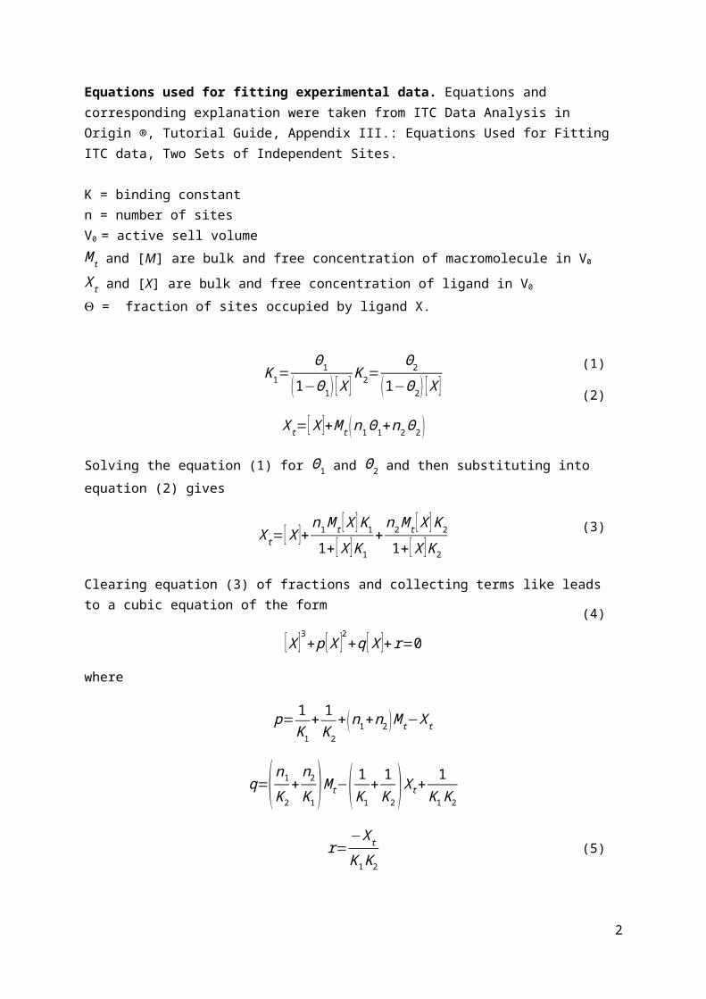
Equations used for fitting experimental data. Equations and corresponding explanation were taken from ITC Data Analysis in Origin ®, Tutorial Guide, Appendix III.: Equations Used for Fitting ITC data, Two Sets of Independent Sites.
K = binding constantn = number of sites V0 = active sell volumeM t and [M] are bulk and free concentration of macromolecule in V0
X t and [X] are bulk and free concentration of ligand in V0
= fraction of sites occupied by ligand X.
K 1=Θ1
(1−Θ1 ) [X ]K2=
Θ2
(1−Θ2 ) [X ]
X t=[X ]+M t (n1Θ1+n2Θ2 )
Solving the equation (1) for Θ1 and Θ2 and then substituting into equation (2) gives
X t=[X ]+n1M t [X ]K1
1+ [X ] K1+n2M t [X ]K2
1+ [X ]K2
Clearing equation (3) of fractions and collecting terms like leads to a cubic equation of the form
[X ]3+ p [X ]2+q [X ]+r=0
where
p= 1K1
+ 1K2
+ (n1+n2 )M t−X t
q=( n1
K2+n2
K 1)M t−( 1
K1+ 1K2 )X t+ 1
K1 K2
r=−X tK1K2
Equations 4 and 5 can be solved for [X ] either in closed form or (as done in Origin) numerically by using Newton’s Method if parameters n1, n2, K1, and K2 are assigned. Both Θ1 and Θ2 may then be obtained from equation (1).
The heat content after any injection i is equal to
Q=M tV 0 (n1Θ1 ΔH 1+n2Θ2 ΔH2 )
After a similar correction for displaced volume, the pertinent calculated heat effect for the i injection is
2
(1)
(2)
(3)
(4)
(5)
(6)
ΔQ ( i )=Q (i )+dV iV 0
[Q (i )+Q ( i−1 )2 ]−Q (i−1 )
which may be used in the Marquardt algorithm to obtain best values for the six fitting parameters.
3
(7)
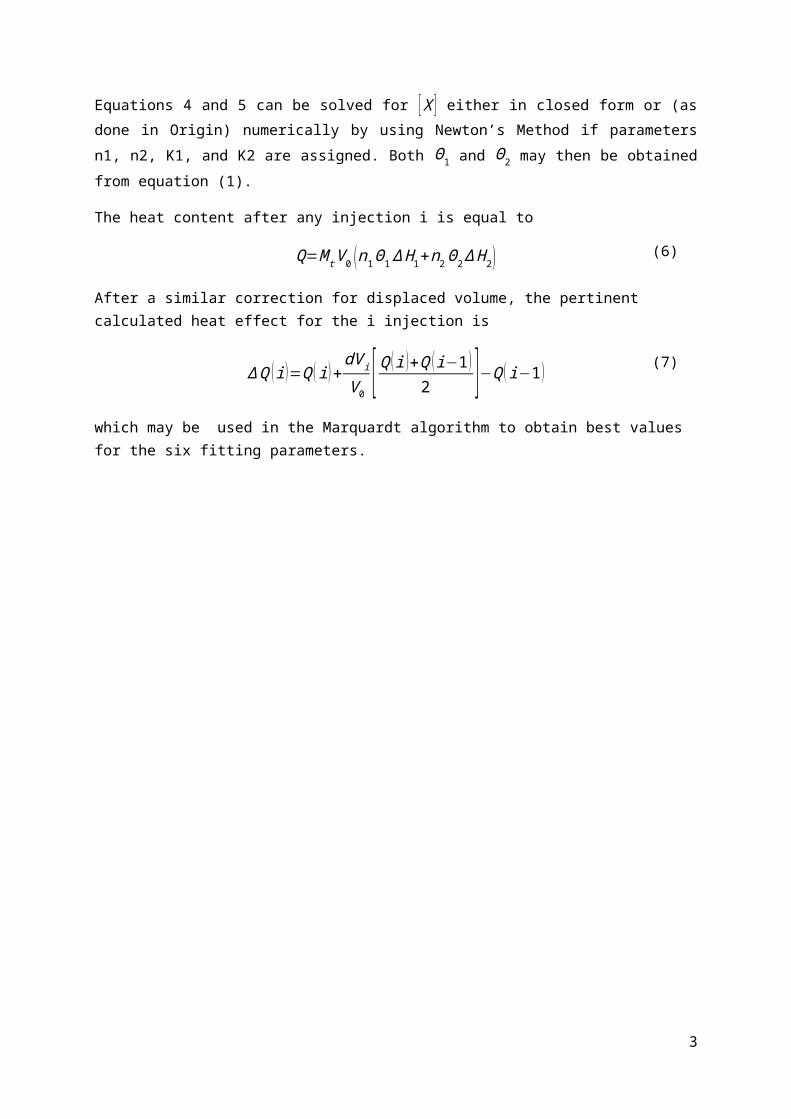
Supporting Fig. 1 Titrations of wt gal-1 with anginex using different concentrations of gal-1. a 15 µM, b 36 µM
and c 72 µM. Raw data (top) and binding isotherms fitted for the two independent binding sites model (bottom)
with integrated experimental data points (■) and fitted values (–) are shown.
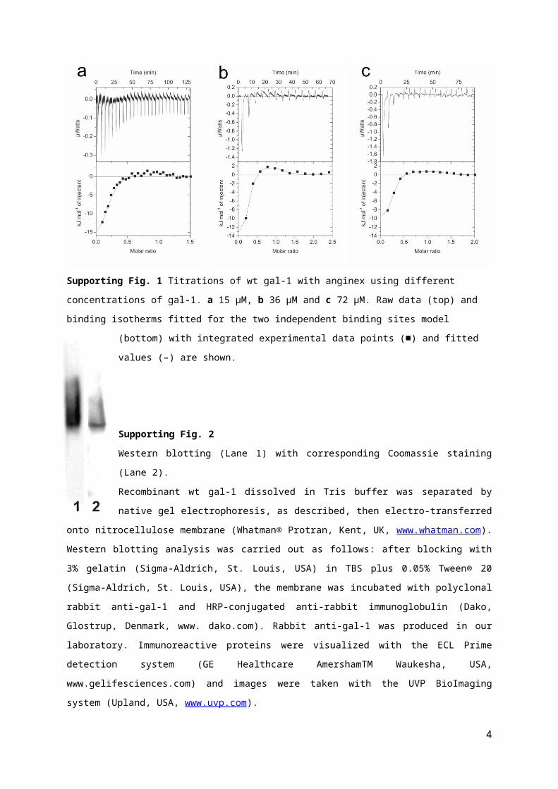
Supporting Fig. 2
Western blotting (Lane 1) with corresponding Coomassie staining (Lane 2).
Recombinant wt gal-1 dissolved in Tris buffer was separated by native gel electrophoresis, as
described, then electro-transferred onto nitrocellulose membrane (Whatman® Protran, Kent, UK,
www.whatman.com). Western blotting analysis was carried out as follows: after blocking with 3%
gelatin (Sigma-Aldrich, St. Louis, USA) in TBS plus 0.05% Tween® 20 (Sigma-Aldrich, St.
Louis, USA), the membrane was incubated with polyclonal rabbit anti-gal-1 and HRP-conjugated
anti-rabbit immunoglobulin (Dako, Glostrup, Denmark, www. dako.com). Rabbit anti-gal-1 was
produced in our laboratory. Immunoreactive proteins were visualized with the ECL Prime
detection system (GE Healthcare AmershamTM Waukesha, USA, www.gelifesciences.com) and images were
taken with the UVP BioImaging system (Upland, USA, www.uvp.com).
4
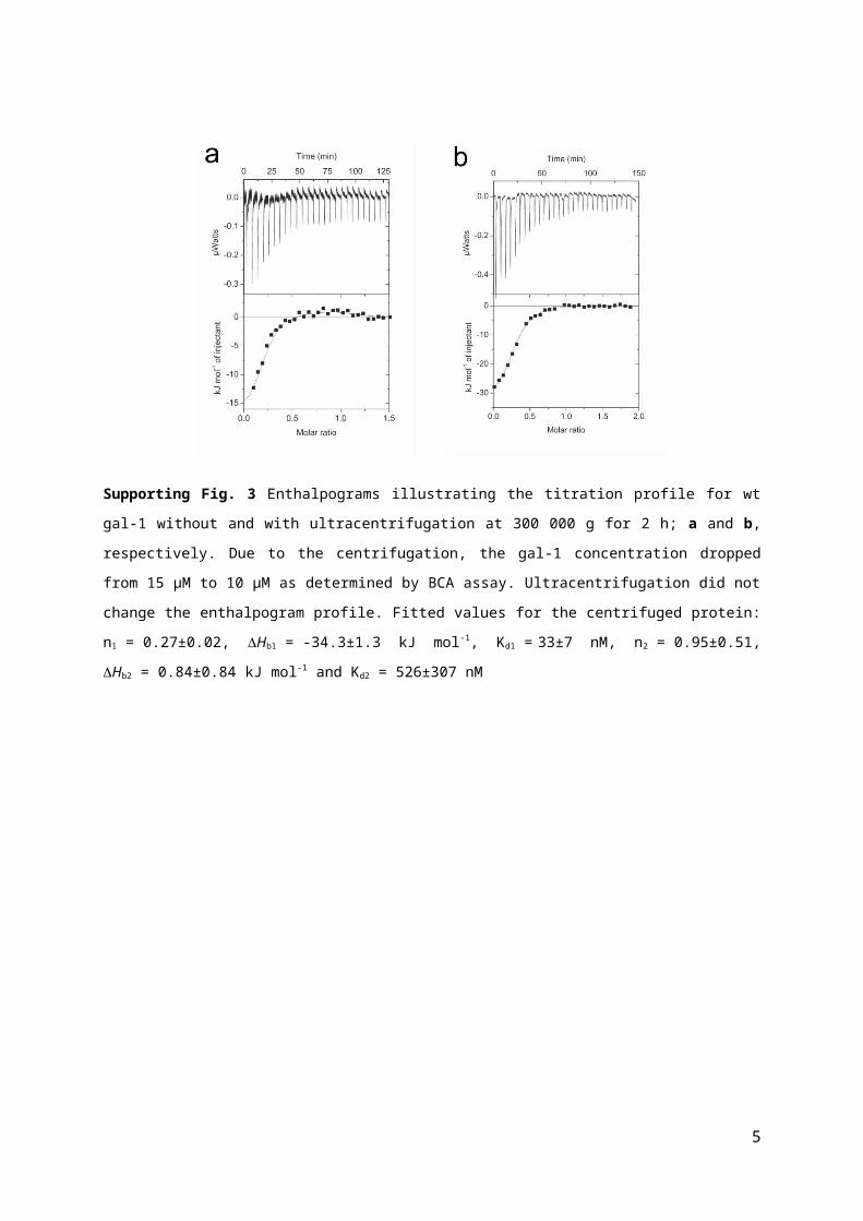
Supporting Fig. 3 Enthalpograms illustrating the titration profile for wt gal-1 without and with
ultracentrifugation at 300 000 g for 2 h; a and b, respectively. Due to the centrifugation, the gal-1 concentration
dropped from 15 µM to 10 µM as determined by BCA assay. Ultracentrifugation did not change the
enthalpogram profile. Fitted values for the centrifuged protein: n1 = 0.27±0.02, Hb1 = -34.3±1.3 kJ mol-1,
Kd1 = 33±7 nM, n2 = 0.95±0.51, Hb2 = 0.84±0.84 kJ mol-1 and Kd2 = 526±307 nM
Supporting Fig. 4 TOF-MS data of wt gal-1. a gal-1 without reducing agent, b after 1 h incubation with 1 mM
TCEP, c after 1 day incubation with 1 mM DTT and d after 1 day incubation with 1 mM BME
5

Peptide characterization
Liophilized anginex was analyzed by ESI MS mass spectrometry and by analitical HPLC measurements on Aeris
Peptide C18 4.6*250 mm column (Phenomenex) with a gradient from 5% A to 80% B over 25 min 1.2 ml/min
flow rate. Eluents were: A: 0,1% TFA/water; B: 0.1% TFA/ 20% water/ 80% ACN Peptide purity was 95%>
according to analytical HPLC measurements.
Mwt: 3992.80, Expected m/z values: [M+3H]3+: 1331.44; [M+4H]4+: 998.83; [M+5H]5+: 799.27; [M+6H]6+:
666.22; [M+7H]7+: 571.19
6
![Potential Hepatoprotective Role of Galectin-3 during HCV ... · cule in cell biology [22, 23]. Galectin-3 is involved in several biological processes including cell proliferation,](https://img.pdfslide.us/doc/110x75/60e40d64a7cbb4423f4233bf/potential-hepatoprotective-role-of-galectin-3-during-hcv-cule-in-cell-biology.jpg)