Embed Size (px)
Citation preview

© 2015. Published by The Company of Biologists Ltd.
SHORT COMMUNICATION
Light sensitivity in a vertebrate mechanoreceptor?
Gary E. Baker1†, Willem J. de Grip2, Michael Turton3, Hans-Joachim Wagner4,
Russell G. Foster3, Ron H. Douglas1*
1 Department of Optometry & Visual Science, School of Health Sciences, City University
London, Northampton Sq, London EC1V 0HB, UK
2 Department of Biochemistry, Radboud Institute for Molecular Life Sciences, Radboud
University Medical Center, Nijmegen, The Netherlands
3 Nuffield Laboratory of Ophthalmology, University of Oxford, Levels 5-6 West Wing, John
Radcliffe Hospital, Headley Way, Oxford, OX3 9DU, UK
4 Anatomisches Institut, Universität Tübingen, Ősterbergstrasse 3, 72074 Tübingen,
Germany
† Deceased
* Author for correspondence ([email protected])
The
Jour
nal o
f Exp
erim
enta
l Bio
logy
– A
CC
EPTE
D A
UTH
OR
MA
NU
SCR
IPT
http://jeb.biologists.org/lookup/doi/10.1242/jeb.125203Access the most recent version at J Exp Biol Advance Online Articles. First posted online on 23 July 2015 as doi:10.1242/jeb.125203http://jeb.biologists.org/lookup/doi/10.1242/jeb.125203Access the most recent version at
First posted online on 23 July 2015 as 10.1242/jeb.125203

ABSTRACT
Using immunohistochemistry and Western blot analysis we demonstrate that melanospin is
localised in cells around the central pore of lateral line neuromasts in the African clawed frog,
Xenopus laevis. Since melanopsin is a known photoreceptor pigment with diverse functions
in vertebrates, we suggest that the lateral line of Xenopus laevis, which is primarily a
mechanorecptor, may also be light sensitive. Potential functions of such photosensitivity are
discussed, including its role in mediating locomotor responses following dermal illumination.
KEY WORDS: Melanopsin, lateral line, mechanoreceptor, photosensitivity,
multimodality, phototaxis
SUMMARY STATEMENT
Lateral lines are sense organs on the bodies of aquatic vertebrates sensitive to water
displacement. In the African clawed frog they contain the photopigment melanopsin,
suggesting they may also be light sensitive.
The
Jour
nal o
f Exp
erim
enta
l Bio
logy
– A
CC
EPTE
D A
UTH
OR
MA
NU
SCR
IPT

INTRODUCTION
Although photoreceptors within the outer retina of vertebrate eyes are used by animals
for image forming (IF) light detection, extraretinal photoreceptors are widespread among
non-mammalian vertebrates, occurring mainly in the brain, but also evident elsewhere in the
body (Foster & Hankins, 2002). Such non-image forming (NIF) photoreceptors serve diverse
functions including; the regulation of circadian rhythms, mediating locomotor responses to
dermal illumination, influencing pigment migration in chromatophores and conferring direct
light sensitivity to muscles within the iris.
Until relatively recently, it has been assumed that the only pigments capable of
conferring photosensitivity to photoreceptors, even those located in structures outside the eye,
use rod and cone opsins. However, in the last two decades a number of opsins have been
identified that are different enough to those of traditional photoreceptors to constitute
separate gene families (Shand and Foster, 1999). One such photopigment opsin is melanopsin
(OPN4). Initially shown to contribute to light-evoked pigment migration within dermal
melanophores of Xenopus laevis (Provencio et al., 1998), melanopsin has since been
implicated in a number of roles including conferring light-sensitivity to a subset of
photoresponsive retinal ganglion cells (pRGCs) in mammals which measure overall
irradiance and underlie various non-imaging photoreceptive tasks (Hankins et al., 2008;
Bailes and Lucas, 2010 ).
A chance observation during an investigation into iris photosensitivity suggested that
the lateral line neuromasts of Xenopus laevis might contain melanopsin. Lateral line
neuromasts are mechanoreceptors sensitive to water displacement, distributed across the body
of many aquatic vertebrates (Dijkgraaf, 1962). In Xenopus laevis they are grouped into raised
‘stitches’ arranged in characteristic patterns on the skin’s surface (Murray, 1955). The
localisation of melanopsin within lateral line neuromasts suggests they may be sensitive to
photic as well as mechanical stimuli.
Here we report on the presence and distribution of melanopsin within Xenopus laevis
lateral lines and speculate on the functional significance of light sensitivity within this
mechanoreceptor.
The
Jour
nal o
f Exp
erim
enta
l Bio
logy
– A
CC
EPTE
D A
UTH
OR
MA
NU
SCR
IPT

RESULTS AND DISCUSSION
Immunostaining using a polyclonal antibody (CERN972), raised against a Xenopus
laevis melanopsin peptide, showed the majority of neuromasts on both dorsal and ventral
surfaces of adult male and female pigmented and albino Xenopus laevis to be
immunopositive (Fig 1A). No differences in distribution of melanopsin were observed
between the different phenotypes.
Individual neuromasts showed dense immunopositive staining surrounding the central
pore, with fine processes radiating outwards (Fig 1B). In light- (Fig 1C) and electron-
microscopic (Fig 1E) sections, dense immunopositive staining was located intracellularly in
epidermal cells at the margins of the neuromast pore. As evident from wholemounts (Fig 1B),
immunostaining was not confined to the margin of the pore. In serial reconstructions of
individual neuromasts we also identified melanopsin in peripheral cells lying slightly deeper
in the neuromast (Fig 1D).
Immunoreactivity was also detected by the CERN972 antibody in a Western blot
analysis of Xenopus brain and stitch samples at a mass consistent with melanopsin (Fig. 2).
This is in agreement with previous identification of melanopsin expression in tadpole
melanophores and adult Xenopus laevis brain and ocular structures (Provencio et al., 1998).
Most samples present an upper immunoreactive band near 55 kDa and a lower band at 45-50
kDa. There are 2 isoforms of melanopsin in Xenopus laevis (OPN4x and
OPN4m)(Bellingham et al., 2006), both of which would be detected by CERN972 and may
be represented by the two bands in the stitch samples (Fig 2). This would indicate that the
two melanopsin orthologs most commonly found in non-mammalian vertebrates (Bellingham
et al., 2006; Davies et al., 2011) are present in Xenopus laevis lateral line stitches. The
predicted mass for OPN4x is 60 kDa, but membrane proteins usually migrate with a
somewhat lower apparent mass in SDS-PAGE. The full sequence of OPN4m is unknown but
comparison of OPN4x and m isoforms in other species suggests that they migrate with a
similar apparent mass in SDS-PAGE (Davies et al., 2011; Bailes and Lucas, 2013).
Bellingham et al (2006) did not detect the OPN4x message in adult Xenopus skin tissue,
which may either be due to the low quantity of OPN4x message, being only expressed in
stitches, or it may indicate that the two strong bands we observe in stitch samples represent
two splice variants of OPN4m. This phenomenon has already been observed in some
mammalian species (Pires et al., 2009), in chicken (Torii et al., 2007) and in elephant shark
(Davies et al., 2012). The immunoreactivity at higher molecular masses is consistent with
The
Jour
nal o
f Exp
erim
enta
l Bio
logy
– A
CC
EPTE
D A
UTH
OR
MA
NU
SCR
IPT
formation of oligomeric complexes (dimers, trimers, etc.) which is common under the
conditions used for SDS PAGE analysis.
Since melanopsin is a known photopigment, the presence of melanospin
immunoreactivity within Xenopus laevis lateral line neuromasts suggests that apart from
being sensitive to mechanical stimuli, these sense organs may also be light sensitive. It is
natural to speculate about the potential functional significance of such lateral line
photosensitivity.
Many animals respond to dermal illumination with locomotor activity (Steven, 1963).
Some previous evidence suggests the lateral line of larval lamprey may mediate such dermal
photosensitivity. Their lateral line nerves generate electrophysiological responses following
illumination of the tail and the lesioning of these nerves disrupts the behavioural response to
such illumination (Deliagina et al., 1995; Ronan and Bodznick, 1991; Young, 1935). Our
results suggest that the photosensitivity of the lateral line might be conferred by melanopsin.
Interestingly, the light-driven electrophysiological response of the lamprey lateral line nerves
have a long latency, high threshold and do not adapt (Ronan and Bodznick, 1991), which are
also characteristics of melanopsin-based retinal photoreceptors in mammals (Bailes & Lucas,
2010; Hughes et al., 2012).
A previous report suggests adult Xenopus laevis are negatively phototactic (Denton
and Pirenne, 1954). However, it is not known if they react to localised dermal illumination
with locomotor activity. We confirmed the negative phototaxis of this species by observing
their behaviour in an aquarium, only half of which was illuminated. In 89.8% trials (n=49)
where the animals started in the lit half of the aquarium they moved to the dark half of the
tank within three minutes (average latency 63 secs). When they started in the dark half of the
aquarium (n=39), on the other hand, the frogs normally remained there for the duration of the
experiment, spending on average 86.6% of their time in darkness and only rarely venturing
into the light for brief periods of time.
We investigated whether focal illumination of the animal’s ventral surface, which
could not be detected by their eyes, would induce a locomotor avoidance response. While
they did appear to react to such stimuli, this was no more frequent than in control animals
simply maintained in darkness. Thus, using focal ventral illumination, there was no evidence
of dermally-induced locomotor activity in adult Xenopus laevis. It could be argued that
ventral illumination is not the ideal stimulus, as in the wild the underside of the animal will
receive less illumination than other areas of the body. However, ventral neuromasts stained as
heavily with melanopsin antibody as neuromasts elsewhere on the body. Furthermore, using
The
Jour
nal o
f Exp
erim
enta
l Bio
logy
– A
CC
EPTE
D A
UTH
OR
MA
NU
SCR
IPT

focal ventral illumination was the only way to be certain that the illumination was not
detected by the dorsally directed eyes of intact animals. Less systematic focal illumination of
other areas of the body also failed to induce consistent locomotor responses
Since focal illumination of the body surface did not induce a behavioural response, it
seems likely that melanospin in lateral line neuromasts of Xenopus laevis serves a function
other than dermally-driven locomotor activity.
The activity of lateral line neuromasts is known to be modulated by the central
nervous system using efferent neurons (Russell, 1971). For example, the activity of toadfish
lateral line nerves is rapidly suppressed by visual stimuli such as the sight of prey species
(Tricas and Highstein, 1991). Outer retinal photoreceptors (rods and cones) are required for
such IF processes as identifying prey and thus efferent innervation is essential if the lateral
line is to be affected by such stimuli. However, the physiological properties of rods and
cones make them less suited for monitoring overall light levels and this is thought to be the
primary reason the mammalian retina contains a population melanopsin-containing pRGCs,
whose sluggish but long lasting responses make them ideal for detecting overall irradiance
(Bailes & Lucas, 2010; Hughes et al., 2012). It is therefore conceivable that melanopsin
within the lateral line serves a similar role and modulates lateral line activity in response to
longer term changes in ambient light levels. Lateral line sensitivity might, for example, be
increased in darkness when photic stimuli are not available. Alternatively, the sensitivity of
neuromasts might be adjusted by variations in light level associated with depth as the nature
of the vibratory information changes.
Co-localisation of mechano and photosensory function is not unique to Xenopus
laevis and lamprey lateral lines. It has also been reported in invertebrates. Larval Drosophila
abdominal mechanosensory neurones also respond to light and contribute to light avoidance
behaviour (Xiang et al., 2010). Based on the distribution of developmental Pax genes, it has
been suggested that ears, mechanoreceptors closely related to lateral lines, and eyes share a
common evolutionary lineage (Fritzsch and Piatigorsky, 2005). Multimodality of sense
organs involving photoreception and mechanoreception might therefore not be that unusual
or surprising.
The
Jour
nal o
f Exp
erim
enta
l Bio
logy
– A
CC
EPTE
D A
UTH
OR
MA
NU
SCR
IPT

MATERIALS AND METHODS
Immunocytochemistry & microscopy
Five Xenopus laevis (Daudin) were euthanized by overdose of tricaine
methanesulfonate (Sigma) followed by decapitation and pithing. The skin was immersed in
phosphate buffered (pH7.3) 4% paraformaldehyde at 4°C for 3-4 hours. Patches containing
lateral line stitches were stored in Phosphate Buffered Saline (PBS) until further processing
or in 30% sucrose for cryosectioning.
For immunostaining, tissue was rinsed in PBS, immersed in 0.3% H2O2-methanol for
30 minutes and rinsed again in PBS. Following immersion for 30 minutes in normal goat
serum diluted in a solution of 1% triton X-100 in PBS, tissue was incubated at 4oC for 24-48
hours in the primary antibody diluted 1:2000 or 1:4000 in PBS (both dilutions produced
identical staining patterns). This polyclonal antibody (CERN972) was raised against a 15-mer
peptide covering residues 216-230 of Xenopus laevis OPN4x (FLAIRSTGRNVQKLG)
(Provencio et al., 1998). The peptide was linked to rabbit serum albumin using SATA-MHS
chemistry (Schielen et al., 1989). The resulting construct was injected in albino female New
Zealand rabbits and processed as previously described (deGrip, 1985).
After primary antibody incubation, labelling was visualized using an avidin-
biotinylated horseradish peroxidase second antibody procedure (Vector Elite ABC kit; Vector
Laboratories, Peterborough, UK) applying diaminobenzidine as the chromagen (Sigma Fast;
Sigma-Aldrich, Gillingham, Dorset, UK).
Skin segments were viewed in wet mount to identify immunopositive regions. Some
were prepared as wholemounts, while segments for fine structural observation were
immersed in 2% aqueous osmium tetroxide for 1 hour, before processing for araldite
embedding. Semithin (1m) sections were cut (Ultracut E; Reichert-Jung, Depew, New York,
USA) and counterstained with toluidine blue. Images were collected using an Olympus BH2
photomicroscope equipped with a Spot RT Color digital camera (Diagnostic Instruments inc.,
Sterling Heights, Michigan, USA). For electron microscopy no further enhancement to the
contrast of the HRP-label was required and sections were viewed on a LEO-EM912 electron
microscope (Zeiss, Oberkochen, Germany) and recorded with a digital camera.
The
Jour
nal o
f Exp
erim
enta
l Bio
logy
– A
CC
EPTE
D A
UTH
OR
MA
NU
SCR
IPT
Molecular analysis
Samples of lateral line stitches, eye, and various brain regions were removed from 2
animals euthanized as described above and frozen. All tissue was ground using a pre-chilled
pestle and mortar prior to homogenisation in 2% (w/v) SDS, 50mM DTT with mini complete
protease inhibitors (Roche). Samples were incubated at room temperature on a shaking
platform for 2h to improve solubilisation. The lysate was centrifuged at 23000xg for 30min at
20°C and the supernatant fraction used for SDS PAGE and Western blotting as described
previously (Pires et al., 2009).
Every effort was made to avoid contamination of lateral line stich samples with
dermal melanopores during dissection. If there was any minor contamination, this is unlikely
to have been sufficient to produce the strong immunoreactive bands observed. We also found
two clear bands in the stitch lanes on SDS-PAGE, while previous studies (Provencio et al.,
1998; Bellingham et al., 2006) only detected one band in Xenopus melanophores. Hence even
if there was some contamination by melanophores, at least one of the observed bands is
derived from lateral line stitches.
Phototaxis
Individual animals were removed from their home tank, during the light phase of their
light/dark cycle, and put in an experimental aquarium (20x30x20 cm). The sides of this
aquarium were covered by black card and animals were observed from above. After 10
minutes acclimation in dim room light the animal was placed in total darkness for 2 mins,
before one half of the aquarium was illuminated (3.41W/m2) from below by a ‘light box’,
consisting of two fluorescent tubes (Phillips 20W/47 Graphic A; Guildford, Surrey, UK)
behind a white diffusing surface, for three minutes followed by 2 minutes of darkness before
being exposed to light once more for another 3 minutes, for a maximum of 10 trials per
animal. The half of the tank that was illuminated was varied randomly. 7 pigmented and 2
albino animals were tested.
We also investigated the ability of focal illumination to induce locomotor activity.
The ventral surface of 4 animals was illuminated using the same protocol as above, but
instead of illuminating half the aquarium the light source was covered except for a 1cm round
aperture that was positioned near the centre of the animals ventral surface when it was resting
on the bottom of the tank. The time of any movement after the spot was turned on was noted
(n=18). A similar number of control observations were made with the stimulating spot in
The
Jour
nal o
f Exp
erim
enta
l Bio
logy
– A
CC
EPTE
D A
UTH
OR
MA
NU
SCR
IPT

position but not switched on. Less systematically, we also tried directing light onto various
parts of the body with both a narrow torch beam and low power lasers and observing any
reaction.
Acknowledgements
We are grateful to Rob Lucas, Simon Laughlin, Dan-Eric Nilsson, David Whitmore and
Kathy Tamai for useful discussion. Vera Moenter helped with the phototactic experiments,
Uli Mattheus provided histological support and Petra Bovee-Geurts aided in the antibody
production.
Competing interests
The authors declare no competing financial interests
Author contributions
G.E.B and R.H.D. conceived the study. G.E.B. performed the immunohistochemistry, for
which W.J.dG. provided the antibody. H.-J.W. performed most of the microscopy. R.G.F.
and M.T. carried out the Western blot analysis and R.H.D. performed the photactic
experiments. All authors contributed to the interpretation of data. R.H.D. drafted the
manuscript , which was edited by all authors, except G.E.B., prior to submission.
Funding
This work was supported by the European Office of Aerospace Research & Development
[F61708-98-W0026 to W.J.dG] and the Wellcome Trust [090684/Z/09/Z and 098461/Z/12/Z
to RGF].
The
Jour
nal o
f Exp
erim
enta
l Bio
logy
– A
CC
EPTE
D A
UTH
OR
MA
NU
SCR
IPT

References
Bailes, H.J. and Lucas, R.J. (2010). Melanopsin and inner retinal photoreception. Cell. Mol.
Life Sci. 67(1), 99-111.
Bailes, H.J. and Lucas, R.J. (2013). Human melanopsin forms a pigment maximally
sensitive to blue light (λmax ~ 479 nm) supporting activation of Gq/11 and Gi/o
signalling cascades. Proc.R.Soc.B 280 (1759), 20122987.
Bellingham, J., Chaurasia S.S., Melyan, Z., Liu, C.M., Cameron, M.A., Tarttelin, E.E.,
Iuvone, P.M., Hankins, M.W., Tosini, G. and Lucas, R.J. (2006). Evolution of
melanopsin photoreceptors: Discovery and characterization of a new melanopsin in
nonmammalian vertebrates. Plos Biology 4 (8),1334-1343.
Davies, W.I.L., Tay, B.-H., Zheng, L., Danks, J.A., Brenner, S., Foster, R.G., Collin,
S.P., Hankins, M.W., Venkatesh, B. and Hunt, D.M. (2012). Evolution and
functional characterisation of melanopsins in a deep-sea chimaera (Elephant shark,
Callorhinchus milii). PLoS ONE 7 (12), e51276.
Davies, W.I.L., Zheng, L., Hughes, S., Tamai, T.K., Turton, M., Halford, S., Foster,
R.G., Whitmore, D. and Hankins, M.W. (2011). Functional diversity of
melanopsins and their global expression in the teleost retina. Cell.Mol.Life Sci. 68
(24), 4115-4132.
deGrip, W.J. (1985). Immunochemistry of rhodopsin. Prog. Retin. Res. 4, 137-180.
Deliagina, T.G., Ullén, F., Gonzalez M.-J., Ehrsson, H., Orlovsky, G.N. and Grillner, S.
(1995). Initiation of locomotion by lateral line photoreceptors in lamprey:
Behavioural and neurophysiological studies. J. Exp. Biol. 198, 2581–2591.
Denton, E.J and Pirenne, M.H. (1954). The visual sensitivity of the toad Xenopus laevis. J.
Physiol. 125, 181-207.
Dijkgraaf, S. (1962). The function and significance of the lateral-line organs. Biol. Rev. 38,
51-105.
Foster, R.G. and Hankins, M.W. (2002). Non-rod, non-cone photoreception in the
vertebrates. Prog. Retin. Eye Res. 21, 507–527.
Fritzsch, B. and Piatigorsky, J. (2005). Ancestry of photonic and mechanic sensation.
Science 308, 1113-1114.
Hankins, M.W., Peirson, S.N. and Foster, R.G. (2008). Melanopsin: an exciting
photopigment. Trends Neurosci. 31, 27-36.
The
Jour
nal o
f Exp
erim
enta
l Bio
logy
– A
CC
EPTE
D A
UTH
OR
MA
NU
SCR
IPT

Hughes, S., Hankins, M.W., Foster, R.G. and Peirson, S.N. (2012). Melanopsin
phototransduction: slowly emerging from the dark. Prog. Brain Res. 199, 19-40.
Murray, R.W. (1955). The lateralis organs and their innervation in Xenopus laevis. Q. J.
Micro. Sci. 96(3), 351-361.
Pires, S.S., Hughes, S., Turton, M., Melyan, Z., Peirson, S.N., Zheng, L., Kosmaoglou,
M., Bellingham, J., Cheetham, M.E., Lucas, R.J., Foster, R.G., Hankins, M.W.
and Halford, S. (2009) Differential expression of two distinct functional isoforms of
melanopsin (Opn4) in the mammalian retina. J. Neurosci. 29(39), 12332-12342.
Provencio, I., Jiang, G., deGrip, W.J., Hayes, W.P. and Rollag, M.D. (1998). Melanopsin:
An opsin in melanophores, brain and eye. Proc. Nat. Acad. Sci. USA 95, 340-345.
Ronan, M. and Bodznick, D. (1991). Behavioural and neurophysiological demonstration of
a lateralis skin sensitivity in larval sea lampreys. J. Exp. Biol. 161, 97-117.
Russell, I.J. (1971). The role of the lateral line efferent system in Xenopus laevis. J. Exp.
Biol. 54, 621-641.
Schielen, W.J.G., Voskuilen, M., Tesser, G.I. and Nieuwenhuizen, W. (1989). The
sequence a-alpha-(148-160) in fibrin, but not in fibrinogen, is accessible to
monoclonal antibodies. Proc. Natl. Acad. Sci. USA 86(22), 8951-8954.
Shand, J. and Foster, R.G. (1999). The extraretinal photoreceptors of non-mammalian
vertebrates. In Adaptive Mechanisms in the Ecology of Vision (ed. S.N. Archer,
M.B.A. Djamgoz, E.R. Loew, J.C. Partridge and S. Vallerga), pp. 197-222.
Dordrecht: Kluwer Academic Publishers.
Steven, D.M. (1963). The dermal light sense. Biol. Rev. 38, 204-240.
Torii, M., Kojima, D., Okano, T., Nakamura, A., Terakita, A., Shichida, Y., Wada, A.
and Fukada, Y. (2007). Two isoforms of chicken melanopsins show blue light
sensitivity FEBS Lett. 581(27), 5327-5331.
Tricas, T.C. and Highstein, S.M. (1991). Action of the octavolateralis efferent system upon
the lateral line of free-swimming toadfish, Opsanus tau. J. Comp. Phys. A 169, 25-37.
Xiang, Y., Yuan, Q., Vogt, N., Looger, L.L., Jan, L.Y. and Jan, Y.N. (2010). Light-
avoidance-mediating photoreceptors tile the Drosophila larval body wall. Nature 468
(7326), 921-U312.
Young, J.Z. (1935). The photoreceptors of lampreys: 1. Light-sensitive fibres in the lateral
line nerves. J. Exp. Biol. 12, 229–238.
The
Jour
nal o
f Exp
erim
enta
l Bio
logy
– A
CC
EPTE
D A
UTH
OR
MA
NU
SCR
IPT

Figures
Figure 1 Melanopsin immunostaining of Xenopus laevis neuromasts
A: Low power wet mount of the dorsal skin showing linear arrays of stained
neuromasts (arrows) forming lateral line stitches.
The
Jour
nal o
f Exp
erim
enta
l Bio
logy
– A
CC
EPTE
D A
UTH
OR
MA
NU
SCR
IPT
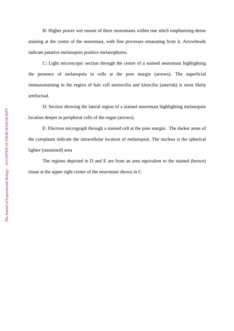
B: Higher power wet mount of three neuromasts within one stitch emphasising dense
staining at the centre of the neuromast, with fine processes emanating from it. Arrowheads
indicate putative melanopsin positive melanophores.
C: Light microscopic section through the centre of a stained neuromast highlighting
the presence of melanopsin in cells at the pore margin (arrows). The superficial
immunostaining in the region of hair cell stereocilia and kinocilia (asterisk) is most likely
artefactual.
D: Section showing the lateral region of a stained neuromast highlighting melanopsin
location deeper in peripheral cells of the organ (arrows).
E: Electron micrograph through a stained cell at the pore margin. The darker areas of
the cytoplasm indicate the intracellular location of melanopsin. The nucleus is the spherical
lighter (unstained) area
The regions depicted in D and E are from an area equivalent to the stained (brown)
tissue at the upper right corner of the neuromast shown in C
The
Jour
nal o
f Exp
erim
enta
l Bio
logy
– A
CC
EPTE
D A
UTH
OR
MA
NU
SCR
IPT
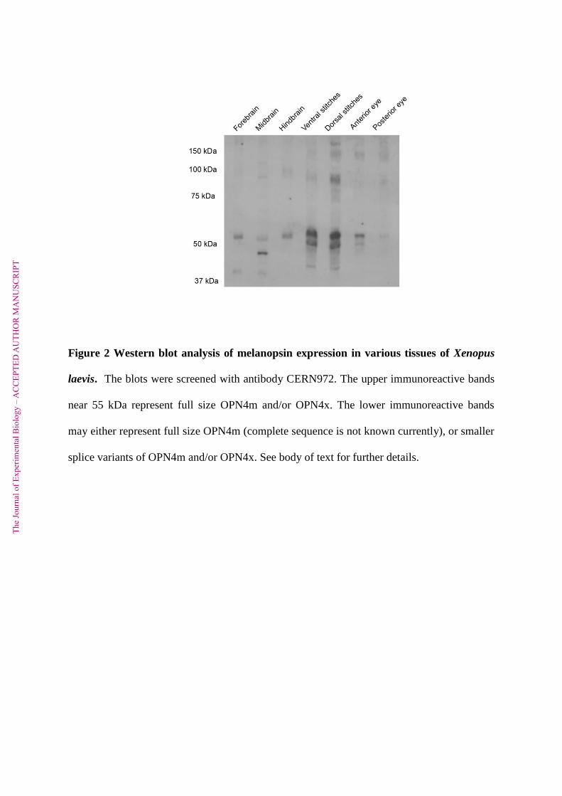
Figure 2 Western blot analysis of melanopsin expression in various tissues of Xenopus
laevis. The blots were screened with antibody CERN972. The upper immunoreactive bands
near 55 kDa represent full size OPN4m and/or OPN4x. The lower immunoreactive bands
may either represent full size OPN4m (complete sequence is not known currently), or smaller
splice variants of OPN4m and/or OPN4x. See body of text for further details.
The
Jour
nal o
f Exp
erim
enta
l Bio
logy
– A
CC
EPTE
D A
UTH
OR
MA
NU
SCR
IPT