Embed Size (px)
Citation preview

HAL Id: hal-01924074https://hal.univ-lorraine.fr/hal-01924074
Submitted on 17 Mar 2021
HAL is a multi-disciplinary open accessarchive for the deposit and dissemination of sci-entific research documents, whether they are pub-lished or not. The documents may come fromteaching and research institutions in France orabroad, or from public or private research centers.
L’archive ouverte pluridisciplinaire HAL, estdestinée au dépôt et à la diffusion de documentsscientifiques de niveau recherche, publiés ou non,émanant des établissements d’enseignement et derecherche français ou étrangers, des laboratoirespublics ou privés.
Light-sensitive dextran-covered PNBA nanoparticles astriggered drug delivery systems: Formulation,
characteristics and cytotoxicityMeriem El Founi, Soliman Mehawed Abdellatif Soliman, Régis Vanderesse,
Samir Acherar, Emmanuel Guedon, Isabelle Chevalot, Jérôme Babin,Jean-Luc Six
To cite this version:Meriem El Founi, Soliman Mehawed Abdellatif Soliman, Régis Vanderesse, Samir Acherar, EmmanuelGuedon, et al.. Light-sensitive dextran-covered PNBA nanoparticles as triggered drug delivery sys-tems: Formulation, characteristics and cytotoxicity. Journal of Colloid and Interface Science, Elsevier,2018, 514, pp.289 - 298. �10.1016/j.jcis.2017.12.036�. �hal-01924074�

1
Light-sensitive dextran-covered PNBA nanoparticles
as triggered drug delivery systems: formulation,
characteristics and cytotoxicity
Meriem EL FOUNI a,b
, Soliman Mehawed Abdellatif SOLIMAN a,b,c
, Régis VANDERESSE a,b
,
Samir ACHERAR a,b
, Emmanuel GUEDONd, Isabelle CHEVALOT
d, Jérôme BABIN
a,b, Jean-Luc
SIX a,b *
a) Université de Lorraine, Laboratoire de Chimie Physique Macromoléculaire LCPM, UMR
7375, Nancy F-54001, France
b) CNRS, Laboratoire de Chimie Physique Macromoléculaire LCPM, UMR 7375, Nancy F-
54001, France
c) Chemistry Department, Faculty of Science, Cairo University, 12613 Giza, Egypt.
d) CNRS, Laboratoire Réactions et Génie des Procédés LRGP, UMR 7274, Nancy F-54001,
France
Corresponding Author : Jean-Luc SIX (E-mail: [email protected])
Submitted to Journal of Colloid and Interface Science

2
ABSTRACT
Hypothesis: For some years, smart nano-objects are one of the main focuses of current research.
In the framework of polymeric nanomedicine, o-nitrobenzyl alcohol derivatives lead to light-
responsive polymeric materials. At this day, nanomedicine based on polysaccharide/poly(o-
nitrobenzyl acrylate) (PNBA) copolymers have never been reported.
Experiments: For the first time, PNBA core/dextran shell nanoparticles (NPs) were formulated
by evaluating two different processes: i) nanoprecipitation of preformed Dextran-g-PNBA
glycopolymers, ii) emulsion/evaporation using azido-functionalized PNBA and alkynated
dextran, carrying out (or not) an interfacial click chemistry reaction. NPs’ characterization,
colloidal stability in the presence of salts and an anionic competitive surfactant (SDS) and light-
induced disruption were assessed. Finally, the potential use of these NPs as photo-responsive
drug delivery systems was investigated by a preliminary in vitro cytotoxicity study using Caco-2
cells.
Findings: Whatever the process, the photosensitive property and the colloidal stability of NPs in
the presence of salts were proved. However, triazole rings between the dextran shell and the
PNBA core avoid the dextran shell desorption in the presence of SDS. NPs' biocompatibility
towards Caco-2 was proved and 100% cell viability was still observed after exposure to NPs
following by 60 sec UV-irradiation.
KEYWORDS: Drug Delivery System; Polysaccharide; Photo-responsive polymer;
Biodegradable; Cancer treatment

3

4
1) INTRODUCTION
For more than a decade, polymeric nanomedicine that includes micelles, nanoparticles (NPs)
or nanocapsules received increasing attention, and many reviews dealing with this subject were
published.[1-5] Drug may be loaded into such nano-objects to reduce dosage, minimize side-
effects, protect drug from degradation and thus to enhance its efficiency. Nevertheless, a long
period in the bloodstream is often expected (case of stealthy nano-objects). To prevent the
adsorption of opsonins (circulating proteins) on the nano-object's surface and its subsequent
trapping by the reticuloendothelial system therefore its removing from blood flow,[6-7]
polyethylene oxide (PEO, also called polyethylene glycol - PEG) is commonly used to cover the
nano-object and to provide it stealthiness. However, although PEO is approved by Food and
Drug Administration (FDA)[8] and after close to nearly half a century of clinical uses, it was
shown that PEO induces hypersensitivity reaction, complement activation and anti-PEO antibody
formation reactions.[8-9] Several neutral hydrophilic polysaccharides were already reported as
promising alternatives of PEO due to their inherent biodegradability, immunogenicity, and
bioactivity.[10] In fact, some polysaccharides as dextran allow the colloidal stability of such
nano-objects,[11-13] prevent interactions with cells and proteins, thus extending the nano-
objects' circulation half-life,[9] and ensuring their stealthiness. After functionalization of such
hydrophilic shell (PEG or dextran for instance) by adequate ligands as antibodies, carbohydrates
or peptides, nano-objects can target specific cells to be treated. These nano-objects were called
drug delivery system (DDS)[14-18] and some of them, based on biodegradable materials, were
approved by FDA and are already commercially available.[19]
After encapsulation of drug into DDS, the drug release occurs by DDS degradation or
swelling, and/or by diffusion outside the nanocarrier. Commonly, biodegradable hydrophobic

5
(co)polyesters as poly(lactic acid) (PLA), poly(glycol acid) (PGA) and their poly(lactide-co-
glycolide) (PLGA) were used to formulate DDS. In these cases, drug release occurs by diffusion
and degradation of the DDS. For some years, scientists took interest to introduce sensitive parts
in polymeric materials to reach DDS that are sensitive to internal or external stimuli. Different
types of smart or stimuli–responsive DDS are reported as thermosensitive DDS, pH-sensitive
DDS, light-sensitive DDS, molecule-responsive DDS, …[20-24] After DDS formulation and
according to adequate stimulation, the loaded drug is released when and where physicians and
clinicians want.
Among all the light-responsive polymeric materials described in literature,[25-30] o-
nitrobenzyl alcohol derivatives are today the most studied in polymer science. o-Nitrobenzyl
ester bonds are known to be cleft by UV or two photons adsorptions, yielding to carboxylic acid
functions.[31] Such bonds were already assessed to light-induce the disassembly of block
copolymer micelles.[32-36] Very recently, some of us have reported the first controlled
polymerization of o-nitrobenzyl acrylate (NBA), [37] then the synthesis of amphiphilic grafted
photosensitive glycopolymers called Dex-g-PNBA, composed on dextran (Dex) as hydrophilic
backbone and poly(o-nitrobenzyl acrylate) (PNBA) as photo-responsive grafts.[38] To the best
of our knowledge, the formulation of nano-objects from polysaccharide/PNBA copolymers has
never been reported. Indeed, only two papers are dealing with the light-disruption of nano-
objects based on diblock copolymers containing one PNBA block and one poly(2-ethyl-2-
oxazoline)[39] or polydimethylacrylamide[40] block.
This study aimed to investigate for the first time the formulation of PNBA core / dextran shell
NPs that can be used as DDS for anticancer treatments. Two different formulation processes
were compared (nanoprecipitation and emulsion/organic solvent evaporation). More precisely,

6
within the emulsion/organic solvent evaporation process, an in situ Huisgen-type Copper(I)-
catalyzed Azide–Alkyne Cycloaddition (CuAAC) click-chemistry was carried out in some
experiments. Then, NPs batches were characterized in term of size, zeta potential, dextran
amount and colloidal stabilities in the presence of salts and of an anionic competitive surfactant.
The photosensitive character of these PNBA-based NPs was evaluated by varying different
irradiation parameters. Finally, and because the colorectal cancer is known to be the third most
common cancer worldwide[41-43], preliminary study on the in vitro cytotoxicity of these
PNBA-based NPs (before and upon UV-irradiation) towards human epithelial colorectal
adenocarcinoma (Caco-2) cells was reported.
2) EXPERIMENTAL
2.1) Materials
Amphiphilic alkynated dextran was derived from dextran.[38] The yield of substitution () of
such alkynated dextran was estimated equal to 15% (15 pending alkynyl groups per 100
glucopyranosic units), and its surface tension property was checked (Figure S1). Azido-
functionalized poly(o-nitrobenzyl acrylate) PNBAM-N3, where M is the number average
molecular weight, was derived from PNBAM-Br obtained via a controlled Single Electron
Transfer-Living Radical Polymerization (SET-LRP) of o-nitrobenzyl acrylate (NBA).[37] Dex-
g-PNBA glycopolymers were obtained by carrying out CuAAC in DMSO.[38] For more
information, such glycopolymers are called Dex()-g-PNBAM, where is the number of
PNBAM grafts per 100 glucopyranosic units. Weight fractions of PNBA (FPNBA) into such Dex-

7
g-PNBA are varying from 40 to 85% as shown in Table 1. Whatever FPNBA, all Dex-g-PNBA are
insoluble in water, but soluble in DMSO or in THF/H2O mixtures.
Tetrahydrofuran (THF), dichloromethane (DCM), copper bromide (CuBr, 99.9%), sodium
dodecylsulfate (SDS), ethylenediaminetetraacetic acid (EDTA) were purchased from Sigma-
Aldrich and used without further purification.
2.2) Elaboration of nanoparticles
2.2.1) Nanoprecipitation
25 mg of Dex-g-PNBA were dissolved in 5 mL of THF/H2O mixture (95/5, v/v; except in case
of Dex(15)-g-3PNBA3,700 where 75/25, v/v was used) for 24 h, then added drop wise (0.1 mL per
min) into 10 mL of distilled water under magnetic stirring. After complete addition, 10 mL of
distilled water were added portion-wise to freeze the NPs dispersion. Finally, THF was removed
by centrifugation (15,000 rpm, 15 °C, 30 min). NPs were washed twice by using distilled water,
and then were freeze-dried.
2.2.2) Emulsion/Organic Solvent Evaporation
25 mg of either PNBA-Br, PNBA-N3 or mixtures (from 0/1 to 1/0) were dissolved in 1 mL of
DCM. Meanwhile, 50 mg of alkynated dextran were dissolved in 10 mL of distilled water
(DCM-saturated). The organic phase was added under vigorous stirring to the aqueous one, then
the mixture was sonicated (pulsed mode, 46 W, 2 min, ice bath) using a Vibracell 75043 model
(Bioblock Scientific). After sonication, DCM was evaporated at 37°C for 2.5 h under stirring.
Suspension was then centrifuged (10,000 rpm, 15°C, 60 min) and the collected NPs were

8
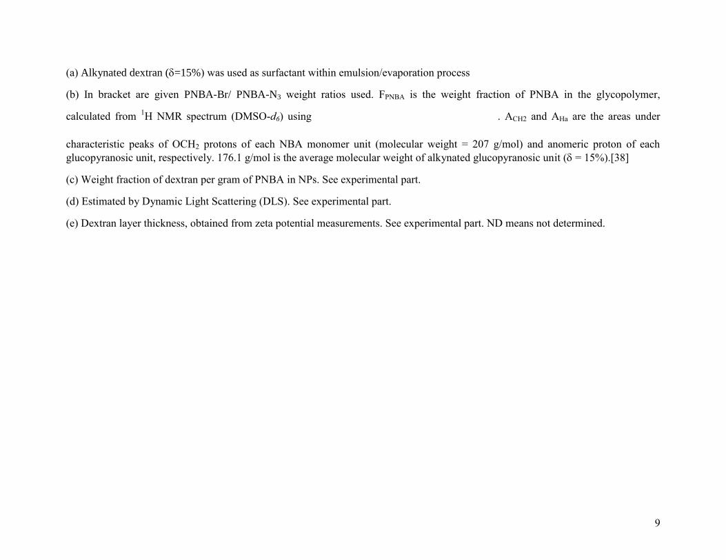
Table 1. Characterizations of nanoparticles.
Run (a)
Method used to produce nanoparticles (b)
Used polymers (c)
Wt of dextran
(mg/g PNBA)
(d) Z-Average
diameter (nm) (d)
PDI (e)
PZ
(nm)
1 Nanoprecipitation Dex(15)-g-3PNBA3,700 - FPNBA = 40% 916 129±3 0.216 10
2 Nanoprecipitation Dex(15)-g-14PNBA3,500 - FPNBA = 75% 310 118±3 0.080 5
3 Nanoprecipitation Dex(15)-g-12PNBA9,800 - FPNBA = 85% 153 185±2 0.040 3
4 Emulsion/Evaporation without CuAAC PNBA7,900-Br / PNBA8,100-N3 (1/0) 140 109±2 0.144 ND
5 Emulsion/Evaporation without CuAAC PNBA7,900-Br / PNBA8,100-N3 (0.5/0.5) 109 132±2 0.124 ND
6 Emulsion/Evaporation without CuAAC PNBA7,900-Br / PNBA8,100-N3 (0/1) 114 140±0 0.170 9
7 Emulsion/Evaporation without CuAAC PNBA10,300-Br / PNBA10,300-N3 (0/1) 109 145±1 0.165 8
8 Emulsion/Evaporation with CuAAC PNBA7,900-Br / PNBA8,100-N3 (0.75/0.25) 174 131±3 0.107 ND
9 Emulsion/Evaporation with CuAAC PNBA7,900-Br / PNBA8,100-N3 (0.5/0.5) 185 125±1 0.069 ND
10 Emulsion/Evaporation with CuAAC PNBA7,900-Br / PNBA8,100-N3 (0.25/0.75) 174 117±1 0.047 ND
11 Emulsion/Evaporation with CuAAC PNBA7,900-Br / PNBA8,100-N3 (0/1) 185 118±1 0.103 10
12 Emulsion/Evaporation with CuAAC PNBA10,300-Br / PNBA10,300-N3 (0/1) 215 141±2 0.181 7
9
(a) Alkynated dextran (=15%) was used as surfactant within emulsion/evaporation process
(b) In bracket are given PNBA-Br/ PNBA-N3 weight ratios used. FPNBA is the weight fraction of PNBA in the glycopolymer,
calculated from 1H NMR spectrum (DMSO-d6) using . ACH2 and AHa are the areas under
characteristic peaks of OCH2 protons of each NBA monomer unit (molecular weight = 207 g/mol) and anomeric proton of each
glucopyranosic unit, respectively. 176.1 g/mol is the average molecular weight of alkynated glucopyranosic unit ( = 15%).[38]
(c) Weight fraction of dextran per gram of PNBA in NPs. See experimental part.
(d) Estimated by Dynamic Light Scattering (DLS). See experimental part.
(e) Dextran layer thickness, obtained from zeta potential measurements. See experimental part. ND means not determined.

10
resuspended in water, centrifuged again in order to remove the non-adsorbed alkynated dextran,
and finally freeze-dried.
In some experiments an in situ CuAAC was carried out by adding 5 mg of CuBr to the first
emulsion under N2 flow and prior to the sonication step. To remove residual copper, EDTA (5 eq
per CuBr) was added to the final washed NPs suspension, which was left under stirring for 24 h
at room temperature, then centrifuged (10,000 rpm, 15°C, 30–60 min), and finally washed twice
with deionized water.
In the case of uncoated NPs, the initial aqueous phase was composed of 3 g/L of SDS instead
of alkynated dextran.
2.3) Characterization of nanoparticles
The Dynamic Light Scattering (DLS) at low concentration was evaluated using a Malvern
High Performance Particle Sizer (HPPS) instrument. The average scattering intensity during the
measurement is called Mean Count Rate (MCR) and the analysis of the intensity fluctuations
allows to estimate the NPs' size and the polydispersity index (PDI). Although this apparatus is
able to measure relatively concentrated samples, 200 µL of NPs suspension were diluted in 2 mL
of NaCl aqueous solution (10−3
M). In case of irradiated NPs (see below), 200 µL of irradiated
suspension were mixed with 2 mL of Phosphate Buffer Saline (PBS, KH2PO4/Na2HPO4, pH =
7)/H2O mixture (50/50, v/v)). The final NPs dispersion concentration was equal to 0.11 mg/mL.
The mean diameter Dz (nm) is the so-called Z-average from cumulated analysis, i.e. an
intensity–average diameter, and was measured three times with deviation remaining below 5 nm.
11
After freeze-drying, NPs were dissolved in DMSO-d6 and the weight of dextran (mg) per gram
of PNBA was estimated from 1H NMR spectrum (Figure 1), according to equation (1).
1H NMR
spectra were recorded on a Bruker Avance 300 apparatus (300.13 MHz, 25°C) in DMSO-d6.
(1)
where AHa (4.7 ppm) and ACH2 (5.25 ppm) are the areas of anomeric protons and PNBA
benzylic methylene ones, respectively. 162 and 207 are the molecular weights (g/mol) of
glucosidic and NBA monomer units, respectively.
2.4) Zeta potential and adsorption layer thickness
The electrophoretic mobility was measured in NaCl solutions of variable concentration (10−5
to
10−2
M) using a Malvern Zetasizer Nano-Z instrument. Calculations of zeta potential () were
done from the electrophoretic mobility using the modified Booth equation.[44] As already
reported,[12,45] this equation allows the calculation of values for any kH and a values, where
kH −1
is the Debye length related to the ionic strength, and a the radius of NPs. On the opposite,
the classical Smoluchowski and Huckel equation are applicable only under two limiting cases,
i.e., (kH a) > 100 and (kH a) < 0.1, respectively. values were used to estimate the thickness
of the adsorption layer (PZ) by using the Eversole and Boardman equation (2).[46]
(2)
where Z = 1 (charge of Na+, Cl
- ions), is the surface potential of the NP, e is the elementary
charge of electron, kB is the Boltzmann constant, T is the absolute temperature and PZ is the

12
Figure 1. 1H NMR spectra (DMSO-d6) of NPs prepared by emulsion/organic solvent evaporation with (A) and without (B) in situ
interfacial CuAAC.

13
distance of the shear plane from the surface of the NP, corresponding approximately to the
adsorbed layer thickness. Thus, the plot of versus kH gave a straight line with
PZ as slope (determined at lower kH values), as already described.[11,12,45]
2.5) Colloidal stability of nanoparticles
The colloidal stability of NPs dispersions in the presence of NaCl was assessed by turbidimetry
using UVikon XL Spectrophotometer (Bio-Tek Instruments). Typically, 0.5 mL of NPs
dispersion (0.11 mg/mL) was added to 3 mL of NaCl solutions (concentrations ranging from
10−4
to 5 M). The samples were allowed to stand in the dark before the analysis. Their optical
density (OD) was measured over the range 450–650 nm at 50 nm intervals. For each NaCl
concentration, the curve log (OD) as a function of log () was plotted, where is the
wavelength. The slope d(log OD)/d called n, was calculated and taken as an indication of NPs
size.[11,12,45] Indeed, the occurrence of flocculation upon increasing ionic strength was
evidenced by a sharp decrease in n values.
The colloidal stability of NPs suspension toward SDS was carried out after adding SDS aqueous
solution (1% weight) to the suspension. The sample was allowed to stand for 24 h under stirring,
in the dark. NPs were then centrifuged, washed twice with deionized water, freeze-dried and
dissolved in DMSO-d6 to quantify the amount of residual dextran on the surface of NPs.[12,45]
The percentage of desorbed dextran was calculated using equation (3), where Wtinital and Wtfinal
correspond to the weight of dextran (mg) per gram of PNBA before and after treatment by SDS,
respectively.

14
(3)
2.6) Light irradiation of PNBA-based nanoparticles
To study the light-induced disruption of PNBA-based NPs, 3 mL of NPs suspension (0.11
mg/mL) in PBS (pH = 7) were irradiated in 1 cm × 1 cm quartz cuvette with a OmniCure®S1000
UV spot cure lamp in the power range of 54-1150 mW/cm2. A light guide of 8 mm diameter
equipped with a 320–500 nm filter was used. After irradiation, the Mean Count Rate value
(MCR) was measured on the one hand. On the other hand, NPs were washed by DCM using
separatory funnel to dissolve and extract o-nitrosobenzaldehyde (Figure S2). Finally, NPs
suspension was freeze dried for 48 h prior to NMR characterizations.
2.7) In vitro cytotoxicity and cells viability
Caco-2 cells were cultivated in an appropriate culture medium (Dulbecco modified eagle's
medium - DMEM) with high glucose concentration (4.5 g/L), 4 mM L-glutamine, supplemented
with 10% foetal bovine serum, and 1% antibiotic antimycotic solution (Sigma). The cells were
usually split when reaching 80% confluence (5–7 days). They were first rinsed with Dulbecco’s
phosphate-buffered saline without calcium (D-PBS) (Sigma), then trypsinized with a solution
containing 0.25% trypsin and 1 mM EDTA (GIBCO).
Caco-2 cells were seeded into 96-well microplates at 1.104 cells/well in 200 µL of culture
medium. After 24 h, two sets of experiments were performed: i) cells were irradiated and
incubated for another 24 h or 48 h at 37 °C, under 5% CO2 atmosphere, ii) cells were incubated
with various NPs dispersion concentrations (< 230 µg/mL), then irradiated (or not) and incubated
for another 24 h or 48 h at 37 C, under 5% CO2 atmosphere. In some experiments, the culture
medium was changed 4 h after the irradiation, then cells were incubated for another 24 h or 48 h

15
at 37 °C, under 5% CO2 atmosphere. Also, cells without irradiation and without NPs addition
were cultivated as controls.
After incubation, culture medium was removed and 200 µL of fresh medium were then added.
The cell viability was determined using MTT assay, using mitochondrial succinate
dehydrogenase activity of viable cells by the reduction of the yellow coloured tetrazolium salt, 3-
(4,5-dimethylthiazol-2-yl)-2,5-diphenyl tetrazolium bromide, into a blue coloured formazan
product.[47] More precisely, 50 µL of MTT solution (2 g/L) were added in each well. After
incubation for 3 h at 37°C, formazan crystals were observed, dissolved with 150 µL of isopropyl
alcohol, then spectrophotometrically quantified at 550 nm using a multi-well plate reader. OD
value was subjected to the percentage of cell viability using equation (4):
(4)
All these cytotoxicity experiments were performed in triplicate. Error bars (see below) represent
standard deviation (SD) of the mean value for two independent experiments. One-way analysis
of variance was performed by ANOVA procedure. Significant differences between means were
determined by Duncan’s Multiple Range tests. Differences at p > 0.05 were considered
significant.
3) RESULTS AND DISCUSSION
3.1) Nanoparticles by nanoprecipitation
None of the Dex-g-PNBA glycopolymers were soluble in acetone/H2O mixtures commonly
used in nanoprecipitation method.[12] Fortunately, THF/H2O mixture can be used to dissolve

16
Dex-g-PNBA with FPNBA higher than or equal to 40% (Table 1) to formulate PNBA-based NPs
by nanoprecipitation. In this process, another surfactant was not required in the water phase
according to the amphiphilic properties of such Dex-g-PNBA. As shown in Table 1, only one
population of NPs was obtained with low PDI, whatever the glycopolymer used. The Z-average
diameter of NPs based on Dex(15)-g-12PNBA9,800 (Run 3, Table 1) was larger than that of the
two other batches (Runs 1-2, Table 1) due to its high weight fraction of PNBA. For each NPs
batch, electrophoretic mobility was measured in different NaCl concentrations and compared to
uncoated PNBA NPs (Figure 2). In the case of low NaCl concentration (10-5
M), we estimated
zeta potential values () equal to -31.7 and -42.6 mV for NPs based on Dex(15)-g-14PNBA3,500
and Dex(15)-g-12PNBA9,800, respectively (Runs 2-3, Table 1, Figure 2), that was higher than the
-60
-50
-40
-30
-20
-10
0
1E-05 2E-03 4E-03 6E-03 8E-03
Zeta
po
ten
tiel
(m
V)
[NaCl] Concentration (mol/L)
Run 2
Run 3
Run 6
Run 7
Run 11
Uncoated Nps
Figure 2. Evolution of zeta potential for NPs made by nanoprecipitation of Dex-g-PNBA (Runs
2 and 3, Table 1) or by emulsion/evaporation with (or not) in situ CuAAC (Runs 6, 7 and 11,
Table 1) against concentration of NaCl.

17
value estimated in case of uncoated NPs. These values indicate the presence of negative charges
at the NPs surface according to the ester groups of PNBA chains that may lead to ionized
carboxylic groups on the surface, as currently reported for polylactide NPs.[12] Nevertheless,
these charges were screened by increasing the ionic strength of NaCl concentration evidencing
the presence of a neutral dextran outer layer covering NPs (not in case of uncoated NPs, Figure
2). This effect was already reported in case of PLA core / dextran shell NPs.[12]
Using Eversole and Boardman equation (2), the outer dextran shell thickness (∆PZ) was
estimated from measurements. Increasing FPNBA in glycopolymer led to decrease both the
weight fraction of dextran per gram of PNBA and the ∆PZ. Indeed, the increase of the
hydrophobicity of such glycopolymers (increase of FPNBA) leads to a lower expansion of the
outer dextran layer shell and decrease of ∆PZ (Table 1).
3.2) Nanoparticles by Emulsion/Organic Solvent Evaporation
Emulsion/organic solvent evaporation method is another technique commonly used to formulate
NPs dispersion with narrow polydispersity.[12,13,48] In the present study, the organic phase was
loaded with either PNBAM-N3, PNBAM-Br or mixtures (from 0/1 to 1/0), while the aqueous one
was containing amphiphilic alkynated dextran. Using such emulsion/evaporation process
provides a colloidal system based on a hydrophobic PNBA core surrounded by a physically
adsorbed alkynated dextran shell. When using PNBA7,900-Br only (run 4, Table 1), a Z-average
diameter of NPs equal to 109 nm and the amount of dextran per gram of PNBA equal to 140 mg
were estimated. When using PNBA7,900-Br / PNBA8,100-N3 (0.5/0.5) mixture or PNBA-N3 only
(PNBA8,100-N3 or PNBA10,300-N3), both similar amounts of dextran per gram of PNBA (around
110 mg) and Z-average diameters of NPs (130-145 nm) were measured, whatever the PNBA
molecular weight (runs 5-7, Table 1). A lower weight fraction of dextran (mg) per gram of

18
PNBA was estimated, that may be due to the lower hydrophobicity of azide end functions
compared to bromide ones.
In some other experiments, "clicked" NPs were prepared by emulsion/evaporation process
carrying out an in situ interfacial CuAAC click reaction.[12,45] For instance, when using
PNBA7,900-Br / PNBA8,100-N3 mixtures, an in situ CuAAC occurred at the liquid/liquid interface
by adding CuBr into the medium, prior to sonication step. Carrying out such in situ click
chemistry between azide and alkyne functions during the sonication step [12,45] leads to produce
amphiphilic Dex(15)-g-PNBA8,100 glycopolymers at the liquid/liquid interface, allowing to
covalently link the dextran shell on the PNBA core via triazole rings. Such Dex(15)-g-
PNBA8,100 in situ produced at this interface are more hydrophobic than alkynated dextran,
leading to increase the amount of dextran (> 170 mg/g PNBA) while similar Z-average diameters
of NPs (around 120 nm) were estimated (runs 8-11, Table 1). Increasing the molecular weight of
PNBA-N3 (run 12, Table 1) increases both the Z-average diameter and the weight fraction of
dextran (mg) per gram of PNBA according to a higher hydrophobic Dex-g-PNBA in situ
produced. Such in situ CuAAC can also be carried out in absence of N2 flow without affecting
the Z-average diameter of NPs, as shown in Figure S3.
For each NPs batch formulated by emulsion/organic solvent evaporation, with and without in
situ CuAAC, electrophoretic mobility was measured in different NaCl concentrations, as already
done in the case of nanoprecipitation process (Figure 2). In the case of low NaCl concentration
(10-5
M), we estimated equal to -15.6, -18.5 and -25.6 mV for NPs made by
emulsion/evaporation process with (or not) in situ CuAAC, respectively (Runs 6, 7 and 11, Table
1, Figure 2). Such charges were screened by increasing the ionic strength of NaCl concentration,
evidencing the presence of a neutral dextran outer layer covering PNBA core NPs. ∆PZ estimated

19
by equation (2) were similar (from 7 to 10 nm) whatever the occurrence of the in situ CuAAC,
which means that carrying out (or not) such a reaction does not significantly influence the ∆PZ. In
situ CuAAC leads to the formation of Dex(15)-g-PNBA8,100 glycopolymers at the liquid/liquid
interface (run 11, Table 1). By comparison with data of run 3 (NPs produced by
nanoprecipitation of Dex(15)-g-12PNBA9,800), we can observe a thicker dextran shell in the case
of run 11. This result could be explained by a faster formation of NPs by nanoprecipitation than
using emulsion/evaporation process, even with CuAAC, in agreement with work done by
Couvreur et al. [50]
3.3) Nanoparticles dispersion stability
Firstly, the colloidal stability of NPs dispersions in NaCl medium was assessed during at least
15 days in the dark by turbidimetry as a function of different ionic strengths. As already reported
for other dextran-covered NPs[11,12,45], all the PNBA-based NPs dispersions were proved to be
stable over the whole NaCl concentrations (from 10-4
to 5 M), whatever the formulation process
used (Figure S4). Same experiments were carried out under exposure to natural light and we
observed a 5 nm decrease in NPs diameter. According to these results, injection of such NPs in
the bloodstream may be considered as ionic strength of blood equal to 0.15. Nevertheless,
circulating proteins may desorb the dextran shell of NPs.
To evaluate the stability of the dextran outer shell towards circulating proteins, NPs suspension
was stirred over 24 h in presence of an anionic drastic competitive surfactant (SDS) that will
mimic the proteins action. Desorption of adsorbed polymers by SDS has already been reported in
the literature to evaluate the strength of amphiphilic copolymers adsorption.[51-53] In recent
papers, we proved that one physically absorbed dextran outer layer onto PLA-core NPs[12] or
onto PLA shell / oily core nanocapsules[45] cannot ensure colloidal stability against such SDS.

20
On the one hand, NPs were formulated by emulsion/evaporation process carrying out an in situ
CuAAC. As above written, triazole rings were produced to covalently link dextran shell onto the
PNBA core. On an opposite way, without interfacial CuAAC, alkynated dextran was only
physically adsorbed at the NPs surface. Figure 3(A,B) shows 1H NMR spectra of such "clicked"
NPs before and after contact with SDS. As one can see, dextran characteristic peaks were still
present in the spectrum after SDS contact, testifying the presence of triazole rings are preventing
the desorption of dextran shell. More precisely, only 4% (15%) of the dextran shell were
desorbed by the competitive SDS when the in situ CuAAC was occurred under N2 flow (without
N2 flow). On the other hand, when NPs were formulated without in situ CuAAC, 85% of the
dextran shell were desorbed after SDS contact. Finally, in the case of NPs produced by
nanoprecipitation, no desorption of the dextran shell was observed according to the triazole links
between dextran and PNBA parts in glycopolymers.
3.4) Light irradiation of PNBA-based nanoparticles
Photosensitive properties of both PNBA[37] and dextran-g-PNBA glycopolymers[38] were
already reported by some of us. We also proved that dextran part in such glycopolymers does not
prevent, but delays the PNBA photolysis. Consequently, Dex-g-PNBA glycopolymers give
dextran-g-poly(acrylic acid) (Dex-g-PAA) ones at total photolysis conversion (Figure S2).
Nevertheless, if photolysis is not quantitative, dextran-g-P(NBA-co-AA) are produced.[38]
To investigate the light irradiation effect on PNBA-based NPs, we arbitrarily selected NPs based
on either Dex(15)-g-14PNBA3,500 (run 2, Table 1) or produced by emulsion/evaporation process
with (or not) in situ CuAAC (runs 5 and 9, Table 1). Firstly, we monitored the pH of the NPs
dispersion during UV irradiation. As shown in Table 2, pH of dispersion decreased, confirming
the formation of free carboxylic groups according to Figure S2. Similar results were observed for

21
Be
fore
SDS
Aft
er
SDS
CH2
(PNBA)Ha
(Dextran)CH2
(PNBA)Ha
(Dextran)CH2
(PNBA)Ha
(Dextran)
A) B) C)
Figure 3. 1H NMR spectra (DMSO-d6) of NPs produced with (A, B) or without (C) in situ CuAAC. Click chemistry was carried out
under (A) or without (B) N2 flow. Spectra are given before and after SDS contact.

22
Table 2. Photolysis conversion of 3 mL dispersion of NPs based on Dex(15)-g-14PNBA3,500
(0.11 mg/mL) versus UV irradiation.
Duration of irradiation (a)
ACH2/AHa (b)
% Photolysis (c)
pH of dispersion
Without irradiation 5.05 0 7.18
2 min 3.38 33 6.40
5 min 0.51 90 5.30
10 min - - 3.81
(a) UV-lamp power = 1150 mW/cm²
(b) ACH2 and AHa are the areas under characteristic peaks of CH2 protons of each NBA
monomer unit and anomeric proton (Ha) of each glucopyranosic unit, respectively.
(c)
both formulation processes. Consequently to simplify the reading, we will show only results for
NPs made by nanoprecipitation.
1H NMR spectra (DMSO-d6) of irradiated NPs show a decrease of ACH2/AHa ratio depending
on the irradiation duration, where ACH2 and AHa are the areas under characteristic peaks of CH2
protons of each NBA monomer unit and anomeric proton (Ha) of each glucopyranosic unit,
respectively. From this ACH2/AHa ratio, the yield of photolysis can be estimated (Table 2). As
shown, 90% photolysis was reached after 5 min UV-irradiation (power 1150 mW/cm²). These
results are in agreement with our previous results wherein no further evolution of the absorbance
at 325 nm (characteristic of o-nitrosobenzaldehyde by-product) was observed after 5 min
irradiation of Dex-g-PNBA under this power irradiation.[38] After 10 min UV irradiation, no
23
peak was observed on the 1H NMR spectrum and we think that PNBA grafts were totally
hydrolyzed in PAA grafts at this time, leading to Dex-g-PAA not soluble in DMSO-d6 or D2O.
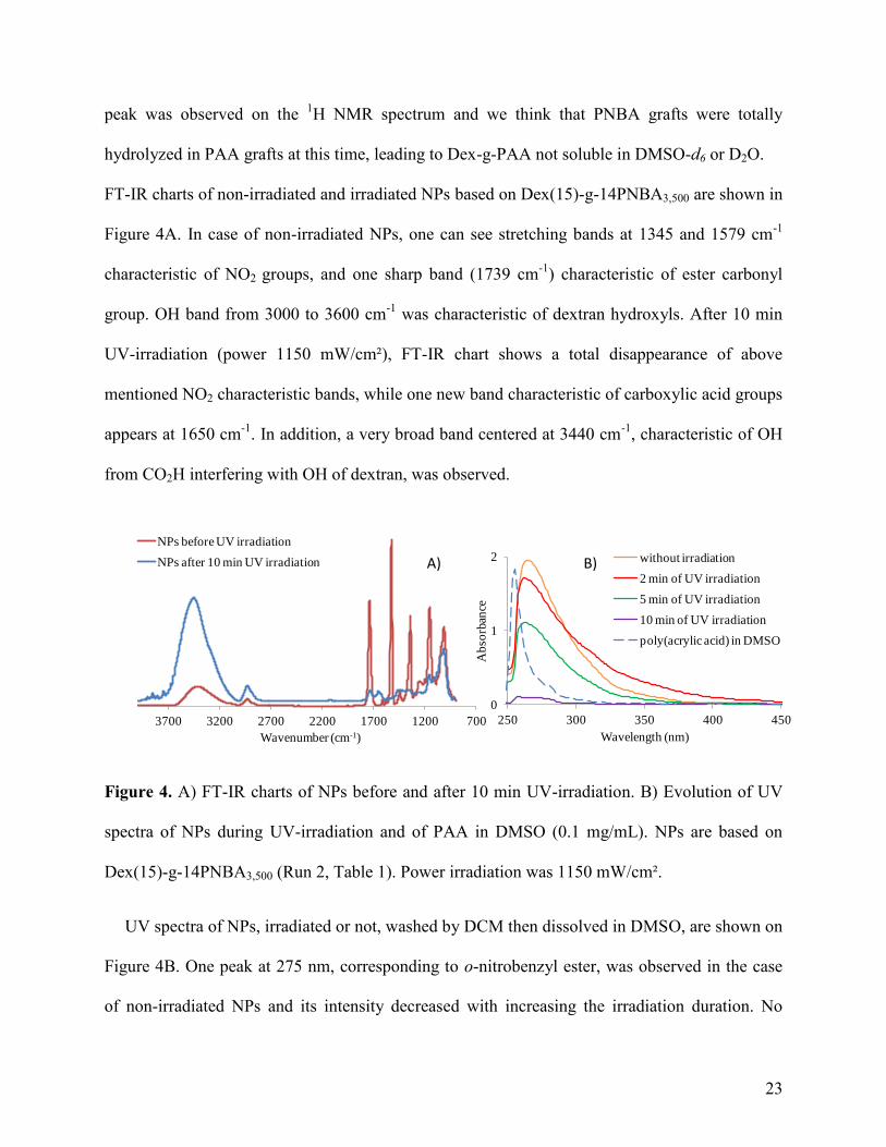
FT-IR charts of non-irradiated and irradiated NPs based on Dex(15)-g-14PNBA3,500 are shown in
Figure 4A. In case of non-irradiated NPs, one can see stretching bands at 1345 and 1579 cm-1
characteristic of NO2 groups, and one sharp band (1739 cm-1
) characteristic of ester carbonyl
group. OH band from 3000 to 3600 cm-1
was characteristic of dextran hydroxyls. After 10 min
UV-irradiation (power 1150 mW/cm²), FT-IR chart shows a total disappearance of above
mentioned NO2 characteristic bands, while one new band characteristic of carboxylic acid groups
appears at 1650 cm-1
. In addition, a very broad band centered at 3440 cm-1
, characteristic of OH
from CO2H interfering with OH of dextran, was observed.
A) B)
700120017002200270032003700
Wavenumber (cm-1)
NPs before UV irradiation
NPs after 10 min UV irradiation
0
1
2
250 300 350 400 450
Ab
sorb
ance
Wavelength (nm)
without irradiation
2 min of UV irradiation
5 min of UV irradiation
10 min of UV irradiation
poly(acrylic acid) in DMSO
Figure 4. A) FT-IR charts of NPs before and after 10 min UV-irradiation. B) Evolution of UV
spectra of NPs during UV-irradiation and of PAA in DMSO (0.1 mg/mL). NPs are based on
Dex(15)-g-14PNBA3,500 (Run 2, Table 1). Power irradiation was 1150 mW/cm².
UV spectra of NPs, irradiated or not, washed by DCM then dissolved in DMSO, are shown on
Figure 4B. One peak at 275 nm, corresponding to o-nitrobenzyl ester, was observed in the case
of non-irradiated NPs and its intensity decreased with increasing the irradiation duration. No

24
band at 325 nm, characteristic of o-nitrosobenzaldehyde by-product, was observed according to
the good DCM washing. After 10 min UV-irradiation, we still observed a small band over range
250-280 nm that may correspond to the electronic transitions as π→π* and n→π* of carboxylic
acid groups (PAA grafts) and of the esters linking grafts onto dextran backbone.[54] Indeed, the
UV spectrum of PAA ( = 1,800 g/mol) in DMSO shows one band over range of wavelengths
from 250 to 280 nm (Figure 4B).
3.4.1) Effect of the medium
We choose to irradiate NPs (made by nanoprecipitation of Dex(15)-g-12PNBA9,800, run 3,
Table 1) in two different media: distilled water and PBS.
Firstly in the case of NPs dispersed in H2O, the evolution of Normalized Mean Count Rate
(MCR/MCRo) with the duration of irradiation (power 1150 mW/cm²) was drawn on Figure 5A.
The Mean Count Rate value (MCR) was measured initially (MCRo) and after each irradiation
duration (30 sec intervals). As shown, normalized MCR decreased until 120 sec irradiation, then
increased according to the swelling of the NPs. Actually, according to our previous results,
PNBA grafts were not totally converted into PAA ones after 60 sec of irradiation under this UV-
lamp power.[38] Consequently, after 60 sec of irradiation, grafts are based on NBA and acrylic
acid monomer units (P(NBA-co-AA) grafts). At full photolysis, PNBA grafts were converted
into PAA ones as shown on Figure S2. But, as PAA is insoluble in pure water, PAA grafts
present onto the dextran backbone, and thus in the core of NPs, lead to swell NPs, scattering
laser beam of DLS as native NPs do.
Secondly, we carried out the same experiments in PBS buffer. When running irradiation, we
observed a continuous decrease of normalized MCR until 90 sec irradiation due to the
.

25
0E+00
2E-01
4E-01
6E-01
8E-01
1E+00
0 50 100 150 200
No
rmali
zed
Mea
n C
ou
nt R
ate
Irradiation duration (s)
50 mW/cm²
250 mW/cm²
320 mW/cm²
620 mW/cm²
1150 mW/cm²
320 mW/cm²
620 mW/cm²
1150 mW/cm²
0E+00
2E-01
4E-01
6E-01
8E-01
1E+00
0 50 100 150 200 250
No
rmali
zed
Mea
n C
ou
nt R
ate
Irradiation duration (s)
320 mW/cm²620 mW/cm²1150 mW/cm²320 mW/cm²620 mW/cm²1150 mW/cm²
B) C)
0E+00
2E-01
4E-01
6E-01
8E-01
1E+00
1E+00
0 50 100 150 200
No
rmali
zed
Mea
n C
ou
nt R
ate
Irradiation duration (s)
in pure water
in PBS
A)
Figure 5. Normalized Mean Count Rate of NPs against duration of irradiation. A) Effect of the dispersion medium, irradiation power
1150 mW/cm² (run 3, Table 1). B) Effect of the irradiation power in PBS. Run 2, Table 1 (solid symbols) and run 3, Table 1 (open
symbols). C) Effect of the irradiation power in PBS. Run 6, Table 1 (solid symbols) and run 11, Table 1 (open symbols).

26
progressive solubilisation of Dex-g-P(NBA-co-AA) glycopolymers in PBS, leading to the
disappearance of NPs (Figure 5A). After 90 sec irradiation, normalized MCR was constant to
low value. At this time, all NPs were disappeared and converted to dissolved Dex-g-P(NBA-co-
AA) glycopolymers. Indeed, in such a buffer, PAA grafts were converted to their salts, that are
readily soluble.
3.4.2) Effects of the irradiation power and of the nanoparticles chemistry
Firstly, NPs based on Dex(15)-g-14PNBA3,500 (FPNBA =75%, run 2, Table 1) were irradiated
under various powers (Figure 5B) in PBS. For one given irradiation power, normalized MCR
decreased with increasing the irradiation duration until reaching a stable value. Secondly, NPs
based on Dex(15)-g-12PNBA9,800 (FPNBA =85%, run 3, Table 1) show a slower decrease of MCR,
that is explained by both the higher number of NBA monomer units in glycopolymer and the
higher hydrophobicity of the NPs. Therefore, for one given irradiation power, higher duration of
irradiation is necessary to obtain water-soluble Dex-g-P(NBA-co-AA) glycopolymers from run 3
compared to run 2 (Table 1). Increasing the irradiation power leads to disrupt a higher NPs
number for the same irradiation time. Same observation was done for NPs made by
emulsion/evaporation process (Figure 5C). Moreover, if we compare the decreasing of
normalized MCR for NPs made with or without an in situ CuAAC (runs 6 and 11, Table 1), we
can conclude that the presence of triazole rings does not prevent the NPs disruption as the
kinetics appeared close. At higher irradiation duration, MCR of irradiated "clicked" NPs (run 11,
Table 1) seems to be higher than that observed in case of NPs produced without CuAAC (run 6,
Table 1). In fact, in the case of NPs produced without CuAAC, total photolysis will lead to a
blend of dextran and PAA chains in comparison with Dex-g-PAA produced after quantitative
photolysis of "clicked" NPs that may organize themselves in aqueous phase.

27
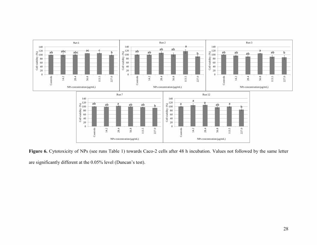
3.5) Light-sensitive nanoparticles cytotoxicity
3.5.1) Nanoparticles cytotoxicity
In order to evaluate the biocompatibility of such PNBA-based NPs, their cytotoxicity was
determined towards Caco-2 cells using MTT assay. In literature, diblock copolymers containing
one PNBA part or some o-nitrobenzyl ester linkages were already proved to be not cytotoxic
towards human breast cancer cells (MDA-MB-231)[55], (MDA-MB-435)[56] or human
umbilical vein endothelial cells (HUVEC cells).[33] But to the best of our knowledge, studies
dealing with the cytotocixity of o-nitrobenzyl-based copolymers or PNBA-based NPs towards
Caco-2 have never been reported. The Caco-2 cells viability was measured after 24 h (Figure S5)
and 48 h (Figure 6) incubation with various NPs batches formulated by either nanoprecipitation
or emulsion/organic solvent evaporation with (or without) in situ CuAAC, and using various NPs
dispersion concentration (less than or equal to 227 µg/mL). As shown in Figure 6, NPs until
113.5 µg/mL do not exhibit cell toxicity (cell viability ~ 100%) whatever the NPs formulation
process or the use of an in situ CuAAC. At the NPs concentration corresponding to 227 µg/mL, a
very low decrease of the cell viability can also be observed for some NPs. Consequently, for
experiments hereafter described, the NPs concentration equal to 113.5 µg/mL will be used as it
has been considered that same results are observed whatever the NPs batch.
3.5.2) Irradiation of Caco-2 cells
The Caco-2 cells viability was investigated after cells exposure to UV-irradiation, by varying
power and duration of irradiation. As shown in Figure 7, 100% Caco-2 cell viability was
observed after 24 h or 48 h incubation by applying 30 sec irradiation with a power of 60
mW/cm2. However, this viability decreased when increasing either the irradiation duration or the
28
0
20
40
60
80
100
120
140
Co
ntr
ols
14
.2
28
.4
56
.8
11
3.5
22
7.0
Cel
l v
iab
ilit
y (
%)
NPs concentration (µg/mL)
Run 1
0
20
40
60
80
100
120
140
Co
ntr
ols
14
.2
28
.4
56
.8
11
3.5
22
7.0
Cel
l v
iab
ilit
y (
%)
NPs concentration (µg/mL)
Run 2
0
20
40
60
80
100
120
140
Co
ntr
ols
14
.2
28
.4
56
.8
11
3.5
22
7.0
Cel
l v
iab
ilit
y (
%)
NPs concentration (µg/mL)
Run 3
0
20
40
60
80
100
120
140
Co
ntr
ols
14
.2
28
.4
56
.8
11
3.5
22
7.0
Cel
l v
iab
ilit
y (
%)
NPs concentration (µg/mL)
Run 7
0
20
40
60
80
100
120
140
Co
ntr
ols
14
.2
28
.4
56
.8
11
3.5
22
7.0
Cel
l v
iab
ilit
y (
%)
NPs concentration (µg/mL)
Run 12
ab abc abcac c
b ab abab ab
a
b ab ab aba
ab b
ab ab a ab ab ba
aa
ab ab
Figure 6. Cytotoxicity of NPs (see runs Table 1) towards Caco-2 cells after 48 h incubation. Values not followed by the same letter
are significantly different at the 0.05% level (Duncan’s test).

29
0
20
40
60
80
100
120
Cel
l v
iab
ilit
y (%
)
Power (mW/cm2) and duration (s) of irradiation
A)
0
20
40
60
80
100
120
Cel
l v
iab
ilit
y (%
)
Power (mW/cm2) and duration (s) of irradiation
B)
a a a
b
cd
e
f
e cef
a a
bb
bcb
cd cdd
cd bcd
Figure 7. Effect of the irradiation power/duration on the Caco-2 cell viability after (A) 24 h and
(B) 48 h incubation. Values not followed by the same letter are significantly different at the
0.05% level (Duncan’s test).
0
20
40
60
80
100
120
0 30 60 180 300
Cel
l v
iab
ilit
y (%
)
Duration of irradiation (s)
Control With culture medium renewal
0
20
40
60
80
100
120
0 30 60 180 300
Cel
l v
iab
ilit
y (%
)
Duration of irradiation (s)
Control With culture medium renewalA) B)
ab ab abaa a
cc
bc
d
a a
b
a a a
b
a
b
a
Figure 8. Effect of the culture medium renewal 4 h after the irradiation treatment (power: 60
mW/cm2) on the Caco-2 cell viability. The viability was estimated after (A) 24 h and (B) 48 h
incubation. Values not followed by the same letter are significantly different at the 0.05% level
(Duncan’s test).
irradiation power. In order to reduce the side-effects of the UV-irradiation treatment, the
replacement of the culture medium was performed 4 h after the irradiation, then cells were
incubated during an additional 24 h or 48 h before carrying the MTT assay. As shown in Figure
8, this change has clear benefits on the cell viability as around 85-90% was still observed after 48

30
h incubation (300 sec of irradiation at power 60 mW/cm2) versus less than 40% without medium
culture renewal.
3.5.3) Irradiation of Caco-2 cells incubated with nanoparticles
Finally, Caco-2 cells were exposed to 0.114 mg/mL of NPs (run 7, Table 1) and to irradiation
(power 60 mW/cm2). In some experiments and as previously done, the culture medium was
renewed 4 h after the irradiation, then cells were incubated during 24 h or 48 h before carrying
the MTT assay. With 30 sec irradiation and when the culture medium was not replaced, cell
viability reached 100% after 24 h incubation (Figure S6) but decreased up to 45% after 48 h
incubation (Figure 9) in agreement with the possible cytotoxicity[55] of the o-nitrobenzaldehyde
by-product produced during the photolysis of PNBA part (Figure S2). Nevertheless, with the
same irradiation duration, a clear improvement of the cell viability can be observed when
renewing the culture medium (Figure 9), which also mimics the dilution of the o-
nitrobenzaldehyde by-product in fluid body. The cell viability still reached 100% after 60 sec
irradiation, but longer irradiation led to the decrease of the cell viability. For instance, up to 55%
of cell viability was observed after 3 min irradiation.
4) CONCLUSIONS
PNBA core/Dex shell NPs were formulated by comparing two different processes:
nanoprecipitation of amphiphilic Dex-g-PNBA glycopolymers or emulsion/organic solvent
evaporation. Within this later process, we carried out (or not) an in situ CuAAC. Whatever the
process used, NPs with average diameter from 120 to 140 nm were obtained, then characterized
in terms of dextran amount per gram of PNBA (from 100 to around 300 mg of dextran / g of

31
0
20
40
60
80
100
120
140
Controls 30 60 180 300
Cel
l v
iab
ilit
y (%
)
Irradiation duration (s)
Control With culture medium renewal
a a
bd
c
bd
c
bdb b
d
Figure 9. Caco-2 cell viability (after 48 h incubation) after exposure to NPs, then UV irradiation
(power: 60 mW/cm2) with various durations. The medium culture was renewed (or not) 4 h after
the irradiation. Concentration of NPs (run 7, Table 1) was 0.114 mg/mL. Values not followed by
the same letter are significantly different at the 0.05% level (Duncan’s test).
PNBA). From the zeta potential values, one dextran surface layer thickness of 3-5 nm was
measured in the case of NPs made by nanoprecipitation, while an 8-10 nm thickness was
estimated using emulsion/organic solvent evaporation, carrying or not in situ CuAAC. The
colloidal stability of all NPs batches in the presence of salt was proved, but in the presence of
SDS we observed 85% desorption of the dextran shell for NPs formulated by emulsion/organic
solvent evaporation without CuAAC. Fortunately, less or equal up to 4% desorption was reached
in the case of NPs made by emulsion/organic solvent evaporation with in situ CuAAC or
nanoprecipitation processes, due to the triazole ring links between the dextran shell and the
PNBA core.
Photosensitive property of such PNBA-based NPs was evaluated under UV light irradiation by
firstly studying the photolysis kinetics, then varying either the suspension medium or the
irradiation parameters. We observed that NPs disappeared in PBS medium and were converted to

32
dissolved Dex-g-P(NBA-co-AA) glycopolymers before their total photolysis. Such photolysis
may be controlled depending on the irradiation mode and the chemical composition of NPs. NPs
disappearance kinetics depend also on the irradiation power: higher the power was, faster the
NPs disruption was.
The biocompatibility of these photosensitive NPs towards Caco-2 cells was proved by MTT
assay, whatever the NPs formulation process and the in situ CuAAC occurrence. In parallel, the
cell viability after UV-irradiation treatment was studied. It was shown that the replacement of the
culture medium 4 h after the irradiation allows an irradiation treatment of the Caco-2 with power
corresponding to 60 mW/cm2 up to 300 sec: after 48 h incubation, the cell viability remained at
85-90%. Finally, after exposure to PNBA-based NPs then to UV-irradiation (power: 60
mW/cm2), 100% cell viability was still observed after 60 sec irradiation. These results open the
way to future experiments dealing with such light-sensitive PNBA/Dex NPs. In the very next
future, the loading of such NPs with anticancer drugs, then their release induced by UV-
irradiation will be investigated. Moreover, we will show that the o-nitrobenzaldehyde side-effect
is negligible in comparison to the anticancer drug activity.
ASSOCIATED CONTENT
Supplementary data related to this article can be found in the online version of this article:
Surface tension of alkynated dextran. Photolysis of Dex-g-PNBA. Characteristics and colloidal
stability of NPs. Caco-2 cells viability after exposure to NPs, then (or not) to UV-irradiation
treatment.
ACKNOWLEDGMENT

33
M. El Founi was supported by a grant of the French Ministry in charge of Research. S.M.A. Soliman
gratefully acknowledges support from an Erasmus Mundus External Cooperation Windows – Flowby
Flow EU-Egypt Bridge Building (FFEEBB) Graduate Research Fellowship. The authors express
their highest gratitude to Marie-Christine Grassiot (LCPM) and Caroline Sejil (LCPM) for SEC
measurements, to Olivier Fabre (LCPM) for NMR measurements and to Bruno Ebel (LRGP) for
statistical analyses.
REFERENCES
[1] J.H. Park, S. Lee, J.H. Kim, K. Park, K. Kim, I.C. Kwon, Polymeric nanomedicine for
cancer therapy, Prog. Polym. Sci. 33 (2008) 113–137
[2] T. Lammers, S. Aime, W.E. Hennink, G. Storm, F. Kiessling, Theranostic nanomedicine,
Account Chem. Res. 44 (2011) 1029–1038
[3] S. Moein Moghimi, A. Christy Hunter, J. Clifford Murray, Nanomedicine: current status and
future prospects, FASEB J. 19 (2005) 311–330
[4] Y. Liu, H. Miyoshi, M. Nakamura, Nanomedicine for drug delivery and imaging: A
promising avenue for cancer therapy and diagnosis using targeted functional nanoparticles,
Int. J. Cancer 120 (2007) 2527–2537
[5] R.K. Jain, T. Stylianopoulos. Delivering nanomedicine to solid tumors, Nat. Rev. Clin.
Oncol. 7 (2010) 653–664
[6] A. Vonarbourg, C. Passirani, P. Saulnier, J.P. Benoit. Parameters influencing the stealthiness
of colloidal drug delivery systems. Biomaterials 27 (2006) 4356–4373
[7] M. D. Howard, M. Jay, T. D. Dziubla, X. Lu. PEGylation of nanocarrier drug delivery
systems: State of the art, J. Biomed. Nanotechnol. 4 (2008) 133–148
[8] E. M. Pelegri-O'Day, E. W. Lin, H. D. Maynard, J. Am. Chem. Soc. 136 (2014) 14323-
14332

34
[9] K. Knop, R. Hoogenboom, D. Fischer, U. S. Schubert, Poly(ethylene glycol) in drug
delivery: pros and cons as well as potential alternatives, Angew. Chem. Int. Ed. 49 (2010)
6288-6308
[10] Z. H. Liu, Y. P. Jiao, Y. F. Wang, C. R. Zhou, Z. Y. Zhang, Polysaccharides-based
nanoparticles as drug delivery systems, Adv. Drug Deliv. Rev. 60 (2008) 1650-1662
[11] C. Gavory, A. Durand, J.-L. Six, C. Nouvel, E. Marie, M. Léonard, Polysaccharide-
covered nanoparticles prepared by nanoprecipitation, Carbohyd. Polym. 84 (2011) 133-140
[12] M. Laville, J. Babin, I. Londono, M. Legros, C. Nouvel, A. Durand, R. Vanderesse, M.
Leonard, J.-L. Six, Polysaccharide-covered nanoparticles with improved shell stability using
synthetic strategies based on click-chemistry, Carbohyd. Polym. 93 (2013) 537-546
[13] C. Nouvel, J. Raynaud, E. Marie, E. Dellacherie, J.-L. Six, A. Durand, Biodegradable
nanoparticles made from polylactide-grafted dextrancopolymers, J. Colloid Interface Sci.
330 (2009) 337–343
[14] A. Kumari, S.K. Yadav, S.C Yada, Biodegradable polymeric nanoparticles based drug
delivery systems, Colloid Surf. B-Biointerfaces 75 (2010) 1-18
[15] E. Locatelli, M. Comes Franchini, Biodegradable PLGA-b-PEG polymeric nanoparticles:
synthesis, properties, and nanomedical applications as drug delivery system, J. Nanopart.
Res. 14 (2012) 1316-1332
[16] D. B. Pacardo, F.S. Ligler, Z. Gu, Programmable nanomedicine: synergistic and
sequential drug delivery systems, Nanoscale 7 (2015) 3381-3391
[17] D. Mishra, J. R. Hubenak, A. B. Mathur, Nanoparticle systems as tools to improve drug
delivery and therapeutic efficacy, J. Biomed. Mater. Res. Part A 101A (2013) 3646–3660.
[18] S. Bamrungsap, Z. Zhao, T. Chen, L. Wang, C. Li, T. Fu, W. Tan, Nanotechnology in
therapeutics: a focus on nanoparticles as a drug delivery system, Nanomedicine, 7 (2012)
1253-1271

35
[19] T.M. Allen, P.R. Cullis, Drug delivery systems: entering the mainstream, Science 303
(2004) 1818-1822
[20] B. Taghizadeh, S. Taranejoo, S. A. Monemian, Z. S. Moghaddam, K. Daliri, H.
Derakhshankhah, Z. Derakhshani, Classification of stimuli–responsive polymers as
anticancer drug delivery systems, Drug Deliv. 22 (2015) 145-155
[21] C. Alvarez-Lorenzo, A. Concheiro, Intelligent drug delivery systems: polymeric micelles
and hydrogels, Mini-Rev. Med. Chem. 8 (2008) 1065-1074
[22] M. Karimi, A. Ghasemi, P. Sahandi Zangabad, R. Rahighi, S. M. Moosavi Basri, H.
Mirshekari, M. Amiri, Z. Shafaei Pishabad, A. Aslani, M. Bozorgomid, D. Ghosh, A.
Beyzavi, A. Vaseghi, A. R. Aref, L. Haghani, S. Bahramia, M. R. Hamblin, Smart
micro/nanoparticles in stimulus-responsive drug/gene delivery systems, Chem. Soc. Rev. 45
(2016) 1457-1501
[23] D. Liu, F. Yang, F. Xiong, N. Gu, The smart drug delivery system and its clinical
potential, Theranostics 6 (2016) 1306-1323
[24] N. Rapoport. Physical stimuli-responsive polymeric micelles for anti-cancer drug
delivery, Prog. Polym. Sci. 32 (2007) 962-990
[25] S. Dai, P. Ravi, K.C. Tam, Thermo- and photo-responsive polymeric systems, Soft
Matter 5 (2009) 2513-2533
[26] F. D. Jochum, P. Theato, Temperature- and light-responsive smart polymer materials,
Chem. Soc. Rev. 42 (2013) 7468-7483
[27] Q. Yan, D. Han, Y. Zhao, Main-chain photoresponsive polymers with controlled location
of light-cleavable units: from synthetic strategies to structural engineering, Polym. Chem. 4
(2013) 5026-5037
[28] G. Liu, W. Liu, C.-M. Dong, UV- and NIR-responsive polymeric nanomedicines for on-
demand drug delivery, Polym. Chem. 4 (2013) 3431–3443

36
[29] O. Bertrand, J.-F. Gohy, Photo-responsive polymers: synthesis and applications, Polym.
Chem. 8 (2017) 52-73
[30] V. Marturano, P. Cerruti, M. Giamberini, B. Tylkowski, V. Ambrogi, Light-responsive
polymer micro- and nano-capsules, Polymers 9 (2017) 8
[31] H. Zhao, E. S. Sterner, E. B. Coughlin, P. Theato, o-Nitrobenzyl alcohol derivatives:
Opportunities in polymer and materials science, Macromolecules 45 (2012) 1723-1736
[32] J. Jiang, X. Tong, D. Morris, Y. Zhao, Toward photocontrolled release using light-
dissociable block copolymer micelles, Macromolecules 39 (2006) 4633-4640
[33] Q. Jin, T. Cai, H. Han, H. Wang, Y. Wang, J. Ji, Light and pH dual-degradable triblock
copolymer micelles for controlled intracellular drug release, Macromol. Rapid Commun. 35
(2014) 1372-1378
[34] G. Jiang, T. Jiang, H. Chen, L. Li, Y. Liu, H. Zhou, Y. Feng, J. Zhou, Preparation of
multi-responsive micelles for controlled release of insulin, Colloid Polym. Sci. 293 (2015)
209-215
[35] S. Jana, A. Bose, A. Saha, T. K. Mandal, Photocleavable and tunable thermoresponsive
amphiphilic random copolymer: self-assembly into micelles, dye encapsulation, and
triggered release, J. Polym. Sci., Part A: Polym. Chem. 55 (2017) 1714-1729
[36] X. Jiang, C. A. Lavender, J. W. Woodcock, B. Zhao, Multiple micellization and
dissociation transitions of thermo- and light-sensitive poly(ethylene oxide)-b-
poly(ethoxytri(ethylene glycol) acrylate-co-o-nitrobenzyl acrylate) in water,
Macromolecules 41 (2008) 2632-2643
[37] S. M. A. Soliman, C. Nouvel, J. Babin, J.-L. Six, o-Nitrobenzyl acrylate is polymerizable
by Single Electron Transfer-Living Radical Polymerization, J. Polym. Sci., Polym. Chem.
52 (2014) 2192-2201
[38] S. M. A. Soliman, L. Colombeau, C. Nouvel, J. Babin, J.-L. Six, Amphiphilic
photoresponsive dextran-g-poly(o-nitrobenzyl acrylate) glycopolymers, Carbohyd. Polym.
136 (2016) 598-608

37
[39] S. Jana, A. Saha, T. K. Paira, T. K. Mandal, Synthesis and self-aggregation of poly(2-
ethyl-2-oxazoline)-based photocleavable block copolymer: micelle, compound micelle,
reverse micelle, and dye encapsulation/release, J. Phys. Chem. B 120 (2016) 813-824
[40] Z. Xu, B. Yan, J. Riordon, Y. Zhao, D. Sinton, M. G. Moffitt, Microfluidic synthesis of
photoresponsive spool-like block copolymer nanoparticles: flow-directed formation and
light triggered dissociation, Chem. Mater. 27 (2015) 8094-8104
[41] D. Royston, D. G. Jackson, Mechanisms of lymphatic metastasis in human colorectal
adenocarcinoma, J. Pathol. 217 (2009) 608-619
[42] L. Baandrup, L. T. Thomsen, T. B. Olesen, K. K. Andersen, B. Norrild, S. K. Kjaer, The
prevalence of human papillomavirus in colorectal adenomas and adenocarcinomas: A
systematic review and meta-analysis, Eur. J. Cancer 50 (2014) 1446-1461
[43] J. J. Smith, N. G. Deane, P. Dhawan, R. D. Beauchamp, Regulation of metastasis in
colorectal adenocarcinoma: A collision between development and tumor biology, Surgery
144 (2008) 353-366
[44] S.R. Deshiikan, K.D. Papadopoulos, Modified Booth equation for the calculation of zeta
potential, Colloid. Polym. Sci. 276 (1998) 117-124
[45] K. Poltorak, A. Durand, M. Leonard, J.-L. Six, C. Nouvel, Interfacial click chemistry for
improving both dextran shell density and stability of biocompatible nanocapsules, Colloid
Surface A 483 (2015) 8-17
[46] W.G. Eversole, W.W. Boardman, The effect of electrostatic forces on electrokinetic
potentials, J. Chem. Phys. 9 (1941) 798–801
[47] T. Mosman, Rapid calorimetric assay for cellular growth and survival: Application to
proliferation and cytotoxicity assays, J. Immunol. Methods 65 (1983) 55-63
[48] D. Chognot, M. Leonard, J.-L. Six, E. Dellacherie, Surfactive water-soluble copolymers
for the preparation of controlled surface nanoparticles by double emulsion/solvent
evaporation, Colloid Surface B, 51 (2006) 86-92

38
[49] P. Cintas, G. Palmisano, G. Cravotto, Power ultrasound in metal-assisted synthesis: From
classical Barbier-like reactions to click chemistry, Ultrason. Sonochem. 18(S1) (2011) 836-
841
[50] M. T. Peracchia, C. Vauthier, D. Desmaele, A. Gulik, J.-C. Dedieu, M. Demoy, J.
d'Angelo, P. Couvreur, Pegylated nanoparticles from a novel methoxypolyethylene glycol
cyanoacrylate-hexadecyl cyanoacrylate amphiphilic copolymer, Pharm. Res. 15 (1998) 550-
556
[51] B. Cattoz, T. Cosgrove, M. Crossman, S.W. Prescott, Surfactant-mediated desorption of
polymer from the nanoparticle interface, Langmuir 28 (2012) 2485-2492
[52] A.M. Blokhus, K. Djurhuus, Adsorption of poly(styrene sulfonate) of different molecular
weights on alpha-alumina: effect of added sodium dodecyl sulfate, J. Colloid Interface Sci.
296 (2006) 64-70
[53] R. A. Lauten, A.-L. Kjøniksen, B. Nystrom, Adsorption and desorption of unmodified
and hydrophobically modified ethyl(hydroxyethyl)cellulose on polystyrene latex particles in
the presence of ionic surfactants using dynamic light scattering, Langmuir 16 (2000) 4478-
4484
[54] S. Kavlak, H. K. Can, A. Guner, Interaction of poly(maleic anhydride-alt-acrylic acid)
with transition metal cations, Ni2, Cu2, and Cd2: a study by UV–Vis spectroscopy and
viscosimetry, J. Appl. Polym. Sci. 92 (2004) 2698-2705
[55] P. Anilkumar, E. Gravel, I. Theodorou, K. Gombert, B. Thézé, F. Ducongé, E. Doris,
Nanometric micelles with photo-triggered cytotoxicity, Adv. Funct. Mater. 24 (2014) 5246-
5252
[56] J. Song, Z. Fang, C. Wang, J. Zhou, B. Duan, L. Pu, H. Duan, Photolabile plasmonic
vesicles assembled from amphiphilic gold nanoparticles for remote-controlled traceable drug
delivery, Nanoscale 5 (2013) 5816-5824


![Sustained-release nanoAR T formulation for the treatment ... · sustained release profile, and application of 2 bilayers ([tenofovir+dextran sulphate] 2 +vorinostat) to magnetic nanoparticles](https://img.pdfslide.us/doc/110x75/5f2fabc4d21df375ea1485f3/sustained-release-nanoar-t-formulation-for-the-treatment-sustained-release-profile.jpg)