Embed Size (px)
Citation preview

Plant Physiol. (1989) 91, 542-5510032-0889/89/91/0542/1 0/$01 .00/0
Received for publication January 4, 1989and in revised form May 18, 1989
Light-induced Spectral Absorbance Changes in Relation toPhotosynthesis and the Epoxidation State of Xanthophyll
Cycle Components in Cotton Leaves1
Wolfgang Bilger*, Olle Bjorkman, and Susan S. ThayerCarnegie Institution of Washington, Department of Plant Biology, Stanford, California 94305
ABSTRACT
When cotton (Gossypium hirsutum L., cv Acala SJC-1) leaveskept in weak light were suddenly exposed to strong red actiniclight a spectral absorbance change took place having the follow-ing prominent characteristics. (a) It was irreversible within thefirst four minute period after darkening. (b) The difference in leafabsorbance between illuminated and predarkened leaves had amajor peak at 505 nanometers, a minor peak at 465 nanometers,a shoulder around 515 nanometers, and minor troughs at 455 and480 nanometers. (c) On the basis of its spectral and kineticcharacteristics this absorbance change can be readily distin-guished from the much faster electrochromic shift which has apeak at 515 nanometers, from the slow, so-called light-scatteringchange which has a broad peak centered around 535 nanometersand is reversed upon darkening, and from absorbance changesassociated with light-induced chloroplast rearrangements. (d)The extent and time course of this absorbance change closelymatched that of the deepoxidation of violaxanthin to zeaxanthinin the same leaves. (e) Both the absorbance change and theability to form zeaxanthin were completely blocked in leaves towhich dithiothreitol (DTT) had been provided through the cutpetlole. DTT treatment also caused strong inhibition of that com-ponent of the 535-nanometer absorbance change which is re-versed in less than 4 minutes upon darkening and considered tobe caused by increased light scattering. Moreover, DTT inhibiteda large part of nonphotochemical quenching of chlorophyll fluo-rescence in the presence of excessive light. However, DTT hadno detectable effect on the photon yield of photosynthesis meas-ured under strictly rate-limiting photon flux densities or on thelight-saturated photosynthetic capacity, at least in the short term.We conclude that it is possible to monitor light-induced violax-anthin de-epoxidation in green intact leaves by measurement ofthe absorbance change at 505 nanometers. Determination ofabsorbance changes in conjunction with measurements of pho-tosynthesis in the presence and absence of DTT provide a systemwell suited for future studies of mechanisms of dissipation ofexcessive excitation energy in intact leaves.
Under most natural conditions leaves are normally exposedto irradiance levels during the course of the day which exceedthe capacity by which the resulting excitation energy can beused in photosynthesis. This excessive energy must be safely
'C.I.W.-D.P.B. Publication No. 1025. This work was supportedby Grant No. 86-CRCR-1-2054 of the Competitive Grants Programof the U.S. Department of Agriculture to 0. B. A Carnegie InstitutionFellowship and a Feodor-Lynen-Fellowship by the Alexander vonHumboldt-Foundation to W. B. is gratefully acknowledged.
dissipated; otherwise the photosynthetic system will sufferphotoinhibitory damage (17). Analysis of Chl fluorescencehas provided strong evidence that leaves adapted to highirradiances have a greater capacity to dissipate excessive en-ergy via a process of NRD2 than leaves adapted to lowirradiances (1). It has been proposed that such NRD, whichis seen as qNP, could protect against photoinhibitory damage(1, 4, 13). It has also been proposed that the molecular basisof 'safe' nonradiative dissipation in part involves the carote-noid zeaxanthin, hypothetically acting as a quencher of ex-cited states of Chl in the antenna (4, 5). Good correlationsbetween NRD (expressed as an increase in qNP or as anincrease in KD, the rate constant for NRD) and zeaxanthincontent of leaves were obtained under a wide range of con-ditions (5).
Zeaxanthin is formed by deepoxidation of violaxanthin(5,6,5',6'-diepoxizeaxanthin) via the intermediate antherax-anthin (5,6-monoepoxizeaxanthin) (for reviews, see refs. 6,25). Both deepoxidation steps are catalyzed by the sameenzyme, violaxanthin deepoxidase (25). This enzyme isthought to be located on the lumen side of the thylakoidmembrane and its activity has a sharp optimum around pH5. Excessive light promotes the deepoxidation of violaxanthinto zeaxanthin, evidently because the resulting buildup of apH gradient across the thylakoid membrane causes acidifica-tion of the lumen, thereby activating the deepoxidase (6, 25).The back reaction (epoxidation ofzeaxanthin to violaxanthin)is catalyzed by an epoxidase thought to be located on theother side of the membrane. Its activity is promoted underthe pH and redox conditions that exist under limiting light(25). Violaxanthin bound to thylakoid membranes appears tobe mainly located in LHCI and LHCII (20). However, withthe methods hereto used for isolating the pigment proteincomplexes, a considerable fraction ofthe violaxanthin appearsin the free pigment band. The distribution of the xanthophyllcycle components in the thylakoid membrane is thereforeuncertain; moreover, these components may also have a highdegree of mobility.
In view of its proposed role in photoprotection, moreinformation about the epoxidation state of the xanthophyll-
2 Abbreviations: NRD, nonradiative energy dissipation; A, anther-axanthin; DTT, dithiothreitol; AA,, absorbance change at wavelengthX; LHC, light harvesting complex; qp, qNP, photochemical and non-photochemical Chl fluorescence quenching, respectively; P[C02],photosynthetic CO2 uptake; P[ET], photosynthetic electron transport;P[02], photosynthetic 02 evolution; PFD, photon flux density (400-700 nm); V, violaxanthin; z, zeaxanthin.
542 www.plantphysiol.orgon November 22, 2018 - Published by Downloaded from Copyright © 1989 American Society of Plant Biologists. All rights reserved.

ABSORBANCE CHANGES AND XANTHOPHYLL CYCLE IN LEAVES
cycle in vivo under conditions of excessive light is needed.The techniques used so far to quantify the xanthophyll com-ponents of this cycle in green leaves, TLC and HPLC, havecertain drawbacks. TLC is complicated and very time con-suming. Although HPLC has many advantages over TLC, theusefulness ofHPLC has been limited primarily because of thedifficulties involved in separating zeaxanthin from luteinwhich is a major component of leaf xanthophylls. Recentimprovements (22) have fully overcome this problem but thefact that these methods are destructive still limits their use inkinetic studies of the operation of the xanthophyll cycle inintact leaves.Yamamoto et al. (26) and Sokolove and Marsho (21)
described an absorbance change in chloroplast suspensionswhich was associated with the conversion of violaxanthin tozeaxanthin. This absorbance change had a peak at 505 nm.Recently, Pfundel and Strasser ( 16), working with photosyn-thetically incompetent etiolated bean leaves, were able toobserve an increase in absorbance at 505 nm over severalhours in darkness that took place after vacuum infiltrationwith ascorbate at pH 5. This slow increase in absorbance wasaccompanied by a large increase in zeaxanthin and decreasein violaxanthin. Measurements of absorbance changes at 505nm in intact, photosynthesizing green leaves are complicatedby their high optical density at this wavelength and by thepresence of light-induced absorbance changes with consider-able spectral overlaps. These are the electrochromic shift,peaking at 515 or 518 nm (24), the so-called, light-scatteringchange, centered around a broad optimum around 530 nm(7, 12), and changes caused by chloroplast rearrangementswithin the leaf (9).
Since light-induced absorbance changes associated withzeaxanthin formation in green leaves have not been previouslyreported, we investigated the potential of measurements ofsuch changes for studies of the kinetics of the operation of thexanthophyll cycle. Using normal green cotton leaves havinga high photosynthetic capacity, we were able to show that thedeepoxidation that occurred when the leaves were illuminatedwith strong red light was clearly associated with a large andspecific light-induced absorbance change with a peak at505 nm. This absorbance change was blocked by a DTT ap-plication that had no effect on short-term photosyntheticperformance.
MATERIALS AND METHODS
Plant Material
Cotton plants (Gossypium hirsutum L. cv Acala SJC-1)were grown in a controlled-growth room in flowing-nutrientculture as described in detail by Schafer and Bj6rkman (18).The incident PFD at leaf level was 700 to 800 Mmol m-2 s-'.Fully expanded leaves were used, usually from the fourth tosixth node, counting from the top of the plant. About 30 minbefore excision, the leaves were shaded by a panel of reflectivealuminum foil, giving a PFD of about 100 ,umol m-2 s-' at theleaf surface. Some experiments were made on leaves fromcotton plants grown in a field just outside the laboratory.These experiments were made during July at which the dailymaximum PFD was approximately 2000 ;tmol m-2 s-'. Thewhole plant had been kept shaded since the night before each
experiment. The PFD at time of leaf excision was approxi-mately 100 ,mol mr2 s'. At this PFD the deepoxidation stateof the xanthophyll cycle in cotton was found to be the sameas in leaves which had been kept in darkness for several hours.
DTT Treatments
In all experiments the petiole was cut either under water(controls) or under a 1 mm solution of reduced or oxidizedDTT with a piece of a sharp razor blade. When DTT oroxidized DTT was applied, the petiole was promptly trans-ferred to a small cylinder containing the same solution. Forthe first 15 or 30 min, the leaves were kept at 100 to 200,,mol m-2 s-' PFD to keep the stomata open so that uptake ofthe DTT solution via the transpiration stream was facilitated.The PFD was then decreased to approximately 15 'mol m-2s-' and maintained at this low PFD until the start of measure-ments. The volume of solution taken up by each leaf wasrecorded hourly. Depending on transpiration rate the timerequired for the leaves to take up an amount of solution equalto their fresh weight varied from 2 to 4 h. On the assumptionthat DTT freely entered the leafwith the transpiration stream,this would yield a concentration in the bulk leaf solution ofat least 1 mM.
Induction of Violaxanthin Deepoxidation in Weak Light inthe Presence of Ascorbate
For the experiment shown in Table II, leaf discs wereinfiltrated under vacuum with a Na-ascorbate buffer (150mM), adjusted to pH 5.0 with citric acid (final concentrationabout 10 mM) with or without 1 mM DTT. After infiltration,the leaf discs were kept floating for 5 h on the same solution.To avoid anaerobiosis, the leaf discs were illuminated by weaklight (approximately 10 ,umol m-2 s-').
Chemicals
Na-ascorbate, citric acid, DTT, and oxidized DTT (trans-4,5-dihydroxy-1,2-dithiane) were purchased from Sigma, St.Louis, MO. Freshly prepared solutions of DTT or oxidizedDTT were used for each experiment. The same batches ofDTT and oxidized DTT were kindly tested by H. Yamamotoand A. Gilmore of the University of Hawaii for the potencyof these compounds as inhibitors of activity of purified vio-laxanthin deepoxidase in vitro. They confirmed that this batchof reduced DTT was a highly effective inhibitor, whereas thebatch of oxidized DTT had no effect on the activity of theisolated enzyme.
Measurements of Light-Induced Absorbance Changes inLeaves
Absorbance measurements were carried out in a PerkinElmer model 356 Dual-Wavelength, Dual-Beam spectropho-tometer. Leaf discs (20 or 28 mm in diameter) were placed inthe primary sample compartment in front of the photomul-tiplier tube and fixed by means ofa small holder. The distancebetween the leaf and the photomultiplier cathode was about15 mm. All measurements of light-induced absorbancechanges reported in this paper were made in the split beam
543
www.plantphysiol.orgon November 22, 2018 - Published by Downloaded from Copyright © 1989 American Society of Plant Biologists. All rights reserved.
Plant Physiol. Vol. 91, 1989
mode. The reference beam, which bypassed the leaf, wasattenuated by neutral density filters to give approximately thesame signal as the beam transmitted through the leaf. Actinicillumination was provided by a cold light fiber optic lightsource (model KL 1500, Schott, Mainz, FRG). The fiber opticentered the sample compartment from the rear side. Theactinic light beam was filtered through a red-transmittingfilter (Coming 2-60) and the photomultiplier tube was pro-tected by insertion ofa blue-green transmitting filter (Coming4-96) immediately in front of it. At an actinic PFD of 1300gmol mM2 s-' this arrangement resulted in a small transientartifact upon light on or light off of an amplitude equal to0.0003 absorbance units, as determined with a Whatman No.1 filter paper in place of the leaf sample. To minimize waterloss during the measurements, a small housing, lined withreflective foil on the outside and with moistened fabric on theinside, was placed around the leaf holder. Some measure-ments of light-induced absorbance changes were also made at505, 515, and 540 nm with a custom-built instrument thatpermitted use ofintact leaves attached to the plant. The resultsobtained in these measurements were very similar to thoseobtained with leaf discs using the system described above.
Photosynthesis Measurements
Photon yields of 02 evolution were determined at 4 to 5%C02, 20 to 21% 02, balance N2, under strictly rate limitingphoton flux densities on leaves that had not been preexposedto excessive light. These measurements were either made on10 cm2 leaf discs in a closed system using a Hansatech (King'sLynn, Norfolk, England) 12 leaf disc O2 electrode as describedin Bjorkman and Demmig (2) or using an open-flow system(18) on a 10 cm2 area of a leaf whose cut petiole was kept inwater or in a DTT solution. In the latter system, a humidifiedstream of 5 C02, 19.9% 02, balance N2, was passed throughthe leaf chamber at a flow rate of 4 mL min-' (using a massflow controller model FC260, Tylan, Carson, CA) and thechange in O2 concentration caused by the leaf was continu-ously measured in the exit gas stream with a model DW1/CBI-D O2 electrode system (Hansatech), modified for gas-phase measurements. No difference was found between thephoton yield values obtained with the open and the closedsystems.
Light-dependence curves and light-saturated rates of pho-tosynthetic O2 evolution were determined with the open-flowsystem only, which is much better suited to measurements ofhigh photosynthetic rates than the closed leaf disc system.The open flow, whole leaf, system also avoids problems causedby heating of the O2 electrode at high irradiance levels sincethe electrode chamber temperature is independent of that ofthe leafchamber. Moreover, since the leaf is permitted to takeup water, any effects caused by transpirational water loss fromthe leaf are minimized. Measurements of light-saturated ratesof 02 evolution were made at a gas flow rate of 14 mL min-',while in the determinations of light dependence curves theflow rate at each photon flux density was adjusted (4-14 mLmin-') to give a suitable change in O2 concentration. Ratesof 02 exchange are given as the sum of net 02 evolution anddark respiration. The light dependence curves were measuredas the response to increased PFDs. The rate at each PFD wastaken after an apparent steady state had been reached which
usually was obtained within 10 to 15 min after each increasein PFD. In the experiments shown in Figure 7, the leaftemperature rose from 27 to 33°C as the PFD was increasedfrom 800 to 2000 ,Amol m-2 sg'. Since the rate of light- andC02-saturated photosynthesis increases with increasing tem-perature in this range, the rates were adjusted for the temper-ature dependence determined at saturating PFD in separateexperiments. Other experiments with comparable cottonleaves in the absence and presence of DTT where the lightdependence of photosynthesis in the range 800 to 2000 ,umolm-2 s- was measured at a constant leaftemperature confirmedthat these adjustments for a rising leaf temperature werecorrect.
Rates of CO2 uptake in the presence and absence of DTTunder normal air concentrations of CO2 were made at a leaftemperature of 30°C as previously described (18). The photonyield of CO2 uptake was first determined at strictly limitingPFDs (35 and 90 '4mol m-2 s-'). The PFD was then raised to1000 ,umol m-2 s-' and after 15 min it was raised to 2000 ,umolm-2 s-'. The rate determined when an apparent steady statehad been reached (after approximately 15 min at this PFD)was taken as the light saturated rate. The sum of the rates ofcarboxylation and oxygenation (P[ET]) was calculated asdescribed ( 18).
Xanthophyll Determinations
Contents of the components of the xanthophyll cycle weremeasured on leaf samples which had been frozen in liquidnitrogen. Extraction was in 80% acetone and separation byHPLC as described in detail (22).
Chl Fluorescence Measurements
Chl fluorescence was measured as previously described ( 18),using a system which incorporates a PAM Chl fluorometersystem (Walz, Effeltrich, FRG). The petioles were immersedin water or a 1 mM DTT solution during these measurements.In experiments where we wanted to prevent gas exchange,both leaf surfaces were covered with a thin layer of silicongrease (Dow Coming high vacuum grease) just before the startof the fluorescence measurements. Nonphotochemical fluo-rescence quenching (qNp) and photochemical quenching (qp)were calculated as outlined in (19). However, the maximumfluorescence yield for each leaf used in the calculations of qNPwas not determined in leaves which had been kept in completedarkness but was instead determined for leaves kept at a lowPFD (cf. 18). The saturating pulses used to cause temporaryclosure of all PSII traps had a PFD of 5000 smol m-2 s-' anda duration of 1 s.
RESULTS
Light-induced Absorbance Changes and ViolaxanthinDeepoxidation
Yamamoto et al. (26) described an absorbance change at505 nm which was associated with zeaxanthin formation inisolated chloroplast preparations. Since strong light inducesdeepoxidation of violaxanthin, we measured light-inducedabsorbance changes in cotton (Gossypium hirsutum L. cvAcala SJC-1) leaves which were depleted of zeaxanthin by
544 BILGER ET AL.
www.plantphysiol.orgon November 22, 2018 - Published by Downloaded from Copyright © 1989 American Society of Plant Biologists. All rights reserved.

ABSORBANCE CHANGES AND XANTHOPHYLL CYCLE IN LEAVES
preincubation in weak light for several hours. In Figure 1 areshown results ofabsorbance change measurements at 505 nm,compared with those at 535 nm, where the so-called lightscattering change usually is measured. At 505 nm, absorbancerose monotonically upon illumination and showed no declineduring the following dark period. After the light was turnedoff, a small further increase in absorbance took place. Asecond illumination produced little further rise, corroboratingthe short-term irreversibility of the first light-induced absorb-ance change. Like the absorbance at 505 nm, that at 535 nmrose during the first 2 min of the first exposure, but exhibiteda longer lag time and the absorbance leveled off earlier. Incontrast to the change at 505 nm, the change at 535 nmdisplayed a pronounced decay upon darkening, although theabsorbance did not return to its initial level. The secondillumination induced a larger change with faster kinetics thanthat observed during the first illumination and it was nowfully reversed upon darkening.
Previous studies on light-induced absorbance changesaround 535 nm have indicated that the maximum extent ofthis change is strongly dependent on the balance between theactivity of ATP-consuming dark reactions and the intensityof the actinic light (7, 1 1). It also seems likely that factorssuch as stomatal opening, leaftemperature, and the activationstate of Calvin cycle enzymes can all affect the amplitude ofthis absorbance change and, therefore, cause a large variationin the response of different leaf samples. However, a stablestate of the photosynthetic apparatus which resulted in goodreproducibility was obtained when the leaves were kept at lowPFD (15 ,umol m2 s-') for 3 to 4 h prior to the absorbancechange measurements.
Figure 1. Original traces of light-induced absorbance changes at 505(upper trace) and 535 nm (lower trace) in cotton leaf discs. Twodifferent discs of the same leaf were used, after a preadaptationperiod at low PFD (15 ,umol m-2 s-1) of 5 h. The arrows denote thetimes at which red actinic light (500 4mol photons m-2 s-1) wasswitched on (T) or off (l). The leaf discs were in air at a temperatureof 26°C during measurement.
As shown in the traces in Figure 1 at both 505 and 535 nm,a small, rapid component is evident upon light on and lightoff. This component is presumably caused by spectral overlapfrom the electrochromic shift which is known to have a peakaround 518 nm. The rise kinetics upon light on are generallysimilar at 505 and 535 nm but a pronounced difference isevident upon light off. The differences in the kinetics betweenthe absorbance changes at these two wavelengths indicatedthat a good point to determine the extent of change inabsorbance that is specific to 505 nm would be after at least3 min following the actinic illumination period.
Figure 2 shows the kinetics ofthe absorbance change at 505nm (AA5o5) in comparison with those of the measured zeax-anthin or zeaxanthin plus antheraxanthin content in differentdiscs of the same leaf. Violaxanthin deepoxidation followed atime course similar to that of AA505 not only in the light butalso in the dark. On these grounds it seemed justified tenta-tively to distinguish absorbance changes, caused by violax-anthin deepoxidation from those caused by light scatteringchanges associated with membrane energization, on the basisof reversibility.From measurements similar to those shown in Figure 1,
the extent of the reversible and the extent of the irreversiblepart of AA, determined as indicated in the inset in Figure 3,were obtained at different wavelengths. The resulting differ-ence spectra are shown in Figure 3. The irreversible part(curve B) shows a pronounced peak at 505 nm with anapparent shoulder at about 515 nm. Sometimes the absorb-ance change at 515 nm was even higher than that at 505 nm.
0.05
Eur 0.04
10
0
< 0.02
0.01
+NL-0
aJ)NJ
0 2 4 6 8Time, min
Figure 2. Comparison of the time courses of the absorbance changeat 505 nm and xanthophyll pigment content in leaf discs uponillumination. Different discs of the same leaf were illuminated (PFD =500 Mmol m 2 s-') in the spectrophotometer for the indicated timesand then frozen in liquid nitrogen within 2 s. Subsequently, pigmentcontents were determined by HPLC. The arrow denotes the timewhen actinic light was tumed off. Pigment contents are given inpercent of the total xanthophyll cycle pool size (V + A + Z). Thediscs had the same size (1.0 cm in diameter) as the actinic light beam.AA505 was determined under identical conditions in samples of thesame leaf. A representative curve from three parallel measurementsis shown.
505
t time3 min
545
www.plantphysiol.orgon November 22, 2018 - Published by Downloaded from Copyright © 1989 American Society of Plant Biologists. All rights reserved.
Plant Physiol. Vol. 91, 1989
The spectrum also has a small peak at 465 nm and two minortroughs at 480 and 455 nm. For comparison, the spectrum ofthe reversible part is also shown (curve A). This spectrumdisplayed a single peak around 535 nm and very small changeswere evident below 500 nm. Although data are not shownhere, the spectrum of the change obtained in the secondillumination (see trace for 535 nm in Fig. 1) measured atmaximal amplitude was almost identical to that shown forthe reversible component in Figure 3. While the irreversiblecomponent contributed markedly to the total absorbancechange at 535 nm, the reversible component contributed littleto the change at 505 nm. That the contribution of the revers-
ible change to the total change at 505 nm was small is alsoindicated by the finding that little additional change occurredupon a second illumination (Fig. 1, top trace). Part of thesmall change in AA5o5 in response to the second illuminationmay be caused by the electrochromic shift (see below).The relationship between AA5o5 and zeaxanthin content is
shown in Figure 4A and that between and zeaxanthin plusantheraxanthin is shown in Figure 4B. In both cases an
approximately linear relationship was obtained. Differentleaves showed somewhat different slopes, presumably becauseof slightly different optical characteristics.
Effects of DTT on Light-induced Absorbance Changes
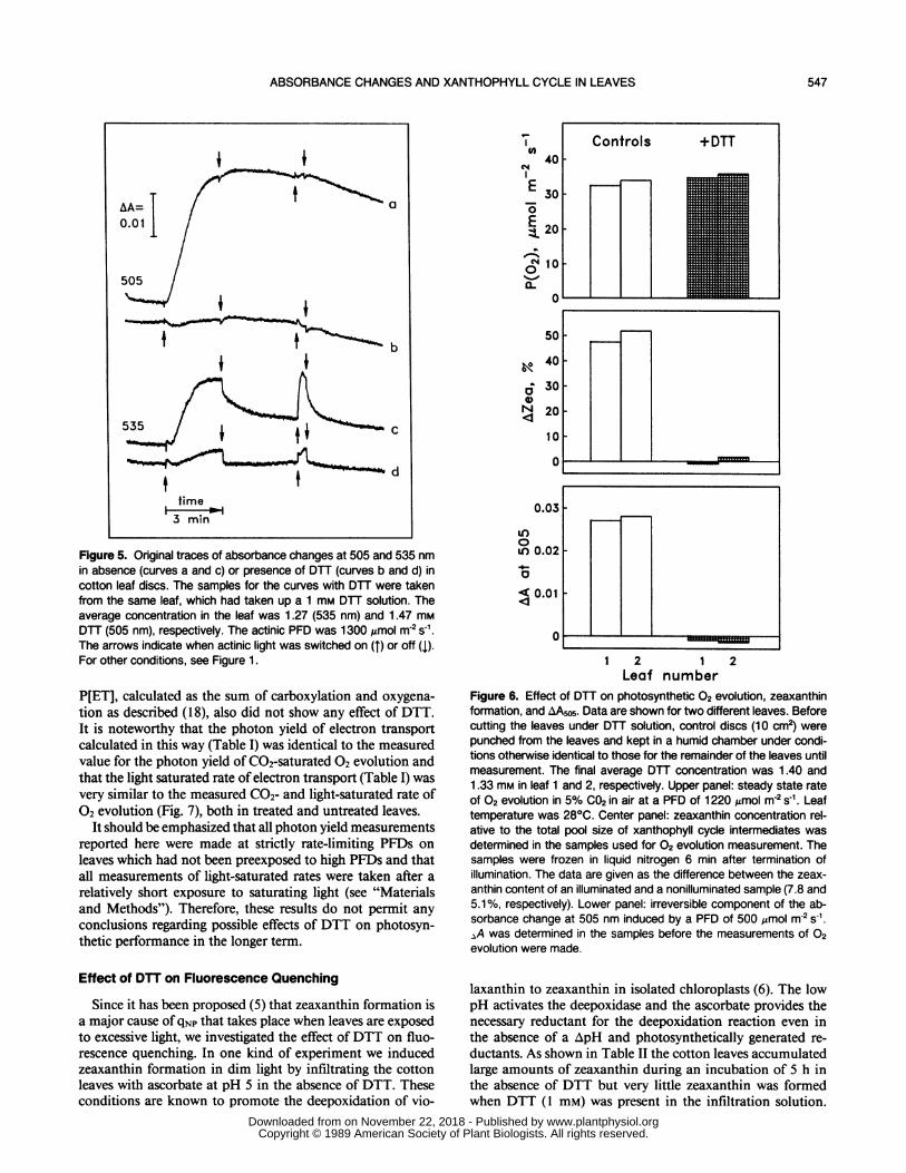
Application ofDTT via the cut petiole invariably preventedlight-induced zeaxanthin formation in cotton leaves (see be-low). When such DTT treated leaves were illuminated, theincrease in absorbance at 505 nm was completely inhibited(Fig. 5, trace b). Only a small absorbance change with rapidinduction and relaxation kinetics remained. DTT treatmentalso inhibited the irreversible component of the light-inducedabsorbance change at 535 nm (Fig. 5, trace d) and at otherwavelengths as well (data not shown). Although a small andslow rise at 535 nm still occurred upon illumination, theslowly reversible transient, usually taken as indicative of lightscattering changes due to membrane energization (11) had
0.06 A
<0.04~ ~~~~BB
8 min
0.02
A .10
440 460 480 500 520 540Wavelength, nm
Figure 3. Difference spectra for light-induced absorbance changesin cotton leaf discs, determined after 4 min of a subsequent darkperiod. The inset, with a trace measured at 535 nm, shows how thereversible (A) and the irreversible (B) component were obtained. TheAA scale also applies to the inset. For the spectrum, a new disc wasused at each wavelength. Actinic PFD was 500 ,mol m-2 S-1 . For otherconditions, see Figure 1.
O.Of
E0.0!E o.osc
e o.o0
.41- 0.0O.
0
< 0.0o
0.0
A6 "/
0'
2 A.0-0 20 3
- Z-10 20 30Z, ,0
10 20 30 40Z+A, %
50 60
Figure 4. Relationship between and amount of zeaxanthin (A) andthe sum of zeaxanthin and antheraxanthin (B) formed during illumi-nation at 500 ,umol photons in-2 s' and during the subsequent darkperod. In addition to the data shown already in a different way inFigure 2 (closed symbols), data from another experiment with a
different leaf are shown (open symbols). Pigment contents are givenin percent of the total xanthophyll cycle pool size (V + A + Z). Themeasuring conditions were the same as in Figure 2.
almost completely disappeared (Fig. 5, trace d). The rapidreversal had approximately the same amplitude as the slowrise. This suggests that the slow rise was caused by a slowbuildup of an electric membrane potential. A similar slowrise can be seen at 505 nm (Fig. 5, trace b), although theamplitude is smaller [cf (24)]. However, the kinetics at 505nm are more complex and obviously composed of the elec-trochromic changes and an additional fourth component. Thelatter displays a decline in the light during the first minuteand may reflect the same process as the relatively rapid riseafter light off, which accounts for most of the kinetics afterlight off in the control as well.
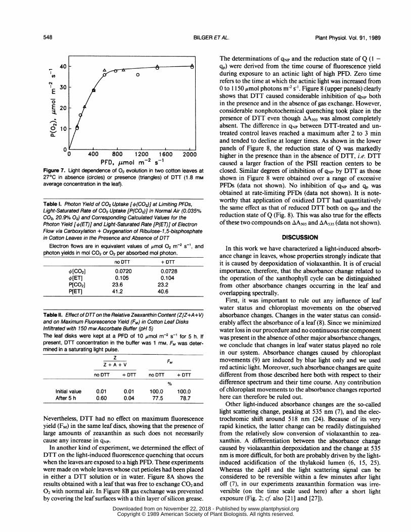
Effect of DTT on Photosynthesis
In spite of the inhibitory effect of DTT treatment on
zeaxanthin formation and on the associated AA505 as well as
on AA535 (Fig. 6, lower panels), such treatments had no effecton the rate of C02-saturated 02 evolution determined on thesame leaves (Fig. 6, upper panel). Moreover, the response ofC02-saturated O2 evolution to increased PFD in the presenceofDTT was indistinguishable from that in the absence of thiscompound (Fig. 7). The DTT treated leaf used in this exper-iment had taken up a relatively high amount of DTT andAA505 was completely eliminated (data not shown). Othermeasurements confirmed that the photon yield of 02 evolu-tion (Oa), determined at limiting PFDs was unaffected byDTT. 4a values of 0.104 ± 0.004 02 photon-' (n = 3) were
obtained for DTT treated leaves compared with 0.105 ± 0.00402 photon-' for untreated leaves [cf. (18)]. No zeaxanthin wasfound in control leaves kept in the range where the rate ofphotosynthesis was linear with PFD.As shown in Table I, measurements of photosynthetic CO2
uptake in air of normal atmospheric CO2 and 02 concentra-tion also did not show any detectable effect on photosynthesisof DTT treatments that fully eliminated AA5o5 and stronglysuppressed light-induced absorbance changes at 535 nm. Nei-ther the photon yield at limiting PFDs nor the light-saturatedrate of CO2 uptake was affected by DTT. The electron flow
B0
,,S0R'
,0 I
I,/If.
I-J '-.
I
546 BILGER ETAL.
www.plantphysiol.orgon November 22, 2018 - Published by Downloaded from Copyright © 1989 American Society of Plant Biologists. All rights reserved.
ABSORBANCE CHANGES AND XANTHOPHYLL CYCLE IN LEAVES
Figure 5. Original traces of absorbance changes at 505 and 535 nmin absence (curves a and c) or presence of DTT (curves b and d) incotton leaf discs. The samples for the curves with DTT were takenfrom the same leaf, which had taken up a 1 mm DTT solution. Theaverage concentration in the leaf was 1.27 (535 nm) and 1.47 mmDTT (505 nm), respectively. The actinic PFD was 1300 Amol m-2 s-1.The arrows indicate when actinic light was switched on (T) or off (1).For other conditions, see Figure 1.
P[ET], calculated as the sum of carboxylation and oxygena-tion as described (18), also did not show any effect of DTT.It is noteworthy that the photon yield of electron transportcalculated in this way (Table I) was identical to the measuredvalue for the photon yield of C02-saturated 02 evolution andthat the light saturated rate of electron transport (Table I) wasvery similar to the measured C02- and light-saturated rate of02 evolution (Fig. 7), both in treated and untreated leaves.
It should be emphasized that all photon yield measurementsreported here were made at strictly rate-limiting PFDs onleaves which had not been preexposed to high PFDs and thatall measurements of light-saturated rates were taken after arelatively short exposure to saturating light (see "Materialsand Methods"). Therefore, these results do not permit anyconclusions regarding possible effects of DTT on photosyn-thetic performance in the longer term.
Effect of DTT on Fluorescence Quenching
Since it has been proposed (5) that zeaxanthin formation isa major cause of qNp that takes place when leaves are exposedto excessive light, we investigated the effect of DTT on fluo-rescence quenching. In one kind of experiment we inducedzeaxanthin formation in dim light by infiltrating the cottonleaves with ascorbate at pH 5 in the absence of DTT. Theseconditions are known to promote the deepoxidation of vio-
(040
E 30
EE 20
0 10
0_%.a.
a0N
4
0.c
LI)0LO0.C4-
.<0.c
io
00o--
I I0 -----
)3
O2===
1 2 1 2Leaf number
Figure 6. Effect of DTT on photosynthetic 02 evolution, zeaxanthinformation, and AAw5. Data are shown for two different leaves. Beforecutting the leaves under DTT solution, control discs (10 cm2) werepunched from the leaves and kept in a humid chamber under condi-tions otherwise identical to those for the remainder of the leaves untilmeasurement. The final average DTT concentration was 1.40 and1.33 mm in leaf 1 and 2, respectively. Upper panel: steady state rateOf 02 evolution in 5% C02 in air at a PFD of 1220 Amol m2 s-1. Leaftemperature was 280C. Center panel: zeaxanthin concentration rel-ative to the total pool size of xanthophyll cycle intermediates wasdetermined in the samples used for 02 evolution measurement. Thesamples were frozen in liquid nitrogen 6 min after termination ofillumination. The data are given as the difference between the zeax-anthin content of an illuminated and a nonilluminated sample (7.8 and5.1%, respectively). Lower panel: irreversible component of the ab-sorbance change at 505 nm induced by a PFD of 500 ,umol m-2 S-1.aA was determined in the samples before the measurements of 02evolution were made.
laxanthin to zeaxanthin in isolated chloroplasts (6). The lowpH activates the deepoxidase and the ascorbate provides thenecessary reductant for the deepoxidation reaction even inthe absence of a ApH and photosynthetically generated re-ductants. As shown in Table II the cotton leaves accumulatedlarge amounts of zeaxanthin during an incubation of 5 h inthe absence of DTT but very little zeaxanthin was formedwhen DTT (1 mM) was present in the infiltration solution.
547
www.plantphysiol.orgon November 22, 2018 - Published by Downloaded from Copyright © 1989 American Society of Plant Biologists. All rights reserved.
Plant Physiol. Vol. 91, 1989
400
I30E
E 20-
a.
p0 400 800 1200 1600 2000
PFD, ltmol m-2 s-1Figure 7. Light dependence of 02 evolution in two cotton leaves at270C in absence (circles) or presence (triangles) of DTT (1.8 mmaverage concentration in the leaf).
Table I. Photon Yield of CO2 Uptake [q5(CO2)J] at Limiting PFDs,Light-Saturated Rate of CO2 Uptake [P(CO2)] in Normal Air (0.035%C02, 20.9% 02) and Corresponding Calculated Values for thePhoton Yield [q5(ET)] and Light-Saturated Rate [P(ET)] of ElectronFlow via Carboxylation + Oxygenation of Ribulose-1,5-bisphosphatein Cotton Leaves in the Presence and Absence of DTT
Electron flows are in equivalent values of umol 02 m-2 s-1, andphoton yields in mol C02 or 02 per absorbed mol photon.
no DTT + DTT
1[CO2] 0.0720 0.07281[ET] 0.105 0.104P[C02] 23.6 23.2P[ET] 41.2 40.6
Table II. Effect of DTTon the Relative Zeaxanthin Content (Z/Z+A+V)and on Maximum Fluorescence Yield (FM) in Cotton Leaf DisksInfiltrated with 150 mMAscorbate Buffer (pH 5)The leaf disks were kept at a PFD of 10 ,umol m-2 S-1 for 5 h. Ifpresent, DTT concentration in the buffer was 1 mM. FM was deter-mined in a saturating light pulse.
zF
Z + A + V FM
no DTT + DTT no DTT + DTT
Initial value 0.01 0.01 100.0 100.0After 5 h 0.60 0.04 77.5 78.7
Nevertheless, DTT had no effect on maximum fluorescenceyield (FM) in the same leaf discs, showing that the presence oflarge amounts of zeaxanthin as such does not necessarilycause any increase in qNP.
In another kind of experiment, we determined the effect ofDTT on the light-induced fluorescence quenching that occurswhen the leaves are exposed to a high PFD. These experimentswere made on whole leaves whose cut petioles had been placedin either a DTT solution or in water. Figure 8A shows theresults obtained with a leaf that was free to exchange CO2 and02 with normal air. In Figure 8B gas exchange was preventedby covering the leaf surfaces with a thin layer of silicon grease.
The determinations of qNp and the reduction state ofQ (1 -qp) were derived from the time course of fluorescence yieldduring exposure to an actinic light of high PFD. Zero timerefers to the time at which the actinic light was increased from0 to 1150 ,mol photons m-2 s-'. Figure 8 (upper panels) clearlyshows that DTT caused considerable inhibition of qNp bothin the presence and in the absence of gas exchange. However,considerable nonphotochemical quenching took place in thepresence of DTT even though AA505 was almost completelyabsent. The difference in qNp between DTT-treated and un-treated control leaves reached a maximum after 2 to 3 minand tended to decline at longer times. As shown in the lowerpanels of Figure 8, the reduction state of Q was markedlyhigher in the presence than in the absence of DTT, i.e. DTTcaused a larger fraction of the PSII reaction centers to beclosed. Similar degrees of inhibition of qNp by DTT as thoseshown in Figure 8 were obtained over a range of excessivePFDs (data not shown). No inhibition of qNp and qp wasobtained at rate-limiting PFDs (data not shown). It is note-worthy that application of oxidized DTT had quantitativelythe same effect as that of reduced DTT both on qNp and thereduction state ofQ (Fig. 8). This was also true for the effectsofthese two compounds on lA505 and AA535 (data not shown).
DISCUSSION
In this work we have characterized a light-induced absorb-ance change in leaves, whose properties strongly indicate thatit is caused by deepoxidation of violaxanthin. It is of crucialimportance, therefore, that the absorbance change related tothe operation of the xanthophyll cycle can be distinguishedfrom other absorbance changes occurring in the leaf andoverlapping spectrally.
First, it was important to rule out any influence of leafwater status and chloroplast movements on the observedabsorbance changes. Changes in the water status can consid-erably affect the absorbance of a leaf (8). Since we minimizedwater loss in our procedure and no continuous rise componentwas present in the absence ofother major absorbance changes,we conclude that changes in leaf water status played no rolein our system. Absorbance changes caused by chloroplastmovements (9) are induced by blue light only and we usedred actinic light. Moreover, such absorbance changes are quitedifferent from those described here both with respect to theirdifference spectrum and their time course. Any contributionof chloroplast movements to the absorbance changes reportedhere can therefore be ruled out.Other light-induced absorbance changes are the so-called
light scattering change, peaking at 535 nm (7), and the elec-trochromic shift around 518 nm (24). Because of its veryrapid kinetics, the latter change can be readily distinguishedfrom the relatively slow conversion of violaxanthin to zea-xanthin. A differentiation between the absorbance changecaused by violaxanthin deepoxidation and the change at 535nm is more difficult, for both are probably driven by the light-induced acidification of the thylakoid lumen (6, 15, 25).Whereas the ApH and the light scattering signal can beconsidered to be reversible within a few minutes after lightoff (7), in our experiments zeaxanthin formation was irre-versible (on the time scale used here) after a short lightexposure (Fig. 2; cf also [21 ] and [27]).
548 BILGER ETAL.
www.plantphysiol.orgon November 22, 2018 - Published by Downloaded from Copyright © 1989 American Society of Plant Biologists. All rights reserved.

ABSORBANCE CHANGES AND XANTHOPHYLL CYCLE IN LEAVES
1.0
0.8
z 0.6
0.4
0
1.0
0.8
n.
0- 0.6
0.4
0.2[
0 2 4 6Time, min
8 10Time
The change in absorbance still present after a dark periodof 4 min has two other properties which indicate that it iscaused by violaxanthin deepoxidation. First, its differencespectrum (Fig. 3) is completely different from spectra forother known absorbance changes, but very similar to thatreported in Yamamoto et al. (26) for violaxanthin deepoxi-dation in isolated chloroplasts. Its peaks are located at 505and 465 nm, its troughs at 480 and 455 nm, while Yamamotoet al. (26) observed peaks at 505 and 468 nm and troughs at482 and 450 nm in the respective wavelength band. There isonly one apparent difference between the spectrum deter-mined by Yamamoto et al. and that determined by us: we
invarably found an additional shoulder, or sometimes a peak,around 515 nm. However, Yamamoto et al. did report a shiftof the maximum from 505 nm to longer wavelengths andtentatively ascribed it to overlapping changes in the absorb-ance by Chl. Alternatively, the shoulder at 515 nm could beexplained by the occurrence of selective scattering (14, 23)caused by the appearance of a new absorption band.The second major evidence for ascribing the changes at 505
nm to violaxanthin deepoxidation is the complete suppressionof the irreversible component by DTT. DTT is a powerfulinhibitor of violaxanthin deepoxidation in chloroplasts (27),etiolated leaves (16), and green leaves. Although it is likelythat DTT does not affect only the xanthophyll cycle (see Fig.5 and comments below), its action still seems quite specific,since photosynthesis was totally unaffected by DTT.Although the spectral change caused by violaxanthin dee-
poxidation is large (Figs. and 3) and also makes an importantcontribution to the total absorbance change observed at 535nm (Fig. 3), it has not been previously reported for intactleaves. To obtain a maximal response it was important thatthe leaf had been kept under conditions that favor a highepoxidation state (weak light or darkness). It is noteworthy
_ Figure 8. Nonphotochemical (qNp) and
A P% photochemical (expressed as 1 - qp) fluo-*6-- -0 rescence quenching in cotton leaves after+DTT DTT treatment. Three different leaves of
the same plant were allowed to take uppure water (control) or a 1 mm solution ofeither reduced (closed circles) or oxidizedDTT (open circles) for 4 h at a low PFD (10Amol m2s-1). Actinic PFD was 1150 ,umolm 2 s-'. Panel B shows the result obtainedwhen different spots of the same leaveswere illuminated while gas exchange wasprevented by application of silicon grease.When AA5o5 was determined under the
+DTT same conditions as in Figure 1 on the sameleaves, the irreversible component was
--- .J suppressed by at least 97% in the leavestreated with reduced or oxidized DTT, com-pared with the control.
6 8 10e, min
that the cotton leaves used in our experiments also had a largepool of xanthophyll cycle components (violaxanthin + an-theraxanthin + zeaxanthin) (22).At 505 nm a relatively large absorbance change was ob-
tained, even for small variations in the deepoxidation state(Fig. 4). At this wavelength the relative contribution fromother processes was small (Fig. 5), so that AA505 could be useddirectly to estimate the deepoxidation state. However, toobtain quantitative data, calibration for each leaf may beneeded since the apparent extinction coefficient may varyfrom leaf to leaf. It still remains to be determined if AA505 iScaused by the difference in absorbance between violaxanthinand zeaxanthin alone or between violaxanthin and zeaxanthinplus antheraxanthin.We found that the inhibitory effects of supplying oxidized
DTT were indistinguishable from the effects of reduced DTTon qNp, qp, AA505 (Fig. 8) and AA535. This finding is in contrastto the effects of the two forms of DTT on the isolatedviolaxanthin deepoxidase, where the reduced form acts asvery potent inhibitor while the oxidized form has no effect(see "Materials and Methods"). A simple explanation for theeffectiveness of exogenously applied oxidized DTT as aninhibitor of violaxanthin deepoxidation in vivo would be thatit is reduced to DTT in the leaf. However, we do not haveany direct evidence that such a reduction takes place.
In addition to inhibition ofviolaxanthin deepoxidation andassociated light-induced absorbance changes, DTT inhibitedthe reversible component of the 535 nm absorbance changewhich is unlikely to be caused by violaxanthin deepoxidation(Fig. 5). The 535 nm absorbance change has been thought tobe caused by altered light scattering properties of the chloro-plasts due to conformational changes (7, 15) induced by theacidification of the thylakoid lumen. Our observations wouldsupport the view that the reversible component of AA535 iS
- A
Control
B-
+DTT
Il
+DTT
Control
a s z | x X wX l l- --
549
www.plantphysiol.orgon November 22, 2018 - Published by Downloaded from Copyright © 1989 American Society of Plant Biologists. All rights reserved.
Plant Physiol. Vol. 91, 1989
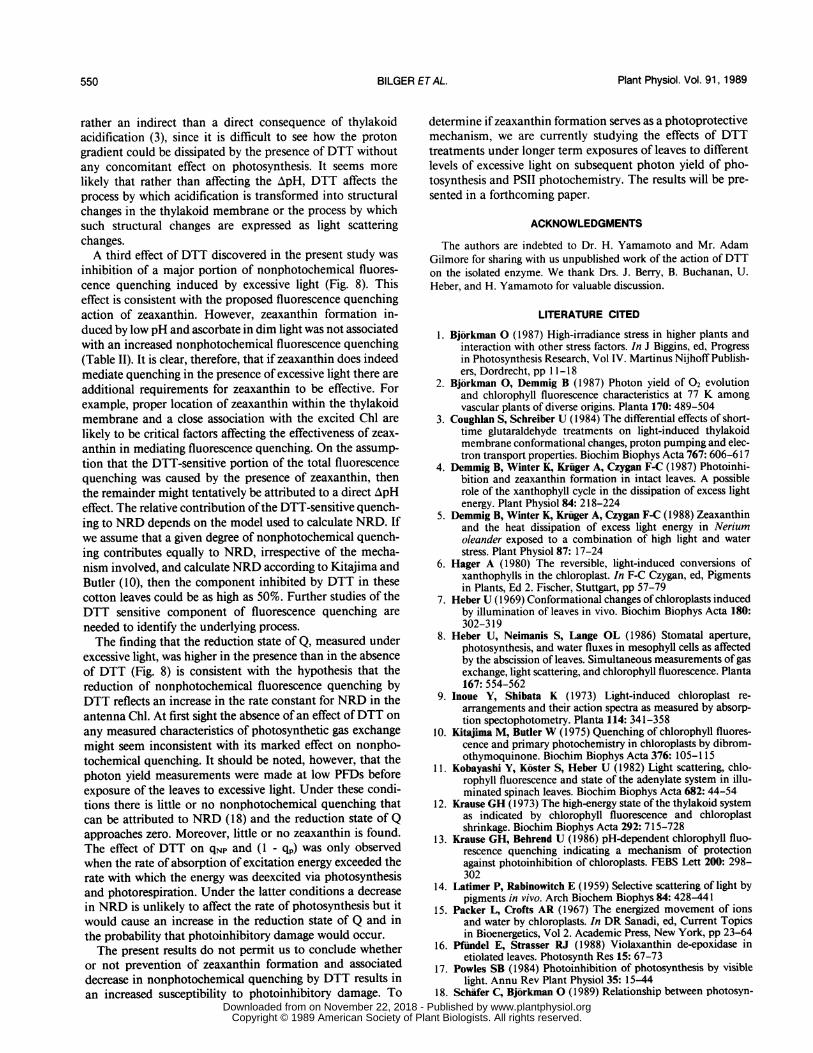
rather an indirect than a direct consequence of thylakoidacidification (3), since it is difficult to see how the protongradient could be dissipated by the presence of DTT withoutany concomitant effect on photosynthesis. It seems more
likely that rather than affecting the zApH, DTT affects theprocess by which acidification is transformed into structuralchanges in the thylakoid membrane or the process by whichsuch structural changes are expressed as light scatteringchanges.A third effect of DTT discovered in the present study was
inhibition of a major portion of nonphotochemical fluores-cence quenching induced by excessive light (Fig. 8). Thiseffect is consistent with the proposed fluorescence quenchingaction of zeaxanthin. However, zeaxanthin formation in-duced by low pH and ascorbate in dim light was not associatedwith an increased nonphotochemical fluorescence quenching(Table II). It is clear, therefore, that if zeaxanthin does indeedmediate quenching in the presence of excessive light there are
additional requirements for zeaxanthin to be effective. Forexample, proper location of zeaxanthin within the thylakoidmembrane and a close association with the excited Chl are
likely to be critical factors affecting the effectiveness of zeax-
anthin in mediating fluorescence quenching. On the assump-tion that the DTT-sensitive portion of the total fluorescencequenching was caused by the presence of zeaxanthin, thenthe remainder might tentatively be attributed to a direct ApHeffect. The relative contribution of the DTT-sensitive quench-ing to NRD depends on the model used to calculate NRD. Ifwe assume that a given degree of nonphotochemical quench-ing contributes equally to NRD, irrespective of the mecha-nism involved, and calculate NRD according to Kitajima andButler (10), then the component inhibited by DTT in thesecotton leaves could be as high as 50%. Further studies of theDTT sensitive component of fluorescence quenching are
needed to identify the underlying process.The finding that the reduction state of Q, measured under
excessive light, was higher in the presence than in the absenceof DTT (Fig. 8) is consistent with the hypothesis that the
reduction of nonphotochemical fluorescence quenching byDTT reflects an increase in the rate constant for NRD in theantenna Chl. At first sight the absence of an effect ofDTT on
any measured characteristics of photosynthetic gas exchangemight seem inconsistent with its marked effect on nonpho-tochemical quenching. It should be noted, however, that the
photon yield measurements were made at low PFDs before
exposure of the leaves to excessive light. Under these condi-
tions there is little or no nonphotochemical quenching that
can be attributed to NRD (18) and the reduction state of Qapproaches zero. Moreover, little or no zeaxanthin is found.
The effect of DTT on qNp and (1 - qp) was only observedwhen the rate of absorption of excitation energy exceeded therate with which the energy was deexcited via photosynthesisand photorespiration. Under the latter conditions a decreasein NRD is unlikely to affect the rate of photosynthesis but it
would cause an increase in the reduction state of Q and in
the probability that photoinhibitory damage would occur.
The present results do not permit us to conclude whether
or not prevention of zeaxanthin formation and associated
decrease in nonphotochemical quenching by DTT results in
an increased susceptibility to photoinhibitory damage. To
determine if zeaxanthin formation serves as a photoprotectivemechanism, we are currently studying the effects of DTTtreatments under longer term exposures of leaves to differentlevels of excessive light on subsequent photon yield of pho-tosynthesis and PSII photochemistry. The results will be pre-sented in a forthcoming paper.
ACKNOWLEDGMENTS
The authors are indebted to Dr. H. Yamamoto and Mr. AdamGilmore for sharing with us unpublished work of the action of DTTon the isolated enzyme. We thank Drs. J. Berry, B. Buchanan, U.Heber, and H. Yamamoto for valuable discussion.
LITERATURE CITED
1. Bjorkman 0 (1987) High-irradiance stress in higher plants andinteraction with other stress factors. In J Biggins, ed, Progressin Photosynthesis Research, Vol IV. Martinus Nijhoff Publish-ers, Dordrecht, pp 11- 18
2. Bjorkman 0, Demmig B (1987) Photon yield of 02 evolutionand chlorophyll fluorescence characteristics at 77 K amongvascular plants of diverse origins. Planta 170: 489-504
3. Coughlan S, Schreiber U (1984) The differential effects of short-time glutaraldehyde treatments on light-induced thylakoidmembrane conformational changes, proton pumping and elec-tron transport properties. Biochim Biophys Acta 767:606-617
4. Demmig B, Winter K, Kruger A, Czygan F-C (1987) Photoinhi-bition and zeaxanthin formation in intact leaves. A possiblerole of the xanthophyll cycle in the dissipation of excess lightenergy. Plant Physiol 84: 218-224
5. Demmig B, Winter K, Kruger A, Czygan F-C (1988) Zeaxanthinand the heat dissipation of excess light energy in Neriumoleander exposed to a combination of high light and waterstress. Plant Physiol 87: 17-24
6. Hager A (1980) The reversible, light-induced conversions ofxanthophylls in the chloroplast. In F-C Czygan, ed, Pigmentsin Plants, Ed 2. Fischer, Stuttgart, pp 57-79
7. Heber U (1969) Conformational changes of chloroplasts inducedby illumination of leaves in vivo. Biochim Biophys Acta 180:302-319
8. Heber U, Neimanis S, Lange OL (1986) Stomatal aperture,photosynthesis, and water fluxes in mesophyll cells as affectedby the abscission of leaves. Simultaneous measurements of gasexchange, light scattering, and chlorophyll fluorescence. Planta167: 554-562
9. Inoue Y, Shibata K (1973) Light-induced chloroplast re-
arrangements and their action spectra as measured by absorp-tion spectophotometry. Planta 114: 341-358
10. Kitajima M, Butler W (1975) Quenching of chlorophyll fluores-cence and primary photochemistry in chloroplasts by dibrom-othymoquinone. Biochim Biophys Acta 376: 105-115
11. Kobayashi Y, Koster S, Heber U (1982) Light scattering, chlo-rophyll fluorescence and state of the adenylate system in illu-minated spinach leaves. Biochim Biophys Acta 682: 44-54
12. Krause GH (1973) The high-energy state of the thylakoid systemas indicated by chlorophyll fluorescence and chloroplastshrinkage. Biochim Biophys Acta 292: 715-728
13. Krause GH, Behrend U (1986) pH-dependent chlorophyll fluo-rescence quenching indicating a mechanism of protectionagainst photoinhibition of chloroplasts. FEBS Lett 200: 298-302
14. Latimer P, Rabinowitch E (1959) Selective scattering of light bypigments in vivo. Arch Biochem Biophys 84: 428-441
15. Packer L, Crofts AR (1967) The energized movement of ionsand water by chloroplasts. In DR Sanadi, ed, Current Topicsin Bioenergetics, Vol 2. Academic Press, New York, pp 23-64
16. Pfiindel E, Strasser RJ (1988) Violaxanthin de-epoxidase inetiolated leaves. Photosynth Res 15: 67-73
17. Powles SB (1984) Photoinhibition of photosynthesis by visiblelight. Annu Rev Plant Physiol 35: 15-44
18. Schafer C, Bjorkman 0 (1989) Relationship between photosyn-
550 BILGER ETAL.
www.plantphysiol.orgon November 22, 2018 - Published by Downloaded from Copyright © 1989 American Society of Plant Biologists. All rights reserved.

ABSORBANCE CHANGES AND XANTHOPHYLL CYCLE IN LEAVES
thetic energy conversion efficiency and chlorophyll fluores-cence quenching in upland cotton (Gossypium hirsutum L.).Planta 178: 367-376
19. Schreiber U, Schliwa U, Bilger W (1986) Continuous recordingof photochemical and non-photochemical chlorophyll fluores-cence quenching with a new type of modulation fluorometer.Photosynth Res 10: 51-62
20. Siefermann-Harms D (1985) Carotenoids in photosynthesis. I.Location in photosynthetic membranes and light-harvestingfunction. Biochim Biophys Acta 811: 325-355
21. Sokolove PM, Marsho TV (1976) Ascorbate-independent carot-enoid de-epoxidation in intact spinach chloroplasts. BiochimBiophys Acta 430: 321-326
22. Thayer SS, Bjorkman 0 (1989) Leaf xanthophyll content andcomposition in sun and shade as determined by HPLC. Pho-tosynth Res (in press)
23. Thorne SW, Horvath G, Kahn A, Boardman NK (1975) Light-dependent absorption and selective scattering changes at 518nm in chloroplast thylakoid membranes. Proc Natl Acad SciUSA 72: 3858-3862
24. Wift HT (1979) Energy conversion in the functional membraneof photosynthesis. Analysis by light pulse and electric pulsemethods. The central role ofthe electric field. Biochim BiophysActa 505: 355-427
25. Yamamoto HY (1979) Biochemistry of the violaxanthin cycle inhigher plants. Pure Appl Chem 51: 639-648
26. Yamamoto HY, Kamite L, Wang Y-Y (1972) An ascorbate-induced absorbance change in chloroplasts from violaxanthinde-epoxidation. Plant Physiol 49: 224-228
27. Yamamoto HY, Kamite L (1972) The effects of dithiothreitol onviolaxanthin de-epoxidation and absorbance changes in the500-nm region. Biochim Biophys Acta 267: 538-543
551
www.plantphysiol.orgon November 22, 2018 - Published by Downloaded from Copyright © 1989 American Society of Plant Biologists. All rights reserved.