Embed Size (px)
Citation preview
MOL #99671
1
Ligand residence time at GPCRs – why we should take our time to study it
C. Hoffmann, M. Castro, A. Rinken, R. Leurs, S.J. Hill, H.F. Vischer
Bio-Imaging-Center/Rudolf-Virchow-Zentrum and Institute of Pharmacology and Toxicology,
University of Würzburg, Würzburg, GERMANY (CH)
Molecular Pharmacology Laboratory. Biofarma Research Group (GI-1685). University of Santiago de
Compostela. Center for Research in Molecular Medicine and Chronic Diseases (CIMUS), 15782
Santiago de Compostela, SPAIN (MC)
Institute of Chemistry, University of Tartu, Tartu, ESTONIA (AR)
Amsterdam Institute for Molecules Medicines and Systems, Division of Medicinal Chemistry, Faculty
of Sciences, VU University, Amsterdam, De Boelelaan 1083, 1081 HV Amsterdam, THE
NETHERLANDS (RL, HFV)
Cell Signalling Research Group, School of Life Sciences, Medical School, Queen’s Medical Centre,
University of Nottingham, Nottingham, UNITED KINGDOM (SJH)
This article has not been copyedited and formatted. The final version may differ from this version.Molecular Pharmacology Fast Forward. Published on July 7, 2015 as DOI: 10.1124/mol.115.099671
at ASPE
T Journals on M
ay 20, 2018m
olpharm.aspetjournals.org
Dow
nloaded from
MOL #99671
2
Running Title: residence time at GPCRs
Corresponding author:
Carsten Hoffmann
Bio-Imaging-Center/ Rudolf-Virchow-Zentrum and
Department of Pharmacology and Toxicology
Universty of Wuerzburg
Versbacher Strasse 9
97078 Würzburg
Germany
Text pages: 14
Table: 1
Figure: 1
References: 92
Abstract word count: 158
Introduction word count: 446
Discussion word count: 4457
Abbreviations:
FCS - fluorescence correlation spectroscopy; FRET - fluorescence resonance energy transfer; GLP -
glucagon-like peptide; GPCR - G-protein-coupled receptor; GTP – guanosine triphosphate; IC50 -
inhibitor concentration that inhibits response by 50%; MAPK - mitogen-activated protein kinase;
PASMC – pulmonary artery smooth muscle cells; PTH - parathormone; QCM – quartz crystal
microbalance; SPR – surface plasmon resonance; SNAP-tag - previously known as genetically
modified AGT: O6-alkylguanine-DNA alkyltransferase; TSH - thyroid-stimulating hormone;
This article has not been copyedited and formatted. The final version may differ from this version.Molecular Pharmacology Fast Forward. Published on July 7, 2015 as DOI: 10.1124/mol.115.099671
at ASPE
T Journals on M
ay 20, 2018m
olpharm.aspetjournals.org
Dow
nloaded from
MOL #99671
3
Abstract
Over the past decade the kinetics of ligand binding to a receptor have received increasing interest. The
concept of drug-target residence time is becoming an invaluable parameter for drug optimization. It
holds great promise for drug-development and its optimization is thought to reduce off-target effects.
The success of long-acting drugs like tiotropium support this hypothesis. Nonetheless, we know
surprisingly little about the dynamics and the molecular detail of the drug binding process. Since
protein dynamics and adaptation during the binding event will change the conformation of the protein,
ligand binding will not be the static process that is often described. This can cause problems since
simple mathematical models often fail to adequately describe the dynamics of the binding process. In
this perspective we will discuss the current situation with an emphasis on GPCRs. This are important
membrane protein drug-targets that undergo conformational changes upon agonist binding in order to
communicate signalling information across the plasma membrane of cells.
This article has not been copyedited and formatted. The final version may differ from this version.Molecular Pharmacology Fast Forward. Published on July 7, 2015 as DOI: 10.1124/mol.115.099671
at ASPE
T Journals on M
ay 20, 2018m
olpharm.aspetjournals.org
Dow
nloaded from
MOL #99671
4
Introduction
G-protein-coupled receptors (GPCRs) represent attractive pharmacological targets. A long tradition of
research in this field has led to the development of several successful drug-classes that make up
almost 30% of all marketed drugs. Such drugs can interfere with a given GPCR by binding to the
receptor and preventing the binding of the endogenous ligand (in which case they are known as
antagonists) or they can mimic the endogenous ligand and stimulate a functional response (in which
case they are agonists). Such simple views are currently still found in many pharmacological
textbooks, and although this simplicity helps to teach beginners the basic principles of receptor
pharmacology, we know that this view is overly simplified. Within the past 20 years or so, we have
witnessed a dramatic increase in our knowledge of how GPCRs function. We have learned that
GPCRs can undergo different conformational changes when different ligands bind to the same
receptor (Nygaard et al., 2013). We have seen a tremendous community-wide effort on GPCR
crystallization achieve substantial success, and to date more than 100 x-ray structures of 28 different
GPCR have become available to the public (Shonberg et al., 2015). It is likely that many more are
available within commercial research groups. We have also learned recently that receptor
internalization does not necessarily stop a GPCR from being able to continuously signal from the
inside of a cell (Irannejad et al., 2013). Nonetheless, our current knowledge of the earliest steps
involved in ligand binding appears to be very rudimentary (Pan et al., 2013), especially when
compared to other aspects of GPCR biology. Ligand binding to a given receptor protein is a dynamic
process and is not distinct in its general rules from enzymes, ligand-gated ion-channels or other
GPCRs (Colquhoun, 1998; Colquhoun, 2006). Although we can learn a lot from equilibrium binding
assays and determine ligand binding affinities, the underlying constant flux in ligand-binding (on-rate)
and unbinding (off-rate) of a ligand has been largely ignored even though this can have a significant
influence in vivo, where equilibrium conditions are rarely achieved.
In this short perspective article we briefly introduce the term ligand residence time and briefly discuss
the currently available assays that are used to study ligand residence time for GPCRs. We will also
critically discuss some known shortcomings and limitations of such assays. Furthermore, we will
discuss recent technical advances that might provide insight into the molecular determinants of ligand
This article has not been copyedited and formatted. The final version may differ from this version.Molecular Pharmacology Fast Forward. Published on July 7, 2015 as DOI: 10.1124/mol.115.099671
at ASPE
T Journals on M
ay 20, 2018m
olpharm.aspetjournals.org
Dow
nloaded from
MOL #99671
5
binding and contribute to a more systematic evaluation of ligand residence time in the future. Finally,
we will outline the potential influence of different ligand residence times on GPCR signalling and
name successful examples were optimization of ligand residence time has improved drug performance
in patients.
The concept of ligand residence time at GPCRs
Within the last ten years several excellent reviews have appeared on the general topic of drug-target
residence time, each covering the topic from a different perspective (Copeland et al., 2006; Tummino
and Copeland, 2008; Lu and Tonge, 2010; Dahl and Akerud, 2013, Vauquelin and Charlton, 2013;
Guo et al., 2014 to name a few), and have highlighted this parameter for drug-discovery. To keep this
perspective article focussed, we will refer the reader to those articles or the recently published book
“Thermodynamics and Kinetics of Drug Binding” (ed. Keserü and Swinney, 2015) for an in-depth
discussion, and we will focus on the concept of residence time at GPCRs.
The signal transduction cascade that is mediated by a GPCR is initiated by the binding of agonist to
the receptor. The newly formed agonist-receptor complex generates a signal in the given cell, and the
lifetime of this complex has a big impact on the efficiency of signal transduction. As a consequence
drug-target residence time has become an important parameter for drug discovery, alongside classical
affinity parameters such as IC50- and Ki-values (Copeland et al., 2006). In an in vivo system, the
residence time becomes crucial if the pharmacokinetic drug elimination is faster than its dissociation
from the receptor complex (Dahl and Akerud, 2013). In this case the residence time directly depends
on the dissociation rate of the drug from its complex with the receptor. The detection of dissociation
rates initially looks straight forward since most often ligand receptor interactions are illustrated in
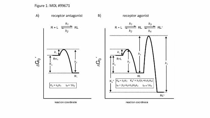
terms of structurally static binding and dissociation events. This is schematically depicted in figure 1A
for the case of an antagonist binding to a GPCR, assuming there is no conformational change
occurring. Since the ligand dissociation-rate constant (k2 , also termed koff) is inversely proportional
to drug residence time (1/koff), the residence time can be experimentally determined by measuring
ligand dissociation-rate constants (Copeland, 2011). However, it has become evident that such
descriptions are inadequate to explain the impact of conformational dynamics on this process
This article has not been copyedited and formatted. The final version may differ from this version.Molecular Pharmacology Fast Forward. Published on July 7, 2015 as DOI: 10.1124/mol.115.099671
at ASPE
T Journals on M
ay 20, 2018m
olpharm.aspetjournals.org
Dow
nloaded from
MOL #99671
6
(Copeland, 2011). It was recently shown that the dynamic of a protein can greatly influence ligand
dissociation (Teague, 2003; Carrol et al., 2012) or in other words conformational adaptation of the
receptor can greatly influence the residence time of a ligand on its receptor, or a drug on its target.
This is schematically depicted in figure 1B for the case of an agonist of a GPCR. If the dissociation
rate constant k4 is small compared to the dissociation rate constant k2 of the inactive receptor complex,
then the active complex will be stable and the ligand residence time will be determined largely by k4.
In case of a GPCR, receptor activation generates a high affinity agonist complex. This phenomenon
can sometimes be observed in binding experiments as a high affinity state, which can be eliminated by
the addition of GTP. The active receptor complex can thus extend significantly the drug’s residence
time (Copeland et al., 2006) and this situation becomes more complex if, in addition to an ortosteric
ligand, allosteric regulators are also present (May et al., 2011; Corriden et al., 2014; Christopoulos,
2014).
State of the art: currently used assays
Radioligand binding assays
As stated above, the ligand dissociation-rate constant (koff) is inversely proportional to drug residence
time (1/koff) and can be experimentally determined by measuring ligand dissociation-rate constants
(Copeland, 2011). The determination of the residence time of a drug in complex with a receptor
became possible only after the development of radioligand binding assays (Paton and Rang, 1965).
Until recently, this was the major method available to assess ligand binding directly, and is still the
most frequently used assay format (see table 1). This approach allows the direct detection of on- and
off-rates for a high-affinity radioligand. This method, however, predominantly gives information
about the labelled ligand itself, although this has been invaluable in the study allosteric regulatory
effects (De Amici et al., 2010). The technique has several limitations since the radioligand-binding
assay requires separation of the bound ligand from the free ligand fraction, and the binding itself may
have several steps. If we are interested in a non-labelled competing ligand, then the situation is more
complex. In such cases, only a fraction of the receptor-ligand complexes might be detected if the
radioligand and test compound do not bind to the same receptor conformational state. Different assay
This article has not been copyedited and formatted. The final version may differ from this version.Molecular Pharmacology Fast Forward. Published on July 7, 2015 as DOI: 10.1124/mol.115.099671
at ASPE
T Journals on M
ay 20, 2018m
olpharm.aspetjournals.org
Dow
nloaded from
MOL #99671
7
formats for competition binding are available that allow radioligand and competitor kinetic binding
constants (e.g. kon and koff) to be determined (Guo et al., 2014). The relative strengths and weaknesses
of each procedure has been described previously (Guo et al. 2014). Nonetheless, if the compound of
interest itself is not labelled, only indirect information of its residence time will be acquired. In
addition, non-homogeneity of this assay system complicates the interpretation of the results obtained.
Further problems might arise if the radiolabelled ligand cannot easily be removed during the assay. In
these cases the phenomenon of rebinding can occur, and this can complicate the determination of koff-
values (Vauquelin and Charlton, 2010). Furthermore, such assays currently ignore conformational
dynamics and hence, are largely unsuitable for GPCR receptor agonists that induce a conformational
change in the receptor. This problem also extends to data fitting procedures, since often researchers
use very simple kinetic models to fit the data that do not account for such details (Motulsky and
Mahan, 1984). Furthermore, radioligand binding assays are often conducted on ice and in non-
physiological buffer conditions and it has been shown that 10-fold shorter residence times of
tiotropium at the M3 acetylcholine receptor are obtained under physiological conditions (Sykes et al.,
2012).
Label-free approaches (see below) have also been used to study ligand protein interaction kinetics
using purified proteins and immobilisation strategies. This has the advantage of using known and
well-characterised proteins, but comes at the cost of the need for detergents and non-native
membranes. For the β2-adrenergic receptor, the local membrane environment has been demonstrated
recently to have a significant influence on receptor-ligand interactions and it has been recommended
that residence time measurements should be conducted using conditions which are as close as possible
to the natural conditions, for example using whole cell experiments or even tissue slices (Sykes et al.,
2014). Notwithstanding the difficulties mentioned above, the concept of residence time optimization
of GPCR ligands has led to the development of antagonist drugs with long-residence times such as
tiotropium (Tautermann et al., 2013). Even for agonists, a positive correlation between agonist
efficacy and residence time has been observed in case of the M3 acetylcholine receptor (Sykes et al.,
2009) and at the adenosine A2A receptor (Guo et al., 2012), although no such correlation was
observed for the adenosine A1 receptor (Louvel et al. 2014).
This article has not been copyedited and formatted. The final version may differ from this version.Molecular Pharmacology Fast Forward. Published on July 7, 2015 as DOI: 10.1124/mol.115.099671
at ASPE
T Journals on M
ay 20, 2018m
olpharm.aspetjournals.org
Dow
nloaded from
MOL #99671
8
Surface plasmon resonance analysis
An alternative biophysical approach that is frequently used to determine kinetic ligand binding in drug
discovery is represented by surface plasmon resonance (SPR) analysis (see table 1). This method can
be considered as a label free method with respect to the ligand. In order to generate a plasmon,
polarized light is directed via a prism onto a gold-coated glass surface on which the sample is bound.
The refractive index of the medium near the gold surface is a major parameter that influences the
critical angle of the polarized light. If the refractive index changes for example during the formation
of the ligand-receptor complex a signal will be detected due to a shift in the critical angle. This
relationship is used to analyse the dynamics of ligand binding. Due to recent technical advancements,
this technique is now capable of detecting the binding of molecules as small as 200 Da (Aristotelous
et al., 2015) and is now well suited to investigate GPCR ligands. The application of this approach to
GPCRs has recently been reviewed (Aristotelous et al., 2015). Currently six GPCRs have been
investigated using this approach (rhodopsin, CXCR4, CCR5, adenosine A2A receptor, β1- and β2-
adrenergic receptors; Aristotelous et al., 2015). One major drawback is the need to use purified
proteins and the purification often limits the application of this approach. Furthermore, the required
immobilisation of the protein on the SPR chip can potentially block the accessibility of the intra- or
extracellular side of the receptor. However, due to the label free approach with respect to the ligand,
both orthosteric and allosteric ligands can be investigated. This approach has been used to investigate
the ligand binding pocket of a stabilized version of the adenosine A2A-receptor (Zhukov et al., 2011)
and to perform a fragment screening at the β1-adrenergic receptor (Christopher et al., 2013) that
identified novel lead-structures for this receptor. Both studies demonstrate the powerful potential of
this approach. The influence of lipid composition upon assay performance was recently investigated
in a comparative study of the adenosine A2A-receptor employing four different reconstitution
approaches (Bocquet et al., 2015). When the receptor was reconstituted in lipid nanodiscs, protein
stability was enhanced and the kinetic data obtained were more similar to native receptors compared
to those solubilized in detergents. Similar results were obtained for the CXCR4 receptor when the
receptor was embedded in lipoparticles largely consisting of native cell membrane (Heym et al.,
This article has not been copyedited and formatted. The final version may differ from this version.Molecular Pharmacology Fast Forward. Published on July 7, 2015 as DOI: 10.1124/mol.115.099671
at ASPE
T Journals on M
ay 20, 2018m
olpharm.aspetjournals.org
Dow
nloaded from
MOL #99671
9
2015). In combination there studies demonstrate the influence of native membrane composition upon
protein performance. Very recently, the application of SPR was extended to investigate binding
kinetics to whole cells using a Herceptin-Her2 combination where the mass increase was detectable in
whole cells (Wang et al., 2014).
Novel approaches and promising developments:
Quartz Crystal Microbalance
A very recent development to study the kinetics of ligand binding to whole cells is provided by Quartz
Crystal Microbalance (QCM) technology. This approach uses changes in the frequency of a quartz
crystal resonator to provide information on mass changes. If a quartz crystal is placed in between two
electrodes in a sandwich like arrangement and an alternating electric potential is applied over the
crystal, the crystal will start to vibrate. At a given frequency, resonance occurs and this forms the
basis of QCM-technology (Aastrup, 2013). The resonance frequency depends on the mass of the total
system and thus, if cells are placed on the crystal, ligand binding will alter the resonance frequency
which forms the basis of signal detection. The principal was originally discovered in 1959, but it is
only recently that commercial devices have become available. One major advantage of this technique
is the ability to use two flow chambers, in which transfected or non-transfected cells can be compared
in a paralleled fashion, to provide an indication of receptor-specific binding (Wright et al., 2014).
Binding of fluorescent ligands by fluorescence intensity
Alternatives to radioligand binding opened up when novel fluorescence methods for the
characterization of ligand binding to GPCRs were implemented. Fluorescent ligands have been used
to characterize GPCRs for almost four decades (Melamed et al., 1976; Atlas and Levitzki, 1977). In
these studies staining patterns were evaluated by fluorescence microscopy which gave valuable
information about receptor localization at the subcellular level, but did not add information about
ligand binding properties. Several attempts to distinguish bound fluorescent ligands from free ligand,
and to quantify their signal after separation were done, but these attempts turned out to be difficult
(Sridharan et al., 2014). If the binding to the receptor changes the fluorescence emission spectrum or
This article has not been copyedited and formatted. The final version may differ from this version.Molecular Pharmacology Fast Forward. Published on July 7, 2015 as DOI: 10.1124/mol.115.099671
at ASPE
T Journals on M
ay 20, 2018m
olpharm.aspetjournals.org
Dow
nloaded from
MOL #99671
10
fluorescence intensity of the ligand, this alteration can be used for quantification of the receptor-
bound ligands. However, due to cellular autofluorescence and high level of non-specific signals, a
wide use of this method was prevented (Sridharan et al., 2014). However, the use of red fluorescent
dyes such as BODIPY-630/650 has enabled quantitative evaluation of ligand-receptor interactions in
the case of a number of GPCRs including the β1-adrenoceptor and the adenosine A1 and A3 receptors
(May et al., 2011; Stoddart et al., 2012; Hill et al., 2014; Gherbi et al., 2014, 2015).
Binding determined by fluorescence anisotropy
An alternative approach is offered by the detection of changes of fluorescence anisotropy (see also
table 1). Binding of labelled ligand to the receptor restricts its freedom to rotate within the lifetime of
the activated fluorophore. Therefore the portion of polarized light emitted by the ligand increases. In
this case one does not have to physically separate bound ligand from unbound, and one can monitor
the binding as process in real time. This method has been used to characterize ligand binding to
receptors of hormones like endothelin (Junge et al., 2010) and melanocortin (Veiksina et al., 2010).
However, it has also been demonstrated for GPCRs of small molecules like acetylcholine for mACh-
receptors (Huwiler et al., 2010) and serotonin (Tõntson et al., 2014). However, the ratiometric nature
of this assay format generates also certain limitations for itself – the changes in anisotropy can only be
detected if the ratio of bound to free fluorescent ligand has significantly altered (Nosjean et al., 2006).
This is only the case when the concentrations of receptor and ligand are in the same order and at the
level of the dissociation constant of the interaction. Such high level of receptor binding sites, in order
to allow reliable measurements, are usually difficult to achieve and one has also to be aware of
substantial background autofluorescence. One possible solution, besides the use of purified proteins,
has been shown by the use of budding baculoviruses which display GPCRs on their surfaces at such
high density that these assays became suitable (Veiksina et al., 2014).
Binding determined by fluorescence correlation spectroscopy
Similar information about fluorescent ligand binding can be obtained if changes in the particle
number and mobility of fluorescently labelled species are detected with Fluorescence Correlation
This article has not been copyedited and formatted. The final version may differ from this version.Molecular Pharmacology Fast Forward. Published on July 7, 2015 as DOI: 10.1124/mol.115.099671
at ASPE
T Journals on M
ay 20, 2018m
olpharm.aspetjournals.org
Dow
nloaded from
MOL #99671
11
Spectroscopy (FCS). This technique measures fluctuations in the fluorescence intensity of
fluorescently labelled particles diffusing through a small illuminated detection volume. This allows
free ligands to be distinguished from slowly diffusing receptor-bound ligands without their physical
separation (Briddon and Hill, 2007). The technique works best at low fluorescent particle numbers
and can therefore be used to monitor binding to endogenously expressed receptors (Briddon and Hill,
2007). Furthermore, low concentrations of fluorescent agonists and antagonists can be used to detect
active (R*) and inactive (R) receptor conformations (Cordeaux et al., 2008; Corriden et al., 2014).
One major advantage of this method is that the actual ligand amounts can be measured. It can be used
at the single cell level and even at the level of single molecules (compare table 1). This approach has
already been used for the characterization of ligand binding to different GPCRs, including, adenosine
A1 and A3 (Cordeaux et al., 2008; Corriden et al., 2014; Middleton et al., 2007) and adrenergic
receptors (Prenner et al., 2007).
Binding determined by resonance energy transfer techniques
Förster Resonance Energy Transfer (FRET) based methods have been widely acknowledged for
studies of GPCRs. This has been mainly used for the characterization of signalling and
oligomerization (van Unen et al., 2015; Lohse et al., 2012). To monitoring direct ligand binding by
FRET at GPCRs this usually requires that the receptor is labelled on the extracellular side with a
fluorophore. This can be achieved by fusing a fluorescence protein (Castro et al., 2005; Fernandez-
Duenas et al., 2012) or a SNAP tag to the N-terminus of the receptor (Lohse et al., 2012). More
recently, a bioluminscent protein (NanoLuc) has been fused to the N-terminus of GPCRs to allow
bioluminescence resonance energy transfer (BRET) to a fluorescent ligand bound to the target GPCR
(Stoddart et al., 2015).
Indirectly, ligand binding can also be monitored by a GPCR-based FRET sensor, which allows the
study of receptor activation to be monitored by FRET and report upon ligand binding by a
conformational change that alters the observed FRET-signal (Lohse et al., 2012). Using such
approaches, kinetic differences in on-rates for ligand binding were observed at the α2a-adrenergic
receptor for ligands with different efficacies (Nikolaev et al. 2006). Very recently, this approach was
This article has not been copyedited and formatted. The final version may differ from this version.Molecular Pharmacology Fast Forward. Published on July 7, 2015 as DOI: 10.1124/mol.115.099671
at ASPE
T Journals on M
ay 20, 2018m
olpharm.aspetjournals.org
Dow
nloaded from
MOL #99671
12
used to study dynamic conformational changes at the M3 acetylcholine receptor and a constitutively
active receptor. Agonists exhibited a higher affinity at the constitutively active receptor with unaltered
ligand on-rates. The major difference observed at both receptor variants was a 10-fold increase in
receptor deactivation time for the constitutively active receptor. This indicated that the observed
higher ligand affinity would solely be due to a decrease in ligand off-rates and hence increase in
ligand residence time (Hoffmann et al., 2012). Such approaches allow protein dynamics to be taken
into account, but currently do not allow ligand binding to be observed directly. When concentration-
dependent receptor activation was analysed under non-equilibrium conditions at different time-points
and in the time range of seconds, it was observed that concentration-effect curves were shifted to
higher affinity in a time dependent manner (Ambrosio and Lohse, 2012). This phenomenon has been
also described in a recent simulation of ligand binding and was predicted to result in a kinetic
discrimination between different receptors (Ventura et al., 2014). Earlier work by the group of
Jennifer J. Linderman has simulated the impact of different ligand off-rates on receptor signalling and
receptor desensitization (Woolf and Linderman, 2003). It was also proposed that these processes
would be differentially affected by different ligand off-rates and could even be used to design biased
agonism to a certain degree.
Examples for biological discrimination by different ligand residence times
Muscarinic receptor antagonists employed as bronchodilators in the treatment of chronic obstructive
pulmonary disease (COPD) constitute perhaps the best example of drugs for which optimization of
binding kinetic parameters is critical for their in vivo profile. Acetylcholine promotes
bronchoconstriction and mucus secretion via stimulation of muscarinic receptors present in the
airways. While blocking M1/M3 receptor subtypes would counteract airway limitation in COPD
patients, blocking presynaptic M2 autoreceptors would be detrimental for this purpose and systemic
M2 antagonism would increase the risk of tachycardia as side effect. The difficulties in finding
muscarinic receptor subtype-selective ligands was successfully overcome by the development of
ipratropium, a short-acting muscarinic antagonist (SAMA), and long-acting muscarinic antagonists
(LAMA) tiotropium (Disse et al., 1993) and the novel aclidinium (Gavaldà et al., 2009) and
This article has not been copyedited and formatted. The final version may differ from this version.Molecular Pharmacology Fast Forward. Published on July 7, 2015 as DOI: 10.1124/mol.115.099671
at ASPE
T Journals on M
ay 20, 2018m
olpharm.aspetjournals.org
Dow
nloaded from
MOL #99671
13
glycopyrronium (Casarosa et al., 2009) that are particularly indicated for maintenance therapy. All
these drugs dissociate more rapidly from M2 than from M3 receptors. Apart from the advantageous
kinetic subtype selectivity of these drugs, the duration of action of the LAMAs was suggested to be
primarily related to their long residence time at M3 receptors (Disse et al., 1993; Casarosa et al.,
2009). Hence, in was shown that the duration of the bronchodilator action in vivo of different
muscarinic antagonists resembles their residence times at M3 receptors determined in vitro under non-
physiological conditions (Gavaldà et al., 2014). However, other factors and particularly rebinding of
the dissociated drug to receptors in the effect compartment, seem likely to contribute to the long
duration of action of LAMAs in vivo (Sykes et al., 2012).
The histamine H1 receptor (H1R) increases vascular permeability and smooth muscle contraction
during allergic responses. The first generation antagonist mepyramine (pyrilamine) competitively
antagonizes histamine-induced increase in intracellular [Ca2+] and guinea pig ileum contraction,
resulting in a right shift of the histamine concentration response curves without affecting the maximal
reponse (Anthes et al., 2002; Slack et al., 2011b). In contrast, other antihistamines such as azelastine,
desloratidine (Aerius®), GSK1004723, and ceterizine (Zyrtec®) inhibited histamine-induced Ca2+
signaling and/or smooth muscle cell contraction in an apparently non-competitive manner, resulting in
an attenuated maximal response (Anthes et al., 2002; Slack et al., 2011a; Slack et al., 2011b). Indeed,
these insurmountable antagonists dissociated at least 70-fold more slowly from the H1R compared to
mepyramine (Gillard et al., 2002; Anthes et al., 2002; Gillard and Chatelain, 2006; Slack et al.,
2011b). Interestingly, the two enantiomers of cetirizine display a 25-fold difference in affinity for the
H1R, which results from different dissociation rate constants. Levocetirizine (Xyzal®) dissociates
from the H1R with a half-time of 142 min, whereas (S)-cetirizine has a dissociation half time of 6 min
(Gillard et al., 2002). The long residence time of levocetirizine on the H1R has been related to the
interaction of its carboxylic moiety with lysine 191(5.39) in transmembrane helix 5 (Wieland et al.,
1999; Gillard et al., 2002). Substitution of lysine 191 with alanine on the receptor side, or the carboxyl
with methyl ester or hydroxyl moieties on the ligand side significantly accelerated the dissociation
rate (Gillard et al., 2002). Although for several of these ligands the long duration of action in vitro and
This article has not been copyedited and formatted. The final version may differ from this version.Molecular Pharmacology Fast Forward. Published on July 7, 2015 as DOI: 10.1124/mol.115.099671
at ASPE
T Journals on M
ay 20, 2018m
olpharm.aspetjournals.org
Dow
nloaded from

MOL #99671
14
in vivo preparations have been linked to the long residence time, for azelastine retention in the airway
epithelium has also been suggested to be implicated (Slack et al., 2011a).
In addition to slow dissociation from the H1R, GSK1004723 is also reported as an insurmountable
antagonist on the histamine H3 receptor (H3R) with slow dissociation kinetics at the human H3R
(Slack, et al., 2011b). So far, no data are available for other H3R ligands, making a direct link with
functional effects difficult. The H4R antagonist JNJ7777120 shows best efficacy in in vivo models as
compared to other classes of H4R antagonists, even though its relatively short half-life time in the
circulation. Indeed, JNJ7777120 has a longer target residence time on the H4R as compared to other
tested antagonists (Smits et al., 2012; Andaloussi et al., 2013).
Dissociation rates might also be related to qualitative differences in the cellular effects of drugs
belonging to the same class. Endothelin receptors mediate calcium signals elicited by endothelin-1 in
pulmonary artery smooth muscle cells (PASMC). These signals are described by a first rapid transient
peak response followed by a sustained and lower magnitude plateau in intracellular calcium
concentration. Sustained Ca2+ signals in PASMC have been related to sustained pulmonary
vasoconstriction and pulmonary vascular remodelling in pulmonary arterial hypertension (PAH)
through PASMC contraction and proliferation (Kuhr et al., 2012). Functional studies in PASMC
indicate that the slowly dissociating endothelin receptor antagonist macitentan is differentiated from
the competitive antagonists bosentan or ambrisentan through its insurmountable antagonism of the
sustained Ca2+ signal elicited by endothelin-1 at least under non-equilibrium conditions. This
difference among drugs was not revealed when the first fast Ca2+ peak in response to ET-1 was
considered (Gatfield et al., 2012). It is conceivable that this qualitative difference in the modulation of
a complex cellular response by these drugs might result in a better control of pathological processes
involving PASMC by the novel drug macitentan.
Along the same lines, there is growing evidence for the relevance of kinetics for the cellular responses
elicited by GPCRs upon interaction with different ligands. Second or third wave signals can occur
(Lohse and Calebiro, 2013), which in some cases include non-classical signals dependent on beta-
This article has not been copyedited and formatted. The final version may differ from this version.Molecular Pharmacology Fast Forward. Published on July 7, 2015 as DOI: 10.1124/mol.115.099671
at ASPE
T Journals on M
ay 20, 2018m
olpharm.aspetjournals.org
Dow
nloaded from
MOL #99671
15
arrestins such as the regulation of mitogen-activated protein kinases (MAPKs) (Shukla et al., 2011).
There is also the potential for sustained second messenger signals from internalized receptors
(Calebiro et al., 2009; Roed et al., 2015), which are internalized in functional complexes together with
their cognate G proteins (Calebiro et al., 2009; Irannejad et al., 2013). These events are being resolved
by performing dynamic measurements of receptor activation and cellular signaling in live cells with
different biosensors and in some cases, by following the intracellular fate of the co-internalized ligand
(Calebiro et al., 2009; Roed et al., 2014). In this context, different receptor conformations resultant
from the interaction of structurally different ligands might account for ligand functional bias, but it is
not clear to what extent ligand-receptor binding/unbinding kinetics also might play a role. Some
examples of functional bias on GPCRs include that of the PTH1R, for which peptide ligands with
different patterns of biased signaling have been described (Gesty-Palmer et al. 2009; Cupp et al.
2013). Human parathyroid hormone (hPTH) and PTH-related protein (hPTHrP), the two endogenous
agonists of PTH1R, elicit different effects on the renal synthesis of 1,25(OH)2 vitamin D and
therefore, on hypercalcemia in humans in continuous infusion (Horwitz et al., 2005), a dose regime
that discards differences in pharmacokinetics as the only explanation for the discordant effects. The
fully active portions of these peptides, PTH-(1-34) and PTHrP-(1-36), associate and dissociate from
the receptor with different kinetics, as determined by radioligand binding assays (Dean et al., 2008)
and by FRET approaches using fluorescent-labeled peptides and receptor tagged with GFP (Castro et
al., 2005; Ferrandon et al., 2009). Furthermore, the slow dissociating PTH, in contrast to the fast
dissociating PTHrP, co-internalized with the receptor and Gs proteins and elicited a sustained
intracellular cAMP signal (Ferrandon et al., 2009). Although the impact of ligand binding kinetics of
PTH1R on the distinct cellular responses promoted by different peptides is not known, these
observations suggest that certain ligands might show a stable binding to a conformational state of the
receptor capable of generating a prolonged cAMP signal by isomerization to a different active
conformation without dissociation of the bound agonist (Vilardaga et al., 2014).
In the case of the glucagon-like peptide-1 receptor (GLP-1R), a therapeutic target in type 2 diabetes, it
was found that it internalizes rapidly and with similar kinetics upon activation with its endogenous
This article has not been copyedited and formatted. The final version may differ from this version.Molecular Pharmacology Fast Forward. Published on July 7, 2015 as DOI: 10.1124/mol.115.099671
at ASPE
T Journals on M
ay 20, 2018m
olpharm.aspetjournals.org
Dow
nloaded from
MOL #99671
16
ligand GLP-1, or with the two stable GLP-1 analogues exendin-4 (exenatide) and liraglutide (Roed et
al., 2014). However, upon interaction with GLP-1, the receptor underwent recycling with 2–3 times
faster kinetics than obtained with the two stable agonists. This observation corresponded with a longer
co-localization of GLP-1R and the internalized ligand in early recycling endosomes in the case of
exendin-4 and liraglutide compared to GLP-1 (Roed et al., 2014). Recent evidence indicate the
requirement of internalized GLP-1R/GLP-1 complexes in endosomes for endosomal cAMP signaling
and regulation of insulin secretion by GLP-1 in pancreatic β-cells (Kuna et al., 2013). It was
suggested that the acidic environment in the endosomal compartment could facilitate the dissociation
of co-internalized receptor-ligand complexes (Lu et al., 2013). In this context, the work of Roed et al
(2014) points to the fact that ligands with different on/off binding kinetics might be able to
differentially promote receptor internalization (as proposed, Woolf and Linderman, 2003), post-
endocytic sorting and/or recycling and thus displaying a “kinetic functional selectivity”. This
paradigm might be interpreted as a further enrichment of previous biased signaling found for this
receptor in yeast, where exenatide displayed a significant bias for the Gi pathway (Weston et al.
2014). Therefore, it would be of interest to know the dissociation rates of co-internalized ligands from
their receptors in intracellular compartments.
In conclusion, we think that it has become clear that studying residence time will add significant
information to our understanding of ligand binding at GPCRs or any other proteins. Successful
examples like tiotropium for drug optimization exist that demonstrate the potential to improve target
selectivity by kinetic optimization. Nonetheless, we think the currently used assays, particularly at
GPCRs, fail to take into account the conformational dynamics of GPCRs, especially for agonists.
Therefore, we need to develop novel assays formats that take into account the conformational changes
upon ligand binding. Such assays should close the gap by ideally detecting binding and
conformational changes in parallel and at the same time as has been shown for ion-channels. This will
be technically challenging, but fluorescence technologies might be helpful in this respect, but all
cautions discussed need to be appropriately addressed. However, we think it will be worth the effort if
better medication can be designed.
This article has not been copyedited and formatted. The final version may differ from this version.Molecular Pharmacology Fast Forward. Published on July 7, 2015 as DOI: 10.1124/mol.115.099671
at ASPE
T Journals on M
ay 20, 2018m
olpharm.aspetjournals.org
Dow
nloaded from
MOL #99671
17
Author contributions
Wrote or contributed to the writing of the manuscript: Hoffmann, Castro, Rinken, Leurs, Hill,
Vischer.
References
Aastrup T (2013) Talking Sense. Innovations in Pharmaceutical Technology 46: 48-51. Ambrosio M and Lohse MJ (2012) Nonequilibrium activation of a g-protein-coupled receptor. Mol
Pharmacol 81(6): 770-777. Andaloussi M, Lim HD, van der Meer T, Sijm M, Poulie CB, de Esch IJ, Leurs R and Smits RA
(2013) A novel series of histamine H4 receptor antagonists based on the pyrido[3,2-d]pyrimidine scaffold: comparison of hERG binding and target residence time with PF-3893787. Bioorganic & medicinal chemistry letters 23(9): 2663-2670.
Anthes JC, Gilchrest H, Richard C, Eckel S, Hesk D, West RE, Jr., Williams SM, Greenfeder S, Billah M, Kreutner W and Egan RE (2002) Biochemical characterization of desloratadine, a potent antagonist of the human histamine H(1) receptor. European journal of pharmacology 449(3): 229-237.
Atlas D and Levitzki A (1977) Probing of beta-adrenergic receptors by novel fluorescent beta-adrenergic blockers. Proceedings of the National Academy of Sciences of the United States of America 74(12): 5290-5294.
Aristotelous T, Hopkins AL and Navratilova I (2015) Surface plasmon resonance analysis of seven-transmembrane receptors. Methods in enzymology 556: 499-525.
Bocquet N, Kohler J, Hug MN, Kusznir EA, Rufer AC, Dawson RJ, Hennig M, Ruf A, Huber W and Huber S (2015) Real-time monitoring of binding events on a thermostabilized human A2A receptor embedded in a lipid bilayer by surface plasmon resonance. Biochim Biophys Acta 1848(5): 1224-1233.
Briddon SJ and Hill SJ (2007) Pharmacology under the microscope: the use of fluorescence correlation spectroscopy to determine the properties of ligand-receptor complexes. Trends Pharmacol Sci 28(12): 637-645.
Calebiro D, Nikolaev VO, Gagliani MC, de Filippis T, Dees C, Tacchetti C, Persani L and Lohse MJ (2009) Persistent cAMP-signals triggered by internalized G-protein-coupled receptors. PLoS Biol 7(8): e1000172.
Carroll MJ, Mauldin RV, Gromova AV, Singleton SF, Collins EJ and Lee AL (2012) Evidence for dynamics in proteins as a mechanism for ligand dissociation. Nat Chem Biol 8(3): 246-252.
Casarosa P, Bouyssou T, Germeyer S, Schnapp A, Gantner F and Pieper M (2009) Preclinical evaluation of long-acting muscarinic antagonists: comparison of tiotropium and investigational drugs. J Pharmacol Exp Ther 330(2): 660-668.
Castro M, Nikolaev VO, Palm D, Lohse MJ and Vilardaga JP (2005) Turn-on switch in parathyroid hormone receptor by a two-step parathyroid hormone binding mechanism. Proceedings of the National Academy of Sciences of the United States of America 102(44): 16084-16089.
Christopher JA, Brown J, Dore AS, Errey JC, Koglin M, Marshall FH, Myszka DG, Rich RL, Tate CG, Tehan B, Warne T and Congreve M (2013) Biophysical fragment screening of the beta1-
This article has not been copyedited and formatted. The final version may differ from this version.Molecular Pharmacology Fast Forward. Published on July 7, 2015 as DOI: 10.1124/mol.115.099671
at ASPE
T Journals on M
ay 20, 2018m
olpharm.aspetjournals.org
Dow
nloaded from
MOL #99671
18
adrenergic receptor: identification of high affinity arylpiperazine leads using structure-based drug design. J Med Chem 56(9): 3446-3455.
Christopoulos A (2014) Advances in g protein-coupled receptor allostery: from function to structure. Mol Pharmacol 86(5): 463-478.
Colquhoun D (1998) Binding, gating, affinity and efficacy: the interpretation of structure-activity relationships for agonists and of the effects of mutating receptors. Br J Pharmacol 125(5): 924-947.
Colquhoun D (2006) The quantitative analysis of drug-receptor interactions: a short history. Trends Pharmacol Sci 27(3): 149-157.
Copeland RA (2011) Conformational adaptation in drug-target interactions and residence time. Future Med Chem 3(12): 1491-1501.
Copeland RA, Pompliano DL and Meek TD (2006) Drug-target residence time and its implications for lead optimization. Nat Rev Drug Discov 5(9): 730-739.
Cordeaux Y, Briddon SJ, Alexander SP, Kellam B and Hill SJ (2008) Agonist-occupied A3 adenosine receptors exist within heterogeneous complexes in membrane microdomains of individual living cells. Faseb J 22(3): 850-860.
Corriden R, Kilpatrick LE, Kellam B, Briddon SJ and Hill SJ (2014) Kinetic analysis of antagonist-occupied adenosine-A3 receptors within membrane microdomains of individual cells provides evidence of receptor dimerization and allosterism. Faseb J 28(10): 4211-4222.
Cupp ME, Nayak SK, Adem AS and Thomsen WJ (2013) Parathyroid hormone (PTH) and PTH-related peptide domains contributing to activation of different PTH receptor-mediated signaling pathways. The Journal of pharmacology and experimental therapeutics 345(3): 404-418.
Dahl G and Akerud T (2013) Pharmacokinetics and the drug-target residence time concept. Drug Discov Today 18(15-16): 697-707.
De Amici M, Dallanoce C, Holzgrabe U, Trankle C and Mohr K (2010) Allosteric ligands for G protein-coupled receptors: a novel strategy with attractive therapeutic opportunities. Med Res Rev 30(3): 463-549.
Dean T, Vilardaga JP, Potts JT, Jr. and Gardella TJ (2008) Altered selectivity of parathyroid hormone (PTH) and PTH-related protein (PTHrP) for distinct conformations of the PTH/PTHrP receptor. Molecular endocrinology 22(1): 156-166.
Disse B, Reichl R, Speck G, Traunecker W, Ludwig Rominger KL and Hammer R (1993) Ba 679 BR, a novel long-acting anticholinergic bronchodilator. Life sciences 52(5-6): 537-544.
Fernandez-Duenas V, Gomez-Soler M, Jacobson KA, Kumar ST, Fuxe K, Borroto-Escuela DO and Ciruela F (2012) Molecular determinants of A(2A) R-D(2) R allosterism: role of the intracellular loop 3 of the D(2) R. J Neurochem 123(3): 373-384.
Ferrandon S, Feinstein TN, Castro M, Wang B, Bouley R, Potts JT, Gardella TJ and Vilardaga JP (2009) Sustained cyclic AMP production by parathyroid hormone receptor endocytosis. Nat Chem Biol 5(10): 734-742.
Gatfield J, Mueller Grandjean C, Sasse T, Clozel M and Nayler O (2012) Slow receptor dissociation kinetics differentiate macitentan from other endothelin receptor antagonists in pulmonary arterial smooth muscle cells. PloS one 7(10): e47662.
Gavalda A, Miralpeix M, Ramos I, Otal R, Carreno C, Vinals M, Domenech T, Carcasona C, Reyes B, Vilella D, Gras J, Cortijo J, Morcillo E, Llenas J, Ryder H and Beleta J (2009) Characterization of aclidinium bromide, a novel inhaled muscarinic antagonist, with long duration of action and a favorable pharmacological profile. J Pharmacol Exp Ther 331(2): 740-751.
Gavalda A, Ramos I, Carcasona C, Calama E, Otal R, Montero JL, Sentellas S, Aparici M, Vilella D, Alberti J, Beleta J and Miralpeix M (2014) The in vitro and in vivo profile of aclidinium bromide in comparison with glycopyrronium bromide. Pulmonary pharmacology & therapeutics 28(2): 114-121.
Gesty-Palmer D, Flannery P, Yuan L, Corsino L, Spurney R, Lefkowitz RJ and Luttrell LM (2009) A beta-arrestin-biased agonist of the parathyroid hormone receptor (PTH1R) promotes bone formation independent of G protein activation. Sci Transl Med 1(1): 1ra1.
This article has not been copyedited and formatted. The final version may differ from this version.Molecular Pharmacology Fast Forward. Published on July 7, 2015 as DOI: 10.1124/mol.115.099671
at ASPE
T Journals on M
ay 20, 2018m
olpharm.aspetjournals.org
Dow
nloaded from
MOL #99671
19
Gherbi K, Briddon SJ and Hill SJ (2014) Detection of the secondary, low-affinity beta1 -adrenoceptor site in living cells using the fluorescent CGP 12177 derivative BODIPY-TMR-CGP. Br J Pharmacol 171(23): 5431-5445.
Gherbi K, May LT, Baker JG, Briddon SJ and Hill SJ (2015) Negative cooperativity across β1-adrenoceptor homodimers provides insights into the nature of the secondary low affinity “CGP 12177” β1-adrenoceptor binding conformation. FASEB J in press.
Gillard M and Chatelain P (2006) Changes in pH differently affect the binding properties of histamine H1 receptor antagonists. European journal of pharmacology 530(3): 205-214.
Gillard M, Van der Perren C, Massingham R and Chatelain P (2002) Binding characteristics of [3H]levocetirizine to cloned human H1-histamine-receptors expressed in CHO cells. Inflammation research : official journal of the European Histamine Research Society [et al] 51 Suppl 1: S77-78.
Guo D, Hillger JM, AP IJ and Heitman LH (2014) Drug-target residence time-a case for g protein-coupled receptors. Med Res Rev 34(4): 856-892.
Guo D, Mulder-Krieger T, Ijzerman AP and Heitman LH (2012) Functional efficacy of adenosine A(2A) receptor agonists is positively correlated to their receptor residence time. Br J Pharmacol 166(6): 1846-1859.
Heym RG, Hornberger WB, Lakics V and Terstappen GC (2015) Label-free detection of small-molecule binding to a GPCR in the membrane environment. Biochim Biophys Acta 1854(8): 979-986.
Hill SJ, May LT, Kellam B and Woolard J (2014) Allosteric interactions at adenosine A1 and A3 receptors: new insights into the role of small molecules and receptor dimerization. Br J Pharmacol 171(5): 1102-1113.
Hoffmann C, Nuber S, Zabel U, Ziegler N, Winkler C, Hein P, Berlot CH, Bünemann M and Lohse MJ (2012) Comparison of the activation kinetics of the m3 acetylcholine receptor and a constitutively active mutant receptor in living cells. Mol Pharmacol 82(2): 236-245.
Horwitz MJ, Tedesco MB, Sereika SM, Syed MA, Garcia-Ocana A, Bisello A, Hollis BW, Rosen CJ, Wysolmerski JJ, Dann P, Gundberg C and Stewart AF (2005) Continuous PTH and PTHrP infusion causes suppression of bone formation and discordant effects on 1,25(OH)2 vitamin D. Journal of bone and mineral research : the official journal of the American Society for Bone and Mineral Research 20(10): 1792-1803.
Huwiler KG, De Rosier T, Hanson B and Vogel KW (2010) A fluorescence anisotropy assay for the muscarinic M1 G-protein-coupled receptor. Assay and drug development technologies 8(3): 356-366.
Irannejad R, Tomshine JC, Tomshine JR, Chevalier M, Mahoney JP, Steyaert J, Rasmussen SG, Sunahara RK, El-Samad H, Huang B and von Zastrow M (2013) Conformational biosensors reveal GPCR signalling from endosomes. Nature 495(7442): 534-538.
Junge F, Luh LM, Proverbio D, Schafer B, Abele R, Beyermann M, Dotsch V and Bernhard F (2010) Modulation of G-protein coupled receptor sample quality by modified cell-free expression protocols: a case study of the human endothelin A receptor. Journal of structural biology 172(1): 94-106.
Keserü GM and Swinney DC (2015) Thermodynamics and Kinetics of Drug Binding. Wiley-VCH Verlag GmbH & Co. KGaA, Heidelberg, Germany.
Kuhr FK, Smith KA, Song MY, Levitan I and Yuan JX (2012) New mechanisms of pulmonary arterial hypertension: role of Ca(2)(+) signaling. American journal of physiology Heart and circulatory physiology 302(8): H1546-1562.
Kuna RS, Girada SB, Asalla S, Vallentyne J, Maddika S, Patterson JT, Smiley DL, DiMarchi RD and Mitra P (2013) Glucagon-like peptide-1 receptor-mediated endosomal cAMP generation promotes glucose-stimulated insulin secretion in pancreatic beta-cells. American journal of physiology Endocrinology and metabolism 305(2): E161-170.
Lohse MJ and Calebiro D (2013) Cell biology: Receptor signals come in waves. Nature 495(7442): 457-458.
Lohse MJ, Nuber S and Hoffmann C (2012) Fluorescence/Bioluminescence resonance energy transfer techniques to study g-protein-coupled receptor activation and signaling. Pharmacol Rev 64(2): 299-336.
This article has not been copyedited and formatted. The final version may differ from this version.Molecular Pharmacology Fast Forward. Published on July 7, 2015 as DOI: 10.1124/mol.115.099671
at ASPE
T Journals on M
ay 20, 2018m
olpharm.aspetjournals.org
Dow
nloaded from
MOL #99671
20
Louvel J, Guo D, Agliardi M, Mocking TA, Kars R, Pham TP, Xia L, de Vries H, Brussee J, Heitman LH and Ijzerman AP (2014) Agonists for the Adenosine A1 Receptor with Tunable Residence Time. A Case for Nonribose 4-Amino-6-aryl-5-cyano-2-thiopyrimidines. J Med Chem 57(8): 3213-3222.
Lu H and Tonge PJ (2010) Drug-target residence time: critical information for lead optimization. Curr Opin Chem Biol 14(4): 467-474.
Lu J., Willars G.B. Endothelin-converting enzyme-1 regulates the re-sensitisation of signalling by the glucagon-like peptide-1 receptor. Acta Pharmacologica Sinica 34:Suppl. S8.29, 2013
May LT, Bridge LJ, Stoddart LA, Briddon SJ and Hill SJ (2011) Allosteric interactions across native adenosine-A3 receptor homodimers: quantification using single-cell ligand-binding kinetics. Faseb J 25(10): 3465-3476.
Melamed E, Lahav M and Atlas D (1976) Direct localisation of beta-adrenoceptor sites in rat cerebellum by a new fluorescent analogue of propranolol. Nature 261(5559): 420-422.
Middleton RJ, Briddon SJ, Cordeaux Y, Yates AS, Dale CL, George MW, Baker JG, Hill SJ and Kellam B (2007) New fluorescent adenosine A1-receptor agonists that allow quantification of ligand-receptor interactions in microdomains of single living cells. J Med Chem 50(4): 782-793.
Motulsky HJ and Mahan LC (1984) The kinetics of competitive radioligand binding predicted by the law of mass action. Mol Pharmacol 25(1): 1-9.
Nikolaev VO, Hoffmann C, Bünemann M, Lohse MJ and Vilardaga JP (2006) Molecular Basis of Partial Agonism at the Neurotransmitter {alpha}2A-Adrenergic Receptor and Gi-protein Heterotrimer. J Biol Chem 281(34): 24506-24511.
Nosjean O, Souchaud S, Deniau C, Geneste O, Cauquil N and Boutin JA (2006) A simple theoretical model for fluorescence polarization binding assay development. Journal of biomolecular screening 11(8): 949-958.
Nygaard R, Zou Y, Dror RO, Mildorf TJ, Arlow DH, Manglik A, Pan AC, Liu CW, Fung JJ, Bokoch MP, Thian FS, Kobilka TS, Shaw DE, Mueller L, Prosser RS and Kobilka BK (2013) The Dynamic Process of beta(2)-Adrenergic Receptor Activation. Cell 152(3): 532-542.
Pan AC, Borhani DW, Dror RO and Shaw DE (2013) Molecular determinants of drug-receptor binding kinetics. Drug Discov Today 18(13-14): 667-673.
Paton WD and Rang HP (1965) The Uptake of Atropine and Related Drugs by Intestinal Smooth Muscle of the Guinea-Pig in Relation to Acetylcholine Receptors. Proceedings of the Royal Society of London Series B, Biological sciences 163: 1-44.
Prenner L, Sieben A, Zeller K, Weiser D and Haberlein H (2007) Reduction of high-affinity beta2-adrenergic receptor binding by hyperforin and hyperoside on rat C6 glioblastoma cells measured by fluorescence correlation spectroscopy. Biochemistry 46(17): 5106-5113.
Roed SN, Nohr AC, Wismann P, Iversen H, Brauner-Osborne H, Knudsen SM and Waldhoer M (2015) Functional Consequences of Glucagon-like Peptide-1 Receptor Cross-talk and Trafficking. J Biol Chem 290(2): 1233-1243.
Roed SN, Wismann P, Underwood CR, Kulahin N, Iversen H, Cappelen KA, Schaffer L, Lehtonen J, Hecksher-Soerensen J, Secher A, Mathiesen JM, Brauner-Osborne H, Whistler JL, Knudsen SM and Waldhoer M (2014) Real-time trafficking and signaling of the glucagon-like peptide-1 receptor. Molecular and cellular endocrinology 382(2): 938-949.
Shonberg J, Kling RC, Gmeiner P and Lober S (2015) GPCR crystal structures: Medicinal chemistry in the pocket. Bioorganic & medicinal chemistry 23(14): 3880-3906.
Shukla AK, Xiao K and Lefkowitz RJ (2011) Emerging paradigms of beta-arrestin-dependent seven transmembrane receptor signaling. Trends Biochem Sci 36(9): 457-469.
Slack RJ, Hart AD, Luttmann MA, Clark KL and Begg M (2011a) In vitro characterisation of the duration of action of the histamine-1 receptor antagonist azelastine. European journal of pharmacology 670(2-3): 586-592.
Slack RJ, Russell LJ, Hall DA, Luttmann MA, Ford AJ, Saunders KA, Hodgson ST, Connor HE, Browning C and Clark KL (2011b) Pharmacological characterization of GSK1004723, a novel, long-acting antagonist at histamine H(1) and H(3) receptors. Br J Pharmacol 164(6): 1627-1641.
This article has not been copyedited and formatted. The final version may differ from this version.Molecular Pharmacology Fast Forward. Published on July 7, 2015 as DOI: 10.1124/mol.115.099671
at ASPE
T Journals on M
ay 20, 2018m
olpharm.aspetjournals.org
Dow
nloaded from
MOL #99671
21
Smits RA, Lim HD, van der Meer T, Kuhne S, Bessembinder K, Zuiderveld OP, Wijtmans M, de Esch IJ and Leurs R (2012) Ligand based design of novel histamine H(4) receptor antagonists; fragment optimization and analysis of binding kinetics. Bioorganic & medicinal chemistry letters 22(1): 461-467.
Sridharan R, Zuber J, Connelly SM, Mathew E and Dumont ME (2014) Fluorescent approaches for understanding interactions of ligands with G protein coupled receptors. Biochim Biophys Acta 1838(1): 15-33.
Stoddart LA, Vernall AJ, Denman JL, Briddon SJ, Kellam B and Hill SJ (2012) Fragment screening at adenosine-A(3) receptors in living cells using a fluorescence-based binding assay. Chem Biol 19(9): 1105-1115.
Stoddart LA, Johnston EKM, Wheal AJ, Goulding J, Robers MB, Machleidt, T, Wood KV, Hill SJ, Pfleger KDG (2015) Application of BRET to monitor ligand-binding to GPCRs. Nature Methods in press.
Sykes DA, Dowling MR and Charlton SJ (2009) Exploring the mechanism of agonist efficacy: a relationship between efficacy and agonist dissociation rate at the muscarinic M3 receptor. Mol Pharmacol 76(3): 543-551.
Sykes DA, Dowling MR, Leighton-Davies J, Kent TC, Fawcett L, Renard E, Trifilieff A and Charlton SJ (2012) The Influence of receptor kinetics on the onset and duration of action and the therapeutic index of NVA237 and tiotropium. J Pharmacol Exp Ther 343(2): 520-528.
Sykes DA, Parry C, Reilly J, Wright P, Fairhurst RA and Charlton SJ (2014) Observed Drug-Receptor Association Rates Are Governed by Membrane Affinity: The Importance of Establishing "Micro-Pharmacokinetic/Pharmacodynamic Relationships" at the beta2-Adrenoceptor. Mol Pharmacol 85(4): 608-617.
Tautermann CS, Kiechle T, Seeliger D, Diehl S, Wex E, Banholzer R, Gantner F, Pieper MP and Casarosa P (2013) Molecular basis for the long duration of action and kinetic selectivity of tiotropium for the muscarinic M3 receptor. J Med Chem 56(21): 8746-8756.
Teague SJ (2003) Implications of protein flexibility for drug discovery. Nat Rev Drug Discov 2(7): 527-541.
Tontson L, Kopanchuk S and Rinken A (2014) Characterization of 5-HT(1)A receptors and their complexes with G-proteins in budded baculovirus particles using fluorescence anisotropy of Bodipy-FL-NAN-190. Neurochemistry international 67: 32-38.
Tummino PJ and Copeland RA (2008) Residence time of receptor-ligand complexes and its effect on biological function. Biochemistry 47(20): 5481-5492.
van Uhnen J, Woolard J, Rinken A, Hoffmann C, Hill SJ, Goedhart J, Bruchas MR, Bouvier M and Adjobo-Hermans (2015) A perspective on studying GPCR signaling with RET biosensors in living organisms MolPharm # 98897 in revision needs to be added
Vauquelin G and Charlton SJ (2010) Long-lasting target binding and rebinding as mechanisms to prolong in vivo drug action. Br J Pharmacol 161(3): 488-508.
Vauquelin G and Charlton SJ (2013) Exploring avidity: understanding the potential gains in functional affinity and target residence time of bivalent and heterobivalent ligands. Br J Pharmacol 168(8): 1771-1785.
Veiksina S, Kopanchuk S and Rinken A (2010) Fluorescence anisotropy assay for pharmacological characterization of ligand binding dynamics to melanocortin 4 receptors. Analytical biochemistry 402(1): 32-39.
Veiksina S, Kopanchuk S and Rinken A (2014) Budded baculoviruses as a tool for a homogeneous fluorescence anisotropy-based assay of ligand binding to G protein-coupled receptors: the case of melanocortin 4 receptors. Biochim Biophys Acta 1838(1 Pt B): 372-381.
Ventura AC, Bush A, Vasen G, Goldin MA, Burkinshaw B, Bhattacharjee N, Folch A, Brent R, Chernomoretz A and Colman-Lerner A (2014) Utilization of extracellular information before ligand-receptor binding reaches equilibrium expands and shifts the input dynamic range. Proceedings of the National Academy of Sciences of the United States of America 111(37): E3860-3869.
Vilardaga JP, Jean-Alphonse FG and Gardella TJ (2014) Endosomal generation of cAMP in GPCR signaling. Nat Chem Biol 10(9): 700-706.
This article has not been copyedited and formatted. The final version may differ from this version.Molecular Pharmacology Fast Forward. Published on July 7, 2015 as DOI: 10.1124/mol.115.099671
at ASPE
T Journals on M
ay 20, 2018m
olpharm.aspetjournals.org
Dow
nloaded from
MOL #99671
22
Wang W, Yin L, Gonzalez-Malerva L, Wang S, Yu X, Eaton S, Zhang S, Chen HY, LaBaer J and Tao N (2014) In situ drug-receptor binding kinetics in single cells: a quantitative label-free study of anti-tumor drug resistance. Scientific reports 4: 6609.
Weston C, Poyner D, Patel V, Dowell S and Ladds G (2014) Investigating G protein signalling bias at the glucagon-like peptide-1 receptor in yeast. British journal of pharmacology 171(15): 3651-3665.
Wieland K, Laak AM, Smit MJ, Kuhne R, Timmerman H and Leurs R (1999) Mutational analysis of the antagonist-binding site of the histamine H(1) receptor. J Biol Chem 274(42): 29994-30000.
Woolf PJ and Linderman JJ (2003) Untangling ligand induced activation and desensitization of G-protein-coupled receptors. Biophys J 84(1): 3-13.
Wright SC, Proverbio D, Valnohova J, Schulte G, Aastrup T (2014) Label-free Cell-based assay for the Characterization of Peptide Receptor Interactions. International pharmaceutical industry 6(2): 54-57
Zhukov A, Andrews SP, Errey JC, Robertson N, Tehan B, Mason JS, Marshall FH, Weir M and Congreve M (2011) Biophysical mapping of the adenosine A2A receptor. J Med Chem 54(13): 4312-4323.
Footnotes
The authors are thankful for financial support by:
The Deutsche Forschungsgemeinschaft (DFG), Transregio 166 (Project C2) to C. Hoffmann.
The Spanish Ministry of Economy and Competitiveness (MINECO) [SAF2014-57138-C2-1-R] to M.
Castro.
The Estonian Ministry of Education and Science (IUT 20-17) to A.Rinken
The innovative medicines initiative grant K4DD “Kinetics for Drug Discovery” to R. Leurs and S.J.
Hill.
The UK Medical Research Council grant [G0800006] to S. J. Hill
The “TOPPUNT grant of the Netherlands Organization of Scientific Research - Chemical Sciences”
to R. Leurs and H.F. Visher.
Conflict of Interests
This article has not been copyedited and formatted. The final version may differ from this version.Molecular Pharmacology Fast Forward. Published on July 7, 2015 as DOI: 10.1124/mol.115.099671
at ASPE
T Journals on M
ay 20, 2018m
olpharm.aspetjournals.org
Dow
nloaded from
MOL #99671
23
The authors declare no conflict of interest
This article has not been copyedited and formatted. The final version may differ from this version.Molecular Pharmacology Fast Forward. Published on July 7, 2015 as DOI: 10.1124/mol.115.099671
at ASPE
T Journals on M
ay 20, 2018m
olpharm.aspetjournals.org
Dow
nloaded from
MOL #99671
24
Figure legends
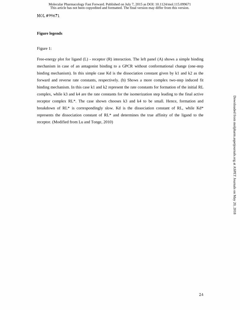
Figure 1:
Free-energy plot for ligand (L) - receptor (R) interaction. The left panel (A) shows a simple binding
mechanism in case of an antagonist binding to a GPCR without conformational change (one-step
binding mechanism). In this simple case Kd is the dissociation constant given by k1 and k2 as the
forward and reverse rate constants, respectively. (b) Shows a more complex two-step induced fit
binding mechanism. In this case k1 and k2 represent the rate constants for formation of the initial RL
complex, while k3 and k4 are the rate constants for the isomerization step leading to the final active
receptor complex RL*. The case shown chooses k3 and k4 to be small. Hence, formation and
breakdown of RL* is correspondingly slow. Kd is the dissociation constant of RL, while Kd*
represents the dissociation constant of RL* and determines the true affinity of the ligand to the
receptor. (Modified from Lu and Tonge, 2010)
This article has not been copyedited and formatted. The final version may differ from this version.Molecular Pharmacology Fast Forward. Published on July 7, 2015 as DOI: 10.1124/mol.115.099671
at ASPE
T Journals on M
ay 20, 2018m
olpharm.aspetjournals.org
Dow
nloaded from
MOL #99671
25
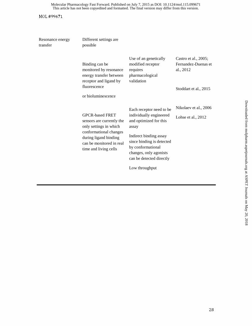
Table 1: Advantages and disadvantages of different methods for kinetic binding experiments
Methods
Advantage
Disadvantage
Reference with respect to GPCRs
Established
Radioligand binding
Widely applicable
Good documentation
Applicable in membranes, cells, tissue slices
A radioligand with high affinity and selectivity is required.
Possible lack of specificity
Radioactive waste
Guo et al., 2014
Surface plasmon resonance
Label free with respect to the ligand
Flow through system
Little material required
Real time detection
Target immobilization required
Purification and stabilization of protein might be required
Often artificial environment
Aristotelous et al., 2015
Christopher et al., 2013
Bocquet et al., 2015
This article has not been copyedited and formatted. The final version may differ from this version.Molecular Pharmacology Fast Forward. Published on July 7, 2015 as DOI: 10.1124/mol.115.099671
at ASPE
T Journals on M
ay 20, 2018m
olpharm.aspetjournals.org
Dow
nloaded from
MOL #99671
26
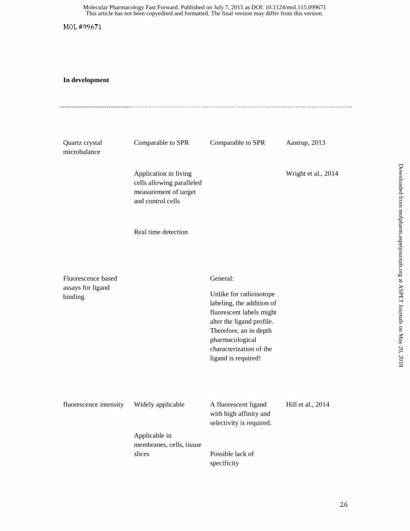
In development
Quartz crystal microbalance
Comparable to SPR
Application in living cells allowing paralleled measurement of target and control cells
Real time detection
Comparable to SPR
Aastrup, 2013
Wright et al., 2014
Fluorescence based assays for ligand binding
General:
Unlike for radioisotope labeling, the addition of fluorescent labels might alter the ligand profile. Therefore, an in depth pharmacological characterization of the ligand is required!
fluorescence intensity
Widely applicable
Applicable in membranes, cells, tissue slices
A fluorescent ligand with high affinity and selectivity is required.
Possible lack of specificity
Hill et al., 2014
This article has not been copyedited and formatted. The final version may differ from this version.Molecular Pharmacology Fast Forward. Published on July 7, 2015 as DOI: 10.1124/mol.115.099671
at ASPE
T Journals on M
ay 20, 2018m
olpharm.aspetjournals.org
Dow
nloaded from
MOL #99671
27
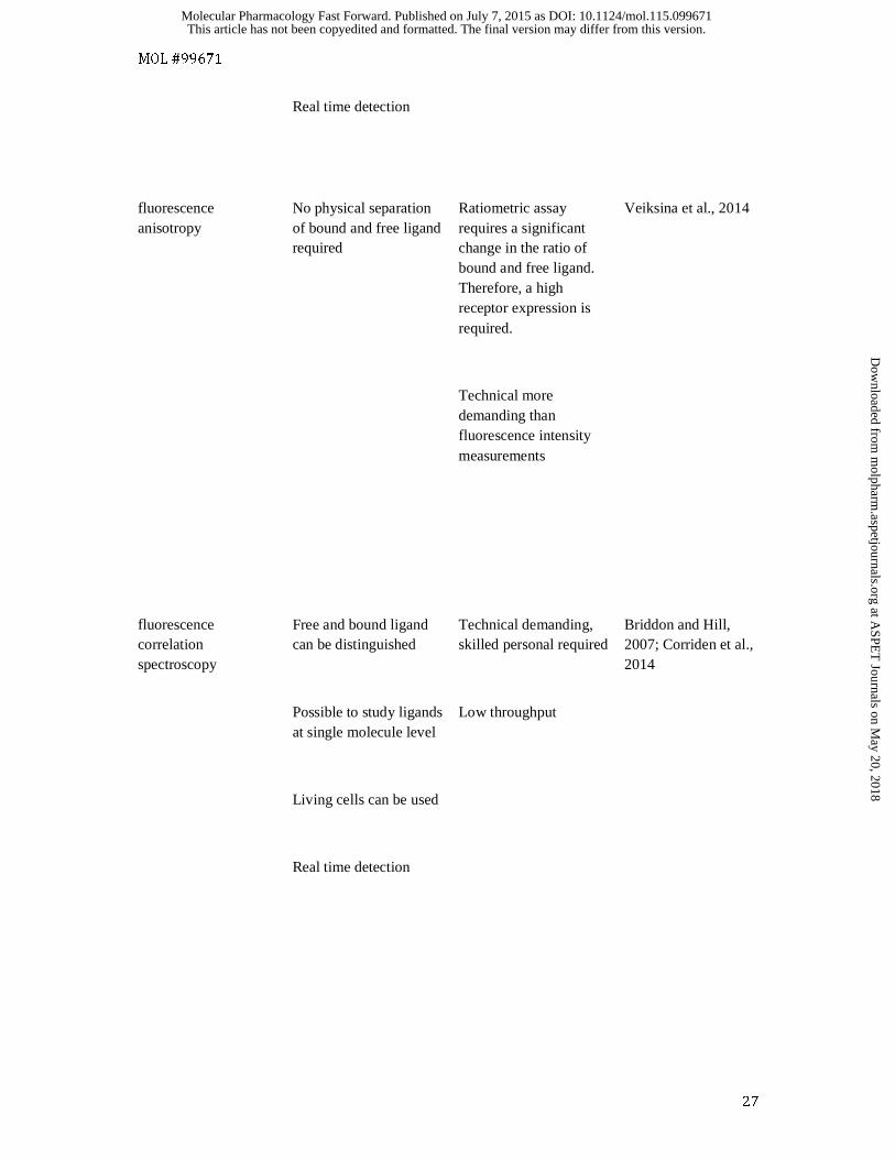
Real time detection
fluorescence anisotropy
No physical separation of bound and free ligand required
Ratiometric assay requires a significant change in the ratio of bound and free ligand. Therefore, a high receptor expression is required.
Technical more demanding than fluorescence intensity measurements
Veiksina et al., 2014
fluorescence correlation spectroscopy
Free and bound ligand can be distinguished
Possible to study ligands at single molecule level
Living cells can be used
Real time detection
Technical demanding, skilled personal required
Low throughput
Briddon and Hill, 2007; Corriden et al., 2014
This article has not been copyedited and formatted. The final version may differ from this version.Molecular Pharmacology Fast Forward. Published on July 7, 2015 as DOI: 10.1124/mol.115.099671
at ASPE
T Journals on M
ay 20, 2018m
olpharm.aspetjournals.org
Dow
nloaded from
MOL #99671
28
Resonance energy transfer
Different settings are possible
Binding can be monitored by resonance energy transfer between receptor and ligand by fluorescence
or bioluminescence
GPCR-based FRET sensors are currently the only settings in which conformational changes during ligand binding can be monitored in real time and living cells
Use of an genetically modified receptor requires pharmacological validation
Each receptor need to be individually engineered and optimized for this assay
Indirect binding assay since binding is detected by conformational changes, only agonists can be detected directly
Low throughput
Castro et al., 2005; Fernandez-Duenas et al., 2012
Stoddart et al., 2015
Nikolaev et al., 2006
Lohse et al., 2012
This article has not been copyedited and formatted. The final version may differ from this version.Molecular Pharmacology Fast Forward. Published on July 7, 2015 as DOI: 10.1124/mol.115.099671
at ASPE
T Journals on M
ay 20, 2018m
olpharm.aspetjournals.org
Dow
nloaded from
This article has not been copyedited and formatted. The final version may differ from this version.Molecular Pharmacology Fast Forward. Published on July 7, 2015 as DOI: 10.1124/mol.115.099671
at ASPE
T Journals on M
ay 20, 2018m
olpharm.aspetjournals.org
Dow
nloaded from