Embed Size (px)
Citation preview

Report 3922(32)
Life Sciences Digest
n0lbl
m
https://ntrs.nasa.gov/search.jsp?R=19900016141 2018-08-25T05:54:56+00:00Z

:x
K
i!:
x
:/i
x
Y

NASA Contractor Report 3922(32)
USSR Space Life Sciences Digest
Issue 27
Edited by
Lydia Razran Stone and Ronald Teeter
Lockheed Engineering & Sciences Company
Washington, D.C.
Victoria Garshnek
The George Washington University
Washington, D. C.
Joseph Rowe
Library of Congress
Washington, D.C.
Prepared for
NASA Office of Space Science and Applications
under Contract NASW-4292
National Aeronautics andSpace Administration
Office of Management
Scientific and TechnicalInformation Division
1990


USSR SPACE LIFE SCIENCES DIGEST ISSUE 27
TABLE OF CONTENTS
Reader Feedback Form vFrom the Editors viADAPTATION 1
Hormones, Adaptation, and Systemic Reactions. 1AVIATION MEDICINE 3
The effect of workload on the functional state of flight crews of ship-based aviation. 3Aviation Medicine.(Book Review) 5
BIOLOGICAL RHYTHMS 8Free running circadian rhythms in the darkling beetle after space flight. 8
BIOSPHERICS 10Modeling of Space Systems for Studying the Earth's Natural Resources 10
BOTANY 13The effects of single hits by heavy ions of galactic cosmic radiation on Lactuca sativaseeds flown on board Salyut-6 and Salyut-7 space stations. 13
CARDIOVASCULAR AND RESPIRATORY SYSTEMS 17Central and regional hemodynamics on long-term space flights. 17Cardiac contractility of guinea pigs exposed to long-term continuous stress. 23Circulation and oxygen pressure in the brains of alert and anesthetized rabbits in a head-down tilt position. 25
EN DOC RINOLOGY 27Morphological research on the adrenal glands of rats after flight on COSMOS-1667. 27Hypothalamus/pituitary neurosecretory system in rats exposed to high-altitude hypoxia. 29Endocrine response to low frequency electromagnetic fields of continuous andintermittent generation. 30
ENZYMOLOGY 32Activity of digestive enzymes in response to immobilization stress and its pharmacologicalcorrection with adrenoreceptor blocking agents. 32
EXOBIOLOGY 34Abiogenic thermal synthesis of nucleotides in the presence of lunar soil. 34
HABITABILITY AND ENVIRONMENT EFFECTS 36Therrnoregulatory responses in humans to moderate levels of hypercapnia. 36
HEMATOLOGY 38Use of a ferrocerone test to measure iron reserves under various living conditions. 38
IMM UNOLOGY 40The immune status of individuals suffering from acute altitude sickness. 40The effect of smoking on human resistance in a pressurized environment. 42On the protective function of the skin. 43
M ETABOLISM 44Endogenous ethanol in the blood and tissues of rats exposed to hypobaric hypoxia. 44Use of the method of thin layer chromatography to study lipid ligands in the serumalbumin of athletes. 46Activity of NADP-dependent cytoplasmic dehydrogenases in the liver and adipose tissueof rats during recovery after hypokinesia. 47
o°°
f III
(_ __J__Lf.. INTENTIONA[.t.YBLAN[

USSR SPACE LIFE SCIENCESDIGEST ISSUE 27
MUSCULOSKELETAL SYSTEM 48Effects of active metabolites of Vitamin D3 on the bones of rats exposed to differenthypokinesia paradigms. 48Changes induced by hydroxydimethyl aminopropylidene diphosphonate in the responseof bone tissue in rats to hypokinesia. 50Changes in human bone under conditions of bedrest and weightlessness: The humanskeleton under conditions of bedrest and weightlessness. 52Changes in human bone under conditions of bedrest and weightlessness: Bonechanges when functional loading of the skeleton is reduced from the standpoint of rapidand slow-developing osteoporosis. 56Changes in human bone under conditions of bedrest and weightlessness:Generalprinciples of bone changes under conditions of weightlessness and its simulations. 57
NEUROPHYSIOLOGY 65Specification for an "ideal" drug to prevent space motion sickness (space adaptationsyndrome. 65Remote ultrastructural changes in cerebellar cortex of rats after exposure to acceleratedcarbon ions. 68Cerebrovascular effects of motion sickness. 69
PSYC HOLOGY 71An analysis of factors determining sex differences in the stress responses of white rats. 71
RADIOBIOLOGY 72Periodicity of hemopoiesis during continuous g-irradiation with low dose rates. 72
SPACE MEDICINE 73The work of the section on Aviation and Space Medicine of the Moscow PhysiologicalSociety (Conference Review). 73In the Interests of Public Health 76
iv

USSR Space Life Sciences Digest: Issue 27Reader Feedback Form
To our readers: We are working in a large number of highly technical, specializedareas for which adequate Russian-English glossaries have yet to be compiled. We askyour help in improving the accuracy and specificity or our English terminology. Pleasefill out the form below whenever you encounter an incomprehensible, incongruous,awkward or otherwise inappropriate term. While we solicit all suggestions forimproved renderings, the statement that a term is inappropriate provides us withuseful information, even when no better alternative can be suggested. A copy of thisform will appear in all future issues of the Digest. Thank you for your help.
Abstract Number Incorrect or inappropriate term ISuggested rendering
PLEASE RETURN TO: Dr. Lydia StoneLockheed Engineeringand Sciences Company
600 Maryland Ave. SWSuite 600, East Wing
Washington, DC 20024
V

USSR SPACE LIFE SCIENCES DIGEST
FROM THE EDITORS
ISSUE 27
This is Issue 27 of the USSR Space Life Sciences Digest. Articles presenting ordiscussing space flight data in this issue are: Biorhythms P1204, Botany P1213,Cardiovascular and Respiratory Systems P1202, Endocrinology P1203, andMusculoskeletal System P1222 and 1224. The coming issue of the Digest will bedouble, i.e., provide abstracts for two issues of the Soviet Space Biology and AerospaceMedicine journal. This issue will also mark the beginning of our use of an "expertpanel" to provide technical editing of the digest. We expect that inclusion of his panelwill improve the accuracy and appropriateness of the technical terminology used in theDigest. We would like to thank Drs. Gary Coulter and and F. Ronald Dutcher for acting asexpert consultants for the current issue.
Address correspondence to:Dr. Lydia Stone
Lockheed Engineeringand Sciences Company
600 Maryland Ave. SWSuite 600, East Wing
Washington, DC 20024
vi

ADAPTATION
MONOGRAPH:
M160(27/90) Vinogradov VV.Gormony, Adaptatsiya i Sistemnyye Reaktsii Organizma; F'opMoH_, Az_anTa_HHH CHCTeMH_ePeaKUHH OpraHH3Ma; [Hormones, Adaptation, and Systemic Reactions.]Minsk: Nauka i Tekhnika; 1989.[223 pages; 19 Tables; 56 Figures; 538 references]Affiliation (book): Institute of Biochemistry; Belorussian Academy of Sciences
KEY WORDS: Adaptation; Endocrinology, Hormones, Stress; Biological Rhythms, SeasonalRhythms; Metabolism; Cardiovascular and Respiratory System
Annotation: With reference to a wide range of experimental and clinical materials, the authoranalyzes the role played by hormones in supporting rhythmic .homeostatic functions responsiblefor adaptation at the cellular level and for the development of systemic reactions to stress.Hormonal mechanisms underlying seasonal rhythms in metabolic functions are discussed. Arationale is presented for using biochemical indicators of the stress response in medicalpractice for diagnosing latent forms of circulatory insufficiency in cardiovascular pathology.Biochemical, physiological, and medical aspects of adaptation, its appropriateness and failuresare discussed. This monograph is intended for biochemists, physiologists, and medicalpersonnel.
CCt_ENTS
Abbreviations (30
Foreword (4)
Chapter 1.1.11.2
1.3
1.4
1.5
Biological Rhythms and Adaptation (7)Hormonal mechanisms underlying seasonal rhythms (7)Biosynthesis of protein in the liver accompanying natural fluctuations in steroid
levels (18)Seasonal rhythms in hormone production in the adrenal glands and energy supply for
protein synthesis in the liver (21)Thermodynamic (informational) approach to the problem of seasonal shifts in
protein metabolism (27)Seasonal rhythms as adaptation to periodic changes in the environment (42)
Chapter 2.2.12.2
2.32.4
2.52.6
The Bioenergetics of Adaptation (59)The generation of energy (59)Endergonic functions of the hepatic mitochondria (71)Ca 2+ transport (83)
The mechanism underlying activation of succinate dehydrogenase in the livers ofrats exposed to stress (100)
Bioenergetics of the adrenal glands of rats exposed to stress (104)A kinetic model of the functioning of succinate dehydrogenase in the adrenal glands
and the mechanisms underlying regulation of enzyme activity (121)

ADAPTATION
Chapter 3. The Systemic Nature of Adaptation (155)3.1 The information value of the stress-response in cardiology (155)3.2 Stress and the functional reserves of the adrenal gland and heart3.3 Hormones and diagnosis of circulatory insufficiency (180)
Conclusion (198)
References (204)
2

AVIATION MEDICINE
PAPER:
P1230(27/90) Mel'nik SG, Shakula AV, Gladkikh FD.The effect of workload on the functional state of flight crews of ship-baseda via tion.
Voyenno-Meditsinksiy Zhurnal.1989(7): 54-57.[No references]Authors' Affiliations: USSR Medical Corps
Aviation Medicine, Functional State, Cardiovascular and Respiratory SystemsHumans, Air Crews, Ship-Based AviationHuman Performance, Workload
Abstract: Traditional physiological methods were used to study the functions of thecardiovascular and respiratory systems and reserve capacities of 26 helicopter pilots andnavigators in ship-based aviation before and after flights performed in the course of long-duration cruises. At low latitudes, mean data suggest a hypotonic reaction to flight conditions,attributed by the authors to heat and long periods of relative hypokinesia. After 2- and 3-hourflights, only systolic pressure decreased for pilots, while both diastolic and systemic pressuredecreased in navigators. Results were of similar magnitude for both durations of flight.Recovery of blood pressure was not complete after 58 hours. Heart rate remained the samepostflight for pilots but increased in navigators. After 2- and 3-hour flights at low latitudes,respiration rate increased significantly and reserve capacity (measured by performance onbreath holding tests) decreased. When flight duration increased from 2 to 3 hours, respirationrate increased still further and reserve capacity decreased. Fifty-eight hours after the 2-hourflight, most respiratory parameters had recovered, but this was not the case for the 3-hourflight.
In an additional study, the same group of six helicopter pilots performed analogous flightmissions at high and low latitudes. Cardiovascular response was adequate at high latitudes, withmoderate increase in systolic and diastolic pressure, and increased heart rate, normalizingafter 16 hours. At low latitudes, blood pressure decreased and heart rate increasedsignificantly, and these changes persisted for more than 16 hours, normalizing completely only58 hours postflight. After flight at high latitudes, breathing rate increased only insignificantlyand reserve capacities decreased moderately, with normalization occurring after 16 hours.Analogous flights at low latitudes led to greater increases in respiration rate and significantdecreases in respiratory reserve.
Results of a questionnaire revealed that pilots and navigators considered the optimal flightworkload to be 3+1 hours per shift, 6+1 hours per week, 17+1 hour per month, and 104+3
hours per year.
Table 1: Dynamics of cardiovascular parameters in helicopter crews flying at high latitudes
Table 2: Dynamics of cardiovascular parameters in helicopter crews flying at low latitudes
Figure 1: Change in parameters of functional state of the cardiovascular system of pilots flyingat high latitudes
Figure 2: Change in parameters of functional state of the cardiovascular system of pilots flyingat low latitudes
3

AVIATION MEDICINE
Figure 3" Change in parameters of functional state of the respiratory system of pilots flying athigh latitudes
Figure 4: Change in parameters of functional state of the respiratory system of pilots flying atlow latitudes
4

AVIATION MEDICINE
BOOK REVIEW:
BR17(27/90)*Gyurdzhian AA.Review of: Ernsting J, and King , et al. (Editors).A viation Medicine.London: Butterworth 1988, 738 pages.Kosmicheskaya Biologiya i Aviakosmicheskaya Meditsina.23(6): 92-94; 1989.
NOTE: Although this is a translation of review of a British, not a Soviet book. It may be of someinterest to our readers to find out what English-language works Soviet scientists are aware ofand how such literature is reviewed.
KEY WORDS: Aviation Medicine, Human Performance, Aviation Psychology, Biodynamics,Thermal Stress; Biological Rhythms
The second edition of the British handbook on aviation medicine is intended for physicians
undergoing postgraduate specialization training in aviation medicine.
Both the scientific editors and the majority of authors (42 of whom participated in preparingthe handbook) - are members of the Medical Corps of the British Royal Air Force, mainly at theInstitute of Aviation Medicine in Farnborough. Although the authors are all British, the workcommendably considers ideas and scientific data of researchers from other nations and also theofficial regulations of the International Organization of Civil Aviation.
The current edition has been revised to such a great extent that it is not strictly accurate to callit the second edition of the handbook published in 1978 (Aviation Medicine, Editor, G. Dhenin[?sic.], London, Tri-Med Books Ltd. 1978). Only 25% of the authors contributing to the secondedition had contributed to the first. Even the few chapters written by the same authors haveundergone fairly major revisions in light of the latest advances in aviation medicine. It is anadded convenience that the second edition has been published as a single volume, while the firstwas in two volumes.
The handbook comprises 10 parts, 55 chapters and 2 appendices. The first five parts aredevoted to issues in aviation physiology, and the last five to clinical problems, workingconditions and hygiene in aviation, and also aviation pathology and investigation of aircraftaccidents.
Reflecting the development of aviation during the last 10 years (after publication of the firstvolume), the main focus in aviation medicine has shifted from study of the effects of flightfactors and conditions on autonomic and somatic functions (respiration, cardiovascularfunctioning, biodynamics, thermal regulation) to research on psychophysiological status andjob performance, particularly in flight crews. For this reason, the greatest differencesbetween the first and second editions can be found in sections dealing with these topics. Forexample, Part 5 "Aviation Psychology," has been rewritten completely, while the chapters inPart 9 devoted to the clinical aspects of aviation medicine have been substantially reworked andexpanded. Part 10, which discusses methods for investigating aircraft accidents and results inaviation pathology is completely new.
Let us turn to analysis of the contents of the handbook. In Part 1 ("Environmental Pressure")data concerning the Earth's atmosphere, air pressure differentials, the principles ofrespiratory physiology, hypoxia and hyperventilation, oxygen equipment,'full pressure suits,pressurized cabins and partial pressure suits, as well as the effects of toxic gases and vaporsare discussed thoroughly and clearly.
5
AVIATION MEDICINE
Part 2 ("Biodynamics") relatively briefly (compared to two other modern handbooks), but insufficient depth, considers the effects of prolonged acceleration of various types and ofvibration, as well as measures taken to protect flight crews against their effects. This sectiondescribes the dynamics and mechanisms of forces acting on humans in aircraft accidents and theassociated injuries (particularly head injuries) in flight crews and passengers, means ofprotection against these forces, and methods for escaping from aircraft.
Part 3 ("Thermal Stress and Survival") is devoted to heat exchange and thermal regulation inhumans, ways to protect flight crews from thermal stress, and survival techniques in adversethermal and climatic conditions, particularly after accidents. The Institute of Aviation Medicinetraditionally sponsors a major research program on thermal regulation and temperature stressin special laboratories in which important and well-designed work is conducted.
Part 4 ("Sense Organs") devoted to issues related to the sense organs is unique. In threechapters one of the most famous specialists in this area, A.J. Benson, considers in detail theproblems of spatial orientation in pilots and also the critical problem of motion sickness. Inaddition, this part contains chapters devoted to current data on pilot vision and the interrelatedissues of noise and communication (radiocommunications) under flight conditions.
Part 5 ("Aviation Psychology") is very impressive. It cites the psychological results mostrelevant to the performance of flight crews. It considers the processes of attention,particularly allocation of pilot attention, cognition, decision-making and memory (e.g.,memory for an immediately preceding flight accident). Part 5 provides a good presentation ofthe problems of stress and approaches to evaluating the workload of a flight crew, problems ofindividual psychophysiological differences, personality traits, selection of personnel andvarious training methods. Significant emphasis is placed on ergonomics, particularly theergonomic characteristics of new display and control systems. For the first time in anyhandbook of aviation medicine there is a chapter devoted to the social psychology of aircraftcrews.
Finally, several chapters in part 5 provide glossaries of relevant terms. This is veryimportant since technical psychological terminology is not always known by aviationphysicians.
Part 6 ("Special Types of Flight") contains very useful information about the medical aspects ofvarious types of flights of civil and military aircraft.
Chapters 7 and 8 ("Commercial Aviation and Health," and "Health and Hygiene") considerproblems of public health relevant to civil aviation. International regulations for maintainingthe health of aircraft crews and passengers, and rules for transporting the sick and handicappedare cited, along with the medical standards for flight certification examinations. Problemsrelated to maintaining health, the hygiene of aircraft flights and airports, quarantine, andimmunological and epidemiological aspects of international flights, regulations concerningtransport of animals, and disinfection and extermination of rats and insects are discussed. Thechapter discussing maintenance of the heath of ground personnel, particularly technical aviationpersonnel, whose working conditions are sometimes very adverse, is very timely.
Part 9 ("Clinical Aspects of Aviation Medicine") will be especially interesting for Sovietspecialists. In Soviet handbooks and proceedings of meetings on aviation medicine, clinicalissues are emphasized much less than in analogous foreign works This contributes to somedegree of isolation of aviation physicians from the rest of medicine and does not assist theirtheoretical and clinical training as doctors and specialists in flight certification examinations.
6
AVIATION MEDICINE
Two chapters in Part 9 are devoted to diurnal biological rhythms, work/rest schedules, sleepand wakefulness, job performance and appropriate medical recommendations. Next there arenine chapters devoted to purely clinical aspects of aviation medicine: diseases of thecardiovascular, respiratory, and digestive systems; kidney disease; diabetes; obesity;thyrotoxicosis; anemia; sickle cell anemia; tumors; psychiatric; neurological;otorhinolaryngological; opththalmological and orthopedic problems; alcoholism; drugdependence; and sexual dysfunction.
It should be noted that all the clinical issues are discussed as they pertain to occupationaldiseases of aviation personnel and to health risk conditions and factors relevant to jobperformance, and the requirements for flight certification examinations. Thus, for example,the chapter devoted to otorhinolaryngology elucidates issues relevant to the harmful effects ofnoise; the chapter on ophthalmology focuses on vision requirements for flight crews; thechapter on orthopedics considers typical skeletal injuries (particularly to the spine) inaircraft accidents and ejection, etc. These discussions consider modern advances in medicalscience and new regulations for flight certification examinations, including those of the ICAO.The section devoted to medical ethics in the work of the medical corps for civil and militaryaviation is interesting.
The final chapter, Chapter 10, ("Investigation of Aircraft Accidents") is new and extremelyuseful. It elucidates the administrative and legal issues involved in such investigations, takingaccount of ICAO requirements and the set of methods used by specialists in aviation pathology,including the methods for identifying plane crash victims by their teeth and dental work.
The first appendix contains seven tables of units of measurement with the appropriate symbols,abbreviations, and coefficients for transforming units from one system to another. The secondappendix contains a 14-page detailed subject index.
An important advantage of this book is its success in combining accurate and simple forms ofpresentation with a high concentration of information and modern factual material in therelevant areas of aviation medicine. At the end of each chapter is a list of cited sources in theliterature to enable deeper study.
The authors' texts are adequately illustrated with graphs, diagrams, figures, and photographs.All this makes the book under review valuable both as a handbook and a reference work. Incompleteness and breadth of coverage the book resembles the handbook on aviation physiology("A Textbook of Aviation Physiology" edited by J.A. Gilles, et al., Oxford, Pergammon Press,1965, 1226 pages) published in England.
The book undoubtedly will be of interest of Soviet aviation and space physicians, and also tothose working in related disciplines.
7

BIOLOGICAL RHYTHMS
PAPER:
P1204(27/90)* Alpatov Am, Yevstratov YuA., Chernyshev VB, Lebedev MI, Zotov VA.Free running circadian rhythms in the darkling beetle after space flight.Kosmicheskaya Biologiya i Aviakosmicheskaya Meditsina.23(6): 31-33; 1989.[9 references; 5 in English]
Biological Rhythms, Circadian Rhythms, Free-Running, Motor ActivityBeetles, DarklingSpace Flight, COSMOS-1887
Abstract: Darkling beetles (Trigonoscelis gigas), which are capable of maintaining free-running rhythms of motor activity for long periods under conditions of constant darkness, wereselected as subjects in this experiment on the effects of space flight on biological rhythms. Twomonths preflight, the beetles were caught and maintained in a cage with a 16:8 light to darknessratio. During the experiment each beetle was housed in an individual cell where motor activitywas recorded. Throughout the experimental period, the insects were maintained in totaldarkness. Motor activity was recorded for 8 days preflight and 10 days postflight and duringthe 14-day flight on COSMOS-1887. After the flight the animals were returned to the cage andtheir circadian rhythms resynchronized. A 24-hour control experiment was run on the groundduring which the insects were exposed to vibration such as occurs during spacecraft take-offand landing, and also chilling at 15° such as occurred immediately after landing.
In space, six of the seven beetles displayed a reliable decrease in the period of their circadianrhythm of motor activity. In two of these, the period gradually recovered to normal in 5-7 daysafter landing. Vibration did not affect the rhythm, and chilling completely depressed the beetles'activity but did not affect subsequent parameters. In one insect chilling did displace the rhythmby 3 hours. No changes in period occurred in the control experiment.
The effect observed here m decrease in the spontaneous period of the circadian rhythm underconditions of weightlessness m is important because period duration is a fundamentalcharacteristic of the circadian system and is known to be little affected by external factors, withthe exception of those that act as time cues (light or, less frequently, temperature). Gravity isnot a time cue, which makes the observed effect somewhat surprising. However, it has beenfound previously that period of circadian rhythms may be increased by prolonged exposure tocentrifugation (in primates). In both cases the effect, while statistically significant, did notexceed the bounds of the norm. This result suggests that in space the biologically optimumduration of a "day" may differ from that on Earth.
Table: Duration of the period (.c, hr) of free-running circadian rhythms of motor activity ininsects
Beetle # Preflight Postflight p (t-test)1 25.0+0.4 23.7+0.2 <0.0252 24.3+0.2 21.6_+0.5" <0.00053 23.7_+0.2 24.3_+0.4 n.s.4 23.8_+0.4 22.0_+0.7* <0.055 23.7_+0.2 22.4_+0.4 <0.016 24.1_+0.3 23.7_+0.2 n.s.7 24.3_+0.2 22.9_+0.4 <0.005
8

BIOLOGICAL RHYTHMS
Raster-analysis
+t =L=eA_=-- ==il,t
: : = =-----= = =1 =
=_ x=s=-= =2st_t=
'_Q00 ==
T_U= 24 . 60 rain
=tta==tz_====mt_
===t_=== ==
==t=== =*a=O_===== = = ==== = =
==00,00===
Space flight
..... _+ .... ---=;:;1_=-=• ===1= l "===== = =_- =.=
== == Za_t_8aI_s=== = -_-::oo_'=
' 2aO_=+ =
Readaptation
::g;:=Ill==
:=ILZ==
:tttl==:
==eft::
::I18:::
Vibration
Chilling
:=OOO*:==li==
IIOI'.m_
:o#::= ,_.;1111 :I
III11111= =
=.0_0===
i
Figure: Free-running circadian rhythm of motor activity of beetle number 2From the top: Day of experiment, darkness of each character corresponds to the totalnumber of movements in an hour.
9

BIOSPHERICS
MONOGRAPH:
M161(27/9) Khantseverov FR, Ostroukhov VV.Modelirovaniye Kosmicheskikh Sistem Izucheniya Prirodnykh Resursov Zemli; Mo_e_HpomaHHeKOCMHqeCKHX CHCTeM H3yqeHH_! rlpHpO_HblX Pecypcos 3eMJ]H; [Modeling of Space Systems forStudying the Earth's Natural Resources]Moscow: Mashinostroyenie; 1989.[264 pages; 69 Figures]
KEY WORDS: Biospherics, Mathematical Modeling, Space Systems, Remote Sensing, NaturalResources
Annotation: This book considers theoretical and practical aspects of modeling space systems forstudying the Earth's natural resource, citing an original set of multifactor models of differentphysical types,with varying capacity and structural complexity. The book is intended forengineers in the area of cosmonautics, and also for specialists working on issues of systemsanalysis and the mathematical and physical modeling of space systems of scientific and economicsignificance.
CONTENTS
Foreword (3)
Chapter 1. Methodological principles for the analysis and synthesis of space systems (5)1.1 The subject and objectives of research on the properties of formalized systems (5)1.2 Mathematical formulation of the objectives of systems analysis and synthesis (21)1.3 Criteria for evaluating the performance of space systems (31)1.4 A systems methodology for investigating the functional dynamics of orbital and ground-
based space systems for studying the Earth's natural resources (42)
Chapter 2. A method of graph-analytic modeling and its applications (47)2.1 Preparation for and design of a modeling experiment (48)2.2 Formalization of the process of satellite movement. Topological model of conditions in
the regions being studied (50)2.3 Techniques for simulating the processes of observation and storage of information on
board a satellite (53)2.4 An analytic model to evaluate the performance of system components (56)2.5 A methodology for experimentally identifying the law underlying the distribution of
natural features within the observation field of on-board remote sensing devices (59)2.6 A methodology for analyzing the efficiency of the information-receiving process in
ground-based stations of a multistation reception system (63)
Chapter 3. Electronic modeling of systems (74)3.1 The concepts of design and technological profile of the modeled device (74)3.2 Principles for reproducing the dynamics of a "satellite-ground station" system (80)3.3 Reproducing the functional dynamics of an on-board video apparatus (89)3.4 Simulation of illumination conditions, cloud cover, and system reliability (91)3.5 General operating scheme of a simulation device (95)3.6 Examples of a model-based evaluation of the capacity of an on-board memory device as a
function of input parameters (97)3.7 A methodology for simulating observation under varying illumination conditions (101)
10

BIOSPHERICS
Chapter 4. Methods for formally describing movement of individual satellites and groups ofsatellites (104)
4.1 Formulation of objectives (104)4.2 Methods for depicting the satellite's entry into the observation region and zones of
radiovisibility (105)4.3 Examples of empirical distribution of communication points, observation cycles, and
information reception periods (114)4.4 Techniques for estimating the satellite's period of radiovisibility to ground-based
reception stations (117)4.5 Methodology for identifying the expected distribution pattern of durations of
information transmissions from space (121)
Chapter 5. A theory for optimizing processes of selective observations of natural features fromspace (127)
5.1 A heuristic model for finding the optimal observation route (127)5.2 Mathematical statement of the problem. Optimization criteria (135)5.3 Mathematical model of an optimal survey of natural features (142)5.4 General theory for optimizing observation of natural resources (148)
Chapter 6. A set of mathematical models of space systems for studying natural resources (156)6.1 Concept and description of a generalized model of a space system (156)6.2 Aggregated models of the functioning of orbital subsystems (166)6.3 Ballistic models of orbital groupings (175)6.4 Investigation of the efficiency of on-board systems of automatic navigation, orientation,
stabilization and focusing of a satellite (179)6.5 A method for investigating an on-board information-processing computer complex
(189)
Chapter 7. Mathematical modeling of the functional dynamics of on-board and ground-basedvideo systems (190)
7.1 A complex model for surveying natural resources and storing and transmitting remotesensing data (190)
7.2 A method for investigating the dynamic characteristics of the focusing apparatus (198)7.3 A simulation model of the crew of a space station (202)7.4 Principles of design and description of a complex model of ground-based video equipment
(206)7.5 A model for optimal management of video information processing procedures on board a
spacecraft and in ground-based reception stations (211)7.6 Simulation and computation of the effect of ground-based spacecraft control (215)7.7 A method for investigating the reliability characteristics of on-board service and
focusing systems (219)
11
BIOSPHERICS
Chapter 8. Principles for complex systems analysis of the functional dynamics of systems forstudying the Earth's natural resource from space (223)
8.1 A methodology for model-based estimation of the distribution of natural resources andtheir "accessibility" to remote sensing (223)
8.2 A methodology for systems analysis of the efficiency and balance of informationgenerated by on-board video devices (227)
8.3 Examples of analysis of the dynamics of queue formation on board a spacecraft andderivation of specifications for storage capacity (231)
8.4 A methodology for deriving the requirements for personnel to interpret scientific videoinformation (235)
8.5 Examples of the analysis of capacitance of a ground-based reception station network as afunction of observation schedules (240)
8.6 Objectives of complex analysis. Initial prerequisites (242)8.7 Examples of estimation of the effects of "Discipline" conditions on the information
capacity of an orbital subsystem (243)8.8 A methodology for the complex analysis of functional dynamics of a "satellite-ground
stations" system (245)8.9 An example of estimation of the integral productivity of a space system (251)8.10 A methodology for general systems analysis of the information capacity of a
multisatellite system for studying the Earth's natural resources when orbitalinclination varies (253)
References (261)
12

BOTANY
PAPER:
P1213(27/90)* Nevzgodina LV, Maksimova YeN, Kaminskaya YeV.The effects of single hits by heavy ions of galactic cosmic radiation on Lactucasativa seeds flown on board Salyut.6 and Salyut-7 space stations.Kosmicheskaya Biologiya i Aviakosmicheskaya Meditsina.23(6): 66-70; 1989.[15 references; 6 in English]
Genetics, AberrationsBotany, Lettuce, SeedsSpace Flight, Long-term, Salyut-6, Salyut-7; Radiobiology, Heavy Ions
Abstract: Air-dried seeds of lettuce, Lactuca sativa were exposed on board Salyut-6 for 123
days and on board Salyut-7 for periods of 40, 201, and 457 days. All of the biostacks used inthese experiments were located in the inhabited portion of the spacecraft and were maintainedunder controlled conditions. Each biostack consisted of layers of seeds placed in "monolayers"alternating with physical detectors that recorded the paths of cosmic heavy ions. Nitracellulosedetectors recorded heavy ions with charge of Z > 6 and LET > 200 keV/p.m. After the sampleswere returned to Earth, the biostacks were disassembled. To identify heavy ion tracks thedetectors were soaked in an alkaline solution and then were examined in a stack which had thesame coordinate system as the ones returned from space. Joint examination of the biologicallayer and detectors made it possible to identify the seeds through which heavy ions had passed.Flux density and fluence of heavy ions were measured. Dosimetry was performed dosimetryusing thermoluminescent detectors. Integral absorbed doses were measured. Two weeks afterthe return to Earth, seeds in all conditions were moistened with tap water and germinated inPetri dishes. One day after hydration and subsequently during growth, cells were fixed in a 1:3mixture of acetic acid and alcohol and a cytogenetic analysis was performed during the firstmitosis. Seed viability (day 3) and germination (day 7) were noted. Radiation effects wereassessed by the presence of various chromosomal restructurings observed during anaphase andtelophase. The following groups were utilized: flight seeds hit and not hit by heavy particles,and ground control seeds.
Dosimetry measurements showed an increase in fluence and dose as flight duration increasedfrom 40 to 457 days.
Germination of the flight seeds was somewhat delayed, especially those seeds hit by heavy ions.Delays were noted for all durations of flight. Viability was depressed in flight seeds on day 3after moistening; however, differences between control and flight groups had disappeared by day7. Cytogenetic analysis of flight seeds showed an increased frequency of aberrant cells for alldurations of exposure to space, with greatest numbers of aberrations found in seeds hit by ions.Frequency of aberration increased with flight duration. To a lesser extent this was also true ofcontrol seeds, which may be attributed to natural aging. Cells with multiple chromosomebreaks occurred most frequently in seeds hit by heavy ions and exposed to spacee for 201 and457 days. Fewer multiple aberrations were found in unhit flight seeds, with fewer still incontrol seeds.
There was a linear relationship between the absorbed dose of radiation and the observed effects(number of aberrant cells, cells with multiple chromosome breaks). The highest frequency ofaberrant cells occurred in flight seeds irradiated with a dose of 63.4 mGy. The data obtainedindicate that the biological effectiveness of heavy ions is considerably greater than would bepredicted on the basis of absorbed dose and fluence. The dose-effect curves for the frequency ofaberrant cells were parallel for hit and unhit seeds; however, this was not the case for the
13

BOTANY
parameter of frequency of cells with multiple chromosome aberrations. In the range of dose,_ of16 - 63.4 mGy, the slope of the curve for hit seeds was greater than for unhit seeds. Thissuggests that as flight duration increases, the frequency of multiple damage due to direct effectsof heavy ions with higher charges also increases. In the Bioblok-3 experiment, with durationsof 201 and 457 days, only heavy ions with charges Z > 16 were counted.
The authors conclude that their data show that the effects that were obtained result from direct
hits by individual heavy nuclei of galactic cosmic radiation. However, both current and previouswork has noted similar effects in seeds not hit by heavy ions. Possible causes of these changesmay be particles with Z < 16, which were not recorded, or nonionized mechanisms, such asacoustic and impact waves, forming as a result of heating and changes in the aggregate state ofthe substance in the particle track. It was recommended that further studies be devoted to theeffects of multicharged ions (produced by an accelerator) with known physical parameters ofeffect and under controlled conditions, and also to investigation of nonionized mechanismsunderlying formation of primary radiobiological effects.
Table: Cytogenetic analysis of lettuce seeds after exposure on Salyut-6 and Salyut-7
Exposure Group # seeds Aberrant Cells with Number ofduration cells, % multiple aberrations
aberrations, % per aber-rant cell
40 Control 5 4 2.23+0.23 0.23_+0.08 1.1 0Flight:
Not Hit 31 0 4.20+0.13 0.31___0.04 1.09Hit 267 4.28+0.12 0.50_+0.04 1.06
66 Control 3 3 1.66_+0.32 0 1.00Flight:
Not Hit 3 1 1.77+0.37 0.07-+0.07 1.04Hit 4 0 3.80-+0.44 0.26+0.12 1.07
123 Control 2 0 2.00_+0.55 0 1.00Flight:
Not Hit: 1 25 3.21-+0.35 0.15_+0.08 1.06Hit 6 2 4.97+0.55 0.26-+0.13 1.13
201 Control 1 20 3.94+0.28 0.32-+0.08 1.11Flight:
Not Hit 230 5.56-+0.39 0.50+0.12 1.09Hit 534 8.40+0.25 1.40_+0.11 1.25
457 Control 4 8 4.90_+0.76 0.61 +0.27 1.12Flight:
Not Hit 75 12.52+1.21 1.22-+0.40 1.11Hit 169 15.15-+0.87 2.61+0.39 1.23
14

BOTANY
40 66 723 20_ 457
'001 o8050 ./
40 ._20 ...
100- b
50_0- "'*"
tO0: c"
80- /60- /
40 " ..
!
I i i r I
I
J ,
I00
8O
6O
_0
2O
d 2...........
e_ °°,"
L • .i .oeTj .,e*"
_ 40 60 80 IO0 /20 I40 160 I80 200 220
Figure 1 : Change in the magnitude of fluence (a) and dose (b) during exposure of cells onSalyut-6 and -7
Abscissa: duration of exposure (days); ordinate: a - fluence (number of particles per
cm2); b - dose (mGy)
Figure 2: Dynamics of germination of seeds in a "monolayer" after exposure to space for 40(a), 66 (b), 201 (c), and 457 (d) days
Abscissa: germination time (hours); ordinate: germination rate (%). I - 3 days; II - 7days; 1 - control; 2- unhit flight seeds; 3 - hit flight seeds
15

BOTANY
'°E°
I0 20 30 40 50 80
Figure 3: Frequency of aberrant cells (a) and ceils with multiple aberrations (b) in lettuceseeds exposed to space as a function of absorbed dose. Rate of spontaneous mutation has beensubtracted.
Abscissa: close (mGy); ordinate: a - aberrant cells (%); b - cells with multipleaberrations (%). 1 - seeds not hit by heavy ions; 2 - seeds hit by heavy ions.
16

CARDIOVASCULAR AND RESPIRATORY SYSTEMS
PAPERS:
P1202(27/90)* Turchaninova VF, Yegorov AD, Domracheva MV.Central and regional hernodynamics on long-term spaceKosmicheskaya Biologiya i Aviakosmicheskaya Meditsina.23(6): 19-26; 1989.[20 references; 6 in English]
flights.
Cardiovascular and Respiratory Systems, Hemodynamics, Central, RegionalHumans, CosmonautsSpace Flight, Long-term, Salyut-6, Salyut-7, Exercise
Abstract: This paper describes results of study of central and regional hemodynamics incosmonauts on long-term flights using the method of tetrapolar impedance plethysmography.The instrument utilized, the "Reograf-2," not only generated a rheographic curve but alsoproduced a calibrated signal proportional to the constant component of impedance. Both signalswere transmitted on the same channel, minimizing possible measurement errors. Impedanceplethysmograms of the forearm and calf, right and left hemispheres (using frontal mastoidleads); and the torso, were recorded. Measurements were made of resting subjects, andsubjects exposed to graded physical exercise (minute 1 after 5 minutes of pedalling on a bicycleergometer with loading of approximately 800 kgm/min). Impedance plethysmograms wererecorded from the trunk. Impedance plethysmograms of the head were recorded during LBNP(-25 mm Hg for 2 minutes, -35 mm Hg for 3 minutes) for all cosmonaut subjects andadditionally of the trunk for members of the Salyut-6-Soyuz, and Salyut-7-Soyuz crews.Cardiac stroke volume and circulatory minute volume were determined from the impedance
plethysmogram of the body (shoulder - shoulder) according to the formula SV=AR/R.P, inwhich R is baseline resistance, _R is the change in resistance, and P is body weight (in grams).The following parameters were computed from the regional impedance plethysmograms: 1)pulsed blood filling index (PBFI), the ratio of the variable and constant components of
impedance in the appropriate region, P: PBFI=,_R/R=Ax/K.10.10 -4, where Ax is the amplitudeof the major plethysmogram wave, and K is the amplitude of the calibrated signal; 2) durationof the rapid rise in the anacrotic portion of the impedance plethysmogram curve, primarilyreflecting tonus of precapillary vessels (small arteries and arterioles); diastolic index of theratio of the amplitude of the dichrotic peak to the amplitude of the major wave of the impedanceplethysmogram, reflecting outflow of blood from the area under consideration and tonusprimarily of the postcapillary vessels.
[Note: Number or identity of cosmonaut subjects and range of flight duration are not directlyspecified in this article. Evidently long-term flights were accomplished on Salyut-6 and -7and lasted 65-237 days.]
At rest on short-term flights (typically on day 2-3 of flight), heart rate was virtuallyunchanged, stroke and circulatory minute volumes decreased nonsignificantly (by 8 and 13%,respectively). On long-term flights, heart rate increased somewhat (from 60 to 66 beats/minin month 2-3), while stroke and minute volume were unchanged. Lack of expected change instroke volume is attributed to initiation of reflex responses from receptors in thecardiopulmonary region. On long-term flights, heart rate increased in minute 1 after exercise:from 59 to 91 beats/min in month 1; and from 68 to 96 beats/rain in months 2-8 (preflightfrom 68 to 97). Stroke volume inflight increased by 5% (not statistically significant) inmonth 2-8 of flight (preflight by 20%), while minute circulatory volume increased by 55-58% (preflight 79%). These results suggest that during space flight the mechanismdetermining minute circulatory volume in response to exercise is different from that on Earth.Preflight the increase in this parameter is attributable both to heart rate and to stroke volume,
17
CARDIOVASCULAR AND RESPIRATORY SYSTEMS
while inflight the role of the chronotropic function of the heart increases. These results suggestthat in flight after exercise there is greater blood pooling in the lower extremities than onEarth.
In response to LBNP, heart rate increased from 68 to 82 beats/minute inflight compared to anincrease from 63 to 77 beats/minute preflight. Response of minute and stroke volumes werevirtually unchanged.
On short-term flights there was a 22% increase in pulsed blood filling in the forearms by 22%and a statistically significant 29% decrease in this parameter in the calves. There was adecrease in tonus of large vessels in both areas, more pronounced in the calves. Pre- andpostcapillaries of the forearm underwent dilation, while fine vessels of the calves underwentonly insignificant changes. On long-term flights, pulsed filling in the forearm was virtuallyunchanged from preflight values, while filling in the calves decreased by 25-26%. At the sametime, tonus of small vessels in the forearms decreased by 32-40% and that of large vessels ofthe calf by 17-19%. Differences between corresponding parameters of the forearms and calvesdecreased considerably in weightlessness.
On short-term flights there was a general absence of regular changes in parameters of bloodfilling and tonus of brain vasculature. On long-term flights pulsed blood filling decreased in theleft hemisphere in months 2-8 by 10% and tended to increase in the right hemisphere, leadingto a moderate level of interhemisphere asymmetry. At the same time the tonus of the pre- andpostcapillaries decreased, leading to significant decrease in dicrotic and diastolic indices in theimpedance plethysmogram. Venous waves synchronous with the atrial complex on the EKG werenoted, especially on the right. These phenomena indirectly suggest impeded venous outflow fromthe skull. The majority of cosmonauts displayed signs of vasodilation in the head, which mayserve to improve outflow and prevent further venous engorgement. When LBNP(-35 mm Hg) was applied both pre- and inflight, parameters of pulsed blood filling decreasedin the brain. No differences were noted in pre- and inflight tests. Tonus of small vessels of thehead did not change significantly in response to LBNP, either preflight or during week 1 offlight. On long-term flights tonus of the small vessels of the head, which was generallyreduced, increased in response to LBNP up to preflight levels. Thus LBNP has a normalizingeffect on these vessels. Mean values of blood filling of brain vessels during LBNP generallyexceeded preflight levels. During months 2-8 of flight, tonus of large vessels andpostcapillaries on the right increased. Inflight application of LBNP decreased individualdifferences in these parameters. The authors conclude that the changes observed in central andregional hemodynamics during space flight result from restructuring of the generalhemodynamic status under conditions of weightlessness and vary as a function of flight durationand location of the area studied with respect to the heart. These changes are considered to reflectadaptation to space flight factors and suggest that the circulatory system still retains adaptivepotential on flights lasting up to 237 days.
18
CARDIOVASCULAR AND RESPIRATORY SYSTEMS
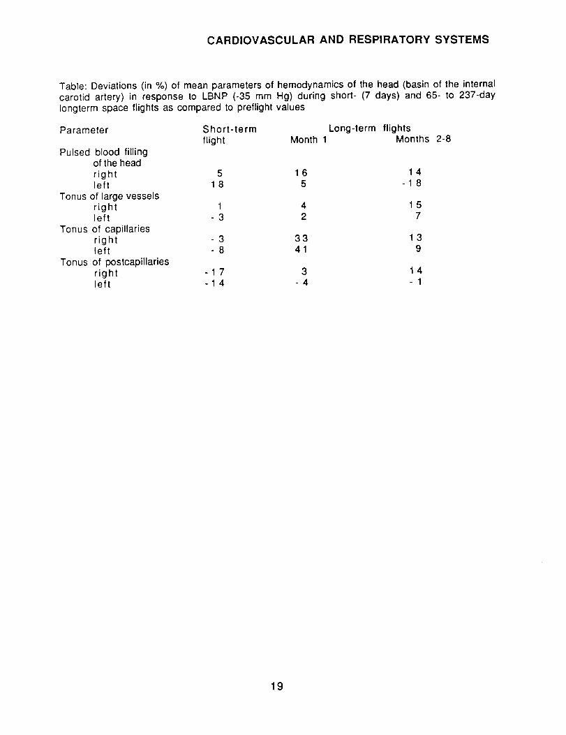
Table: Deviations (in %) of mean parameters of hemodynamics of the head (basin of the internalcarotid artery) in response to LBNP (-35 mm Hg) during short- (7 days) and 65- to 237-dayIongterm space flights as compared to preflight values
Parameter Short-term Long-term flightsflight Month 1 Months 2-8
Pulsed blood fillingof the headright 5 1 6 1 4left 1 8 5 - 1 8
Tonus of large vesselsright 1 4 1 5left 3 2 7
Tonus of capillariesright 3 33 1 3left - 8 4 1 9
Tonus of postcapillariesright - 1 7 3 1 4left -1 4 - 4 - 1
19

CARDIOVASCULAR AND RESPIRATORY SYSTEMS
HR, bts/min
v,/•
' _ "//:1 F
a
SV,
I00 I-
i11
".//. 80 t.-
gll
515fl/
y_
F
HR, bts/minIO0 r
i
........ VXA70 _- _.f-f-= VIAleO1- .... ,.//.4 IV..//]
IN50{ . g";'._1
SV, % a b c
125_
' .... r_50 = .....
CO, % a b c
150 _ ....I.'/1.1
125 _- =.//.i .... V/I///.4 i/ //.,i
.... ,....Y//2 1_lOO_- _/,/J ,----._//_
_''.2 .... I>>;. I /Aa b c
%
COP
ii
*01-"
J
80 - 5///,,.//A ,'//// i / _ //1"/
Y//A Y/l".... eo - 5//.4.///A ///.
9"/.._ 123;I//A _//i//7.i ,111
,//A i///.4 _O - 5////.... ////IlIA t11/i/.._ _ ////
.... ,'///
_////I
_,.__
5"//._
F
Figure 1: Change in mean values of heart rate (HR), stroke volume (SV), and cardiac output(CO) before and during long-term space flights (65-237 days)
A- in response to graded physical exercise test (minute 1 of recovery); b- in responseto LBNP (-35 mm Hg)Here and in Figure 4: white bars - before the test; hatched bars - after the test. a -preflight, b - month 1 inflight; c - month 2-8 of flight F - flight
20

CARDIOVASCULAR AND RESPIRATORY SYSTEMS
PBP,units b
_L
5_
5_
PBP,units'o- _ b
8_7L
5;
A
_tr, "s. b
If
Io
9
7
B
/o
o
7
$
Dcl, %7o_ a b
50
_0 P
30
20_
Dcl, %b s0- Q b
#0
Figure 2: Change in mean value of pulsed blood filling parameter (PBP), tonus of large vessels(odT) and precapillaries (Dcl) of vessels of the forearm (a) and calves (b) at rest and duringshort-term - 7 day (A) and long-term - 65-237 day flights (B)
A: white bars - preflight, hatched bars -flight. B: white bars - preflight; hatchedbars - month 1 of flight; cross hatched bars - months 2-8 of flight
21

PBP,
units a
.oLi
r IVA
:!I-
CARDIOVASCULAR AND RESPIRATORY SYSTEMS
b _Ir.
8-
¢,-,-,
r,2_
NV/AV/AI,'/11
_,,_ s-
a b % Q b
p'/,
Dcl,
m
// i
i.."
PBP, BDcl, %
a/r % a b 4o- a bunits _ b I
:I_"7 20- !
i Y/, Io- "
Figure 3: Change in mean value of pulsed blood filling parameter, tonus of large vessels (ou'T)and precapillaries (Dcl) of vessels of the head (basin of the internal carotid artery, a - righthemisphere; b - left hemisphere) at rest and during short-term - 7 day (A) and long-term -65-to 237-day flights (B)
Legend same as Figure 2.
PBP,PBP, units units
3
Dcl, %
I30 i _/
2o !V
• wl
O
F a F o b c a b c
-- z/_4
//i
F
g
;z
O F
B Dcl
lop
% i
a b c a b c
Figure 4: Change in mean value of pulsed blood filling parameter, tonus of large vessels (_T)and precapillaries (Dcl) of vessels of the head (basin of the internal carotid artery, I - righthemisphere; II - left hemisphere) in response to LBNP (-35 mm Hg) before and during short-term - 7 day (A) and long-term -65-to 237-day flights (B)
22
CARDIOVASCULAR AND RESPIRATORY SYSTEMS
P1208(27/90)* Kuznetsov VI.Cardiac contractility of guinea pigs exposed to long-term continuous stress.Kosmicheskaya Biologiya i Aviakosmicheskaya Meditsina.23(6): 42-47; 1989.[18 references; 3 in English]
Cardiovascular and Respiratory Systems, Cardiac ContractilityGuinea PigsPsychology, Stress
Abstract: Experiments were performed on 52 female guinea pigs. Subjects were stressed byplacing them in groups of 3-4 in confining cages for periods of 60 and 100 days. Matchedcontrol groups were used. Food and water were not restricted. During the first stage of theexperiment cardiac contractility was measured in anesthetized animals after opening of thechest cavity. Pressure in the left ventricle was recorded using an electromanometer. To testresistance to isometric loading, the aorta was occluded for 30 seconds, with pressure recordedat seconds 5 and 25. The pressure curve was used to compute the following parameters: heartrate, developed pressure, maximal rate of contraction and relaxation of the left ventricle. Ratepressure product was computed as the product of heart rate and developed pressure, while theindex of structural function was considered the product of heart rate and developed pressure perunit dry mass of the mydocardium of the left ventricle. During the second stage, the heart wasrapidly removed from anesthetized guinea pigs and placed in an ice-cold normal saline solutionuntil contraction stopped. The heart was then perfused with an oxygenated bicarbonate Krebs-Henselate buffer, with pH of 7.3-7.4 and temperature maintained at 34°C. A metal cannula wasinserted in the left auricle, through which the solution entered the left ventricle. The aortalcannula was attached with a glass tube with outlets every 10 cm allowing graded estimate ofaortal resistance. Studies of the hearts of untreated and chronically stressed guinea pigs were
performed under conditions of spontaneous heart rate and with aortal pressure fixed at 80 cmH20. The muscle fibers of the ventricle were subjected to stretching by increasing pressure inthe left auricle fro 5 to 25 cm H20. Data were tested using Student's t.
After 60 days of stress, the body weight of the guinea pigs was depressed by 12%, and after 100days by 14%. The dry mass of the left ventricle had not changed significantly after 60 days ofstress, but had decreased by 10% after 100 days. Relative mass of the ventricle did not differfrom controls. When cardiac contractility was studied with the heart in the body, it was foundthat after 60 days of stress, heart rate and intraventricular pressure did not differ fromcontrol, although developed pressure showed a tendency to increase, causing the parameter ofintensity of contractile function to increase by 29%. Maximal rates of contraction andrelaxation were unaltered, while the parameter of intensity of structural function was elevatedby 39%. In second 5 after occlusion of the aorta, heart rate, developed pressure, rate pressureproduct and index of structural functioning were unchanged, while maximal rate of contractionand relaxation had increased by 34 and 35%.
After 100 days of stress, all the above parameters previously undergoing changes had returnedto control values at rest and at second 5 of occlusion. The exception was index of structural
functioning which was elevated by 32 and 27% at rest and second 5 of occlusion. This effect wasassociated with a slight increase in heart rate and a slight decrease in dry mass of the leftventricle.
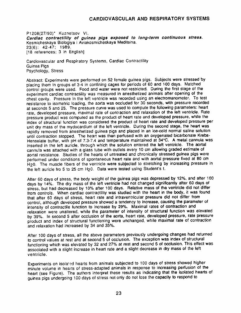
Experiments on isolated hearts from animals subjected to 100 days of stress showed higherminute volume in hearts of stress-adapted animals in response to increasing perfusion of theheart (see Figure). The authors interpret these results as indicating that the isolated hearts ofguinea pigs undergoing 100 days of stress not only do not lose the capacity to respond to
23

CARDIOVASCULAR AND RESPIRATORY SYSTEMS
increased filling pressure by increasing minute volume, but even at some pressures showincreased output relative to controls. The authors conclude that continuous 60- to 100-daystress leads to development of adaptive processes in the heart, manifested in increasedcontractile function of the myocardium in mature animals that (unlike rats) have ceased togrow. The effects of adaptation can be observed at the level of the organism as a whole as well asin isolated hearts, suggesting that adaptation involves central mechanisms of regulation, as wellas autoregulation.
Table 1" Change in myocardial contractile function in guinea pigs undergoing long-term chronicstress
Table 2: Change in minute volume of isolated hearts in control and stress-adapted animals atvarious loading levels
24
2t
/8
15
12
9
6
7
]//t_'\
,/
Figure: Filling pressure and minute volume in experiments on isolated hearts of control guineapigs (a) and those undergoing a 100-day period of stress
Abscissa - filling pressure in the left ventricle (in cm H20); ordinate - minute volume(in ml/min per 1 kg moist tissue of the left ventricle. * - significant differences
24
CARDIOVASCULAR AND RESPIRATORY SYSTEMS
P1226(27/90) Beketov AI, Konyayeva Yel.Circulation and oxygen pressure in the brains of alert and anesthetized rabbitsin a head-down tilt position.Fiziologicheskiy Zhurnal SSSR im I.M. Sechenova75(11): 1548-1553.Authors' affiliations: Medical Institute of Crimea, Simferopol; Sechenov Institute ofEvolutionary Physiology and Biochemistry, Leningrad
Cardiovascular and Respiratory Systems, Circulation, Oxygen Pressure, BrainsRabbits, Alert, AnesthetizedHead-Down Tilt
Abstract: The goal of this work was to analyze changes in local blood flow and oxygen pressure invarious regions of the cerebral cortex and total brain blood flow in alert and anesthetizedrabbits in head-down tilt position. Subjects were 23 alert and 4 anesthetized rabbits. Changesin local and total brain blood flow were measured using the hydrogen clearance method with ananovolt ammeter and a loop oscillograph, oxygen pressure was measured polarographically.Animals breathed hydrogen in a 5% mixture with air for 1 minute. Specific blood flow rate wascomputed using the "initial slope" method and expressed in mill00 g/min; oxygen pressurewas expressed in percent of baseline. Local blood flow and PO2 were measured using platinumneedle electrodes, inserted through holes drilled in the skull of the parietal, frontal, occipital,and left and right temporal lobes of the cerebral cortex. Overall brain blood flow was computedfrom venous outflow from the skull and P02 in venous blood was measured using electrodes incontact with the dura mater in the confluence of the venous sinuses. In the chronic experimentelectrodes were implanted 2 weeks before the experiment and, in the acute experimentperformed on anesthetized animals, inserted immediately before the experiment. Bloodpressure was measured in the short-term experiment in the femural artery, and in the long-term one through an indirect method based on the pulse in the femural artery. EKGs were alsorecorded. Blood flow was measured every 5-10 minutes in the initial position, during head-down tilt (-45 ° for 1 hour), and after return to the horizontal position. At these sameintervals changes in PO2, blood pressure, and EKG were also recorded.
In alert animals during head-down tilt blood flow changed in different ways in different parts ofthe cortex. Immediately after the animals were placed in this position, blood flow increasedinsignificantly in the parietal, left and right temporal, and decreased insignificantly in theoccipital lobes. After 10 minutes it significantly increased in the right temporal and frontalareas of the cortex and decreased significantly in the left temporal area. No change occurred inthe parietal and occipital areas. After 30 minutes and continuing until the end of the tilt periodmost regions of the cortex displayed decreased blood flow. At the onset of head-down tilt, therewas a 55% increase in blood flow in the confluence of venous sinuses of the brain, which wasreplaced after 10 minutes by a decrease (to 77% baseline). Blood pressure changes alsounderwent two phases: increase to 108% at minute 5 and an insignificant decrease toward theend of the tilt period. No change in EKG was noted, although heart rate gradually decreased.
After the animals were moved into a horizontal position, there was a significant increase inblood flow in the parietal and frontal cortex and a statistically insignificant increase in theoccipital and left temporal areas. Blood flow in the right temporal area decreased significantly,while blood flow in the sinus confluence increased. Change into the horizontal position led to arapid but short-lived hypertensive reaction. When 30 minutes had elapsed after termination ofhead-down tilt, the majority of parameters (except for the parietal region, were belowbaseline). Onset of head-down tilt led to decreased PO2 in all regions of the cortex and in theconfluence of the brain sinuses. After return to horizontal position, PO2 in the parietal and left
temporal areas rapidly returned to baseline and then decreased again. After 30 minutes, PO2
25

CARDIOVASCULAR AND RESPIRATORY SYSTEMS
remained below normal in the cortex and had decreased still further in the confluence of thesinus.
In anesthetized animals onset of head-down tilt led to changes in different directions in thevarious regions of the cerebral cortex. In the parietal and temporal regions there was first ashort-term, insignificant decrease in blood flow and then an increase lasting 10-20 minutes,followed by another decrease. In all other areas and in the confluence of the sinuses there was adecrease in blood flow throughout the period of head-down tilt.
The authors conclude that the most extreme changes in systemic and organ hemodynamics occurduring the first few minutes after change of position. The most characteristic and pronouncedfluctuations in blood flow and pressure are seen during these periods and may be considered theinitial response to postural factors. Subsequent changes in hemodynamics may be a result ofinteraction between the disturbing element and compensatory mechanisms serving to increasefunctional stability of the brain circulation system. Thus, stimulating compensatorymechanisms through various measures, including drugs, may facilitate adaptation of cerebralhemodynamics to extreme conditions.
Figure 1: Blood flow in various regions of the brain in alert rabbits during head-down tilt
Figure 2: Changes in blood flow rate in various regions of the brain in alert and anesthetizedrabbits during exposure to head-down tilt
26

ENDOCRINOLOGY
PAPER:
P1203(27/90)* Prodan NG (USSR) and Baranska V (Poland).Morphological research on the adrenal glands of rats after flight on1667.Kosmicheskaya Biologiya i Aviakosmicheskaya Meditsina.23(6): 27-30; 1989.[15 references; 1 in English]
COSMOS-
Endocrinology, Adrenal Glands, MorphologyRats, MalesSpace Flight, COSMOS-1667
Abstract: The adrenal glands of 7 adult male rats of the Wistar line sacrificed 4-8 hours aftercompletion of a 7-day space flight and 7 rats of a vivarium control group were cleaned of fattissue, weighed, and fixed. After fixation the entire gland was embedded in paraffin. Sections4-5 p.m thick were prepared from the central portion of the gland (with ,a maximum of corticalsubstance) and stained to allow differentiation of cells secreting adrenalin (A-cells) andnoradrenalin (N-cells). Measurements were made of the relative amounts of cortical andmedullary substance, size of cytoplasm and nuclei of cells in different zones of the cortex; thearea occupied by the A- and N- cells, size of nuclei and volume density of medullary cells. Animage analyzer was utilized for measurement. Data were tested using Student's t, with a p <0.05 significance level.
No differences were found between flight and control animals with respect to adrenal weight andvolume of cortical and medullary substance. Histological examination did not reveal significantchanges in flight animals, with the possible exception of a slight narrowing of the glomerularzone. Cortical hyperemia was noted, with capillaries dilated in the reticular and fascicularzones. Judging by vacuolization of the cytoplasm, there was a slight decrease in lipids in flightrats. There was a slight but significant increase in the areas of cytoplasm and nuclei in theglomerular zone, without the ratio of the two being altered. Size of nuclei of cells in thefascicular zone increased. In medullary substance, area occupied by N-cells decreased by 28%in flight rats, while area occupied by A-cells was unchanged. In flight rats size of medullarycell nuclei decreased, while the volume density of the nuclei increased considerably.
The authors conclude that the stress response engendered by short-term exposure toweightlessness was slight, as evidenced by absence of hypertrophy of the cortical substanceoverall and in the fascicular zone. Increase in cell size in this zone evidently resulted fromshifts in fluid electrolyte balance during early exposure to space. The results of morphologicalstudy of the medullary substance suggest that weightlessness does not act as a stressor of thiscomponent of the sympathetic system, since there is no sign of hypertrophy and indeed areaoccupied by N-cells and nuclei of medullary cells was diminished.
Table 1: Weight of the adrenal glands and relative amounts of cortical and medullary substancein rats in the flight and vivarium control groups
Condition Adrenal weight,mg per 100 g bodyweight
Flight 12.7___0.36Control 12.8+0.48
Relative Volume, %cortical medullarysubstance substance
83.62+0.77 16.38+0.7784.70+0.70 15.30_+0.70
Differences are not statistically significant.
27

ENDOCRINOLOGY
Table 2: Results of morphometric studies of the cortices of rats after a 7-day space flight onbiosatellite COSMOS-1667
Parameter
Volume of cell cytoplasm, _m 3
Volume of cell nuclei, p.m3Ratio of nuclei and cytoplasm,%
Volume of cell cytoplasm, p.m3
Volume of cell nuclei, t_m3Ratio of nuclei and cytoplasm,%
Volume of cell cytoplasm, _m 3
Volume of cell nuclei, _m 3Ratio of nuclei and cytoplasm,%
Control Group Flight Group pGIomerular zone
201.51_+11.7 223.91_+12.96 <0.0585,50-+1.69 94.96-+1.69 <0.0542.43-+1.62 42.41 -+1,70 >0.1
Fascicular zone
324.10_+20.97 310.7__.18.46 >0.1114.60_+1.71 139.97-+2.97 <0,05
35.36_+1.76 45.05_+1.72 <0.05Reticular zone
261.49_20.63 258.67_+14.55 >0.1
88.90+2.83 94.14_+2,14 >0.133.99_+1.59 36.39_+1.22 >0.1
Table 3: Results of morphometric investigation of medullary substance of adrenal glands of ratafter space flight on biosatellite COSMOS-1667
Parameters Total area, mm 3A-cells N-cells
Vivarium control 0.700_+0.002 0.070_+0.001
p<0.05Flight 0.700+0.002 0.050-+0.002
Volume of Volumecell nuclei, density ofmm 3 nuclei, %
144.70-+3.10 6.48_+0.41p<0.05 p<0.05
128.51 _+2.37 16.87_+1.03
28

ENDOCRINOLOGY
P1211(27/90)* Yangalycheva EA.Hypothalamus/pituitary neurosecretory system inhypoxia.Kosmicheskaya Biologiya i Aviakosmicheskaya Meditsina.23(6): 54-62; 1990,[21 references; 3 in English]
rats exposed to high.altitude
Endocrinology, Hypothalamus, Pituitary; Neurophysiology, Neurosecretory Apparatus;Morphology
RatsAdaptation, Hypoxia, High-Altitude
Abstract: Morphological changes were studied in the hypothalamic/pituitary neurosecretorysystem in 37 white rats during a period of adaptation to high altitude (3200 m above sea level)conditions during the summer. Animals were sacrificed for study on day 3 (n=6), 10 (n=4),20 (n=6), 30 (n--5), 40 (n-6), and 60 (n=4) of adaptation. The hypothalamus and thepituitary were isolated and fixed in Bouin's fixative. Frontal and sagittal sections were laid outin an incremental series and stained to reveal nucleic acids. The functional status of the systemwas estimated from a number of quantitative and semiquantitative parameters. Functionalsignificance of migration of Gomori-positive granules of the neurosecretory substance wascomputed by formula from parameters, such as the concentration of neurosecretory substancein neurosecretory cells, the supraoptic nucleus, and supraoptic terminals, and secretion of typela neurosecretory substance by the supraoptic nucleus.
The topography and form of the glial nuclei in the supraoptic nucleus and neurohypophysisrevealed clear phase-related differences: if the nucleus was enlarged, then the number ofspherical nuclei (active phase of adaptation) was elevated, and vice versa. Close structuralinteractions among glial nuclei, neurosecretory cells and their axons, and terminals in theneurohypophysis determine the characteristics of the functioning of thehypothalamus/pituitary neurosecretory system under conditions of high altitude.
Table 1: Major characteristics of the supraoptical nuclei of rats adapting to high altitude
hypoxia
Table 2: Changes over time in neurosecretory substance in the neurohypophysis in rats duringadaptation to high altitude
Table 3: Statistical characteristics of the state of the supraoptic nucleus and neurohypophysis ofrats under conditions of high altitudes
Figure 1: Hypothalamus-pituitary system of rats undergoing adaptation to high altitudes
Figure 2: Dynamics of morphological changes in the neurohypophysis of rats during adaptationto high altitudes.
29

ENDOCRINOLOGY
P1219(27/90)* Zagorskaya YeA.Endocrine response to low frequency electromagneticintermittent generation.Kosmicheskaya Biologiya i Aviakosmicheskaya Meditsina.23(6): 4-15; 1990,[126 references; 54 in English]
fields of continuous and
Endocrinology, Endocrine ResponseHumans and Animals, Review ArticleRadiobiology, Low-Frequency Electromagnetic Fields, Constant, Intermittent
Abstract: This work reviews recent literature on the biological effects of exposure to low-frequency contintuous and intermittent electromagnetic fields, especially on the endocrinesystem. A number of works have demonstrated that the hypothalamus is one of the mostsensitive organs to electromagnetic fields, and that the hypothalamus-hypophysis region is oneof the first portions of the body to react to such stimulation. Even with weak intermittentelectromagnetic fields and short exposure concentration of ACTH in the pituitary has been shownto increase substantially after exposure. Morphological study of the adenohypophysis of ratsrepeatedly exposed to intermittent electromagnetic fields revealed signs of activation or strainon the functions of the gonadotropic cells and those that synthesize thyrotropin. Exposure to alow frequency constant electromagnetic field leads to cyclic changes in the posterior lobe of thehypophysis, supraoptic nucleus, hypothalamus and adrenal cortex, with alternation of elevatedand depressed hormonal activation. These effects are interpreted as indicating decreasedregulatory potential of the neurohumoral and pituitary-adrenal gland system.
Electromagnetic fields have been found to have significant effects on melatonin, a regulator ofmany endocrine reactions. These effects are associated with disrupted circadian rhythms. Afterchronic exposure to certain intermittent fields the cortical layer of the adrenal gland increasedin width due to cells of the fascicular zone. 11-OCS was found to increase in the blood of ratsafter exposure to a constant electromagnetic field. Response of the adrenal gland in mice variedas a function of duration of exposure to the field. There was some evidence of adaptation toconstant long duration exposure. Effects were found to vary qualitatively as a function of fieldstrength and other conditions of exposure.
Humans exposed to low frequency constant magnetic fields close in field strength and frequencyto that of a natural magnetic storm displayed changes in the functional activity of thepituitary/adrenal system. Differences were noted between effects of single and multipleexposures and individual differences were marked It seems likely that long-term exposure willdiminish the reserve capacities of the systems.
Many studies have demonstrated that the testes of animals are sensitive to the fields underdiscussion. Changes found have included hemodynamic disturbances, disruption ofcytoarchitecture of the spermatogenic epithelium, and depressed spermatogenesis.Morphological change evoked by repeated or chronic exposure did not always normalize, evenafter 1-2 months. Human studies have shown increased concentrations of testosterone after
exposure to such fields. Results of exposure to these fields on reproductive capacities ofanimals have been contradictory, possibly due to differences in field intensities or otherdiscrepancies in conditions. Results concerning effects on female reproductive function havealso been contradictory.
Animal results have indicated that low frequency constant magnetic fields can affect the thyroidglands, with both depression and activation of thyroid activity noted under different conditionsof field strength, exposure, and delay. Human results have included increased levels of
30

ENDOCRINOLOGY
thyrotropin, and free and total thyroxin, as well as decrease in thyroxin in blood. Some of theseeffects have been relatively long-lasting.
Data on pancreatic effects of low frequency magnetic fields are very sparse. Insulininsufficiency has been reported in rats.
Reported effects on the sympathetic adrenal system have included decreased adrenalin in theadrenal gland, decreased noradrenalin in the brain, hypertrophy of chromaffin cells in theadrenal cortex, and decreased noradrenalin in the hypothalamus.
The authors conclude that these data attest to the sensitivity of the endocrine system to exposureto low-frequency constant electromagnetic fields. Evidently, alteration of the functionalactivity of the pituitary-adrenal, pituitary-thyroid, and sex hormone systems, and of thepancreas, which are regulated by higher levels of the neuroendocrine systems may change as afunction of intensity and duration of exposure, sensitivity of the gland, and individualsensitivity. Intermittent electromagnetic fields of the same induction and frequency havegreater biological effectiveness than constant ones. The response evidently is not specific and issimilar to that noted in the general adaptation syndrome. Duration of symptoms of endocrinestress suggest that complete adaptation does not occur to this factor. It is possible that the testesare more sensitive than the ovaries to the effect of low-frequency constant magnetic fields, butthere is insufficient data to support this conclusion. The pancreas is highly sensitive toconstant electromagnetic fields with 20mT, 50 Hz, but effects of fields with other parametershave not been studied.
Exposure to constant magnetic fields with specially selected parameters has been tested as ameans to increase general resistance to various adverse factors. Such exposure has been foundto help rats withstand hypokinesia. In clinical practice, continuous and intermittent low-frequency electromagnetic fields have been used therapeutically for ischemic heart disease, highblood pressure, and inflammation of the fallopian tubes and ovaries.
It has been demonstrated that exposure to low-frequency electromagnetic fields is associatedwith decrease in rate of catabolic processes, increase in anaerobic glycolysis, disruption of thepermeability of cell membranes, and development of hypoxia in the cells of certain tissues.Repeated exposure had been associated with changes in nucleic acid and nitrogen metabolism invarious tissues. It has not been determined whether the initial effects of these fields occur at thelevel of the whole organism through regulatory structures of the hypothalamus, pituitary, andadrenal glands, or directly at the cellular level, through the sensitive cell membranes andmitochondria. There is no appropriate model of the effects of low-frequency electromagneticfields on the nervous system.
According to the author, the data he cites suggest that, by virtue of their primary interactionswith the cellular membrane, constant electromagnetic fields can be applied to affect adenylatecyclase and thus alter synthesis of cAMP and/or the response of "secondary mediators." Heargues that electromagnetic fields in all likelihood influence the organism through a set ofchanges at various levels of organization. It is possible that as intensity of the stimulationincreases, the key role played by certain mechanisms will increase. He concludes that increasein research on various aspects of hormone-receptor interactions and transmission of hormonalsignals to the cell during exposure to low frequency constant magnetic fields will make animportant contribution to understanding how this factor operates on biological subjects.
31

ENZYMOLOGY
PAPER:
P1225(27/90) Shepotinovskiy Vl, Rogoznyaya NA, Mikashinovich Zl.Activity of digestive enzymes in response to immobilization stress and itspharmacological correction with adrenoreceptor blocking agents.Patologicheskaya Fiziologiya i Eksperimental'naya Terapiya.1989(6):49-52.[10 references; none in English]Authors' Affiliation: Central Scientific Research Laboratory, Rostov Medical Institute
Enzymology, Digestive Enzymes; Gastrointestinal SystemRatsAdaptation; Immobilization Stress; Pharmacological Countermeasures; Adrenoblockers
Abstract: This experiment was performed on 80 white rats of both sexes. Stress was induced inhungry animals by immobilizing them on their backs for 3 or 24 hours. In another condition,animals were adapted to stress by gradually increasing immobilization for from 15 minutes to3 hours over the course of 10 days. The a-blocker, pyrroxan (6-[B-(3-Phenylpyrroprolidyl-l') propionyl-benzodioxan-l,4 hydrochloride) in a' dose of 1 mg/100g) and the 13-blocker obzidan (Inderal) in the same dose, were administered subdurally to someanimals. One group of these subjects was then exposed to 3-hours of immobilization stress,while the other group received no further treatment. The effects of the drugs on intestinaldigestion were studied 3 hours after their administration in both groups. Sour milk (30 g) wasfed to both groups and after 1 hour enzyme activity of the epithelium of the upper third of thesmall intestine was measured. Activity of enterokinase was measured in a homogenate of ascraping from the mucous membrane in a ratio of 1:10 of distilled water. Activity of amylasewas measured in plasma and expressed in grams of salt hydrolyzed by the enzyme in 1 I ofplasma. Activity of the intestinal isoenzyme of alkaline phosphatase was measured in thepresence of L-phenylalanine, which is an inhibitor of the intestinal fraction of serum alkalinephosphatase. Isoenzyme activity was expressed in micromoles of inorganic phosphorus per 1 mlplasma after 1 hour of incubation.
Food loading evoked significant activation of amylase in blood serum. Activity of intestinalalkaline phosphatase decreased by a factor of 1.8, and enterokinase by 1.7. Animals exposed toimmobilization stress before feeding displayed sharp increases (by a factor of 2) in activity ofamylase and enterokinase, interpreted as a part of the general adaptation syndrome and aconsequence of induction of adrenal hormone activity. In these subjects, activity of alkalinephosphatase was 1/10 that of intact animals. Increase of immobilization duration to 24 hoursled to normalization of enterokinase and alkaline phosphate activity and decrease in amylaseactivity to a level intermediate between intact and 3-hour stressed animals. The stressadaptation procedure significantly decreased gastric response to stress, but fell short ofnormalizing it.
The effect of feeding after 3 hours of immobilization can be described as anti-stress. Activity ofenterokinase dropped almost to that of intact fed animals. Activity of amylases was less elevatedthan before feeding and activity of alkaline phosphatase increased by a factor of 9. Evidentlythe leading role of exogenous food stimulation on digestive processes is maintained after 3 hoursof immobilization stress, despite the changes occurring in endogenous regulatory factors.
In intact animals, administration of adrenoblockers altered activity of the enzymes studied.Pyrroxan, which has a selective effect on co-receptors independent of their localization,decreased activity of all enzymes by a factor of 2.4 to 3. Obzidan, a blocker of 13-receptors, hada less marked inhibiting effect. When immobilized animals were injected with the drugs, the
32

ENZYMOLOGY
stress response of digestive enzymes was virtually eliminated, with the B-blocker obzidanhaving a greater effect.
The authors conclude that stress leads to a rapid change in activity of intestinal enzymes, whichis adaptive in nature. The magnitude of the responses of the digestive system to stress isdetermined by the state of the central mechanisms of regulation, and also the degree ofparticipation of the enteric? and hormonal systems, which through numerous feedback loopsinteract with the general hormonal system. Utilization of a- and _- blockers decrease theactivation of the adrenergic regulatory system in response to stress and thus alter activitylevels of pancreatic and intestinal hormones.
Table 1: Activity of intestinal enzymes in intact and immobilized animals
Table 2: Effects of a- and _-blockers on the activity of enzymes in intact and immobilized rats
33

EXOBIOLOGY
PAPER:
P1229(27/90) Kuzicheva YeA, Malko IL.
Abiogenic thermal synthesis of nucleotides in the presence of lunar soft.Zhurnal Evolyutsionnoy Biokhimii i Fiziologii.XXV(6):697-701; 1989.[13 references; 4 in English]Authors' Affiliation: Institute of Cytology, USSR Academy of Sciences, Leningrad
Exobiology; Abiogenic Synthesis; ThermalNucleotidesLunar Soil
Abstract: This work investigated the biogenic thermal synthesis of nucleotides in the presence oflunar soil and its analogue tholeitic basalt. The lunar soil used had been sampled by the Luna-16unmanned station from the Sea of Fecundity. The sample was composed of dark gray regolitewith particle diameter of approximately 0.2 mm. Chemicals used included labelled uridine14C, unlabeled uridine-5'-monophosphate, cyclic uridine-2',3'-monophosphate, uridine-5'-diphosphate, uridine-5'-triphosphate disodium salts, ion-exchange tar DEAE-sephadex,sephadex G-10, unpurified snake venom, and tholeitic basalt. Thermal synthesis of nucleotideswas conducted using an undescribed method. Solid films were prepared from the solution ofuridine 14C and inorganic phosphate in the presence of lunar soil and basalt (and in theirabsence) on a rotor condenser. A column with a solid film of the reactive medium was evacuatedand then placed in a glycerine bath and heated for 2 hours at a temperature of 160°C. The filmwas dissolved in hot distilled water, the lunar soil and basalt filtered out, and the remainderanalyzed. Analysis involved dissolving,initial purification using DEAE-sephadex A-25,selection of the fraction with Ve corresponding to Ve of natural nucleotides, lyophilization,second purification using Sephadex G-10, enzymatic hydrolysis of 5'-bonds, selection of thefractions that did not disintegrate in snake venom, hydrolysis with alkaline phosphatase, andseparation on Sephadex G-10. The nucleotides with an elution volume equal to the elutionvolume of cUMP after two purifications were also incubated with alkaline phosphatase todetermine the amount of cyclic derivative they contained. The yield of products formed wascomputed in terms of initial quantity of nucleoside.
Heating the mixture of uridine and inorganic phosphate in the absence of a substrate led toformation of six synthesis products. The overall yield was 17.8%, and the quantity of uridinenot entering into the reaction was approximately 65%. It was assumed that the remaininguridine disintegrated in response to heat. After the first purification it was established that themajor products were uridine monophosphates. Further processing revealed that fraction Aconsisted of two components A1 and A2. A1 was present in a quantity 3.6% of the total yield, andcontained no compounds with natural bonds, which is logical in synthesis of biochemicallyimportant substances using abiogenic techniques. Yield of A2 was 5.6%. It contained 61%cU-2',3'-MP. Fraction B after the second purification was found to be homogeneous andamounted to 4.6% of the total yield. After enzyme hydrolysis with alkaline phosphatase it wasfound to contain 70% cU-2',3'-MP. Fraction C (2.2% yield) contained 49% U-5'MP and 5%U-2'(3')-MP. Fraction D (yield 1%) contained 10% U-5'-MP and 44% U-2'(3')-MP. Inaddition, fractions E and F contained traces of uridine and uridine monophosphates.
Performance of thermal synthesis in the presence of basalt and lunar soil significantlydecreased the total yields of reactions to 4.15% and 1.62%, respectively. The composition ofthe reaction products changed too. None of the products contained uridine or uridinemonophosphate. In the presence of lunar soil a substance was formed that had the molecularweight of uridine-monophosphate, but contained no natural bonds.
34

EXOBIOLOGY
The authors conclude that lunar soil is not inert, and they hypothesize that the absence ofbiologically important compounds in lunar samples is due partially to the inhibiting effects oflunar soil.
Table: Characteristics of nucelotides synthesized by heating a mixture of uridine and inorganicphosphate in the absence of a substrate and in the presence of basalt and lunar soil
5O
ZO
10
IIIIII
,4
IIII
C!IIIII IIIjII II I
II II
i_. ,. i F
,I 'i
--'-_"" - t'_'---=-_ _r- .... , --,--_OO _00 FOO 800 1000
Figure: Chromatogram of nucleotides synthesized by heating uridine and inorganic phosphatewithout a substrate (1), in the presence of basalt (2), or lunar soil (3)
Column with DEAE-sephadex A=25 (4X28 cm). Elution using a concave gradient fromdistilled water up to 0.4 M ammonium bicarbonate. Rate of elution 180 ml/hr. Arrowsindicate the position of volumes of elution standards. Ratio of uridine
14C+NaH2PO4.H2):107 i_g+68 I_g. Ordinate: radioactivity of the fractions
(imp/min.10-3); Abscissa - elution volume (ml).
35

HABITABILITY AND ENVIRONMENT EFFECTS
PAPER:
P1210(27/90)* Mukhamediyeva LN, Savina VP, Nikitin Yel.Thermoregulatory responses in humans to moderateKosmicheskaya Biologiya i Aviakosmicheskaya Meditsina.23(6): 51-54; 1990.[20 references; 7 in English]
levels of hypercapnia.
Thermoregulatory ResponseHumans, MalesHabitability and Environment Effects, Hypercapnia; Pressurized Environment
Abstract: This work examined the effects of moderate hypercapnia on thermal regulation inhumans. A total of 20 apparently heathy male volunteers participated in this study. Theirthermal status was studied on the basis of measurement of body temperature and skintemperature at 11 points, weighted average mean temperature, mean body temperature andchange in heat content. Heat production was estimated on the basis of gas exchange. Externalrespiration was monitored on the "Spirolit-2" apparatus. Acid base equilibrium ofarterialized blood was estimated on the basis of partial CO2 pressure, concentration of basebuffer, and base excess. Activity of catalases were measured in blood, as were pyruvate,lactate, and number of erythrocytes. The 10 subjects in the experimental group spent 40 daysin a pressurized chamber with a constant level of 1.1-1.6% CO2 and 19-20% oxygen. Airtemperature was maintained at 21-23 ° and humidity at 50-70%. The control group spent 40days in an analogous chamber with CO2 level of 0.2-0.4% and all other conditions the same asfor the experimental group.
Body temperature decreased on day 19 of hypercapnia, but this change only reached 0.5 ° on day39. Beginning on day 3 of exposure, gas exchange increased, remaining at the elevated levelthroughout the treatment period. Acid-base balance displayed changes characteristic ofcompensated respiratory acidosis. The greatest changes in acid-base balance occurred on day 3of treatment. Number of erythrocytes were virtually unchanged throughout the period,suggesting maintenance of adequate blood oxygen capacitance. Level of lactate in blood decreasedsignificantly starting on day 3 of treatment, possibly suggesting inhibition of glycolysis. Ashypercapnia exposure increased, there was a gradual decrease in the heat content of the bodybefore day 19 attributable to decrease of skin temperature and subsequently to coretemperature decreases as well. Starting on day 3 of exposure skin temperature decreased inthe distal areas of the limbs. Foot temperature decreases were most extreme, and were 2.2 °below baseline on day 39. Control subjects also displayed a (less marked) decrease in mean skintemperature, which the authors attribute to hypodynamia. The authors conclude that the resultsshowed that continuous exposure to hypercapnica (1.1-1.6% CO2) for 40 days in athermoneutral zone was accompanied by a "cooling" effect along with an increase in heatproduction starting on day 3. The subsequent significant decrease in lactate in blood wascorrelated with decreased body temperature (r=+0.79). Additional changes includedrespiratory acidosis, a tendency for excess base buffers to decrease in blood, and disruption ofthermal balance as manifested in decreases in body temperature and body heat content, whileheat production remained stable. These results do not preclude the possibility that hypercapnialimits thermogenesis at the stage of glycolytic synthesis of ATP.
Table 1: Parameters of the functions of external respiration, acid-base balance of arterializedblood and biochemical parameters of blood in exposure to moderate levels of hypercapnia
Table 2: Thermal parameters of subjects in the control and hypercapnic groups
36

HABITABILITY AND ENVIRONMENT EFFECTS
Figure: Body temperature, skin temperature, foot temperature and their ratio underexposure to moderate hypoxia
37

HEMATOLOGY
PAPER:
P1215(27/90)* Orlova TA, Kiselev RK.Use of a ferrocerone test to measure iron reservesconditions.
Kosmicheskaya Biologiya i Aviakosmicheskaya Meditsina.23(6): 77-81; 1990.[15 references; 2 in English]
under various living
Hematology, Nutrition, Blood Iron; Ferrocerone TestHumansHypokinesia with Head-Down Tilt
Abstract: The objective of this work was to develop a methodology for evaluating reserves ofiron in the body and to test it on healthy individuals under normal conditions and whileundergoing head-down tilt of varying duration. Existing measures for testing iron reserves areeither too complex or can only be used when iron deposits are elevated. The authors proposed amethod of assessing iron reserves using ferrocerone (a sodium salt oforthocarboxybenzoylferrocene), which is utilized clinically as a supplement to correct irondeficiency. The reasoning was that if the body's supply of stored iron was limited, then ironsupplements would be utilized rather than excreted. The particular supplement (ferrocerone)was chosen because it is highly soluble in water and readily absorbable by the mucousmembrane of the gastrointestinal tract, enters the bone marrow rapidly, and is well toleratedwithout side effects. To test dynamics of iron excretion in healthy individuals under conditionsof normal activity, a total of 24 healthy young men (group 1) were given ferrocerone in theform of a tablet containing 0.04 g iron in the morning after breakfast. Urine was collectedduring the following time periods: 09:00-13:00, 13:00-16:00, 16:00-20:00, 20:00-09:00, and iron levels were measured in each sample, on the basis of reaction withbathophenylanthroline. The study of iron levels under conditions of hypokinesia and head-downtilt utilized 20 individuals, 6 of whom spent 50 days in head-down (-6 °) hypokinesia whiletaking folicobalamine, a stimulant of hemopoiesis (group 2). The 14 individuals in group 3spent 120 days at a head-down angle of -4.5 °. Group 2 was studied on days 25 to 15 beforetreatment, on days 25 and 46 of head down tilt, and days 12 and 28 afterwards. Group 3 wasstudied before treatment, on days 28, 30, 60-70, and 105-110 during treatment, and 4-5days after it. Hemoglobin mass was also studied in subjects undergoing hypokinesia.
After individuals living under normal conditions took the dose of ferrocerone, they excreted atotal of approximately 50% with urine. In these subjects two temporal patterns of excretionwere observed: in one type. excretion peaked during the first 4 hours after taking the drug, andin the other, during hours 4-8. Analysis of the subjects exposed to hypokinesia revealedsimilar results for the two hypokinesia experiments: 1)before hypokinesia, renal excretion ofiron was analogous to the norm 2)during days 25-30 of hypokinesia with head-down tilt,excretion of iron after ingestion of ferrocerone was virtually the same as baseline, while massof hemoglobin decreased; 3) on day 45 excretion of iron began to decrease, with hemoglobineither increasing slightly or continuing to decrease; 4) on days 65-75 for the 120-day studyand day 13 of readaptation for the 50-day study, iron retention was greatest, with 68% of ironadministered retained in group 2 and 62% in group 3 retained. Subsequently iron excretionwas at approximately normal levels, regardless of duration of hypokinesia with head-down tiltor stimulating substance used. Recovery of hemoglobin mass differed somewhat in the twogroups. The authors recommend the ferrocerone test as a technique for evaluating iron reservesbefore iron deficiency is manifest in the blood.
Table: Types of fractional renal excretion of iron in healthy humans after administration of 300mg ferrocerone
38

HEMATOLOGY
Figure: Changes in renal excretion of iron after ferrocerrone loading and hemoglobin massunder conditions of hypokinesia with head-down tilt
39
IMMUNOLOGY
PAPERS:
P1212(27/90) Mirrakhimov MM, Kitayev MI, Tokhtabayev AG.The immune status of individuals suffering from acute altitude sickness.Kosmicheskaya Biologiya i Aviakosmicheskaya Meditsina.23(6): 62-66; 1990.[18 references; 9 in English]
Immunology, Immune StatusHumans, Males, Individual DifferencesAdaptation, High Altitude Sickness
Abstract: A total of 235 healthy males, aged 18-20, were observed during a 150-day period ofadaptation to an altitude of 3600 m above sea level. The subjects were divided into two groupson the basis of adaptive response. Group 1 contained 179 people showing satisfactoryadaptation, and group 2 contained 56 men who during the first 3 days of the study developedsigns of mild or moderate acute altitude sickness. Diagnosis of altitude sickness was only madeif the symptoms persisted for 3 days or more. Immunological examinations were conducted onall subjects at an altitude of 1543 m above sea level. T- and B-cells were identified andmeasured in lymphocytes extracted from peripheral blood. T-lymphocytes were identified usingthe method of spontaneous rosette-formation with sheep erythrocytes (E-RFC). B-lymphocytes were identified on the basis of their capacity to form rosettes in response to theerythrocyte antibody complex EAC-RFC. Null (killer) cells were computed by subtraction.Absolute (per 1 ml blood) and relative (in %) levels of E- and EAC-RFC and null-cells werecomputed. Subpopulations (T,f and Tp.) of T-lymphocytes were identified through presence ontheir membranes of Fc-receptors reacting with IgG and IgM. Functional status of the T-lymphocytes was assessed using the blast-transformation response with PHA and concavalin A.B-lymphocyte functioning was evaluated on the basis of level of immunoglobulins in bloodserum using the radial immunodiffusion method.
It was found that during the first few days of exposure to high altitudes all subjects displayed astatistically significant decrease in total circulation of E-RFC and blast-transformationresponse to PHA. Changes were more pronounced in subjects who suffered from high altitudesickness. Normalization occurred on days 25-30 in individuals adapting successfully, but onlyon day 90 for those who displayed symptoms of high altitude sickness. Absolute concentration ofE-RFC was still depressed in the latter subjects after 150 days. T-lymphocyte functioning wasalso depressed in these individuals, normalizing only after 90 days compared to 25-30 days insuccessful adapters. Blast transformation response was not depressed at all at high altitudes insuccessful adapters, but was significantly diminished in those suffering from high altitudesickness. Subjects who developed high altitude sickness had decreased numbers of Tycells andelevated numbers of Tp. cells during their initial 3 months at high altitudes. Concentrations ofEAC-RFC were depressed in subjects with mountain sickness compared to those adaptingsuccessfully. A similar pattern of depression was noted when functional activity of B-cells wasmeasured. Concentrations of null cells were elevated in those with mountain sickness, anddifferences persisted for 25-30 or 90 days, depending on whether level was considered relativeto 1 ml blood, or total amount of circulating blood. It was found that subjects developingmountain sickness had higher baseline levels of null cells, and lower levels of E-RFC and TI_cells. This may be of significance for selecting individuals likely to adapt well to conditions ofhigh altitude.
Table 1: Concentration of E-RFC in subjects suffering from high altitude sickness during
adaptation
40

IMMUNOLOGY
Table 2: Blast transformation response with PHA and Con A in patients with high altitudesickness during adaptation
Table 3: Concentration of EAC-RFC in patients with high altitude sickness during adaptation
Figure 1" T'y cells in patients with high altitude sickness and individuals adapting successfully tohigh altitude conditions
Figure 2: Tp. cells in patients with high altitude sickness and individuals adapting successfully tohigh altitude conditions
1 2 7 2 t 2
Figure 3: Baseline studies of E-rosette-forming lymphocytes, null lymphocytes and Tp. cells(helpers) in patients with high altitude sickness (1) and those adapting successfully (2) tohigh altitude conditions (3600 m above sea level)
Ordinate - number of cells (in %). Hatched bars - null-lymphocytes; Black bars - E-rosette forming lymphocytes; white bars - Tp.-cells (helpers)
41

IMMUNOLOGY
P1216(27/90)* Novikov VS, Bortinsovskiy VN.The effect of smoking on human resistance in a pressurized environment.Kosmicheskaya Biologiya i Aviakosmicheskaya Meditsina.23(6): 81-85; 1990.[20 references; 8 in English]
Immunology, Resistance; Cardiovascular and Respiratory Systems; Exercise ToleranceHumans, MenSmoking, Habitability and Environment Effects; Pressurized Environment
Abstract: Research was performed on healthy young men spending 2-months in a pressurizedenvironment, performing stressful operator work and undergoing hypokinesia under conditionsof only insignificant changes in temperature, humidity, and atmospheric components. Theexperimental group consisted of 38 smokers, who had smoked for an average of 5 years,andsmoked from 10 to 20 cigarettes per day during the experimental period. The control groupconsisted of 36 nonsmokers. The cellular factor of nonspecific protection, microbial resistanceof skin, functional status of the cardiorespiratory system, exercise tolerance, self assessmentof state, and incidence of illness were studied. Methods used included determining the absorbingand digestion capacity of leukocytes, skin automicroflora, breath holding on exhalation, vitalcapacity of the lungs, blood pressure, heart rate at rest and during graded physical activity,written self assessment of health complaints, and symptoms of diseases. Measurements weremade 1 month before exposure to experimental conditions and on day 15, and during months 15,20, and 50 of the experiment. Health complaints and parameters were additionally measured in240 smokers and 220 nonsmokers working under analogous conditions. Data were testedstatistically using Student's t test.
At the beginning (15 days) of their work in the pressurized environment all subjects displayedsigns of stress, including increased leukocyte absorbing function and decreased efficiency ofintracellular digestion. Effects of experimental conditions were more pronounced in thesmokers. After 30 days in the environment, decrease in protective functions was noted, to amore pronounced degree in the smokers. Skin automicroflora levels were higher at allexperimental periods in the smokers. On day 60 of treatment, parameters of rate ofphagocytosis and effectiveness of intracellular digestion were 16-34% lower in smokers thannonsmokers. Effects on bacteriocidal capacity of lymphocytes were interpreted as due toinhibition of oxygen utilization in smokers. Smokers (52% of the subjects) accounted for79.5% of the recorded illnesses and had more severe illnesses. Smokers had lower vitalcapacity of the lungs, higher heart rates, and higher blood pressure, and lower exercisetolerance than nonsmokers. Smokers were more likely to complain of fatigue, bad moods,headaches, and insomnia, as well as having more rapid heartbeat and more frequent angina
Table 1: Parameters of cellular factors of nonspecific protection in smokers working inpressurized environments
Table 2: Parameters of incidence of disease in smokers working in pressurized environments
Figure 1: Automicroflora of the skin in nonsmokers and smokers working in pressurizedenvironments
Figure 2: Level of exercise tolerance in smokers and nonsmokers working in pressurizedenvironments
42

IMMUNOLOGY
P1220(27/90)* Ignatova OV, Berlin AA, Pak ZP, Popov IG.On the protective function of the skin.Kosmicheskaya Biologiya i Aviakosmicheskaya Meditsina.23(6): 15-19; 1990.[:36 references; 17 in English]
Immunology, Skin, Protective FunctionHumansOperational Medicine
Abstract: This article reviews the literature on the protective function of the skin and how itrelates to support of the health of manned space flights. The authors conclude that this function,on one hand, depends on the general resistance of the macroorganism, and on the other, on thecomposition and biological properties of skin autoflora and is determined as a result of a set offactors: 1) the physico-chemical and biological characteristics of the external environment; 2)the quantitative and qualitative composition of autoflora; 3) the quantitative and qualitativecomposition of the film of moisture and oils on the skin's surface; 4) the morphologicalintegrity of the epidermis. This must be considered when a system of sanitation and hygiene isdeveloped for spacecraft crews. The third factor, which defines skin type, may be of particularimportance for individually tailoring recommendations for selection of cleansing agents forparticular crewmembers. Optimal conditions for maintaining the normal functional status ofthe skin can only be accomplished by considering all of these factors, as well as flight duration.
43
METABOLISM
PAPERS:
P1209(27/90)* Tarasov YuA, Ostovskiy YuM, Satanovskaya Vl, Uopo Av, Velichko MG,Abakumov GZ.
Endogenous ethanol in the blood and tissues of rats exposed to hypobarichypoxla.Kosmicheskaya Biologiya i Aviakosmicheskaya Meditsina.23(6): 47-51; 1990.[29 references; 11 in English]
Metabolism, Endogenous EthanolRats
Hypoxia, Hypobaric
Abstract: In this work the authors studied the level of endogenous ethanol in the blood and tissuesof 18 white male rats as an indicator of response to hypoxia. Animals were divided into threegroups, all of which spent 1 hour in a barochamber. The control group spent the time at normalair pressure of 1020 gPa, experimental group 2 at an altitude equivalent of 2500 m (746.6gPa), and group 3 at a height equivalent of 5000 m (540.3 gPa). After descent at a rate of 30m/second, the animals were decapitated and a portion of tissue immediately frozen in liquidnitrogen. Endogenous ethanol was measured using gas-chromatography. Activity of alcoholdehydrogenase was measured spectrophotometrically using ethanol or acetylaldehyde as thesubstrate. The source of enzymes was a supernatant of tissue homogenate first frozen in liquidnitrogen. Activity of NAD-dependent aldehyde dehydrogenase was assessed in the liver andactivity of aldehyde dehydrogenase in the bone. Total protein, pyruvate, and lactate and the ratioof lactate to pyruvate were measured in the liver and blood. Antioxidant activity of bloodplasma was monitored in the blood and adrenal glands.
Level of endogenous ethanol in blood and tissues turned out to be a sensitive indicator of evenshort exposure to relatively low levels of hypoxia. This parameter does not seem to be relatedto the onset of a stress responses, since 11-OCS only increased in animals exposed to the higherhypoxia levels. Parameters of lipid peroxidation likewise increased only in response to thehigher level of hypoxia. Changes in levels of ethanol cannot be explained with reference toactivity of enzymes metabolizing ethanol, since the latter did not change in a regular fashion.The authors conclude that the concentration of endogenous ethanol in blood and tissues is a moresensitive indicator of exposure to hypoxia than parameters of lactate, lipid peroxidation, or11-OCS.
Table 1: Activity of ethanol- and acetylaldehyde metabolizing enzymes in the liver and brain ofrats exposed to varying levels of hypobaric hypoxia
Table 2: The effects of various levels of hypobaric hypoxia on concentration of lactic andpyruvic acid in the liver and blood of rats.
Table 3: Lipid peroxidation in blood plasma in rats exposed to varying levels of hypobarichypoxia
44

METABOLISM
I50
%
'50
1iiii ,oo50 <. > _ . 30
0 Q b c
Figure 1: Endogenous ethanol in the liver (a), brain (b) and blood (c) of rats exposed tovarying levels of hypobaric hypoxia
Parameters of rats exposed in the barochamber at 765 mm Hg (1) are taken as 100%: 2560 mm Hg (2500 m); 3 - 405 mm Hg (5000 m);
<*> p < 0.1>0.05; * p<0.05
Figure 2: Concentration of 11-oxycorticoids in blood (a) and adrenal glands (b) of rats exposedto varying levels of hypoxia. Key: as for Figure 1.
45

METABOLISM
P1217(27/90)* Tolkacheva NV, Levachev MM, Kobozev GV, Safronova LG, Sorokina AG.Use of the method of thin layer chromatography to study lipid Iigands in theserum albumin of athletes.Kosmicheskaya Biologiya i Aviakosmicheskaya Meditsina.23(6): 85-88; 1989.[23 references; 2 in English]
Metabolism, Lipid Peroxidation, Serum LigandsHumans, AthletesExercise
Abstract: Studies were performed on human serum albumin obtained using electrophoresis in apolyacrilamide gel. Lipid extracts were obtained with an undescribed method and theirconcentration measured. Two methods of thin layer chromatography were used to separate outindividual lipids. In the first, fractions of free fatty acids were separated with a silica gel in asystem of solvents of petroleum ether-ice cold acetic acid (90:10) and stained by spraying onlayers of concentrated HCI, with the lipid spots identified using a standard reference spot ofether of free fatty acids and a mixture of standard lipids. The second method was used forstudying fractions of phospholipids on plates of the Czech substance "Silufol" in a solvantsystem consisting of a chloroform-methanol-25% water solution of ammonia (60:35:5); toreveal the fractions, the layers were stained with a solution of rhodamine yellow, and lipidswere identified with ultraviolet light. Subject samples were compared with that of a controlgroup. Products of lipid peroxidation were identified by an undescribed method. A group of 22highly qualified bicyclists, aged 17 to 25, served as subjects in a transitional period of theyear's training cycle, during which they were engaging in moderate exercise in team races of100 km. Blood was taken from the ulnar vein before and after exercise. For comparativepurposes blood was studied from 40 normal subjects and 19 subjects suffering from high bloodpressure.
The serum albumin of highly trained athletes displayed four additional fractions ofphospholipids: phosphatidylethanolamine, sphingomyelin and the minor fraction ofphosphatidylinosite, and lysophosphatidylcholine absent in the albumin of the control group.Thus the molecules of this protein were saturated with phospholipids. The rate of the process ofdiene conjugates and diene ketones increased in the athletes and was associated directly withtheir level of training. Products of lipid peroxidation decreased in serum albumin as theprocesses of adaptation to various levels of athletic training progressed. In the pathologicalstate, associated with increased hydroperoxides especially in high blood pressure, suchadaptation does not occur.
Table 1: Chemical composition of lipids in serum albumin of subjects
Table 2: Concentration of lipids and products of lipid peroxidation in blood plasma and serumalbumin of athletes
46

METABOLISM
P1218(27/90)* Potapov PP.Activity of NADP-dependent cytoplasmic dehydrogenases in the liver and adiposetissue of rats during recovery after hypokinesia.Kosmicheskaya Biologiya i Aviakosmicheskaya Meditsina.23(6): 89-90; 1989.[11 references; none in English]
Metabolism, Cytoplasmic Dehydrogenases, Liver, FatRatsHypokinesia, Recovery
Abstract: The study of metabolic processes during the period of readaptation after hypokinesia isof interest for its relevance to optimization of countermeasures. This experiment wasperformed on 75 white rats (60 experimental and 15 controls). Animals' motor activity waslimited by placing them in very small individual cages for 30 days. After the experimentalperiod terminated, animals were placed in ordinary group cages. One rat was sacrificed on day30 of hypokinesia and then one a day for the next 25 days of recovery. Data was processed usingthe moving average method. Significance of differences was tested using t,he Wilcoxon MannWhitney non-parametric test. In the first experiment, activity of isocitrate dehydrogenase(ICDH) and glucose-6-phosphate dehydrogenase (G-6-PDH) were measured in the liver andadipose tissue. In the second experiment, activity of NADP-dependent malate dehydrogenase(MDH) was measured in the liver. The cytoplasmic fraction was isolated in a refrigeratedchamber at a temperature of 0-4°C.
On day 30, activity of G-6-PDH was elevated by 33% in the liver compared to control animals.During the initial portion of the recovery period, activity of ISDH and G-6-PDH wasstatistically significantly elevated in liver and adipose tissue compared both to control levelsand levels during hypokinesia. Activity of liver G-6-PDH remained elevated until day 19 ofrecovery, and dropped below normal on days 21-23. ICDH activity in the liver normalized atthe end of the first week of recovery, dropped below normal on days 9-18, and subsequentlynormalized. Activity of MDH was depressed during the first week of readaptation and elevatedfrom days 10 to 19. In adipose tissue, activity of G-6-PDH continued to be elevated until day17 and that of ICDH only until day 6. Activation of ICDH and G-6-PDH in the liver during theearly adaptation period may be considered an appropriate metabolic reaction providingincreased production of NAPDH, while depressed activity of cytoplasmic ICDH between days 9and 18 of recovery may be associated with consumption of ICDH on other metabolic paths.Decreased activity of NADP-dependent MDH during early readaptation may be associated withincreased gluconeogenesis. It should be borne in mind that activity of ICDH is significantlyhigher than activity of G-6-PDH and MDH, thus potential production of NAPDH in the liver isonly elevated during the early period after removal of animals from hypokinesia. Duringreadaptation animals' need for reducing synthesis is elevated. After hypokinesia this might leadto a relative deficit in NAPDH, which should be considered an adverse change in metabolism.
Figure 1: Changes in activity of cytoplasmic NADP-dependent dehydrogenases in the liver inrats during readaptation after hypokinesia
Figure 2: Changes in activity of cytoplasmic NADP-dependent dehydrogenases in fatty tissue inrats during readaptation after hypokinesia
47

MUSCULOSKELETAL SYSTEM
P1206(27/90)* Belakovskiy MS, Khaidakov MS.Effects of active metabolites of Vitamin D3 on the bones of rats exposed todifferent hypokinesia paradigms.Kosmicheskaya Biologiya i Aviakosmicheskaya Meditsina.23(6): 36-39; 1989.[18 references; 11 in English]
Musculoskeletal System, Bones; Metabolism, Calcium, PhosphorusRats, MaleNutrition, Vitamin D3 Metabolites, Hypokinesia, Tail Suspension, Immobilization Cages
Abstract: Previous experiments have shown that active metabolites of vitamin D3 help tooptimize calcium-phosphorus metabolism and the state of bone tissue in hypokinesia createdusing immobilization cages. The current experiment compared these effects to those obtainedusing the tail suspension paradigm. Subjects were male Wistar line rats. The animals receiveda semisynthetic diet with a 0.6% concentration of calcium and phosphorus. Hypokinesia wascreated either through immobilization cages or tail suspension for a period of 6 weeks. Some ofthe animals received oral doses of 1,25(OH)2 and/or 24,25(OH)2D 3 in doses of 0.025 and1.25 mg/day. After treatment, the animals were sacrificed and measurements made of totalcalcium, inorganic phosphate, and PTH in blood serum; and volume, density, and levels ofcalcium, phosphorus in mineralisates of epiphyses and central areas of the diaphyses of thefemur bones. Active transport of calcium in the small intestine was assessed on the basis of
differer, ces in absorption of 45Ca by disks from the duodenum when incubated in a mediumperfused with carbon or nitrogen.
Tail suspension was associated with some inhibition of rate of weight increase and a moderatebut insignificant increase in PTH and some increase'in absorption of calcium in the intestine.The distal epiphyses of the femur in these animals displayed a slight decrease in volume, and asharp decrease in density and calcium and phosphorus levels; while the central regions of thediaphysis displayed an increase in density and mineral levels. Maintenance in theimmobilization cages was associated with a reliable decrease in body weight and the developmentof hypocalcemia with PTH unchanged and calcium absorption in the intestine diminished. Thevolume of the epiphyses decreased substantially. The proximal epiphyses displayed both adecrease in density and concentration of minerals, while in the distal epiphyses theseparameters remained at control level. Changes in the diaphyses were analogous to thoseoccurring in the tail-suspension paradigm.
Oral administration of active metabolites did not lead to changes in body weight, either alone orin combination. In suspended animals receiving metabolites, no changes were noted in bloodcalcium or phosphorus, or rate of accumulation of calcium in the intestine. PTH decreased wheneither or especially both metabolites were administered. Animals of all groups responded to themetabolites with marked increase in density and volume concentration of calcium andphosphorus in the distal epiphysis and diaphyses.
Combined administration of D3 metabolites to animals immobilized in cages led to normalizationof total calcium in blood and some increase in rate of accumulation of calcium in the intestine.
There was a significant increase in density and mineral levels in the proximal epiphyses, whilethere were virtually no changes in the distal epiphyses or diaphyses. The authors conclude thatbone changes resulting from hypokinesia are mainly produced by local factors that increasebone sensitivity to systemic influences.
48

MUSCULOSKELETAL SYSTEM
Table 1: Effects of 1,25(OH)2D3 and 24,25(OH)2D3 on increase in body weight, concentration
of calcium, phosphorus, and PTH in blood serum and absorption of 45Ca by the mucousmembrane of the small intestine of rats exposed to different experimental hypokinesiaparadigms
Table 2: Effects of 1,25(OH)2D 3 and 24,25(OH)2D 3 on volume, density and levels of calciumand phosphorus in the epiphyses of rats exposed to different experimental paradigms ofhypokinesia
Table 3: Effects of 1,25(OH)2D 3 and 24,25(OH)2D 3 on length, density and levels of calciumand phosphorus in the diaphyses of rats exposed to different experimental paradigms ofhypokinesia
49

MUSCULOSKELETAL SYSTEM
P1207(27/90)* Shvets VN, Pankova AS, Gordovskaya MD. Vnukova ZE.Changes induced by hydroxydimethyl aminopropylidene diphosphonate inresponse of bone tissue in rats to hypoMnesla.Kosmicheskaya Biologiya i Aviakosmicheskaya Meditsina.23(6): 39-42; 1989.[14 references; 10 in English]
the
Musculoskeletal System, Bone Tissue; Metabolism, CalciumRats, MaleHypokinesia, Immobilization Cages, Hydroxymethyl Aminopropylidene Diphosphonate
Abstract: Hydroxydimethyl aminopropylidene diphosphonate (Russian abbreviation: AMOK) hasbeen suggested as an agent to modify disorders of calcium metabolism. This study investigatedits effects on bone changes associated with hypokinesia in a total of 180 male Wistar rats. Inexperiment 1, subjects were injected subcutaneously with 5 mg/kg AMOK for a period of 10days, after which 10 were sacrificed and their bones used as a baseline control. The remainingrats were divided into 2 equal groups. One group was housed in immobilization cages. Theothers were maintained under ordinary vivarium conditions. On days 7, 15, and 35 10 ratsfrom both groups were sacrificed. In experiment 2, animals were housed in immobilizationcages. One group was injected daily with 0.01 mg/kg AMOK. The other group served as acontrol. On days 7, 15, and 35 10 rats from each group were sacrificed. An intact group of 40rats served as an additional control. All animals ate the same diet. The tibia bone and lumbarvertebra were isolated from each rat sacrificed. Material was fixed, decalcified, and embeddedin paraffin. Histological sections 5-7 I_m thick were made laterally through the center of thebone, and stained.
Using an ocular test system, measurements were made of volume density of spongy bone in theproximal area of the metaphysis of the tibia and throughout the body of the vertebra. Volumedensity of spongy bone was measured at a magnification of 280 X in the zone of the primaryspongiosa throughout its length. Volume density of bone was computed according to the formula:V = (N/289).100%, where V is volume density and N is the number of points in the structureof the trabeculae. Student's t was used to test data for significance.
Volume density of spongy bone decreased as early as day 7 of hypokinesia, and then remained atthis new lower level. In week 1 decrease in bone mass was associated with the stress response,while failure of the loss to continue was due to adaptation of bone tissue to hypokinesia. WhereAMOK in a dose of 0.01 mg/kg was injected daily starting at the start of hypokinesia, then thereaction of bone tissue was completely prevented and, indeed weight of spongy bone even began toexceed baseline. This was due to inhibition of resorption and growth in bone length. Wheninjection of AMOK for 10 days preceded hypokinesia, bone mass increased substantiallycompared to control. When animals with increased bone mass were then subjected tohypokinesia, the response was similar to that of "pure" hypokinesia, confirming that initialloss was due to stress. However, after the first week of hypokinesia, bone mass recovered tobaseline in the animals treated with AMOK. Animals treated with AMOK and maintained undernormal conditions continued to show elevated bone mass. Thus, AMOK did not prevent the stressresponse of bone, but did interfere with "adaptive" decrease in its mass.
5O

MUSCULOSKELETAL SYSTEM
o b
70
50 L
,,_. _- 'oI p! _ , L, i
0 7 15 25 0 7 _i 35 0 7 15 35 0 _ ,5 J5
Figure 1 Volume density of all spongy bone in the metaphysis of the tibia bone (a) and body ofthe vertebra (b)
Here and in Figure 2:1 - "pure hypokinesia:; 2 - intact control; 3 - AMOK injectedduring hypokinesia in a dose of 0.01 mg/kg; 4 - AMOK, injected 10 days beforehypokinesia in a dose of 5 mg/kg. Abscissa - day of the experiment; ordinate - absolutevalue of volume density of bone (in %)
Figure 2: Volume density of spongy bone localized in the primary spongiosa of the tibia (a) andvertebra body (b)
51
MUSCULOSKELETAL SYSTEM
P1222(27/90) Stupakov GP, Volozhin AI.Changes in human bone under conditions of bedrest and weightlessness: Thehuman skeleton under conditions of bedrest and weightlessness.136- 147In: M157(26/90) Stupakov GP, Volozhin AI.Kostnaya Sistema i Nevesomost'; KOCTH&RCHCTeMa.H HeBeCOMOCTb[The Skeletal System and Weightlessness].Volume 63 In series: Problemy Kosmicheskoy Biologii,Leningrad: Nauka; 1989.
Musculoskeletal System, Bone; Mechanical Properties; Bone MineralsHumans, Cosmonauts, Patients, OsteoporosisSpace Flight, Salyut-6, Bedrest
Abstract: This section deals with results of studies of the mechanical properties of the humanvertebra and the rate of development of osteoporosis in real and simulated microgravity usingparameters that enabled the quantitative assessment of changes. One study utilized autopsiedmaterial from patients who had died after 20-40 days of bedrest and also. subjects in bedrestexperiments and cosmonauts on Salyut-6.
Autopsied material was used to study the mechanical properties of human bone vertebrae andparameters of their physical/chemical properties. In the bedrest experiment and incosmonauts the density of the structure of the head bone was studied using direct photonabsorptiometry.
The sternal and lumbar vertebrae (Tlo-L3) were isolated from 9 corpses of individuals, aged22 to 46. Of these 5 had undergone therapeutic bedrest 20 to 42 days in duration before dyingof sudden complications, such as myocardial infarction. Control material came from thevertebrae of 4 individuals who had died of accidents and had not been restricted in motoractivity. Bone material was isolated no later than 24 hours after death, and each vertebra wascarefully stripped of soft tissue. The mechanical testing of the vertebrae for compressionstrength utilized a standard machine with rate of loading of 10 mm/min. In addition tomechanical properties, mineral saturation of the vertebrae and the elements contained in itsmineral components were determined.
It was discovered that the vertebrae of individuals who had undergone bedrest displayeddiminished resistance to destruction and deformation, as indicated by decreased bearingcapacity, rigidity, and energy of elastic deformation. In people who had died of accidents thebearing capacity of the vertebrae was a direct function of the amount of mineral substance inthem. In those undergoing bedrest, this relationship still held, although for a given quantity ofminerals vertebra strength was lower than in the normal activity groups. There was nodifference between the mineral contents of the vertebrae of both groups. In the group that hadengaged in normal activity before death, maximum strength, specific energy, and modulus of
elasticity were 68.9, 1.48, and 1700.2, kgf/cm 2, respectively. Corresponding values for thebedrest group were 47.1, 1.01, and 1169.4 kgf/cm 2- These differences were highlysignificant. Only the parameters of relative deformation were the same in both groups.
When the mineral composition of bone was studied, it was found that bones of the bedrestsubjects had diminished concentrations of Ca, but retained normal P and Mg with Na andparticularly K elevated. The possibility that altered mineral composition had affected vertebraestrength was tested through derivation of multiple linear regressions. In normal subjects dyingin accidents, the highest association was between ultimate strength of the vertebrae and mineralsaturation (r=0.739). In the bedrest group this correlation was diminished (r=0.302).
52

MUSCULOSKELETAL SYSTEM
Multiple regression with concentrations of Ca, P, and Mg was considerably higher (r=0.779)for the bedrest group (control group r=0.886). The authors interpret this as indicating thatdecreased vertebra strength in the bedrest group can be attributed largely to altered mineralcomposition (particularly the decrease in Ca) of bone substance. Addition of Na and K to themultiple regression increased r only slightly.
The authors hypothesize that during the early stage of unloading, corresponding to the activationof resorption processes, there is a partial restructuring of bone tissue involving the formationof defective crystals of hydroxyapatite, or else the loss of Ca ions from the crystal surfaceoccurs independently of restructuring. In either case the strong ionic and hydrogen bondsbetween organic and mineral components are weakened, in turn weakening the stiffness ofcollagen, and increasing deformability of bone substance and decreasing its strength.
The authors argue that the phase of decreased bone strength without loss of mass is transient innature, and thus of limited practical significance compared to loss of strength due toosteoporosis. However, this stage may be of significance for short-term (approximately 1month) space flights.
Loss of bone mass and thus associated mineral component occurring in osteoporosis result innegative calcium and phosphorus balance. Thus quantitative analyses of bone density andmineral balance should be performed to assess rate and severity of structural skeletal changesin real and simulated weightlessness. Decreased bone density has been observed throughphotodensitometry in cosmonauts and astronauts after relatively short space flights. However,results suggesting that up to 20% of minerals are lost from certain bones turned out to beinflated due to an error inherent in the measurement technique.
The method of direct photon absorptiometry enabled more accurate determination of the extentof bone demineralization occurring in space. The authors summarize American data from flightsof Skylab, Gemini-7, Apollo-7 and 11, as well as Western hypokinesia data. The conclusionthey draw from this work is that relatively slight values of negative Ca balance may beassociated with substantial loss of mass of spongy bone, indicating inhomogeneity of developmentof csteodystrophy in various areas of the skeleton. They emphasize the importance of individualdifferences in such processes. In their own research they have noted that density of structuralcomposition, as estimated from mineral saturation, varies in healthy individuals aged 20-25within broad limits from 0.135 to 320 g/cm 3. Individuals with high initiai bone mineralsaturation may suffer loss of bone mass that would not be revealed using x-ray techniques.
The authors studied the effects of 30 and 120 days of experimental bedrest on concentration ofminerals in the heel bone, using direct photon absorptiometry (Bone-Scanner-7102instrument). In the 30-day condition, subjects were examined twice, on the days before andafter treatment. In the 120-day conditions mineral levels were measured several times duringtreatment and used to compute bone atrophy in percentage terms as a function of time, usingregression analysis for each subject. This method enabled a more accurate measurement ofmean monthly loss of mineral substance. Results, in the table below, suggest that mean monthlybone changes were higher for the 30-day condition than the 120-day one. Subjects in theformer were younger and the difference may be due to their faster metabolic rate.
A study of Salyut-6 cosmonauts included individuals exposed to space for 140, 175, 184 and 75days. Examinations were made 1 month preflight, with the exception of crewmembers for the140-day flight. For these two cosmonauts baseline data were measured 1.5 and 2 yearspreflight. Postflight examinations were performed 2-4 days after landing. The results arepresented in the table below. On the whole, the mean monthly extent of bone atrophy incosmonauts was lower than in the bedrest studies for subjects with equivalent metabolic rates.
53

MUSCULOSKELETAL SYSTEM
This lower rate of dystrophy may be associated with the use of prophylactic measures on boardthe station.
It should be noted that in crewmembers participating in two flights (captain of the first andfourth, flight engineer of the second and third), the mean monthly parameters of bone atrophywere very close for both cases, although the former estimated he had devoted more work toprophylactic measures on his second flight. This may show the leading role played by individualdifferences in rate of development of osteoporosis during exposure to space.
Table 21: Changes in mechanical properties of bones in humans after 20-40 day periods ofbedrest
Table 22: Multiple regression analysis of the ultimate strength of vertebrae, their mineralsaturation and mineral composition
Table 23: Results of experiment (Hansson et a1.,1975)
Table 24: Results of examinations of subjects undergoing 30- and 120-day period of bedrest
Bedrest Subject Age,yrs Weight, kg Height, cm Metabolism Heelboneduration, cal/kg/day atrophy,%days30 1 20 68.5 1 70 25.20 6.49
2 22 67.5 1 78 25.75 2.393 20 63.6 171 26.15 7.114 20 75.0 1 80 24.88 1.15 20 63.0 1 65 25.79 3.916 20 67.0 165 25.07 9.3
Mean 20.3 67.4 171.5 25.47 5.05
Bedrest Subject Age,yrs Weight, kg Height, cm Metabolismdu ration, cal/kg.daydays1 20 1 30 75.3 181 23.8
2 44 89.8 172 20.83 32 80.8 173 22.64 36 84.0 177 22.25 35 68.6 164 23.36 30 91.5 1 76 22.0
Heel boneatrophy,%
Total Monthly15.6 3 912.2 3 05
3.98 1 013.9 3 517.2 4 321.7 5 4
Mean 34.5 81.7 173.8 22.45 3.53
54

MUSCULOSKELETAL SYSTEM
Table 25: Results of examination of Salyut-6 crewmembers
Crew Flight Cosmonaut Age,yr Weight, kgduration
Height,cm Metabolism Heel bone dystrophy.%cal/kg/day total mean monthly
1 1 4 0 Captain 3 6 84Engineer 3 8 7 6
2 1 75 Captain 3 8 7 9Engineer 3 9 8 4
3 1 84 Captain 3 5 71Engineer 4 0 8 5
4 7 5 Captain 3 9 8 3Engineer 4 1 7 2
Mean 38.3 79.3
1 72 20.88 3.0 0.651 77 24.93 19.8 4.31 69 22.41 9.8 1.71 82 21.58 3.0 0.521 72 24.72 9.6 1.591 82 21.33 4.4 0.731 72 20.67 0.9 0.371 75 21.0 3.2 1.3
175.1 22.19 1.40
kgf _L " __° !
HO0,O_ • o •
;.,Z:., °t
700,0[ = °o"-_ ° "_"°o o
-°°I300,Oh i i i i L
i,O I¢_0 m,O f_O g
Figure 39: Carrying capacity of vertebrae as a function of mass of mineral substances theycontain
1 - normal motor activity, 2 - after 20-40 days of bedrest1 y = 221.790+51.006x, p<0.01, r=0.879;2- y2=153.465+38.614x, p<0.01, r=0.767
55

MUSCULOSKELETAL SYSTEM
P1223(27/90) Stupakov GP, Volozhin AI.Changes in human bone under conditions of bedrest and weightlessness: Bonechanges when functional loading of the skeleton is reduced from the standpointof rapid and slow-developing osteoporosis.147-150.In: M157(26/90) Stupakov GP, Volozhin AI.Kostnaya Sistema i Nevesomost'; KOCTI-la_ICHCTeM&H HeBeGOMOCTb[The Skeletal System and Weightlessness].Volume 63 In series: Problemy Kosmicheskoy Biologii,Leningrad: Nauka; 1989.
Musculoskeletal System, BoneHumans
Reduced Functional Loading, Osteoporosis, Rapid and SlowDeveloping
Abstract: Rate of development of osteoporosis may vary considerably in different functional andpathological states. The typical example of slow-developing osteoporosis, involutional orphysiological osteoporosis, involves slow, generally equal loses of bone mass from the spongyand compact structures of both the peripheral and central skeleton. In rapid-developingosteoporosis, due to various metabolic shifts, loss of bone mass begins in the center of the bodyand only later spreads to the periphery. The earliest and swiftest changes occur in thevertebrae, then in the bones of the pelvis and long bones, and significantly later in the compactbones, when loss of mass in the spine has already ceased. Bone loss in real and simulatedweightlessness should be classified as rapidly developing. However, the biophysical andbiomechanical properties of human bone have only been studied in slow-developingosteoporosis, although the changes in the skeleton associated with involutional processes on theone hand and decreased functional loading of bones on the other appear identical. In the lattercase more profound structural changes are likely, accompanied by drastic decrease in strength.For this reason changes in bone strength in old age evidently cannot be considered a completeanalog of changes due to disuse.
56

MUSCULOSKELETAL SYSTEM
P1224(27/90)Stupakov GP, Volozhin AI.Changes in human bone under conditions of bedrest and weightlessness: Generalprinciples of bone changes under conditions of weightlessness and itssimulations.150- 158.In: M157(26/90) Stupakov GP, Volozhin AI.Kostnaya Sistema i Nevesomost'; KOCTHa.RCHCTeMaH HeBecOMOCTb[The Skeletal System and Weightlessness].Volume 63 In series: Problemy Kosmicheskoy Biologii,Leningrad: Nauka; 1989.
Musculoskeletal System, Bone; Metabolism, Metabolic Rate, CalciumHumans, Rats, DogsWeightlessness, Simulated Weightlessnsess
Abstract: The variability in increase in negative calcium balance in response to unloading of theweight-bearing system in cosmonauts and experimental subjects has been shown to relate to twofactors: initial density of bone structures and rate of metabolic processes,, both general and localthat determine the activity of processes of physiological restructuring of bone. Two methodsmay be used to determine the rate of resorption of various bone structures in humans: one basedon an intraspecies model of bone atrophy and the other based on an interspecies model.
To develop the intraspecies model, a quantitative evaluation must be performed of the extent ofnegative calcium balance as a function of time, serving as an equivalent to loss of bone mass. Todo this. American data on calcium balance were used from 85 subjects spending varying amountsof time in strict bedrest and from 9 astronauts completing 28- to 84-day Skylab flights. Byintegrating data in the literature concerning calcium balance over time, a function relating totalcalcium loss to duration of bedrest or space flight was constructed (Figure 40). This figureshows that loss of calcium varies broadly from individual to individual, although its level inspace flight lies virtually within the error bar for bedrest. This allows bedrest to beconsidered a completely adequate model of weightlessness with respect to the bone system.
Loss of calcium of up to 40 g for 9 months of bedrest amounts to approximately 4% of its totallevel in the skeleton. Loss of this magnitude does not present a threat if it reflects homogeneousresorption of bone substance throughout the entire skeleton, but could become dangerous if itprogressed.
Rats develop osteoporosis of the epiphysis of the femur in weightlessness (distal epiphysis,head), i.e., in areas of spongy substance. Loss of bone mass estimated on the basis of mineralsaturation reached 18-21% without any signs of osteoporosis in the compact substance of the
diaphysis. Analogously, when dogs were placed in conditions where the supporting function ofthe hind legs was eliminated for 3 months, atrophy of the spongy structures in the distalepimataphysis reached an average of 33% and had virtually reached a plateau, while in thediaphysis only the initial signs of osteoporosis could be seen. This corroborates data in theliterature attesting to the fact that in rapidly developing osteoporosis in humans, atrophicchanges in compact bone occur significantly after stabilization of atrophy of spongy bone.
Non-weight-bearing bones, even in their spongy component, do not undergo osteoporosis duringspace flight or bedrest. Thus, there is basis for a hypothesis that the negative calcium balanceoccurring with elimination of weight loading relatively early is due mainly to resorption ofspongy bone of the axial skeletal. Rate of resorption may differ for various portions of theskeleton, as will rate of physiological restructuring of one or another structure.
57

MUSCULOSKELETAL SYSTEM
Presuming the interrelationship of these processes, the authors developed a methodology forevaluating the severity of atrophy in the spongy structures of the axial skeleton, which took intoaccount data on the mass of spongy substance, concentration of calcium and rate of physiologicalrestructuring. For this the authors used material cited by experts in the InternationalCommission on Radiation Protection and V. Schneider's results of research on calcium balance in90 subjects undergoing strict bedrest, 12 for 20 weeks. Mean loss of calcium during this timewas 23 g. Two assumptions were made when using this data. First, because of absence ofinformation on the quantity of spongy substance separately for the distal and proximalepimetiphysary areas of the femur bones, they were assumed to be equal. Second, the meanparameter for renewal of analogous structures of the femur bone was taken as the rate ofrenewal of spongy tissue in the tibia and foot bones. Results of the analysis are given in Table26. In addition, the authors directly evaluated total calcium in the bodies of vertebrae T1-L5 inthree men with average anthropometric parameters. Good agreement with the data in the tablewas achieved.
It is completely justified to use the amount of calcium loss from specific bones as a measure ofbone atrophy, since the rat and dog experiments described above showed that the extent ofmineralization of bone substance (ash content, %) and its ratio of Ca:P, changes insignificantlyif at all and here can be ignored.
Computation of rate of atrophy of bone tissue (Vxl) in general is described by the followingequation::
vxi=m4'vi/T..m3i, where T..m3i=T.m li'v'a/1 04.
where i - is the bone index, m4 is the total loss of calcium for the period studied, in g; vi is therate of physiological restructuring, in % per year. _-m3i is the total mass of renewed calcium ing/year, mi is the mass of spongy bone, g, and a is the concentration of calcium in it in %.
This estimate of rate of atrophy for bones of the pelvis and foot agree well with data on direct orinstrument based evaluations of the rate of thinning of structures of the iliac and heel bones.Thus, based on study of 34 bone fragments taken from the iliac of patients undergoing strictbedrest, atrophy averaged 6%, and in the heel bone at various times of the experiment invarious numbers of subjects fluctuated between 0 and 8.4%, averaging 4% per month. Datafrom studies of the vertebrae of patients undergoing long-term bedrest were close to computedrate of atrophy for the vertebral column.
Available data on extent of bone atrophy in the vertebral column obtained by a new method ofcomputer tomography, are contradictory. Some data show rate of resorption of bone substance inthe vertebrae exceeding by a factor of 5 the corresponding parameter for spongy structures ofthe peripheral skeleton, while other data assess these parameters as relatively close. Dataobtained from the authors' calculations puts the ratio at approximately 2:1.
It may be demonstrated graphically that rate of atrophy of various structures during the periodof active development of osteoporosis is a function of total loss of calcium by the body. As Figure41 shows, experimental and computed parameters on extent of atrophy of structures of the heelbone as a function of total loss of calcium by the body correspond. This graph can be used as anomogram to determine loss of bone mass as a function of magnitude of negative calcium balance.To do this one also needs to measure concentration of calcium in the vertebral column. Based ondata from Figure 41, one may retrospectively assess the results of work to identify X-rayindicators of osteoporosis in the iliac bone and proximal epiphysis of the femur. Osteoporosiswas diagnosed for loss of calcium from 15 to 36 g, which according to the function citedcorresponds to bone atrophy in the areas studied of approximately 18-40% (for mean initialdensity of the bone structure). Evidently this level of bone atrophy in the area studied may bediagnosed using X-rays, since the total thickness of the trabecular structures is small here.
58

MUSCULOSKELETAL SYSTEM
Thus there is good agreement between the measured and derived extents of loss of bone mass inthe heel and lilac bones, proximal metaphysis of the femur and vertebral column. The data inthis model show that the most rapid resorption occurs in the vertebral column, which,moreover, contains the major portion of spongy substance of the whole axial skeleton. Thus, thenegative calcium balance is due largely to atrophic changes in the vertebral column. It is noaccident that, in 30 subjects studied during 60-day periods of bedrest in which they werepermitted to sit up, not a single instance of negative calcium balance was found. Taking intoaccount that the processes of atrophy of spongy structures ceases relatively early, after lessthan a year, negative calcium balance persisting after that period is evidently associated withthe development of processes of resorption of compact substances.
Before ending the consideration of the intraspecies model, it should be noted that the conditionsof weight unloading that occurs during bedrest are not the same for all areas of the axialskeleton, since passive muscle tonus does not increase the level of purely internal stress in thevertebral column, characteristic of other bones of the skeleton. This circumstance is anadditional reason to also consider interspecies models of bone atrophy.
The interspecies model entails consideration of two principles which the authors haveestablished: the relative rate of atrophy of bone tissue is an inverse function of the initialconcentration of mineral substances in spongy bone, and the extent of osteoporosis is directlyproportional to the rate of basal metabolism, which follows from interspecies functions.
Before considering the model, it would be useful to consider interspecies characteristics of thestate of the mineral phase of bone, since it may be associated with rate of physiologicalrestructuring and at the same time determine the mechanical properties of bone. Existing dataattest to interspecies differences in parameters of mineral saturation of the bone structuresstudied, with smaller differences in the extent of mineralization and concentration of the basicelements composing hydroxyapatite: Ca and P.
It should be noted that the extent of mineralization and the quantitative characteristics ofmineral composition are accounted for in the integral parameters of mineral saturation, whichalso reflect compactness of composition of trabecular structures. It is no accident that thedynamic strength of the vertebra Y1 in humans (studied by subjecting segments T11-L3 totriangular impact of 50-70 msec in duration), the static strength of vertebra Y2, and also thestatic strength of spongy structures in dogs (distal epimetaphysis zone of the femur, Y3) and inrats (head of the femur bone, Y4) are closely related to their mineral saturation. Theassociation between these values can be described by equations of the exponential or parametrictypes:
Y1--1(O.O696-O.399x1+O.64x 21)
Y2--16.7.1022.7 x, r=0.884
Y3=1110.8x 1,99 r=0.933
Y4=52.36.58.88 x r---0.982
r=0.912
where x - mineral saturation in g/cm 3, Xl and x2 are bodies TlO and Tll of the vertebra,respectively, and x3 and x4 are the corresponding areas of the femur bones of dogs and rats.
Strength parameters (Y) for these equation are expressed in kgf/cm 2.
Figure 42 shows the general function relating the mechanical properties of bone to mineralsaturation. Not only do these functions correspond to parameters of normal and osteoporoticbone, they appear to be universal for different species. This fact is highly consistent with dataon the mineral phase of bone. It also may be that the level of mineral saturation itself is a
59

MUSCULOSKELETAL SYSTEM
function of rate of physiological restructuring of bone tissue, which also follows from theequation presented in Table 25.
Construction of the interspecies model involved creation of an index reflecting the ratio of basalmetabolism to mineral saturation, since these two parameters have opposite effects on rate ofatrophic changes. In rats, mineral saturation of the distal epiphysis and the head of the femur,and also rate of resorption of bone were approximately the same, allowing the mean parameters(0.600 g/cm 3 and 17.8% per month) to be utilized in the ratio. In dogs, mineral saturation of
the bone in the distal epimetaphysial zone was 0.253 g/cm 3 with a rate of atrophy of 11% per
month, while the the corresponding values for the head of the femur were 0.372 g/cm 3 and6.2%. Thus ratios for the dog were computed separately. Resulting functions of rats, dogs, andhumans are linear in nature (Figure 42). Human data on atrophy rate of the heel bone wereobtained from a 20-week bedrest experiment performed by Schneider. Using the derived
function and the estimate that mineral saturation of the human vertebrae is 0.226 g/cm 3, theindex is equal to 113.6 and the predicted atrophy rate is an average of 7.3% per month.Individual data on metabolic rate and mineral saturation of bones can be used to predictindividual rates of dystrophic processes.
The prediction function was tested for the heel bone of 6 healthy young subjects spending 1month in strict bedrest. Results of the prediction were virtually identical to data frominstrument-based measurement of the heel bone. Mean-monthly development of osteoporosis inthe heel of cosmonauts was well predicted by the model. However, this was not the case for thevertebrae. American data (Cann, 1982) show loss of mineral substances from the vertebrae ofapproximately 1/4 of the predicted value. The authors attempt to explain this discrepancy withreference to osmotic pressure and increased hydration of the disks. It would be expected thatsubjects in whom hydration is abnormally low would show osteoporosis of the vertebrae close tothat predicted.
Table 26: Data on rate of dystrophic changes in the spongy structures of the axial skeleton inresponse to decreased weight loading
8o13o
Vertebralcolumn
Pelvisbones:
Femurproximal
distalSacrum
tibiafibula
Foot bones
Spongy Ca, g Restruct, g Ca re- % Ca re- % Ca lost Atrophy in Bone atrophybone, g %/yr newed/ yr newed/yr in 20 wks 20 wks,% %/month
330 38 8.3 3.15 55.2 12.7 33.5 7.3
40 4.6 6.5 0.30 5.2 1.2 26.1 5.7
13013025
1605
15
1 5 5.7 0.85 15.0 3.4 23.0 5.01 5 2.5 0.37 6.5 1.5 10.0 2.22.9 6.5 0.19 3.3 0.71 26.6 5.818.4 4.1 0.75 13.2 3.13 16.4 3.60.6 4.1 0.02 0.4 0.095 16.2 3.51.7 4.1 0.07 1.2 0.283 16.9 3.7
60

MUSCULOSKELETAL SYSTEM
Table 27: Parameters of the mineral phase of spongy bone
Subject Bone Mineral den- Ash con-sity, g/cm 3 tent%
Concentration of elements per 100 g ash
Ca, g P,g Na, mg K,mg
Humans Body of 0.218 46.0vertebra
Dogs Distal epimeta- 0.253 59.48
physis of femurRats Head of femur 0.600 60.0
42.70 14.30 601.0 64.2
41.13 14.68 516.0 44.9
43.5 17.2 380.0 40.0
g7#
lz
8
#
d
g
25
15
5t__
2 J q 5" 5 ? 8 g 10111Zweeks
]3
Z 5 10 I_ 18 ZZ Z5 ,.,'0weeks
Figure 40: Loss of calcium as a function of time. A - in equal periods of bedrest (1) andspaceflight (2), B - in bedrest of up to 30 weeks.
61

MUSCULOSKELETAL SYSTEM
32 Av I
Za
z@ _ j_ z,3
02 6 70 lq 18 22
Ca, g
Figure 41- Extent of atrophy of spongy structures of the axial skeleton as a function of loss oforganic calcium
A- computed results, B - research data: vertebral column; 2, 3 - bones of the pelvis,sacrum; 4 - proximal epiphysis of the femur; 5, 6, 7 - tibia, fibular, foot bones; 8 -distal epiphysis of the femur; 2a = iliac (Minaire, 1974); 5a - heel bone (Donaldson etal., 1970; Schneider, 1977).
62
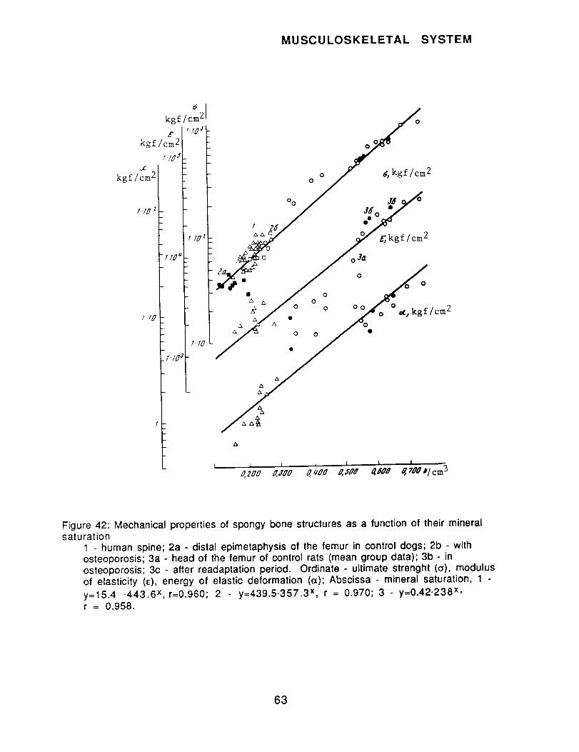
MUSCULOSKELETAL SYSTEM
kgf/cm 2
kgf/cm 2
I10 z
i 10
kgf /cm _
E "lOJ
II0 zl
1"10
.I'I0
0 2
i !_o "7I zs/ e_
7 f,kgflcm2
/o Z °!;,,,,.oOo
&
I I I I I I I
o,,200 O,,JO0 o, ,oo O,EOO _#Oo _ 700 #/cm 3
Figure 42: Mechanical properties of spongy bone structures as a function of their mineralsaturation
1 - human spine; 2a - distal epimetaphysis of the femur in control dogs; 2b - withosteoporosis; 3a - head of the femur of control rats (mean group data); 3b - inosteoporosis; 3c - after readaptation period. Ordinate - ultimate strenght (o), modulusof elasticity (Q, energy of elastic deformation (a); Abscissa - mineral saturation, 1
y=15.4 -443.6 x,r=0.960; 2 y=439.5.357.3 x, r = 0.970; 3 - y=0.42.238 x,r = 0.958.
63

MUSCULOSKELETAL SYSTEM
% month
"5
,r2
8
_7_I ¸
° ' '_o ......... '9o'I/0 130 f50 770 !BMIMS index
Ash, %
:f
_Z
/0
V
J* g_ ,'.2 '5 2.0 _:_ I, SMS, glcm 3
Figure 43: Rate of dystrophy of bone structures (in % month) as a function of the relative indexof osteoporosis (basal metabolic rate/mineral saturation)
1- humans (Schneider, 1977); 2 - humans (weightlessness); 3 - dogs; 4 - rats; lightand dark circles indicate error limits of mean values - for hypodynamia, andweightlessness, respectively
Figure 44: Relationship between mineral saturation and ash content:1 - demineralized collagen; 2 - humans, bone tissue of vertebrae; 3 - dogs, bonetissue of the proximal epiphysis of the femur. 5 - hydroxyapatite. Abscissa: mineral
saturation, g/cm3; Ordinate - ash content, %
64

NEUROPHYSIOLOGY
PAPERS:
P1205(27/90)* Karkishchenko NN.Specification for an "ideal" drug to prevent space motion sickness (spaceadaptation syndrome.Kosmicheskaya Biologiya i Aviakosmicheskaya Meditsina.23(6): 33-36; 1989.[16 references; 3 in English]
Neurophysiology, Space Adaptation Syndrome; Human Performance, Mental Work CapacityHumans, Patients, Vestibular Tolerance, DiminishedRotation, Pharmacological Countermeasures
Abstract: The goal of this work was to identify the effects and mechanisms underlying the actionof drugs holding promise for use in prevention of space motion sickness.
A total of 179 subjects, aged 19-26, with diminished vestibular tolerance participated in theexperiment. Motion sickness was induced through a rotation and vestibular stimulation.Rotation was continued until the subjects developed symptoms of level I1-111in severity. Thesignal to terminate vestibular stimulation was the onset of nausea or vomiting. Subjects wereassigned to drug conditions (single or multiple doses taken internally) by a double blind methodincluding a placebo condition, with scopolamine in a dose of 1 mg used as a standard. Theantimotion sickness efficacy of the drugs was assessed by recording length of time a subjecttolerated exposure to acceleration, the dynamics of development of clinical symptoms of motionsickness, and changes in pulse rate during motion sickness induction and spectralcharacteristics of EEG. Cognitive performance was studied through a computerized test. Theresults were processed and tested using the Wilcoxon paired measurements test. Drugs used andtheir efficacy are described in the table.
A single dose of diphenin (dilantin, phenytoin) or dapkene (valproic acid) [bothanticonvulsants], and repeated daily doses of potassium orotate and pyracetame (describedelsewhere as a cyclic analogue of GABA) were associated with significantly longer tolerance ofcomplex acceleration compared to scopolamine. The anti-motion sickness effects of these drugsalso included faster reduction of symptoms of vestibuloautonomic disturbance, smaller changesin pulse rate and in the amplitude of spectral EEG characteristics compared to motion sicknessinduction (in which no drugs were used).
The mechanisms underlying the protective effect of potassium orotate may involve not onlyactivation of nucleic acid synthesis, but also accumulation of GABA in brain tissue accompaniedby decreased glutamate. The interaction of antiepileptics and neuroactive amino acids may alsoplay an important role, since diphenin and depakene have an activating effect on GABAnergicsystems, and a depressing effect on glutamate and aspartatergic systems. In addition, depakeneinhibits glycine metabolism, and the effects of glycine and excitatory amino acids affect releaseof acetylcholine. The therapeutic effects of diphenin and depakene may also be related to theirtendency to depress the hypothalamic beta-endorphin system through mediation by GABAnergicneurons. At the same time, GABA, glutamate, glycines, endogenous agonists of opiate receptors,and acetylcholine participate at the level of the vestibular nuclei in the processing ofinformation about movement of the body in space. Their effects on the opiate, dopamine- andhistaminergic mechanisms may be significant for the suppression of the emetic reflex. An"ideal" pharmacological agent for preventing space motion sickness should have two propertiesin addition to inhibition of vestibular reflexes. It should increase antiorthostatic tolerance and
improve mental and physical performance.
65

NEUROPHYSIOLOGY
Used in combination, the most effective drugs exert a powerful corrective effect on mentalperformance, as assessed by performance on the "missing number test."
The effects and mechanisms of action of the ideal antimotion sickness drug, derived from thecurrent results and data in the literature, are presented in the figure.
Table The effects of drugs on endurance time for complex acceleration
Drug
PlaceboScopolamineDiphenin, 200 mgDiphenin, 300 mgPlacebo
ScopolamineDepakene, 300 mgDepakene, 450 mgPlaceboScopolaminePantogam, 1000 mgPantogam, 1500 mgPlaceboScopolaminePotassium orotate,
5-7 days12-14 days
PlaceboScopolaminePyracetame, 2000 mg
one dose3 days7 days14 days
Endurance time, seconds M+_m1 2 3
118.3+10.7 123.3__+11.3 104.2+9.6136.1+9.2 167.8+10.0"* 123.3+7.3123.2__+11.9 187.8_+22.3" 146.3+17.4104.4+16.0 170.0-+28.3"* 162.8_+27.11 44.1 -+7.8 153.2_+7.6 1 06.5+5.3139.3+8.2 162.7+11.4* 116.8-+8.2
99.1 +9.6 131.7-+1 8.7 132.9+18.799.3+12.0 166.4+__17.3" 167.6+17.5
112.5+8.2 115.0+9.4 102.2+8.4120.511.5 144.5__+12.6" 119.8+10.5118.8+_13.2 133.1+26.9 112.1+22.6116.9-+9.9 141.9+18.5 121.4_+15.9143.8+17.6 171.3_+13.6 119.2+9.4135.6+6.7 149.4+22.4 110.2+16.5
130.0-+18.8 131.3+13.2 101.0_+10.197.5+15.8 239.5+44.5** 245.6+45.6
125.9+17.8 138.4+16.5 109.9+13.1128.5+15.9 161.8+22.7"* 125.9+17.6
120.0_+22.0 230.0-+57.7 191.7+48.1105.0+12.4 212.5+40.4" 202.4_+38.5127.9_+16.4 280.8+46.1"* 219.5+36.0104.5_+13.3 144.5_+26.1"* 138.3_+5.0
1 - endurance time before drug (seconds)2 - endurance time after drug (seconds)3 - 2 as a percentage of 1
Repeated doses: three doses per day - potassium orotate in a daily dose of 40 mg per 1 kg bodyweight; pyracetame in a daily dose of 30 mg per 1 kg body weight.
* - p < 0.05; ** p < 0.01
66

NEUROPHYSIOLOGY
Higrl affinity
STABILIZATION OF NEURONEXCITABILITY
BLOCKING OFOPIATE
DOPAMINERGIC
RECEPTORS
IH MUSCARINE
ISTAMINERICJ
ACTIVATION OF SYSTEMS:
GABAnergic .,_
GLYCINERGIC ._
ISTIMULATION OF ADAPTIVE
INHIBITION OF VESTIBULARREFLEXES
INCREASE INANTITHOSTATIC
TOLERANCE
INCREASE INPERFORMANCE
PROCESSES
ACTIVATION OFCATECHOLAMI
NERGICSYSTEMS
ACTIVATION OF RNA
SYNTHESIS ANDENERGY
METABOLISM
Figure: Effects and mechanisms of the "ideal" pharmacological agent for prevention of spacemotion sickness
67

NEUROPHYSIOLOGY
P1214(27/90)* Verbitskaya LB, Fedorenko VS, Kabitsyna RA..Remote ultrastructural changes in cerebellar cortex of ratsaccelerated carbon ions.
Kosmicheskaya Biologiya i Aviakosmicheskaya Meditsina.23(6): 71-77; 1989.[2 references; 1 in English]
after exposure to
Neurophysiology, Cerebral CortexRats
Radiobiology, Accelerated Carbon Ions, y-Radiation; Remote Effects
Abstract: Experiments were performed on sexually mature male Wistar rats. One group wasirradiated with carbon ions with a flux rate of 104 particles/cm 2. Animals in a second groupwere irradiated with y-radiation using 60Co in a dose of 1.0 Gy, and 1 day later with the abovedose of carbon ions. Group 3 were irradiated only with y-radiation, while group 4 was anunirradiated control. The rats were irradiated with carbon in a synchrophasotron. Following a1-, 3- or 6-month period after irradiation, the animals were sacrificed and the brain tissueprocessed for electron microscopic study. Semi- and ultrathin sections were prepared andstudied using light and electron microscopes.
Irradiation led to dystrophic damage to neurons and glial cells. However, destructive changeswere severe in the groups exposed to carbon ions. Indeed, carbon ions with flux of 104
particles/cm 2 (LET = 1 kequiv/l_m) were more effective than 7-radiation in a dose of 1.0 Gy.Exposure to y-irradiation followed by carbon ion irradiation did not lead to further worseningof pathological processes. Structural damage to the various types of nerve and glial cellsdiffered in nature. Changes in the Purkinje cells were of the "dark" type, while adjacentBergman glial cells showed changes of the "light" type with edema, and swelling of the cellbodies, their nuclei and outgrowths in the granular layer. In contrast, nerve granule cellsshowed "light" changes, while adjacent glial cells -- the oligodendrites -- showed thickening ofkaryo- and cytoplasm, and heightened osmiophilia. It is suggested that these changes inneurocytes and glial elements serve to maintain impaired functions of the neurons, includingcell interactions. Study of changes over time in the cerebella of irradiated animalsdemonstrated that destructive changes progress during the remote period after irradiation. Thestudy is interpreted as demonstrating the possibility of remote damage to cells of the centralnervous system, after exposure to even small fluxes of heavy ions.
Figure 1: Purkinje cells and the internal granular layer of the cerebellar codex of controlanimals and rats 1 month after irradiation with accelerated carbon ions
Figure 2: Ultrastructural changes in granule cells and synaptic glomerules of the internalgranular layer of the cerebellar cortex of rats 1 month after irradiation with acceleratedcarbon ions
Figure 3: Formation of pathological inclusions, the development of "fissures" in the cytoplasmof Purkinje cells of the cerebellar cortex of rats
Figure 4: Ultrastructure undergoing "dark" type alteration of Purkinje cells and vacuolizationof the dendrites of Purkinje cells in the molecular layer
68

NEUROPHYSIOLOGY
P1227(27/90) Moskalenko YuYe, Beketov AI, Maksimiuk VF, Skoromnyy NA.Cerebrovsscular effects of motion sickness.Fiziologicheskiy Zhurnal SSSR im I.M.Sechenova.75(11): 1560-1567; 1989.[12 references; 2 in English]Authors' Affiliations: Institute of Evolutionary Physiology and Biochemistry, Leningrad;Crimean Medical Institute, Simferopol
Neurophysiology, Cerebrovascular Effects; Cardiovascular and Respiratory Systems, CerebralCirculationRabbitsMotion Sickness, Head-Down Tilt
Abstract: Past results have suggested that cerebrovascular reactivity data provide indirectinformation on the compensatory capabilities of the mechanism regulating circulation in thebrain. Cerebrovascular activity was assessed from active responses of the cerebral vasculatureto provocative Ioadings, and was expressed as the ratio of a series of cerebral circulatoryparameters before and during exposure to the provocative stimulus, motion sickness induction.The effects of motion sickness on the compensatory capacities of the cerebral circulation systemwere studied in a long-term experiment on rabbits with implanted electrodes in various areasof the frontal and occipital lobes. Experiments were performed on alert animals looselyrestrained so as not to cause discomfort. Three types of provocative tests were selected for usein assessment of cerebrovascular activity. The first involved a 1.5-minute period of inhalation
of gas containing 10% CO2 in air, in which reactivity was measured by comparing levels oflocal cerebral blood flow before and after inhalation using the method of hydrogen clearancefrom implanted electrodes. The second method involved graded compression of the abdominalcavity for a period of 30 seconds. Vascular response in this case was evaluated using impedanceplethysmography recorded from the same electrodes. Reactivity was assessed from dynamics ofpulse wave amplitudes on a cerebral plethysmogram. The third test utilized light stimulation.In this case clearance was used to measure intensity of local cerebral blood flow, whileimpedance plethysmography served to indirectly determine vascular response. Motion sicknesswas induced using a swing with an arm of 1.3 m and a frequency of 50-55 swings per minute.In another condition motion sickness induction was combined with head-down tilt (-30°).Rabbits exposed to motion sickness induction (swinging) and head-down tilt alone or incombination were examined before and during the provocative stimulation described above.Parameters measured were local cerebral blood flow in the areas of the electrodes and the stateof fluid balance in the same areas of brain tissue. The first parameter was measured using
hydrogen clearance and the second estimated from results of impedance plethysmography.
Results confirmed that 2 hours of swinging leads to some increase (10-25%) in local cerebralblood flow. As a rule, the reactivity of brain vasculature decreased to all three provocativetests after swinging. An exception was retention of vascular reactivity in the visual cortex inresponse to light stimulation. These results, combined with the general tendency for local brainblood flow to increase, suggested a hypothesis that the motion sickness induction proceduredecreased the adaptive activity of central mechanisms for regulating cerebral blood flow, butleft local regulation intact. If these changes are neurogenic (activation of cholinergic systemshas been demonstrated in motion sickness), it is likely that there would also be a change in thefluid balance of brain tissues, impeding outflow of blood from the skull. If motion sickness isaccompanied by factors leading to impeded outflow from the skull, then the compensatoryeffects will be disrupted. The latter will lead to hydration of brain tissue and increase in
intracranial pressure.
69
NEUROPHYSIOLOGY
Effects of head-down tilt are typically well compensated, but at the cost of some strain onregulatory mechanisms. If motion sickness does diminish the adaptive capacity of the cerebralcirculation system, if combined with head-down tilt uncompensated effects are likely to befound, including hydration of brain tissue. To test this hypotheses, rabbits were exposed to acombination of head-down tilt and swinging, and information on brain tissue hydration obtainedindirectly from impedance plethysmograms. Results showed significant impedanceplethysmographic changes consistent with increased hydration in response to the combination ofhead-down tilt and swinging, greater than changes occurring after head-down tilt alone, andmost marked in the visual cortex. Normalization occurred only 1.5-2.0 hours after treatmentterminated.
The authors conclude that their results show that the effect of motion sickness induction on thecomplicated system of regulatory processes responsible for maintaining adequate blood supplyto brain tissue under changing environmental conditions may be considered the majorcerebrovascular effect of this factor. When motion sickness is induced, in all likelihood,coordination among the individual components of the mechanisms regulating cerebralcirculation is disrupted and there is a tendency for central regulatory effects to be depressed,while local ones are retained. General changes in level of local cerebral blood flow that can beobserved in motion sickness, most likely reflect the lack of coordination among individualcomponents of the mechanism regulating cerebral circulation. An important consequence ofthese changes occurring in response to motion sickness is a tendency for physical homeostasis ofneural tissue to be disrupted, increasing the risk of brain edema.
Figure 1 : Changes in reactivity of the vasculature of the sensorimotor and visual areas of thecerebral cortex in response to light stimulation and abdominal compression as estimated fromthe ratio of pulse waves in a local impedance plethysmogram at rest and during the provocativetest
Figure 2: Change in cerebral blood flow in inhalation of carbogenic gas in the sensorimotor areaof the brain
Figure 3: Change in componnts of impedance - extracellular, intracellular, and capacitance --in the sensorimotor and visual areas of the brain in response to head-down tilt, motion sicknessinduction and their combination
Figure 4: Pulse waves of a local impedance plethysmogram in the sensorimotor and visualcortices at rest in head-down tilt, motion sickness and their combination 30 and 60 minutesafter onset of exposure
70
PSYCHOLOGY
PAPER:
P1228(27/90) Anishchenko TG, Burshina SN, Shorina LN.An analysis of factors determining sex differences inwhite rats.
Byulleten' Eksperimental'noy Biologii i Meditsiny.1989(1 1):61 6-61 8.[15 references; 3 in English]Authors' affiliation: N.G. Chernyshevskiy University, Saratov
the stress responses of
Psychology, Stress ResponseRats, Males and FemalesGender Differences, Genetics
Abstract: This work studied stress responses of infantile, normal adult, and castrated male andfemale white rats, and female rats androgenized in infancy, as a means to determine the factorsresponsible for sex differences in these responses. "Emotional-pain stress" was created byimmobilizing animals for 10 minutes; "emotional" stress was created in animals paired withthe immobilized ones by the sight and cries of their "partners." Intensity of the stressresponse was assessed by changes in concentrations of corticosterone in the adrenal gland andblood plasma measured fluorometrically. Stress responses were studied in sexually immaturerats, adult untreated rats, rats castrated 3 weeks before the experiment and adult females thathad been injected at the age of 1 day with 1 mg testosterone propionate. A total of 362 animalswere used.
In adult rats, sex differences in stress responses were marked. In the emotional(observational) stress situation level of corticosterone increased by a factor of 2 in females and1.4 in males. In the emotional situation the females displayed heightened motor activity,showing clear "resonance" with the other animal. The males displayed a passive defensereaction of the "freeze" type. In the emotional-pain stress (actual immobilization) situation,plasma corticosterone increased by a factor of 4 in females and 2.3 in males. Castrated animalsretained the sex differences in response to stress. In the females in the emotional stressparadigm, corticosterone increased in the adrenal gland and plasma by factors of 2 and 2.5,respectively. In the males the corresponding factors were 1.4 and 1.6. In the pain-stressparadigm, castration led to decreased increases in secretion of hormones for both sexes, but sexdifferences were retained. In sexually immature.animals corticosterone increased in females inthe emotional stress paradigm by a factor of 1.5 and 1.4, butin males did not increase in eitherthe adrenal glands or the plasma. In the pain-stress paradigm, synthesis and secretion of thehormone increased by a factor of 1.7 and 2.3 in the immature females while only secretionincreased (by a factor of 2.3) in the males, with adrenal concentration not changing. Thus, inimmature animals the adaptive capacity as indexed by intensity of the stress response isdiminished. However, females display appropriate responses to adverse conditions, whileinfantile males do so to a much lower extent. These results suggest the ontogenio nature of sexdifferences. Female rats androgenized as neonates leading to masculinization of the brain stilldisplayed a female type of stress response. The authors conclude that sex differences in stressresponse are genetically determined and not due to sex hormones or sexual differentiation of thebrain.
Figure 1: Reactions of intact rats to emotional stress and emotional-pain stress
Figure 2: Reactions of castrated rats to emotional stress and emotional pain stress
Figure 3: Reactions of sexually immature rats to emotional stress and pain stress
71

RADIOBIOLOGY
PAPERS:
P1221(27/90) Zukhbaya TM.Periodicity of hemopoiesis during continuous 7-irradiation with low dose rates.In:l Vsesoyuznyy Radiologicheskiy S"yezd: Tezisy Dokladov;I BcecoJozH,-,AP_HO.nOFHqeC..KHRCbe3,,_: Te3HC_ _OK.na_os Io [I All-Union Radiobiological Congress: Paper Abstracts] Moscow, 21-27 August 1989.Pushchino: USSR Academy of Sciences; 1989.Author's Affiliation: Institute for Biomedical Problems, Moscow
Hematology, Hemopoiesis, PerodicityRatsRadiobiology, 7-Irradiation, Continuous
Translation of abstract: It is well known that after acute radiation in sublethal doses, recoveryof the hemopoietic system occurs in phases. Similar fluctuations have been observed in theerythroid population in blood letting, hypoxia and various stress situations. We found that ananalogous pattern of change over time occurs in the major hemopoietic bone marrow cells andthe lymphoid organs during continuous irradiation of rats.
Thus, during a 3-month period of 7-irradiation (22 hours per day) with a dose rate of 0.4Gy/day (total cumulative exposure approximately 40 Gy), four periods of sequential changes inthe size of populations of different hemopoietic cells were observed. In each period the minimalvalues of quantities of erythroid and granulopoietic elements in bone marrow were half that ofthe norm, while maximal values of these same parameters approached baseline. When dose ratewas increased to 1.0-2.0 Gy/day fluctuations were damped, while at a greater dose rate of 4.0Gy/day they disappeared entirely.
The first period in the fuctuating process was analogous to that described in radiobiology as "anabortive increase" and observed in all populations of bone marrow cells (except for thelymphoid), and also the thymus and the spleen. Each cell line differed with respect to intrinsicduration of period and amplitude of fluctuations, which changed as a function of radiationexposure schedule.
The second wave of hemopoietic recovery started on day 10-15 of continuous radiation andcovered all hemopoietic cells (including the lymphoid elements of bone marrow) at dose rate of0.4 Gy/day, attesting to the participation of pluripotent stem cells. At the same time, when doserate was 1.0 G/day, repeated regenerative surges were observed only in the erythroid andneutrophiloid series, which evidently, are associated with the predominant differentiation ofstem hemopoietic cells in this direction to support hemostasis of the most vital elements inperipheral blood.
The phasic nature of changes in the hemopoietic system during continuous exposure to radiationattests to the rhythmic activity of the hemopoietic precursors of the stem and semi-stem type.Prediction of the duration of the period and amplitude of fluctuation of a regenerative wave inexposure to various schedules of continuous radiation may be important in radiation clinics.
72
SPACE MEDICINE
CONFERENCEREVIEW:
CR13(27/90)* Gyurdzhian AA, Zorile VI.The work of the section on Aviation and Space Medicine of the MoscowPhysiological Society.Kosmicheskaya Biologiya i Aviakosmicheskaya Meditsina.23(6): 91-92; 1989.
KEY WORDS: Space Medicine; Aviation Medicine
The section on aviation and space medicine of the Moscow Physiological Society unitesapproximately 300 specialists (physicians, psychologists, engineers, biologists, and others)interested in the problems of aviation and space medicine. Participants include representativesof civil and military aviation, the aviation industry, institutes of higher learning, and scientificand clinical institutions (Institute of Biomedical Problems of the USSR Ministry of Health, TheCentral Scientific Research Aviation Hospital, the Yu.A. Gagarin Center for Cosmonaut Training,the central and clinical hospitals and polyclinics of the Ministry of Civil Aviation, The CivilAviation Scientific Research Institute, the Department of Aviation Medicine of the CentralMedical School, etc.).
The work of the section is directed by a bureau, chaired by N.M. Rudniy, with V.I. Zorile asscientific secretary. Sessions are generally held once a month in the quarters of the Departmentof Aviation Medicine of the Central Medical School (7 Peschanaya Street, Moscow). Jointsessions are also held with other sections of the Moscow Physiological Society, for example, thesection on occupational physiology.
In 1988 there were 10 meetings in which more than 30 papers were delivered and discussed.Two sessions were devoted to the International Day of Aviation and Cosmonautics and wereconducted at the regular session of the 18th Gagarin Lectures.
Among the papers discussed were "New Methods for Evaluating Performance of a Flight Crew"(Ye.A. Yevseyev, S.L. Shapovalov, T.I. Milyavskaya); "Electroencephalographic Correlates ofPilot Job Performance" (D.I. Shnachenko); "Lipid Metabolism in Flight Personnel and Methodsfor Identifying Hyperlipidemia" (S.A. Bugrov, R.K. Kiselev, T.A. Orlova); and other works ofsignificant scientific and practical importance for medical support of flight personnel.
In 1988 members of the section produced more than 10 monographs, 15 texts, and manuals forflight surgeons, flight crews, and cosmonauts. Among these were: "Reserves of the HumanPsyche" (L.P. Grimak, published in a mass edition by Politizdat); "Biological Effects of LunarSoil," (V.V. Kustov, V.I. Belkin, G.G. Kruglikov, Leningrad, Nauka, 1988); and "A Handbook onFunctional Rehabilitation of the Vision of Flight Personnel in Civil Aircraft,"(V.F. Tokarev, S.L.Shapovalov, T.I. Milyavskaya, Y.A. Yevseyev, Moscow, Vozdushniy Transport, 1988).
The meeting of the section on 26 October 1988 was devoted to analysis of the articles in thejournal "Space Biology and Aerospace Medicine" for 1987. Professor N.M. Rudnyj presented areport on the topic. Eight participants took part in a discussion.
They noted that the journal is of great practical and theoretical assistance to specialists inaerospace medicine and interdisciplinary areas. The unanimous opinion of those who spoke wasthat the journal generally meets the needs of its readers.
However, along with the general positive evaluation of the journal, important criticisms andsuggestions focused on further improving the efficiency of the editorial board and the scientificand practical significance of the articles published. In particular, opinions were expressed
73

SPACE MEDICINE
that the articles in the journal are predominantly theoretical, narrowly specialized works onspace biology, with few articles on aviation medicine (N.A. Razsolov). It was also stated thatlittle attention is paid to the psychophysiology of flight tasks, selection and training of pilots andcosmonauts, improvement of the system of medical monitoring, and increasing flight safety (V.I.Zorile, K. A. Pimenova). Publication of articles on these issues would attract the attention ofmany specialists in aviation and would thus increase the number of readers, subscribers andauthors who are flight surgeons and/or scientists working in related areas. This is not the firsttime complaints were heard concerning the length of time it takes for a manuscript to bepublished on the pages of the journal. Increase in efficiency in the publication of articles,among other measures, would help to turn the journal into a monthly publication, (instead ofbimonthly).
P.V. Vasil'yev, a member of the journars editorial board, noted in his talk that few articlesdealing with aviation are submitted, although the editors try to give them preference inpublication.
A major part in increasing reader participation must be played by the section on aviation andspace medicine of the Moscow Physiological Society, which can serve as a vehicle for operationalscientific communication among specialists in aviation and space medicine. The editors canmake broad use of the section meetings to obtain reader feedback. A number of special measurescan be undertaken to acheive this. For example, a section could recommend that material thathas been presented and discussed be published as articles or abstracts in the journal. Theeditorial board of the journal could suggest particularly critical and complex issues in aviationmedici,_e for discussion at section meetings. It would be desirable if reports on past and plannedsection meetings were published in the pages of the journal.
The election of members of our section to Academies -- Professor A.I. Grigor'yev as acorresponding member of the USSR Academy of Medicine as a specialist in "Space Biology andMedicine"; and Professor V.A. Ponomarenko as a corresponding member of the Academy ofPedagogical Sciences as a specialist in "Occupational Training" and member of the presidium ofthat organization -- are acknowledgements by the scientific community of the role of aerospacemedicine in the development of Soviet science. A representative of space pharmacology, V.S.Shashkov has been elected corresponding member of the USSR Academy of Medicine as aspecialist in "Pharmacology."
Membership of the section on aviation and space medicine have elected a representative to the ofUSSR Congress of Peoples Deputies. At the Scientific Conference of Scientific Societies andAssociations at the USSR Academy of Sciences, held 22 and 23 March 1989 in the Moscow Houseof Scientists, N.P. Bekhterev, academician and director of the Scientific and Research Instituteof Experimental Medicine of the USSR Academy of Medicine, and member of the presidium of theI.P. PavIov All-Union Physiological Society, and O.G. Gazenko, consultant to the director of theInstitute of Biomedical Problems of the USSR Ministry of Health, and president of the I.P.PavIov All-Union Physiological Society, were elected as people's deputies.
N.A. Razsolov, K.A. Pimenova, S.L. Shapovalov, Ye.Ye. Nikolayevskij, I.N. Chernyakov, V.V.Chuntul, and A.A. Shishkov participated actively in the work of the section in 1988.
In 1988, 12 scientists, graduate students, adjuncts, flight surgeons, and clinical specialistswere accepted as members of our section of the Moscow Physiological Society. In 1989, planscall for holding nine meetings of the section on aviation and space medicine. Topics to be coveredinclude diagnosis of functional state of the cardiovascular system and visual systems of humansduring flight, and prevention of fatigue and improvement of techniques for restoringperformance levels of crews after long flights. In addition, a symposium is planned on the topicof "psychophysiological aspects of increasing the safety of flights on modern flight vehicles"
74

SPACE MEDICINE
with participation by specialists in various institutes. One meeting will be devoted to thescientific work of medical doctor, professor and winner of the USSR State Prize, P.K. Isakov, inhonor of his 80th birthday.
Finally, certain initiatives and accomplishments of the I.P. Pavlov All-Union PhysiologicalSociety should be noted. The publication of a new informational series, "Physiology.Informational material of the I.P. Pavlov All-Union Physiological Society," edited by thepresident of the society, academician O.G. Gazenko is an outstanding accomplishment. Thesepublications provide timely information concerning the work and decisions of the Division ofPhysiology of the USSR Academy of Sciences, the Presidium and Central Soviet, and the republicsocieties concerning congresses, conferences, symposia, seminars, meetings, and schools inphysiology, and also bibliographic information about books to be published in physiology,medicine, biology and mixed disciplines.
The international ties of the All-Union Physiological Society have increased significantly. In1988 for the first time for a society of the USSR Academy of Sciences, an agreement was signedto enable scientific collaboration between the physiological societies of the USSR and USA, inwhich physiologists from Czechoslovakia, Bulgaria, Finland, and the People's Republic of Chinawill also participate.
75

SPACE MEDICINE
Special Feature: Essay by N. Gurovskiy; In the Interests of Public HealthIn: Aviatsiya i Kosmonavtika; 11/89
KEY WORDS: Space Medicine, Operational Medicine, Public Health, Equipment andInstrumentation, Personnel Selection, Hematology, Biological Rhythms
Today there is not a single area of human activity that has failed to feel the stimulating effects ofcosmonautics. This field has enriched not only our knowledge of space and the Earth, but alsopublic health. As manned space flight has developed, techniques for monitoring the health statusof cosmonauts have improved, along with understanding of human potential and what is requiredin order to prepare humans to undergo the rigorous ordeal of space flight. Now space medicineis providing health benefits to people on Earth too.
Here are some concrete examples of the interrelationship of space and terrestrial medicine.
In its early days, space medicine had to solve the problems involved in selecting and trainingcosmonauts for flight, developing specifications for the environment in weightlessness and theassociated life support systems, and assessing physiological responses to space flight factors.Scientists beginning work in this area were brought up against the inadequacy of our knowledgeconcerning the responses of a healthy individual in various situations and the optimalconcentrations of harmful contaminants in the spacecraft atmosphere. The need to answer suchquestions led to special research to determine the boundaries between pathology and the norm.The results of these studies began to be used by clinicians in treating their patients.
In examining potential cosmonauts, existing methods of pilot selection were made more precise,while new methods were developed to reveal latent pathology. Now these methods have beenadopted in clinical practice, for example, in the study of the vestibular system and identificationof latent coronary insufficiency.
As is well known, weightlessness induces deconditioning of the muscle system, changes incardiovascular functioning, bone tissue, and mineral metabolism. To improve understanding ofthese phenomena, research was undertaken to study the physiological effects of restrictingmotor activity. The bedrest paradigm is currently used as a laboratory model of weightlessness.
It would seem natural to question the possible relevance of these studies for public health.has turned out that they are of direct relevance. Hypokinesia is an extremely significantetiological factor in the development of diseases, especially of the cardiovascular system.
Research on healthy human subjects revealed a number of symptoms characteristic of the staleof hypokinesia. It is essential that clinicians be aware of these symptoms when, while treating apatient, they must differentiate between the symptoms attributable to the primary disease andthose due merely to limited motor activity.
The use in space medicine of constant remote monitoring has found practical application inclinics, health resorts, and athletics. In addition, special equipment has been developed toembody the principles utilized in space medicine. Such systems are used in the postoperativeand resuscitation (intensive care) units of many hospitals. The "Telekont" biotelemetricsystem, designed for monitoring the condition of patients who have suffered myocardialinfarction, is currently being tested.
It would be reasonable to use satellite communication lines for hospitals remote from theleading medical centers of the country. There is currently nothing that in principle prohibitsimplementation of such "remote diagnosis." The prototype of such a system was validated duringthe work of the 28th Soviet expedition to the Antarctic. Using portable medical instruments, a
76
SPACE MEDICINE
physician in the tent recorded an electrocardiogram and measured the blood pressure of oneparticipant in the expedition. These data, in the form of a digital radiogram, were transmittedby satellite to Moscow where they were decoded and analyzed and the conclusions drawn sentback via satellite to the physicianin the Antarctic.
The principles of "remote diagnosis" formed the basis for the operation of a mobile automatedlaboratory ("Avtosan") for mass examination of the population. This laboratory was equippedwith virtually the same medical apparatus as the Salyut space station.
Examination of a single patient, involving measurement or derivation of approximately 15parameters, requires about 15 minutes. Two portable computers perform analysis of the data,automatically take the patients history, and process all information. All the informationgenerated by an examination may be entered into the data base and used for future annualexaminations.
Unfortunately, at present the number of "space methods" available is far from sufficient tocover the entire population.
The "Oxymeter" device developed to study oxygen supply to cosmonauts is now used clinicallyfor diagnosis of a number of diseases: in dentistry for periodontosis, for plastic surgery, andfor ischemic heart disease.
Specialists in space medicine have developed a methodology for studying intestinal microflorathat is used in clinical practice for studying the condition of patients with chronic diseases ofthe gastrointestinal tract.
A method of detoxifying donated blood for long-term storage involves passing blood that hasundergone preliminary incubation with adenine and inosine, through a column with a sorbent(type SKN-100). Blood treated this way may be stored for up to 52 days. In clinical practice,donated blood preserved with a glucose-citron preservative can be kept for 10-15 days. Thedifference is significant. The Committee on Discoveries and Inventions has evaluated theproposed method, concluding that is has great economic significance, providing enormoussavings in the cost of blood storage.
In space there is no natural alternation of day and night and the synchronizers for manyphysiological functions are no longer present. The study of biological rhythms is critical forpassenger aircraft flight across time zones and the numerous jobs involving shift work. Workon chronobiology performed in the context of space biology and medicine has made an importantcontribution to this problem.
In conclusion, I would emphasize one more issue. In selection of cosmonauts, psychologicalexamination helps us identify latent and atypical disturbances, predict behavioral and emotionalresponses of an individual to stress, define a number of principles relevant to humaninteractions within a group, and find rationales for principles of group selection. This hasallowed us to successfully select the members of a group for joint performance, particularly inteam formation in sports, selection of groups for expeditions to the North Pole or to climbmountains.
Thus, space medicine, besides stimulating the development of general biology and the practice ofpublic health, has enriched these areas with new methods and ideas.
77

KEY WORD INDEX
Abiogenic Synthesis 34Accelerated Carbon Ions 68Adaptation 1, 29, 32, 40Adrenal Glands 27Adrenoblockers 32Air Crews 3Athletes 46Aviation Medicine 3 - 5Aviation Psychology 5Bedrest 52Beetles, Darkling 8Biodynamics 5Biological Rhythms 1, 5, 8, 76Biospherics 10Blood Iron 38Bone 50, 52, 56, 57Bone Minerals 52Botany 13Brains 25Calcium 48, 50, 57Cardiac Contractility 23Cardiovascular and Respiratory Systems 1, 3, 17 - 25, 42, 69Cerebral Cortex 68Cerebrovascular Effects 69Circadian Rhythms 8Circulation 25Cosmonauts 17, 52COSMOS-1667 27COSMOS-1887 8Cytoplasmic Dehydrogenases 47Digestive Enzymes 32Dogs 57Endocrine Response 30Endocrinology 1, 27, 29 - 30Endogenous Ethanol 44Enzymology 32Equipment and Instrumentation 76Exercise 17, 46Exercise Tolerance 42Exobiology 34Fat 47Ferrocerone Test 38Functional State 3G-Radiation 68Gastrointestinal System 32Gender Differences 71Genetics 13, 71Guinea Pigs 23Habitability and Environment Effects 36, 42Head-Down Tilt 25, 69Heavy Ions 13Hematology 38, 72, 76Hemodynamics, Central, Regional 17Hemopoiesis 72High Altitude Sickness 40High-Altitude 29Hormones 1Human Performance 3, 5, 65
78

SPACE MEDICINE
Humans 3, 17, 30, 36, 38, 40, 42, 43, 52, 56, 57, 65
Hydroxymethyl Aminopropylidene Diphosphonate 50Hypercapnia 36Hypobaria 44Hypokinesia 23, 47, 48, 50
Hypokinesia with Head-Down Tilt 38Hypothalamus 29Hypoxia 29, 44Immobilization Cages 48, 50Immobilization Stress 2Immune Status 40
Immunology 40- 43Individual Differences 40
Lettuce, Seeds 13Lipid Peroxidation 46Liver 47Low-Frequency Electromagnetic Fields, Constant, Intermittent 30Lunar Soil 34
Males 27, 36, 40, 42, 48, 50Males and Females 71
Mathematical Modeling 10Mechanical Properties 52Mental Work Capacity 65Metabolic Rate 57Metabolism 1, 44 - 47, 50, 57Morphology 27, 29Motion Sickness 69
Motor Activity 8Musculoskeletal System 48 - 57Natural Resources 10
Neurophysiology 29, 65-69Neurosecretory Apparatus 29Nucleotides 34
Nutrition 38, 48Operational Medicine 43, 76Osteoporosis 52, 56Oxygen Pressure 25Patients 52, 65Perodicity 72Personnel Selection 76
Pharmacological Countermeasures 32, 65
Phosphorus 48Pituitary 29Pressurized Environment 36, 42Protective Function 43
Psychology 23, 71Public Health 76Rabbits 25, 69
Radiobiology 13, 30, 68, 72Rats 27, 29, 32, 44, 47, 48, 50, 57, 68, 71, 72Recovery 47Reduced Functional Loading 56Remote Effects 68
Remote Sensing 10Resistance 42Review Article 30Rotation 65
Salyut-6 13, 17, 52
79

KEY WORD INDEX
Salyut-6 13, 17, 52Salyut-7 13, 17Seasonal Rhythms 1Serum Ligandsi.Humans 46Simulated Weightlessnsess 57Smoking 42Space Adaptation Syndrome 65Space Flight 8, 13, 17, 27, 52Space Medicine 73-76Space Systems 10Stress 1, 23Stress Response 71Tail Suspension 48Thermal 34Thermal Stress 5Thermoregulatory Response 36Vestibular Tolerance, Diminished 65Vitamin D3 Metabolites 48Weightlessness 57Workload 3
8O


Nallyel Aef0reu|cs andS_tce Ad_r_slraion
1. Report No.
NASA CR-3922(32)
4. Title and Subtitle
Report Documentation Page
2. Government Accession No. 3. Recipient's Catalog No.
USSR Space Life Sciences Digest - Issue 27
Victoria Garshnek,*
7. Author(s)
Lydia Razran Stone, Ronald Teeter,
and Joseph Rowe,** Editors
9. Performing Organization Name and Address
Lockheed Engineering and Sciences Company
600 Maryland Avenue SW, Suite 600
Washington, DC 20024
12. Spon_ringAgencyNameandAddre_
Office of Space Science and Applications
National Aeronautics and Space Administration
Washington, DC 20546-0OO1
5. Re_rt Date
July 1990
6. Performing Organization Code
8. Performing Organization Report No.
10. Work Unit No.
15. Supplementa_ Notes
*George Washington University, Washington, D.C.
**Library of Congress, Washington, D.C.
11. Contract or Grant No.
NASW-4292
13. TypeofReportandPeriodCovered
Contractor Report
14. Sponsoring Agency Code
SB
16. Abstract
This issue of the USSR Space Life Sciences Digest contains abstracts of 30
journal papers or book chapters published in Russian and of 2 Soviet
monographs. Selected abstracts are illustrated with figures and tables from the
original. A Soviet book review of a British handbook of aviation medicine and
description of the work of the division on aviation and space medicine of the
Moscow Physiological Society are also included.
17. Key Words(Sug_st_ by Author(s))
space biology
aerospace medicine
flight experiments
flight simulations
microgravity
19. SecuriW Cla_if. (of this report)
Unclassified
NASA FORM 1626 OCT 86 Ava i 1able
USSR
18. Distribution Statement
Unclassified - Unlimited
'_.SecuriW Cla_if.(ofthispage)
Unclassified
from the National Technical
Subject Category 51
21. No. of pages _. Price
86 AO6
Information Service, Springfield,
VA 22161
NASA-i_ag_, 1990


National Aeronautics andSpace AdministrationCode NTT-4
Washington, D.C.20546-0001
Offic;al Bus_ness
Penalty for Prlvale USe, $300
SPECIAL FOURTH-CLASS RATE
POSTAGE & FEES PAIDNASA
Permit No. G-27
i
x
POSTMASTER: If Undelivecable (Section 1 $8
Postal Manual) Do Not RetUrn
ii
i


![Documented Code for Glossaries v3.01 [Nicola L.C. Talbot] [School of Computing Sciences, University of East Anglia] [12!04!2011]](https://img.pdfslide.us/doc/110x75/55721000497959fc0b8ca38f/documented-code-for-glossaries-v301-nicola-lc-talbot-school-of-computing-sciences-university-of-east-anglia-12042011.jpg)