Embed Size (px)
Citation preview
23. Radionov, V. F., Bryazgin, N. N. & Aleksandrov, Y. I. The Snow Cover of the Arctic Basin
(Gidrometeoizdat, St. Petersburg, 1996).
24. Babko, O., Rothrock, D. A. & Maykut, G. A. Role of rafting in the mechanical redistribution of sea ice
thickness. J. Geophys. Res. 107, doi:10.1029/1999JC000190 (2002).
25. Wadhams, P. & Horne, R. J. An analysis of ice profiles obtained by submarine sonar in the Beaufort sea.
J. Glaciol. 25, 401–424 (1980).
26. Lindsay, R. W. & Rothrock, D. A. Arctic sea ice leads from advanced very high resolution radiometer
images. J. Geophys. Res. 100, 4533–4544 (1995).
27. Bourke, R. H. & McLaren, A. S. Contour mapping of Arctic Basin ice draft and roughness parameters.
J. Geophys. Res. 97, 17715–17728 (1992).
28. Haas, C. & Eicken, H. Interannual variability of summer sea ice thickness in the Siberian and
central Arctic under different atmospheric circulation regimes. J. Geophys. Res. 106, 4449–4462
(2001).
29. Gregory, J. M. et al. Recent and future changes in Arctic sea ice simulated by the HadCM3 AOGCM.
Geophys. Res. Lett. 29, doi:10.1029/2001GL014575 (2002).
30. Eicken, H., Tucker, W. B. & Perovich, D. K. Indirect measurements of the mass balance of
summer Arctic sea ice with an electromagnetic induction technique. Ann. Glaciol. 33, 194–200
(2001).
Acknowledgements We thank the European Space Agency for the provision of ERS data, and the
National Snow and Ice Data Centre for submarine and passive microwave data. We also thank
J. Mansley for assistance with data processing, and D. Wingham for comments. This work was
supported by the UK Natural Environmental Research Council and the European Union.
Competing interests statement The authors declare that they have no competing financial
interests.
Correspondence and requests for materials should be addressed to S.L. ([email protected]).
..............................................................
Neuroanatomy of flying reptilesand implications for flight,posture and behaviourLawrence M. Witmer1, Sankar Chatterjee2, Jonathan Franzosa3
& Timothy Rowe3
1Department of Biomedical Sciences, College of Osteopathic Medicine,Ohio University, Athens, Ohio 45701, USA2Museum of Texas Tech University, Box 43191, Lubbock, Texas 79409, USA3Jackson School of Geosciences, University of Texas, Austin, Texas 78712, USA.............................................................................................................................................................................
Comparison of birds and pterosaurs, the two archosaurian flyers,sheds light on adaptation to an aerial lifestyle. The neurologicalbasis of control holds particular interest in that flight demandson sensory integration, equilibrium, and muscular coordinationare acute1–8. Here we compare the brain and vestibular apparatusin two pterosaurs based on high-resolution computed tomo-graphic (CT) scans from which we constructed digital endocasts.Although general neural organization resembles birds, ptero-saurs had smaller brains relative to body mass than do birds. Thisdifference probably has more to do with phylogeny than flight, inthat birds evolved from nonavian theropods that had alreadyestablished trends for greater encephalization5,9. Orientation ofthe osseous labyrinth relative to the long axis of the skull wasdifferent in these two pterosaur species, suggesting very differenthead postures and reflecting differing behaviours. Their enlargedsemicircular canals reflect a highly refined organ of equilibrium,which is concordant with pterosaurs being visually based, aerialpredators. Their enormous cerebellar floccular lobes may suggestneural integration of extensive sensory information from thewing, further enhancing eye- and neck-based reflex mechanismsfor stabilizing gaze.
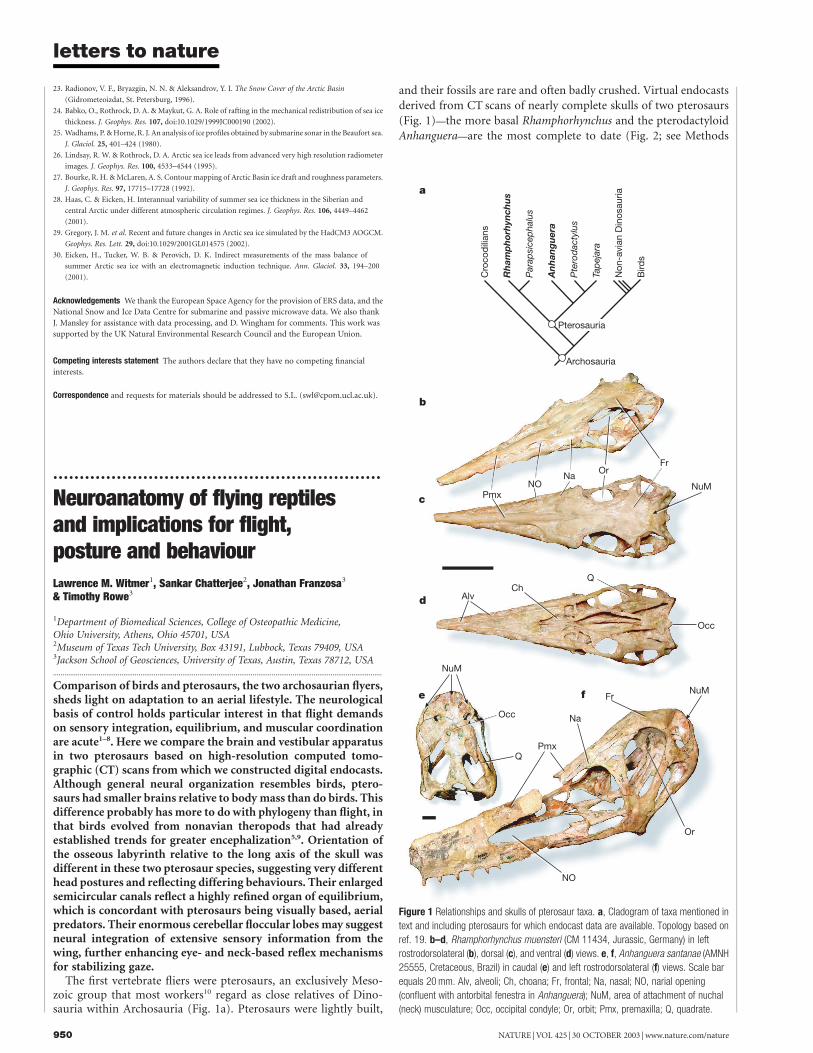
The first vertebrate fliers were pterosaurs, an exclusively Meso-zoic group that most workers10 regard as close relatives of Dino-sauria within Archosauria (Fig. 1a). Pterosaurs were lightly built,
and their fossils are rare and often badly crushed. Virtual endocastsderived from CT scans of nearly complete skulls of two pterosaurs(Fig. 1)—the more basal Rhamphorhynchus and the pterodactyloidAnhanguera—are the most complete to date (Fig. 2; see Methods
Figure 1 Relationships and skulls of pterosaur taxa. a, Cladogram of taxa mentioned in
text and including pterosaurs for which endocast data are available. Topology based on
ref. 19. b–d, Rhamphorhynchus muensteri (CM 11434, Jurassic, Germany) in left
rostrodorsolateral (b), dorsal (c), and ventral (d) views. e, f, Anhanguera santanae (AMNH
25555, Cretaceous, Brazil) in caudal (e) and left rostrodorsolateral (f) views. Scale bar
equals 20 mm. Alv, alveoli; Ch, choana; Fr, frontal; Na, nasal; NO, narial opening
(confluent with antorbital fenestra in Anhanguera); NuM, area of attachment of nuchal
(neck) musculature; Occ, occipital condyle; Or, orbit; Pmx, premaxilla; Q, quadrate.
letters to nature
NATURE | VOL 425 | 30 OCTOBER 2003 | www.nature.com/nature950
and Supplementary Information). They confirm some previousfindings of birdlike attributes1–8: expansion of the cerebrum andcerebellum, displacing the enlarged optic tecta (lobes) ventro-laterally; small olfactory areas; and enlarged flocculi (cerebellarauricles) (Fig. 2). Despite these structural similarities, the brainsof Rhamphorhynchus and Anhanguera, relative to body mass, do notfall within the range of extant birds, although they were enlargedrelative to extant nonavian reptiles4,5,11 (Fig. 3; see Methods).Moreover, comparisons of total brain mass do not reveal differencesin relative size of brain components (and hence underlying neuralorganization). For example, the enormous flocculi of pterosaursprobably outweighed the optic tecta, whereas the reverse is certainlytrue in birds.
Nevertheless, pterosaurs do possess a number of avian neuro-anatomical traits that may well be associated with the sensoryand coordination functions necessary for flight. Jerison4 suggestedthat avian brains were relatively larger than those of pterosaursbecause birds evolved in the environmentally complex and neuro-logically challenging arboreal habitat that required greater neuralprocessing and hence greater mass. That may be true, but anotherfactor is that birds and pterosaurs had different phylogeneticstarting points: pterosaurs evolved from relatively very small-brained basal archosaurs, whereas birds evolved from theropoddinosaurs that had already initiated a substantial trend of brainexpansion5,9.
The virtual endocasts include the semicircular canals (Fig. 2),which had previously been only partially known for one pterosaur,Parapsicephalus1. The entire osseous labyrinth is preserved bilater-ally in Anhanguera and the large majority of it is preserved inRhamphorhynchus. The semicircular canal system is greatlyexpanded, with the long canals encircling the flocculus. Its generalarrangement closely resembles that of birds and some other dino-saurs5, but, whereas it is relatively modest in these groups, the
vestibular apparatus is relatively much larger in the pterosaurs.The well preserved osseous labyrinths in Rhamphorhynchus andAnhanguera provide an opportunity to test behavioural hypothesesof head orientation and posture. Researchers tend to reconstructthe head orientation of extinct animals with the skull’s longaxis (often the jawline) horizontal. Animals, of course, adopt avariety of head postures. There is a rich and taxonomically diverseliterature supporting a robust empirical relationship betweenthe planar elevation of the lateral semicircular canal and preferredhead orientation12–16. Determining ‘preferred’ head posture mayseem problematic at first, but most vertebrates adopt a verystereotyped ‘alert’ posture13,17 and, moreover, retain this posturethrough a variety of behaviours15. Most studies agree that thispreferred head orientation involves maintaining the lateral semi-circular canal approximately level with the horizon (that is, 08inclination) or elevated slightly in the front (5–108 inclination)12–16.
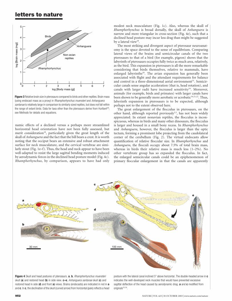
Applying a conservative 58 inclination to pterosaurs shows thatthe long axis of the skull of Rhamphorhynchus indeed has a more orless horizontal orientation, as is typically portrayed6. However,bringing the lateral semicircular canal of Anhanguera to a positionof 58 inclination results in the skull’s long axis being strongly down-turned (Fig. 4c). This dramatically different posture impacts onbehavioural hypotheses relating to feeding and locomotion, perhapsallowing lateral scanning movements of the head (that is, in theplane of the lateral canals) to operate with optimal sensitivity13 orperhaps even simply allowing for a less obstructed view and greateroverlap of the visual fields (binocular vision; Fig. 4e). Differences inhead orientation may correlate with differences in body postureduring terrestrial quadrupedal locomotion in that Rhamphor-hynchus, with its relatively shorter forelimbs, must have adopted amore horizontal trunk, whereas Anhanguera had longer forelimbsand so had a more upright body posture6,18,19
, which in turnrequired a compensatory down-turning of the head. The aerody-
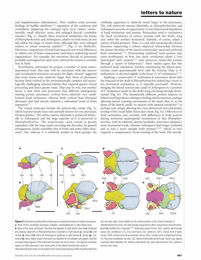
Figure 2 Endocasts and labyrinths of pterosaurs compared to brains of extant archosaurs.
a, Brain of the crocodilian archosaur Alligator mississippiensis in right lateral view.
b, Brain of the avian archosaur Columba livia (pigeon) in right lateral view. c–e, Endocast
and osseous labyrinth of Rhamphorhynchus muensteri in right lateral (c), dorsal (d), and
ventral (e) views. f–h, Same of Anhanguera santanae in right lateral (f), dorsal (g), and
ventral (h) views. Major areas of the brain are labelled for the alligator and pigeon, and the
corresponding regions of the endocasts are given the same colour. The lagenar (cochlear)
regions of both pterosaurs and some parts of the lateral semicircular canal of
Rhamphorhynchus were not included in the virtual endocasts but were reconstructed from
the raw slice data. Some details of the ventral portion of the virtual endocast of
Rhamphorhynchus were not well enough preserved to allow unequivocal reconstruction.
a and b modified from originals29,30. Scale bars equal 10 mm. AnC, anterior semicircular
canal; Cbl, cerebellum; CC, crus communis; Cer, cerebrum; CN II, cranial nerve II (optic
nerve); CN III, cranial nerve III (oculomotor nerve); CN V, cranial nerve V (trigeminal nerve);
Flo, flocculus (cerebellar auricle); LaC, lateral (horizontal) semicircular canal; Lag, lagena
(cochlea); Med, Medulla; Olf, olfactory lobe (bulb); Op, optic lobe (tectum); PoC, posterior
semicircular canal.
letters to nature
NATURE | VOL 425 | 30 OCTOBER 2003 | www.nature.com/nature 951
namic effects of a declined versus a perhaps more streamlinedhorizontal head orientation have not been fully assessed, butmerit consideration18, particularly given the great length of theskull of Anhanguera and the fact that the bill bears a crest. It is worthnoting that the occiput bears an extensive and robust attachmentsurface for neck musculature, and the cervical vertebrae are simi-larly stout (Fig. 1e–f). Thus, the head and neck appear to have beenwell-adapted to resist the large sagittal bending moments inducedby aerodynamic forces in the declined head posture model (Fig. 4c).Rhamphorhynchus, by comparison, appears to have had only
modest neck musculature (Fig. 1c). Also, whereas the skull ofRhamphorhynchus is broad dorsally, the skull of Anhanguera isnarrow and more triangular in cross-section (Fig. 4e), such that adeclined head posture may incur less drag than might be suggestedby a lateral view18.
The most striking and divergent aspect of pterosaur neuroanat-omy is the space devoted to the sense of equilibrium. Comparinglateral views of the brains and semicircular canals of the twopterosaurs to that of a bird (for example, pigeon) shows that thelabyrinth of pterosaurs occupies fully twice as much area, relatively,as the bird. This expansion in pterosaurs is all the more remarkableconsidering that birds themselves, relative to mammals, haveenlarged labyrinths20. The avian expansion has generally beenassociated with flight and the attendant requirements for balanceand control in a three-dimensional aerial environment21. Semicir-cular canals sense angular acceleration (that is, head rotation), andcanals with larger radii have increased sensitivity16. Moreover,animals (for example, birds and primates) with larger canals havebeen shown to be generally more aerobatic or acrobatic16,21,22. Thus,labyrinth expansion in pterosaurs is to be expected, althoughperhaps not to the extent observed here.
The great enlargement of the flocculus in pterosaurs, on theother hand, although reported previously2,5, has not been widelyappreciated. In extant nonavian reptiles, the flocculus is incon-spicuous, whereas in birds and many other dinosaurs, the flocculusis larger and housed in a small bony recess. In Rhamphorhynchusand Anhanguera, however, the flocculus is larger than the optictectum, forming a prominent lobe projecting from the caudolateralcorner of the cerebellum (Fig. 2). The virtual endocasts allowquantification of relative floccular size. In Rhamphorhynchus andAnhanguera, the flocculi occupy about 7.5% of total brain mass,whereas in birds their relative mass is much less (1–2%). Noother vertebrate group has so expanded the flocculus. In fact,the enlarged semicircular canals could be an epiphenomenon ofprimary floccular enlargement in that the canals are apparently
Figure 3 Relative brain size in pterosaurs compared to birds and other reptiles. Brain mass
(using endocast mass as a proxy) in Rhamphorhynchus muensteri and Anhanguera
santanae is relatively large in comparison to similarly sized reptiles, but does not fall within
the range of extant birds. Data for taxa other than the pterosaurs derive from Hurlbert28;
see Methods for details and equations.
Figure 4 Skull and head postures of pterosaurs. a, b, Rhamphorhynchus muensteri
skull (a) and restored head (b) in side view. c–e, Anhanguera santanae skull (c) and
restored head in side (d) and front (e) views. Brains (endocasts) are indicated in red in a
and c. In c, the declination of the skull (curved arrow) from horizontal (pale) reflects a head
posture with the lateral canal inclined 58 above horizontal. The double-headed arrow in c
indicates the well-developed neck muscles that would have prevented excessive
sagittal deflection of the head caused by aerodynamic drag. a and c modified from
originals25,26.
letters to nature
NATURE | VOL 425 | 30 OCTOBER 2003 | www.nature.com/nature952

constrained to encircle the flocculus.The flocculus has important connections with the vestibular
system (via brainstem nuclei), the eye muscles (which are approxi-mately coplanar with the canals), and, in some taxa, the neckmuscles23,24. This circuitry is best understood in the context of thevestibulo-ocular (VOR) and vestibulocollic (VCR) reflexes wherebycoordination of head, eye and neck movements ensures stabilizationof an image on the retina, preventing blurring16,24. These reflexesallow a cheetah or a hawk to maintain a rock-steady gaze as itpursues its prey. Because some of the processing takes place in theflocculus, it would seem that pterosaurs devoted (perhaps evendiverted) considerable neural resources to the integration of thesegaze-stabilization mechanisms. Enhancement of such mechanismsseems reasonable in these two visually oriented pterosaurs giventheir apparent foraging style of aerial fish-eating6,25,26.
However, there is no reason to believe that pterosaurs were moreagile or aerobatic than birds (including avian aerial piscivores), andhence the size of the flocculus and semicircular canals remainsenigmatic. Under the principle of proper mass (the amount ofneural tissue in a structure is proportional to the amount ofprocessing4), it would seem that the flocculus of pterosaurs was thesite of neural processing unlike that seen in extant vertebrates,suggesting that an explanation should be sought among the unusualfeatures of pterosaurs, such as the large, skin-covered flight mem-brane of the wing. In birds and mammals, the flocculus receivesinputs carrying proprioceptive and cutaneous information (with arelay in the inferior olivary nucleus)24. Thus, the pterosaur flocculusmay have processed an unusually high volume of proprioceptiveand somatosensory information associated with the wing mem-brane that stretched between the limbs, as well as with the limb jointsthemselves, consequently having a direct impact on the VOR/VCRand flight control. Support for enhanced proprioceptive input frompterosaur wings comes from the recent finding that the flightmembrane incorporated muscle and tendon27, which would havesent proprioceptive (muscle spindle) fibres back to the centralnervous system, potentially for integration in the flocculus. Thus,the enlarged flocculus may be causally linked to the pterosaurintegumentary wing membrane, with the wing providing to theflocculus potentially massive amounts of sensory data on attitudeand body orientation, resulting in enhanced compensatory reflexesfor maintaining the fixation of gaze upon a target. A
MethodsImagingInformation on the brains of extinct organisms has traditionally come from naturallyoccurring endocasts of the brain cavity1, latex endocasts7,8 made after the rock has beenremoved, and reconstructions of ground thin-sections of skulls4. We used a newer,noninvasive technique employing X-ray computed tomography (CT) to reconstruct adigital or ‘virtual’ endocast from the transverse CT slices of the brain cavity and vestibularapparatus. One skull each of Rhamphorhynchus muensteri (CM 11434; from the UpperJurassic Solnhofen Lithographic Limestones of southern Germany) and Anhanguerasantanae (AMNH 25555; from the Lower Cretaceous Santana formation of northeasternBrazil) was acid-prepared to remove surrounding matrix and then CT-scanned.Rhamphorhynchus was scanned along the coronal axis for a total of 476 slices, each slice0.25-mm thick, with an interslice spacing of 0.2 mm (for a slice overlap of 0.05 mm).Anhanguera was scanned along the coronal axis for a total of 595 slices, each slice 0.50-mmthick with an interslice spacing of 0.45 mm (for a slice overlap of 0.05 mm). Raw slice data,reconstructed skulls, and virtual endocast animations are provided in the SupplementaryInformation.
Relative brain size calculationsAllometric scaling of brain mass (M Br) and body mass (M Bd) provides a means ofcomparing relative brain size. Jerison4 facilitated comparison by devising a simple metric,the encephalization quotient (EQ), which is a ratio of actual brain mass to the predictedbrain mass (based on allometry) for the animal’s reference group (for example, mammals,reptiles). We employ Hurlburt’s28 modifications of Jerison’s method, such that for reptiles,EQ ¼ M Br/(0.155(M Bd)0.55), whereas for birds, EQ ¼ M Br/(0.117(M Bd)0.59). We use theavian equation to estimate the pterosaur EQs because pterosaur brains filled theendocranial cavity (as in birds, but unlike in reptiles). Mass estimation forRhamphorhynchus (CM 11434) and Anhanguera (AMNH 25555) was complicated by lackof associated postcranial skeletons. We obtained body masses from comparably sizedcomplete skeletons: Rhamphorhynchus muensteri (SMF R412825) and Anhanguera piscator
(NSN-PV 1989226). Using a principal-components analysis method for estimating bodymass18, M Bd for Rhamphorhynchus is 136 g, and M Bd for Anhanguera is 7,600 g. Brainvolume (determined from CT) multiplied by density (1.036 g cm23) yields M Br of 0.83 gfor Rhamphorhynchus and 7.72 g for Anhanguera. EQ is 0.39 for Rhamphorhynchus and0.34 for Anhanguera. Plotting log-transformed values on Hurlburt’s28 graph of brainversus body-mass shows that these pterosaurs fall between the reptile and avian polygons,with their EQs below those of birds (Fig. 3).
Received 14 March; accepted 2 September 2003; doi:10.1038/nature02048.
1. Newton, E. T. On the skull, brain, and auditory organ of a new species of pterosaurian (Scaphognathus
purdoni), from the Upper Lias near Whitby Yorkshire. Phil. Trans. R. Soc. Lond. B 179, 503–537
(1888).
2. Edinger, T. Das Gehirn der Pterosaurier. Z. Anat. Entwicklungsgesch. 82, 105–112 (1927).
3. Edinger, T. The brain of Pterodactylus. Am. J. Sci. 239, 665–682 (1941).
4. Jerison, H. J. Evolution of the Brain and Intelligence 482 (Academic, New York, 1973).
5. Hopson, J. A. in Biology of the Reptilia Vol. 9 Neurology A (eds Gans, C., Northcutt, R. G. & Ulinki, P.)
39–146 (Academic, New York, 1979).
6. Wellnhofer, P. The Illustrated Encyclopedia of Pterosaurs 192 (Crescent, New York, 1991).
7. Bennett, S. C. The osteology and functional morphology of the Late Cretaceous pterosaur Pteranodon.
Part I. General description of osteology. Palaeontogr. Abt. A 260, 1–112 (2001).
8. Wharton, D. S. The Evolution of the Avian Brain 343. PhD thesis, Univ. Bristol (2002).
9. Larsson, H. C. E., Sereno, P. C. & Wilson, J. A. Forebrain enlargement among nonavian theropod
dinosaurs. J. Vert. Paleontol. 20, 615–618 (2000).
10. Brochu, C. A. Progress and future directions in archosaur phylogenetics. J. Paleontol. 75, 1185–1201
(2001).
11. Kellner, A. W. A. Description of the braincase of two Early Cretaceous pterosaurs (Pterodactyloidea)
from Brazil. Am. Mus. Novit. 3175, 1–34 (1996).
12. Lebedkin, S. Uber die Lage des Canalis semicircularis lateralis bei Saugern. Anat. Anz. 58, 447–460
(1924).
13. Duijm, M. On the head posture of some birds and its relation to some anatomical features. Proc.
Koninkl. Nederl. Akad. Wetensch. C 54, 202–211, 260–271 (1951).
14. Blanks, R. H. I., Curthoys, I. S. & Markham, C. H. Planar relationships of semicircular canals in the
cat. Am. J. Physiol. 223, 55–62 (1972).
15. Erichsen, J. T., Hodos, W., Evinger, C., Bessette, B. B. & Phillips, S. J. Head orientation in pigeons:
postural, locomotor and visual determinants. Brain Behav. Evol. 33, 268–278 (1989).
16. Spoor, F. & Zonneveld, F. Comparative review of the human bony labyrinth. Yearbook Phys. Anthropol.
41, 211–251 (1998).
17. de Beer, G. R. How animals hold their heads. Proc. Linn. Soc. Lond. 159, 125–139 (1947).
18. Chatterjee, S. & Templin, R. J. Posture, locomotion and paleoecology of pterosaurs. Geol. Soc. Am.
Spec. Pap. (in the press).
19. Unwin, D. M., Lu, J. & Bakhurina, N. N. On the systematic and stratigraphic significance of pterosaurs
from the Lower Cretaceous Yixian Formation (Jehol Group) of Liaoning, China. Mitt. Mus. Naturk.
Berlin Geowiss. Reihe 3, 181–206 (2000).
20. Jones, G. M. & Spells, K. E. A theoretical and comparative study of the functional dependence of the
semicircular canal upon its physical dimensions. Proc. R. Soc. Lond. B 157, 403–419 (1963).
21. Turkewitsch, B. G. Zur Anatomie des Gehororgans der Vogel (Canales semicirculares). Z. Anat.
Entwicklungsgesch. 103, 551–608 (1934).
22. Spoor, F., Bajpal, S., Hussain, S. T., Kumar, K. & Thewissen, J. G. M. Vestibular evidence for the
evolution of aquatic behavior in early cetaceans. Nature 417, 163–166 (2002).
23. Butler, A. B. & Hodos, W. Comparative Vertebrate Neuroanatomy: Evolution and Adaptation 514
(Wiley-Liss, New York, 1996).
24. Winship, I. R. & Wylie, D. R. W. Zonal organization of the vestibulocerebellum in pigeons (Columba
livia): I. Climbing fiber input to the flocculus. J. Comp. Neurol. 456, 127–139 (2003).
25. Wellnhofer, P. Die Rhamphorhynchoidea (Pterosauria) der Oberjura-Plattenkalke Suddeutschlands.
Teil I. Allgemeine Skelettmorphologie. Palaeontogr. Abt. A 148, 1–33 (1975).
26. Kellner, A. W. A. & Tomida, Y. Description of a new species of Anhangueridae (Pterodactyloidea) with
comments on the pterosaur fauna from the Santana Formation (Aptian-Albian), northeastern Brazil.
Nat. Sci. Mus. Monogr. 17, 1–135 (2000).
27. Tischlinger, H. & Frey, E. Ein Rhamphorhynchus (Pterosauria, Reptilia) mit ungewohnlicher
Flughauterhaltung aus dem Solnhofener Plattenkalk. Archaeopteryx 20, 1–20 (2002).
28. Hurlburt, G. R.. Relative Brain Size in Recent and Fossil Amniotes: Determination and Interpretation
250. PhD thesis, Univ. Toronto (1996).
29. Romer, A. S. Osteology of the Reptiles 772 (Univ. Chicago Press, Chicago, 1956).
30. Proctor, N. S. & Lynch, P. J. Manual of Ornithology 340 (Yale Univ. Press, New Haven, 1993).
Supplementary Information accompanies the paper on www.nature.com/nature.
Acknowledgements D. S. Berman (Carnegie Museum of Natural History) and J. Maisey
(American Museum of Natural History) agreed to the loan and preparation of the pterosaur
specimens. M. Atanassov assisted with body mass estimates and other morphometrics. Z. Zheng
acid-prepared the fossils. M. Colbert, J. Humphries, R. Ketcham, and J. Maisano assisted with the
CTscanning, data processing, and web delivery. Figures were drafted by R. Ridgely (Figs 2 and 3a,
c) and K. McQuilkin (Fig. 3b, d, e and Fig. 4). We thank G. R. Hurlburt and D. S. Wharton for
sharing data in their doctoral dissertations. We thank G. R. Hurlburt, P. M. O’Connor, E. Weber,
and D. S. Wharton for fruitful discussion of pterosaurs and neuroscience, and R. J. Templin for
providing aerodynamic expertise. The manuscript benefited from comments provided by
D. M. Unwin & S. C. Bennett. Funding was provided by NSF grants to L.M.W. and T.R. and by
Texas Tech University to S.C.
Competing interests statement The authors declare that they have no competing financial
interests.
Correspondence and requests for materials should be addressed to L.M.W. ([email protected]).
letters to nature
NATURE | VOL 425 | 30 OCTOBER 2003 | www.nature.com/nature 953