Embed Size (px)
Citation preview
Volume No: 2 (2015), Issue No: 4 (April) April 2015 www.ijmetmr.com Page 389
ISSN No: 2348-4845International Journal & Magazine of Engineering,
Technology, Management and ResearchA Peer Reviewed Open Access International Journal
Abstract:
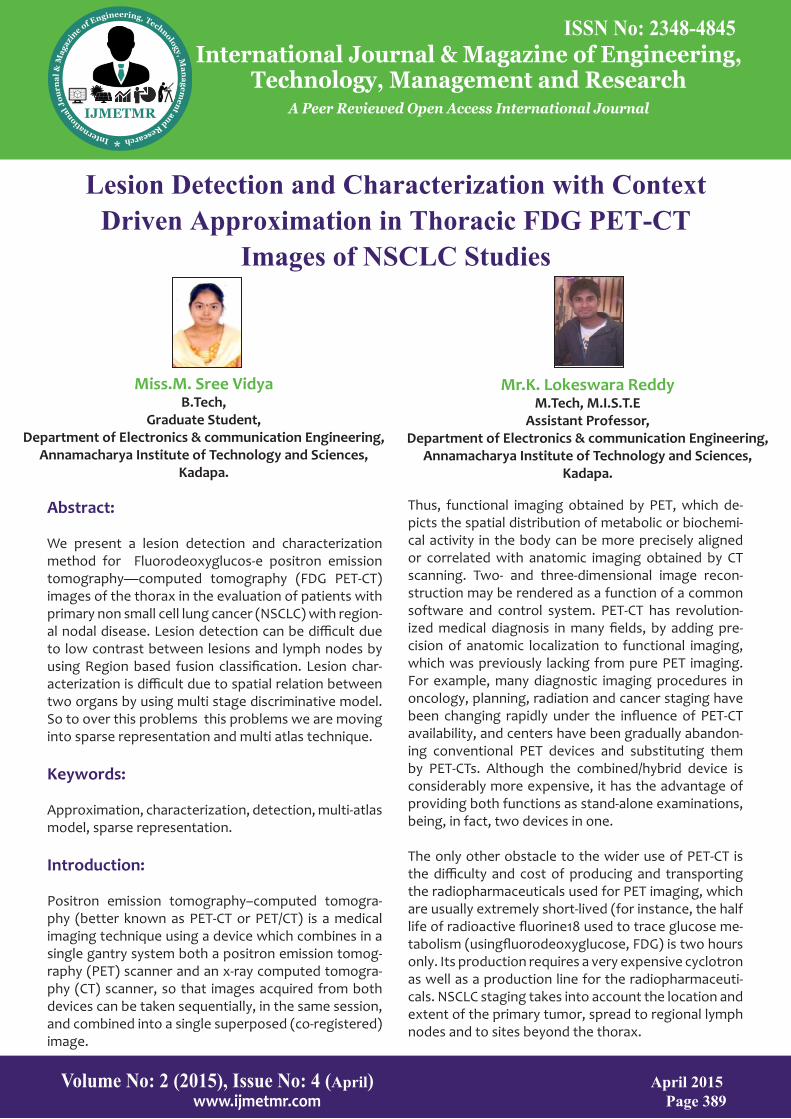
We present a lesion detection and characterization method for Fluorodeoxyglucos-e positron emission tomography—computed tomography (FDG PET-CT) images of the thorax in the evaluation of patients with primary non small cell lung cancer (NSCLC) with region-al nodal disease. Lesion detection can be difficult due to low contrast between lesions and lymph nodes by using Region based fusion classification. Lesion char-acterization is difficult due to spatial relation between two organs by using multi stage discriminative model. So to over this problems this problems we are moving into sparse representation and multi atlas technique.
Keywords:
Approximation, characterization, detection, multi-atlas model, sparse representation.
Introduction:
Positron emission tomography–computed tomogra-phy (better known as PET-CT or PET/CT) is a medical imaging technique using a device which combines in a single gantry system both a positron emission tomog-raphy (PET) scanner and an x-ray computed tomogra-phy (CT) scanner, so that images acquired from both devices can be taken sequentially, in the same session, and combined into a single superposed (co-registered) image.
Miss.M. Sree VidyaB.Tech,
Graduate Student,Department of Electronics & communication Engineering,
Annamacharya Institute of Technology and Sciences,Kadapa.
Mr.K. Lokeswara ReddyM.Tech, M.I.S.T.E
Assistant Professor,Department of Electronics & communication Engineering,
Annamacharya Institute of Technology and Sciences,Kadapa.
Thus, functional imaging obtained by PET, which de-picts the spatial distribution of metabolic or biochemi-cal activity in the body can be more precisely aligned or correlated with anatomic imaging obtained by CT scanning. Two- and three-dimensional image recon-struction may be rendered as a function of a common software and control system. PET-CT has revolution-ized medical diagnosis in many fields, by adding pre-cision of anatomic localization to functional imaging, which was previously lacking from pure PET imaging. For example, many diagnostic imaging procedures in oncology, planning, radiation and cancer staging have been changing rapidly under the influence of PET-CT availability, and centers have been gradually abandon-ing conventional PET devices and substituting them by PET-CTs. Although the combined/hybrid device is considerably more expensive, it has the advantage of providing both functions as stand-alone examinations, being, in fact, two devices in one.
The only other obstacle to the wider use of PET-CT is the difficulty and cost of producing and transporting the radiopharmaceuticals used for PET imaging, which are usually extremely short-lived (for instance, the half life of radioactive fluorine18 used to trace glucose me-tabolism (usingfluorodeoxyglucose, FDG) is two hours only. Its production requires a very expensive cyclotron as well as a production line for the radiopharmaceuti-cals. NSCLC staging takes into account the location and extent of the primary tumor, spread to regional lymph nodes and to sites beyond the thorax.
Lesion Detection and Characterization with Context Driven Approximation in Thoracic FDG PET-CT
Images of NSCLC Studies
Volume No: 2 (2015), Issue No: 4 (April) April 2015 www.ijmetmr.com Page 390
ISSN No: 2348-4845International Journal & Magazine of Engineering,
Technology, Management and ResearchA Peer Reviewed Open Access International Journal
FDG PET-CT is the most accurate imaging modality for lung cancer staging. While FDG PET was a valuable non-invasive imaging technique to detect functional rather than anatomical data, its main limitation was the lack of spatial resolution. The combination of PET and CT in one device helps overcome this limitation. Manual interpretation of a PET-CT study is time-consuming due to the large volume of data and requires extensive ex-perience and so can suffer from inter-observer differ-ences.
In thoracic PET-CT studies, the main challenges to au-tomated lesion detection are low contrast between normal anatomical structures and lesions, and inter-subject variations in temporal FDG uptake. On CT, le-sions usually appear similar to the soft tissues, which is problematic when lesions are located adjacent to the chest wall or meditational structures. For PET, al-though lesions are typically more FDG -avid than sur-rounding structures, some lesions have only mild FDG uptake; and there can be elevated FDG uptake in nor-mal structures.
Such low contrast implies that detecting lesions based on the within -subject information can be complicated. lung tumors can invade the mediastinum and lymph nodes can be adjacent to the lung parenchyma. In both examples, the two lesion types have similar spa-tial characteristics and it is difficult to differentiate be-tween them.
Existing system:
In the existing system region based fusion classifica-tion and multi stage discriminative model techniques are used. In region based fusion classification charac-teristics of each region are calculated and region based approach is used to fuse the images region-by-region, in the wavelet domain. The multi stage discriminative model is used for automatically detect both tumors and abnormal lymph nodes simultaneously from PET-CT images. But in the existing system noise can’t be eliminated. So the proposed system can overcome the above drawback.
Methods to find FDG PET-CT:
Lesion detection methods on PET-CT images are based on thresholding.
The standard uptake value (SUV), which is a semi-quan-titative measure of FDG uptake, is widely used to de-termine the threshold.Adaptive threshold values have been proposed to better accommodate the subject or region-level characteristics, such as the contrast based threshold and the iterative threshold. Some stud-ies have reported on the detection of only one lesion type—the primary lung tumor or lymph nodes—with the assumption that there is only single lesion type in the image. Without such an assumption, the lung fields can be firstly segmented, and then lung tumors are de-tected within the segmented lung fields. However, in cases where the lung tumors invade the mediastinum, the segmentation of the lung fields is often unreliable. With the multi-atlas method, reference data are re-ferred to as atlases. Majority voting or weighted com-bination of multiple atlases transfers to the labeling of a testing image. Most commonly the weights are de-termined based on predefined formulas, such as local similarity between atlases and the test image. Sparse representation has been successfully applied to solve classification problems with applications in face rec-ognition , and recently in the medical imaging domain . Brie fl y, a reference set is constructed to represent each class, and a reconstruction difference is comput-ed for the test data based on each reference set. The class corresponding to the lowest difference is then the class label of the test data.
In multi-atlas and sparse representation approaches, improvement over the basic sparse regularization has mainly focused on spatial constraints, such as group sparsity. Another technique is to incorporate diction-ary learning in place of the raw reference data. Howev-er, in these methods, the optimization is usually to im-prove the reconstruction, which might not correspond to better classification.
Benefaction of FDG PET-CT:
we propose a lesion detection and characterization method for thoracic FDG PET-CT images in patients with NSCLC. For lesion detection, image patches that clearly represent the lung fields, mediastinum or the lung lesion are first de-tected with thresholding opera-tions. The remaining patches are then labeled as lesion or mediastinum based on their approximation of the detected lesion and mediastinum patches.
Volume No: 2 (2015), Issue No: 4 (April) April 2015 www.ijmetmr.com Page 391
ISSN No: 2348-4845International Journal & Magazine of Engineering,
Technology, Management and ResearchA Peer Reviewed Open Access International Journal
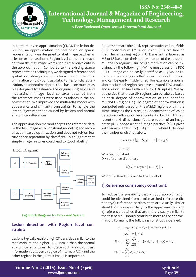
In context driven approximation (CDA). For lesion de-tection, an approximation method based on sparse representation was designed to label image patches as a lesion or mediastinum. Region-level contexts extract-ed from the test image were used as reference data in the ap-proximation. Compared to the existing sparse representation techniques, we designed reference and spatial consistency constraints for a more effective dis-crimination of low - contrast data. For lesion character-ization, an approximation method based on multi-atlas was designed to estimate the original lung fields and mediastinum. Image -level contexts obtained from the reference images were used as atlases in the ap-proximation. We improved the multi-atlas model with appearance and similarity constraints, to handle the inter-subject variations caused by lesions and normal anatomical differences.
The approximation method adapts the reference data to the test image with constraint modeling and recon-struction-based optimization, and does not rely on fea-ture space separation by classifiers. This suggests that simple image features could lead to good labeling.
Fig: Block Diagram for Proposed System
Lesion detection with Region level con-straint:
Lesions typically exhibit high CT densities similar to the mediastinum and higher FDG uptake than the normal anatomical structures. To locate such areas, contrast information between a region of interest (ROI) and the other regions in the 3-D test image is important.
Regions that are obviously representative of lung fields (LF), mediastinum (MS), or lesion (LS) are labeled first. The remaining regions (UN) are further labeled as MS or LS based on their approximation of the detected MS and LS regions. Our design motivation can be ex-plained by the following. 1) While most areas on a FDG PET-CT image can be easily identified as LF, MS, or LS, there are some regions that show in-distinct features and can be easily misidentified. For example, a non le-sion mediastinal region can have elevated FDG uptake, and a lesion can have relatively low FDG uptake. We hy-pothe-size that these UN regions can be labeled based on their degree of approximation of the identified MS and LS regions. 2) The degree of approximation is computed only based on the MS/LS regions within the same image as the UN region.Approximation of lesion detection with region level contexts: Let fi€RHx1 rep-resent the H -dimensional feature vector of an image patch pi. Suppose a set of image patches {pi} is given with known labels L(pi)=l € {1,…L} , where L denotes the number of distinct labels.
Where c=constantDl= reference dictionary
Where fx- flx=difference between labels
1) Reference consistency constraint:
To reduce the possibility that a good approximation could be obtained from a mismatched reference dic-tionary.1) reference patches that are visually similar should contribute similarly to the approximation; and 2) reference patches that are more visually similar to the test patch should contribute more to the approxi-mation. Formally, the following construct is defined:
Volume No: 2 (2015), Issue No: 4 (April) April 2015 www.ijmetmr.com Page 390
ISSN No: 2348-4845International Journal & Magazine of Engineering,
Technology, Management and ResearchA Peer Reviewed Open Access International Journal
FDG PET-CT is the most accurate imaging modality for lung cancer staging. While FDG PET was a valuable non-invasive imaging technique to detect functional rather than anatomical data, its main limitation was the lack of spatial resolution. The combination of PET and CT in one device helps overcome this limitation. Manual interpretation of a PET-CT study is time-consuming due to the large volume of data and requires extensive ex-perience and so can suffer from inter-observer differ-ences.
In thoracic PET-CT studies, the main challenges to au-tomated lesion detection are low contrast between normal anatomical structures and lesions, and inter-subject variations in temporal FDG uptake. On CT, le-sions usually appear similar to the soft tissues, which is problematic when lesions are located adjacent to the chest wall or meditational structures. For PET, al-though lesions are typically more FDG -avid than sur-rounding structures, some lesions have only mild FDG uptake; and there can be elevated FDG uptake in nor-mal structures.
Such low contrast implies that detecting lesions based on the within -subject information can be complicated. lung tumors can invade the mediastinum and lymph nodes can be adjacent to the lung parenchyma. In both examples, the two lesion types have similar spa-tial characteristics and it is difficult to differentiate be-tween them.
Existing system:
In the existing system region based fusion classifica-tion and multi stage discriminative model techniques are used. In region based fusion classification charac-teristics of each region are calculated and region based approach is used to fuse the images region-by-region, in the wavelet domain. The multi stage discriminative model is used for automatically detect both tumors and abnormal lymph nodes simultaneously from PET-CT images. But in the existing system noise can’t be eliminated. So the proposed system can overcome the above drawback.
Methods to find FDG PET-CT:
Lesion detection methods on PET-CT images are based on thresholding.
The standard uptake value (SUV), which is a semi-quan-titative measure of FDG uptake, is widely used to de-termine the threshold.Adaptive threshold values have been proposed to better accommodate the subject or region-level characteristics, such as the contrast based threshold and the iterative threshold. Some stud-ies have reported on the detection of only one lesion type—the primary lung tumor or lymph nodes—with the assumption that there is only single lesion type in the image. Without such an assumption, the lung fields can be firstly segmented, and then lung tumors are de-tected within the segmented lung fields. However, in cases where the lung tumors invade the mediastinum, the segmentation of the lung fields is often unreliable. With the multi-atlas method, reference data are re-ferred to as atlases. Majority voting or weighted com-bination of multiple atlases transfers to the labeling of a testing image. Most commonly the weights are de-termined based on predefined formulas, such as local similarity between atlases and the test image. Sparse representation has been successfully applied to solve classification problems with applications in face rec-ognition , and recently in the medical imaging domain . Brie fl y, a reference set is constructed to represent each class, and a reconstruction difference is comput-ed for the test data based on each reference set. The class corresponding to the lowest difference is then the class label of the test data.
In multi-atlas and sparse representation approaches, improvement over the basic sparse regularization has mainly focused on spatial constraints, such as group sparsity. Another technique is to incorporate diction-ary learning in place of the raw reference data. Howev-er, in these methods, the optimization is usually to im-prove the reconstruction, which might not correspond to better classification.
Benefaction of FDG PET-CT:
we propose a lesion detection and characterization method for thoracic FDG PET-CT images in patients with NSCLC. For lesion detection, image patches that clearly represent the lung fields, mediastinum or the lung lesion are first de-tected with thresholding opera-tions. The remaining patches are then labeled as lesion or mediastinum based on their approximation of the detected lesion and mediastinum patches.
Volume No: 2 (2015), Issue No: 4 (April) April 2015 www.ijmetmr.com Page 391
ISSN No: 2348-4845International Journal & Magazine of Engineering,
Technology, Management and ResearchA Peer Reviewed Open Access International Journal
In context driven approximation (CDA). For lesion de-tection, an approximation method based on sparse representation was designed to label image patches as a lesion or mediastinum. Region-level contexts extract-ed from the test image were used as reference data in the ap-proximation. Compared to the existing sparse representation techniques, we designed reference and spatial consistency constraints for a more effective dis-crimination of low - contrast data. For lesion character-ization, an approximation method based on multi-atlas was designed to estimate the original lung fields and mediastinum. Image -level contexts obtained from the reference images were used as atlases in the ap-proximation. We improved the multi-atlas model with appearance and similarity constraints, to handle the inter-subject variations caused by lesions and normal anatomical differences.
The approximation method adapts the reference data to the test image with constraint modeling and recon-struction-based optimization, and does not rely on fea-ture space separation by classifiers. This suggests that simple image features could lead to good labeling.
Fig: Block Diagram for Proposed System
Lesion detection with Region level con-straint:
Lesions typically exhibit high CT densities similar to the mediastinum and higher FDG uptake than the normal anatomical structures. To locate such areas, contrast information between a region of interest (ROI) and the other regions in the 3-D test image is important.
Regions that are obviously representative of lung fields (LF), mediastinum (MS), or lesion (LS) are labeled first. The remaining regions (UN) are further labeled as MS or LS based on their approximation of the detected MS and LS regions. Our design motivation can be ex-plained by the following. 1) While most areas on a FDG PET-CT image can be easily identified as LF, MS, or LS, there are some regions that show in-distinct features and can be easily misidentified. For example, a non le-sion mediastinal region can have elevated FDG uptake, and a lesion can have relatively low FDG uptake. We hy-pothe-size that these UN regions can be labeled based on their degree of approximation of the identified MS and LS regions. 2) The degree of approximation is computed only based on the MS/LS regions within the same image as the UN region.Approximation of lesion detection with region level contexts: Let fi€RHx1 rep-resent the H -dimensional feature vector of an image patch pi. Suppose a set of image patches {pi} is given with known labels L(pi)=l € {1,…L} , where L denotes the number of distinct labels.
Where c=constantDl= reference dictionary
Where fx- flx=difference between labels
1) Reference consistency constraint:
To reduce the possibility that a good approximation could be obtained from a mismatched reference dic-tionary.1) reference patches that are visually similar should contribute similarly to the approximation; and 2) reference patches that are more visually similar to the test patch should contribute more to the approxi-mation. Formally, the following construct is defined:
CT Pre Processing Sparse technique
PET Pre processing
Multi atlas technique
Reference consistency
Spatial consistency
Appearance constraint
Similarity constraint
Labeling outputs
Lesion detection
& characteri
zation
Block Diagram:
Volume No: 2 (2015), Issue No: 4 (April) April 2015 www.ijmetmr.com Page 392
ISSN No: 2348-4845International Journal & Magazine of Engineering,
Technology, Management and ResearchA Peer Reviewed Open Access International Journal
2) Spatial consistency constraint:
To encourage spatially consistent labels in a local re-gion, our idea is to encode the spatial consistency preference into the labeling phase. In particular, rather than determining the label of a test patch px based on the approximation difference only as in (2),we pro-pose that the label of px should also be affected by the surrounding patches, which could contain other test patches or patches with known labels. If px is visually similar to the surrounding patches, similar labels should be assigned among them. A local region is defined as a connected component comprising A number of image patches {px} with unknown Labels and R is surround-ing by images patches {pi} with known labels.
Lesion characterization:The detected lesions in thoracic FDG PET- CT images com-prise two types: primary lung tumor (LT) and ab-normal lymph nodes (LN). There can also be false posi-tive detection that is actually high FDG uptake in the myocardium (MC), which is normal and nonpathologi-cal. LT lies inside the lung fields, LNs are in the hilar or mediastinal regions, and MC is a large area in the medi-astinum near the left lung field.
Therefore, our underlying algorithm identifies the lung fields and mediastinum to extract the spatial character-istics. The LF/MS regions labeled during lesion detec-tion exclude the lesions.These considerations suggest that we need to estimate the actual lung fields and the mediastinum from the image.
Approximation of lesion characterization with image level contexts:
This is based on the observation that there is great sim-ilarity in the normal anatomical appearances between images, even though patient-specific conditions intro-duce variation. Therefore, given a test image , the ap-proach is to estimate the lung fields and mediastinum based on reference images. The estimation is equiva-lent to relabing the patches in I as LF or MS. For each axial Is the test image I, a feature vector gx € RQx1 is computed by concatenating the patch –wise labels derived during lesion detection. In gx, each element gx(Pi) represents the labels(LF,MS, or LS) of patch Pi € Is, with numeric values 1,2, or 1.5. The value 1.5 means that the LS patches can be relabeled as LF or MS with equal probabilities. A multi-atlas model with sparse regularization is then formulated to derive the LF/MS labels for s.
where is a weight vector with nonzero elements, and is a constant. The vector contains real numbers that are approximations of LF/MS labels. To further enhance the labeling performance, we designed two additional constraints—appearance and similarity con-straints.
1) Appearance Constraint:
One limitation with the multi-atlas model in (12) is that, the feature vector contains the label information only. It also restricts the approximation target in accommo-dating the appearance variations between subjects. We thus include the patch-wise average CT density as a second feature vector. PET data are not used given the low spa-tial resolution and relatively large inter-subject variations compared to the CT data.

Volume No: 2 (2015), Issue No: 4 (April) April 2015 www.ijmetmr.com Page 393
ISSN No: 2348-4845International Journal & Magazine of Engineering,
Technology, Management and ResearchA Peer Reviewed Open Access International Journal
2) Similarity constraint:
Image Jk,s is corresponds to a single weight element wk in w. This means that all patches in Jk,s would con-tribute equally to the approximation. However, due to the nonrigid structure of the thorax and presence of abnormalities, it is normal that only a portion of is similar to , patch-wise similarity information between the test image and the reference images to allocate dif-ferent weights to different patches.
Patch based detection:
The patch - based labeling method for the lesion detec-tion.The objective is to label the patches into LF,MS or LS categories: , the LS patches would be lesions de-tected. we perform a four-class labeling: , , , , where UN represents the patches of un-sure (i.e., MS or LS) category. Specifically, Otsu thresholding is used to separate the LF patches from the rest based on the average CT densities of patches, since the lung fields exhibit much lower CT densities compared to the other three categories.
The PET image is then used to differentiate MS, LS, and UN. , the UN patches {px} {pi} are further labeled as MS or LS [Fig. 2(b)]. To do this, each image patch is represented by a four-dimensional feature vector : its mean and standard deviation of CT densities, and mean and standard deviation of SUV. Next, for a UN patch , two reference dictionaries are constructed based on the labeled MS and LS patches in an image I:DMS and DLS. Two features approximations FxMS are thus de-rived using.
Results and Discussion:
CT input image:
CT input image with lung cancerPET input image:
PET input image with lung cancer
Preprocessing image:
The PET-CT image doesn’t consists of noise, reflec-
tions and masking. Sparse image:
CT image with high Contrast dataSpatial output image:
CT image with improved detection
PerformanceNoisy image:
CT image which represents noiseMedian output:
Volume No: 2 (2015), Issue No: 4 (April) April 2015 www.ijmetmr.com Page 394
ISSN No: 2348-4845International Journal & Magazine of Engineering,
Technology, Management and ResearchA Peer Reviewed Open Access International Journal
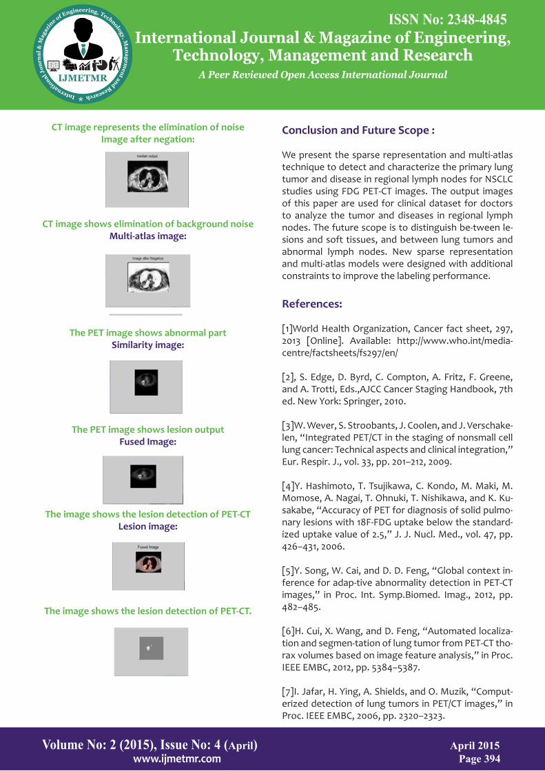
CT image represents the elimination of noiseImage after negation:
CT image shows elimination of background noise
Multi-atlas image:
The PET image shows abnormal part
Similarity image:
The PET image shows lesion output
Fused Image:
The image shows the lesion detection of PET-CT
Lesion image:
The image shows the lesion detection of PET-CT.
Conclusion and Future Scope :
We present the sparse representation and multi-atlas technique to detect and characterize the primary lung tumor and disease in regional lymph nodes for NSCLC studies using FDG PET-CT images. The output images of this paper are used for clinical dataset for doctors to analyze the tumor and diseases in regional lymph nodes. The future scope is to distinguish be-tween le-sions and soft tissues, and between lung tumors and abnormal lymph nodes. New sparse representation and multi-atlas models were designed with additional constraints to improve the labeling performance.
References:
[1]World Health Organization, Cancer fact sheet, 297, 2013 [Online]. Available: http://www.who.int/media-centre/factsheets/fs297/en/
[2], S. Edge, D. Byrd, C. Compton, A. Fritz, F. Greene, and A. Trotti, Eds.,AJCC Cancer Staging Handbook, 7th ed. New York: Springer, 2010.
[3]W. Wever, S. Stroobants, J. Coolen, and J. Verschake-len, “Integrated PET/CT in the staging of nonsmall cell lung cancer: Technical aspects and clinical integration,” Eur. Respir. J., vol. 33, pp. 201–212, 2009.
[4]Y. Hashimoto, T. Tsujikawa, C. Kondo, M. Maki, M. Momose, A. Nagai, T. Ohnuki, T. Nishikawa, and K. Ku-sakabe, “Accuracy of PET for diagnosis of solid pulmo-nary lesions with 18F-FDG uptake below the standard-ized uptake value of 2.5,” J. J. Nucl. Med., vol. 47, pp. 426–431, 2006.
[5]Y. Song, W. Cai, and D. D. Feng, “Global context in-ference for adap-tive abnormality detection in PET-CT images,” in Proc. Int. Symp.Biomed. Imag., 2012, pp. 482–485.
[6]H. Cui, X. Wang, and D. Feng, “Automated localiza-tion and segmen-tation of lung tumor from PET-CT tho-rax volumes based on image feature analysis,” in Proc. IEEE EMBC, 2012, pp. 5384–5387.
[7]I. Jafar, H. Ying, A. Shields, and O. Muzik, “Comput-erized detection of lung tumors in PET/CT images,” in Proc. IEEE EMBC, 2006, pp. 2320–2323.
Volume No: 2 (2015), Issue No: 4 (April) April 2015 www.ijmetmr.com Page 395
ISSN No: 2348-4845International Journal & Magazine of Engineering,
Technology, Management and ResearchA Peer Reviewed Open Access International Journal
About Author’s:
M.Sree Vidya
persuing final year B.Tech in Electronics and commu-nication Engg from Annamacharya Institute of Tech-nology and sciences, kadapa. Her Main focus areas of Interests are Biomedical Image Processing, Embedded System
K Lokeswara Reddy
received B.Tech in Electronics and Communication Engineering and M.Tech in Embedded Systems (ECE) from JNT University, Annantapur. He Is Currently a As-sistant Professor in Annamacharya Institute of Tech-nology and Sciences, Kadapa. He Presented Papers in International and National Conferences and Published papers in International Journals. His current research interests are Biomedical Image processing and Digital Image Processing.

Volume No: 2 (2015), Issue No: 4 (April) April 2015 www.ijmetmr.com Page 394
ISSN No: 2348-4845International Journal & Magazine of Engineering,
Technology, Management and ResearchA Peer Reviewed Open Access International Journal
CT image represents the elimination of noiseImage after negation:
CT image shows elimination of background noise
Multi-atlas image:
The PET image shows abnormal part
Similarity image:
The PET image shows lesion output
Fused Image:
The image shows the lesion detection of PET-CT
Lesion image:
The image shows the lesion detection of PET-CT.
Conclusion and Future Scope :
We present the sparse representation and multi-atlas technique to detect and characterize the primary lung tumor and disease in regional lymph nodes for NSCLC studies using FDG PET-CT images. The output images of this paper are used for clinical dataset for doctors to analyze the tumor and diseases in regional lymph nodes. The future scope is to distinguish be-tween le-sions and soft tissues, and between lung tumors and abnormal lymph nodes. New sparse representation and multi-atlas models were designed with additional constraints to improve the labeling performance.
References:
[1]World Health Organization, Cancer fact sheet, 297, 2013 [Online]. Available: http://www.who.int/media-centre/factsheets/fs297/en/
[2], S. Edge, D. Byrd, C. Compton, A. Fritz, F. Greene, and A. Trotti, Eds.,AJCC Cancer Staging Handbook, 7th ed. New York: Springer, 2010.
[3]W. Wever, S. Stroobants, J. Coolen, and J. Verschake-len, “Integrated PET/CT in the staging of nonsmall cell lung cancer: Technical aspects and clinical integration,” Eur. Respir. J., vol. 33, pp. 201–212, 2009.
[4]Y. Hashimoto, T. Tsujikawa, C. Kondo, M. Maki, M. Momose, A. Nagai, T. Ohnuki, T. Nishikawa, and K. Ku-sakabe, “Accuracy of PET for diagnosis of solid pulmo-nary lesions with 18F-FDG uptake below the standard-ized uptake value of 2.5,” J. J. Nucl. Med., vol. 47, pp. 426–431, 2006.
[5]Y. Song, W. Cai, and D. D. Feng, “Global context in-ference for adap-tive abnormality detection in PET-CT images,” in Proc. Int. Symp.Biomed. Imag., 2012, pp. 482–485.
[6]H. Cui, X. Wang, and D. Feng, “Automated localiza-tion and segmen-tation of lung tumor from PET-CT tho-rax volumes based on image feature analysis,” in Proc. IEEE EMBC, 2012, pp. 5384–5387.
[7]I. Jafar, H. Ying, A. Shields, and O. Muzik, “Comput-erized detection of lung tumors in PET/CT images,” in Proc. IEEE EMBC, 2006, pp. 2320–2323.
Volume No: 2 (2015), Issue No: 4 (April) April 2015 www.ijmetmr.com Page 395
ISSN No: 2348-4845International Journal & Magazine of Engineering,
Technology, Management and ResearchA Peer Reviewed Open Access International Journal
About Author’s:
M.Sree Vidya
persuing final year B.Tech in Electronics and commu-nication Engg from Annamacharya Institute of Tech-nology and sciences, kadapa. Her Main focus areas of Interests are Biomedical Image Processing, Embedded System
K Lokeswara Reddy
received B.Tech in Electronics and Communication Engineering and M.Tech in Embedded Systems (ECE) from JNT University, Annantapur. He Is Currently a As-sistant Professor in Annamacharya Institute of Tech-nology and Sciences, Kadapa. He Presented Papers in International and National Conferences and Published papers in International Journals. His current research interests are Biomedical Image processing and Digital Image Processing.