Embed Size (px)
Citation preview
Id
VTADa
b
c
Rd
e
a
ARRAA
KIILMP
I
gaic
O2
BIR
d
h0
Immunobiology 220 (2015) 437–444
Contents lists available at ScienceDirect
Immunobiology
jo ur nal ho me page: www.elsev ier .com/ locate / imbio
L-27 enhances Leishmania amazonensis infection via ds-RNAependent kinase (PKR) and IL-10 signaling
ictor Barreto-de-Souzaa, Pedro L.C. Ferreiraa, Aislan de Carvalho Vivarinib,eresa Calegari-Silvab, Deivid Costa Soaresc, Eduardo G. Regisa, Renata M.S. Pereirab,ristóbolo M. Silvad, Elvira M. Saraivac, Ulisses G. Lopesb,e,∗∗,umith Chequer Bou-Habiba,∗
Laboratory on Thymus Research, Oswaldo Cruz Institute, FIOCRUZ, Rio de Janeiro, BrazilLaboratory of Molecular Parasitology, Carlos Chagas Filho Institute of Biophysics, Federal University of Rio de Janeiro, Rio de Janeiro, BrazilLaboratory of Immunobiology of Leishmaniasis, Paulo de Goes Institute of Microbiology, Department of Immunology, Federal University of Rio de Janeiro,io de Janeiro, BrazilLaboratory of Inflammatory Genes, Institute of Biological Sciences, Federal University of Minas Gerais, Belo Horizonte, BrazilNational Institute of Science and Technology, Translational Research in Health and Environment in the Amazon Region (INCT-IMPeTAM), Brazil
r t i c l e i n f o
rticle history:eceived 1 September 2014eceived in revised form 7 November 2014ccepted 9 November 2014vailable online 15 November 2014
eywords:L-27L-10eishmania
a b s t r a c t
The protozoan parasite Leishmania infects and replicates in macrophages, causing a spectrum of diseasesin the human host, varying from cutaneous to visceral clinical forms. It is known that cytokines modulatethe immunological response against Leishmania and are relevant for infection resolution. Here, we reportthat Interleukin (IL)-27 increases Leishmania amazonensis replication in human and murine macrophagesand that the blockage of the IL-10 receptor on the surface of infected cells abolished the IL-27-mediatedenhancement of Leishmania growth. IL-27 induced the activation/phosphorylation of protein kinase R(PKR) in macrophages, and PKR blockage or PKR gene deletion abrogated the enhancement of the parasitegrowth driven by IL-27, as well as the L. amazonensis-induced macrophage production of IL-27. We alsoobserved that L. amazonensis-induced expression of IL-27 depends on type I interferon signaling and the
acrophagesKR
engagement of Toll-like receptor 2. Treatment of Leishmania-infected mice with IL-27 increased lesionsize and parasite loads in the footpad and lymph nodes of infected animals, indicating that this cytokineexerts a local and a systemic effect on parasite growth and propagation. In conclusion, we show thatIL-27 is a L. amazonensis-enhancing factor and that the PKR/IFN1 axis and IL-10 are critical mediators ofthis IL-27 induced effect.
ntroduction
Leishmania is a kinetoplastid protozoan parasite that under-oes its life cycle in both invertebrate and vertebrate hosts (Kaye
nd Scott, 2011). Once in macrophages, the parasite differentiatesnto amastigote forms and extensively proliferates inside the intra-ellular phagolysosome, eventually lysing the infected cells and∗ Corresponding author at: Laboratório de Pesquisas sobre o Timo, Institutoswaldo Cruz/Fiocruz, Av. Brasil 4365, Manguinhos – Pav. Leonidas Deane/510,1040-360 Rio de Janeiro, RJ, Brazil. Tel.: +55 21 3865 8116; fax: +55 21 2209 4110.∗∗ Corresponding author at: Laboratório de Parasitologia Molecular, Instituto deiofísica Carlos Chagas Filho, Av. Carlos Chagas Filho s/n, Centro de Ciências da Saúde,
lha do Fundão, Universidade Federal do Rio de Janeiro, 21949-902 Rio de Janeiro,J, Brazil. Tel.: +55 21 2562 6536; fax: +55 21 2290 8193.
E-mail addresses: [email protected] (U.G. Lopes), [email protected],[email protected] (D.C. Bou-Habib).
ttp://dx.doi.org/10.1016/j.imbio.2014.11.006171-2985/© 2014 Elsevier GmbH. All rights reserved.
© 2014 Elsevier GmbH. All rights reserved.
subsequently invading neighboring macrophages (Kaye and Scott,2011). Leishmania spp. infections can cause a spectrum of clinicalforms in the human host, varying from cutaneous lesions to visceraldiseases (Kaye and Scott, 2011). It is estimated that leishmaniasisaffects approximately 12 million people worldwide (World HealthOrganization, 2010).
It has been reported that cytokines play an important rolein the control of Leishmania growth in macrophages (Sacks andNoben-Trauth, 2002), modulating the immunological responseagainst the parasite and influencing infection resolution (Sacksand Noben-Trauth, 2002). Studies have shown that an immuneresponse with a predominating production of T helper (Th)-2 cytokines contributes to replication of Leishmania (Sacks and
Noben-Trauth, 2002), whereas, in contrast, the development of aT helper (Th) 1 response is detrimental to Leishmania survival andreplication within macrophages. The presence and combination ofcytokines and the moment and the context that they bind to cells
4 muno
(rbLCIrmcp
svIW(2faeIecrReetapdcrpIi2
(rsd(saaf(liboeIhpPWg(ogtLtf
38 V. Barreto-de-Souza et al. / Im
e.g., during antigen presentation) also influence the overallesponse against Leishmania. To date, a plethora of cytokines haseen described to influence the establishment or replication ofeishmania (Kaye and Scott, 2011; Sacks and Noben-Trauth, 2002;ummings et al., 2010; Locksley et al., 1987; Kurtzhals et al., 1994).
n this study, we show that Interleukin (IL)-27 directly promotes theeplication of Leishmania amazonensis in both human and murineacrophages, thus unveiling a novel biological function for this
ytokine in the context of the biology of the infection by protozoanarasites.
The biologically active form of IL-27 is a heterodimeric cytokinetructurally related to IL-12 and is constituted by the Epstein–Barrirus-induced gene 3 (EBI-3) and protein 28 (p28) subunits. TheL-27 receptor (IL-27R) is heterodimeric as well, formed by the
SX-1 (also called IL-27 receptor � chain) and glycoprotein 130gp130) molecules (Yoshida and Miyazaki, 2008; Yoshida et al.,009). IL-27 is produced mostly by dendritic cells and macrophagesollowing different stimuli, such as Toll-like receptor (TLR) lig-nds, statins or type 1 Interferons (IFN1) (Wirtz et al., 2005; Hauset al., 2007; Molle et al., 2007; Iyer et al., 2010). IL-27 regulatesL-10 production (Iyer et al., 2010; Batten et al., 2008; Ansarit al., 2011; Sun et al., 2011; Wang et al., 2011) and signifi-antly contributes to suppress inflammatory processes, as recentlyeported in the immunopathology of severe malaria (Freitas doosário et al., 2012). It has been shown that IL-27 presents a rel-vant role in the early development of the Th1 response (Pflanzt al., 2002), thus, deficiency of IL-27 or its receptor can favorhe growth and establishment of parasitic infections in whichn initial Th1 response is detrimental to parasites, such as Toxo-lasma gondii, Trypanosoma cruzi, Leishmania major and Leishmaniaonovani (Hunter et al., 2004). On the other hand, mice geneti-ally deficient of the IL-27R subunit WSX-1 develop an early Th2esponse that prevents gut colonization by Trichuris muris, whichromotes a prompt expulsion of the worms (Bancroft et al., 2004).
L-27 is also endowed with potent inhibitory effect against humanmmunodeficiency virus type-1 (HIV-1) (Greenwell-Wild et al.,009).
The IFN-induced double-stranded RNA-activated protein kinasePKR) contains two functionally distinct domains, including aegulatory amino-terminal domain that consists of two double-tranded RNA-binding sites, and a catalytic carboxy-terminalomain that mediates serine and threonine phosphorylationMeurs et al., 1990; Sadler and Williams, 2007). PKR is con-titutively expressed in lymphocytes and macrophages, and itsctivation can occur after cellular exposure to different stimulind stresses, such as double-stranded viral RNAs, dextran sul-ate, chondroitin sulfate, heparin, growth factors and cytokinesSadler and Williams, 2007). Upon activation, PKR phosphory-ates the serine 51 located at the alpha subunit of eukaryoticnitiation factor 2� (eIF2�), which leads to protein synthesis inhi-ition. A cascade of signaling events that modulate the activityf other proteins also occurs upon PKR activation, including thexpression of genes involved in immune responses (e.g., IFN-� andL-10) (Sadler and Williams, 2007; Raven and Koromilas, 2008). Itas been reported that IL-27 initiates the transcription of multi-le IFN-induced genes in macrophages and T cells, including theKR/EIF2AK gene (Imamichi et al., 2008, 2012; Silva et al., 2004).e recently described that PKR activation favors the intracellular
rowth of L. amazonensis, due to parasite-induced IL-10 expressionPereira et al., 2010), and we also showed the pivotal importancef the PKR/IFN1 axis and the engagement of TLR2 in L. amazonensisrowth in macrophages (Vivarini et al., 2011). Taking into account
he evidence of a close association between IL-27, IL-10, PKR andeishmania, we investigated whether IL-27 could induce PKR activa-ion and modulate L. amazonensis infection. In this work, we report,or the first time, that IL-27 promotes the growth of L. amazonensisbiology 220 (2015) 437–444
through a signaling pathway that encompasses PKR/IFN1 axis andIL-10 signaling.
Materials and methods
Human primary macrophages and murine RAW 264.7 cells
Primary human monocyte-derived macrophages were obtainedfrom peripheral blood mononuclear cells (PBMCs) isolated bydensity gradient centrifugation (GE Healthcare) of buffy coatpreparations from healthy blood donors, as described elsewhere(Barreto-de-Souza et al., 2006). In brief, PBMCs were suspended inDulbecco’s Modified Eagle Medium (DMEM) supplemented with10% human serum (HS; Millipore) and then plated in permanoxchambers slides (Lab-Tek®, Nalge Nunc; 8 × 10⁵ cells/500 �L ofmedium) and incubated at 37 ◦C, 5% CO2 for one week. Then, thenon-adherent cells were washed out, fresh medium was addedback, and the mature macrophage monolayer was maintainedin culture for two more days at the same conditions mentionedabove. The macrophage purity was ≥95% as determined by flowcytometry (BD FACS Canto II) with the use of labeled anti-CD68 anti-bodies (Santa Cruz Biotechnology). All experimental proceduresinvolving human cells were approved by the Oswaldo Cruz Founda-tion/Fiocruz Research Ethics Committee (Rio de Janeiro, RJ, Brazil),under the number 397-07.
RAW 264.7 cells transfected with either empty (RAW-bla) orPKR K296R (RAW-DN-PKR) containing plasmids were generatedas described previously (Imamichi et al., 2008) and maintainedin RPMI medium supplemented with 10% fetal calf serum (FCS;Hyclone) and 1 �g/mL basticidin for plasmid selection. Twenty-fourhours before experimentation, the cells were detached from cultureflasks and seeded into chamber slide wells (Lab-tek®, NalgeNuncInternational) at 3 × 10⁴ cells/well.
Murine peritoneal macrophages
Ten-week-old male 129/SvEv PKR−/− (PKR KO) mice and theirrespective wild type littermates (WT) were used for experimentsinvolving PKR. Briefly, four days before peritoneal lavage, 2 mL of3% thioglycolate were intra-peritoneally injected in each mouse.Peritoneal macrophages were harvested after 10 mL injection ofcold DMEM, and total peritoneal cells were suspended in DMEMsupplemented with 10% FCS and plated into chamber slides at3 × 105 cells/well. Following overnight incubation, non-adherentcells were removed after a wash with warmed PBS. For assaysrelated to IFN1 and TLR2, 10-week-old-male 129/SvEv (IFNR1-KO)and C57BL/6 (TLR2-KO) mice and their respective wild type litter-mates were used according to the same protocol described above.The experimental protocols using mice were approved by the Fed-eral University of Rio de Janeiro Committee for Animal Use (permitnumbers: IMPPG 024 and IBCCF171).
Parasites and macrophage infection
Promatigote forms of L. amazonensis (WHOM/BR/75/Josefa)were maintained in Schneider’s insect medium (Sigma–Aldrich)supplemented with 10% FCS, at 26 ◦C. To infect human or murinemacrophages, 4- to 5-day-old stationary promastigotes werewashed twice, suspended in DMEM and added to cultures at aratio of 6 parasites per macrophage. Infections were carried outfor 4–6 h at 35 ◦C. Then, non-internalized parasites were washed
out and fresh DMEM with 5% HS (human macrophages) or 10%FCS (murine cells) was replenished to the infected cultures. Next,Leishmania-infected cells were treated with different reagents,as follows, according to the specific protocol. Anti-human IL-27
muno
(t5rrucmLtlpp2
W
mepa(naw
Qr
iS1c1TCCafGcbfitRSwdcrs
I
ppiawtsi
V. Barreto-de-Souza et al. / Im
R&D Systems) and anti-human-IL-10 receptor (anti-IL-10R) neu-ralizing antibodies (Peprotech, USA) and the PKR inhibitor (PKRi,27450, Merck), were used at 0.5 �g/mL, 2 �g/mL and 300 nM,espectively. Recombinant murine and human IL-27 (rmIL-27 andhIL-27, respectively; R&D Systems, USA) were used at 1 ng/mL,nless otherwise stated. Infected cells were kept at standardulture conditions for 3–4 days and then washed, fixed withethanol and stained with Panótico (Laborclin; Pinhais, PR, Brazil).
eishmania-infected and non-infected macrophages, as well ashe amount of parasites inside of the cells, were counted usingight microscopy, and the infection index was calculated multi-lying the percentage of infected macrophages by the averagearasite number per cell, as described (Barreto-de-Souza et al.,006).
estern blot analysis of PKR activation
To investigate whether IL-27 could activate PKR, 106 human pri-ary macrophages were maintained in tissue culture flasks and
xposed to rhIL-27 (1 ng/mL; R&D Systems) during different timeoints at 37 ◦C, 5% CO2. To detect the total PKR levels and PKRctivation, we carried out immunoblotting analysis with anti-PKRCell Signaling) or anti-Thr451 phospho-PKR antibodies [MBL Inter-ational Corporation], respectively. The load of protein extractspplied to the immunoblots was normalized relative to the reactionith anti-GAPDH or anti-�-actin antibodies [Cell Signaling].
uantitative real-time reverse transcriptase polymerase chaineaction
Following L. amazonensis infection for 4 h, the total RNA of per-toneal macrophages (106 cells) was extracted with an Invitrap®
pin Universal RNA Mini Kit (Invitek, Berlin, Germany) and a �g aliquot of total RNA was reverse transcribed into first-strandDNA with ImProm-II (Promega, Madison, WI) and an oligo(dT)2–18 primers, according to the manufacturer’s instructions.he following primers were used: IL-27p28 forward: 5′-TCTGCTTCCTCGCTACCAC-3′ and reverse: 5′-GGGGCAGCTTCTTTT-TTCT-3′; EBI3 forward: 5′-TCCAAGCTGCTCTTCCTGTCACTT-3′
nd reverse: 5′-ATACCGAGAAGCATGGCATTGCAC-3′; GAPDHorward: 5′-TGCACCACCAACTGCTTAGC-3′ and reverse: 5′-GCATGGACTGTGGTCATGAG-3′. Quantitative PCR (qPCR) wasonducted using 95 ◦C (10 min) to denature DNA strands followedy 95 ◦C (30 s), then 60 ◦C (30 s) for 40 cycles. Amplicon speci-city was carefully verified by the presence of a single meltingemperature peak in dissociation curves. Real time quantitativeT-PCR (qRT-PCR) was performed using the Applied BiosystemstepOneTM detection system (Applied Biosystems, Foster City, CA)ith GoTaq® qPCR Master Mix (Promega). qRT-PCR experimentalata were normalized using GAPDH primers as an endogenousontrol. All expression ratios were computed via analysis of theelative gene expression ��Ct method through the StepOneoftware version 2.0 (Applied Biosystems).
L-27 production measurement
RAW-bla and RAW-DN-PKR cells were cultured in 48-welllates, infected with L. amazonensis at a ratio of 6 parasiteser cell and incubated for 4–6 h at 37 ◦C, 5% CO2. Next, non-
nternalized parasites were removed by washing with PBS (pH 7.2)nd infected cultures were maintained in DMEM supplemented
ith 10% FCS (Sigma) for 24 h at 37 ◦C, 5% CO2. Then, IL-27 produc-ion was measured in the cell culture supernatants using a specificandwich ELISA (R&D Systems), according to the manufacturer’snstructions.
biology 220 (2015) 437–444 439
IL-27 treatment of Leishmania-infected mice
C57BL/6 mice (10 per group) were infected in the left hind foot-pad with 5 × 10⁵ L. amazonensis promastigotes and, then, injectedin the infected footpad with rmIL-27 (100 ng) or PBS (both in 10 �lvolume) on days 2, 4 and 6 after infection. Lesion sizes were mea-sured weekly with a digital caliper. At the indicated time-points,the mice were euthanized to determine the parasite burden by alimiting dilution assay, as previously described (Lima et al., 1997).
Statistical analyses
Results were analyzed using the Graphpad Prism v4.0 software(San Diego, CA) and are presented as the mean values ± standarderror of the mean (SEM). Statistical analyses were performed usingStudent’s t test, and two-way ANOVA for quantitative real-time PCRdata. Comparisons between means were considered statisticallysignificant when the p value was less than 0.05.
Results
L. amazonensis infection enhances macrophage expression ofIL-27, which increases the parasite replication via IL-10 signaling
It has been reported that plasma levels of IL-27 are elevated inpatients with visceral leishmaniasis and that PBMCs from Leish-mania braziliensis infected individuals synthesize IL-27 mRNA exvivo in response to soluble Leishmania antigens (Ansari et al., 2011;Novoa et al., 2011). Thus, we hypothesized that L. amazonensisinfection could induce IL-27 expression. To test that assumption,we infected peritoneal macrophages with promastigotes of L. ama-zonensis, and 4 h later the total RNA was extracted to analyze themessenger RNA levels for the p28 and EBI3 subunits. As shown inFig. 1A, L. amazonensis-infected macrophages exhibited increasedlevels of mRNA for both EBI3 and p28 units, which corroborated thenotion that IL-27 expression is indeed augmented in macrophagesinfected with this parasite.
Because we observed that IL-27 is induced by L. amazonensis-infected macrophages, we next sought to investigate whether IL-27could affect parasite proliferation. Initially, human macrophagesinfected with L. amazonensis promastigotes were exposed to dif-ferent rhIL-27 concentrations. Three to four days after the cytokineaddition we determined the cellular infection index, as describedin Section “Materials and methods”. As depicted in Fig. 1B, bothtested concentrations of IL-27 (1 ng/mL and 5 ng/mL) were capableof increasing parasite growth in culture (1 ng/mL doubled the infec-tion index). Of note, IL-27 significantly augmented the percentageof infected cells (the absolute values of intracellular parasite growthand parasite propagation in macrophages can be observed in Suppl.Fig. 1), suggesting that IL-27 not only increases Leishmania mul-tiplication in macrophages but also drives parasite propagationin culture. The addition of anti-IL-27 neutralizing antibodies tothe infected cell cultures significantly reduced the parasite mul-tiplication (Fig. 1C), meaning that the naturally released IL-27 isindeed a favoring mediator of L. amazonensis growth in infectedmacrophages.
It is well known that IL-27 elicits IL-10 expression inmacrophages and T cells in a variety of in vivo and in vitro mod-els (Yoshida and Miyazaki, 2008; Yoshida et al., 2009; Freitas doRosario et al., 2012). We have also previously demonstrated thatPKR activation in the presence of L. amazonensis leads to IL-10
expression, which, in turn, favors parasite replication (Pereira et al.,2010). Thus, we addressed the contribution of IL-10 in the observedenhanced parasite multiplication triggered by IL-27. Accordingly,we treated human macrophages infected with L. amazonensis with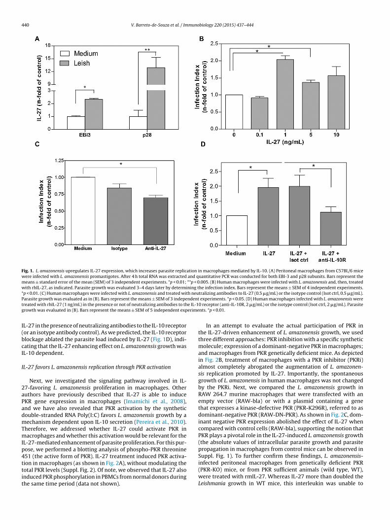
440 V. Barreto-de-Souza et al. / Immunobiology 220 (2015) 437–444
Fig. 1. L. amazonensis upregulates IL-27 expression, which increases parasite replication in macrophages mediated by IL-10. (A) Peritoneal macrophages from C57BL/6 micewere infected with L. amazonensis promastigotes. After 4 h total RNA was extracted and quantitative PCR was conducted for both EBI-3 and p28 subunits. Bars represent themeans ± standard error of the mean (SEM) of 3 independent experiments. *p < 0.01; **p < 0.005. (B) Human macrophages were infected with L. amazonensis and, then, treatedwith rhIL-27, as indicated. Parasite growth was evaluated 3–4 days later by determining the infection index. Bars represent the means ± SEM of 4 independent experiments.*p < 0.01. (C) Human macrophages were infected with L. amazonensis and treated with neutralizing antibodies to IL-27 (0.5 �g/mL) or the isotype control (Isot ctrl, 0.5 �g/mL).P ndentt e IL-1g erime
I(bcI
I
2aPadmTmIp4ttit
arasite growth was evaluated as in (B). Bars represent the means ± SEM of 3 indepereated with rhIL-27 (1 ng/mL) in the presence or not of neutralizing antibodies to throwth was evaluated in (B). Bars represent the means ± SEM of 5 independent exp
L-27 in the presence of neutralizing antibodies to the IL-10 receptoror an isotype antibody control). As we predicted, the IL-10 receptorlockage ablated the parasite load induced by IL-27 (Fig. 1D), indi-ating that the IL-27 enhancing effect on L. amazonensis growth wasL-10 dependent.
L-27 favors L. amazonensis replication through PKR activation
Next, we investigated the signaling pathway involved in IL-7-favoring L. amazonensis proliferation in macrophages. Otheruthors have previously described that IL-27 is able to induceKR gene expression in macrophages (Imamichi et al., 2008),nd we have also revealed that PKR activation by the syntheticouble-stranded RNA Poly(I:C) favors L. amazonensis growth by aechanism dependent upon IL-10 secretion (Pereira et al., 2010).
herefore, we addressed whether IL-27 could activate PKR inacrophages and whether this activation would be relevant for the
L-27-mediated enhancement of parasite proliferation. For this pur-ose, we performed a blotting analysis of phospho-PKR threonine51 (the active form of PKR). IL-27 treatment induced PKR activa-
ion in macrophages (as shown in Fig. 2A), without modulating theotal PKR levels (Suppl. Fig. 2). Of note, we observed that IL-27 alsonduced PKR phosphorylation in PBMCs from normal donors duringhe same time period (data not shown).experiments. *p < 0.05. (D) Human macrophages infected with L. amazonensis were0 receptor (anti-IL-10R, 2 �g/mL) or the isotype control (Isot ctrl, 2 �g/mL). Parasitents. *p < 0.01.
In an attempt to evaluate the actual participation of PKR inthe IL-27-driven enhancement of L. amazonensis growth, we usedthree different approaches: PKR inhibition with a specific syntheticmolecule; expression of a dominant-negative PKR in macrophages;and macrophages from PKR genetically deficient mice. As depictedin Fig. 2B, treatment of macrophages with a PKR inhibitor (PKRi)almost completely abrogated the augmentation of L. amazonen-sis replication promoted by IL-27. Importantly, the spontaneousgrowth of L. amazonensis in human macrophages was not changedby the PKRi. Next, we compared the L. amazonensis growth inRAW 264.7 murine macrophages that were transfected with anempty vector (RAW-bla) or with a plasmid containing a genethat expresses a kinase-defective PKR (PKR-K296R), referred to asdominant-negative PKR (RAW-DN-PKR). As shown in Fig. 2C, dom-inant negative PKR expression abolished the effect of IL-27 whencompared with control cells (RAW-bla), supporting the notion thatPKR plays a pivotal role in the IL-27-induced L. amazonensis growth(the absolute values of intracellular parasite growth and parasitepropagation in macrophages from control mice can be observed inSuppl. Fig. 1). To further confirm these findings, L. amazonensis-
infected peritoneal macrophages from genetically deficient PKR(PKR-KO) mice, or from PKR sufficient animals (wild type, WT),were treated with rmIL-27. Whereas IL-27 more than doubled theLeishmania growth in WT mice, this interleukin was unable to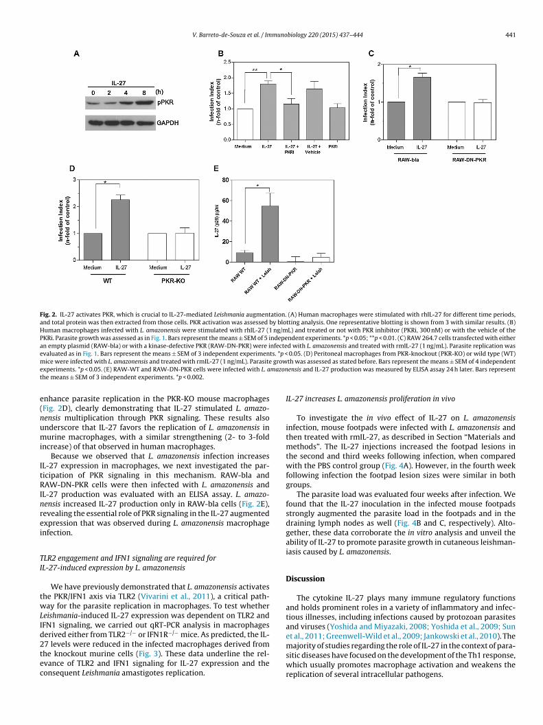
V. Barreto-de-Souza et al. / Immunobiology 220 (2015) 437–444 441
Fig. 2. IL-27 activates PKR, which is crucial to IL-27-mediated Leishmania augmentation. (A) Human macrophages were stimulated with rhIL-27 for different time periods,and total protein was then extracted from those cells. PKR activation was assessed by blotting analysis. One representative blotting is shown from 3 with similar results. (B)Human macrophages infected with L. amazonensis were stimulated with rhIL-27 (1 ng/mL) and treated or not with PKR inhibitor (PKRi, 300 nM) or with the vehicle of thePKRi. Parasite growth was assessed as in Fig. 1. Bars represent the means ± SEM of 5 independent experiments. *p < 0.05; **p < 0.01. (C) RAW 264.7 cells transfected with eitheran empty plasmid (RAW-bla) or with a kinase-defective PKR (RAW-DN-PKR) were infected with L. amazonensis and treated with rmIL-27 (1 ng/mL). Parasite replication wasevaluated as in Fig. 1. Bars represent the means ± SEM of 3 independent experiments. *p < 0.05. (D) Peritoneal macrophages from PKR-knockout (PKR-KO) or wild type (WT)m growe mazont
e(numi
ItRInrei
TI
twLId2tec
ice were infected with L. amazonensis and treated with rmIL-27 (1 ng/mL). Parasitexperiments. *p < 0.05. (E) RAW-WT and RAW-DN-PKR cells were infected with L. ahe means ± SEM of 3 independent experiments. *p < 0.002.
nhance parasite replication in the PKR-KO mouse macrophagesFig. 2D), clearly demonstrating that IL-27 stimulated L. amazo-ensis multiplication through PKR signaling. These results alsonderscore that IL-27 favors the replication of L. amazonensis inurine macrophages, with a similar strengthening (2- to 3-fold
ncrease) of that observed in human macrophages.Because we observed that L. amazonensis infection increases
L-27 expression in macrophages, we next investigated the par-icipation of PKR signaling in this mechanism. RAW-bla andAW-DN-PKR cells were then infected with L. amazonensis and
L-27 production was evaluated with an ELISA assay. L. amazo-ensis increased IL-27 production only in RAW-bla cells (Fig. 2E),evealing the essential role of PKR signaling in the IL-27 augmentedxpression that was observed during L. amazonensis macrophagenfection.
LR2 engagement and IFN1 signaling are required forL-27-induced expression by L. amazonensis
We have previously demonstrated that L. amazonensis activateshe PKR/IFN1 axis via TLR2 (Vivarini et al., 2011), a critical path-ay for the parasite replication in macrophages. To test whether
eishmania-induced IL-27 expression was dependent on TLR2 andFN1 signaling, we carried out qRT-PCR analysis in macrophageserived either from TLR2−/− or IFN1R−/− mice. As predicted, the IL-
7 levels were reduced in the infected macrophages derived fromhe knockout murine cells (Fig. 3). These data underline the rel-vance of TLR2 and IFN1 signaling for IL-27 expression and theonsequent Leishmania amastigotes replication.th was assessed as stated before. Bars represent the means ± SEM of 4 independentensis and IL-27 production was measured by ELISA assay 24 h later. Bars represent
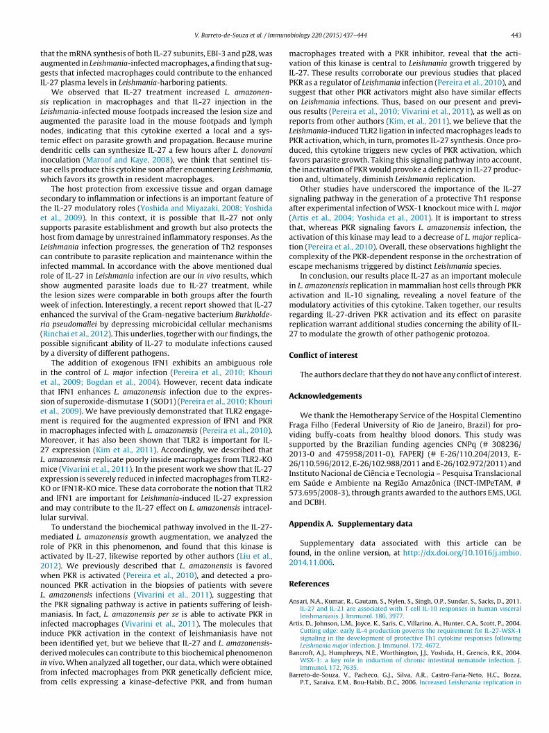
IL-27 increases L. amazonensis proliferation in vivo
To investigate the in vivo effect of IL-27 on L. amazonensisinfection, mouse footpads were infected with L. amazonensis andthen treated with rmIL-27, as described in Section “Materials andmethods”. The IL-27 injections increased the footpad lesions inthe second and third weeks following infection, when comparedwith the PBS control group (Fig. 4A). However, in the fourth weekfollowing infection the footpad lesion sizes were similar in bothgroups.
The parasite load was evaluated four weeks after infection. Wefound that the IL-27 inoculation in the infected mouse footpadsstrongly augmented the parasite load in the footpads and in thedraining lymph nodes as well (Fig. 4B and C, respectively). Alto-gether, these data corroborate the in vitro analysis and unveil theability of IL-27 to promote parasite growth in cutaneous leishman-iasis caused by L. amazonensis.
Discussion
The cytokine IL-27 plays many immune regulatory functionsand holds prominent roles in a variety of inflammatory and infec-tious illnesses, including infections caused by protozoan parasitesand viruses (Yoshida and Miyazaki, 2008; Yoshida et al., 2009; Sunet al., 2011; Greenwell-Wild et al., 2009; Jankowski et al., 2010). The
majority of studies regarding the role of IL-27 in the context of para-sitic diseases have focused on the development of the Th1 response,which usually promotes macrophage activation and weakens thereplication of several intracellular pathogens.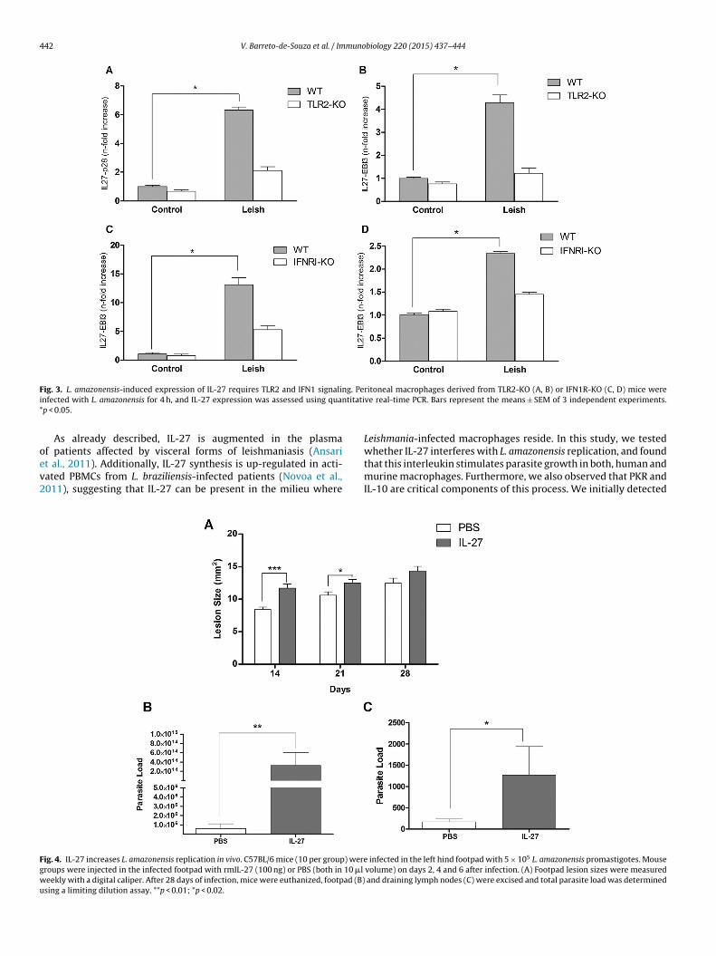
442 V. Barreto-de-Souza et al. / Immunobiology 220 (2015) 437–444
F g. Pei titati*
oev2
Fgwu
ig. 3. L. amazonensis-induced expression of IL-27 requires TLR2 and IFN1 signalinnfected with L. amazonensis for 4 h, and IL-27 expression was assessed using quanp < 0.05.
As already described, IL-27 is augmented in the plasma
f patients affected by visceral forms of leishmaniasis (Ansarit al., 2011). Additionally, IL-27 synthesis is up-regulated in acti-ated PBMCs from L. braziliensis-infected patients (Novoa et al.,011), suggesting that IL-27 can be present in the milieu whereig. 4. IL-27 increases L. amazonensis replication in vivo. C57BL/6 mice (10 per group) wereroups were injected in the infected footpad with rmIL-27 (100 ng) or PBS (both in 10 �l
eekly with a digital caliper. After 28 days of infection, mice were euthanized, footpad (B)sing a limiting dilution assay. **p < 0.01; *p < 0.02.
ritoneal macrophages derived from TLR2-KO (A, B) or IFN1R-KO (C, D) mice wereve real-time PCR. Bars represent the means ± SEM of 3 independent experiments.
Leishmania-infected macrophages reside. In this study, we tested
whether IL-27 interferes with L. amazonensis replication, and foundthat this interleukin stimulates parasite growth in both, human andmurine macrophages. Furthermore, we also observed that PKR andIL-10 are critical components of this process. We initially detectedinfected in the left hind footpad with 5 × 10⁵ L. amazonensis promastigotes. Mousevolume) on days 2, 4 and 6 after infection. (A) Footpad lesion sizes were measured
and draining lymph nodes (C) were excised and total parasite load was determined
muno
tagI
sLantdisw
steshLcirstwer(pb
ietsemiM2LmeKaal
mra2wnLtmiibdiff
V. Barreto-de-Souza et al. / Im
hat the mRNA synthesis of both IL-27 subunits, EBI-3 and p28, wasugmented in Leishmania-infected macrophages, a finding that sug-ests that infected macrophages could contribute to the enhancedL-27 plasma levels in Leishmania-harboring patients.
We observed that IL-27 treatment increased L. amazonen-is replication in macrophages and that IL-27 injection in theeishmania-infected mouse footpads increased the lesion size andugmented the parasite load in the mouse footpads and lymphodes, indicating that this cytokine exerted a local and a sys-emic effect on parasite growth and propagation. Because murineendritic cells can synthesize IL-27 a few hours after L. donovani
noculation (Maroof and Kaye, 2008), we think that sentinel tis-ue cells produce this cytokine soon after encountering Leishmania,hich favors its growth in resident macrophages.
The host protection from excessive tissue and organ damageecondary to inflammation or infections is an important feature ofhe IL-27 modulatory roles (Yoshida and Miyazaki, 2008; Yoshidat al., 2009). In this context, it is possible that IL-27 not onlyupports parasite establishment and growth but also protects theost from damage by unrestrained inflammatory responses. As theeishmania infection progresses, the generation of Th2 responsesan contribute to parasite replication and maintenance within thenfected mammal. In accordance with the above mentioned dualole of IL-27 in Leishmania infection are our in vivo results, whichhow augmented parasite loads due to IL-27 treatment, whilehe lesion sizes were comparable in both groups after the fourtheek of infection. Interestingly, a recent report showed that IL-27
nhanced the survival of the Gram-negative bacterium Burkholde-ia pseudomallei by depressing microbicidal cellular mechanismsRinchai et al., 2012). This underlies, together with our findings, theossible significant ability of IL-27 to modulate infections causedy a diversity of different pathogens.
The addition of exogenous IFN1 exhibits an ambiguous rolen the control of L. major infection (Pereira et al., 2010; Khourit al., 2009; Bogdan et al., 2004). However, recent data indicatehat IFN1 enhances L. amazonensis infection due to the expres-ion of superoxide-dismutase 1 (SOD1) (Pereira et al., 2010; Khourit al., 2009). We have previously demonstrated that TLR2 engage-ent is required for the augmented expression of IFN1 and PKR
n macrophages infected with L. amazonensis (Pereira et al., 2010).oreover, it has also been shown that TLR2 is important for IL-
7 expression (Kim et al., 2011). Accordingly, we described that. amazonensis replicate poorly inside macrophages from TLR2-KOice (Vivarini et al., 2011). In the present work we show that IL-27
xpression is severely reduced in infected macrophages from TLR2-O or IFN1R-KO mice. These data corroborate the notion that TLR2nd IFN1 are important for Leishmania-induced IL-27 expressionnd may contribute to the IL-27 effect on L. amazonensis intracel-ular survival.
To understand the biochemical pathway involved in the IL-27-ediated L. amazonensis growth augmentation, we analyzed the
ole of PKR in this phenomenon, and found that this kinase isctivated by IL-27, likewise reported by other authors (Liu et al.,012). We previously described that L. amazonensis is favoredhen PKR is activated (Pereira et al., 2010), and detected a pro-ounced PKR activation in the biopsies of patients with severe. amazonensis infections (Vivarini et al., 2011), suggesting thathe PKR signaling pathway is active in patients suffering of leish-
aniasis. In fact, L. amazonensis per se is able to activate PKR innfected macrophages (Vivarini et al., 2011). The molecules thatnduce PKR activation in the context of leishmaniasis have noteen identified yet, but we believe that IL-27 and L. amazonensis-
erived molecules can contribute to this biochemical phenomenonn vivo. When analyzed all together, our data, which were obtainedrom infected macrophages from PKR genetically deficient mice,rom cells expressing a kinase-defective PKR, and from human
biology 220 (2015) 437–444 443
macrophages treated with a PKR inhibitor, reveal that the acti-vation of this kinase is central to Leishmania growth triggered byIL-27. These results corroborate our previous studies that placedPKR as a regulator of Leishmania infection (Pereira et al., 2010), andsuggest that other PKR activators might also have similar effectson Leishmania infections. Thus, based on our present and previ-ous results (Pereira et al., 2010; Vivarini et al., 2011), as well as onreports from other authors (Kim, et al., 2011), we believe that theLeishmania-induced TLR2 ligation in infected macrophages leads toPKR activation, which, in turn, promotes IL-27 synthesis. Once pro-duced, this cytokine triggers new cycles of PKR activation, whichfavors parasite growth. Taking this signaling pathway into account,the inactivation of PKR would provoke a deficiency in IL-27 produc-tion and, ultimately, diminish Leishmania replication.
Other studies have underscored the importance of the IL-27signaling pathway in the generation of a protective Th1 responseafter experimental infection of WSX-1 knockout mice with L. major(Artis et al., 2004; Yoshida et al., 2001). It is important to stressthat, whereas PKR signaling favors L. amazonensis infection, theactivation of this kinase may lead to a decrease of L. major replica-tion (Pereira et al., 2010). Overall, these observations highlight thecomplexity of the PKR-dependent response in the orchestration ofescape mechanisms triggered by distinct Leishmania species.
In conclusion, our results place IL-27 as an important moleculein L. amazonensis replication in mammalian host cells through PKRactivation and IL-10 signaling, revealing a novel feature of themodulatory activities of this cytokine. Taken together, our resultsregarding IL-27-driven PKR activation and its effect on parasitereplication warrant additional studies concerning the ability of IL-27 to modulate the growth of other pathogenic protozoa.
Conflict of interest
The authors declare that they do not have any conflict of interest.
Acknowledgements
We thank the Hemotherapy Service of the Hospital ClementinoFraga Filho (Federal University of Rio de Janeiro, Brazil) for pro-viding buffy-coats from healthy blood donors. This study wassupported by the Brazilian funding agencies CNPq (# 308236/2013-0 and 475958/2011-0), FAPERJ (# E-26/110.204/2013, E-26/110.596/2012, E-26/102.988/2011 and E-26/102.972/2011) andInstituto Nacional de Ciência e Tecnologia – Pesquisa Translacionalem Saúde e Ambiente na Região Amazônica (INCT-IMPeTAM, #573.695/2008-3), through grants awarded to the authors EMS, UGLand DCBH.
Appendix A. Supplementary data
Supplementary data associated with this article can befound, in the online version, at http://dx.doi.org/10.1016/j.imbio.2014.11.006.
References
Ansari, N.A., Kumar, R., Gautam, S., Nylen, S., Singh, O.P., Sundar, S., Sacks, D., 2011.IL-27 and IL-21 are associated with T cell IL-10 responses in human visceralleishmaniasis. J. Immunol. 186, 3977.
Artis, D., Johnson, L.M., Joyce, K., Saris, C., Villarino, A., Hunter, C.A., Scott, P., 2004.Cutting edge: early IL-4 production governs the requirement for IL-27-WSX-1signaling in the development of protective Th1 cytokine responses followingLeishmania major infection. J. Immunol. 172, 4672.
Bancroft, A.J., Humphreys, N.E., Worthington, J.J., Yoshida, H., Grencis, R.K., 2004.WSX-1: a key role in induction of chronic intestinal nematode infection. J.Immunol. 172, 7635.
Barreto-de-Souza, V., Pacheco, G.J., Silva, A.R., Castro-Faria-Neto, H.C., Bozza,P.T., Saraiva, E.M., Bou-Habib, D.C., 2006. Increased Leishmania replication in
4 muno
B
B
C
F
G
H
H
I
I
I
J
K
K
K
K
L
L
L
M
M
44 V. Barreto-de-Souza et al. / Im
HIV-1-infected macrophages is mediated by tat protein through cyclooxy-genase-2 expression and prostaglandin E2 synthesis. J. Infect. Dis. 194, 846.
atten, M., Kljavin, N.M., Li, J., Walter, M.J., de Sauvage, F.J., Ghilardi, N., 2008. Cut-ting edge: IL-27 is a potent inducer of IL-10 but not FoxP3 in murine T cells. J.Immunol. 180, 2752.
ogdan, C., Mattner, J., Schleicher, U., 2004. The role of type I interferons in non-viralinfections. Immunol. Rev. 202, 33.
ummings, H.E., Tuladhar, R., Satoskar, A.R., 2010. Cytokines and their STATs incutaneous and visceral leishmaniasis. J. Biomed. Biotechnol. 2010, 294389.
reitas do Rosário, A.P., Lamb, T., Spence, P., Stephens, R., Lang, A., Roers, A.,Muller, W., O’Garra, A., Langhorne, J., 2012. IL-27 promotes IL-10 productionby effector Th1 CD4+ T cells: a critical mechanism for protection from severeimmunopathology during malaria infection. J Immunol. 188, 1178.
reenwell-Wild, T., Vazquez, N., Jin, W., Rangel, Z., Munson, P.J., Wahl, S.M., 2009.Interleukin-27 inhibition of HIV-1 involves an intermediate induction of type Iinterferon. Blood 114, 1864.
ause, L., Al-Salleeh, F.M., Petro, T.M., 2007. Expression of IL-27 p28 by Theiler’svirus-infected macrophages depends on TLR3 and TLR7 activation of JNK-MAP-kinases. Antiviral Res. 76, 159.
unter, C.A., Villarino, A., Artis, D., Scott, P., 2004. The role of IL-27 in the develop-ment of T-cell responses during parasitic infections. Immunol. Rev. 202, 106.
mamichi, T., Yang, J., Huang, D., Brann, W., Fullmer, T.W., Adelsberger, B.A., Lem-picki, J.W., Baseler, R.A., Lane, M.W.H.C., 2008. IL-27, a novel anti-HIV cytokine,activates multiple interferon-inducible genes in macrophages. AIDS 22, 39.
mamichi, T., Yang, J., Huang, D.W., Sherman, B., Lempicki, R.A., 2012. Interleukin-27induces interferon-inducible genes: analysis of gene expression profiles usingAffymetrix microarray and DAVID. Methods Mol. Biol. 820, 25.
yer, S.S., Ghaffari, A.A., Cheng, G., 2010. Lipopolysaccharide-mediated IL-10 trans-criptional regulation requires sequential induction of type I IFNs and IL-27 inmacrophages. J. Immunol. 185, 6599.
ankowski, M., Kopinski, P., Goc, A., 2010. Interleukin-27: biological properties andclinical application. Arch. Immunol. Ther. Exp. (Warsz) 58, 417.
aye, P., Scott, P., 2011. Leishmaniasis: complexity at the host–pathogen interface.Nat. Rev. Microbiol. 9, 604.
houri, R., Bafica, A., Silva Mda, P., Noronha, A., Kolb, J.P., Wietzerbin, J., Barral,A., Barral-Netto, M., Van Weyenbergh, J., 2009. IFN-beta impairs superoxide-dependent parasite killing in human macrophages: evidence for a deleteriousrole of SOD1 in cutaneous leishmaniasis. J. Immunol. 182, 2525.
im, H.S., Go, H., Akira, S., Chung, D.H., 2011. TLR2-mediated production of IL-27and chemokines by respiratory epithelial cells promotes bleomycin-inducedpulmonary fibrosis in mice. J. Immunol. 187, 4007.
urtzhals, J.A., Hey, A.S., Jardim, A., Kemp, M., Schaefer, K.U., Odera, E.O., Christensen,C.B., Githure, J.I., Olafson, R.W., Theander, T.G., et al., 1994. Dichotomy of thehuman T cell response to Leishmania antigens. II. Absent or Th2-like responseto gp63 and Th1-like response to lipophosphoglycan-associated protein in cellsfrom cured visceral leishmaniasis patients. Clin. Exp. Immunol. 96, 416.
ima, H.C., Bleyenberg, J.A., Titus, R.G., 1997. A simple method for quantifying Leish-mania in tissues of infected animals. Parasitol. Today 13, 80.
iu, L., Cao, Z., Chen, J., Li, R., Cao, Y., Zhu, C., Wu, K., Wu, J., Liu, F., Zhu, Y., 2012.Influenza A virus induces interleukin-27 through cyclooxygenase-2 and proteinkinase A signaling. J. Biol. Chem. 287, 11899.
ocksley, R.M., Heinzel, F.P., Sadick, M.D., Holaday, B.J., Gardner Jr., K.D., 1987. Murinecutaneous leishmaniasis: susceptibility correlates with differential expansion ofhelper T-cell subsets. Ann. Inst. Pasteur Immunol. 138, 744.
aroof, A., Kaye, P.M., 2008. Temporal regulation of interleukin-12p70 (IL-12p70)and IL-12-related cytokines in splenic dendritic cell subsets during Leishmaniadonovani infection. Infect. Immun. 76, 239.
eurs, E., Chong, K., Galabru, J., Thomas, N.S., Kerr, I.M., Williams, B.R., Hov-anessian, A.G., 1990. Molecular cloning and characterization of the human
biology 220 (2015) 437–444
double-stranded RNA-activated protein kinase induced by interferon. Cell 62,379.
Molle, C., Nguyen, M., Flamand, V., Renneson, J., Trottein, F., De Wit, D., Willems, F.,Goldman, M., Goriely, S., 2007. IL-27 synthesis induced by TLR ligation criticallydepends on IFN regulatory factor 3. J. Immunol. 178, 7607.
Novoa, R., Bacellar, O., Nascimento, M., Cardoso, T.M., Ramasawmy, R., Oliveira, W.N.,Schriefer, A., Carvalho, E.M., 2011. IL-17 and regulatory cytokines (IL-10 andIL-27) in L. braziliensis infection. Parasite Immunol. 33, 132.
Pereira, R.M., Teixeira, K.L., Barreto-de-Souza, V., Calegari-Silva, T.C., De-Melo, L.D.,Soares, D.C., Bou-Habib, D.C., Silva, A.M., Saraiva, E.M., Lopes, U.G., 2010. Novelrole for the double-stranded RNA-activated protein kinase PKR: modulationof macrophage infection by the protozoan parasite Leishmania. FASEB J. 24,617.
Pflanz, S., Timans, J.C., Cheung, J., Rosales, R., Kanzler, H., Gilbert, J., Hibbert, L., Chu-rakova, T., Travis, M., Vaisberg, E., Blumenschein, W.M., Mattson, J.D., Wagner,J.L., To, W., Zurawski, S., McClanahan, T.K., Gorman, D.M., Bazan, J.F., de WaalMalefyt, R., Rennick, D., Kastelein, R.A., 2002. IL-27, a heterodimeric cytokinecomposed of EBI3 and p28 protein, induces proliferation of naive CD4+ T cells.Immunity 16, 779.
Raven, J.F., Koromilas, A.E., 2008. PERK and PKR: old kinases learn new tricks. CellCycle 7, 1146.
Rinchai, D., Khaenam, P., Kewcharoenwong, C., Buddhisa, S., Pankla, R., Chaussabel,D., Bancroft, G.J., Lertmemongkolchai, G., 2012. Production of interleukin-27 byhuman neutrophils regulates their function during bacterial infection. Eur. J.Immunol. 42, 3280.
Sacks, D., Noben-Trauth, N., 2002. The immunology of susceptibility and resistanceto Leishmania major in mice. Nat. Rev. Immunol. 2, 845.
Sadler, A.J., Williams, B.R., 2007. Structure and function of the protein kinase R. Curr.Top. Microbiol. Immunol. 316, 253.
Silva, A.M., Whitmore, M., Xu, Z., Jiang, Z., Li, X., Williams, B.R., 2004. Protein kinaseR (PKR) interacts with and activates mitogen-activated protein kinase kinase6 (MKK6) in response to double-stranded RNA stimulation. J. Biol. Chem. 279,37670.
Sun, J., Dodd, H., Moser, E.K., Sharma, R., Braciale, T.J., 2011. CD4+ T cell help andinnate-derived IL-27 induce Blimp-1-dependent IL-10 production by antiviralCTLs. Nat. Immunol. 12, 327.
Vivarini, A.deC., Pereira, R.deM., Teixeira, K.L., Calegari-Silva, T.C., Bellio, M., Lau-renti, M.D., Corbett, C.E., Gomes, C.M., Soares, R.P., Silva, A.M., Silveira, F.T., Lopes,U.G., 2011. Human cutaneous leishmaniasis: interferon-dependent expressionof double-stranded RNA-dependent protein kinase (PKR) via TLR2. FASEB J. 25,4162.
Wang, H., Meng, R., Li, Z., Yang, B., Liu, Y., Huang, F., Zhang, J., Chen, H., Wu, C., 2011.IL-27 induces the differentiation of Tr1-like cells from human naive CD4+ T cellsvia the phosphorylation of STAT1 and STAT3. Immunol. Lett. 136, 21.
Wirtz, S., Becker, C., Fantini, M.C., Nieuwenhuis, E.E., Tubbe, I., Galle, P.R., Schild,H.J., Birkenbach, M., Blumberg, R.S., Neurath, M.F., 2005. EBV-induced gene 3transcription is induced by TLR signaling in primary dendritic cells via NF-kappaB activation. J. Immunol. 174, 2814.
World Health Organization, 2010. World Health Organization Working to Overcomethe Global Impact of Neglected Tropical Disease. First WHO Report on NeglectedDisease. World Health Organization, Geneva. Switzerland.
Yoshida, H., Miyazaki, Y., 2008. Regulation of immune responses by interleukin-27.Immunol. Rev. 226, 234.
Yoshida, H., Hamano, S., Senaldi, G., Covey, T., Faggioni, R., Mu, S., Xia, M., Wakeham,
A.C., Nishina, H., Potter, J., Saris, C.J., Mak, T.W., 2001. WSX-1 is required for theinitiation of Th1 responses and resistance to L. major infection. Immunity 15,569.Yoshida, H., Nakaya, M., Miyazaki, Y., 2009. Interleukin 27: a double-edged swordfor offense and defense. J. Leukoc. Biol. 86, 1295.