Embed Size (px)
Citation preview
Legge – Calve – Perthese disease
S.M. Mazloumi
Associate professor in orthopedics
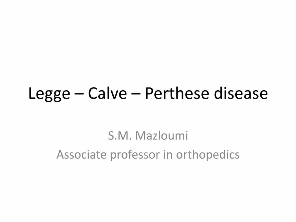
Legg- Calve – Perthes disease
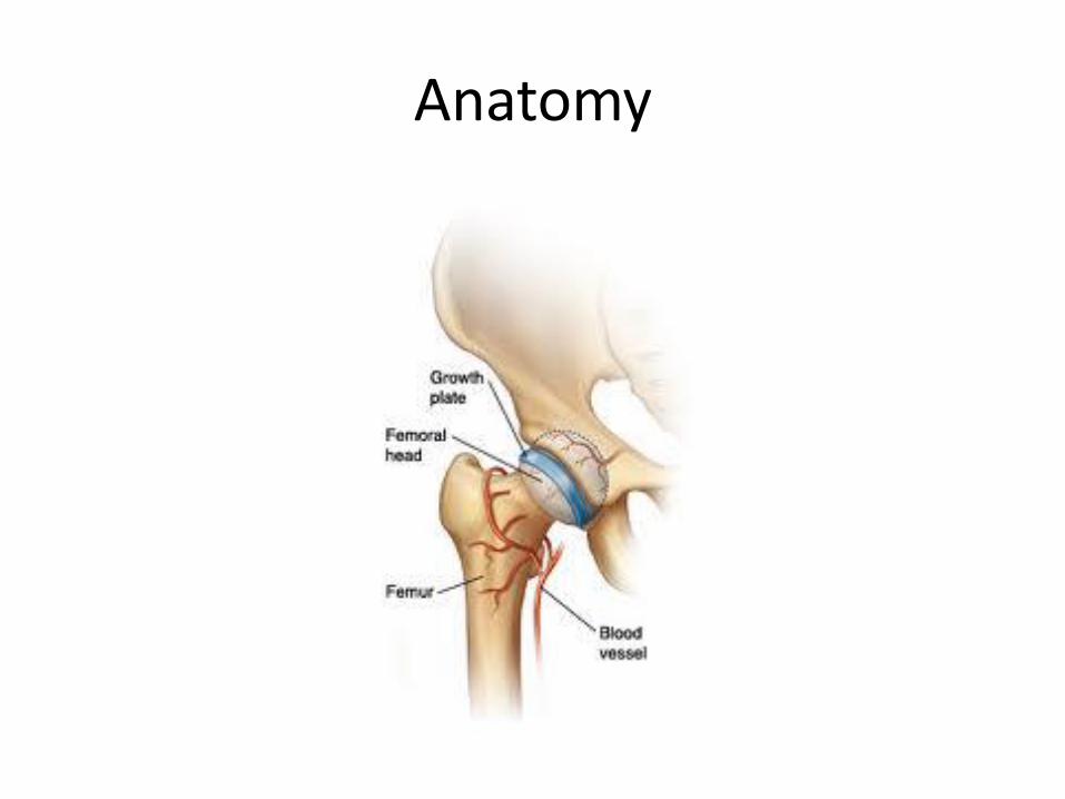
Anatomy
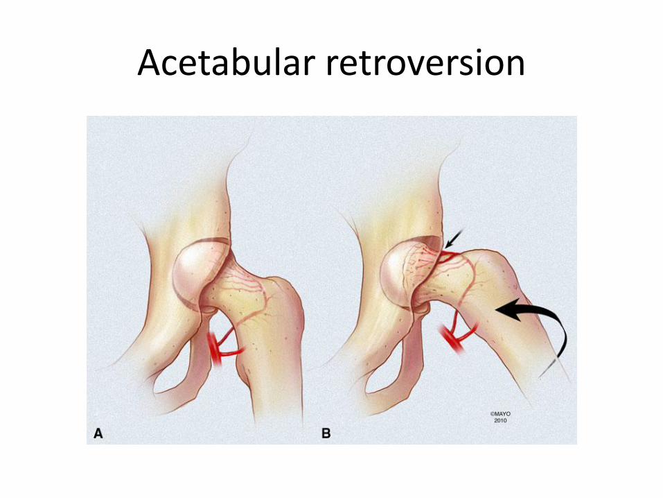
Acetabular retroversion
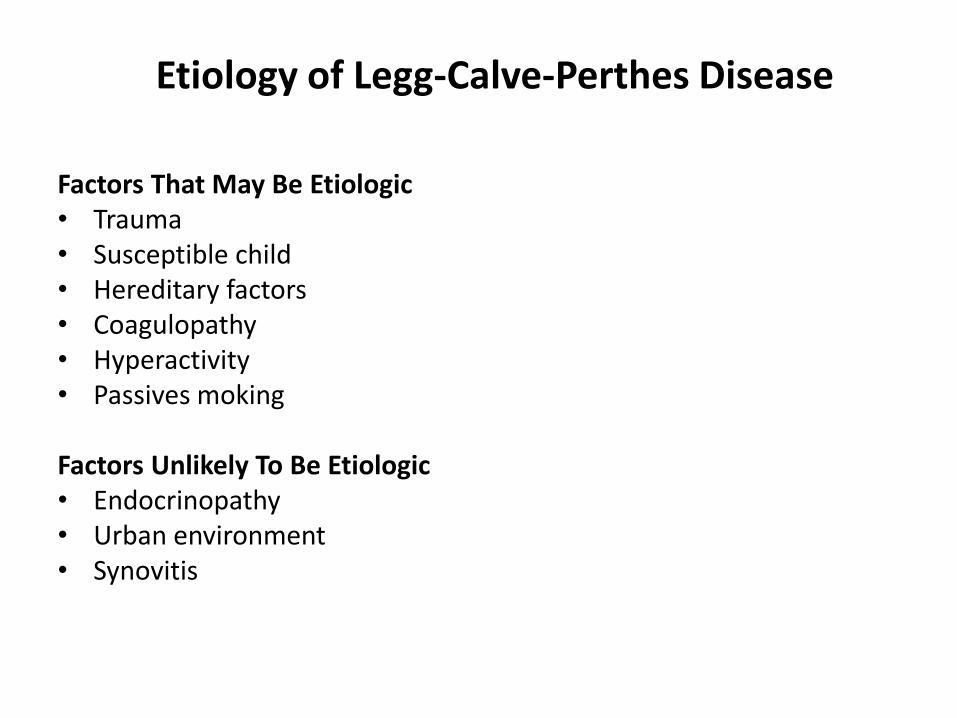
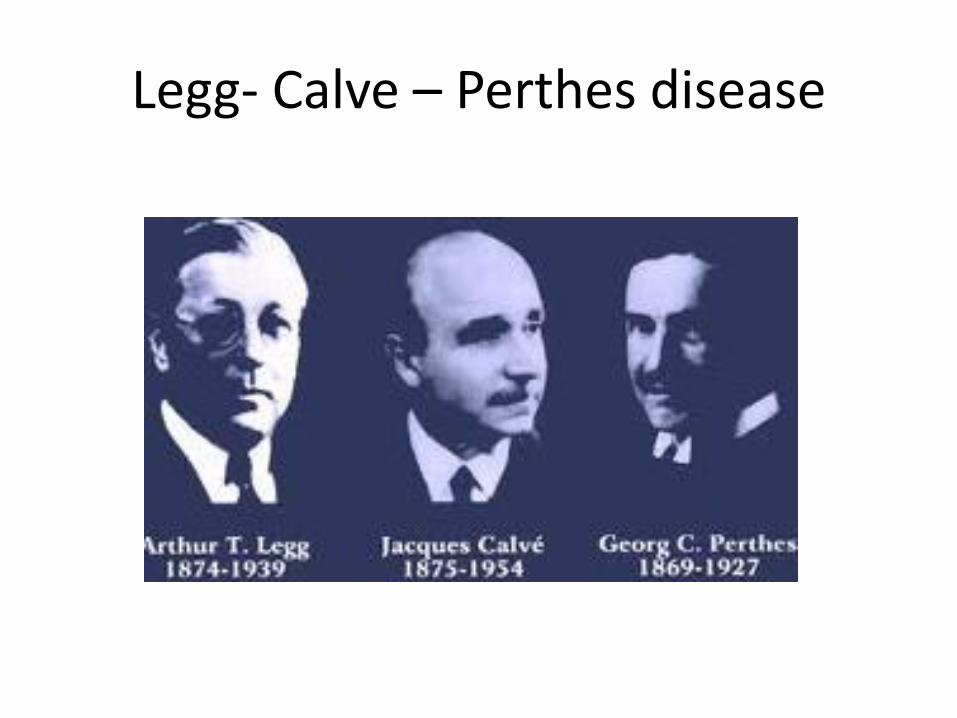
Etiology of Legg-Calve-Perthes Disease
Factors That May Be Etiologic • Trauma • Susceptible child • Hereditary factors • Coagulopathy • Hyperactivity • Passives moking Factors Unlikely To Be Etiologic • Endocrinopathy • Urban environment • Synovitis
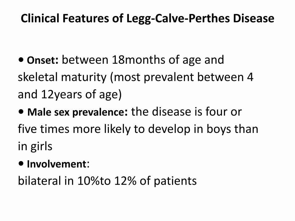
Clinical Features of Legg-Calve-Perthes Disease
• Onset: between 18months of age and
skeletal maturity (most prevalent between 4
and 12years of age)
• Male sex prevalence: the disease is four or
five times more likely to develop in boys than
in girls
• Involvement:
bilateral in 10%to 12% of patients
Clinical Features of Legg-Calve-Perthes Disease
Symptoms
-limp that is exacerbated by activity and alleviated with rest
-pain, which may be located in the groin,anterior hip region, medial knee joint or laterally around the greater Trochanter
- history of antecedent trauma
Clinical Features of Legg-Calve-Perthes Disease
Signs
- Abductor limp
- Decreased range of motion of the hip, especially on abduction and internal rotation
- Flexion/extension less affected
Symptoms and Signs of Legg-Calve- Perthes Disease
Symptoms • Limping • Hip pain • Knee pain • History of trauma (?) Signs • Limp • Decreased hip range of motion • Spasm of long muscle around hip joint
Pathologic Findings of Legg-Calve- Perthes Disease
Early Stage • Dead trabecular bone , Collapsed trabeculae • Thickened articular cartilage , Physeal disruption • Cartilage extending from the physis into the
metaphysis Fragmentation Stage • Invasion of vascular granulation tissue • New bone forming on old trabeculae • Woven new bone formation Healing Stage • New bone, woven and lamellar • Return to normai architecture
Differential Diagnosis for Legg-Calve- Perthes Disease
Other Causes of Avascular Necrosis
• Sickle cell disease
• Other hemoglobinopathies
• Thalassemia
• Steroid medication
• After traumatic hip dislocation
• Treatment of developmental dysplasia of
• the hip
Differential Diagnosis for Legg-Calve- Perthes Disease
Epiphyseal Dysplasias
• Muitiple epiphyseal dysplasia
• Spondyloepiphyseal dysplasia
• Mucopolysaccharidoses
• Hypothyroidism
Differential Diagnosis for Legg-Calve- Perthes Disease
Other Syndromes
• Osteochondromatosis
• Metachondromatosis
• Schwartz-Jam pel syndrome
• Trichorhinophalangeal syndrome
• Maroteaux-Lamy syndrome
Caterall classification
• Group I, partial head or less than half head involvement;
• Groups II and III, more than half head involvement and sequestrum formation
• Group IV, involvement of the entire epiphysis
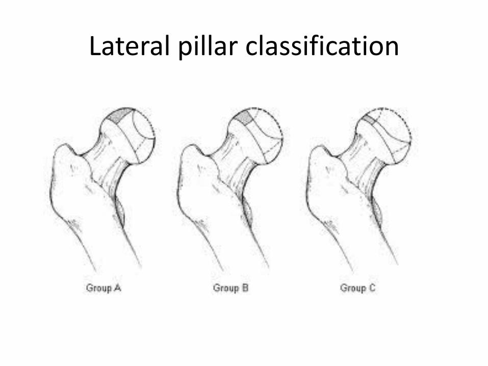
Lateral pillar classification
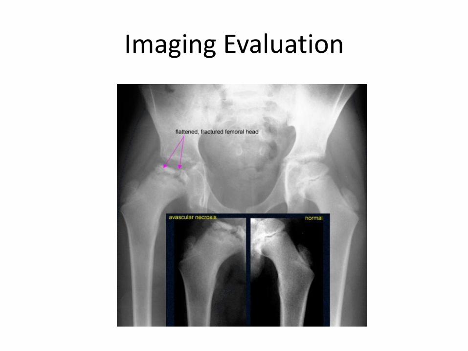
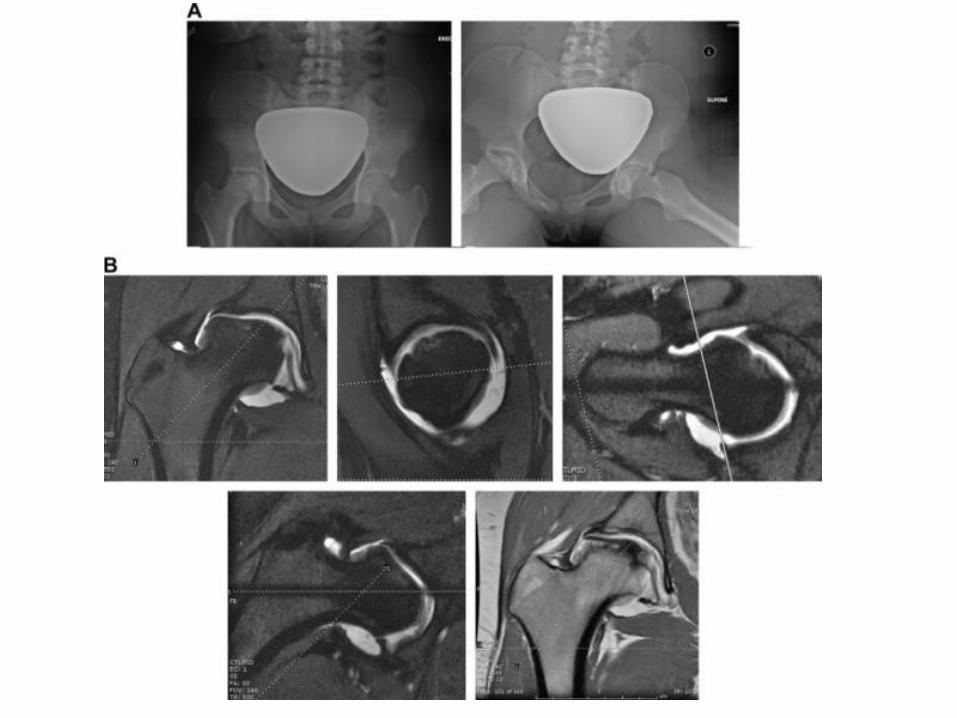
Imaging Evaluation
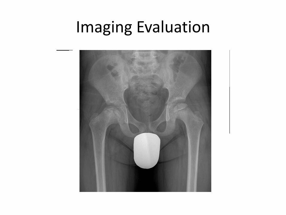
Imaging Evaluation

X-Ray
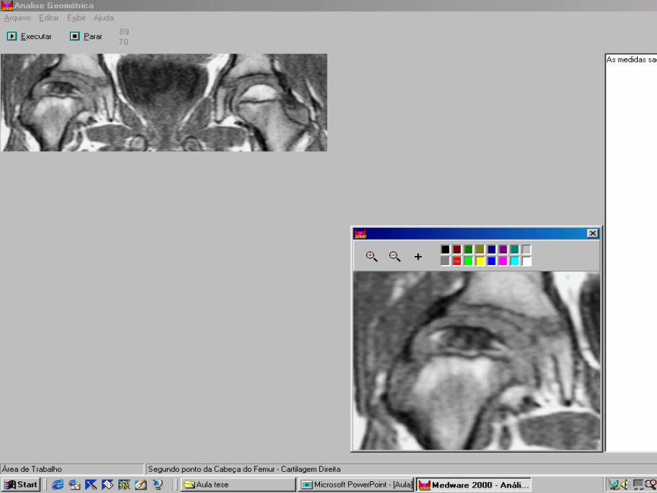
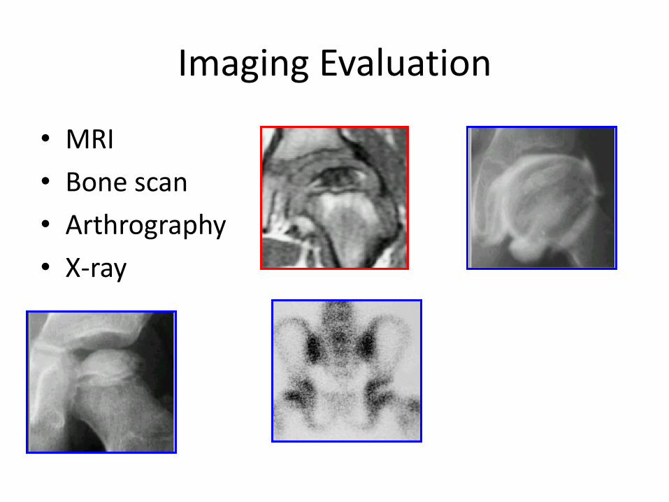
Imaging Evaluation
• MRI
• Bone scan
• Arthrography
• X-ray
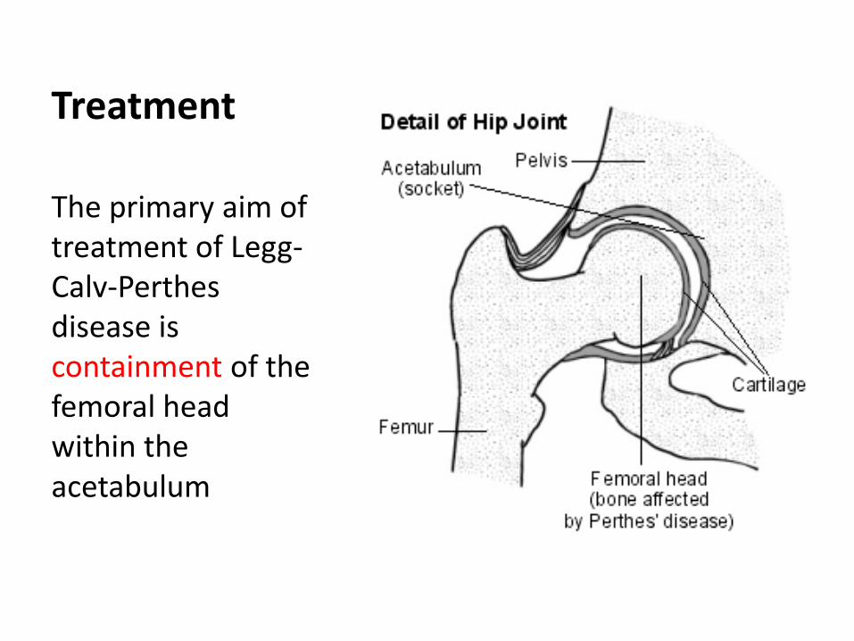
Treatment
The primary aim of treatment of Legg-Calv-Perthes disease is containment of the femoral head within the acetabulum

Bracing
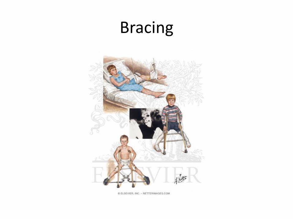
Bracing

Varus Derotational Osteotomy
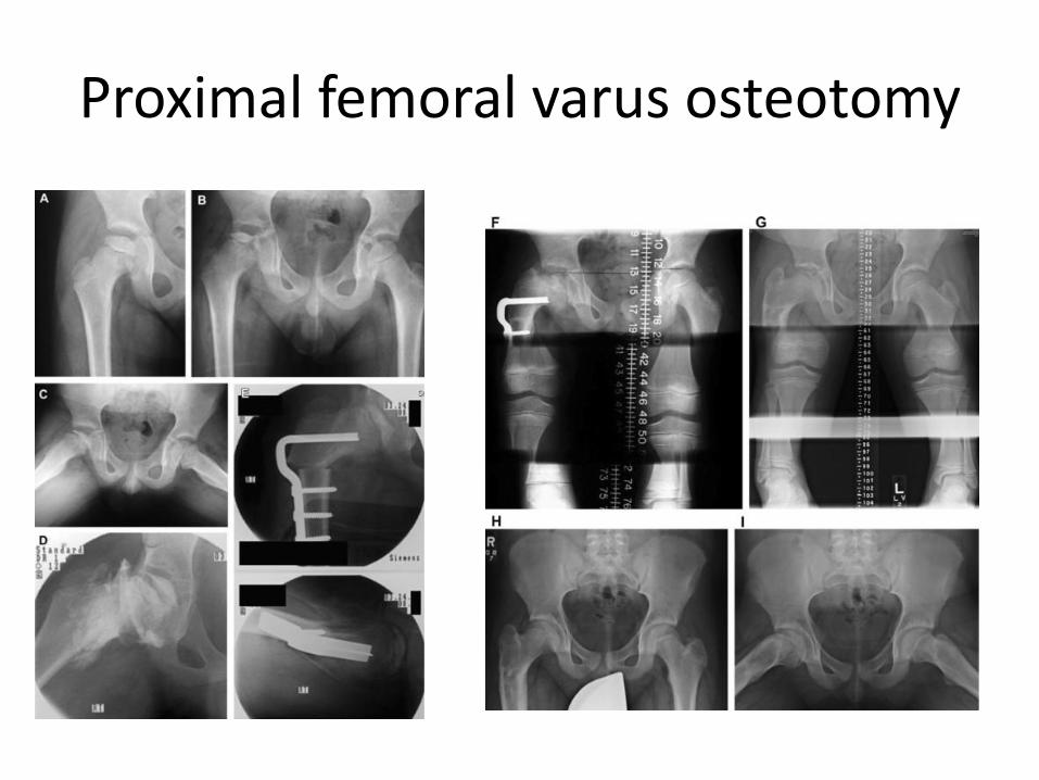
Proximal femoral varus osteotomy

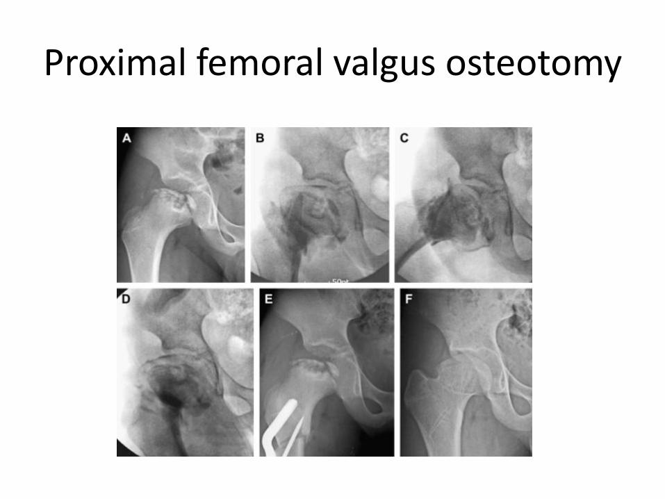
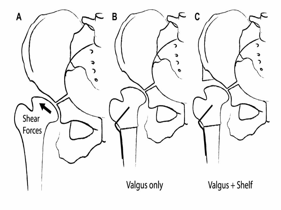
Proximal femoral valgus osteotomy
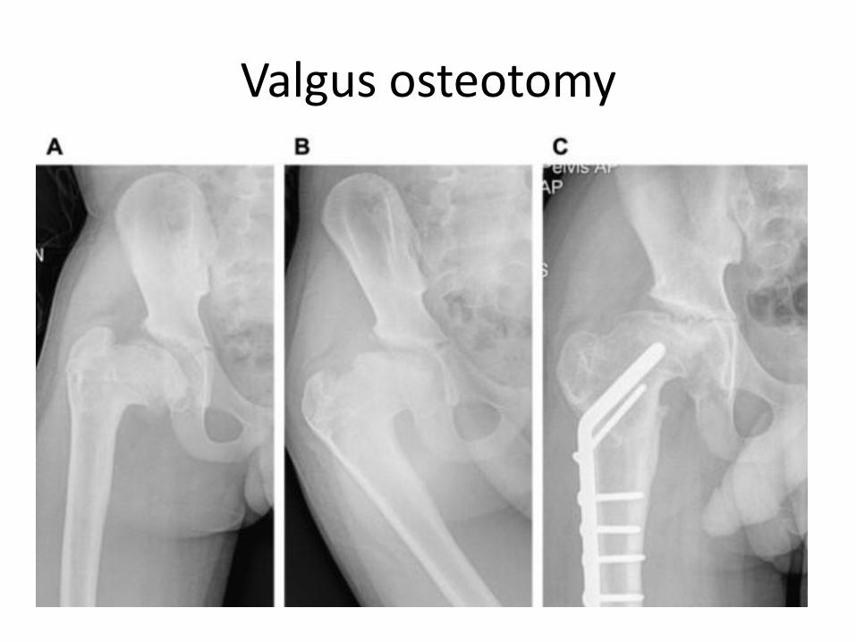
Valgus osteotomy
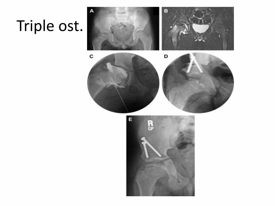
Triple ost.
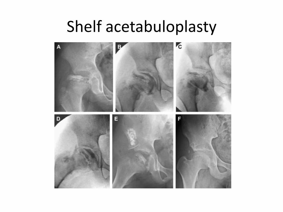
Shelf acetabuloplasty
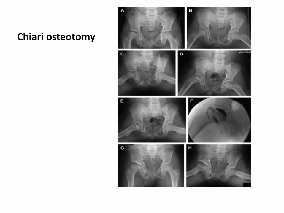
Chiari osteotomy
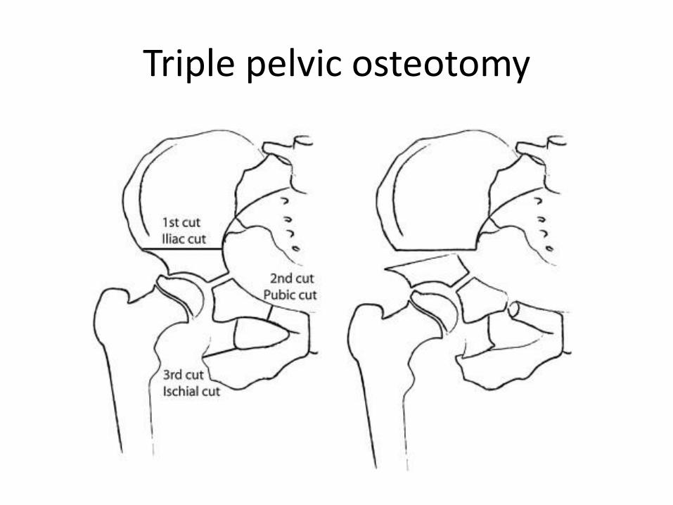
Triple pelvic osteotomy

Double-level osteotomy
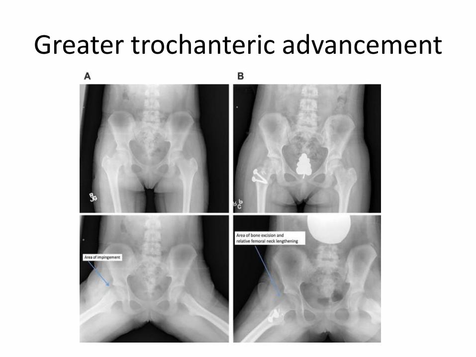
Greater trochanteric advancement
Treatment
1. Most patients can be treated by noncontainment methods and obtain good results (84%). 2. Satisfactory clinical results frequently can be obtained at long-term follow-up despite an unsatisfactory radiographic appearance (nine hips). 3. The Catterall classification is a valid indicator of results, but is not applicable as a therapeutic guide for an average of 8.1 months after onset

Treatment
4. Head-at-risk signs added little to the Catterall classification as a prognostic indicator or therapeutic guide. 5. All of the fair and poor results were in patients with Catterall III or IV involvement and onset of the disease at age 6 or older. (A Catterall III or IV classification is equivalent to Herring groups B and C.)
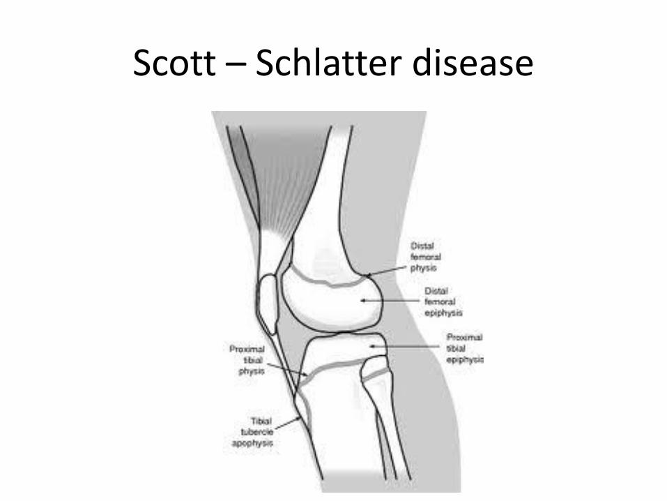
Scott – Schlatter disease
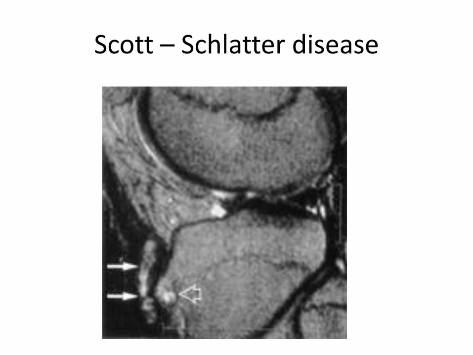
Scott – Schlatter disease