Embed Size (px)
Citation preview

Proceedings of a workshop
sponsored by THE CENTER FOR ADVANCED STUDIES
IN THE SPACE LIFE SCIENCES AT THE MBL
15- 17 November 1996
Marine Biological Laboratory, Woods Hole, Massachusetts
Funded by THE NATIONAL AERONAUTICS
I R- The Biological Bulletin, Vol. 194, June 1998 Printed in USA
https://ntrs.nasa.gov/search.jsp?R=19990054485 2020-04-15T12:26:14+00:00Z
THE CYTOSKELETON: MECHANICAL, PHYSICAL,
AND BIOLOGICAL INTERACTIONS
Proceedings of a workshop
sponsored by THE CENTER FOR ADVANCED STUDIES
IN THE SPACE LIFE SCIENCES AT THE MBL
15-17 November 1996
Marine Biological Laboratory, Woods Hole, Massachusetts
Funded by THE NATIONAL AERONAUTICS AND SPACE ADMINISTRATION under Cooperative Agreement NCC 2-896

CONTENTS The Cytoskehton:
Mechanical, Physical, and Biological Interactions
INTRODUCTION by E. A. Dawidowicz . . . . . . . . . . . . . .
Ingber, Donald E. Cellular basis of mechanotransduction . . . . . . . . . . .
Forgacs, Gabor Surface tension and viscoelastic properties of embry-
. . . . . . . . . . onic tissues depend on the cytoskeleton Bod, David H.
Two-dimensional cytoskeletons under stress . . . . . . . Janmey, Paul A., Josef Kas, Jagesh V. Shah, Philip G. Allen, and Jay X. Tang
Cytoskeletal networks and filament bundles: regulation by proteins and polycations . . . . . . . . . . . . . . . . . .
STRUCTURAL, MECHANICAL, AND BIOLOGICAL PROPERTIES OF THE C ~ K E L E T O N
Steinmetz, Michel O., Daniel Stoffler, and Ueli Aebi Actin: dissecting the structural basis of its oligomeriza-
. . . . . . . . . tion, polymerization, and polymorphism Stewart, Murray, Thomas M. Roberts, Joseph E. Ital- iano, Karen L. King, Robin Hammel, G. Parathasa- thy, Timothy L. Bullock, Airlee J. McCoy, Helen Kent, Andreas Haaf, and David Neuhaus
Amoeboid motility without actin: insights into the mo- lecular mechanism of locomotion using the major
. . . . . . . . . . . . . sperm protein (MSP) of nematodes Luna, Elizabeth J., Anne L. Hitt, Damon Shutt, Debo- rah Wessels, David Soll, Pat Jay, Chris Hug, Elliot L. Elson, Alex Vesley, Gregory P. Downey, Michael Wang, Steven M. Block, Wade Sigurdson, and Freder- ick Sachs
Role of ponticulin in pseudopod dynamics, cell-cell adhesion, and mechanical stability- of an amoeboid membrane skeleton . . . . . . . . . . . . . . . . . . . . . . . . 345
Pelham, Robert J. Jr., and Yu-li Wang 7
Cell locomotion and focal adhesions are regulated by . . . . . . . . the mechanical properties of the substrate 348
MICROTUBULES AND VISCOELASTICITY OF ACTIN
MacKintosh, Frederick Theoretical models of viscoelasticity of actin solutions and the actin cortex . . . . . . . . . . . . . . . . . . . . . . . . 351
Nguyen, H. L., D. Gruber, T. McGraw, M. P. Sheetz, ,
and J. Chloe Bulinski Stabilization and functional modulation of rnicrotu-
. . . . . . . . bules by microtubule-associated protein 4 354
Gunderson, Gregg, Geri Kreitzer, Tiffani Cook, and Guojuan Liao
Microtubules as determinants of cellular polarity . . .
' INTERMEDIATE FILAMENTS, HEMIDESMOSOMES, AND DESMOSO
Goldman, Robert D., S. Clement, S. Khuon, R. Moii., A. Trejo-Skalli, T. Spann, and M. Yoon
Intermediate filament cytoskeletal system: dynamic and mechanical properties . . . . . . . . . . . . . . . . . . .
Coulombe, Pierre A., Matthew Wawersik, Rudolph D. Paladhi, and Erick Noensie
Type I keratin 16 forms relatively unstable tetameric assembly subunits with various Type I . keratin part- ners: biochemical basis and functional implications
Steinert, Peter M. Structural-mechanical intergration of keratin interme- diate filaments with cell peripheral structures in the
. . . . . . . . . . . . . . . cornified epidermal keratinocyte Jones, Jonathan C. R, Omar Skalli, Robert D. Gold- man, and Scott E. Baker
What links laminin-5 to the keratin cytoskeleton in epithelial cells? . . . . . . . . . . . . . . . . . . . . . . . . . . .
Green, Kathleen J., Andrew P. Kowalczyk, ELayne k Bomslaeger, Helena L. Palka, and Suzanne M. Nor- vell
Desmosomes: integrators of mechanical integrity in . . . . . . . . . . . . . . . . . . . . . . . . . . . . . . . . . . tissues
Meng, Jin-jun, Elayne Bornslaeger, Kathleen J. Green, and Wallace Ip
Protein-protein interactions in intermediate filament structure and anchorage to the cell surface . . . . . .
Wiche, Gerhard Domain structure and transcript diversity of plectin 381
Fujiwara, Keigi, Michitaka Masuda, Masaki Osawa, 1 % Kazuo Katoh, Yumiko Kano, Noboru Harada, and Rosangela B. Lopes
Response of vascular endothelial cells to fluid flow 384 Otey, Carol A. /4
A role for ~ ~ 1 2 5 ~ ~ ~ in suppression of apoptosis in fibroblasts . . . . . . . . . . . . . . . . . . . . . . . . . . . . . . . 387
Chien, Shu, and John Y. J. Shyy 2 Q Effects of hemodynamic forces on gene expression and signal transduction in endothelial cells . . . . . . . . . . . 390

320 THE CYTOSKELETON
McIntire, Larry V., John E. Wagner, Maria Papadaki, Peggy A. Whitson, and Suzanne G. Eskin
Effect of flow on gene regulation in smooth muscle cells and macromolecular transport across endothelial cell monolayers . . . . . . . . . . . . . . . . . . . . . . . . . . . 394
BIOCHEMICAL PROPERTIES OF THE CYTOSKELETON az?
Baker, Scott E., and Jonathan C. R. Jones Identification of a functional domain in laminin-5 400
Shah, Jagesh V., Louise Z. Wang, Peter Traub, and Paul 23 A. Ja-ey
Interaction of vimentin with actin and phospholipids 402
Tang, Jay X, and Paul A. Janmey Two distinct mechanisms of actin bundle formation
Svitkha, Tatyana M., Alexander B. Verkhovsky, and Gary B. Borisy
Plectin sidearms mediate interactions of intermedi- ate filaments withr microtubules and other compo- nents of the cytoskeleton . . . . . . . . . . . . . . . . . . . .
CONCL,UD~G REMARXs by Robert Goldman . . . . . . . . . . . PVBLISSHED BY T~*LE ONLY . . . . . . . . . . . . . . . . . . . . . . CHAIRS AND SPEAKERS . . . . . . . . . . . . . . . . . . . . . . . . . LIST OF PARTICIPANTS .- . . . . . . . . . . . . . . . . . . . . . . . .

Reference: Biol. Bull. 194: 321-322. (June, 1998)
Introduction
This workshop, entitled "The Cytoskeleton: Me- chanical, Physical, and Biological Interactions," was sponsored by the Center for Advanced Studies in the Space Life Sciences at the Marine Biological Labora- tory. This Center was established through a cooperative agreement between the MBL and the Life Sciences Di- vision of the National Aeronautics and Space Adminis- tration. The Center is charged to act as an interface between NASA and the basic science community, pro- moting interactions and discussions in areas of basic biology that are of mutual interest. To achieve these goals, the Center sponsors a series of workshops on various topics in the life sciences, including cell biol- ogy, developmental biology, evolutionary biology, mo- lecular biology, neurobiology, plant biology, and sys- tems biology.
Elements of the cytoskeleton have been implicated in the effects of gravity on the growth of plants and fungi. An intriguing finding in this regard is the report by Wayne et al. (1992) indicating that an integrin-like protein may be the gravireceptor in the internodal cells of Chara. Involvement of the cytoskeleton in cellular graviperception of the basidiomycete Flammulina velu- tipes has also been reported (Monzer, 1995). Although the responses of mammalian cells to gravity are not well documented, Ingber (199 1) has proposed that inte- grins-which are involved in both transmembrane sig- naling and the formation of structural connections be- tween the extracellular matrix and the cytoskeleton (Sastry and Horwitz, 1993)-can act as mechanochem- ical transducers in mammalian cells. Ever increasing evidence supports this notion (Shyy and Chien, 1997).
At a previous workshop at the MBL, on the "Future of Aquatic Research in Space," Baxter attempted to
This paper was originally presented at a workshop titled The Cytoskel- eton: Mechanical, Physical, and Biological Interactions. The workshop, which was held at the Marine Biological Laboratory, Woods Hole, Massachusetts, from 15-17 November 1996, was sponsored by the Center for Advanced Studies in the Space Life Sciences at MBL and funded by the National Aeronautics and Space Administration under Cooperative Agreement NCC 2-896.
reconcile the differences between theoretical predic- tions and empirical findings about gravity-dependent changes in cellular activities (Baxter and Byrne, 1997). A potential similarity between the effects of micrograv- ity and shear stress on mammalian cells (Schmitt et al., 1996; Hu and Chien, 1997) may provide the clues we require to resolve this apparent dichotomy. Thus, whereas Schmitt et al. (1996) have shown that the dis- tribution of protein kinase C in human leukocytes is altered in microgravity, Hu and Chien (1997) have shown that shear stress affects the distribution of pro- tein kinase C in endothelial cells.
Mechanical stress induced by shear force produces a rapid reorganization of the cytoskeleton, including rearrangement of actin and vimentin filaments in endo- thelial cells (Davies et al., 1997; Goldman, discussion at this workshop). This cellular response to mechanical stress is reminiscent of alterations in the cytoskeleton detected in response to heat shock (Morimoto, at this workshop [See list, "Published by Title Only"]; Welch et al., 1985; Walter et al., 1990) and related stress (Haskin et al., 1993). In his introductory remarks at this workshop on the cytoskeleton, Bob Goldman pointed out that understanding the molecular bases of the cellular responses to mechanical stress in ground- based studies is currently the best available approach to delineating the potential role of microgravity at the cellular level. Goldman further indicated that since lit- tle is known about the integrated mechanical and physi- cal properties of cytoplasm, this workshop would be the best place to begin developing interdisciplinary ap- proaches to the effects of mechanical stresses on cells and on their most likely responsive cytoplasmic ele- ments-the fibrous proteins comprising the cyto- skeleton.
The program for this meeting, arranged by Bob Gold- man and Paul Janmey, brought many of the world's leading authorities to Woods Hole in an attempt to es- tablish communication links amongst physicists, bio- chemists, and cell biologists, all approaching this prob- lem from different perspectives.

322 THE CYTOSKELETON
Bob Goldman concluded his introduction to the Literature Cited meeting with the following statement: Baxter, D. A., and J. H. Byrne. 1997. Complex oscillations in simple
It is especially appropriate that this meeting is be- neural 'ystems. Biol. 192: 167-169. Davies, P. F., K. A. Barbee, M. V. Volin, A. Robotewsky, J. Chen,
ing here at the Marine Labors- L. Joseph, M. L. Griem, M. N. Wernick, E. Jacobs, D. C. Pola- tory-and in this particular Lillie Auditorium, cek, N. DePaola, and A. I. Barakat. 1997. Spatial relationships since this is the place in which Jacques Loeb, Frank in early signaling events of flow-mediated endothelial mechanotrans-
Lillie, Charles whitman and others first attempted duction. Annu. Rev. Physiol. 59: 527-549.
to integrate chemistry and physics into studies of Haskin, C. L., K. A. Athanasiou, R. Klebe, and I. L. Cameron. 1993. A heat-shock-like response with cytoskeletal disruption occurs fol-
cellular structure. lowing hydrostatic pressure in MG-63 osteosarcoma cells. Biochem.
E.A. DAWIDOWICZ Cell. Biol. 71: 361-371. Hu, Y-L., and S. Chien. 1997. Effects of shear stress on protein
Center for Advanced Studies in the kinase C distribution in endothelial cells. J. Histochem Cytochem. Space Life Sciences at the MBL 45: 237-249. Woods Hole, Massachusetts Ingber, D. 1991. Integrins as mechanochemical transducers. Curr.
Opin. Cell Biol. 3: 841-848. Monzer, J. 1995. Actin filaments are involved in cellular gravipercep-
tion of the basidiomycete Flammulina velutipes. Eur. J. Cell Biol. 66: 151-156.
Sastry, S. K., and A. F. Horwitz. 1993. Integrin cytoplasmic do- mains: mediators of cytoskeletal linkages and extra- and intra-cellu- lar initiated transmembrane signaling. Curr. Opin. Cell Biol. 5: 819- 831.
Schmitt, D. A., J. Hatton, C. Emond, D. Chaput, H. Paris, T. Levade, J-P. Cazenave, and L. Schafar. 1996. The distribution of protein kinase C in human leukocytes is altered in microgravity. FASEB J. 10: 1627-1634.
Shyy, J. Y-J., and S. Chien. 1997. Role of integrins in cellular re- sponses to mechanical stress and adhesion. Curr. Opin. Cell Biol. 9: 707-713.
van Bergen en Henegouwen, P.M., and A.M. Linnemans. 1987. Heat shock gene expression and cytoskeletal alterations in mouse neuroblastoma cells. Exp. Cell Res. 171: 367-375.
Walter, M. F., N. S. Petersen, and H. Biessmann. 1990. Heat shock causes the collapse of the intermediate filament cytoskeleton in Dro- sophila embryos. Dev. Gen. ll: 270-279.
Wayne, R., M. P. Staves, and A. C. Leopold. 1992. The contribution of the extracellular matrix to gravisensing in characean cells. J. Cell Sci. 101: 611-623.
Welch, W. J., J. R. Feramisco, and S. H. Blose. 1985. The mamma- lian stress response and the cytoskeleton: alterations in intermediate filaments. Ann. N. Y. Acad. Sci. 455: 57-67.

Reference: Biol. Bull. 194: 323-327. (June, 1998)
Cellular Basis of Mechanotransduction .
DONALD E. INGBER
Departments of Pathology and Surgery, Children's Hospital and Haward Medical School, Boston, Massachusetts 021 15
Physical forces, such as those due to gravity, are funda- mental regulators of tissue development. To influence morphogenesis, mechanical forces must alter growth and function. Yet little is known about how cells convert me- chanical signals into a chemical response. This presenta- tion attempts to place the potential molecular mediators of mechanotransduction within the context of the structural complexity of living cells.
Our experimental approach is based on the hypothesis that cells use tensegrity architecture to structure them- selves (Ingber, 1993, 1998; Ingber and Jamieson, 1985). Most man-made structures gain their stability through continuous compression; one element weighs down on the element below due to the force of gravity. In contrast, tensegrity structures stabilize themselves through continu- ous tension that is distributed across all of the structural elements and balanced by a subset of these elements that resist compression locally. These internal struts generate an internal tension or "prestress" that mechanically stabi- lizes the entire structure. Tensegrity cell models com- posed of sticks and elastic string (Fig. 1) predict many complex cell behaviors, including how cells change shape when they adhere to rigid or flexible extracellular matrices (Ingber, 1993, 1998; Ingber and Jamieson, 1985). Tenseg- rity models also predict that cells and nuclei are hard- wired to respond immediately to mechanical stresses transmitted over cell surface receptors that physically cou- ple the cytoskeleton to the extracellular matrix and to other cells.
We recently developed a technique to apply controlled
This paper was originally presented at a workshop titled The Cytoskel- eton: Mechanical, Physical, and Biological Interactions. The workshop, which was held at the Marine Biological Laboratory, Woods Hole, Massachusetts,'from 15-17 November 1996, was sponsored by the Center for Advanced Studies in the Space Life Sciences at MBL and funded by the National Aeronautics and Space Administration under Cooperative Agreement NCC 2-896.
mechanical forces (rotational shearstresses) to cell sur- face receptors in living cells. In brief, magnetic micro- spheres are coated with specific receptor ligands and are thus bound to the cell surface. The microspheres are mag- netically twisted, and their rotation (angular strain) is si- multaneously quantified. Using this method, magnetic twisting cytometry (Wang et al., 1993; Wang and Ingber, 1995), we have been able to confirm that extracellular matrix receptors, such as integrins, and cell-cell adhesion receptors (e.g., E-selectin) provide preferred paths for me- chanical signal transfer across the cell surface and to the internal cytoskeleton (Wang et al., 1993; Wang and Ingber, 1995; Yoshida et al., 1996). We also were able to show directly that living cells behave mechanically as if they were tensegrity structures. Our evidence includes a demonstration of linear stiffening behavior; results indi- cating that cell stiffness depends on internal prestress in the cytoskeleton; and data showing that microtubules re- sist lateral compression in the cytoplasm (Wang et al., 1993; Wang and Ingber, 1994, 1995; Stamenovic et al., 1996; Maniotis et al., 1997; Tagawa et al., 1997; Lee et al., 1998). In addition, we have been able to demonstrate that pulling on cell surface integrins with matrix-coated micropipettes in living cells results in immediate realign- ment of cytoskeletal filaments, as well as tension-depen- dent changes in structure inside the nucleus (Maniotis et al., 1997). This latter finding directly confirms the exis- tence of hard-wiring (mediated by intermediate filaments and actin microfilaments) in cells and emphasizes that conventional biomechanical models of the cell based on a viscous cytosol surrounded by an elastic membrane are not accurate or useful when considering the molecular basis of cell mechanics.
The finding that integrins mediate the transfer of me- chanical signals across cellular membranes is important for tissue physiology because integrins also coordinate other forms of signal transduction in the cell. Many sig-

324 THE CYT( DSKELETON
\
Rgure 1. A tensegrity model composed of sticks and elastic strings. This model rounds up on a flexible substrate (upper panel), but it spreads when attached to a rigid foundation (lower panel), much like a living cell (Ingber, 1993, 1998; Ingber and Jamieson, 1985).
nal-transducing molecules absociate with cytoAkeletal scaffolds within the focal adhesion complex and appear to concentrate at the site of integrin binding (Plopper et al., 1995). Importantly, these same signaling components mediate the cellular effebts of soluble growth factors and insoluble extracellular matrix, as well as mechanical forces. Thus, integrins are perfectly poised to mediate mechanochemical transduction. We have found, in fact, that cells can be switched between programs of growth, aifferentiationi- and apoptosis by changing the balance of forces across cell surface integrins and thus altering cell shape (Ingber and Folkman, 1989; Ingber, 1990; Singhvi et al., 1994; Chen et al., 1997). These results demonstrate that while extracellular matrix, growth factors, and me- chanical forces all contribute to cellular regulation, me- chanical signals are the dominant regulators. ,
Mechanical stresses may be integrated with other envi-
ronmental signals and transduced into a biochemical re- sponse through force-dependent changes in cytoskeletal scaffold geometry or through local changes in molecular shape that alter chemical potential and thereby influence thermodynamic parameters (Ingber, 1997; Chicurel et al., 1998a). For example, we have recently found that increas- ing tension across integrins results in cytoskeletal restruc- turing events that lead to the creation of a cellular micro- compartment specialized for local protein synthesis at the site of integrin binding (Chicurel et al., 1998b). Taken together, our results suggest that tensegrity provides a mechanism to focus mechanical energy on critical molec- ular transducers and to both orchestrate and tune the cellu- lar response to mechanical stress (Ingber, 1993, 1997, 1998; Chicurel et al., 1998a). Tensegrity also may explain how stresses are transmitted through tissues, and how cellular responses are integrated within the hierarchical complexity of living tissues and organs (Ingber and Jamieson, 1985; Ingber, 1993, 1998; Chen and Ingber, 1998).
Acknowledgments
This work was supported by grants from NASA and NIH.
Literawre Cited
Chen, C. S., and D. E. Ingber. 1998. Tensegrity and mechanoregula- tion: from skeleton to cytoskeleton. Osteoarthritis Articular Carti- lage (in press).
Chen, C. S., M. Mrksich, S. Huang, 6. Whitesides, and D. E. Ingber. 1997. Geometric control of cell life and death. Science 276: 1425- 1428.
Chicurel, M., C. S. Chen, and D. E. Ingber. 1998a. Cellular control lies in the balance of forces. Curr. Opin. Cell Biol. 10: 232-239.
Chicurel, M. E., R. H. Singer, C. J. Meyer, and D. E. Ingber. 1998b. Integrin binding and mechanical tension induce movement of mRNA and ribosomes to focal adhesions. Nature 392: 730-733.
Ingber, D. E. 1990. Fibronectin controls capillary endothelial cell growth by modulating cell shape. Proc. Natl. Acad. Sci. USA 87: 3579-3583.
Ingber, D. E. 1993. Cellular tensegrity: defining new rules of biologi- cal design that govern the cytoskeleton. J. Cell Sci. 104: 613-627.
Ingber, D. E. 1997. Tensegrity: The architectural basis of cellular mechanotransduction. Annu. Rev. Physiol. 59: 575-599.
Ingber, D. E. 1998. The architecture of life. Sci. Am. 278: 48-57. Ingber, D. E., and J. Folkman. 1989. Mechanochemical switching
between growth and differentiation during fibroblast growth factor- stimulated angiogenesis in vitro: role of extracellnlar matrix. J. Cell Biol. 109: 317-330.
Ingber, D. E., and J. D. Jamieson. 1985. Cells as tensegrity struc- tures: architectural regulation of histodifferentiation by physical forces transduced over basement membrane. Pp. 13-32 in Gene Expression During Normal and Malignant Differentiation, L. C. An- dersson, C. G. Gahmberg, and P. Ekblom, eds. Academic Press, Orlando, FL.
Lee, K.-M., K. Tsai, N. Wang, and D. E. Ingber. 1998. Extracellular matrix and pulmonary hypertension: control of vascular smooth mus- cle cell contractility. Am. J. Physiol. 274: H76-H82.

PHYSICAL PROPERTIES OF THE CYTOSKELETON 325
Maniotis, A., C. Chen, and D. E. Ingber. 1997. Demonstration of mechanical connections between integrins, cytoskeletal filaments and nuceloplasm that stabilize nuclear structure. Proc. Natl. Acad. Sci. USA 94: 849-854.
Plopper, G., H. McNarnee, L. Dike, K. Bojanowski, and D. E. Ingber. 1995. Convergence of integrin and growth factor receptor signaling pathways within the focal adhesion complex. Mol. Biol. Cell 6: 1349-1365.
Sighvi, R., A. Kumar, G. Lopez, G. N. Stephanopoulos, D. I. C. Wang, G. M. Whitesides, and D. E. Ingber. 1994. Engineering cell shape and function. Science 264: 696-698.
Stamenovic, D., J. Fredberg, N. Wang, J. Butler, and D. Ingber. 1996. A microstructural approach to cytoskeletal mechanics based on tensegrity. J. Theor. Biol. 181: 125-136.
Tagawa, H., N. Wang, T. Narishige, D. E. Ingber, M. R. Zile, and
G. Cooper IV. 1997. Cytoskeletal mechanics in pressure overload cardiac hypertrophy. Circ Res. 80: 281 -289.
Wang, N., and D. E. Ingber. 1994. Control of cytoskeletal mechanics by extracellular matrix, cell shape, and mechanical tension. Biophys. J. 66: 2181-2189.
Wang, N., and D. E. Ingber. 1995. Probing transmembrane mechani- cal coupling and cytomechanics using magnetic twisting cytometry. Biochem. Cell Biol. 73: 1-9.
Wang, N., J. P. Butler, and D. E. Ingber. 1993. Mechanotransduc- tion across the cell surface and through the cytoskeleton. Science 260: 1124-1127.
Yoshida, M., W. F. Westli , N. Wang, D. E. Ingber, A. Rosenweig, N. Resnick, and M. Gimbrone. 1996. Leukocyte adhesion to vas- cular endothelium induces e-selectin association with the actin cy- toskeleton. J. Cell Biol. 133: 445-455.
Discussion
BORISY: HOW do you imagine that the nucleus is receiving and transmitting the mechanical signal into a chemical signal, especially since there are no intermediate filaments, microtu- bules. or actin filaments within the nucleus?
INGBER: We believe that mechanical connections to the nu- cleus effect changes in chemical signals at the plasma mem- brane. There are also data suggesting that nuclear pore size and nuclear transport rates are being affected at the nucleus. Some studies suggest that the nuclear pores are distorted when nuclei spread, affecting the efficiency of nuclear transport. This is of interest to us because we find that cells need to spread late in G1 to get into S phase, and nuclear transport of large enzyme complexes is a requirement for S phase entry late in GI. We can harpoon the nucleus, pull out all of the nucleoplasm on a string in interphase or pull out all the chromosomes on a string in metaphase, then add a small amount of magnesium, and all these structures unwind. After dilution of the magnesium, they all rewind to reconstruct their original form and position (Mani- otis et al., 1997. J. Cellul. Biochem. 65: 114- 130). This effect is not nonspecific; rather it is DNA-based. The structure of DNA and its nuclear matrix scaffoldings are being affected. The literature tells how DNA is wound on the nuclear matrix, which dictates its regulation. I think that there are things in the nucleus that are load-bearing and by pulling on them we may change their kinetics and thermodynamics. This also may in- ,
crease the efficiency, possibly allowing specific transcription factors to enter certain sites on parts of transcriptionally active DNA near the nuclear matrix. But it is not going to be a simple on-off process. We need to continue developing the techniques to study these effects.
MAC~TOSH: Are there any other ways to look for the develop- ment of nonrandom networks of this kind? Obviously one could look for order of some kind. My second question relates to prestressed fibers. How do you visualize prestressed fibers in a
network where many of the crosslinks are highly dynamic and transient?
INGBER: Our view of the cytoskeleton is almost exclusively based on the use of immunofluorescence microscopy, which presents a problem of limited resolution. We think that actin stress fibers are "floating" in a black sea of cytoplasm. That sea is filled with a continuous network of actin filaments in loose polygonal arrangements, perhaps more actin than in stress fibers. Dynamic polymerization on a stress fiber can be de- scribed in terms of a molecular rope made up of many smaller ropes, with the group in the middle maintaining mechanical connectedness as the outer rope components "polymerize" on and off. In terms of cross-links and dynamics, I believe that the actin cytoskeleton is a tensegrity structure which immediately responds to a quick pull on its attachments to the cell surface by slightly realigning all its elements; through tensegrity you get flexibility out of a structure, even when it contains nonexten- sible or rigid elements. There also may be some regions that exhibit relatively increased distortion which may change molec- ular shape and thus alter local thermodynamic parameters and, hence, influence molecular biochemistry (Ingber, 1997. Annu. Rev. Physiol. 59: 575-599). For example, this could influence rates of cross-link breakage and reformation or alter cytoskeletal filament polymerization as has been observed for microtubules. If the mechanical stress is sustained, as might be expected for an adhesive interaction with a substrate, then this process would proceed in an iterative manner and result in progressively greater levels of cytoskeletal restructuring as is observed in spreading cells.
In response to your first question about patterns: there is a lot of order in the cytoskeleton. Mathematical descriptions of our tensegrity model predict the linear stiffening behavior we observe in living cells whether the models incorporate elastic elements and rigid struts or nonextensible tensile elements and buckleable struts. I believe this latter configuration is really how

326 THE CYTOSKELETON
it works with microtubules or cross-linked bundles of actin filaments acting as the buckleable struts. This is now clear from the work of Andrew Matus (Kaech et al., 1996. Neuron 17: 1189- 1199). Intermediate filaments are also coils that are basi- cally extensible structures that can change in length and that mechanically couple the nucleus to cell surface receptors (Mani- otis et al., 1997. Proc. Natl. Acad. Sci. USA 94: 849-854). Many cells also contain titin, which is a highly elastic molecule. I think the cell builds hierarchically; it's not just six struts in a cell. That's why after a cell is cut with a microneedle, each piece has the properties of the whole system, such as the ability to move, as shown many years ago by Gunter Albrecht-Buehler.
STEWART: It seems to me that there are two elements in this about the idea of mechanical transduction in the nucleus. What concerns me is that the elements in the nucleus that are involved in gene expression are not likely to be bearing the loads that are going to come down through the nucleus.
INGBER: We have data to show that we reorient the mitotic spindle by pulling on integrins in a mitotic cell. So we are getting force to every chromosome.
STEWART: Yes, but the mitotic spindle is not involved in transcription.
INGBER: I showed pictures where we have analyzed SC35 splicing sites. Don Coffey and co-workers have shown (Pienta et al., 1991. Crit. Rev. Eukaryot. Gene Expr. 1: 355-385) that the parts of the genome actively involved in transcription are on the nuclear matrix. He has mapped the genes on a prostate cell that is sensitive to androgen and finds that they are all at the base of DNA loops on the nuclear matrix, which is probably part of a load-bearing scaffolding. After castration the animal loses androgen sensitivity, becoming estrogen sensitive within hours. Within a matter of hours, those same genes are at the tip of the loop. These genes that have been turned on were at the bottom, in physical interconnection with the load-bearing system. You may have your conception, but I believe that we have actual data to show that it is not correct.
STEWART: HOW can this happen by just applying a mechanical stress? It seems to me that there are well-documented pathways involving chemical messages that could easily come from the cytoskeleton. One of the principal regulatory roles in terms of communication between the nucleus and the cytoplasm is that elements are immobilized on the cytoskeleton-NFkappaB, for example. You could easily imagine that being released and transported.
INGBER: I completely agree with that. That is why the first thing I said in answer to your question was that the initial effect of mechanical force is to change chemicals in the cytoplasm. It is not one or the other, it is both. I think most cells have specialized structures, like mechanoreceptor cells, to take the load. A pressure-sensitive cell in your skin has lots of matrix so when you press once it feels it, then the stress dissipates. Different cells are structured so that stress may never get to the nucleus, in terms of causing a change. I'm not saying that when you stretch the nucleus you make it grow. In G2 phase the nucleus spreads all the time, you don't get S phase. My point
is that if you have all the chemicals coming from the cytoplasm, you don't get the same result; it will depend on the structure of the nucleus. All of these factors are necessary, but not suffi- cient; and they are all interdependent. This is just another poten- tial way to feed in information. Half of my lab work is based on what you are talking about (that is, chemical signaling) be- cause we think it is equally important.
STEWART: TO make it plausible you need to first show that the forces are being distributed to the elements that are working, as opposed to the nucleus itself. You have to provide some sort of mechanism whereby those forces can produce realistic effects. You need to think of the magnitude of the forces com- pared to the elements that are involved in actually changing the structure of the chromatin. I am concerned that the forces that are involved and are going to produce the regulatory changes of the nucleus are rather large compared to the mechanical forces that you are likely to be able to concentrate.
INGBER: We don't necessarily have to distort anything to affect function. If you have a spring that vibrates and you change the center of gravity of that spring, you change its vibration; you change kinetics. If you slightly distort the spring you can change kinetics without having to distort the whole thing. But I agree with you. It has taken a number of years for me to reach this point. We have had to combat the arguments that you can't get force to the nucleus, by testing it. Right now, I don't know how this works at the level of transcription. However, my only point is that forces applied to the cell surface can get to the nucleus. Just because we can't envision a mechanism doesn't mean that it doesn't happen.
FORGACS: I wish to propose an alternative idea which is based on percolative networks. This is more random than tensegrity. Tensegrity structures appear more ordered than percolative structures. I would like to point out that the linear stress-strain relationship is a generic feature of connective networks. Perco- lative structures possess exactly the same behavior.
INGBER: Only if they are prestressed, and we are talking about linear stress-stiffness curves here, not linear stress-strain.
FORGACS: Percolative structure can basically produce the same thing once you fix the network somehow, which you may refer to as prestressed. My question concerns how mechanical forces can induce relevant changes. We had the model calcula- tion which shows that mechanical forces of the magnitude that can be produced in percolative networks can really kick mole- cules bound to the cytoskeleton and bring them from one molec- ular or quantum energy level to another. I interpret this as going from one conformation to another. It would be nice if someone would design an experiment to test this theoretical possibility.
INGBER: Percolation presents a complementary view to under- stand the connectivity of these networks: how you go from losing connections to having connections, and how signals transmit over this. However, I don't think that percolation can predict the patterning and mechanical response of these struc- tures in living cells (Ingber, 1998. Proceedings of the Les Houches Meeting on Dynamical Networks in Physics and Biol- ogy. France, Springer-Verlag. In press).

PHYSICAL PROPERTIES
GUNDERSEN: AS a biochemist, I believe there are mechanical effects on cells and that this is an important component of how cells respond to their environment. Your tensegrity models seem to predict fairly well some of the basic properties of cells. However, you never labeled your models. What are the struts and what are the elements tying them? I am interested in whether there is a 1:l correspondence between your rods and the tie elements to some cytoskeletal structure? Is this a good representation of the behavior of those elements in the cell? Is it possible that there may be other things in the cell, for example, the dynamics of the filaments, that contribute to the behavior of your tensegrity models?
INGBER: In the video I showed, our modeled networks of actomyosin and those individual struts are 3.6 pm in length. The geodesic nets and linear stress fibers created by those mod- els exhibit structural features that are exactly those predicted from analysis of the actomyosin network in living cells based on thin section transmission electron microscopy (Lazarides, 1976. J. Cell Biol. 68: 202-219; Osborn et al., 1978. Cell 14: 477-488). Our model is exactly precise, strut for strut, vertex for vertex, at least in this context. In terms of the compression elements, Steve Heidemann and co-workers (Joshi et al., 1985, J. Cell Biol. 101: 697-705) have shown that bundles of microtu- bules in the neurite act like compression struts. Andrew Matus has recently shown this directly in cells containing microtubules labeled with green fluorescent protein. They are being pulled by actin and balanced by matrix tethers, just as we are saying here. The matrix itself consists of local compression struts, due to the distribution of forces between focal adhesion at either end of the same stress fiber, resulting in the stability of the whole cell, which is globally tensile. Thus the cell is a tensegrity structure, based on definition at the whole cell level. I have shown you that connecting single microtubules with many acto- myosin filament nets with dimensions of 3.6 pm creates hierar- chical structures, again with tensegrity-based mechanical stabil- ity. We are just beginning to develop testable hypotheses. If that is true, we should be able to determine curvature of a microtubule on a specific size scale and ranges of amplitude, and how changing contractility affects that. That is where we are heading.
GUNDERSEN: DO YOU think you could isolate, in a biochemical sense, something that would behave like your stick and strut models? What I am asking is, can you do the biochemistry behind the tensegrity models? Do you think that is possible?
OF THE CYTOSKELETON 327
INGBER: Steve Heidemann has used tensegrity to define a thermodynamic model that explains how microtubule polymer- ization is regulated. This also explains microtubule polymeriza- tion in hepatocytes, as we have published. I must emphasize that as cells stick and spread they go from round to a pancake. We have measured this and find no correlation between the total amount of actin, microtubule or intermediate filaments, and spreading.
GUNDERSEN: Maybe it is in their dynamics. The dynamics of all these filaments is very sensitive to all kinds of different changes.
INGBER: What I am saying is that actin polymerization goes up 20-fold when an isolated hepatocyte attaches to a matrix- coated dish, with no change in shape. It then goes down 20- fold, with no change in shape. When the cell goes from round to a pancake, microtubules are constant; intermediate filaments are constant. You could argue that it is changing in a local domain. This may be so, but it is not a global, viscous polymer- ization. It does change when the cell is moving and forming ruffling edges; I totally agree with that. However, I don't believe that it is possible to explain mechanotransduction and higher order integration on the basis of a single molecule. If you are asking whether we can identify an assemblage of these elements that have certain mechanical properties, my answer is "I hope so." One possible approach would be to look at self-assembly reactions in whole cell extracts. This has been done with the mitotic spindle, which we believe behaves in similar ways; that is, it is a unit that stiffens by global transmission of tensile forces that are resisted internally by multiple microtubule struts.
BARAKAT: There is evidence that small forces, such as shear stresses over endothelial cells of a magnitude less than 1, and even as low as 0.1 dyne per square centimeter, can elicit bio- chemical responses. These forces are thought to be significantly smaller than what is required to induce mechanical deformation in certain cytoskeletal elements. Do you think that the fact that these small forces elicit biochemical responses is consistent with the notion of tensegrity?
INGBER: IS there any knowledge about the frequency of those stimulations? One can change the harmonics without changing the deformation and get some of the same things. These struc- tures are coupled harmonic oscillators; by banging the whole cell the nucleus starts moving with the same frequency.

Reference: Biol. Bull. 194: 328-330. (June, 1998)
Surface Tension and Viscoelastic Properties of Embryonic Tissues Depend on the Cytoskeleton
GABOR FORGACS
Departments of Physics and Biology, Clarkson University, Potsdam, New York 13676
A number of morphogenetic phenomena in early devel- opment, as well as in vitro experiments, suggest that em- bryonic tissues in many respect behave as liquids. A small chunk of such tissue, originally of arbitrary shape, will eventually assume an almost perfectly spherical shape when left alone in the medium. When two such tissues are placed contiguously, a state reminiscent of that of immiscible fluids of different surface tensions (in the ab- sence of gravity) is reached: one tissue spreads, engulfs, and eventually surrounds the other. The same final con- figuration can be attained in a sorting out assay, when the cells of the two tissues are initially intermixed. The properties leading to the final states in the engulfment and sorting out experiments are transitive: if tissue A is spread upon by tissue B, and B spread upon by C, then A will be spread upon by C if the two tissues are mutually adhesive. The prediction of transitivity in the mutual spreading preferences of embryonic tissues was the basis of a test of the "differential adhesion hypothesis (DAH) (Steinberg, 1970); i.e., the liquid-like behavior of cell populations is attributed to the surface tensions of the tissues, which is postulated to arise from adhesive and cohesive interactions of their component cells. Surface and interfacial tensions are equilibrium properties govern- ing the final configurations assumed by the tissues. If embryonic tissues indeed possess liquid properties, it is their viscoelastic characteristics that determine how equi- librium is reached.
Here I describe a method for defining and simultane-
This paper was originally presented at a workshop titled The Cytoskel- eton: Mechanical, Physical, and Biological Interactions. The workshop, which was held at the Marine Biological Laboratory, Woods Hole, Massachusetts, from 15-17 November 1996, was sponsored by the Center for Advanced Studies in the Space Life Sciences at MBL and funded by the National Aeronautics and Space Administration under Cooperative Agreement NCC 2-896.
ously measuring the surface tensions and viscoelastic properties of tissues. Spherical cell aggregates are placed between the plates of a specifically designed parallel plate apparatus (Fig. I), compressed with a known force, and allowed to equilibrate (Fig. 2). The surface tension is determined from the equilibrium force and the change in shape of the aggregate using Laplace's equation (Foty et al., 1994). Measurements of the surface tension of several embryonic tissues are presented and correlated with the mutual spreading behavior of these tissues (Foty et al., 1996). It is demonstrated that tissue surface tension is indeed a well-defined intensive physical parameter: it does not depend on sample variability or the specific con- ditions under which it is measured. In particular, it is independent of the size of the aggregate and the magni- tude of the compressive force.
Viscoelastic properties are modeled by a generalized Kelvin body, extensively used to interpret viscoelasticity in biological materials (Fung, 1993). The Kelvin body is an appropriately constructed circuit of springs (to model elasticity) and dashpots (to model viscosity). The predic- tion of the model is compared with the force relaxation curve obtained after compression. The analysis shows that embryonic tissues are very well characterized in terms of two relaxation times: a shorter one defined by the early elastic response, and a longer one defined by the later viscous response.
As postulated in the differential adhesion hypothesis, the surface tension is correlated with the number of cell adhesion molecules, most of which are transmembrane proteins attached to the cytoskeleton. Recent experimental results suggest that the measured values of the tensions may strongly depend on the state of the cytoskeleton, the interconnected, intracellular, filamentous structure of macromolecules. The measured physical parameters (sur- face tension, viscosity, elastic constants, and relaxation times) can be related to biologically relevant quantities

PHYSICAL PROPERTIES OF THE CYTOSKELETON 329
Figure 1. Schematic representation of the compression plate appa- ratus. Spherical cell aggregates (A) are positioned between the upper and lower compression plates (UCP and LCP, respectively). The UCP is suspended from the arm of an electrobalance, which records the compressive force that is exerted on the aggregate when the lower assembly (LA) is turned. The evolution of the compressive force with time is continuously recorded by a computer.
Figure 2. A characteristic compressive relaxation curve as recorded by the computer. The one shown comes from a chick embryonic heart aggregate. This curve is matched with the one predicted by the Kelvin body in terms of the physical parameters mentioned in the text.
like the strength of binding between cell adhesion mole- cules and their characteristic lifetimes.
Literature Cited
Foty, R., G. Forgacs, C. M. Pleger, and M. Steinberg. 1994. Liquid properties of embryonic tissues: measurement of interfacial tensions. Phys. Rev. Lett. 72: 2298-2301.
Foty, R., C. M. Pleger, G. Forgacs, and M. Steinberg. 1996. Surface tension of embryonic tissues predict their mutual envelopment be- havior. Development 122: 161 1 - 1620.
Fung, Y. C. 1993. Biomechanics. Springer-Verlag, New York. Steinberg, M. S. 1970. Does differential adhesion govern self-assem-
bly processes in histogenesis? Equilibrium configurations and the emergence of a hierarchy among populations of embryonic cells. J. Exp. Zool. 173: 395-434.
Discussion
SCHWARTZ: HOW do you know whether cytochalasin lowers surface tension by reducing tension inside the cell or by affect- ing cadherins and cell-cell adhesion?
FORGACS: Since cadherins are known to be coupled to the actin cytoskeleton, I believe cytochalasin affects them both.
COULOMBE: HOW much time is required for these cells to sort out? Does this require de novo gene expression, or de novo protein synthesis? Does it occur in the presence of cyclohexa- mide?
FORGACS: I can tell you that it takes about 20 h for these cells to divide under the conditions of the experiment, and it takes anywhere from 2 to 15 h for the compression force to fully relax, depending on the tissue type; neural retina, for example,
relaxes in about 2 h. We have not performed experiments in the presence of cyclohexarnide.
BRUINSMA: Your finding that the effect of surface tension for all these cells is proportional to the number of cadherins would naively suggest that cadherins are not cooperative but work independently of one another. (Forgacs: Not necessarily.) It has been shown that cadherins have a strong tendency to aggregate in strings. In general, with focal adhesion you wouldn't expect the effect of tension to be proportional to the number of adhesion molecules.
FORGACS: Those experiments show that this tension, which we call effective surface tension, is linearly proportional to the number of cadherins. You are correct in saying that there is

THE CYTOSKELETON
strong evidence that cadherins bundle up. When we use fluo- rescence microscopy to look at the cell surface to see what happens to the cadherins during relaxation, we detect fluorescent patches over the whole surface. This could reflect how the ag- gregates are prepared, which may not be the same as in the body. I expect that the connections are stronger when there are more cadherins in a focal contact. Even when cadherins are bundled, the overall cohesiveness of the tissue manifested in the value of the measured surface tension may still be propor- tional to the number of cadherins. Attachment of cadherins to the cytoskeleton is also a factor.
JANEVIEY: In your model, it looks as though recovery of shape soon after deformation is the result of passive mechanical or elastic recovery. In that case, you might be able to separate the cytochalasin effect from its effect on selectin efficiency or function by determining whether cytochalasin alters the first elastic recovery. If this sits for a long time, recovery to the spherical state requires cell migration and reformation of cell- cell contacts that are not necessary after the quick recovery. If you look at those two kinds of relaxations, do you see systematic differences?
FORGACS: These findings are recent. We have not checked the effect of cytochalasin on viscosity. We do see the early response in terms of the short relaxation time, which I interpret as an elastic response. We also see much longer relaxation times, which I interpret in terms of viscous relaxation. Our ability to fit these experimental results with two relaxation times, two exponentials, fits nicely with local changes at the level of a single cell followed by cooperative phenomena as the cells line up with each other.
BORISY: I would like you to go into the formalism of surface tension and viscosity. I am worried that unless we penetrate the formalism and try to explain it in molecular terms, the formal- ism may be misleading. Could you comment on what you think is responsible for the behavior that gives rise to this formalism? Surface tension could be a counter for some minimization or maximization principle. With an oil droplet in water, for exam-
ple, we can talk about maximization of hydrogen bonds of water molecules as driving the spherical shape. There may be a similar principle that can account for the behavior of cells in these aggregates which could also be described in terms of surface tension. What we would like to understand is what are these minimization principles? What is operating? What do you think is responsible for this formalism?
FORGACS: YOU are asking a difficult question. The ultimate goal, of course, is to relate measurable physical properties such as surface tension to molecular mechanisms. At this point we take the physicists' attitude, namely we use the simplest possible formalism to explain our experimental results, and that is sur- face tension. What about the molecular details? Recent experi- mental results suggest that the number of cadherins and the resultant strength of cohesion are important contributions to what we call surface tension, but this is not the full story. Although we have some idea of the forces involved, we are unable, at this point, to interpret surface tension in terms of these forces alone. This does not mean that our formalism is wrong; it means that there are certain factors that we don't understand. We do not know, for example, how to quantify the effect of cytoskeletal attachments.
CHEN: I am trying to extend your analysis to understand what is going on. By analogy with properties of liquids, I think that your measurements of surface tension reflect attractive forces between cells, and that your determinations of viscosity are a measure of how easily the cells can move past one another. It seems that the sorting process you describe may be considered a diffusion-limited process of sorting in a highly viscous fluid. Have you looked at the rate of sorting as some measure of diffusion?
FORGACS: This is precisely what we are now doing. We have learned from following the sorting process as a function of time that the process is basically nucleation, which is diffusion limited. I don't want to present our final results, but it is interest- ing that standard theories of nucleation hold true. I believe that we can learn a great deal from these studies.

Reference: Biol. Bull. 194: 331-333. (June, 1998)
Two-Dimensional Cytoskeletons Under Stress
DAVID H. BOAL
Department of Physics, Simon Fraser University, Burnaby, British Columbia, V5A lS6, Canada
Planar triangular networks under stress are predicted to have several interesting properties: a first-order transi- tion to a collapsed state for a range of compressive stresses, and a negative Poisson ratio for a range of ten- sions (i.e., they expand transversely when stretched longi- tudinally). When these two-dimensional nets are allowed to fluctuate in three dimensions, they are predicted to be asymptotically rigid at long length scales and to have a universally negative Poisson ratio, even at zero stress (reviewed in Boal, 1996). There are many examples of two-dimensional networks in nature: auditory outer hair cells (Tolomeo et al., 1996) and bacterial cell walls (Ghuysen, 1968) contain few or many layers of networks with square or honeycomb symmetry. Further, not all networks are isotropic: the peptidoglycan network of the bacterial cell wall is anisotropic in the network plane, being stiff in one direction but soft in the other. -
One well-studied network is the membrane-associated cytoskeleton of the human red blood cell-a two-dimen- sional network whose elements are tetramers of the pro- tein spectrin. Although the contour length of a spectrin tetramer is approximately 200 nm, the average separation between the sixfold junctions Linking the tetramers is closer to 70 nm (Steck, 1989). Thus, one picture of the erythrocyte cytoskeleton is that of a triangular network of convoluted chains, as shown by the simulation in Fig- ure 1. By mechanically manipulating the erythrocyte, measurements can be made of the shear modulus p and compression modulus K, of its cytoskeleton in the lipid bilayer plane to which the network is attached (Discher' et al., 1994).
This paper was originally presented at a workshop titled The Cytoskel- eton: Mechanical, Physical, and Biological Interactions. The workshop, which was held at the Marine Biological Laboratory, Woods Hole, Massachusetts, from 15-17 November 1996, was sponsored by the
Although the cytoskeleton chains appear convoluted in the simulation, the chain junctions (the white disks in Fig. 1) fluctuate only slightly around their mean positions. Indeed, the junctions in the simulation behave like those of a spring network with a reduced temperature of kBT/ K,,S,2 = 1/30, where k, is Boltzmann's constant, K,, is the network spring constant, and So is the equilibrium spring length. At low temperature, the elastic moduli of such a network are p/Ksp = J3 (1 - d3 . P/K,)/4, and Ka/ K,, = J3 (I + P/[J~K,,])/~, where P is the in-plane pres- sure, defined to be negative for networks under tension. These expressions are in rough agreement with experi- ment if K,, is estimated from the properties of polymer chains. When stretched, the erythrocyte cytoskeleton is predicted to lie close to the bilayer plane and to restrict
Center for Advanced Studies in the Space Life Sciences at MBL and Figure 1. Polymer chain model of the erythrocyte cytoskeleton. The funded by the National Aeronautics and Space Administration under large white disks indicate the locations of the sixfold junction vertices Cooperative Agreement NCC 2-896. of the chains.

332 THE CYTOSKELETON
mechanical properties from those of networks with perfect triangular, square, or honeycomb symmetry. For example, while a network whose connectivity is sixfold on average may have near-ideal properties, bond-depleted networks may be weak to the point of failure (Mohandas and Evans, 1994). Percolation theory has provided a qualitative de- scription of how the elastic moduli decrease as the aver- age connectivity of the network decreases (reviewed in Saxton, 1990).
Literature Cited
Boal, D. H. 1996. Statistical physics of membranes and lamellar sys- tems. Pp. 541-562 in Encyclopedia of Applied Physics, Vol. 19, G. Trigg, ed. VCH Publishers and American Institute of Physics, New York.
Discher, D. E., N. Mohandas, and E. A. Evans. 1994. Molecular maps of red cell deformations: hidden elasticity and in situ connec- tivity. Science 266: 1032- 1035.
Ghuysen, J.-M. 1968. Use of bacteriolytic enzymes in determination Figure 2. Simulation of randomly diffusing proteins in the bilayer of wall structure and their role in cell metabolism. Bacteriol. Rev. plane, showing locations of the proteins separated by constant time 32: 425-464. intervals. The proteins are segregated into corrals by their interaction Mohandas, N., and E. A. Evans. 1994. Mechanical properties of the with the cytoskeleton. red cell membrane in relation to molecular structure and genetic
defects. Annu. Rev. Biophys. Biomol. Struct. 23: 787-818. Saxton, M. 1990. The membrane skeleton of erythrocytes: a percola-
the motion of membrane proteins that extend significantly tion model. Biophys. J. 57: 1167-1177. into the cvto~lasm. As shown in Figure 2. membrane Steck, T. L. 1989. Red cell shape. Pp. 205-246 in Cell Shape: Deter-
d L " proteins that are otherwise freely diffusing may become minants, Regulation and Regulatory Role, W . Stein and F. Bronner,
eds. Academic Press, New York. restricted to localized "corrals" because of their repulsive Tolomeo, J. C. Steele, and C. Holley. Mechanical
interactions with the cytoskeleton. properties of the lateral cortex of mammalian auditow outer hair A A
Biological networks contain defects that may alter the cells. Biophys. J. 71: 421-429.
Discussion
TAYLOR: Could you clarify what you mean by low tempera- ture in relationship to spectrin?
BOAL: If YOU look at the motion of the nodes rather than the floppy chains and follow the movement of those nodes, the rms (root mean square) dispersion in the position of these nodes resembles motions at low temperature. The energy scale in this system is provided by K,,S:, where &, is the effective spring constant of the network and So is the equilibrium spring length. In these units, the temperature kT is equal to 1/30, which is very low.
SCHWARTZ: I want to see if I understand the implication of your model. When the cytoskeletal network is under stress, and the density of sites restricting diffusion increases, you would predict that molecular diffusion would slow. On the other hand, if molecules were confined in a restricted area, would reaction rates increase by stretching the network?
BOAL: Yes, there is an increase in the local density of pro- teins. and hence there would be an increase in the reaction rates.
SCHWARTZ: In principle, you could effect signaling by mole- cules that are not actually attached to the cytoskeletal network?
BOAL: Yes. Let me comment on diffusion. There are two effects in Figure 2: the network is stretched out compared to
'the equilibrium configuration, so the overall protein density is lower; however, the proteins are concentrated in corrals, so their local density may be higher. One can expect that some effect would arise from the stretching of the network alone. The cor- ralling phenomenon is real.
SCHWARTZ: For those of us who think of signaling molecules as being attached to those networks, that is an interesting impli- cation.
BOAL: If these molecules are attached to the net, they are

PHYSICAL PROPERTIES OF THE CYTOSKELETON 333
going to spread out more. On the other hand, if they are cor- ralled, they will bump into each other frequently.
STEWART: I want to follow up on Ed Taylor's question. In your equation PKsPS2 - 30, what are the units you used? Is the spring constant (K,,) in that expression on the order of kT? Or, depending upon the units, is it much less than kT, perhaps two or more orders of magnitude less?
B o a : K,, and kT have different units. K,, is in jouleslsquare meter, so one must use an appropriate length scale to make K, and kT comparable. The product K,,S;, which is an energy, is 30 times kT. The compression modulus K,, and the shear modu- lus p, are both within a factor of two of K,,.
STEWART: If we applied the sort of energy involved in kT to the system, would this produce a large or small change in terms of the difference between nodes?
B o a : A small change. Basically, the nodes are vibrating around slowly, although the chains themselves are oscillating wildly.
enhancing the modulus, without weakening the material. These are rather special cases, yet they are supported by simulations. This is a fundamental property of entropic elasticity.
B o a : There has been a lot of work on generic changes to the triangulation of triangulated nets; for example, having five- fold and sevenfold coordinated sites. This produces modest changes in the moduli, but not the huge differences seen when the nets are depleted.
MACKINTOSH: The examples that I'm thinking of are net- works that have zero shear modulus at zero temperature, like a square lattice.
INGBER: Studies on lipid domains and stretch-activated ion channels are looking for the type of information that you have. It might be interesting to see how your kinetic phenomena match up with some of those channel systems. In the type of experiments you have described, it seems that most investigators pull on the outer curvature of the red blood cell. Does the dimple in the middle of the cell have the same mechanical properties as the outer rim?
B o a : I do not think they differ at all. Even in our studies, GUNDERSEN: I am very interested in the effect of stretching
there is a slightly different average connectivity at the edges on the potential corralling of molecules. When vesicles pinch
compared to the center. But when you inflate the cell first and off from membranes-for example, in the flow of proteins
then pull, there are no differences. A question would be, has from endoplasmic to Golgi reticulum-such a corralling of
the cytoskeleton relaxed during the inflation process such that molecules may occur. I'm wondering if you have any comments
an initially inhomogeneous connectivity has relaxed away? on this?
B o a : I cannot comment on that in my own research, but I am familiar with experiments on normal rat kidney cells. These cells show a strong tendency to form corals or domains. The domains are typically 500-700 nm, reflecting the fact that the cytoskeletons in these kidney cells are presumably much looser, or of a much larger scale system, than in the erythrocyte. How- ever, similar measurements of domain size in erythrocytes are not possible because the size of the beads used in these experi- ments is comparable to the domain size in the erythrocyte.
GUNDERSEN: With respect to the pinching off of vesicles, proteins on the vesicles may actually be affecting the clustering phenomenon.
MACKINTOSH: Although your talk focused primarily on spec- trin networks, you also mentioned anisotropic stresses. Can you look at anisotropic stresses in the lamellopodium?
B o a : Not yet. We have done some general work on aniso- tropic stresses. The statistical mechanics have not been suffi- ciently investigated and, before studying biological systems, that is where my laboratory has been focusing. In principle, ,
there is no reason why we cannot study these stiffer, longer systems, such as the lamellopodium.
MACKINTOSH: Several people have suggested that you can create defects in polymer networks, removing cross-links and
INGBER: There must be some prestress or internal stress, to maintain that kind of curvature.
B o a : If we compare our stretched cytoskeletons with aspi- ration experiments (involving huge deformations), we have to add some prestress to the stretched cytoskeletons, in order to get better agreement.
SHAFRIR: YOU cited discrete percolation theory, but from what I saw in your picture of this network (Fig. I), it does not appear to be discrete. Did you try to simulate that (Boal: That is not my work.) with a continuous percolation model?
B o a : Mike Saxton, at University of California-Davis, has looked at a variety of percolation models. As I recall, in no cases did the predicted value of the shear modulus agree with the experimentally observed value for spectrin-depleted erythro- cytes. This may just mean that percolation theory can't be ap- plied to this system because of the structure of the spectrin network. For example, connectivity in spectrin-depleted red blood cells may be different from that in the normal blood cell. However, there may be some experimental bias in these measurements. When researchers collect the samples on which to conduct the aspiration, they select blood cells where they can attach the micropipette onto the surface. Even though the sample has a global average spectrin content, the specific cells chosen for investigation may not have the same spectrin content as the global average.

Reference: Biol. Bull. 194: 334-336. (June, 1998)
Cytoskeletal Networks and Filament Bundles: Regulation by Proteins and Polycations
PAUL A. JANMEY, JOSEF a s , JAGESH V. SHAH, PHILIP G. ALLEN, AND JAY X. TANG
Experimental Medicine Division, Brigham and Women's Hospital, Haward Medical School, 221 Longwood Avenue, Boston, Massachusetts 02115
The three-dimensional polymer network formed by the cytoskeleton is the main determinant of cellular mechan- ics (Elson, 1988; Maniotis et al., 1997) and is required for the cell to resist external forces as well as to generate and transmit the forces used during cell motility (Stossel, 1994). Three types of protein filaments-microtubules, F-actin, and intermediate filaments-form the basis of the cytoskeleton. Certain types of polymers tend to con- centrate in separate regions of the cell; typically actin is concentrated at the cell cortex, whereas the rnicrotubules and intermediate filaments are more centrally localized. However, the three types of cytoskeletal filaments can also interpenetrate and form contacts with each other and with specialized structures in cell membranes to provide mechanical continuity throughout the cell. The architec- ture of these networks depends on local activation of specific regulatory elements, and the variety of structures they form have distinct mechanical characteristics (Satcher and Dewey, 1996). Two distinct types of cy- toskeletal assembly are open meshworks of single fila- ments, and asymmetric assemblies of filament bundles.
The viscoelastic properties of networks formed by F- actin, rnicrotubules, and various intermediate filament types (e.g., vimentin and neurofilaments) differ strongly from each other, as shown in Figure 1. At biologically relevant stresses (e.g., from the 10 dyne/cm2 of fluid shear
'
stress at the artery wall, to the greater stresses needed
This paper was originally presented at a workshop titled The Cytoskel- eton: Mechanical, Physical, and Biological Interactions. The workshop, which was held at the Marine Biological Laboratory, Woods Hole, Massachusetts, from 15-17 November 1996, was sponsored by the Center for Advanced Studies in the Space Life Sciences at MBL and funded by the National Aeronautics and Space Administration under Cooperative Agreement NCC 2-896.
for phagocytosis and locomotion) the different types of purified cytoskeletal polymer networks exhibit very dif- ferent mechanical responses. At a constant weight con- centration (2 mglml), microtubule networks deform to the largest extent, presumably because there are no bonds to keep the rigid polymers from sliding past each other. In contrast, networks of long actin filaments or of short actin filaments linked to each other by the divalent ABP280 crosslinker initially exhibit very little deformation, but at larger stresses these networks appear to rupture. Vimentin intermediate filaments are more deformable at smaller stresses than F-actin but resist much larger stresses with- out rupture. This ability to deform without damage, and the downward curvature of the strainlstress plot indicative of strain hardening is also observed in the extracellular fibrin network. The mechanical differences suggest some aspects of the possible biological function of these cy- toskeletal elements; they also suggest the molecular basis of the elasticity of such networks, which differs radically from the viscoelastic properties of rubber-like materials (MacKintosh et al., 1995; Kroy and Frey, 1996; Maggs, 1997).
Bundles of filamentous polymers are also a common feature of biological tissues, ranging from partly ordered structures (such as stress fibers), to well-ordered struc- tures (such as sarcomeres and the paracrystalline arrays of actin filaments in microvilli and rnicrotubules in fla- gella). The formation of such structures in vivo is gener- ally thought to be orchestrated by the activity of specific binding proteins (Otto, 1994); but the thermodynamic driving force for the formation of bundles is largely un- known (Grazi, 1994; Tang et al., 1997). Like DNA, all of the cytoskeletal filaments are anionic, with linear charge densities sufficiently high to stabilize electrostatic interac- tions with polyanions even at physiological ionic strength

PHYSICAL PROPERTIES OF THE CYTOSKELETON
Stress-strain response of four biopolymer networks 100
Microtubules rupture
F-actin +I- ABP
40 rupture
.. - 0 20 4 0 60 80 100
STRESS (dyneIcm2)
Figure 1. Shear strain of 2 mglml samples of polymerized biopoly- mer networks. The measurements were made 10 s after a range of shear stresses were imposed with a torsion pendulum, as previously described (Janmey, 1991).
(Tang et al., 1996). Theories of polyelectrolytes devel- oped to account for cation-induced condensation of DNA (1) apply equally well to F-actin, microtubules, intermedi- ate filaments, and some filamentous viruses; and these theories provide an explanation for the ability of specific polycationic proteins to be efficient bundling factors for all of these diverse filament types. The effects of metal ions and polyvalent protein ligands on the structure and rheology of cytoskeletal networks likewise provide data relevant to both the biological function of the networks and the molecular structures underlying their mechanical properties.
Literature Cited
Anderson, C. F., and M. J. Record. 1990. Ion distributions around DNA and other cylindrical polyions: theoretical descriptions and physical implications. Annu. Rev. Biophys. Biophys. Chem. 19: 423- 465.
Elson, E. L. 1988. Cellular mechanics as an indicator of cytoskeletal structure and function. Annu. Rev. Biophys. Biophys. Chem. 17: 397-430.
Grazi, E. 1994. Cytoskeleton, motile structures and macromolecular crowding. Adv. Exp. Med. Biol. 358: 123-30.
Janmey, P. A. 1991. A torsion pendulum for measurement of the viscoelasticity of biopolymers and its application to actin networks. J. Biochem. Biophys. Methods 22: 41-53.
Kroy, K., and E. Frey. 1996. Force-extension relation and plateau modulus for wormlike chains. Phys. Rev. Lett. 77: 306-309.
MacKintosh, F., J. Kas, and P. Janmey. 1995. Elasticity of semi- flexible biopolymer networks. Phys. Rev. Lett. 75: 4425-4428.
Maggs, A. 1997. Two plateau moduli for actin gels. Phys. Rev. A. 55: 7396-7400.
Maniotis, A. J., C. S. Chen, and D. E. Ingber. 1997. Demonstration of mechanical connections between integrins, cytoskeletal filaments, and nucleoplasm that stabilize nuclear structure. Proc. Natl. Acad. Sci. USA 94: 849-854.
Otto, J. J. 1994. Actin-bundling proteins. Curr. Opin. Cell Biol. 6: 105-109.
Satcher, R., and C. Dewey. 1996. Theoretical estimates of mechani- cal properties of the endothelial cell cytoskeleton. Biophys. J. 71: 109-118.
Stossel, T. P. 1994. The machinery of cell crawling. Sci. Am. 271: 54-55, 58-63.
Tang, J., S. Wong, P. Tran, and P. Janmey. 1996. Counterion in- duced bundle formation of rodlike polyelectrolytes. Ber. Bunsenges. Phys. Chem. 100: 796-806.
Tang, J. X., T. Ito, T. Tao, P. Traub, and P. A. Janmey. 1997. Oppo- site effects of electrostatics and steric exclusion on bundle formation by f-actin and other filamentous polyelectrolytes. Biochemistry 36: 12600- 12607.
Discussion
COULOMBE: I have a question about the behavior of individ- ual filaments in a network of similar filaments. If the filament surface is charged, how do you explain node formation at dis- crete points along the length of the network?
JANMEY: If the filaments are all anionically charged, you might expect them to be so electrostatically repulsive that they would never come near enough to one another to make nodes. There are two possibilities. One is that some of those nodes are simply kinks in which two filaments, that may be repulsive to each other, are caught in something like a local knot. What we are then looking at is the time it takes for that kind of knot to unravel. A second possibility is that, although the filaments repel each other in a vacuum, when they are in a medium full of counter ions attractive interactions can be created between
polymers with like charges. The attraction could be based on the sharing of counterion clouds, or on fluctuations in that cloud. In this way, dipole-dipole interactions may in some cases over- come electrostatic repulsion between the polymers. This is an experimental surprise to us, but it holds up consistently.
FORGACS: If YOU shear or deform a microfilament network, is it possible to change some rate of polymerization?
JANMEY: I'm not sure that is known for actin. In the case of microtubules, there is evidence that assembly and disassembly rates of tubulin dimers on the microtubule end can be affected by something that looks like a force-generating mechanism. In an actin system it is more likely that you would generate break- age of actin filaments, and accelerate either polymerization or

336 THE CYTOSKELETON
depolymerization. The short answer to your question is that we do not really know.
FORGACS: YOU mentioned that microtubule and actin net- works do not interact in your assays, whereas intermediate fil- aments and actin do. Do we know anything about these interac- tions in the cell?
JANMEY: The filament systems are intimately related to one another-it is very difficult to manipulate one without manipu- lating the others. The question is whether that kind of connectiv- ity is directly mediated by polymer-polymer contact, or whether it works through specific regulatory proteins that mediate this "talking" of one filament system to another. That is the kind of question we would like to address. In the case of microtubules and actin, it looks as though the interaction must be mediated by some third protein.
FORGACS: If those filaments are all negatively charged, is it possible that some signaling molecules, which are positively charged, can slide or diffuse within those filaments, thereby contributing to their stability?
JANMEY: That is an appealing concept, but I do not know if it has been experimentally tested and verified. There is an interesting split in thinking of the cytoskeleton from a purely mechanical view, which is our lab's prejudice, dissociating it from the connectivity made by a percolated network. That is why the split between making a connective network or a struc- ture rigid enough to resist or support a mechanical stress pre- sented in David Boal's talk is so interesting. We do not know how to separate those two features.
CHEN: Does tagging the actin with a fluorescent group change its stiffness or affect surface interactions with other actin polymers?
JANMEY: That is a really good question. We cannot detect differences in flexibility between a fluorescently tagged actin
filament and a non-tagged filament by using techniques such as dynamic light scattering or electron microscopy. Some fluoro- phores carry their own electrostatic charge. Therefore, we should be able to assemble populations of filaments consisting of the same protein, yet differing by 10%-20% in electrostatic surface charge, due to the fluorophore that we couple to them. If the hypothesis regarding surface charges is correct, then the fluorescently tagged filament types should behave differently. We have yet to test this. In answer to your question, there is no obvious alarm that fluorescent tagging of actin filaments is a problem.
GOLDMAN: Did YOU mix microtubules and intermediate fil- aments, especially neurofilaments?
JANMEY: Yes, we have mixed them. One of our problems with intermediate filaments and other filament types is how to take polymerized systems and instill another polymer into them. We start by placing preformed neurofilaments and preformed microtubules next to each other and gently trying to get them to interpenetrate. One of the really interesting findings, which Shah will present, concerns vimentin. We have tried to polymer- ize vimentin around very low concentrations of labeled actin filaments. The polymerization process breaks the filaments into small pieces. This surprising result might be an interesting me- chanical problem. We know it is not simply due to a chemical poison effect, because before the vimentin subunits form a net- work they don't do anything to destabilize the actin. Only after vimentin begins to polymerize and form a visco-elastic network does it break up the actin filaments. We can polymerize tubulin around actin, but we cannot polymerize vimentin around actin without breaking up the actin. Therefore, it is possible that some of the work of polymerization has the consequence of breaking actin filaments.
TAYLOR: Actin and myosin subfragments have a net negative charge, yet they still interact because of local charge distribu- tions on a protein surface.

Reference: Biol. Bull. 194: 337-341. (June, 1998)
Actin: Dissecting the Structural Basis of Its Oligomerization, Polymerization, and Polymorphism
MICHEL 0. STEINMETZ, DANIEL STOFFLER, AND UELI AEBI
M.E. Miiller Institute, Biozentrum, Base1 University, CH-4056, Basel, Switzerland
At first glance, the "actin polymerization" problem may appear to have been solved: that is, it involves a simple nucleation-condensation mechanism following pseudo-first-order assembly kinetics leading to a steady state (Oosawa et al., 1975; Carlier, 1991). Although there is general agreement that one of the first steps in the polymerization reaction of G-actin into F-actin filaments involves dimerization of a significant fraction of the monomer pool, evidence has been presented that this di- mer-called the "lower dimer" (LD)-is in a G-like conformation and is, by itself, unable to polymerize into F-actin filaments (Millonig et al., 1988). Hence LD for- mation may represent an unproductive side reaction simi- lar to the "ring' ' formation occurring during the oscillat- ing cycle of assembly and disassembly seen in microtu- bules (Mandelkow et al., 1991).
We have now demonstrated that LD, while being un- productive by itself, can add to growing F-actin filaments via one of its subunits (Steinmetz et al., 1997a). Slowly but definitely, the surplus monomers dissociate from these partially "LD-decorated" filaments to yield, eventually, bonaJide F-actin filaments at steady state. Taken together, these findings strongly suggest that F-actin polymeriza- tion may involve multiple pathways rather than a simple nucleation-condensation mechanism. The role of the LD should therefore be seriously considered in any future model or mechanism that attempts to describe the poly- merization of G-actin into F-actin filaments. The func-
This paper was originally presented at a workshop titled The Cytoskel- eton: Mechanical, Physical, and Biological Interactions. The workshop, which was held at the Marine Biological Laboratory, Woods Hole, Massachusetts, from 15-17 November 1996, was sponsored by the Center for Advanced Studies in the Space Life Sciences at MBL and funded by the National Aeronautics and Space Administration under Cooperative Agreement NCC 2-896.
tional significance of the LD is also supported by the occurrence of actin-binding proteins and molecules that bind to an LD-type dimer with high affinity (Hesterkamp et al., 1993; Bubb et al., 1994, 1995).
The Gd3+-induced polymorphic crystalline actin sheets (Aebi et al., 1981; Smith et al., 1983) are made from actin dimers thought to be in an LD-like conformation (Millonig et al., 1988); we have therefore computed 3-D reconstructions from tilt series of negatively stained crys- talline actin tubes. We are now in the process of building an atomic model of the actin sheet dimer by fitting the atomic structure of the actin molecule (Kabsch et al., 1990) into its EM-based 3-D mass density map (Bremer et al., 1994).
When phalloidin (a bicyclic heptapeptide toxin of the toadstool Amanita phalloides) was present in a 2: 1 molar excess over actin, we observed a drastic enhancement of the nucleation rate, with a more minor effect on the elongation rate. Moreover, this effect occurred regardless of the divalent cation (i.e., Mg2+, ca2+, or none) bound to the high-affinity binding site (HAS) of the G-actin monomer. We also found that slow polymerization of Mg-G-actin into filaments could also be achieved with stoichiometric amounts of phalloidin, even without Kt. These data indicate that the mechanisms of action of K+ and phalloidin on the polymerization reaction of G-actin into F-actin filaments are very different. As phalloidin does not bind to monomeric actin, it appears not to acti- vate G-actin significantly under low-salt conditions. How- ever, halloidin tightly binds to, and thereby strongly stabi- lizes, nuclei and growing filaments (i.e., by a "locking- in" mechanism), so that slowly but definitely the net equilibrium is shifted toward F-actin filament formation.
Moreover, we have systematically investigated the ef- fect of the divalent cation (i.e., M ~ ~ + , Ca2+, or none) bound to the HAS of the G-actin molecule in terms of

THE CYTOSKELETON

STRUCTURAL, MECHANICAL, AND BIOLOGICAL PROPERTIES OF THE CYTOSKELETON 339
the mechanical properties and 3-D structure of the re- sulting F-actin filaments (i.e., polymerized in the presence of 100 mM KC1). Judged from 3-D helical reconstruc- tions, the overall filament conformation appears rather robust to this variation at the 25 A resolution level. In accord with a recent report (Isambert et al., 1995), but at variance with earlier results (Orlova and Egelman, 1993), we were unable to depict any significant dependence of the apparent persistence length of the resulting F-actin filaments on the divalent cation present in the HAS. But compared with native filaments, phalloidin-stabilized F- actin filaments appeared stiffer, as judged by a typically 2-fold increase of their apparent persistence length. As suggested first by EM data (Bremer et al., 1991) and proposed later by an atomic model (Lorenz et al. 1993), 3-D helical reconstructions of negatively stained phalloi- din-stabilized F-actin filaments yielded subtle but signifi- cant and reproducible changes in their intersubunit contact pattern both along and between the two long-pitch helical strands (Steinmetz et al., 1997a).
To determine the exact location and orientation of the stoichiometrically bound phalloidin molecule within the F-actin filament, we collaborated with Dr. Faulstich (Max-Planck Institute for Medical Research, Heidelberg, Germany) in chemically engineering an undeca-gold (Au,,)-tagged phalloidin derivative (Au,,-phalloidin). F- actin filaments-unstained freeze-dried native (Fig. IB), and Au, ,-phalloidin-stabilized (Fig. 1 C) -were then im- aged in a scanning transmission electron microscope (STEM) by the annular dark-field (ADF) detector at high magnification. To our surprise, the 1 1-gold-atom cluster was directly visible at distinct sites every 5.5 nm along
the two long-pitch helical strands of derivatized F-actin filaments (Fig. ID; see also Steinmetz et al., 1997b). Moreover, 3-D reconstructions (Fig. 1E) of negatively stained Au,,-phalloidin-stabilized filament stretches (Fig. lA), compared to un-tagged phalloidin-stabilized fila- ments, revealed a highly significant increase in mass den- sity at distinct sites located near the interface between the two long-pitch helical strands (Steinmetz et al., 1998). The phalloidin-binding site within F-actin, as thus identi- fied, agrees well with the site proposed by the atomic model of Lorenz et al. (1993). However, atomic modeling of the Au,,-phalloidin derivative within the refined and averaged filament reconstruction (Fig. 1F) yields an orien- tation of the toxin within its binding site that is distinct from that obtained by Lorenz et al. (1993). As demon- strated in Figure lG, if the Au,,-cluster, which is linked to the phalloidin moiety by a spacer of about 17 A, is not to collide with an adjacent intersubunit contact, the phalloidin molecule must be rotated by roughly 180" around an axis parallel to the filament axis compared to the orientation proposed by Lorenz et al. (1993).
Literature Cited
Aebi, U., W. E. Fowler, G. Isenberg, T. D. Pollard, and P. R. Smith. 1981. Crystalline actin sheets: their structure and polymorphism. J. Cell Biol. 91: 340-351.
Bremer, A., R. C. Millonig, R. Siitterlin, A. Engel, T. D. Pollard, and U. Aebi. 1991. The structural basis for the intrinsic disorder of the F-actin filament: the lateral slipping model. J. Cell Biol. 115: 689-703.
Bremer, A., C. Henn, K. N. Goldie, A. Engel, P. R. Smith, and U. Aebi. 1994. Towards atomic interpretation of 3-D reconstructions of F-actin filaments. J. Mol. Biol. 242: 683-700.
Figure 1. Locating the phalloidin-binding site within F-actin filaments by stoichiometric binding of an undeca-gold (Aull)-tagged phalloidin derivative (Au,,-PHD) to the filaments (A-E), and orientation of the PHD moiety within its F-actin-binding site (F, G). STEM (scanning transmission electron microscope) annular dark-field (ADF) micrographs of (A) a negatively stained (i.e., with 0.75% uranyl formate, pH 4.25) Au, ,-PHD stabilized F-actin filament stretch; (B) a freeze-dried and unstained PHD stabilized F-actin filament stretch; and (C) a freeze-dried and unstained Au,,-PHD stabilized F-actin filament stretch. (D) Same data as in (C) but contrast adjusted (i.e., by toplbottom slicing) to display the highest intensities only, which correspond to single undeca-gold clusters (diameter - 1 nm). The image reveals single gold clusters about every 5.5 nm along the two long-pitch helical strands which are staggered by 2.75 nm (see yellow bars). (E) An averaged and refined 3-D helical reconstruction computed from negatively stained Aull-PHD stabilized F- actin filament stretches (see A) has been surface-rendered to include 100% of its nominal molecular mass. The location of the undeca-gold clusters has been dete+ned from a difference map (i.e., Aull-PHD stabilized F-actin filament reconstruction minus PHD stabilized F-actin filament reconstruction) and visualized by 1- nm-diameter gold spheres. (F) Alignment and overlay of an atomic PHD:F-actin trimer (yellow ribbon; data from Lorenz et al., 1993) on the Aull-PHD:F-actin 3-D reconstruction displayed in (E). Compared to the orientation proposed by Lorenz et al. (1993), the PHD molecule was rotated by -180" about an axis roughly parallel to the filament axis and then replaced by our Aull-PHD derivative (violet CPK with a golden sphere). The experimentally determined Au,, density peak is marked by the red dashed contour. (G) On the left, a top view of the atomic Aul,-PHD:F-actin trimer as displayed in (F) is shown; on the right, the same F-actin trimer is displayed, but this time with the PHD moiety oriented as proposed by Lorenz et al. (1993). In this latter orientation, the Aull cluster collides with an actin monomer near the filament axis (yellow cross). Scale bar, 20 nm (A-C).

340 THE CYTOSKELETON
Bubb, M. R., J. R. Knutson, D. K. Porter, and E. D. Korn. 1994. Actobindin induces the accumulation of actin dimers that neither nucleate polymerization nor self-associate. J. Biol. Chem. 269: 25592-25597.
Bubb, M. R., I. Spector, A. D. Bershadsk, and E. D. Korn. 1995. Swinholide A is a microfilament disrupting marine toxin that stabi- lizes actin dimers and severs actin filaments. J. Biol. Chem. 270: 3463-3466.
Carlier, M.-F. 1991. Actin: protein structure and filament dynamics. J. Biol. Chem. 266: 1-4.
Hesterkamp, T., A. G. Weeds, and H. G. Mannherz. 1993. The actin monomers in the ternary gelsolin: 2 actin complex are in an antiparallel orientation. Eur. J. Biochem. 218: 507-513.
Isambert, H., P. Venier, A. Maggs, A. Fattoum, R. Kassab, D. Panta- loni, and M.-F. Carlier. 1995. Flexibility of actin filaments de- rived from thermal fluctuations. J. Biol. Chem. 270: 1 1437- 11444.
Kabsch, W., H. G. Mannherz, D. Suck, E. Pai, and K. C. Holmes. 1990. Atomic structure of the actin: DNase I complex. Nature 347: 37-44.
Lorenz, M., D. Popp, and K. C. Holmes. 1993. Refinement of the F-actin model against X-ray fiber diffraction data by the use of a directed mutation algorithm. J. Mol. Biol. 234: 826-836.
Mandelkow, E.-M., E. Mandelkow, and R. A. Milligan. 1991. Mi-
crotubule dynamics and microtubule caps: a time resolved cryo- electron microscopy study. J. Cell Biol. 114: 977-991.
Millonig, R. C., H. Salvo, and U. Aebi. 1988. Probing actin polymer- ization by intermolecular crosslinking. J. Cell Biol. 106: 785-796.
Oosawa, F., and S. Askura. 1975. Thermodynamics of the Polymer- ization of Proteins. Academic Press, New York.
Orlova, A., and E. H. Egelman. 1993. A conformational change in the actin subunit can change the flexibility of the actin filament. J. Mol. Biol. 232: 334-341.
Smith, P. R., W. E. Fowler, T. D. Pollard, and U. Aebi. 1983. Struc- ture of the actin molecule determined from electron micrographs of crystalline actin sheets with a tentative alignment of the molecule in the actin filament. J. Mol. Biol. 167: 641-660.
Steinmetz, M., K. N. Goldie, and U. Aebi. 1997a. A correlative anal- ysis of actin filament assembly, structure and dynamics. J. Cell Biol. 138: 559-574.
Steinmetz, M., D. Stoffler, A. Hoenger, A. Bremer, and U. Aebi. 1997b. Actin filaments: From cell biology to atomic detail. J. Struct. Biol. 119: 295-320.
Steinmetz, M. O., D. Stoffler, S. A. Miiller, W. Jahn, B. Wolpen- singer, K. N. Goldie, A. Engel, H. Faulstich, and U. Aebi. 1998. Evaluating atomic models of F-actin with an undecagold-tagged phalloidin derivative. J. Mol. Biol. 276: 1-6.
Discussion
GUNDERSEN: Have you looked to see if any actin-binding proteins affect the distribution of lower or upper dimers?
AEBI: NO, we have not done this yet. Using the cross-linking assay, we are currently looking for this type of protein in ex- tracts from various cells. Although it is present, I cannot provide quantitative data at this stage. As I indicated, other researchers have shown that the gelsolin segment 2 acts in vitro to stabilize the lower dimer rather than an F-actin oligomer (Hesterkamp et al., 1993. Eur J. Biochem. 218: 507-513).
CHISHOLM: There is at least one proposal for an alternative structure of actin filaments. Could the lower dimers that you see relate to that alternative structure?
AEBI: We were very interested in this structure proposed by Schutt and co-workers (1995. Biophys. J. 69: 12s-18s) where actin in a 1:l complex with profilin forms a "ribbon." Schutt et al. believe that this ribbon is structurally related to the helical actin filament and may undergo a transition into the helical . actin filament. In fact, Schutt et al. think that such ribbon-to- helix transitions may have a more specific application in a new theory of force generation (see also Schutt et al., 1997. Nut. Struct. Biol. 4: 169-172). We have looked very carefully and find that these ribbons cannot be made out of lower dimers. However, we can assemble actin into a type of folded ribbons which are built of this dimer (Millonig et al., 1988. J. Cell Biol. 106: 785-796; see also Steinmetz et al., 1997. J. Struct. Biol 119:295-320). In fact, by solving the 3-D structure of our Gd3+-
induced crystalline actin sheets by electron crystallography we have built an atomic model of the lower dimer that I did not present here (Steinmetz et al., 1998. J. Mol. Biol. 278: 703- 711).
ALLEN: The reduced fluorescence at steady state for calcium andlor EGTA compared to magnesium that you described for polymerization-was that due to a reduced critical concentra- tion?
AEBI: There is a very small, reproducible attenuation of the fluorescence at steady state for Ca2+- and EGTA-actin compared to Mg2'-actin, but I don't know what this means. It could be a quenching effect.
ALLEN: Does that imply differences in structure that are smaller than your resolution?
AEBI: There are probably significant structural differences between Mg-G-actin and Ca-G-actin. However, once actin has switched from its G- to its F-conformation, the structural differ- ences between Mg-F-actin and Ca-F-actin appear to be rather small so that we cannot depict them at 25 A resolution.
ALLEN: IS there any evidence that the divalent cations influ- ence the generation of the lower dimer?
AEBI: Yes, as we have previously shown (Millonig et al., 1988. J. Cell Biol. 106: 785-796), cations do influence genera- tion of the lower dimer. For example, when actin is polymerized
STRUCTURAL, MECHANICAL, AND BIOLOGICAL PROPERTIES OF THE CYTOSKELETON 34 1
with 2 mM CaCl,, the yield of lower dimer is significantly larger than when the same polymerization is conducted in the presence of 2 mM MgC1,. Also, when Mg-G-actin is polymer- ized with phalloidin alone (i.e., without addition of any salt), we cannot detect any significant amounts of lower dimer during the course of polymerization (Steinmetz et al., 1997. J. Cell Biol. 138: 559-574). The lower dimer, which is not a filament dimer, has been obsemed by many techniques. There are condi- tions (e.g., polymerization with 50 rnM CaCl,) where more than 95% of the actin forms lower dimer during the first few seconds. In fact, under sheet- or ribbon-forming conditions, 100% of the actin forms lower dimer.
short enough that you do not get significant delocalization of the undeca-gold cluster. I would guess from the strength of its binding that it is in the right position. However, as I showed during my talk, for the undeca-gold cluster to be in the right position (i.e., as revealed by 3-D reconstruction of Aull-phalloi- din labeled F-actin filaments), we had to rotate the phalloidin moiety by roughly 180" about an axis parallel to the filament axis compared to the orientation proposed by Lorenz et al. (1993. J. Mol. Biol. 234: 826-836).
BONSY: In your introduction you describe actin as a passive molecule. The researchers in this area who study actin dynam- ics, as opposed to structure, might disagree. They might point out that actin polymerization is the driving force for cell protru-
ALLEN: IS there any possibility that the gold-labeled phalloi- sion, listerial locomotion, and other viral and bacterial particles,
din has a different structure from native phalloidin? and for acrosome extension.
AEBI: According to all our criteria, coupling of undeca-gold AEBI: I agree with. you. It is just a matter of the right seman- (Aull) to phalloidin via a 17-.&-long spacer has no significant tics. I use "passive" to refer to the actin-myosin type of force effect on its structure or conformation. The spacer is long generation. You can generate a lot of force through polymeriza- enough so that the binding affinity of phalloidin to actin is only tion in non-muscle cells. Murray Stewart will show in his talk an order of magnitude lower than that of native phalloidin but that motility can be generated without motors. . -

Reference: Biol. Bull. 194: 342-344. (June, 1998)
&+?&:. c. f, -/'
Amoeboid Motility Without Actin: Insights into the . 2 6.4 ' Molecular Mechanism of Locomotion Using the
Major Sperm Protein (MSP) of Nematodes
MURRAY STEWART*, THOMAS M. ROBERTS?, JOSEPH E. ITALIANO, JR.?, KAREN L. KING?, ROBIN HAMMELT, G. PARATHASATHY?, TIMOTHY L. BULLOCK*,
AIRLEE J. MCCOY*, KELEN KENT*, ANDREAS HAAF*, AND DAVID NEUHAUS*
* Medical Research Council Laboratory of Molecular Biology, Hills Road, Cambridge, CB2 2QH, England; and ?Department of Biological Science, Florida State University,
Tallahassee, Florida, 32306
Because of their simplicity and specialization, nema- tode sperm are a powerful system with which to investi- gate the molecular principles underlying amoeboid cell motility (reviewed by Theriot, 1996; Roberts and Stewart, 1995, 1997). These sperm crawl at up to 50 pmlmin by extending a pseudopod packed with bundles of cytoskele- tal filaments that can be observed in vivo by light micros- copy (Roberts and Stewart, 1995, 1997).
The cytoskeleton in nematode sperm is based on the 14-kD major sperm protein (MSP) rather than on actin. Locomotion in this system is generated by the vectorial assembly of MSP filaments and their bundling into mac- rofibers, and motor proteins do not appear to play a major role (Roberts and Stewart, 1997). MSP filaments are formed in vivo and in vitro from two helical subfilaments that wrap around one another. Because of their unique structure, MSP filaments have an inherent capacity to form bundles (King et al., 1994; Stewart et al., 1994), and this is crucial to their function. In solution, MSP forms stable dimers (Haaf et al., 1996), and X-ray crystal- lography shows that the MSP polypeptide chain fold is based on a seven-stranded beta sandwich that closely re-, sembles the fold of immunoglobulins, and especially that
This paper was originally presented at a workshop titled The Cytoskel- eton: Mechanical, Physical, and Biological Interactions. The workshop, which was held at the Marine Biological Laboratory, Woods Hole, Massachusetts, from 15-17 November 1996, was sponsored by the Center for Advanced Studies in the Space Life Sciences at MBL and funded by the National Aeronautics and Space Administration under Cooperative Agreement NCC 2-896.
of the bacterial chaperonin PapD (Bullock et al., 1996a, b). X-ray crystallography has also been used to determine the structure of the helices of the MSP dimers; these dimers are indistinguishable from the subfilaments from which filaments are constructed (Bullock et al., 1998). Because these filaments have no overall polarity, pseudo- pod extension is not likely to be generated by motor pro- teins acting on MSP filaments (Bullock et al., 1998). To investigate the polymerization and bundling of MSP in greater detail, we constructed an in vitro motility system in which MSP filament assembly and bundling is able to move membrane vesicles (Italian0 et al., 1996). With this system we have shown that locomotion requires a soluble factor in addition to MSP, membrane vesicles derived from the sperm plasma membrane, and ATP. Hydrostatic pressure reduces the rate of polymerization in vitro and inhibits whole sperm locomotion. When the pressure is increased, fewer MSP filaments are produced more slowly (Roberts et al., 1998). Moreover, these pressure studies show that MSP polymerization is restricted to the irnmedi- ate vicinity of the membrane vesicle and appears to re- quire the generation of an activated species, MSP*, through the interaction of a soluble factor with a protein associated with the vesicle surface (Roberts et al., 1998).
The way in which nematode sperm crawl is closely analogous to the way in which actin-based amoeboid cells move over substrates. Moreover, the in vitro MSP-based motility system closely resembles the "comet tails" by which Listeria generates motility (see Theriot, 1996). Therefore, both actin-based and MSP-based systems probably generate protrusive force by similar mecha-

STRUCTURAL, MECHANICAL, AND BIOLOGICAL PROPERTIES OF THE CYTOSKELETON 343
nisms. Our studies on the molecular mechanism of nema- tode sperm amoeboid motility have emphasized the con- tributions made by vectorial assembly and filament bundling, and it is likely that these features also make a major contribution to motility in actin-based systems (Roberts and Stewart, 1995, 1997).
Acknowledgments
Supported in part by NIH GM 29994.
Literature Cited
Bullock, T. L., S. Parthasarathy, H. M. Kent, T. M. Roberts, and M. Stewart. 1996a. New crystal forms of the motile major sperm protein (MSP) of Ascaris suum. J. Struct. Biol. 116: 432-437.
Bullock, T. L., T. M. Roberts, and M. Stewart. 1991%. 2.5 A resolu- tion crystal structure of the motile major sperm protein (MSP) of Ascaris suum J. Mol. Biol. 263: 284-296.
Bullock, T. L., A. J. McCoy, H. M. Kent, T. M. Roberts, and M.
Stewart. 1998. Structural basis for amoeboid motility in nematode sperm. Nature Struct. Biol. 5: 184-189.
Haaf, A., P. J. G. Butler, T. M. Roberts, D. Neuhaus, and M. Stew- art. 1996. The motile major sperm protein (MSP) from Ascaris suum is a symmetric dimer in solution. J. Mol. Biol. 260: 251-260.
Italiano, J. E., T. M. Roberts, M. Stewart, and C. A. Fontana. 1996. Reconstitution in vitro of the motile apparatus from the amoeboid sperm of Ascaris: direct evidenq that filament assembly and bundling move membranes. Cell. 84: 105-114.
King, K. L., M. Stewart, and T. M. Roberts. 1994. Supramolecular assemblies of the Ascaris suum major sperm protein associated with amoeboid cell motility. J. Cell Sci. 107: 631-638.
Roberts, T. M., and M. Stewart. 1995. Nematode sperm locomotion. Curr. Opin. Cell Biol. 7: 13-17.
Roberts, T. M., and M. Stewart. 1997. Nematode sperm: amoeboid movement without actin. Trends Cell Biol. 7: 368-373.
Roberts, T. M., E. D. Salmon, and M. Stewart. 1998. Hydrostatic pressure shows that larnellipodial motility in Ascaris sperm requires membrane-associated major sperm protein filament nucleation and elongation. J. Cell Biol. 140: 367-375.
Stewart, M., K. L. King, and T. M. Roberts. 1994. The motile major sperm protein of Ascaris suum forms filaments constructed from two helical subfilaments. J. Mol. Biol. 243: 60-71.
Theriot, J. 1996. Worm sperm and advances in cell locomotion. Cell 84: 1-4.
Discussion
CHISHOLM: If there is filament assembly at the front end of the cell, and disassembly at the rear end, how do the monomers get back to the front end?
STEWART: I think part of it can get back by diffusion. In addition, the amount of the MSP (major sperm protein) that is assembled at any one time will be as great as it seems when observed by light microscopy-probably about 10%-20% of the total MSP. We think that enough diffusion can occur by recycling alone, because the cells themselves are moderately small. At the moment, we haven't been forced to postulate something more involved than that.
CHISHOLM: Even when the sperm move at the rate that you stated-35 pdmin?
STEWART: They move at 35 pdmin, but this means that a small amount of protein is just being added at the front each time and taken off at the back. I think this can probably be
'
done without having to transport the protein.
WADE: IS there a relationship between the membrane and the fiber in terms of phosphorylation?
STEWART: The generation of fibers in our in vitro system is very intimately associated with that membrane vesicle. When we use pH jump experiments in vivo, we can see that growth of new material is very intimately associated with the leading edge of the pseudopod. I am somewhat skeptical of a mecha-
nism that invokes a molecule on the membrane taking in indi- vidual monomers and polymerizing them. I think that the pro- cess is mediated by a molecule present on that vesicle, possibly transmembrane integrin-like molecules or receptor-like mole- cules. We do not have smoking-gun evidence for that. We do have a 70-Kd protein on a gel, but no sequence. A protein like that could interact through GTP-binding proteins to stimulate polymerization in the immediate vicinity. At the moment 'we can't distinguish between these two possible models.
SCHWARTZ: Does this 70-Kd protein bind GTP?
STEWART: We are currently trying to test that by isolating large quantities of protein to determine the direct binding of GTP.
JANMEY: Could you comment on the hydrostatic pressure experiments? Are you suggesting that the effect of the hydro- static pressure might be the release of water associated with the polymerization process? Do you think there is something involved in the protrusion of the vesicle at the edge of the bundle?
STEWART: Most proteins have a large shell of water that is bound moderately strongly around the surface. Some people refer to this as crystallizing around the surface. The volume of these bound water molecules is significantly lower than that of free water in solution. When two molecules come together and form a polymer, they usually do so by c o m b i i g hydrophobic

344 THE CYT(
faces with the exclusion of water. A large amount of water is released in the process, resulting in .a transient volume change. That would be a way of rectifying a thermal-action-type mecha- nism. There are a whole series of variations on these mecha- nisms that George Oster has proposed. Another aspect may be important: if a transient volume change occurs in this immediate vicinity-particularly since the rest of the cytoplasm is a gel- then this volume change might be used to move the membrane more readily, making it easier to put in another molecule. I do not want to give you the impression that we have definitive data to support this suggestion. But the idea would be consistent with the pressure effect; increasing pressure would tend to de- crease effects that rely on positive volume changes, according to Le Chatelier's Principle. We can actually stop the cells mov- ing with about 70 atm pressure. This pressure is rather low compared to an actin-based system.
TAYLOR: I am still confused about the energy source. Actin treadmilling is possible because that is coupled to ATP hydroly- sis.
STEWART: YOU are correct. We can't say how ATP is being used by MSP.
TAYLOR: The subunits have to diffuse to the front of the cell, which would result in a higher concentration at the back of the cell. This suggests a linkage. What is the G-protein doing? Is it in the GTP-bound state and released later?
STEWART: Even though this is cell biology, it is unwise to violate the first and second laws of thermodynamics. It is reas- suring that energy must be added to make this system work. What puzzles me is how the energy is actually being utilized. We have not been able to show that it is directly utilized by the MSP. There is no evidence of any nucleotide binding to MSP. We suspect that the energy is being used in the form of ATP, but it could also be used in the form of GTP with an exchange system in the cell. Recent evidence indicates that GTP, not ATP, is used in nuclear cytoplasmic transport. The energy could be used by the G-protein cycling between two states. A large number of G-proteins and motor proteins switch between two states. It is not clear whether these proteins pro- duce motion via such a process. It could be that the G-protein
is switching and activating some other protein that could be breaking the MSP dimer into monomers. We do not know whether the filaments are made up from MSP monomers or dimers. Although our prejudice is dimers, the energy could be used to form monomers. The energy could be used to phosphor- ylate MSP, which would result in a small cap of phosphorylated MSP. We have been trying to demonstrate phosphorylation of MSP but without success, although this may be due to technical difficulties. Less than 0.1% of the MSP is actually in the fibers, even though these are seen everywhere in our in vitro assay. The bottom line is, we don't know exactly where the energy is being used, but at least it is being used, which is better than violating the laws of thermodynamics.
GUNDERSEN: A treadmilling model suggests a polar filament. Are these filaments polar?
STEWART: We don't know. We know that the MSP exists as a dirner in solution, with a twofold rotation axis. Based on symmetry, the filament will lack polarity if that twofold rotation axis is perpendicular to the axis of the filament. In the models that we have generated from our C2 crystals, this twofold axis is close to being perpendicular to the axis of the filament. There- fore, it may well be that the filaments have no net polarity. If that was the case it would be very difficult to imagine how motor proteins might function, because they wouldn't know which way to go. We still have to establish that fact. It looks as if ,there is not much polarity here; however, there may be polarity, but it's just below the level of detection.
GUNDERSEN: HOW does the cell determine to start the fila- ment at its front end? Do you know of any treatments that disorganize this?
STEWART: Acids disorganize this. There is a chemotactic re- sponse. These cells know where the eggs are and crawl after them, but this has not been characterized in molecular terms. When sperm are removed from the vas deferens, they begin to crawl after treatment with a PBS extract of the vas deferens. The mechanism of activation is not understood, although it is probably chemotactic. Once cell movement is established, it appears that there are molecules present in the leading edge that maintain the movement.

Reference: Biol. Bull. 194: 345-347. (June, 1998)
Role of Ponticulin in Pseudopod Dynamics, Cell-Cell Adhesion, and Mechanical Stability
of an Amoeboid Membrane Skeleton
ELIZABETH J. LUNA', ANNE L. HITTI, DAMON SHUTT~, DEBORAH WESSELS~, DAVID SOLL2, PAT JAY3, CHRIS HUG^, ELLIOT L. EL SON^, ALEX VESLEY4,
GREGORY P. DOWNEY~, MICHAEL WANG~, STEVEN M. BLOCK^, WADE SIGURDSON6, AND FREDERICK SACHS~
' University of Massachusetts Medical Center, Worcester Foundation Campus, 222 Maple Ave, Shrewsbury, Massachusetts 01545; Department of Biological Sciences, University of Iowa, Iowa
City, Iowa 52242; Department of Biological Chemistry, Washington University School of Medicine, St. Louis, Missouri 63 110; Department of Medicine, University of Toronto, Ontario, ON M5S 1A8, Canada; Department of Molecular Biology, Princeton University, Princeton, New Jersey
08544; and Department of Biophysics, SUNY at Buffalo, Buffalo, New York 14214
Ponticulin is a transmembrane protein that constitutes the major high-affinity link between the actin cytoskele- ton and the plasma membrane of the soil amoeba Dictyo- stelium discoideum. As one of the few membrane proteins known to contain both a cytoplasmic domain and a glyco- syl anchor, ponticulin accounts for about 90% of the actin- binding and actin-nucleating activities of isolated plasma membranes.
The function of ponticulin in vivo is being deduced by analyzing mutant amoebae in which the single-copy ponticulin gene has been disrupted by homologous recom- bination. These cells are deficient in high-affinity actin- membrane binding, as determined by co-sedimentation of actin and membrane vesicles from freshly broken cells, electron microscopic analysis, and in vitro assays of actin- membrane binding and membrane-mediated actin nucle- ation. Thus ponticulin's role as a major link between the actin cytoskeleton and the plasma membrane has been . confirmed both in vivo and in vitro. Because ponticulin
This paper was originally presented at a workshop titled The Cytoskel- eton: Mechanical, Physical, and Biological Interactions. The workshop, which was held at the Marine Biological Laboratory, Woods Hole, Massachusetts, from 15-17 November 1996, was sponsored by the Center for Advanced Studies in the Space Life Sciences at MBL and funded by the National Aeronautics and Space Administration under Cooperative Agreement NCC 2-896.
is an integral membrane protein, its absence is expected to affect only the integrity of the membrane attachment to the underlying cortex, and not the extent of cross- linking between cortical actin filaments. This property distinguishes ponticulin from other well-known proteins of the membrane skeleton, such as spectrin, dystrophin, and filamin, all of which are structural elements of both the membrane skeleton and the subjacent cortical cy- toskeleton.
The role of ponticulin, and thus of the membrane skele- ton, in cell behaviors is being explored in a number of functional and mechanical assays. Cells lacking pon- ticulin are less efficient in migrating up chemotactic gradi- ents, apparently due to a loss of positional control of both anterior and lateral pseudopods. The pseudopods in ponticulin-minus cells appear to form normally; but dur- ing cell movement they undergo dramatic shifts in posi- tion relative to the substratum, a behavior never seen in cells containing ponticulin.
Despite their reduced chemotactic ability, ponticulin- minus cells can aggregate sooner than the parental strain during starvation-induced multicellular development. So- lution changes that enhance cell adhesiveness cause all cells, mutant and wild type, to aggregate with the faster, mutant time course. These observations suggest that one role of the ponticulin-based membrane skeleton is to mod- ulate or to negatively regulate cell-cell adhesion.

346 THE CYTOSKELETON
In other experiments, we have been analyzing the role Bibliography of the membrane skeleton during cellular resistance to Chia, C. P., A. L. Hitt, and E. J. Luna. 1991. Direct binding of F various types of mechanical stress. Parental cells and mu- actin to ponticulin, an integral membrane glycoprotein. Cell Motil.
Cytoskel. 18: 164-179. lacking ponticulin have been prodded with glass Chia, (7. p., A. &&hariff., S. A. Savage, and E. J. Luna. 1993. me
rods and forced through Nucleopore filters, techniques integral membrane protein, ponticulin, acts as a monomer in nucleat- that apply forces downward into the cell and parallel to ing actin assembly. J. Cell Biol. 120: 909-922.
the membrane-cortex boundarv. res~ectivelv. Outward Hin, A. L., J. H. Hartwig, and E. J. Luna. 1994. Ponticulin is the ., A
forces have been applied by using optical eeapping of major high-affinity link between the plasma membrane and the corti- cal actin network in Dictyostelium. J. Cell Biol. 126: 1433-1444.
membrane-attached silica beads and suction pipetting. To Hitt, A. L., T. Lu, and E. J. Luna. 1994. Ponticulin is an atrpical date, no significant difference between parental and mu- membrane protein. J. Cell Biol. 126: 1421 - 143 1. tant cells has been observed. Given the large differences Shariff, A., and E. J. Luna. 1990. Dictyostelium discoideum plasma - in membrane-actin attachment observed in cells lacking membranes contain an actin-nucleating activity that requires pon-
ponticulin, these results suggest that all of the mechanical ticulin, an integral membrane glycoprotein. J. Cell Biol. 110: 681 - 693 -<-.
techniques measure properties the Shutt, D. C., D. Wessels, K. Wagenknecht, A. Chandrasekhar, A. cell cortex, rather than of the membrane skeleton. Ongo- Hitt, E. J. Luna, and D. R. Soil. 1995. Ponticulin plays a role in
ing research is focused on understanding the function the , --, positional stabilization of pseudopods. J. Cell Biol. 131: 1495-
and regulation of ponticulin during cell movement and 1SUO.
Wuestehube, L. J., and E. J. Luna. 1987. F-actin binds to the cyto- adhesion and on the identification of structural and func- plasmic surface of ponticulin, a 17kD glycoprotein from Dictyoste- tional analogs of ponticulin in higher organisms. lium discoideum plasma membranes. J. Cell Biol. 105: 1741 - 1751.
Discussion
CHISHOLM: YOU suggested that your assay measures cortical CHISHOLM: IS there really 10%-20% of the nucleation activ- rather than membrane effects. ity remaining or is that just the limit of the assay?
LUNA: That is one possibility; there may be others. LUNA: This is hard to measure reproducibly. Maybe another
cHISHOLM: that case, the idea would be that ponticulin way of looking at it is to say that what we have left might be
anchors the cortex to the membrane. D~ you think that the the Dictyosteliurn equivalent of the big sticks in Don Ingber's cortex and membrane are floating completely independently of model for tensegrity. Perhaps what we have done is remove each other? some of the small elastic moduli that are right at the membrane.
Maybe the big sticks are what matter when pulling vectorially LUN.4: We are trying to figure out what is slipping. It appears and for a relatively long time frame, relative to the cell's ability
that the whole pseudopod is moving when we look at the images to remodel its cortex. However, this is obviously all speculation. of the flying pseudopods with DIC (differential interference contrast). It does not appear that the membrane is moving over INGBER: YOU are basically correct. The magnetic twisting the pseudopod. Our studies have been hampered because we technique used by our group produces results that are different do not have an extracellular epitope unique to ponticulin. Anti- from those produced by techniques that non-specifically poke bodies to the outside of the protein react with carbohydrates or pull on the surface membrane, such as those that you tried. that are present on most glycosylated membrane proteins. Dr. In the same cells, we have used our method, and then Eliot Anne Hitt has generated transfected cells that we hope will Elson's group used their poking technique, which only measures express Myc-tagged ponticulin, with Myc on the outside sur- ' cortical stiffness. Since we can obtain different measurements face. We plan to follow what is moving by using fluorescence with different receptors in the same cell, we know that we are quenching or activation of fluorescence using antibodies di- measuring membrane stiffness with metabolic receptor ligands rected against that epitope. Poking the cell would not give us and internal cytoskeletal stiffness with integrin ligands. You an effect, because this measures cortical stiffness. I would have presented human analogies of how cells move with "elbows," thought that pushing the cells through filters might have made and perhaps that is what is going on. There may be different a difference. I'm really perplexed by the action of the suction structurally stabilized microdomains that are coordinated by pipette. If anything would pull a membrane away from an under- some orienting scaffolding provided by the membrane. An anal- lying cortex, I would have thought that it would be the suction ogy would be if my arm system was working but wasn't coordi- pipette, although the high forces generated by this technique nated with my nervous system. You need a way to measure the may well be acting on deeper cortical structures. mechanics of the microdomains of the cortex rather than the

STRUCTURAL, MECHANICAL, AND BIOLOGICAL PROPERTIES OF THE CYTOSKELETON 347
whole cell, to determine whether each has its own frequencies kind of protein meshwork you will either increase the flickering of movement, which act as one integrated system after insertion motion of the lipid bilayer or you will increase the amount of of your protein. surface area in contact with a smooth substrate. In the Dictyo-
LUNA: If we can use the anti-Myc antibody as a probe of the external domain of ponticulin, and also perform comparative confocal microscopy with this antibody and with our existing antibody against the cytoplasmic domain, we can find out by difference where ponticulin is bound to the actin cytoskeleton. This is what we really want to know-where it is functional, not just where it is.
JANMEY: By analogy with the membrane of the red blood cell, you may expect that if you detach the lipid bilayer from any
stelium mutants, have you ever looked at the number of cells that are in close contact with a glass surface?
LUNA: There is a lot of wobble in the front end of these cells: they look really sloppy when they move, compared to the normal cells. I do not know if that is because the front end of the cell has a series of pseudopods and extensions that are just not well organized. However, we never see extensive smooth areas with rounded contours that I would call "blebs." Looking at the percentage of surface area in close contact with a glass surface is a really good idea, but we haven't tried it yet.
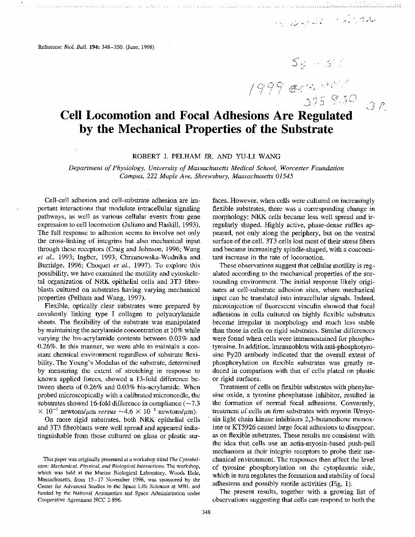
Reference: Biol. Bull. 194: 348-350. (June, 1998)
*.
Cell Locomotion and Focal Adhesions Are Regulated by the Mechanical Properties of the Substrate
ROBERT J. PELHAM JR. AND W - L I WANG
Department of Physiology, University of Massachusetts Medical School, Worcester Foundation Campus, 222 Maple Ave, Shrewsbury, Massachusetts 01545
Cell-cell adhesion and cell-substrate adhesion are im- portant interactions that modulate intracellular signaling pathways, as well as various cellular events from gene expression to cell locomotion (Juliano and Haskill, 1993). The full response to adhesion seems to involve not only the cross-linking of integrins but also mechanical input through these receptors (Craig and Johnson, 1996; Wang et al., 1993; Ingber, 1993; Chrzanowska-Wodnika and Burridge, 1996; Choquet et al., 1997). To explore this possibility, we have examined the motility and cytoskele- tal organization of NRK epithelial cells and 3T3 fibro- blasts cultured on substrates having varying mechanical properties (Pelham and Wang, 1997).
Flexible, optically clear substrates were prepared by covalently linking type I collagen to polyacrylamide sheets. The flexibility of the substrate was manipulated by maintaining the acrylamide concentration at 10% while varying the bis-acrylarnide contents between 0.03% and 0.26%. In this manner, we were able to maintain a con- stant chemical environment regardless of substrate flexi- bility. The Young's Modulus of the substrate, determined by measuring the extent of stretching in response to known applied forces, showed a 13-fold difference be- tween sheets of 0.26% and 0.03% bis-acrylamide. When probed microscopically with a calibrated microneedle, the substrates showed 16-fold difference in compliance (-7.3 x newtonslpm versus -4.6 x lop8 newtonslpm).
On more rigid substrates, both NRK epithelial cells '
and 3T3 fibroblasts were well spread and appeared indis- tinguishable from those cultured on glass or plastic sur-
This paper was originally presented at a workshop titled The Cytoskel- eton: Mechanical, Physical, and Biological Interactions. The workshop, which was held at the Marine Biological Laboratory, Woods Hole, Massachusetts, from 15-17 November 1996, was sponsored by the Center for Advanced Studies in the Space Life Sciences at MBL and funded by the National Aeronautics and Space Administration under Cooperative Agreement NCC 2-896.
faces. However, when cells were cultured on increasingly flexible substrates, there was a corresponding change in morphology: NRK cells became less well spread and ir- regularly shaped. Highly active, phase-dense ruffles ap- peared, not only along the periphery, but on the ventral surface of the cell. 3T3 cells lost most of their stress fibers and became increasingly spindle-shaped, with a concomi- tant increase in the rate of locomotion.
These observations suggest that cellular motility is reg- ulated according to the mechanical properties of the sur- rounding environment. The initial response likely origi- nates at cell-substrate adhesion sites, where mechanical input can be translated into intracellular signals. Indeed, microinjection of fluorescent vinculin showed that focal adhesions in cells cultured on highly flexible substrates become irregular in morphology and much less stable than those in cells on rigid substrates. Similar differences were found when cells were immunostained for phospho- tyrosine. In addition, immunoblots with anti-phosphotyro- sine Py20 antibody indicated that the overall extent of phosphorylation on flexible substrates was greatly re- duced in comparison with that of cells plated on plastic or rigid surfaces.
Treatment of cells on flexible substrates with phenylar- sine oxide, a tyrosine phosphatase inhibitor, resulted in the formation of normal focal adhesions. Conversely, treatment of cells on firm substrates with myosin IIImyo- sin light chain kinase inhibitors 2,3-butanedione monox- ime or KT5926 caused large focal adhesions to disappear, as on flexible substrates. These results are consistent with the idea that cells use an actin-myosin-based push-pull mechanism at their integrin receptors to probe their me- chanical environment. The responses then affect the level of tyrosine phosphorylation on the cytoplasmic side, which in turn regulates the formation and stability of focal adhesions and possibly motile activities (Fig. 1).
The present results, together with a growing list of observations suggesting that cells can respond to both the
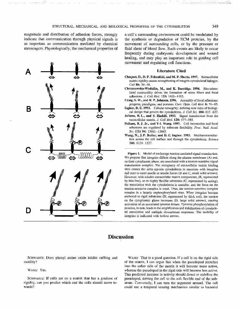
STRUCTURAL, MECHANICAL, AND BIOLOGICAL PROPERTIES OF THE CYTOSKELETON 349
magnitude and distribution of adhesion forces, strongly indicate that communication through physical signals is as important as communication mediated by chemical messengers. Physiologically, the mechanical properties of
a cell's surrounding environment could be modulated by the synthesis or degradation of ECM proteins, by the movement of surrounding cells, or by the pressure or fluid shear of blood flow. Such events are likely to occur frequently during embryonic development and wound healing, and may play an important role in guiding cell movement and regulating cell functions.
Literature Cited Choquet, D., D. P. Felsenfeld, and M. P. Sheetz. 1997. Extracellular
matrix rigidity causes strengthening of integrin-cytoskeletal linkages. Cell 88: 39-48.
Chrzanowska-Wodnika, M., and K. Burridge. 1996. Rho-stimu- lated contractility drives the formation of stress fibers and focal adhesions. J. Cell Biol. 133: 1403- 1415.
Craig, S. W., and R. P. Johnson. 1996. Assembly of focal adhesions: progress, paradigms, and portents. Curr. Opin. Cell Biol. 8: 74-85.
Ingber, D. E. 1993. Cellular tensegrity: defining new rules of biologi- cal design that govern the cytoskeleton. J. Cell Sci. 104: 613-627.
Juliano, R. L., and S. Haskill. 1993. Signal transduction from the extracellular matrix. J. Cell Biol. 120: 577-585.
Pelham, R. J. Jr., and Y-I. Wang. 1997. Cell locomotion and focal adhesions are regulated by substrate flexibility. Proc. Natl. Acad. Sci. USA 94: 13661-13665.
Wang, N., J. P. Butler, and D. E. Ingber. 1993. Mechanotransduc- tion across the cell surface and through the cytoskeleton. Science 260: 1124-1 127.
Figure 1. Model of anchorage-tension mediated signal transduction. We propose that integrins diffuse along the plasma membrane (A) and. on their cytoplasmic phase, are associated with a tension-sensitive signal transduction complex. The occupancy of extracellular matrix binding sites causes the actin-myosin cytoskeleton to associate with integrins and start to exert motile or tensile forces (B and C, small solid arrows). However, with soluble extracellular matrix components, (B, represented by thin line), or on highly flexible substrates (C, represented by spring), the association with the cytoskeleton is unstable, and the force on the tension-sensitive complex is weak. Thus, the tension-sensitive complex remains in a largely unphosphorylated state. When integrins become anchored to rigid substrates (D, represented by thick rod), the tension on the cytoplasmic phase increases (D, large solid arrows), causing activation of an associated tyrosine kinase. Tyrosine phosphorylation of proteins, in turn, leads to the amplification and stabilization of cytoskele- tal association and multiple downstream responses. The mobility of integrins is indicated with hollow arrows.
Discussion
SCHWARTZ: Does phenyl arsine oxide inhibit ruffling and WANG: That is a good question. If a cell is on the rigid side motility? of the matrix, I can argue that when the pseudopod stretches
into the softer side of the matrix it will become more active, WANG: Yes. whereas the pseudopod in the rigid side will become less active.
This predicted increase in activity should direct or stabilize the SCHWARTZ: If cells are on a matrix that has a gradient of pseudopod, driving the cell to the soft flexible end of the sub-
rigidity, can you predict which end the cells should move to- strate. Conversely, I can turn the argument around. The cell wards? could use a temporal sensing mechanism similar to bacterial

350 THE CYTOSKELETON
chemotaxis. When motile cells that are on the flexible side of the matrix enter the rigid side, they could become immobilized. This would lead to accumulation of cells on the rigid side of the matrix. The answer to your question must come from the experiments.
BRUINSMA: YOU said that the cell is sensing the shear modu- lus of the polyacrylamide gel. (Wang: That is correct). If you destroy the cross linking in the polyacrylamide gel to form a very viscous polymer melt, what would happen? Do the cells
only sense the shear modulus, or do they perhaps also sense regions of different viscosity?
WANG: Based on our current observations, where we have been looking at a rigid surface, I cannot predict what will happen with changes in viscosity. We haven't looked at cells plated on a viscous fluid, which is what it would take to answer your question. The experiment that you suggest is a good one. This will require a substrate where we can modulate flexibility to follow cell behavior from the entirely cross linked to the fluid state.

Reference: Biol. Bull. 194: 351-353. (June, 1998)
Theoretical Models of Viscoelasticity of Actin %.., .I: ,,. , Sollutions and the Actin Cortex
F. C. MACKINTOSH
Department of Physics and Biophysics Research Division, University of Michigan, Ann Arbor, Michigan 48109-1120
The elastic response of plant and animal cells depends on a network of protein filaments that form the cytoskele- ton. This is a complex and highly dynamic composite of filamentous proteins, together with a range of accessory proteigs for initiating and terminating polymerization, in- troducing cross-links, and forming lateral arrays or bun- dles of filaments (Alberts, 1994). A principal component of this is the actin cortex, which is itself an entangled and cross-linked network of F-actin. This cortex appears to be responsible for the mechanical stability and resis- tance of eukaryotic cells to external stresses. Coordinated assembly and disassembly of this network in response to cellular signals also appears to play a crucial role in cell locomotion (Stossel, 1994).
The actin cortex, consisting of entangled or cross- linked actin filaments (F-actin), resembles solutions and gels of common synthetic polymers. However, the actin cortex in vivo-as well as in vitro models of the actin cortex, consisting of solutions of reconstituted F-actin- exhibit unique properties that cannot be accounted for by the well-established models of synthetic polymer gels and solutions (DeGennes, 1979; Doi, 1988). For instance, the elastic moduli (specifically, the shear modulus) of actin networks can be several orders of magnitude larger than for comparable synthetic polymer systems (e.g., at the same concentration). This is a key property of actin net- works, as many types of cells must withstand shear stresses as large as 1000 Pa, or even more. Furthermore,
This paper was originally presented at a workshop titled The Cytoskel- eton: Mechanical, Physical, and Biological Interactions. The workshop, which was held at the Marine Biological Laboratory, Woods Hole, Massachusetts, from 15-17 November 1996, was sponsored by the Center for Advanced Studies in the Space Life Sciences at MBL and funded by the National Aeronautics and Space Administration under Cooperative Agreement NCC 2-896.
the results of several experiments in which different ele- ments of the cytoskeleton were disrupted suggest that the mechanical stability of cells and their elastic resistance to such applied stresses is primarily due to the actin cor- tex. Thus, the origin of high-shear moduli of the actin cortex poses an important problem for cell biology.
Progress has been made, recently, in defining the un- usual rheology (including the elastic shear modulus) of solutions containing actin filaments purified from muscle; and theoretical models have been devised to explain the viscoelastic properties of these systems (MacKintosh, 1995; Isambert, 1996; Krov, 1996; Satcher, 1996). All of these models are based, in part, on the known rigidity of actin filaments, as compared with conventional polymer systems. This rigidity (or semi-flexibility) is characterized by apersistence length-the typical length at which ther- mal fluctuations begin to bend the polymer in different directions. For actin, this length is several micrometers (Gittes, 1993; Ott, 1993). However, rigid constituent fil- aments do not necessarily produce entangled solutions or gels with increased rigidity. Indeed, increased polymer stiffness can actually reduce dramatically the shear modu- lus of a solution (Isambert, 1996), and this is due to reduced entanglement (Semenov, 1986).
Here, we describe a model that can account for the large shear moduli observed for actin networks. Specifi- cally, we predict a shear modulus
for densely crosslinked gels, where K is the filament stiff- ness, k is Boltzmann's constant, T is the temperature, J is the so-called mesh size (or typical spacing between polymers), c~ is the concentration of actin monomers of size a comprising the filaments. For entangled (un-

352 THE CYTOSKELETON
to 10 kHz, both storage and loss shear moduli can be 10000 0 1 determined as functions of frequency (Gittes, 1997;
A / $ Schnurr, 1997).
Schmidt. 1997. Microscopic viscoelasticity: shear moduli of soft Figure 1. The measured shear elastic modulus G of actin networks materials determined from thermal fluctuations. Phys. Rev. Lett. 79:
as a function of concentration in mglrnl (Janmey, 1991). The predicted 3286-3289. scaling of modulus with concentration cA is shown as the solid line Isambert, and A. C. Maggs. 1996. Dynamics and rheology of (MacKintosh, 1995). Here, G a cA1'". actin solutions. Macromolecules 29: 1036-1040.
(0
?3
10
l o o
Janmey,P. A.,U. Euteneuer,P. Traub, and M. Schliwa. 1991. Visco- elastic properties of vimentin compared with other filamentous bio-
crosslinked) filaments, somewhat weaker concentration polymer networks. J. Cell Biol. 113: 155-160. and filament stiffness dependencies are predicted: Kroy, K., and E. Frey. 1996. Force-extension relation and plateau
1 rigidity of microtubules and actin filaments measured from thermal 0.1 1 .o Oe0 fluctuations in shape. J. Cell Biol. 120: 923-934.
actin concentration (mg/ml) Gittes, F., B. Schnurr, P. D. Olmsted, F. C. MacKintosh, and C. F.
/ , , , ,,, i The latter concentration dependence of the shear modulus is in accord with recent experiments on in vitro solutions of F-actin (Janmey, 1991); see Figure 1.
We also report on ongoing experimental and theoretical characterizations of local viscoelasticity of actin solu- tions, as observed with embedded micrometer-size parti- cles. These methods can be used to measure the shear and compression moduli, as well as the dynamics of solu- tions and gels at the micrometer scale. Small, inert, spheri- cal beads of 0.5 to 5.0 pm diameter are placed in actin and polyacrylamide (used as control) gels. Through sensitive detection (resolution about 1 nm) of the thermal motion of individual beads, over frequencies ranging from 0.1 Hz
Literature Cited Alberts, B., D. Bray, J. Lewis, M. Raff, K. Roberts, and J. D. Wat-
son. 1994. Molecular Biology of the Cell. Garland, New York. DeGennes, P.-G. 1979. Scaling Concepts in Polymer Physics. Cornell
Doi, Gittes, Univ. Clarendon, M., F., and Press, B. S. Mickey, Oxford. F. Ithaca, Edwards. J. NY. Nettleton, 1988. and The Theory J. Howard. of Polymer 1993. Dynamics. Flexural
modulus for wonnlike chains. Phys. Rev. Lett. 77: 306-309. MacKintosh, F. C., J. Kas, and P. A. Janmey. 1995. Elasticity of
semiflexible biopolymer networks. Phys. Rev. Lett. 75: 4425-8. Ott, A., M. Magnasco, A. Simon, and A. Libchaber. 1993. Measure-
ment of the persistence length of polymerized actin using fluores- cence microscopy. Phys. Rev. E (Statistical Physics, Plasmas, Fluids, and Related Interdisciplinary Topics) 48: R1642-R1645.
Satcher, R. L., and C. F. Dewey. 1996. Theoretical estimates of me- chanical properties of the endothelial cell cytoskeleton. Biophys. J. 71: 109-118.
Schnurr, B., F. Gittes, P. D. Olmsted, C. F. Schmidt, and F. C. MacKintosh. 1997. Determining microscopic viscoelasticity in flexible and semiflexible polymer networks from thermal fluctua- tions. Macromolecules 30: 7781 -7792.
Semenov, A. N. 1986. Dynamics of concentrated solutions of rigid- chain polymers, part 1: Brownian motion of persistent macromole- cules in isotropic solution. J. Chem. Soc. Faraday Soc. Trans. 2. 82: 317-329.
Stossel, T. P. 1994. The machinery of cell crawling. Sci. Am. 271: 54-63.
Discussion
BOAL: A similar ideal expression for the shear modulus is applied in studies of two-dimensional polymeric nets. The shear is proportional to the density of nodes. But when steric interac- tions and the self-avoidance of polymers are accounted for, then the proportionality changes from density to the power one to density to the power 1.2 or 1.25. You showed a plot of the shear modulus as a function of density. You did not comment on what the exponent was, or what the scaling factor is. Is it very much different from one?
MACKINTOSH: The change that you mentioned is not quite
as dramatic in three dimensions. The scaling of the modulus with concentration for polymer chains in a good solvent in three dimensions, as predicted by established models of flexible polymers, is with a power of about 2.2. Specifically, the shear modulus G should be proportional to c2.'. The model I presented predicts an exponent of 2.5 for densely crosslinked networks of filaments such as actin. I mentioned an interesting subtlety about going from cross-linked networks to entangled networks. For conventional (flexible) polymer systems, there is no differ- ence between the two in the scaling exponent with concentra- tion. For systems such as actin, I get an exponent of about ''I5

MICROTUBULES AND V
= 2.2 if there are no cross-links-i.e., a different exponent from that predicted for cross-linked actin networks. In this case, the power law, not just the amplitude, changes. The figure I showed was for an exponent of ''I5, the exponent predicted for entangled semiflexible chains such as actin. This apparent qualitative distinction between entangled and crosslinked semi- flexible polymers is a subtle point that is not yet well under- stood. But, I think it is a subtle problem to pursue.
CHIEN: YOU measure the microviscosity in a dynamic way, but the macro-rheology is done in a static way. Did you also do the dynamic measurement to see how phase angle and visco- elasticity compare with the micro measurements?
MACKINTOSH: Measurements for solutions of this kind by conventional macroscopic rheology techniques, as carried out, for example, by Paul Janmey's group, can be done only for frequencies less than 100 Hz at most. To my knowledge no measurements of any kind have been made for actin above this range, although in principle it can be done with more specialized instruments. The frequency dependence of the shear moduli was studied by the Munich group in the range below 100 Hz. The data were quite limited, but a small increase with frequency was reported. The reported frequency dependence is not inconsistent with the behavior expected for conventional polymer networks (e.g., a power law dependence on frequency with exponent of about 0.5), even though the magnitude of the shear modulus is way off relative to conventional polymers. This is one of the interesting puzzles, for which no one yet seems to have a very good microscopic understanding. Although the origin of elastic- ity is quite different, the dynamics turn out to be very similar at high frequency. A value for the exponent of 0.7 is distinguish- able from conventional systems. Generalization of the model that predicts an increase of the shear modulus with the power 0.5 frequency to semi-flexible systems would predict a power of 0.25. That is still surprising to me.
CHIEN: It is known that with shear the endothelial cell will reorganize its cytoskeletal fibers. In your model, how does the organization affect the shear modulus-for example, alignment of the fibers with flow?
MACK~TOSH: What I have been talking about is the linear response of the network to small strains. The effect you men-
tioned is one of two things. It is either a nonlinear response (as evidenced by the alignment that results from shearing the net- work beyond a certain amount), or low-frequency behavior in which the network responds and flows. I did not try to address either of those Limits. I only discussed the linear response of the network to small strains and at moderately high frequencies, relative to the effects you mentioned.
STEWART: What YOU have described seems to be a wonderful explanation in accounting for actin at 1 mglml. I was wondering how easily this can be extrapolated to the actin concentrations that might be found in a cell where the free water concentration will be down to 5- 10 M. Can this be extrapolated to 10-20 mgl rnl F-actin?
MACKINTOSH: AS YOU probably know, if you take these net- works and concentrate them they form an equilibrium nematic phase of aligned filaments. The reason for this is that the molec- ular weight in these in vitro models is much higher than in the cell. If you break the filaments down to smaller molecular weight, formation of the nematic phase doesn't become a prob- lem, even at elevated concentrations. That is one way of extrap- olating our model to higher concentrations while maintaining isotropic networks. The network is also highly dynamic, but that is beyond the scope of our current work where we are simply interested in understanding the basic polymeric-like re- sponses of the networks.
BRIUNSMA: What would happen if you had stiff impurities in your actin system, such as randomly dispersed microtubules? How would the elastic properties of such an inhomogeneous system differ from the homogeneous structure? Could the stiff- ness that results from the presence of stiff rods of microtubules be magnified elsewhere in the system?
MACKINTOSH: You might imagine that a shear response would result from bending the microtubule filaments. However, if you apply a uniform macroscopic strain to the sample, you get no bending of such inclusions. It may seem counterintuitive, but filaments do not bend when subject to uniform strain. If the strain is not uniform, the response may be nonlinear, since the degree of nonuniforrnity would itself be an increasing function of strain. Therefore, I would expect a change in the nonlineari- ties of the response, but not necessarily in the linear response.

Reference: Biol. Bull. 194: 354-357. (June, 1998)
Stabilization and Functional Modulation of Microtubules by Microtubule-Associated Protein 4
H. L. NGUYEN, D. GRUBER, T. MCGRAW, M.P. SHEETZ, AND J. C. BULINSKI
Departments of Anatomy and Cell Biology and Pathology, Columbia University, College of Physicians and Surgeons, New York, New York 10032
Microtubules (MTs) are hollow cytoplasmic fibers that are composed of a dimeric protein called tubulin, as well as several MT-associated proteins (MAPs) bound along their surface. MTs are believed to play important roles in a variety of cellular processes, including mitosis, cell motility, and intracellular vesicle transport. Both in vitro and in vivo, individual MTs are dynamic; that is, they undergo alternating periods of polymerization and depo- lymerization from their ends, a process known as dynamic instability. The dynamic behavior of MTs is thought to play an important role both in cell cycle events and in cell differentiation.
MAPs have been postulated to function as in vivo regu- lators of the dynamics and functions of MTs. Based on in vitro studies, several MAPs have been classified as assembly-promoting MAPs, because they stimulate MT polymerization. Tau and MAP2 are assembly-promoting MAPs expressed almost exclusively in nervous tissue; MAP4, the subject of our research, is another assembly- promoting MAP of -210 kDa, which is expressed throughout all tissues of all vertebrate organisms that have been examined. MAP4 is the most abundant non-tubulin component of MTs in proliferating cells and non-neuronal tissues, but it is down-regulated in its expression during neuronal differentiation. Because MAP4 is present along the length of MTs that are performing functions as diverse as organizing and transporting organelles and vesicles in
This paper was originally presented at a workshop titled The Cytoskel- eton: Mechanical, Physical, and Biological Interactions. The workshop, which was held at the Marine Biological Laboratory, Woods Hole, Massachusetts, from 15-17 November 1996, was sponsored by the Center for Advanced Studies in the Space Life Sciences at MBL and funded by the National Aeronautics and Space Administration under Cooperative Agreement NCC 2-896.
interphase cells, rearranging the MT array and altering MT dynamics for mitosis, changing cell shape, and speci- fying directional motility, we and others have hypothe- sized that MAP4 plays roles in the regulation of MT dynamics, MT organization, and MT-based transport pro- cesses in vivo. In turn MAP4, itself, is subject to regula- tion as the MT system adapts to changes in the cell cycle or the cell's environment.
To investigate the in vivo functions of MAP4, we pre- pared stably transfected clonal mouse Lfk- cell lines ex- pressing either full-length MAP4 (L-MAP4 cells) or its MT-binding domain (L-MTB cells); see Figure 1. Al- though transfectants showed no dramatic defect in mor- phology or level of MT polymer, as compared with ndive Lk- cells or L-MOCK cells (transfected with vector alone), MTs in L-MAP4 and L-MTB cells showed greater stability than those in control cells. Stability was moni- tored by the level of post-translationally detyrosinated a- tubulin and by a quantitative nocodazole-resistance assay. In vivo, the MT-binding domain of MAP4 stabilized MTs less potently than full-length MAP4, in contrast to the equivalent efficacy demonstrated previously in studies of in vitro MT polymerization. L-MAP4 and L-MTB cells grew significantly more slowly than control cells; this growth inhibition was not due to mitotic arrest or cell death. L-MAP4 and L-MTB cells also exhibited greater tolerance to the MT-depolymerizing agent nocodazole, but not to the MT-polymerizing agent taxol. Results with these transfected cell lines demonstrated that MAP4 and its MT-binding domain are capable of MT stabilization in vivo, and that increasing the intracellular level of MAP4 affects cell growth parameters.
The Lk- cell lines we have generated, which inducibly overexpress different levels and domains of MAP4, have allowed us to address further questions about the

MICROTUBULES AND VISCOELASTICITY OF ACTIN
Overexpression of MAP4 Inhibits Cell Growth
Days After Addition of Dexamethasone
-
-
L-MOCK
s Ltk- mock
v L-MTB #8-4 A L-WP4-4f #13-2
L-MTB
I I I I I I I I
Figure 1. The top portion of the figure shows a growth curve of naiive L*- cells and those expressing empty plasmid, MAP4-MTB, and full-length MAP4 (called L-MOCK or MOCK, L-MTB #8-4, and L- MAP4-4r #13-2, respectively). The cells expressing MAP4 constructs are clearly inhibited in their growth. Expression of MAP4 in the transfectants can be visualized in the immunofluorescence micrographs shown at the bottom of the figure. Cells were stained with a primate-specific antibody to MAP4; the MAP4-MTB and full-length MAP4 present in L-MTB and L-MAP4 cells respectively display bright microtubule staining, while those cells expressing only plasmid (L-MOCK cells) exhibit only dimmer, diffuse staining with the MAP4 antibody.

356 THE CYTOSKELETON
functional interactions of MAP4 in vivo. For example, we examined the distribution of various organelles in L- MAP4 and L-MTB cells, hoping that any alterations in cytoplasmic organization would suggest binding interac- tions of MAP4, which we could then test. In these studies, we noted no change in organelle position, cytoplasmic organization, or cell morphology when we labeled L- MAP4 or L-MTB cells with anti-mannose-6-P, wheat germ agglutinin, anti-cytochrome c, anti-actin, or anti- vimentin antibodies to localize various organelles.
Previous in vitro work had demonstrated that assembly- promoting MAPS can inhibit organellar trafficking along MTs, by sterically hindering kinesin or dynein motility (Lopes and Sheetz, 1993; and unpubl. data, same authors). Thus, we reasoned that, although the steady-state distribu- tion of cellular organelles might appear to be normal, there might nonetheless be defects in in vivo organellar trafficking. Such a defect might affect cell growth, per- haps partially explaining the cell growth defects we had measured. Yet, since the deficit in organellar trafficking predicted from in vitro work would be a kinetic decrease rather than a permanent blockage, no obvious defect in morphology or organellar distribution would be expected.
We tested this hypothesis by examining in vivo organ- elle motility in L-MAP4 and L-MTB cells under high resolution DIC. In both high- and low-expressing L- MAP4 and L-MTB cells, movements of organelles were severely decreased, as compared with L-MOCK or Lk- cells, and the inhibition was greater in the lines that ex- pressed higher levels of MAP4 or MTB. MTB was almost as effective at inhibiting in vivo motility as L-MAP4, but was not as potent as the full-length molecule. We next asked whether in vivo vesicle motility was inhibited by the presence of MTB or MAP4, per se, or whether the inhibition could be ascribed to MT stabilization alone. To answer this question, we treated cells with several concentrations of the MT stabilizing drug taxol. Our re- sults showed that MT stabilization alone was not nearly as effective as MAP4 or MTB at inhibiting movement.
To elucidate the types of transport events altered by overexpression of MAP4 or MTB, we used fluorescent markers to perform several assays of cell-sorting func- tions. We noted defects or retardation in endocytosis (monitoring recycling of Cy3-transferrin), LDL uptake and processing (using di-I LDL), and sorting to lysosomes (observing movement and intensity of FITC-dextran). These data suggested that either MAP4 or MTB, coated onto cellular MTs, was capable of inhibiting normal MT- based transport events in vivo.
As before, we asked whether the inhibition of cellular activities dependent on organellar motility was attributable to the presence of extra MAP4 or MTB, or whether it was due to the MAP-induced alteration in MT stability. Again,
we found that the former possibility was the correct one, because concentrations of taxol that produced equivalent MT stability had virtually no effect on trafficking events. This was particularly exciting to us, for two reasons: First, our results suggested that cells could regulate transport along individual MTs in a single cell by producing high or low local concentrations of MAP4 along different fibers. Moreover, at least one kinase capable of releasing MAP4 from MTs, called MARK (MT-affinity regulating kinase; Drewes et al., 1997), has been described. Alternatively, however, extensive end dynamics, in which MTs depoly- merize near their ends and MAP4 rebinds at higher con- centration to the shortened MT, could provide a stochastic heterogeneity in MAP4 composition that would be capable of altering transport efficiency on individual MTs. Indeed, heterogeneity in MAP4 level along MTs has been observed previously (Chapin and Bulinski, 1994). The second excit- ing aspect of these results is that the observed inhibition of transport properties does not appear in the absence of MTs or at the expense of MT dynamics. In most previous studies, by contrast, inhibition of vesicle transport or traf- ficking events has been measured in the presence of con- centrations of nocodazole or taxol that, respectively, com- pletely depolymerize or stabilize MTs. Our data suggest that the control of MAP4 content along an MT is a potential regulator of transport events along that fiber; this is an area of current study in our laboratory.
In summary, we have used a system in which human MAP4 or its MT-binding domain is inducibly overex- pressed in a cultured line of rodent cells to investigate the in vivo functions of MAP4. We have found that over- expressed MAP4 can contribute to MT stabilization, can affect cell growth parameters, and can inhibit vesicle mo- tility in vivo. Our results suggest that alterations in MAP4 expression, or MT stabilization itself, may be physiologi- cally significant mechanisms for altering cell growth and intracellular transport in vivo; these MAP4 alterations may occur during the cell growth or development, or during experimental manipulations or pathologies.
Acknowledgments
Portions of this work have been published in Bulinski et al. (1997) and Nguyen et al. (1997).
Literature Cited
Bulinski, J. C., T. McGraw, D. Gruber, H. L. Nguyen, and M. P. Sheetz. 1997. . Overexpression of MAP4 inhibits organelle motility in vivo. J. Cell. Sci. 110: 3055-3064.
Chapin, S., and J. C. Bulinski. 1994. Cellular microtubules heteroge- neous in their content of MAP4 (210d MAP). Cell Motil. Cytoskel. 27: 133-149.
Drewes, G., A. Ebneth, U. Preuss, E.-M. Mandelkow, and E. Mandel- kow. 1997. MARK, a novel family of protein kinases that phos-

MICROTUBULES AND VISCOELASTICITY OF ACTLN 357
phorylate microtubule-associated proteins and trigger microtubule Nguyen, H.-L., S. Chari, D. Gruber, C.-M. Lue, S. J. Chapin, and disruption. Cell 89: 297-308. J. C. Bulinski. 1997. Overexpression of full- or partial-length
Lopez, L., and M. P. Sheetz. 1993. Steric inhibition of cytoplasmic- MA4 stabilizes microtubules and alters cell growth. J. Cell Sci. 110: dynein and kinesin motility by MAP2. Cell Motil. Cytoskel. 24: 1-16. 281-294.
Discussion
GOLDMAN: Have you looked at the rate of individual organ- elle translocations, or did you say fewer particles move in cells that overexpress MAP4? A second question is, do any of the motor molecules compete with MAP4 for binding sites on mi- crotubules?
BULINSKI: We don't have the resolution to look at rate of organelle translocations. In answer to your second question, we have not yet done the work to see whether motor molecules compete with MAP4 for those binding sites. Since the microtu- bule-binding domain of MAP4 is homologous to those of MAP2 and Tau, we can extrapolate from studies done on those MAPs. However, there is a conflict in the literature about this. In the original research, Lopez and Sheetz compared three molecules: full-length MAP2, microtubule-binding domain of MAP2 cre- ated by protease digestion, and Tau, which has the same micro- tubule-binding domain but only a small projection domain. They found virtually no inhibition by Tau or the MAP2 microtubule- binding domain, and concluded that inhibition was due to steric factors. However, these studies were conducted at a very low ratio of MAP to tubulin. There is contradictory evidence in another paper from a different group. However, there are other experiments that show fairly good competition data. In our cells we have a high ratio of MAP to tubulin. We've calculated that if the vesicle is 0.1 pm in diameter and there are 50 MAP4 molecules along a microtubule, the vesicle will run into a num- ber of the MAP4 molecules as it transits along the microtubule. In the cells where we have increased MAP content sevenfold, this movement will be inhibited.
GOLDMAN: Could that account for saltatory movement?
BULINSKI: We cannot see the individual tracks the vesicle moves on in vivo. This can be seen in the in vitro system where one can measure run time, the time that the vesicle stays on a microtubule before it falls off. The presence of MAP molecules decreases this run time.
LIAO: Are these microtubule-dependent transport events? '
BULINSKI: Yes, they are similar to transferrin receptor move- ments, which have been studied with microtubule active drugs. There was no vesicle movement for the in vivo motility when the cells were treated with nocadazole. There was decreased movement when they were treated with vanadate, an inhibitor of dynein. These events are microtubule-based movement.
LUNA: Are there lower levels of MAP4 or other MAPs in the melanocytes? Could this represent an instance of microtubule motors run amok? Controlling access to the number of binding sites through steric hindrance would slightly reduce motility of a vesicle along a microtubule; consequently, you would expect it to cause greater reduction in the ability of the vesicles to serve as multivalent crosslinkers.
BULINSKI: We don't know anything about the level of non- motor MAPs, also called assembly-promoting MAPs or struc- tural MAPS (such as MAP4) in melanocytes. I have discussed MAP4 in my presentation. However, there are other MAPS in these cells that may have the same or similar properties. When we increase the level of one MAP, this doesn't tell us about the other MAPS that are present and may function in the same way. There are also kinases, such as MARK (microtubule-affin- ity regulating) kinase, that can phosphorylate a single site on MAP4 and remove it from the microtubule. If that is a regulated event, it could help pigment transport or process formation.
ALBRECHT-BUEHLER: IS it cause or effect that is observed, and could it be that you found the handle on the cellular clock?
BULINSKI: The cyclin people think that they have the comer on the market of the cell cycle. I don't think it is likely that cells would normally regulate their growth this way. But it is a possibility, I suppose, that regulation of MAPS on MTs could have an effect on cell growth. I was hoping that significant effects on transport would be produced by stabilizing the micro- tubules. That would have had implications for how some of these chemotherapeutic drugs work, at concentrations lower than those required to alter mitosis, by altering growth-related transport phenomena. So in some ways I was disappointed that microtubule stability had so little effect.
CHEN: YOU had no trouble loading LDL or transferrin, which implies that the kinesin pathway works fine, but the dynein does not.
BULINSKI: When we've used iodinated transferrin or iodin- ated LDL, they both seem to load up fairly rapidly. That sug- gests that it's not microtubule-based transport, or that it would not be very inhibitory to the dynein motor. But it's still not clear that an hour would be a limiting time frame for a kinetic limitation of transport by MAPs.

Reference: Biol. Bull. 194: 358-360. (June, 1998)
Microtubules as Determinants of Cellular Polarity
GREGG G. GUNDERSEN, GERI KREITZER, TIWANI COOK, AND GUOJUAN LIAO
Departments of Cell Biology & Anatomy and Pathology, Columbia University, New York, New York 10032
Most eukaryotic cells respond to developmental or en- vironmental stimuli with a change in their polarity. Inher- ent in this behavior is the ability of the cell to detect the extracellular cue, to transmit a signal across a plasma membrane, and to translate the signal into molecules that can produce the requisite architecture of change. The overall response could be modulated by the expression of new genes, but since many such responses can be generated without new genetic input, this is not an essen- tial feature of the response. We and others have been asking, in particular, how elements of the cytoskeleton, especially microtubules (MTs), contribute to the genera- tion of cellular polarity by responding to environmental cues and transmitting the information to other cellular constituents. Unlike other signal transduction systems (e.g., growth factor activation of gene expression), the one involving MTs mediates a cellular response that re- flects the spatial information provided by the original ex- tracellular cue. Individual elements of this MT system will be described, and the possible mechanisms by which cells achieve functional polarity will be discussed.
The biological system that we use in most of our studies is an in vitro wound-healing model. Fibroblasts are grown in culture until confluent, and narrow strips of cells are then scraped off the substratum. The cells at the wound edge are initially unpolarized, but in response to wounding, they become polarized with their long axis perpendicular to the wound and with an active leading edge at the portion of the cell facing the cell-free area.
This paper was originally presented at a workshop titled The Cytoskel- eton: Mechanical, Physical, and Biological Interactions. The workshop, which was held at the Marine Biological Laboratory, Woods Hole, Massachusetts, from 15-17 November 1996, was sponsored by the Center for Advanced Studies in the Space Life Sciences at MBL and funded by the National Aeronautics and Space Administration under Cooperative Agreement NCC 2-896.
As a result of this polarization, the cells are able to move directionally into the wound. This model system has a number of advantages for the study of cell polarization: the cellular response is rapid (on the order of minutes); the cells respond synchronously; and polarization can be measured both structurally (cell shape parameters) and functionally (cell migration parameters).
The state of the MTs in cultured fibroblasts before wounding is typical of the MTs in many nonpolarized cells grown in culture. The MTs radiate from a central focus near the nucleus and fill the cytoplasm without any particular bias to one portion of the cell. By directly imaging MTs in cells that have been microinjected with a fluorescent rhodamine-derivative of tubulin (the subunit protein of MTs), we observed that MTs in cells exhibit dynamic instability (periods of growth and shrinkage, with infrequent and rapid transitions between the two phases), a condition similar to that exhibited by MTs in many nonpolarized cells. The dynamic instability of MTs in nonpolarized cells is thought to contribute to the short half-life of these MTs, which has been reported to be 5-10 min (Saxton et al., 1984; Schulze and Kirschner, 1986).
One of the first responses to wounding is the stabilization of a subset of dynamic MTs. In particular, MTs that are oriented toward the wound become stabilized (Gundersen and Bulinski, 1988; Nagasaki et al., 1992; Gundersen et
val., 1994). With a rapid assay to detect MT stabilization, we have observed the formation of stable MTs in as little as 5 min after the addition of growth factors to wounded monolayers of serum-starved cells. This suggests that MTs are poised to respond rapidly to extracellular cues and can function on a time scale that is consistent with many cell behaviors. We are currently trying to identify the factors that are involved in stabilizing the MTs in a localized area and have found that activated rho (a ras-related, small

MICROTUBULES AND VISC
GTPase) is both necessary and sufficient to mediate the selective stabilization of MTs at the wound edge (Cook et al., 1998). We propose that rho is part of a signaling cascade that locally activates or recruits MT stabilizing factors in the leading edge of the cell. We are attempting to identify other factors in this signaling pathway, as well as the factors mediating the MT stabilization.
Shortly after the MTs become stabilized, they accumu- late a post-translationally modified form of tubulin known as detyrosinated tubulin. This modification involves the removal of a tyrosine residue from the C-terminus of a- tubulin by a specific tubulin carboxypeptidase, and this occurs predominantly on polymeric tubulin (Hallak et al., 1977). Detyrosination is reversible, and tyrosine is re- added by a second enzyme, tubulin tyrosine ligase (Ray- bin and Flavin, 1977). This enzyme is active only on soluble tubulin and is responsible for keeping the mono- mer pool of tubulin completely tyrosinated in vivo (Gun- dersen et al., 1987). In cells, therefore, newly polymerized MTs are composed of tyrosinated tubulin. If these MTs turn over rapidly, they are not significantly detyrosinated by the carboxypeptidase, but if they are stabilized, the carboxypeptidase can generate MTs with high levels of detyrosinated tubulin. A subset of MTs with elevated lev- els of detyrosinated tubulin (termed Glu MTs after the C-terminal Glutamate residue of detyrosinated tubulin) has been demonstrated; and these MTs are also stable (reviewed in Bulinski and Gundersen, 1991). In the wound-edge cells, the Glu MTs are oriented toward the wound. Thus, the stabilization of MTs at the leading edge of wounded cells generates a polarized array of MTs that are biochemically distinct from the randomly distributed dynamic MTs.
What is the consequence of detyrosinating the tubulin that constitutes stable MTs? Recently, we have found that intermediate filaments, another type of cytoskeletal filament, are preferentially aligned with the Glu MTs in wound-edge cells (Gurland and Gundersen, 1995). This preferential coalignrnent was disrupted by microinjecting cells with antibodies to detyrosinated tubulin, but not with antibodies to tyrosinated tubulin. More recently, we have found that a nonpolymerizable form of detyrosinated tu- bulin acts as a dominant negative inhibitor of the interme- diate filament-MT interaction when microinjected into cells (Kreitzer and Gundersen, pers. comm). Nonpoly- ,
merizable tyrosinated tubulin had no effect when microin- jected. Since the nonpolymerizable detyrosinated tubulin remained in the monomer pool and did not affect the endogenous, stable Glu MTs, these results show directly that detyrosination is involved in signaling or in recruiting intermediate filaments onto stable Glu MTs. The func- tional consequence of the association of intermediate fil- aments with Glu MT is unknown, but it may rigidify the
leading lamella of the cell so that it is able to resist fluid flow or other mechanical stresses. In any case, the para- digm we have established for intermediate filaments may be applicable to other organelles that are known to depend on MTs for their location in cells.
To identify the molecules that specifically recognize detyrosinated tubulin and mediate the interaction between MTs and intermediate filaments, we attempted to block the binding of proteins to MTs with antibodies to detyrosi- nated or tyrosinated tubulin. We added crude preparations of brain MT-associated proteins (MAPs) or MT motor proteins to MTs composed of a mixture of detyrosinated and tyrosinated tubulin, together with saturating levels of antibody to either detyrosinated or tyrosinated tubulin. Neither antibody significantly affected the binding of brain MAPs. But antibody to detyrosinated tubulin did block the MT-binding of a protein of - 120 kD from the brain MT motor preparations (Liao and Gunderson, 1998). We identified this protein as a kinesin based on its apparent molecular weight, its reactivity with several kinesin antibodies, and its ATP-dependent binding to MTs. To test directly whether kinesin would bind prefer- entially to detyrosinated tubulin, we examined the binding of a recombinant kinesin head fragment to detyrosinated and tyrosinated tubulin from cultured HeLa cells. We found that there is a 2- to 3-fold tighter binding of kinesin to detyrosinated tubulin (Liao and Gunderson, 1998). These data suggest that detyrosination of tubulin may regulate the interaction of kinesin with MTs, and they support the idea that kinesin may mediate the interaction between MTs and intermediate filaments. We are cur- rently determining whether kinesin moves differently on detyrosinated and tyrosinated tubulin, and whether kinesin can move intermediate filaments along MTs.
In summary, our data support a model in which MTs play a central role in generating cellular polarity. The rapid dynamics of MTs allows MTs to act as a sensor for changes in the extracellular milieu. In a process that we still do not understand completely, dynamic MTs are con- verted to stabilized MTs in portions of the cell adjacent to the extracellular stimulus. After stabilization, the MTs are modified by post-transitional modification. This step does not alter the three-dimensional information resident in the array of stabilized MTs. Rather, it translates the change in MT dynamics into a biochemical signal that can be interpreted by other cellular constituents. It also ensues that the other cellular constituents are not able to interfere with the process of MT dynamics; i.e., the other cellular corkstituents are incapable of interacting with ty- rosinated tubulin monomer or with newly polymerized MTs. There are at last six other tubulin post-translational modifications, and we suggest that these may also be involved in regulating MT interactions.

THE CYTOSKELETON
Literature Cited Bulinski, J. C., and G. G. Gundersen. 1991. Stabilization of post-
translational modification of microtubules during cellular morpho- genesis. Bioessays 13: 285-293.
Cook, T. A., T. Nagasaki, and G. G. Gunderson. 1998. Rho guanosine triphosphatase mediates the selective stabilization of microtubules induced by lysophosphatidic acid. J. Cell Biol. 141: 175-185.
Gundersen, G. G., and J. C. Bulinski. 1988. Selective stabilization of microtubules oriented toward the direction of cell migration. Proc. Natl. Acad. Sci. USA 85: 5946-5950.
Gundersen, G. G., S. Khawaja, and J. C. Bulinski. 1987. Postpoly- merization detyrosination of a-tubnlin: a mechanism for the subcel- lular differentiation of microtubules. J. Cell Biol. 105: 251 -264.
Gundersen, G. G., I. Kim, and C. J. Chapin. 1994. Induction of stable microtubules in 3T3 fibroblasts by TGF-P and serum. J. Cell Sci. 107: 645-659.
Gurland, G., and G. G. Gundersen. 1995. Stable, detyrosinated mi- crotubules function to localize vimentin intermediate filaments in fibroblasts. J. Cell Biol. 131: 1275-1290.
Hallak, M. E., J. A. Rodriguez, H. S. Barra, and R. Caputto. 1977. Release of tyrosine from tyrosinated tubulin. Some common factors that affect this process and the assembly of tubulin. FEBS Lett. 73: 147-150.
Liao, G., and G. G. Gunderson. 1998. Kinesin is a candidate for crossbridging microtubules and intermediate filaments: selective binding of kinesin to detyrosinated tubulin and vimentin. J. Biol. Chem. 273: 9797-9803.
Nagasaki, T., C. J. Chapin, and G. G. Gundersen. 1992. Distribu- tion of detyrosinated microtubules in motile NRK fibroblasts is rap- idly altered upon cell-cell contact: implications for contact inhibition of cell locomotion. Cell Motil. Cytoskel. 23: 45-60.
Raybin, D., and M. Flavin. 1977. Enzyme which specifically adds tyrosine to the alpha chain of tubulin. Biochemistry 16: 2189-2194.
Saxton, W. M., D. L. Stemple, R. J. Leslie, E. D. Salmon, M. Zavor- tink, and J. R. McIntosh. 1984. Tubulin dynamics in cultured mammalian cells. J. Cell Biol. 99: 2175-2186.
Schulze, E., and M. Kirschner. 1986. Microtubule dynamics in in- terphase cells. J. Cell Biol. 102: 1020-1031.
Discussion
GOLDMAN: IS there evidence that vimentin binds kinesin?
GUNDERSEN: We have done the experiment and can detect kinesin sedimenting with the intermediate filaments. We haven't done experiments to test whether this reflects a specific interac- tion, but kinesin doesn't sediment by itself.
GREEN: Several laboratories have published data that the tu- mor-suppressor gene-product APC binds to microtubules. Re- cent research has produced some evidence that this association occurs at the leading edge of cells, and it may have something to do with motility. Have you looked at where APC is located with respect to stable microtubules?
GUNDERSEN: Yes, we have, and we are very interested in the mechanism by which the microtubules are selectively stabilized. We have indirect evidence for a specific capping phenomenon: during time-lapse recordings of microtubules in cells, we have observed microtubules to contact something and then stop their dynamics. This may be why stable microtubules behave as they do when diluted. Their ends are no longer active, so they don't lose subunits. We don't know yet what is capping the microtu- bules. It does not seem to be APC. James Nelson has provided us with an antibody to APC, and we have colocalized APC with
Glu microtubules in a number of different cell types. We have not seen a correspondence between APC staining and the ends of Glu microtubules.
TAYLOR: HOW big is the kinesin that you were using to show binding to the intermediate filament?
GUNDERSEN: I believe it was 110 kDa. It is the major kinesin in our crude motor preparations.
TAYLOR: But that's the whole molecule.
GUNDERSEN: Yes. It appears that the kinesin heavy chain is behaving this way.
TAYLOR: With something else attached to the bottom of that heavy chain?
GUNDERSEN: I don't know. The recombinant head alone binds differentially to Glu and Tyr tubulin. Thus, I don't think we need to invoke a light chain or some other associated mole- cule to account for the difference in binding to Glu and Tyr tubulin. We do not know, at this point, whether kinesin heavy chain interacts with intermediate filaments or whether it's a Light chain or some other associated subunit that is responsible. We are looking into this question.

Reference: Biol. Bull. 194: 361-363. (June, 1998)
Intermediate Filament Cytoskeletal System: . %./ ,. i'
Dynamic and Mechanical Properties
R. D. GOLDMAN, S. CLEMENT, S. KHUON, R. MOIR, A. TREJO-SKALLI, T. SPANN, AND M. YOON
Department of Cell and Molecular Biology, Northwestern University Medical School, 303 E. Chicago Avenue, Chicago, Illinois 60611
Intermediate filaments (IF) are major cytoskeletal con- stituents of animal cells. For many years they were thought to be the most stable of all of the different cy- toskeletal systems. Recently, however, IF have been shown to be in a state of dynamic equilibrium in growing cells. In fibroblasts and epithelial cells, this has been dem- onstrated in vivo by the finding that microinjected, soluble vimentin and keratin subunits are incorporated into en- dogenous IF (Vikstrom et al., 1989; Miller et al., 1991). Other experiments involving fluorescence recovery after photobleaching (FRAP) have demonstrated the existence of a steady-state equilibrium between polymerized IF and their subunits in vivo (Vikstrom et al., 1992). Further- more, the exchange between IF subunits and their poly- mers appears to be regulated by phosphorylation cata- lyzed by different kinases and dephosphorylation cata- lyzed by phosphatases (Eriksson et al., 1992). Recently, we have exploited this equilibrium state in vivo through the use of mimetic peptides that are known to both drive vimentin IF disassembly and to inhibit subunit polymer- ization into IF at 1:l molar ratios in vitro. The sequence of these peptides is derived from the helix 1A initiation domain of the central rod region of either keratin or vi- mentin. The same 1A peptide carrying a single point mu- tation has no obvious effect on vimentin IF assembly in vitro up to a 10-fold molar excess. We have also shown . that there are no detectable effects of the wild-type pep-
This paper was originally presented at a workshop titled The Cytoskel-
tide on either the stability or polymerization of microtu- bules and actin at 3-5 molar excesses in vitro. When these peptides are injected into live cells containing vi- mentin IF, they rapidly induce the disassembly of IF net- works. This disassembly of IF in vivo is accompanied by dramatic changes in cell shape and mechanical properties. More specifically, cells lose their asymmetric shapes within 15-45 min as they are transformed into a round shape. Under these conditions, cell-substrate adhesions are lost and there is an induction of microtubule and microfilament disassembly. The rounded cells exhibit ac- tive blebbing for short time intervals, but within 4-5 h they have respread into their normal shapes and have reestablished their cell-substrate contacts. These latter changes are coincident with the reassembly of IF, micro- tubule, and microfilament networks. An important control for these experiments has involved the microinjection of the mutant peptide at the same concentrations. The results demonstrate no significant changes in IF assembly, cell shape, or microtubules and microfilaments (Goldman et al., 1996).
We have also carried out numerous additional controls to make certain that the peptides are primarily targeting IF. One of these controls has involved treatment of 3T3 cells with colchicine to depolymerize microtubules. Thirty minutes after the addition of colchicine, microtu- bules are not detected, and many IF remain extended from the nuclear region to the cell surface. If the 1A peptide is injected into these cells, the effect on cell shape and on actin-microfilaments appears to be the same as in un-
eton: Mechanical, Physical, and Biological Interactions. The workshop, treated cells. Therefore, effects on cell shape do not which was held at the Marine Biological Laboratory, Woods Hole, depend on polymerized microtubules. ~h~ 1~ peptide has Massachusetts, from 15-17 November 1996, was sponsored by the Center for Advanced Studies in the Space Life Sciences at MBL and also been injected into fibroblasts derived from vimentin funded by the National Aeronautics and Space Administration under knockout mice. These cells contain no detectable IF, but Cooperative Agreement NCC 2-896. they do contain extensive arrays of microtubules and mi-

362 THE CYT(
crofilaments. When the peptide is injected, no changes in cell morphology are detected, and the microtubule and microfilament systems remained unperturbed. All of these experimental observations indicate that the peptide-in- duced disruption is specific for IF and has no obvious effects on microtubules and microfilaments. Therefore, in growing cells containing IF, the induction of their disas- sembly leads to alterations in the stability of the other major cytoskeletal components and a dramatic loss of cellular mechanical properties and shape. These observa- tions could be in part due to IF associated proteins (IFAPs) that form bridges between IF and other cytoskele- tal components. For example, BPAGlnIdystonin and plectin both have actin binding domains as well as IF binding domains (for review see Chou et al., 1997).
It has been suggested that the injection of these 1A peptides in vivo might lead to toxic side effects due to the sudden release of large amounts of small oligomers and monomers of vimentin. This does not appear to be the case. For example, in 3T3 cells we have calculated that molar ratios of 1:20 (peptide:vimentin) in vivo are very effective in disassembling the IF network, altering cell shape and microtubule/microfilament assembly (Goldman et al., 1996). At this concentration, we are quite certain that the IF network disassembles into large oligomeric complexes that should not have a significant deleterious effect on overall cell physiology. This is also indicated by the fact that cells treated with the peptide fully recover within a few hours. In contrast, it has also been demonstrated that the perturbation of the normal organization of IF networks by a variety of different agents does not have similar effects on cell shape and cytoskeletal integrity. These include the reorganization of
IF following the disassembly of microtubules with inhibi- tors such as colchicine, the microinjection of IF antibodies and protein kinase A, and following heat shock (for dis- cussion see Goldman et al., 1997). However, these treat- ments do not specifically disassemble IF networks; rather they appear to induce the relocation of existing polymer- ized IF. The latter changes do not mimic the profound changes in IF assembly states that occur following peptide injection. Based on these considerations, we feel that the 1A peptide is a reliable probe for studying the functions of IF in vivo.
Acknowledgments
This work is supported by the NIGMS.
Literature Cited
Chou, Y. -H., 0 . Skalli, and R.D. Goldman. 1997. Intermediate filaments and cytoplasmic networking: new connections and more functions. Curr. Opin. Cell Biol. 9: 49-53.
Eriksson, J. E., D. L. Brautigan, R. Vallee, J. Olmsted, H. Fujiki, and R. D. Goldrnan. 1992. Cytoskeletal integrity in interphase cells requires protein phosphatase activity. Proc. Nut. Acad. Sci. USA 89: 11093-1 1097.
Goldman, R. D., S. Khuon, Y.-H. Chou, P. Opal, and P. M. Steinert. 1996. The function of intermediate filaments in cell shape and cytoskeletal integrity. J. Cell Biol. 134: 971-983.
Miller, R. K., K. Vikstrom, and R. D. Goldman. 1991. Keratin in- corporation into intermediate filament networks is a rapid process. J. Cell Biol. 113: 843-855.
Vikstrom, K. L., G. G. Borisy, and R. D. Goldman. 1989. Dynamic aspects of intermediate filament networks in BHK-21 cells. Proc. Nut. Acad. Sci. USA 86: 549-553.
Vikstrorn, K. L., S. S. Lim, R. D. Goldman, and G. G. Borisy. 1992. Steady state dynamics of intermediate filament networks. J. Cell Biol. 118: 121-129.
Discussion
GREEN: What are the differences in mechanical properties between keratin filaments and type LII filaments? Can we distin- guish differences in mechanical properties in the bundles versus individual keratin filaments?
GOLDMAN: Paul Janmey is just beginning those experiments. . I can tell you one interesting preliminary finding. When we inject the 1A peptide into an epithelial cell, like a PTK cell which normally does not move, the cells round up. All focal adhesions disappear and the cells migrate numerous cell diame- ters to another part of the cover slip. It looks as though the motor systems can turn on when we disassemble keratin, and the cells become actively migratory. As the keratin network re- assembles, because it is quite reversible, we get a static cell again. These epithelial cells usually grow in little islands and are very stable; consequently, they don't move very much. So
it's possible that keratin could be acting as a brake on motility. That's an important function because most tissue cells don't move. In fact, they are terminally differentiated. My guess is that a lot of the dynamic activities that we see, including those of all cytoskeletal systems, are more related to actively growing cells in culture.
FORGACS: At this meeting good evidence has been presented to show that the microtubule and the intermediate filament net- work interconnect directly. You have shown us that actin and intermediate filaments colocalize. Do you have any evidence that this is more than just colocalization? If they do interact, what would be the culprit for that?
GOLDMAN: One interesting candidate, recently discovered by Brown et al. (1995. Nut. Genetics, 10: 301-306) is dystonin.

INTERMEDIATE FILAMENTS, HEMIDESMOSOMES, AND DESMOSOMES 363
Mutation in this gene is responsible for Dystonia musculorum, a mutation in mice, which is a severe nervous disorder. In a recent paper from Elaine Fuchs' lab (Yang et al. 1996. Cell 86: 655-65), BPAG1, a neuronal variant of bullous pemphigoid antigen, is described as the same gene. The gene product has never been isolated, but it has an actin-binding domain and an intermediate filament-binding domain. So there is at least one possible cross-bridging element in addition to plectin. Research presented by Gerhard Wiche and Gary Borisy and others at this meeting show that it is very important to begin looking for interactions among the different cytoskeletal systems.
CHEN: When you inject the peptide, the intermediate fila- ments disassemble, all the other polymers disassemble, and the cells round up. In contrast, the vimentin-free cells show the opposite reaction. Can you interpret these results?
GOLDMAN: The vimentin-free cells don't have any intermedi- ate filaments, so there's nothing to disassemble. This serves as a control for our peptide experiment. It looks to us as if the
GUNDERSEN: In previous studies of intermediate filament function, antibodies to intermediate filaments were injected into cells, causing the collapse of the intermediate filaments back toward the nucleus. In these experiments, cell motility was not affected, and the microtubules were fine. In your experiments, when you injected a peptide it disrupted the intermediate fila- ments, producing all these effects. Why do you think these two experiments give such really disparate results?
GOLDMAN: In our hands antibody injections do not retract all of the intermediate filaments towards the nucleus (unpubl. obs). Approximately 20% to 30% of the intermediate filament population is resistant, even when treated with colchicine or other antimitotic drugs. Perhaps you don't need many filaments to provide appropriate mechanical properties at the periphery of the cell. Furthermore, the IF polymer doesn't appear to change when you inject antibodies; only its organization changes. It is still present in cells. This is very different from the disassembly effects of the peptides.
cells have a compensatory system which allows them to overex- STEWART: When you disassemble intermediate filaments in
press other cytoskeletal proteins. vitro with your peptide, have you observed them with electron - - -
CHEN: Why is that not happening when you depolymerize microscopy to see what it looks like? the intermediate filaments in the normal cells?
GOLDMAN: I'll let Peter (Steinert) answer that. GOLDMAN: Because these reactions occur in 30 minutes, and
the recovery takes place in a few hours. Everything is already STEINERT: Yes, we have. The filaments basically unravel and there, and the cell just goes through a rapid reversal. fall apart into subfilamentous units.
Reference: Biol. Bull. 194: 364-366. (June, 1998)
..J ', Type I Keratin 16 Forms Relatively Unstable
Tetrameric Assembly Subunits With Various Type I1 Keratin Partners: Biochemical Basis
and Functional Implications
PIERRE A. COULOMBE, MATTHEW WAWERSIK, RUDOLPH D. PALADIN, AND ERICK NOENSIE
Departments of Biological Chemistry and Dermatology, The Johns Hopkins University School of Medicine, Baltimore, Maryland 21205
Type I1 keratin 6 (K6) and type I keratins 16 and 17 (K16 and K17) are intermediate-filament (IF) proteins that are induced in wound-edge keratinocytes as early as 4-6 h after injury to skin, either human or mouse. This induction occurs at the expense of the keratin proteins that are normally expressed in differentiating epidermal keratinocytes. Correlated with these changes in protein expression, keratinocytes-for 24 h following injury- undergo major cytoarchitectural alterations that affect their shape, intracellular organization, surface morphol- ogy, and adhesion properties. We recently proposed that the intrinsic properties of K16 are compatible with a direct role in "keratinocyte activation" at the wound edge (Pal- adini et al., 1996). Unlike K14, a related type I keratin that is constitutively expressed in epidermis, we found that K16 forms unstable heterotetramer subunits that po- lymerize into shorter filaments when paired with a variety of type I1 keratin partners (e.g., K5, K6b, Kg). Such prop- erties are of particular interest because it has been shown in a number of studies that the tetramer subunit predomi- nates in the soluble pool of IF subunits in epithelial and nonepithelial cell lines in culture. The tetramer-forming ,
properties of K16 may thus influence its dynamic parti-
This paper was originally presented at a workshop titled The Cytoskel- eton: Mechanical, Physical, and Biological Interactions. The workshop, which was held at the Marine Biological Laboratory, Woods Hole, Massachusetts, from 15-17 November 1996, was sponsored by the Center for Advanced Studies in the Space Life Sciences at MBL and funded by the National Aeronautics and Space Administration under Cooperative Agreement NCC 2-896.
tioning between the polymer and soluble pools and, as such, play a key role in determining the overall impact of its presence on the cytoarchitecture of epidermal kera- tinocytes.
The main objective of this study was to determine the biochemical basis of the unique tetramer-forming proper- ties of K16 as a first step toward a full understanding of its significance in the regulation and function of this kera- tin. We showed that K16-containing tetramers are not only less stable than K14-containing ones, but also less stable than K17- and K19-containing ones, irrespective of the type I1 keratin partner used. We exploited the re- markable degree of primary sequence identity between K14 and K16 to construct a series of chimeric K14-K16 cDNAs, obtaining the corresponding proteins in a purified recombinant form and testing them for tetramer formation using anion-exchange chromatography and chemical cross-linking under denaturing buffer conditions. These studies allowed us to define a segment, of about 200 amino acid residues, within the central a-helical rod do- main (3 10 amino acid residues) that is responsible for the difference in stability. Such a finding was expected, as the rod domain has been identified as the main determinant in the assembly of coiled-coil parallel dimer and anti-parallel tetramer subunits. Alignment of the sequences for human K14, K16, K17, and K19 over this 200-residue segment led to the identification of a candidate residue, '88Pro, which is unique to K16 and has properties that are consis- tent with a local disruption of the a-helical secondary structure. Consistent with this prediction, site-directed

INTERMEDIATE FILAMENTS, HEMIDESMOSOMES, AND DESMOSOMES 365
mutagenesis revealed that 'aspro accounts quantitatively for the instability of K16-containing heterotetramers un- der denaturing conditions. Of note, introduction of a pro- line residue at corresponding positions in K14 ( ls6~al-+- Pro) and K17 (15511e+~ro) rendered these keratins K16- like in their tetramer-forming properties. W e believe that la8Pro contributes to the properties and functions of K16, either by influencing its partitioning between the soluble and polymer pools in the cytoplasm, or by causing it to enter the assembly pathway as a heterodimer instead of a heterotetramer. In addition, these data provide insights into the assembly-productive conformation of the keratin tetramer, a matter of contention in the literature, as well as into the unique interdependency of type I and type I1 keratin sequences for their assembly into 10-nrn filaments.
Acknowledgments
These studies were supported by grants from the NIH and NSF.
Bibliography Coulombe, P. A. 1997. Towards a molecular definition of keratino-
cyte activation after acute injury to stratified epithelia. Biochem. Biophys. Res. Comm. 236: 231-238.
Fuchs, E., and K. Weber. 1994. Intermediate filaments: structure, dynamics, function, and disease. Ann. Rev. Biochem. 63: 345-382.
Heins, S., and U. Aebi. 1994. Making heads and tail of intermediate filament assembly, dynamics, and networks. Curr. Opin. Cell Biol. 6: 25-33.
Paladini, R. D., K. Takahashi, N. S. Bravo, and P. A. Coulombe. 1996. Onset of re-epithelialization after skin injury correlates with a reorganization of keratin filaments in wound edge keratinocytes: defining a potential role for keratin 16. J. Cell. Biol. 132: 381-398.
Discussion
GREEN: In tissues where keratin K16 is expressed at a low level, how much of that is in an insoluble pool in the filament system, and how much is in another type of pool that it is not capable of being incorporated into?
COULOMBE: This is something we really have to look into. From our findings one could predict that the partitioning of K16-containing subunits between the polymer pool and the sol- uble pool should be different from other epidermal keratins. This issue needs to be formally examined in an appropriate set of experiments.
AEBI: The destabilization of your tetramers by proline 188 in keratin 16 suggests that these tetramers are really N,N-type half-stacked tetramers.
COULOMBE: Yes, I would agree with that. This particular proline mutation could tell us that the type of tetramer confor- mation you are referring to is dominant. Conversely, this substi- tution may favor the formation of other tetramer conformations that would have different assembly properties. This may explain why K16 forms relatively short filaments. These issues need to be examined.
AEBI: When you get this overexpression of keratin K16, ei- . ther in your transfected cells or after wounding, what happens to K1 and K6, which form the heterodimers? Does this also increase?
COULOMBE: Yes, they do go up. It's K16, K17 as type I keratins, as well as K6, a type I1 keratin. Now we know that if you make K5, K6, K14, K16, and K17 compete for one another under conditions such that the type I1 are limiting, the one that is left out as type I is 16. The monomer fractions in a wounded
keratin site must be looked at to see which keratin is going to be predominant in the soluble pool. That's another issue we need to examine.
GOLDMAN: Can you speculate about what this might mean in terms of cell physiology? What you are proposing is that K16 is almost a keratin poison. It makes shorter, poorly structured filaments, which might increase motility during wound healing. Why go to all this trouble when there are other ways to do this?
COULOMBE: Your work has shown that phosphorylation could produce comparable changes in filament structure. Gregg Gundersen showed data at this meeting that suggest that one might promote such reorganization of keratin filaments in a fashion completely independent of keratin de novo synthesis, simply by disrupting the interaction with microtubules. I think that the polymer pool and the unpolymerized pool might have separate functions. If one were to look at the in vivo peptide disruption experiment that was performed in your laboratory, I think that a possible mechanism for reorganization of the entire cytoskeleton is that a soluble pool of tremendous size is sud- denly created which the cell normally never sees. This causes the other polymer systems to eventually come apart. In my opinion, the application of molecular genetic approaches will reveal that tetramers are binding to all kinds of cellular compo- nents.
LUNA: If keratins, or possibly all intermediate filaments, are inherently apolar, how do you get motility? How do you get the kinds of movements that Bob Goldman saw? Are there motors, interactions with microtubules, or is this the kind of collapse of biopolymers that we heard about earlier?
COULOMBE: It's clear that intermediate filaments in general

366 THE CYTOSKELETON
lack polarity, which likely makes them unsuitable for use as band. Secondly, keratin does not respond to colchicine or any tracks for motor-based translocation. Moreover, they are very other antimitotic drug the way other intermediate filaments do. convoluted, so it would be very inefficient for the cell to use STEWART: l-he fact that you have the two chains antiparallel them as a track for specific transport events. means that it is possible that the filaments lack polarity. It does
GREEN: DO we really know that for all of the intermediate filaments?
COULOMBE: Yes, I think so. The only possible polymeric exceptions are neurofilaments, since it features those side arms sticking out. What that means in terms of polarity might depend on the way the subunits are integrated along the filament wall. In terms of the basic character of the intermediate filament fiber, it is an apolar structure because the two dimers come together in antiparallel fashion to form a tetramer.
GOLDMAN: I think the use of the term "collapse" is incor- rect. Firstly, it requires energy to move the filaments back, and this takes a long time. They don't collapse; it's not like a rubber
not mean that they lack polarity-they can still be quite polar with the chains antiparallel. It is very important to realize that.
ALBRECHT-BUEHLER: A quick comment. I want to remind you that many epithelial cells migrate as groups of 8-10 cells tied together, not as single cells. I showed this in tissue culture years ago. My point is that if you look for migration, you may not find it in the single cells; you may find it in a whole group of cells. Any changes in the keratin and intermediate filaments may be expressed in the group and not in a single cell in the usual way.
COULOMBE: Although I did not talk about migration of PTK, cells, your point is well taken. As I mentioned in my presenta- tion, epidermal keratinocytes do migrate into a wound site as a stratified epithelial sheet.

Reference: Biol. Bull. 194: 367-370. (June, 1998)
\ b ; ) / / ' /yy > I
3 i 3 - 7 ,-$.
Structural-Mechanical Integration of Keratin Intermediate Filaments With Cell Peripheral
Structures in the Cornified Epidermal Keratinocyte
PETER M. STEINERT
Laboratory of Skin Biology, National Institute of Arthritis and Musculoskeletal and Skin Diseases, NZH, Bethesda, Maryland 20892-2752
The outermost visible layer of the skin consists of ter- minally differentiated, flattened, dead, cornified cells (squames). These squames consist predominantly of kera- tin intermediate filaments (KIF) embedded in a matrix of filaggrin, and these are contained within a specialized thickened insoluble cell peripheral structure termed the cornified cell envelope (CE). The primary function of these cells is to provide a barrier against the environment. We are interested in how these components are assembled and integrated, since it now seems that any defect in either component will cause a serious epidermal disorder, generally an ichthyosiform disease.
One approach has been to study the structure of the CE. The CE is a 15-20 nm-thick layer of insoluble pro- tein formed on the inside of the plasma membrane during terminal differentiation in the epidennis (as well as in other stratified squamous epithelia). It constitutes about 10% of the mass of the cornified cell. Its insolubility is largely attributable to cross-linking of certain structural proteins by a series of transglutaminase (TGase) enzymes which form an M(y-glutamy1)lysine isopeptide bond be- tween the y-amide sidechain of a donor glutamine residue and the E-NH, sidechain group of an acceptor lysine resi- due. Since this bond cannot be cleaved in nature, the result is an insoluble macromolecular complex. In addition, a ,
This paper was originally presented at a workshop titled The Cytoskel- eton: Mechanical, Physical, and Biological Interactions. The workshop, which was held at the Marine Biological Laboratory, Woods Hole, Massachusetts, from 15-17 November 1996, was sponsored by the Center for Advanced Studies in the Space Life Sciences at MBL and funded by the National Aeronautics and Space Administration under Cooperative Agreement NCC 2-896.
series of w-hydroxy ceramides become attached by ester linkages to the cross-linked proteins on the extracellular surface of the cornified cells. This composite protein- lipid CE structure replaces the plasma membrane, and its integrity is vital for barrier function.
Using limited proteolysis procedures, we have isolated and sequenced large numbers of peptides that contain cross-links, and have addressed such questions as which proteins are involved; which glutamine and lysine resi- dues are used on what parts of the proteins; what is the temporal order of protein deposition; and what is the mechanism of assembly of the CE. Quantitatively, most cross-links are intra- and inter-chain links between lori- crin, which is the most abundant CE protein (70%-80%). This protein is unusual in that its content of glycine resi- dues is the highest of any known protein in biology. The glycine sequences are clustered into domains that are thought to be configured as highly flexible loops, inter- spersed by glutamine and lysine rich domains that are the sites of cross-linking. In addition, some cross-links are between loricrin and representatives of the small proline- rich protein (SPR) class (about 5% of total). Most of these linkages suggest that the SPRs serve as cross-bridging proteins. The SPRs contain more proline than any proteins known in biology, and include least 10 different proteins that are differentially expressed in the epidermis of differ- ent body sites. The prolines are distributed among peptide repeats of 8 or 9 residues; the repeats are located in a central domain, and each domain comprises from about 3 (smallest SPR protein) to 23 (largest SPR protein) repeats. These peptide repeats are thought to form a relatively stiff structure. The glutamines and lysines used for cross- linking are located on the head (amino-terminal) and tail

368 THE CYT(
(carboxy-terminal) domains of the SPRs. Together, lori- crin and the SPRs constitute 85% of the protein mass of the CE of the epidermis. Protein expression data show that loricrin and the SPR proteins are located on the intra- cellular side of the CE; thus these proteins must have been accumulated onto the CE structure during the last stages of its assembly. Thus the bulk of the CE consists of a "solution" of SPRs in loricrin. Although the total amount of loricrin + SPRs remains constant in the epider- mis of different body sites, we find the content of SPRs is highest in the epidermis of the palms and soles, with decreasing amounts in elbows, lips, foreskin, and trunk. This series is correlated with decreasing epidermal thick- ness and with the relative amount of mechanical stress or trauma that these epidermal sites undergo during their normal function.
This variability in composition is familiar to materials science, wherein variations in the amounts of a minor cross-bridging component can greatly change the me- chanical properties of a composite material. We therefore speculate that the differential expression of the SPRs re- flects the required biomechanical properties of the epider- mis of different body sites.
When the ceramide lipids were removed by mild alka- line hydrolysis, we found that a number of other CE proteins were exposed for immunogold electron micros- copy. These proteins presumably represent the innermost components of the CE, corresponding to its initial stages of assembly. By additional proteolysis and sequencing, we have found many cross-links within involucrin and between such proteins as involucrin and desmoplakin, or cystatin a, or envoplakin, or loricrin and SPRs.
Some cross-linking also involved the type I1 keratins K1, K2e, K5, and K6. Only one lysine residue in these keratin chains was used for cross-linking: it is located in a conserved stretch of sequences in the V1 head domain. This residue is important in two ways. First, in in vitro crosslinking reactions, TGase enzymes specifically utilize this lysine residue to cross-link synthetic peptides. Sec- ond, we have discovered a family with autosomal domi- nant non-epidermolytic palmar plantar keratoderma whose affected members have a single point mutation that results in the loss of this residue. The patients have a severe scaling disorder of their palms and soles and of other thickened epidermal sites. When viewed by electron microscopy, the affected cornified cells are highly irregu- lar in shape, and there is an abnormal accumulation and
distribution of the lamellar granules that export the lipids. The failed distribution of the lipids is presumably the cause of the hyperkeratosis and scaling phenotype.
Together, these data suggest that CE assembly is initi- ated at, or near, the site where KIF meet desmosomes. In addition, they suggest that certain proteins can mediate an indirect association between KIF and desmoplakin. Moreover, these studies offer a mechanism by which the KIF cytoskeleton of the cornified cell is mechanically integrated with the CE. We conclude that the terminal differentiation program exploits the existing KIF cy- toskeleton-desmosome machinery to build the novel and vital CE barrier structure. Failure to implement proper attachment by cross-linking has resulted in severe me- chanical and biological consequences.
Bibliography Candi, E., E. Tarcsa, J. J. DiGiovanna, J. G. Compton, P. M. Elias,
L. N. Marekov, and P. M. Steinert. 1998. Identification of a highly conserved lysine residue on type I1 keratins which is essential for the structural coordination of keratin intermediate filaments and the cornified cell envelope through isopeptide crosslinking by trans- glutaminases. Proc. Natl. Acad. Sci. USA 95: 2067-2072.
Hohl, D., U. Lichti, M. L. Turner, D. R. Roop, and P.M. Steinert. 1991. Characterization of human loricrin: structure and function of a new class of epidermal cell envelope proteins. J. Biol. Chem. 266: 6626-6636.
Jarnik, M., T. Kartasova, P. M. Steinert, U. Lichti, and A. C. Steven. 1996. Differential expression and cell envelope incorporation of small proline rich protein 1 in different cornified epithelia. J. Cell Sci. 109: 1381-1391.
Kartasova, T., Y. Kohno, H. Koizumi, S. Osada, N. Huh, U. Lichti, P.M. Steinert, and T. Kuroki. 1996. Sequence and expression patterns of mouse SPRI: correlation of expression with epithelial function. J. Invest. Dermatol. 106: 294-304.
Kimonis, V., J. J. DiGiovanna, J.-M. Yang, S. Z. Doyle, S. J. Bale, and J. G. Compton. 1994. A mutation in the V1 end domain of keratin 1 causing non-epidermolytic palmar-plantar keratoderma. J. Invest. Dermatol. 103: 764-769.
Steinert, P. M., and L. N. Marekov. 1995. The proteins elafin, filag- grin, keratin intermediate filaments, loricrin and SPRs are isodipep- tide crosslinked components of the human epidermal cornified cell envelope. J. Biol. Chem. 270: 17702-17711.
Steinert, P. M., and L. N. Marekov. 1997. Involucrin is an important early component in the assembly of the epidermal cornified cell envelope. J. Biol. Chem. 272: 2021-2030.
Steinert, P.M., E. Candi, T. Kartasova, and L. N. Marekov. 1998. Small Proline Rich Proteins are cross-bridging proteins in the comi- fied cell envelopes of stratified squamous epithelia. J. Stmct. Biol. (in press).
' Steinert, P. M., T. Kartasova, and L. N. Marekov. 1998. Biochemi- cal evidence that the small proline rich proteins and trichohyalin function in epithelia by modulation of the biochemical properties of their cornified cell envelopes. J. Biol. Chem. 273: 11758-11769.

INTERMEDIATE FTLAMENTS, HEMIDESMOSOMES, AND DESMOSOMES 369
Discussion
COULOMBE: Am I correct that non-epidermal PPK features hyper-proliferation in the basal compartment?
STEINERT: Yes.
COULOMBE: Since the structural defect involved in this dis- ease is a late event, in terms of the differentiation pathways, there must be some means of signaling between suprabasal cells and basal cells in the absence of lysis. Would you care to speculate on this?
STEINERT: I can only speculate. Dr. Peter Elias, a skin biolo- gist and lipid expert, believes that there is a complex interplay between lipid synthesis in these lamellar granules and cell pro- liferation. He is particularly interested in ceramides, a major lipid component of the lipid envelope, which are known to be second messengers.
GOLDMAN: This is remarkable remodeling of already existing cellular components. The speed at which a cell becomes enucle- ated, presumably by going through some part of apoptosis, and then reutilizes all of these proteins by cross-linking to become a part of that fortified envelope structure is quite remarkable. Desmosomal components, and keratin, and other normal com- ponents are being reorganized into a highly cross-linked struc- ture that bears very little resemblance to desmosomes and other structures in the super basal cells. Can you speculate on how long it takes to go through the process?
STEINERT: On the basis of our studies of cross-linking and immunogold electron microscopy, I believe that we can make intelligent guesses as to how assembly of this complex structure is initiated. We think that this occurs at the site of the desmo- some. What probably happens first is association of things like envoplakin and involucrin to the site of the desmosome by cross-linking-more specifically, to the tail of desmoplakin, which extends 100 nrn from the main plaque of the desmosome into the cytoplasm. Proteins such as involucrin and envoplakin then form interdesmosomal sheets at the focal points of enve- lope assembly. This produces a layer of protein in the granular layer of the epidermis. At about this time mayhem breaks loose and everything within the cell is dissolved. This includes all of the cytoplasmic constituents and the plasma membrane. These events are finalized by the addition of loricrin to the existing involucrin-envoplakin scaffold. We do not yet understand when and how the lipid granules, which are of critical importance to both extra- and intracellular lipids, can escape before the whole structure closes down.
GREEN: YOU mentioned a compromised keratin attachment, but you place more emphasis on the role of the lipid organiza- tion problem. Am I correct in presuming that these are two
separate functions? If so, what are their related roles for the amino terminus?
STEINERT: I assume you are refemng to the amino terminus of desmoplakin? We have no information about its cross-linking to the cell envelope. Perhaps this is buried and may be lost before the envelope is assembled. We have seen only certain desmoplakin sequences near its carboxy terminus, which you have predicted project far into the cytoplasm. However, I want to answer a slightly different question that is important because it bears on what you ask. The amount of keratin cross-linked to the cell envelope represents only about 0.1% of the envelope protein mass. We have calculated what this means in terms of the extent of cross-linking of keratin filaments. Assuming that keratin filaments are 15 nm wide, and assuming a model in which the filaments completely line the intracellular surface of the cell envelope, we calculate about one crosslink per 100 nrn of filament length. This is actually a high level, and it suggests that the keratin filament cytoskeleton really gets glued tightly and permanently onto the cell periphery. We have also seen that the amount of keratin cross-linked to the cell envelope of mouse forestomach tissue is about 10 times higher, which is really an incredible amount. This must reflect the biomechanical requirements of a tissue that is subjected to extraordinarily rigor- ous stresses and trauma. Also, the disease we talk about is autosomal dominant, so we don't know what would happen if both alleles were knocked out-perhaps not a viable fetus. To check this experimentally is of course difficult because, in cell culture, you get upregulation of K5 and K6. A mouse with this lysine residue knocked out in both alleles could be made. We believe that to make only 50% of the cross-links seriously com- promises the structural interface between the cytoplasmic kera- tin filament network and the cell periphery, or the growing cell envelope. It is highly convoluted, which increases the surface area of the cellular connections. Peter Elias hypothesizes that this greater surface area prevents lipids from distributing prop- erly, resulting in decreased barrier function and thus a hyperker- atotic response.
JANMEY: IS the transglutaminase, which crosslinks the pro- teins together, constitutively active?
STEINERT: Yes the transglutaminase enzymes are very active during the stages when the cell envelope barrier is assembled. The transglutaminase story is complicated. There are a number of different enzymes functioning simultaneously. Some of these enzymes exist in multiple different post-translationally modified forms, each possibly with a different function. What we are trying to unravel now is which enzyme does what. Preliminary data suggest that transglutaminase 3 strongly favors cross-link- ing of loricrin and SPRs. Apparently one or two forms of trans-
370 THE CYTOSKELETON
glutarninase 1 are involved in the earlier stages of cross-linking of involucrin and envoplakin to the tails of desmoplakin. We also have data that transglutaminase 1 works synergistically with transglutaminase 3 to cross-link loricrin and SPRs. The studies needed here involve systematic in vitro analyses of the preferences of the enzymes for the numerous substrate proteins, and comparisons with the in vivo data. I f the transglutaminase 1 enzyme is knocked out you get the naturally occurring, autoso- mal recessive human disease called lamellar ichthyosis. This is
a life-threatening disease because it produces a serious loss of skin barrier function. The mouse knockout model results in death of the newborn within 6 hours of birth because of dehydra- tion; that is, there has been complete breakdown of skin barrier function.
BORISY: We are cheered by this presentation. If intermediate filaments have no purpose in life, they now have a purpose after death.

Reference: Biol. Bull. 194: 371-373. (June, 1998)
What Links Larninin-5 to the Keratin Cytoskeleton in Epithelial Cells?
JONATHAN C. R. JONES, OMAR SKALLI, ROBERT D. GOLDMAN, AND SCOTT E. BAKER
Cell and Molecular Biology, Northwestern University Medical School, 303 E. Chicago Avenue, Chicago, Illinois 60611
The extracellular matrix plays an important role in tis- sue morphogenesis. It does so, in part, by interacting with a family of heterodimeric cell surface receptors called integrins. Indeed, the integrins are considered to play an important role, not only in adhesion of cells to matrix, but also in signal transduction between the matrix and the cells (Hynes, 1992). Signal transduction via integrins involves the cytoskeleton and cytoskeleton-associated proteins which bind to the cytoplasmic domain of the integrin subunits.
Our particular interest is in the a6P4 integrin hetero- dimer. This integrin is unusual because it is involved in keratin intermediate filament (IF)/cell surface anchorage at the site of hemidesmosomes-structures that play an important role in epithelial cell-matrix linkage (Quaranta and Jones, 1991; Jones et al., 1994). Since the major matrix element of the hemidesmosome is the laminin iso- form called laminin-5, we have been investigating the molecular links between the keratin cytoskeleton, inte- grins, and laminin-5 matrix. In the current analyses, we have used FG epithelial cells, derived from a pancreatic tumor.
FG cells characteristically grow in mounds and fail to flatten efficiently onto their substrate. In such cells, kera- tin intermediate filaments (IFs) are concentrated in the perinuclear region. Furthermore, the IF-associated protein
This paper was originally presented at a workshop titled The Cytoskel-
IFAP300 (a relative of plectin and possibly identical to a protein termed HD1) primarily localizes along these keratin bundles. Additionally, a604 integrin heterodimers localize in streaks or spots towards the free edges of cells, whereas a 3 p l integrin is predominant at the opposed surfaces of adjacent cells; neither integrin shows any ob- vious interaction with IF. Note that these FG cells express very little, if any, laminin-5, the matrix ligand for both the a6P4 and a3Pl integrin receptors.
We have discovered that we can induce a reorganiza- tion of the cytoskeleton of FG cells, as well as an associa- tion of IF with integrins, by simply adding rat laminin-5 to the medium in which we grow the FG cells (Baker et al., 1997). When FG cells are plated into this medium, they no longer form mounds, but rapidly adhere and spread onto the substrate. They do so, apparently by "cap- turing" rat laminin-5 and placing it basally in circles or arcs at areas of cell-substrate interaction. Double-label irnrnunofluorescence microscopy reveals that IFAP300, a6P4 integrin, and a3p l integrin are all colocalized with the polarized laminin-5, indicating that the FG cells are using these receptors to interact with the exogenously added laminin-5 protein. Concomitantly, a 6 integrin un- dergoes dephosphorylation on serine residue 104 1 ; we are intrigued by the possibility that this event plays a crucial role in mediating subsequent cytoskeleton rear- rangement.
Rapid adhesion, induced by laminin-5, can be blocked by antibodies against the a 3 integrin subunit. In contrast,
eton: Mechanical, Physical, and Biological Interactions. The workshop, although raised to a6 integrin do not block which was held at the Marine Biological Laboratory, Woods Hole, laminin-5-induced rapid adhesion, they do prevent FG Massachusetts, from 15-17 November 1996, was sponsored by the Center for Advanced Studies in the Space Life Sciences at MBL from assuming an morpholog~' This funded by the National Aeronautics and Space Administration under Suggests that a3Pl and a6P4 integrins have distinct func- Cooperative Agreement NCC 2-896. tions: a 3 p l integrin appears to mediate rapid adhesion

THE CYTOSKELETON
dium, keratin IF bundles "move" from their perinuclear location and are induced to associate with IFAP300- a6P4la3Pl integrin complexes along the surface of the cell that is attached to the substratum. Furthermore, the results of coprecipitation experiments suggest that, within IFAF'300-integrin complexes, IFAP300 is associated with the P4 integrin subunit. Based on our results and on evi- dence that IFAP300 binds keratin in vitro (Skalli et al., 1994), we propose that, when a6P4 integrin binds to its matrix ligand laminin-5, IFs indirectly interact with the cytoplasmic domain of a6P4 integrin at the cell surface. The latter interaction is mediated by IFAF'300. In other words, we have identified one potential series of rnole- cules that link laminin-5 indirectly to the IF cytoskeleton. We show this putative interaction diagrammatically in Figure 1.
Acknowledgments
I L ' . Supported by the NM.
Figure 1. FG cells maintained in a laminin-5-rich medium "cap- ture" the laminin-5 and incorporate it into their extracellular matrix. The molecular interactions of the captured laminin-5 are indicated here. Laminin-5 (represented as an inverted t) binds the extracellular domain of the a6P4 integrin. The cell membrane is represented by a dashed line. In this scheme, IFAP300 interacts with the cytoplasmic domain of the P4 integrin subunit and mediates the interaction of the IF cytoskele- ton with the cell surface.
Literature Cited
Baker, S. E., 0. Skalli, R. D. Goldman, and J. C. R. Jones. 1997. Laminin-5 and modulation of keratin cytoskeleton arrangement in FG pancreatic carcinoma cells: involvement of IFAP3OO and evi- dence that laminin-51cell interactions correlate with a dephosphory- lation of a6A integrin. Cell Motil. Cyto. 37: 271 -286.
Hynes, R. 0 . 1992. Integrins: versatility, modulation, and signaling in cell adhesion. Cell 69: 11-25.
Jones, J. C. R., J. Asmuth, S. E. Baker, M. Langhofer, S. I. Roth, and S. B. Hopkinson. 1994. Hemidesmosomes: extracellular ma- trixtintennediate filament connectors. Exp. Cell Res. 213: 1 - ll .
Quaranta, V., and J. C. R. Jones. 1991. The internal affairs of an integrin. Trends Cell Biol. 1: 2-4.
Skalli, b . , J. C. Jones, R. Gagescu, and R. D. Goldman. 1994. of FG cells to laminin-5, whereas a6P4 integrin appears IFAP300 is common to desmosomes and hemidesmosomes and is to mediate cell-spreading on the laminin-5 matrix. a possible linker of intermediate filaments to the junctions. J. Cell
In FG cells maintained in laminin-5-containing me- Biol. 125: 159-170.
Discussion
COULOMBE: Would you agree that the recent work of Elaine ,
Fuchs' lab shows that BAG1 or BP230, the bullous pemphigoid antigen, is involved in anchoring keratin filaments at the cell surface with sites of hernidesmosomes?
JONES: Yes, I think that is true. If you knock out BP230, her pictures would imply that there is a reduction in intermediate filaments at that site in transgenic mouse skin. However, we have used FG cells since they don't possess any of their own BP230. Thus the filament associations that we have described were in the absence of BP230. We speculate that there could
be cooperation between IFAP300 and BP230, in that there may be multiple proteins linking intermediate filaments at the cell surface in intact, complete hemidesmosomes.
WICHE: DQ YOU have evidence for direct interaction of 04 with IFAP300?
KOWALCZYK: YOU showed that when the cells adhere to lam- inin-5, there was dephosphorylation of a6. Can you get that to

INTERMEDIATE FILAMENTS, HEMIDESMOSOMES, AND DESMOSOMES 373
happen in suspension, or do the cells have to adhere to the the filament cytoskeleton. Presumably a firmer attachment to immobilized substrate? substrates is established when this molecular interaction occurs.
JONES: We haven't done it in suspension, so I don't know. BORISY: IS laminin-5 captured from solution?
BORISY: Could you expand a little bit on the functional sig- JONES: We don't think that the laminin-5 has been captured
nificance of that peripheral association of the FAP300 in the from solution. We think, in fact, that laminin-5 possibly rapidly
cells grown on laminin-5? What is its function for the cell? coats the surface upon which the cell sits. The cell then reorga- nizes the laminin-5, and pulls it in. What we observe is the
JONES: At the cell periphery, laminin-5 binds to a6P4 inte- initial aspects of pulling in this laminin-5, which is why this grin, which itself binds to IFAP300, which then is bound to protein is located more toward the cell periphery at that time.

Reference: Biol. Bull. 194: 374-377. (June, 1998)
Desmosomes: Integrators of Mechanical Integrity in Tissues
KATHLEEN J. GREEN, ANDREW P. KOWALCZYK, ELAYNE A. BORNSLAEGER, HELENA L. PALKA, AND SUZANNE M. NORVELL
Department of Pathology, Northwestern University Medical School, Chicago, Illinois 60611
The most prominent cell surface attachment site for intermediate filaments (IF) at epithelial cell-cell interfaces is the desmosome (reviewed in Collins and Garrod, 1994; Schmidt et al., 1994; Garrod et al., 1996; Green and Jones, 1996); see Figure 1. By anchoring IF at sites of strong intercellular adhesion, desmosomes create a trans- cellular network throughout the tissue, and this network is thought to resist forces of mechanical stress. The impor- tance of the IF-cell surface complex has been highlighted by the discovery of mutations in genes encoding IF struc- tural proteins, as well as cell junction proteins that result in blistering diseases of the skin (Steinert and Bale, 1993; Fuchs, 1994; McLean and Lane, 1995). Furthermore, au- toimmune antibodies that specifically attack the desmoso- ma1 cadherin component of desmosomes result in a family of blistering diseases of the skin and oral cavity known as pemphigus (Stanley, 1995). Thus, these cell-cell adhesive structures and their underlying cytoskeletal attachments are clearly important for the integrity of tissues.
Desmosomes are related to another class of adhesive junctions called adherens junctions, which mediate attach- ment of the microfilament cytoskeleton to the cell surface (Kowalczyk and Green, 1996). In adherens junctions, classic calcium-dependent cell-cell adhesion molecules (e.g., E-cadherin) are anchored to the actin cytoskeleton through a complex that includes the armadillo protein family member p-catenin. p-catenin binds directly to both E-cadherin and to the actin-associated protein a-catenin. In desmosomes, two classes of desmosomal cadherins,
This paper was originally presented at a workshop titled The Cytoskel- eton: Mechanical, Physical, and Biological Interactions. The workshop, which was held at the Marine Biological Laboratory, Woods Hole, Massachusetts, from 15-17 November 1996, was sponsored by the Center for Advanced Studies in the Space Life Sciences at MBL and funded by the National Aeronautics and Space Administration under Cooperative Agreement NCC 2-896.
the desmogleins and desmocollins, bind to the p-catenin- related protein called plakoglobin. Unlike classic cadher- ins, desmogleins and desmocollins have not yet been ex- perimentally demonstrated to be adhesion molecules. To examine the adhesive potential of desmosomal cadherins, we expressed desmogleins and desmocollins along with their associated protein plakoglobin, individually and to- gether, in normally nonadherent L cell fibroblasts. Unlike E-cadherin, desmoglein and desmocollin were unable to confer adhesive properties on these cells in several adhe- sion assays, including formation of cell-cell aggregates in suspension (Kowalczyk et al., 1996). This may be due to the complexity of cadherin forms, both within a single desmosome and within tissues; it could also reflect the requirement for additional noncadherin molecules in the adhesive complex.
Co-immunoprecipitation of myc-tagged proteins from L cell lines revealed that plakoglobin binds with an un- usual 6:l stoichiometry to desmoglein, whereas it binds with a 1:l stoichiometry to desmocollin (Kowalczyk et al., 1996). The functional significance of this dramatic difference in stoichiometry is currently unknown; but cells expressing dominant negative mutants of plako- globin that affect the ratio of binding to desmoglein also exhibit alterations in desmosome structure, consistent with a role in desmosome assembly. Plakoglobin can also bind directly to a-catenin and E-cadherin, which leads us to ask how specificity of filament attachment is achieved during junction assembly. We have demonstrated that a- catenin is not associated with plakoglobin bound to either desmoglein or desmocollin. Along with observations from other labs demonstrating that the a-catenin binding site on plakoglobin overlaps with that for desmosomal cadherins, these data suggest a possible means of achieving cytoskel- etal specificity.
Although the mechanism by which the desmosomal cadherin-plakoglobin complex is coupled specifically to

INTERMEDIATE FILAMENTS, HEMTDESMOSOMES, AND DESMOSOMES 375
I Adherens Junction I Microfdaments
Figure 1. Diagram comparing the major molecular constituents of the two intercellular adhesive junc- tions found in epithelial tissues. The adhesion molecules in both junctions are members of the larger cadherin family of calcium-dependent cell adhesion molecules. These are desmogleins and desmocollins in the desmosome (top), and classic cadherins, such as E-cadherin, in the adherens junction (bottom). Members of the armadillo gene family, plakoglobin and p-catenin, associate with the cytoplasmic tails of the cadherins and link the membrane molecules to the cytoskeleton through adapter proteins. In the desmosome, desmoplakin is an adapter protein that anchors the cadherin complex specifically to the intermediate filament cytoskeleton, and in the adherens junction a-catenin anchors the cadherin complex specifically to the microfilament cytoskeleton.
the IF cytoskeleton is unknown, desmoplakin (DP) is a putative link. Our previous work demonstrated that the DP C-terminus associates with IF networks, whereas the DP N-terminus is required for incorporation into desmo- somes. To directly test whether DP is required to link IF to the desmosome, a dominant negative mutant compris- ing 70 kDa of the DP N-terminus (DP-NTP) was stably expressed in A-431 epithelial cells (Bohnslaeger et al., '
1996). DP-NTP dramatically perturbed endogenous DP, likely by competing for interaction with transmembrane adhesive complexes. Ultrastructural analysis revealed junctional structures that were largely lacking associated IF bundles. Adherens junction components, such as a- catenin and E-cadherin, coassembled into these structures, along with desmosomal components and DP-NTP. This observation suggests that sequences in full length DP, not present in DP-NTP, and perhaps anchorage to the IF cytoskeleton, are required for the normal segregation of
desmosomal and classic cadherins during junction as- sembly.
To begin examining whether DP-NTP could associate directly with the desmosomal cadherin-plakoglobin com- plex, junction proteins were coexpressed in L cell fibro- blasts lacking desmosomal components. In cell lines with- out DP-NTP, plakoglobin codistributed diffusely along the membrane with desmosomal cadherins. But in cells coexpressing DP-NTP, desmosomal cadherins and pla- koglobin were redistributed into punctate structures con- taining DP-NTP. The similarity of these clusters to na- scent junctional plaques was more easily assessed by employing a chimeric molecule containing both the E- cadherin extracellular domain and a desmosomal cadherin cytoplasmic domain. When we expressed an E-cadherin1 Dsgl chimera along with plakoglobin and DP-NTP, plaque-like structures reminiscent of epithelial cell des- mosomes were observed at some cell-cell interfaces. Fur-

376 THE CYTOSKELETON
thermore, an antibody directed against the DP N-terminus mosomal plaque protein desmoplakin from cell-cell interfaces dis-
co-immunoprecipitate~ plakoglobin in a complex with rupts anchorage of intermediate filament bundles and alters intercel- lular junction assembly. J. Cell Biol. 134: 985-1002.
DP-NTP9 suggesting that plak0globin provides a link to Collins, J. E., and D. R. Garrod. 1994. Molecular Biology of Desmo- the cadherin-based desmosomal core. Plakoglobin dele- somes and Hemidesmosomes. Pp. 1 - 13 1. R. G. Landes, Austin, TX. tions in which the N- or C-terminus was removed also Fuchs, E. 1994. Intermediate filaments and disease: mutations that
associated with DP, indicating that the central armadillo repeats of plakoglobin contain sequences that mediate complex formation. Together, these results suggest, not only that desmosomal cadherins and plakoglobin form a complex with DP-NTP, but that DP-NTP is also capable of clustering these transmembrane desmosomal cadherin complexes.
The results summarized here provide insights into the protein-protein interactions required for the assembly of a normal desmosome and for anchoring IF to the desmo- soma1 plaque. Further experiments, currently underway, are targeting dominant negative mutations of desmosomal molecules to the epidermis of transgenic mice. This work is helping define the importance of IF-cell surface attach- ment for the integrity of complex epithelia, and will pro- vide a framework for understanding the underlying basis of certain inherited skin diseases.
Literature Cited
Bornslaeger, E. B., C. M. Corcoran, T. S. Stappenbeck, and K. J. Green. 1996. Breaking the connection: Displacement of the des-
cripple cell strength. J. Cell Biol. 125: 51 1-516. Garrod, D., M. Chidgey, and A. North. 1996. Desmosomes: differ-
entiation, development, dynamics and disease. Curr. Opin. Cell Biol. 8: 670-678.
Green, K. J., and J. C. R. Jones. 1996. Desmosomes and hemides- mosomes: structure and function of molecular components. FASEB J. 10: 871-881.
Kowalczyk, A. P., and K. J. Green. 1996. The desmosome: a compo- nent system for adhesion and intermediate filament attachment. Curr. Top. Membr. 43: 187-209.
Kowalczyk, A. P., J. E. Borgwardt, and K. J. Green. 1996. Analysis of desmosomal cadherin-adhesive function and stoichiometry of des- mosomal cadherin-plakoglobin complexes. J. Invest. Dennatol. 107: 293-300.
McLean, W. H. I., and E. B. Lane. 1995. Intermediate filaments in disease. Curr. Opin. Cell Biol. 7: 118-125.
Schmidt, A., H. W. Heid, S. Schafer, U. A. Nuber, R. Zimbelmann, and W. W. Franke. 1994. Desmosomes and cytoskeletal architec- ture in epithelial differentiation: cell type-specific plaque compo- nents and intermediate filament anchorage. Eur. J. Cell Biol. 65: 229-245.
Stanley, J. R. 1995. Autoantibodies against adhesion molecules and structures in blistering skin diseases. J. Exp. Med. 181: 1-4.
Steinert, P.M., and S. J. Bale. 1993. Genetic skin diseases caused by mutations in keratin intermediate filaments. Trends Genet. 9: 280-284.
Discussion
BORISY: Could you explain the terms "armadillo" and "ar- madillo repeat" for the benefit of our physics colleagues?
GREEN: Armadillo is the name of a gene product involved in the "wingless" signaling pathway that governs the establish- ment of segment polarity in Drosophila. It is now known that the vertebrate proteins p-catenin and plakoglobin are both very similar to armadillo. This growing protein family functions both in cell-cell junctions as well as in signaling during development and likely in the adult as well. These proteins consist of a central domain made up of multiple copies of a repeating motif, the "arm" repeat, named after the armadillo gene product. The arm repeats contain binding sites for a number of structural and signaling proteins, and it is thought that the identity of binding partners regulates their function in the junction or in signaling.
BAKER: DO L cells express the catenins? If so, are they caten- ins found in those junctions, or where you see the cell-cell staining?
herin is expressed in L cells, a- and p-catenin are stabilized, and the adhesive complex is reconstituted. Presumably the mRNA is being made, but the protein breaks down unless you transfect in E-cadherin.
BAKER: What about desmosomal cadherins?
GREEN: There is a very minute amount of plakoglobin in L cells. Transfection of desmosomal cadherins leads to stabiliza- tion of this endogenous plakoglobin. But to detect some of the things that we see, we must increase the level of plakoglobin by transfection because there's just vanishingly small amounts of this protein in these cells. Desmosomal cadherins do not stabilize a- or p-catenin at all.
GUNDERSEN: Am I correct that, in your desmoplakin domi- nant negative cell line, the intermediate filaments do not extend out to the cell periphery?
GREEN: Yes.
GREEN: L cells can express a- and p-catenin if you transfect GUNDERSEN: We know that keratin intermediate filaments do a classic cadherin into them. Normally L cells don't exhibit not redistribute to a perinuclear area-that is, collapse-when cadherin-mediated function or adhesion. However, if E-cad- you break down microtubules. One possible reason for that

INTERMEDIATE FILAMENTS, HEMIDESMOSOMES, AND DESMOSOMES 377
might be that they're connected to junctional proteins. If you GREEN: Yes. Several laboratories have mapped plakoglobin break down microtubules in your cell lines, do the intermediate binding sites for classic and desmosomal cadherins. filaments redistribute to a perinuclear area?
IP: Are they very different? GREEN: We haven't done that. We have looked at microtu-
bules just to make sure that we weren't also affecting other filament systems, but we have not treated these cells with colchi- cine or nocodazole.
ALBRECHT-BUEHLER: When you speak of the clustering of these components, it sounds as though you envision a model where the components are free to float laterally in the mem- brane, meet by accident, then cluster and interact. Is that what you have in mind?
GREEN: That could be one way of interpreting it. We do know that, in epithelial cells maintained in low calcium, for instance, cadherins are constantly being synthesized, are translo- cated to the cell surface where they are present in a diffuse distribution, and are then rapidly turned over. This could be due to the fact that clustering and metabolic stabilization do not occur under these conditions.
ALBRECHT-BUEHLER: In the pictures that I have seen, the clusters seem to appear only on the edges of cells. Have you ever seen them floating around on the dorsal or ventral side?
GREEN: Yes, in the L cell system clusters do appear on all surfaces. Although in normal epithelial cells the protein is usu- ally seen at lateral margins, half desmosomes have- been ob- served to recycle in and out of epithelial cells under certain
GREEN: Yes. The plakoglobin binding site for desmosomal cadherins appears to be at the ends of the plakoglobin armadillo repeats, whereas classic cadherins bind to the more central ar- madillo repeats.
QUESTION: IS the actin-mediated adherens junction disrupted by the presence of desmosomal cadherins that shouldn't be able to link up? I picture these two islands of cadherins, which are normally separate, coming together. Do they come together as islands, or do they interpenetrate?
GREEN: Based on immunogold EM observations, we think that they coassemble into the same type of junction. We're trying to look at the actual assembly properties during that process, and think that desmosome and adherens junction com- ponents are intimately intermingled. We think that they form a new, mixed type of junction. Along rhese lines it is interesting to note that in knockout mice lacking plakoglobin, "mixed junctions" containing both desmosomal and adherens junction components were also observed. This is somewhat different from our system, where plakoglobin is still present but the IF cytoskeleton is detached.
LIAO: DO cells with intermediate filaments detach from the junction? Did you check the organization of microtubules? Is it disrupted?
conditions, and these can be seen on the dorsal surface. So, to answer the question: yes, you can see them. GREEN: We have looked at microtubules, and although there
may be subtle changes that we did not detect with fluorescence, IP: Is anything known about how plakoglobin binds to two they don't appear to be significantly altered. We have yet to
classes of cadherins in terms of their sequence and domains? treat them with colchicine or nocodazole.
Reference: Biol. Bull. 194: 378-380. (June, 1998)
Protein-Protein Interactions in Intermediate ' ,
Filament Structure and Anchorage to the Cell Surface
JIN-JUN MENG*, ELAYNE BORNSLAEGERT, KATHLEEN J. GREEN?, AND WALLACE IP*
"Department of Cell Biology, Neurobiology and Anatomy, University of Cincinnati College of Medicine; and f Department of Pathology, Northwestern University Medical School
Many biological processes involve dramatic changes in cellular morphology. These changes are attributable, in large measure, to dynamic remodeling that occurs as a result of interactions between the plasma membrane and the underpinning filamentous cytoskeleton. How the plasma membrane interacts with actin-containing micro- filaments has been studied extensively, but considerably less is known about how it interacts with the other two classes of cytoskeletal elements-microtubules and inter- mediate filaments (IFs). Understanding how IFs are built and how they interact with the cell surface has been a focus of our laboratories for the past several years.
IFs are widely believed to perform structural functions, such as maintaining cell and tissue integrity. The evidence for this is twofold. First, mutations in keratin genes that abolish the ability of the proteins to assemble into IFs are the genetic basis for many hereditary skin blistering diseases. The second kind of evidence comes from studies of the physical characteristics of gels made in vitro from actin, tubulin, and vimentin. Of these three classes of cytoskeletal elements, the rheologic properties of IF net- works are the most compatible with a role in structural support; moreover, mixed IF and actin networks produce a degree of resiliency and strength unattainable by either . of the two alone.
This paper was originally presented at a workshop titled The Cytoskel- eton: Mechanical, Physical, and Biological Interactions. The workshop, which was held at the Marine Biological Laboratory, Woods Hole, Massachusetts, from 15-17 November 1996, was sponsored by the Center for Advanced Studies in the Space Life Sciences at MBL and funded by the National Aeronautics and Space Administration under Cooperative Agreement NCC 2-896.
The family of proteins that make up IFs share a com- mon structural motif that features a central a-helical rod domain and globular head and tail domains. The assembly of these highly asymmetrical molecules into 10-nm-wide filaments takes place in several experimentally identifi- able steps; these include the formation of dimers, tetra- mers, and higher order oligomers, although a continuum of oligomers is more likely to exist beyond the dimer stage. Many kinds of interactions among the three do- mains of the IF polypeptide occur during IF assembly. The most well known of these is the lateral association of the a-helical rod domains through hydrophobic interac- tions to form dimeric coiled-coils. Equally significant are electrostatic interactions among charged zones on the sur- face of dimeric coiled-coils; these drive the formation of tetramers and higher order structures. Using a two-hybrid cloning approach, we recently attempted to identify re- gions along the IF protein molecule that undergo true protein-protein interactions leading to filament formation. These studies showed that the rod domain-especially helices 1B and 2B-had the highest propensity to form homotypic dimers and tetramers, whereas interactions in- volving the head and tail domains were considerably weaker. These results suggest that growth in filament length and girth are largely a function of the overlap of the rod domain alone. The end domains may play a role in other aspects of filament assembly, but their contribu- tion to filament growth is likely to be minimal.
In many tissues, IFs associate with the cell surface at desmosomes. This association between the IF cytoskele- ton and these adhesive cell surface specializations is the structural basis for a transcellular cytoskeleton-plasma

INTERMEDIATE FILAMENTS, HEMIDESMOSOMES AND DESMOSOMES 379
membrane superstructure that stabilizes the entire tissue. Yet, although the IF-desmosome association provides me- chanical integrity in every tissue, the interactions between desmosomes and IF proteins from particular tissues ap- pear to have different structural requirements. The inter- action between the C-terminus of desmoplakin-the ma- jor protein in the cytoplasmic portion of the desmosomes (DP.CT)-and various IF family members has been ac- cessed by yeast two-hybrid analyses. These studies have revealed the most robust interaction to be that between DP.CT and an epidermal keratin. In agreement with previ- ous reports, this interaction requires participation of the
ma1 keratin. Whether dimerization of the Type I11 IF pro- tein is required for this interaction is not known.
These observations have begun to reveal interesting information as to how IFs are constructed, and how they interact with other cytoplasmic components to bring about mechanical stabilization of cells and tissues. They also raise the possibility that, although IFs are believed to serve primarily a structural function in all tissues in which they are found, the underlying molecular basis for each case may be different. Identifying such differences should bring new insights to the larger problem of tissues forma- tion and maintenance.
head domain of the keratin polypeptide. Gteraction be- Bibliography tween DP.CT and epithelial keratins is weaker and re-
Fuchs, E., and K. Weber. 1994. Intermediate filaments: structure, quires that both a Type I and Type 11 keratin be present, dynamics, function and disease. Annu. Rev. Biochem. 63: 345-382. suggesting that dimerization is a prerequisite. In contrast Meng, J.-J., S. A. Khan, and W. Ip. 1996. Intermediate filament to the case of the e~idermal keratin, this interaction does domain interaction during assembly as revealed by two-hybrid clon-
not appear to be mediated by the head domains of either ing. J. Biol. Chem. 271:-1599-1604. Meng, J.-J., E. B. Bornslaeger, K. J. Green, P.M. Steinert, and W.
the Or 'I keratin' DP'CT with 1997. Two hybrid analysis reveals hndmental diffepnces in the Type m IF proteins vimentin and desmin, but these direct interactions between desmoplakin and cell type specific inter- interactions are also weaker than that involving an epider- mediate filaments. J. Biol. Chem. 272: 21495-21503.
Discussion
CHEN: In your two-hybrid system you only use the C-tenni- nus of desmoplakin. Do you think your results would change if you put the C-terminus in the context of the whole molecule? Do you think that you would have more or less interaction with vimentin or the keratins?
IP: This presents a technical problem. We are already push- ing the limits of the size of the cDNA construct in these yeast two-hybrid vectors, by inserting the largest construct reported in the literature. Since dimerization of desmoplakin is mediated by dimerization of its rod domain, we had hoped that expression of the entire rod domain plus the C-terminus would enhance the activity somewhat. However, that's a total of over 1800 amino acids. Although we have cloned it into the vectors, the constructs have not worked.
LIAO: Based on measurements of fluorescence intensity, you deduced which of these interactions is weaker than the other.
'
It is difficult to conclude that certain interactions are stronger than the others without normalizing the expression level, be- cause the level of proteins that you have in the cells are probably very different.
IP: I agree that this is a complex issue. The data are not meant to provide absolute measures of the strength of individual interactions, but to compare the relative strengths of a series of related interactions. As demonstrated nicely by Estojak et al.
(1995. Mol. Cell. Biol. 15:5820-5829), there is not a reporter in two hybrid assays for which the amount of gene expression linearly reflects affinity measured in vitro, but the level of gene expression does provide an indication of the strength of the interaction. Thus, reporter gene expression levels of, say, 300, 200, and 100 cannot be taken to mean interaction strengths of 3x, 2 x and l x , but it does mean that the first interaction is the strongest, the third one is the weakest, and the second is in between. This is particularly useful for comparing a series of interactions of the type (X + Y), ( X + Yr), (X + Yt'), and (X + Yt"), where Yr, Yr', and Yr t t are derivatives of Y. That is what we have tried to do.
It is important to bear in mind that GALA is a transcription factor. For this reason, the amount of GAIA reconstituted by interaction of TA and DB fusion proteins is not as important as the length of time that the reconstituted GAL4 stays reconsti- tuted, provided that enough of it is expressed to activate tran- scription. The stronger the protein-protein interaction, the more robust is the reconstituted GALA and the longer (statistically) it will reside on the DNA to activate transcription, which in turn leads to higher levels of reporter gene expression.
COULOMBE: HOW can you draw conclusions about tetrarner structure based on the two-hybrid system? I can see the direct relevance to dimer formation, but I would like you to explain
THE CYT
that for tetramers. Secondly, do you know for a fact that the so-called serine 23 is phosphorylated in yeast?
IP: In our 1996 paper (Meng et al. 1996. J. Biol. Chem. 27 1: 1599-1604) we showed that both dimers and tetramers are formed in transformed yeast cells. This was shown by ex- panding a positive colony, making a cytosolic extract of the yeast cells, chemically cross-linking it with glutaraldehyde, and then identifying the oligomeric species by western blotting. We found species with molecular sizes consistent with vimentin dimers as well as tetramers.
Regarding phosphorylation of ser-23, the serine at position -23 from the C-terminus: we do not have direct evidence that it is phosphorylated in yeast cells. However, Stappenbeck et al. (1994. J. Biol. Chem. 269: 29351) showed that this residue is phosphorylated in mammalian cells. Ser-23 is situated within an A kinase consensus sequence, and forskolin stimulates its phosphorylation; these observations suggest that the kinase re- sponsible is A kinase. While we cannot say whether ser-23 is phosphorylated in yeast, A kinase certainly is found in yeast cells.
COULOMBE: What about dimers versus tetramers?
IP: We did try that. We picked a positive colony, made a yeast extract followed by cross-Linking, and tried to identify the species present using a western blot analysis. From that analysis we know that both the tetramer and dimer exist.
CHISHOLM: When you add the Type I and Type 11 keratin together with the desmoplakin tail, is the idea that you are actually forming a dimer between those two, and that somehow that dimer is now providing the DNA-binding domain together with the desmoplakin tail and the activating domain?
IP: This study is described in detail in our 1997 paper (Meng et al. 1997. J. Biol. Chem. 272: 21495-21503). Briefly, the Type I and Type 11 keratins (K18 and K8 in this case) were expressed as separate fusion proteins with the trans-activating domain (TA), and the desmoplakin tail was expressed as a fusion with the DNA-binding domain (DB). We know from previous studies that the two keratins interact, but in this experi- ment the keratin interaction would not activate reporter gene
'OSKELETON
transcription because they were both fused with the activating domain only. We also know from previous studies that neither K8 nor K18 alone would interact with the DP tail. Therefore, if P-galactosidase activity is elevated in the three-hybrid trans- formation, it must result from interaction among all three fusion proteins, the KS-TA, K18-TA, and DP,CT-DB.
CHISHOLM: But they're expressed as separate polypeptides. You're forming a h e r ; therefore the argument would be that you require that dimer in order to get enough of the activating domain together with the binding domain.
IP: That is correct.
CHISHOLM: A second question concerns your interpretation of the negative result-when you don't see fluorescence. This has to do with the geometry of association of the DNA-binding domain with the activating domain. If the two proteins were to associate, let's say, in one direction-parallel-I can imagine that you would have the activating domain and the DNA-bind- ing domain in one relationship to each other. If the proteins were to bind anti-parallel, I would imagine that those two activi- ties would be in another physical disposition to each other. Is there any reason to think that they could lead to a negative result in the case of a direct protein-protein interaction?
IP: The scenario you describe could conceivably occur if the molecules involved are highly extended (e.g., very long) rods. In the case of IF proteins and the DP tail, it is unlikely for two reasons. First, IF proteins dimerize in parallel and in axial register. Since, in our constructs, the IF proteinldomain is al- ways fused to the N-terminus of either the GALA TA or DB domain, dimerization of an IF protein always puts the GALA domains in close proximity to one another regardless of how large the IF protein is. Second, DP.CT is not highly extended.
AEBI: AS you certainly know, you can often get false posi- tives with the two-hybrid system. It is therefore important when working at this level to evaluate a positive interaction by in vitro binding assays with the expressed proteins. Have you ex- pressed your positive constructs in E. coli, for example, and then performed in vitro binding experiments?
IP: We have done the filament assembly experiment. We haven't done this with desmoplakins.

Reference: Biol. Bull. 194: 381-383. (June, 1998)
<. L P .* ?(> yf I I
Domain Structure and Transcript Diversity of Plectin
GERHARD WICHE
Institute of Biochemistry and Molecular Cell Biology, University of Vienna, Dr. Bohrgasse 9, A-1030 Vienna, Austria
Plectin, a cytoskeleton-associated protein of exception- ally large size, is abundantly expressed in a wide variety of mammalian tissues and cell types. It is codistributed with different types of intermediate filaments (IFs) and is prominently located at the plasma membrane attach- ment sites of IFs and of microfilaments, such as hemides- mosomes (Wiche et al., 1984), Z-line structures and dense plaques of striated and smooth muscle (Wiche et al., 1983), intercalated discs of cardiac muscle (Zernig and Wiche, 1985), and focal contacts (Seifert et al., 1992). Furthermore, in several tissues, including brain (Errante et al., 1994) and kidney (Yaoita et al., 1996), plectin expression is prominent in cells forming tissue layers at the interface of tissue and fluid-filled cavities. These ob- servations are consistent with a model in which the role of plectin is to strengthen cells against mechanical stress both along their surfaces and at their internal anchorage sites for cytoskeletal filaments. This concept is supported by recent reports demonstrating defective expression of plectin in epidermolysis bullosa simplex (EBS)-Ogna, an autosomal dominant disease that produces severe skin blistering (Koss-Harnes et al., 1997), and EBS-MD, an autosomal recessive disease, characterized by skin blis- tering combined with muscular dystrophy (Gache et al., 1996; MacLean et al., 1996; Smith et al., 1996).
We have cloned and sequenced plectin from rat (Wiche et al., 1991) and man (Liu et al., 1996). Secondary struc-
'
ture predictions based on the deduced amino acid se-
This paper was originally presented at a workshop titled The Cytoskel- eton: Mechanical, Physical, and Biological Interactions. The workshop, which was held at the Marine Biological Laboratory, Woods Hole, Massachusetts, from 15-17 November 1996, was sponsored by the Center for Advanced Studies in the Space Life Sciences at MBL and funded by the National Aeronautics and Space Administration under Cooperative Agreement NCC 2-896.
quences of cDNAs and genomic clones, as well as on electron microscopy of the protein (Foisner and Wiche, 1987), revealed a multi-domain structure composed of a central -200 nm long, a-helical coiled-coil structure flanked by large globular domains. The structure of the carboxy-terminal domain is dominated by six highly ho- mologous repeats that also occur in lesser number in des- moplakin (3 repeats; Green et al., 1990), bullous pemphi- goid antigen (BPAG) 1 (2 repeats; Sawamura et al., 1991), and the recently identified envoplakin (1 repeat; Ruhrberg et al., 1996). Analysis of the human gene locus revealed a complex organization of 32 exons spanning 31 kb of DNA located in the telomeric region (q24) of chromosome 8 (Liu et al., 1996).
On the molecular level, plectin binds to a variety of cytoskeletal proteins, including cytoplasmic and nuclear IF subunit proteins (vimentin, GFAP, cytokeratins, neuro- filament proteins, lamin B), subplasma membrane pro- teins (fodrin and a-spectrin), and high molecular weight microtubule-associated proteins MAP1 and MAP2 (Herr- mann and Wiche, 1987; Foisner et al., 1988; Wiche et al., 1993). The expression of mutant forms of plectin in cell lines transiently transfected with cDNA constructs led to the conclusion that the C-terminal globular domain of plectin is involved in the binding to IFs (Foisner et al., 1991). Recently, we mapped the binding site of plectin to vimentin, and to keratinocyte cytokeratins, to a stretch of -50 amino acid residues within plectin's terminal re- peat 5-domain; and a basic amino acid residue cluster within a functional nuclear targeting sequence motif was identified as an essential element of this site (Nikolic et al., 1996). Moreover, we found that a dystrophinlp- spectrin-like, actin-binding domain of plectin-located in its aminoterminal region (encoded by exons 2-8)-is functional.
Plectin is characterized by versatile binding activities,

382 THE CYTOSKELETON
prominence at distinct strategically important locations and 2 and to the 240-kilodalton subunit of spectrin. J. Biol. Chem.
within the cytoarchitecture (such as cytoskeleton anchor- 262: 1320-1325. Koss-Harnes, D., F. L. Jahnsen, G. Wiche, E. S~yland, P. Brandt-
age junctions)* exOn-intron and zaeg, and T. Gedde-Dahl, Jr. 1997. Plectin abnormality in epider-
differential staining of tissues and cells as revealed by a molvsis bullosa sim~lex Oma: non-resoonsiveness of basal keratino- - battery of monoclonal antibodies raised to the protein. cytes to some anti-rat plectin antibodies. Exp. Dennatol. 6: 41-48. These features suggest that different plectin isofoks ex- Liu, C.-g., C. Maercker, M. J. Castaiibn, R. Hau~tmann, and G.
ist, that they perform different cellular tasks and, thus, Wiche. 1996. Human plectin: organization of the gene, sequence analysis, and chromosome localization (8q24). Proc. Natl. Acad. Sci.
have different subcellular localizations. Recently, we USA 93: 4278-4283. have found several such variant transcripts in rat and man. MacLean, W. H. I., L. Pulldrinen, F. D. J. smith, E. L. ~ u g g , E. B. Of particular interest was the identification of four distinct Lane, F. Bullrich, R. E. Burgeson, S. Amano, D. L. Hudson, K.
first coding exons, all of which splice into a common successive exon 2. RNase protection mapping of tran- scripts containing three of the four identified alternative first exons revealed their coexpression in rat glioma C6 cells, and in a series of different rat tissues. However, significant variations in the expression levels of first exons indicated tissue-specific promoters for at least some of them. Multiple transcriptional start sites and a preceding,
. nontranscribed GC-rich sequence lacking any TATA ele- ment suggested that expression of exon 1 transcripts in- volves a promoter characteristic of housekeeping genes. In addition, plectin splice variants lacking exon 31 (>3 kb), which encodes the entire rod domain of the molecule, were identified by RT-PCR in a variety of cells and tis- sues. These findings lend further support to the hypothesis that plectin is a versatile organizing element of the cy- toskeleton, and they provide first insights into a complex gene regulatory machinery.
Literature Cited
Errante, L. D., G. Wiche, and G. Shaw. 1994. Distribution of plec- tin, an intermediate filament-associated protein, in the adult rat cen- tral nervous system. J. Neurosci. Res. 37: 515-528.
Foisner, R., and G. Wiche. 1987. Structure and hydrodynamic prop- erties of plectin molecules. J. Mol. Biol. 198: 515-531.
Foisner, R., F. E. Leichtfried, H. Herrmann, J. V. Small, D. Lawson, and G. Wiche. 1988. Cytoskeleton-associated plectin: in situ lo- calization, in vitro reconstitution, and binding to immobilized inter- mediate filament proteins. J. Cell Biol. 106: 723-733.
Foisner, R., P. Traub, and G. Wiche. 1991. Protein kinase A- and protein kinase C-regulated interaction of plectin with larnin B and vimentin. Proc. Natl. Acad. Sci. USA 88: 3812-3816.
Gache, Y., S. Chavanas, J.P. Lacour, G. Wiche, K. Owaribe, G. Meneguzzi, and J. P. Ortonne. 1996. Defective expression of plectin/HDl in epidermolysis bullosa simplex with muscular dystro- phy. J. Clin. Invest. 97: 2289-2298.
Green, K. J., D. A. D. Parry, P.M. Steinert, M. L. A. Virata, R. M. Wagner, B. D. Angst, and L. A. Niles. 1990. Structure of the human desmoplakins. Implications for function in the desmosomal plaque. J. Biol. Chem. 265: 2603-2612.
Herrmann, H., and G. Wiche. 1987. Plectin and IFAP-300K are homologous proteins binding to microtubule-associated proteins 1
Owaribe, J. A. McGrath, J. R. McMillan, R. A. J. Eady, I. M. Leigh, A.M. Christiano, and J. Uitto. 1996. Loss of plectin causes epidermolysis bullosa with muscular dystrophy: cDNA clon- ing and genomic organization. Genes Dev. 10: 1724-1735.
Nikolic, B., E. MacNulty, B. Mir, and G. Wiche. 1996. Basic amino acid residue cluster within nuclear targeting sequence motif is essen- tial for cytoplasmic plectin-vimentin network junctions. J. Cell Biol. 134: 1455-1467.
Ruhrberg, C., M. A. Nasser Hajibagheri, M. Simon, T. P. Dooley, and F. M. Watt. 1996. Envoplakin, a novel precursor of the corni- fied envelope that has homology to desmoplakin. J. Cell Biol. 134: 715-729.
Sawamura, D., K. Li, M.-L. Chou, and J. Uitto. 1991. Human bul- lous pemphigoid antigen (BPAGI). Amino acid sequences deduced from cloned cDNA's predict biologically important peptide seg- ments and protein domains. J. Biol. Chem. 266: 17784-17790.
Seifert, G. J., D. Lawson, and G. Wiche. 1992. Immunolocalization of the intermediate filament-associated protein plectin at focal con- tacts and actin stress fibers. Eur. J. Cell Biol. 59: 138-147.
Smith, F. J. D., R. A. J. Eady, I. M. Leigh, J. R. McMillan, E. L. Rugg, D. P. Kelsell, S. P. Bryant, N. K. Spurr, J. F. Geddes, G. Kirtschig, G. Milana, A. G. de Bono, K. Owaribe, G. Wiche, L. Pulkkinen, J. Uitto, W. H. I. McLean, and E. B. Lane. 1996. Plectin deficiency results in muscular dystrophy with epidermolysis bullosa. Nut. Genet. 13: 450-457.
Wiche, G., R. Krepler, U. Artlieb, R. Pytela, and H. Denk. 1983. Occurrence and immunolocalization of plectin in tissues. J. Cell Biol. 97: 887-901.
Wiche, G., R. Krepler, U. Artlieb, R. Pytela, and W. Aberer. 1984. Identification of plectin in different human cell types and immunolo- calization at epithelial basal cell surface membranes. Exp. Cell Res. 155: 43-49.
Wiche, G., B. Becker, K. Luber, G. Weitzer, M. J. Castaiibn, R. Hauptmann, C. Stratowa, and M. Stewart. 1991. Cloning and sequencing of rat plectin indicates a 466-kD polypeptide chain with a three-domain structure based on a central alpha-helical coiled coil. J. Cell Biol. 114: 83-99.
Wiche, G., D. Gromov, A. Donovan, M. J. Castaiibn, and E. Fuchs. 1993. Expression of plectin mutant cDNA in cultured cells indi- cates a role of COOH-terminal domain in intermediate filament asso- ciation. J. Cell Biol. 121: 607-619.
Yaoita, E., G. Wiche, T. Yamamoto, K. Kawasaki, and I. Kihara. 1996. Perinuclear distribution of plectin characterizes visceral epi- thelial cells of rat glomeruli. Am. J. Pathol. 149: 319-327.
Zernig, G., and G. Wiche. 1985. Morphological integrity of single adult cardiac myocytes isolated by collagenase treatment: immunolo- calization of tubulin, microtubule-associated proteins 1 and 2, plec- tin, vimentin, and vinculin. Eur. J. Cell Biol. 38: 113-122.

BIOLOGICAL INTERACTIONS OF THE CYTOSKELETON
Discussion
LUNA: YOU said your plectin knockout mice had a severe phenotype; would you elaborate on that?
WICHE: They have a severe phenotype, with skin blistering from which they die within two days after birth. We are looking to see if they show additional phenotypes.
ALBRECHT-BUEHLER: Plectin seems to be a dangerous mole- cule for a cell to have. As you described it, it acts basically like a fixative, so a cell must control this molecule if it wants to live. The isoforms you describe are one of the possible ways for taking advantage of the multivalency of such a fixative. I would expect that there is much more to it. Is there any evidence for some kind of chaperone, some kind of sequestering proteins, that deliver plectin to places rather than letting it freely fix any protein that it meets? Have you ever looked for something like this?
WICHE: NO we haven't.
BORISY: YOU talked about all of the different molecules that plectin interacts with, and then you told us about the possibility of many plectin isoforms. Do you think that individual plectin molecules can bind to all of the different targets, or do you
think that the spectrum of binding of a particular isoform is more limited, with some of the multiplicity of binding being due to different isoforms?
WICHE: Yes, I would predict that the spectrum of binding of the molecule is limited. Different molecules have different tasks; one molecule cannot do everything. By forming com- plexes, even by dimerization of different isoforms, different functional properties of the formed molecule may be obtained.
BORISY: A second question is, if intermediate filament pro- teins are the major binding partner for plectins, how do you explain the phenotype of the vimentin knockout mouse reported in the literature?
WICHE: I wouldn't say that intermediate filaments are neces- sarily the major interaction partner. That was the conclusion based on early experiments, where we found and isolated iso- forms of plectin due to their binding to intermediate filaments. When we characterize more of these forms, this picture will probably change. Regarding the vimentin knockout mouse, per- haps plectin is more important than the vimentin in this. We have studied plectin expression and organization in the cyto- plasm of vimentin-negative cells. Plectin is there as a network, even without vimentin.

Reference: Biol. Bull. 194: 384-386. (June, 1998)
9 ' ,;" " ---
Response of Vascular Endothelial Cells \ I , - '. ' ' .\
1-P . ,
to Fluid Flow
' i , KEIGI FUJIWARA, MICHITAKA MASUDA, MASAKI OSAWA, KAZUO KATOH, " r r t
YUMIKO KANO, NOBORU HARADA, AND ROSANGELA B. LOPES
Department of Structural Analysis, National Cardiovascular Center Research Institute, Suita, Osaka, 565-8565 Japan
Fluid flow triggers a variety of responses in vascular endothelial cells (ECs), such as initiation of signal trans- duction, modulation of gene expression, and remodeling of cytoskeletal and related structures. However, the pri- mary steps of mechanosensing are not known. Because fluid flow is a mechanical (thus vectorial) stimulus, we decided to study how ECs respond in a vectorial manner. Among the various types of responses (for review, see Davies, 1995), those involving the cytoskeleton are clearly vectorial.
Using a parallel plate flow chamber mounted on a light microscope, we first analyzed morphological responses of ECs to laminar flow (Masuda and Fujiwara, 1993a, b). We found that, in addition to the already known morpho- logical responses of ECs to flow (i.e., the elongation and alignment of ECs parallel to the direction of flow and alignment of stress fibers in the flow direction), flow in- duced preferential development of lamellipodia in the di- rection of flow. This latter response caused ECs to migrate preferentially in the flow direction. Although it takes many hours for both the cell shape change and the align- ment responses to become recognizable, the motility pat- tern change was detectable in 5- 10 min. This is the fastest morphology-related response of ECs exposed to flow.
ECs exhibit little morphologically detectable responses when exposed to fluid shear stress of less than 0.4 Pa (4 dyn/cm2), although it is known that many signal-transduc-
This paper was originally presented at a workshop titled The Cytoskel- eton: Mechanical, Physical, and Biological Interactions. The workshop, which was held at the Marine Biological Laboratory, Woods Hole, Massachusetts, from 15-17 November 1996, was sponsored by the Center for Advanced Studies in the Space Life Sciences at MBL and funded by the National Aeronautics and Space Administration under Cooperative Agreement NCC 2-896.
ing events and gene expressions are activated by much smaller levels of flow. To consistently cause the reorgani- zation of stress fibers, the cell elongation-alignment, and the preferential migration in the flow direction, ECs have to be exposed to a fluid shear stress of over 0.6 Pa. To identify biochemical events occurring during these mor- phological responses, we have investigated changes in the phosphorylation level of some membrane proteins.
We have found that a 128-kDa glycoprotein is tyrosine- phosphorylated within minutes when cultured bovine ECs are exposed to the levels of fluid flow (i.e., >0.5 Pa) that elicit morphological responses (Harada et al., 1995). This protein is now purified and partially sequenced, and using RT-PCR, we have cloned a 3.4-kb cDNA encoding the protein (Osawa et al., 1997). The amino acid and cDNA sequence data show that it is a bovine homolog of platelet endothelial cell adhesion molecule-1 (PECAM-I), con- sisting of 738 amino acids and showing 71% and 63% identity with human and mouse PECAM- 1, respectively. Because it has been shown that fluid flow deforms the cell surface (Liu et al., 1994), we used osmotic changes to deform the cell surface. Although there are various ways to cause deformation of cells, this is the easiest way. We found that PECAM-1 was also tyrosine-phosphory- lated in ECs that were placed in a hyper- or hypo-osmotic medium. However, PECAM-1 is not phosphorylated when ECs are treated with chemical reagents such as thrombin, acetylcholine, ATP, IL-1, TNF-alpha, Ca-iono- phores, PMA and various growth factors, suggesting that this phosphorylation occurs specifically when ECs are mechanically stimulated.
An autophosphorylatable band comigrating with c-Src is present in the anti-PECAM-1 immunoprecipitate, and c-Src phosphorylates and binds to a GST fusion protein containing the PECAM- 1 cytoplasmic domain. A spliced

BIOLOGICAL INTERACTIONS OF THE CYTOSKELETON 385
mRNA form lacking the amino acid residues 703-721 in the cytoplasmic domain is also expressed in bovine ECs. Present within this missing domain is one of the six tyro- sine residues, and c-Src fails to phosphorylate and bind to a GST fusion protein containing the cytoplasmic region of the spliced form. Because a GST fusion protein con- taining an SH2 domain of Fyn can bind to PECAM-1 but an SH3 domain of Fyn cannot, the SH2 domain of c- Src appears to be involved in c-Src binding to tyrosine- phosphorylated PECAM- 1. These results suggest that the YSEI motif in the PECAM-1 sequence, which includes Tyr 713, is the site of tyrosine phosphorylation by the Src family kinase as well as the site of SH2 binding. They also suggest that both PECAM-1 and the Src family kinases are involved in signal transduction of mechanical stimuli in ECs.
The simplest mechanism for transmitting information from the cell surface to the interior of the cell about the direction of flow would enlist a structure that runs be- tween the apical cell surface and other parts of the cell such as the basal surface. We have shown that although the majority of stress fibers are associated with focal adhe- sions located in the basal portion of cells (the basal stress fiber), some terminate at the apical surfaces (the apical stress fiber). Apical stress fibers are anchored to the apical plasma membrane at a structure similar to the focal adhe- sion, and we have proposed to call this apical stress fiber attachment site the apical plaque. The molecular composi-
adhesion plaque where ECs attach firmly to the substrate and the cell-cell adhesion site where PECAM-1 is local- ized.
The PECAM-1 tyrosine phosphorylation does not oc- cur when ECs are treated with cytochalasin D. ECs treated with an actin filament stabilizing agent phalloidin and then stimulated by mechanical means showed a greatly increased level of PECAM-1 tyrosine phosphorylation. Cholchicine did not change the PECAM-1 phosphoryla- tion level. These results suggest that the actin cytoskele- ton is involved in the PECAM-1 tyrosine phosphorylation induced by mechanical stimuli. We are now analyzing what role the actin cytoskeletal system plays in the PECAM-1 tyrosine phosphorylation.
Literature Cited Davies, P. F. 1995. Flow-mediated endothelial mechanotransduction.
Physiol. Rev. 75: 519-560. Harada, N., M. Masuda, and K. Fujiwara. 1995. Fluid flow and
osmotic stress induce tyrosine phosphorylation of an endothelial cell 128 kDa surface glycoprotein. Biochem. Biophys. Res. Commun. 214: 69-74.
Kano, Y., K. Katoh, M. Masuda, and K. Fujiwara. 1996. Macromo- lecular composition of stress fiber plasma membrane attachment sites in endothelial cells in situ. Circulation Res. 79: 1000-1006.
Katoh, K., M. Masuda, and K. Fujiwara. 1995. Focal adhesion proteins associated with apical stress fibers of human fibroblasts. Cell Motil. Cytoskel. 31: 177-195.
Liu, S. Q., M. Yen, and Y. G. Fung. 1994. On measuring the third dimension of cultured endothelial cells in shear flow. Proc. Nut. Acad. Sci. USA 91: 8782-8786. -
tion of the apical plaque is surprisingly similar to that of Masuda, M., and K. Fujiwara. 1993a. The biased lamellipodium
the focal adhesion, but some differences are apparent, development and microtubule organizing center position in vascular endothelial cells migrating under the influence of fluid flow. Biol.
such as the absence of FAK and vitronectin receptors in Cell. 77: 237-245. the apical plaque (Katoh et al., 1995). Our iInmunoflu0- Masuda, M., and K. Fujiwara. 1993b. Morphological responses of
rescence and electron microscope studies show that both single endothelial cells exposed to physiological levels of fluid shear
apical stress fibers and apical plaques are present in ECs stress. Front. Med. Biol. Eng. 5: 79-87. Osawa, M., M. Masuda, N. Harada, R. B. Lopes, and K. Fujiwara.
in sit24 (Kano et al., 1996). At present, we are investigating 1997. Tyrosine phosp~ory~ation of platelet endothelial cell adhe- the precise location of the other end of the apical stress sion molecule-1 (PECAM-1, CD3 1) in mechanically stimulated vas- fiber. Locations we are particularly interested in are the cular endothelial cells. Eur. J. Cell Biol. 72: 229-237.
Discussion
BRUINSIMA: I was somewhat surprised by the high variation in surface stresses you showed in your talk. Did you determine those stresses from a theoretical estimate?
FUJIWARA: It's an estimate generated by computer; we made the contour map of a cell and then applied known flow over it.
BRUINSMA: HOW is the stress sensing taking place? Is it be- cause PECAM is an adhesion molecule, and the shear stress, or osmotic stress, puts tension on the cell-cell contact, and the
PECAM senses stress between the cells? Or, can the PECAM independently sense stress by shear flow applied to a free PECAM on ,the surface?
FUJIWARA: We don't know the answer to any of those ques- tions. Our thinking is that there are several places where the force from fluid shear stress could be concentrated, like the site of cell-cell or cell-substrate adhesion. Mechanical sensors may be present at these sites. We are also thinking about stress fibers

386 THE CYTOSKELETON
that are attached to the luminal surface. These stress fibers are FUJIWARA: We have not done long-term studies. It does ap- contractile structures that are always under tension. If they can pear to come down a bit in a span of about 20 to 30 minutes. be moved, the bottom and the rest bf the cell can directly sense
-
which way the thing is moving. We envision a very simple BARAKAT: The comment has been raised regarding possible
mechanical thing, but have no idea how PECAM-1 phosphory- deformation of the sensors by flow. If you do a Stokes flow-
lation fits in. calculation around a sphere that is a typical distance away from the cell surface, you find that the extent of mechanical deforma-
SHW: This is a wonderful model. You show that alignment tion is smaller than thermal fluctuations. I don't think that will of the extracellular matrix can be regulated by flow, both in be a major player in the sensing of the initial signal. vivo and in vitro. What mechanism for shear stress regulates the alignment of extracellular matrix molecules that are located underneath the endothelial cells?
FUJIWARA: Dick Hynes showed, a long time ago, that stress fibers and fibronectin fibers are nicely aligned in tissue culture cells under certain circumstances. Therefore, I would think that stress fiber organization is related to fibronectin organization underneath the cell.
LUNA: When I see the staining, and knowing that PECAM is also isolated on microvilli, I think of stereocilia, which are long projections involved in how we perceive sound. I wonder if this is related to stereocilia, and could it be the microvilli and the motion of these small projections that is the real sensing element?
FUJIWARA: It is possible. We tried to locate those microvilli, but they are very few. However, we haven't ruled out the possi- bility that they may be involved.
ALBRECHT-BUEHLER: YOU have ignored intermediate fila- ments. Research on the guinea pig aorta and in other vascular endothelia revealed a prominent ring of intermediate filaments surrounding the nucleus parallel to the surface of the epithelium. This is very enigmatic because it is not found anywhere else except in these high flow situations. Is it possible that these rings, or intermediate filaments in particular, play a role?
FUJIWARA: We have not studied intermediate filaments. There are some studies on microtubules which suggest that centriole positions are interesting-their position in the blood vessel. As far as our motility assays are concerned, we can treat the cells with colchicine, and they respond in a normal way. They can sense the flow. Microtubules are not that important, in terms of sensing this flow stimulus. I don't know about intermediate filaments.
CHISHOLM: Which comes first, reorganization of fibronectin or reorganization of actin?
FUJIWARA: We have followed developing vessels: organiza- tion of actin stress fibers comes first, followed by organization of fibronectin.
BARAKAT: DO YOU see evidence of an adaptive mechanism in the phosphorylation? Does it ever go back down to baseline with sustained flow?
BRUINSMA: I think your molecule is smarter than you think it is. You suggest that it is just measuring the surface stress. However, this molecule moves left or right depending on the flow. Surface stress is identical if you reverse the flow from left to right. Your molecule must be doing more than measuring surface stress-it measures a direction as well, and it must do this by measuring a tension gradient on the surface created by viscous stress along the cell. It even has to measure the differ- ence in tension between the front and back part of the cell in order to move.
FUJIWARA: We are thinking about two things. The level of fluid shear stress may be monitored by this molecule by phos- phorylation. The direction may be sensed mechanically by using these stress fibers that run from the top to the bottom.
TAYLOR: YOU said that stress fibers seem to exert a force. Do you have any information that supports this?
FUJIWARA: We have isolated stress fibers. We can just take them out from the cell, add ATP, and they contract at the rate of about 10 pmlmin.
TAYLOR: When you remove the force by stopping the flow, do your cells start to contract?
F~JIWARA: Dr. Chien has information on that. You can see the movement on the surface of the cell. Whether there is this change, I do not know.
WANG: DO endothelial cells normally move inside the blood vessel in vivo?
FUJIWARA: That is an interesting question. We have labeled a section of blood vessels. When we opened them after a few months we saw that the labeled area does not disperse. They did not go toward the upstream. If they do move, they move very little in the direction of flow.
GOLDMAN: I would like to make a comment. We have done some of these shear experiments with Peter Davies at the Uni- versity of Chicago. In collaboration with a colleague of mine, Eric Flitney from Scotland, we find that the intermediate fila- ments of the vimentin system are exquisitely sensitive to shear. In fact, you can see changes in the IF earlier than you see changes in actin. It looks like the first reaction is disassembly and then reorganization or reassembly. This seems to be coinci- dent and prerequisite to shape changes.

Reference: Biol. Bull. 194: 387-389. (June, 1998)
2 , /? : - - .? I . / -A / ..# )' k y L* - P'
FAK ; 1
A Role for pp125 in Suppression of Apoptosis in Fibroblasts
CAROL A. OTEY
Department of Cell Biology, University of Virginia, Charlottesville, Virginia 22908
Integrins are the transmembrane receptors that serve to anchor the cell to the extracellular matrix. Integrins are found clustered in specialized membrane domains called focal adhesions. In addition to cell-to-matrix adhesion, a number of functions have been attributed to focal adhe- sions, including anchorage of the actin cytoskeleton to the membrane and bi-directional signal transduction across the membrane. The integrins are thought to partici- pate in all of these functions; but integrins do not possess any endogenous catalytic activity. Therefore, the signal transduction function of integrins must involve binding partners that can serve to generate second messengers within the cytoplasm. ~ ~ 1 2 5 ~ ~ ~ is a tyrosine kinase that may be involved in such a secondary messenger cascade. FAK colocalizes with integrins in the focal adhesions, and FAK's kinase activity is up-regulated when integrins bind to the extracellular matrix as cells are spreading. The precise downstream functions of FAK are not known, but they could include the initiation of focal adhesion assembly or the regulation of actin attachment to integrins (for review, see Otey, 1996).
Because the FAK-binding site on the cytoplasmic tail of the integrin subunit was recently mapped (Schaller et al., 1995), we reasoned that such a peptide would be able to bind to FAK but would not contain sufficient conformation to activate FAK. Thus, the peptide might function as a "dominant negative" form of integrin. The synthetic integrin peptide was coupled to carrier protein .
This paper was originally presented at a workshop titled The Cytoskel- eton: Mechanical, Physical, and Biological Interactions. The workshop, which was held at the Marine Biological Laboratory, Woods Hole, Massachusetts, from 15-17 November 1996, was sponsored by the Center for Advanced Studies in the Space Life Sciences at MBL and funded by the National Aeronautics and Space Administration under Cooperative Agreement NCC 2-896.
and injected into freshly plated fibroblasts while the cells were still rounded. Two hours after injection, the cells were fixed and stained with rhodamine phalloidin to deter- mine whether peptide-injected cells could assemble actin stress fibers and focal adhesions. It was observed that cells injected with the integrin peptide failed to spread and failed to assemble stress fibers and focal adhesions, whereas cells injected with a scrambled control peptide had spread normally in the same period. We also observed that the nuclei of cells injected with the integrin peptide appeared to be condensed and lobular, an indication that these cells might be in an early stage of apoptosis. Since cleavage of DNA is a defining characteristic of apoptotic cells, peptide-injected cells were stained with a reagent (Apoptag) that detects free DNA ends and thus stains apoptotic nuclei with great specificity. This staining pro- tocol revealed that 95% of cells injected with the integrin peptide were apoptotic. None of the control-injected cells were found to be apoptotic. These results suggest that FAK plays a role in cell spreading and in the assembly of focal adhesions and stress fibers; in addition, FAK activation may be required to suppress apoptotic death in anchorage-depended cells.
To confirm these conclusions, cells were injected with a monoclonal antibody specific for the C-terminus of FAK (antibody 2A7, a gift from Dr. J. Thomas Parsons). The epitope for this antibody is proximal to the FAK localiza- tion sequence, a region in the FAK C-terminal domain that is required for efficient recruitment of FAK to the focal adhesions. When rounded cells were injected with the 2A7 antibody, they spread partially, but then began to apoptose. An unrelated control antibody had no effect on cell spreading or cell survival. This result supports the interpretation that one of the most important roles for FAK may be to convey signals from the focal adhesions to the nucleus; these signals inform the nucleus that the

388 THE CYTOSKELETON
cell is attached to the extracellular matrix. In the absence suppress apoptosis. The latter signal is perhaps conveyed of the FAK signal, anchorage-dependent cells are pro- by the assembled actin cytoskeleton. Tumorigenic cancer grarnmed to undergo apoptosis as a default pathway. cells possess higher levels of active FAK, which sup-
If this interpretation is correct, then one would predict presses apoptosis regardless of cell attachment, and per-
that constituitive activation of FAK would release cells mits the cells to survive and grow in suspension.
from anchorage dependency. In other words, if the FAK signal to the nucleus is always on, regardless of integrin occupancy, then cells would be able to survive in suspen- sion. This prediction was tested by Stephen Frisch and co-workers (Frisch et al., 1996), who transfected cultured epithelial cells with a constituitively active form of FAK and found that the cells could then survive in suspension and were also tumorigenic in nude mice. These results are in good agreement with those of the Cance lab (Xu et al., 1996), who found higher levels of FAK expression in many types of human tumors. Collectively, these data support the following model: normal cells bind to the extracellular matrix through integrins, which results in
. the activation of FAK, which then signals the nucleus to
Bibliography Frisch, S. M., K. Vuori, E. Ruoslahti, and P. Y. Chan-Hui. 1996.
Control of adhesion-dependent cell survival by focal adhesion ki- nase. J. Cell Biol. 134: 793-799.
Hungerford, J. E., M. T. Compton, M. L. Matter, B. G. Hoffstrom, and C. A. Otey. 1996. Inhibition of ~ ~ 1 2 5 ~ ~ ~ in cultured fibro- blasts results in apoptosis. J. Cell Biol. 135: 1383-1390.
Otey, Carol A. 1996. ~ ~ 1 2 5 ~ ~ ~ in the focal adhesion. Inter. Rev. Cytol. 167: 161 - 183.
Schaller, M. D., C. A. Otey, J. Hildebrand, and J. T. Parsons. 1995. Focal adhesion kinase and paxillin bind to peptides mimicking 0 integrin cytoplasmic domains. J. Cell Biol. 130: 1181 - 1188.
Xu, L.-H., L. V. Owens, G. C. Sturge, X. Y. Yang, E. T. Liu, R. J. Craven, and W. G. Cance. 1996. Attenuation of the expression of the focal adhesion kinase induces apoptosis in tumor cells. Cell Growth Differ. 7: 413-418.
Discussion
JANEVIEY: It seems that the kinase activity of FAK is im- portant, either for sending the signal to the nucleus or for seques- tering some factor and preventing it from going into the nucleus and turning some process on. Can you distinguish between those two?
OTEY: In our experiments we can't distinguish between them. We don't know what pathways are being affected down- stream from FAK activation. At this meeting, Martin Schwartz showed that Abl seems to have a similar property in regulating anchorage dependency (see list, "Published by Title Only"). His slides showed that Abl is being translocated to the nucleus. An appealing model is that FAK activation is perhaps the proxi- mal event at the membrane, and Abl activation then occurs downstream from FAK and transmits the information to the nucleus. We don't know that to be the case; it is simply wild conjecture.
WADE: In the cells that overexpress the FAK, is actin playing any role in signal transduction?
OTEY: We don't know whether those serum factors are acti- vating these exact same FAK-mediated pathways that the inte- grins are activating. I think it would be interesting to ask Don Ingber if he thinks that actin can be playing a role in activating FAK and transmitting a signal to the nucleus.
ALLEN: In the expression of the constitutively active FAK, cells in suspension did not apoptose. Were they able to grow in suspension, or is that a separate pathway?
OTEY: That is the work of Steven Frisch (Frisch et al. 1996 J. Cell Biol. 134: 793-799), who found that these cells were not only able to grow in suspension in soft agar, but were also able to generate tumors in nude mice. So, it seems they have almost generated a metastatic phenotype by constitutively acti- vating FAK.
ALLEN: YOU have done all of your microinjections in cells that are round or spreading and are obviously low in focal .adhesions. Do you get the same results if the cells have already established many focal adhesions?
OTEY: Actin seems to play a role in FAK activation. This OTEY: men are fully spread before we inject and they has been studied 'sing mainly growth factors in the absence of already have focal adhesions and suess fibers, there is no appar- serum, because different factors present in serum may activate ent effect of either the peptide or the antibody. FAK. We don't know if this happens through the integrins or is independent of integrins, but we know that these growth ALLEN: Does FAK leave the focal adhesion under those con- factor effects only occur when the actin cytoskeleton is intact. ditions?
WADE: What about the signal going to the nucleus? OTEY: NO, FAK is still there. Possibly, when a cell is fully

BIOLOGICAL INTERACTIONS
spread, the focal adhesions are crowded with the protein. Per- haps once the focal adhesions are made, the focal adhesion kinase is no longer accessible to binding by the antibody.
ALBRECHT-BUEHLER: I hope I did not misunderstand you. You are not saying that any cell in soft agar will automatically die? (OTEY: No, I am not saying that.). The cells didn't prolifer- ate in any of these assays, but they didn't die. If you change the adhesiveness of substrates and do the same thing, will you get the same result? You can have cells that spread to different extents depending on how you coat substrates. Do you get the whole effect or half effects? Is it an all-or-none situation?
OTEY: I don't know the answer to your question. We've only done injection experiments on rigid planar substrates coated with fibronectin. Most of what is known about FAK, including FAK activation when cells are spreading, has been studied in cells that were plated onto rigid planar substrates coated with fibronectin. I don't know whether FAK would even be activated if cells were spreading on a more flexible substrate.
ALBREC~-BUEHLER: It's not the flexibility that I'm after. If you use bacterial plates, for example, where the cells would adhere very poorly, you will get one result. If you use metalized plates or sulfonated plates, you obtain very different results even though you have the same inhibitor concentration.
OTEY: I don't know the answer to your question.
WANG: YOU mentioned that Keith Burridge's group have microinjected pre-spread cells and yet they can see the FAK.
OTEY: Andy Gilrnore (who is present at this meeting) did those injection experiments, so maybe he would like to com- ment on this question. [Addressing Andrew Gilmore] Andy, he's asking about your experimental design. You injected cells that were spread and then you rounded them up, and plated them down again, correct? That was a little different from the way that we did it.
OF THE CYTOSKELETON 389
GUMORE: Yes; however, we have also injected cells that were rounded and in the process of spreading.
WANG: YOU rounded the cells artificially and then let them spread out?
GUMORE: Yes. If we inject as the cells spread, leave them spread and don't round them up, we still see a reduction in tyrosine phosphorylation and focal adhesions (Gilmore and Romer. 1996. Mol. Biol. Cell 7 : 1209-1224). It doesn't seem to have any effect whether or not the cells are forming new focal adhesions or whether they are just left as they are.
WANG: My second question concerns a publication on FAK knockout.
OTEY: Yes, a FAK knockout mouse has been made. That experiment shows us that FAK is important because it was an embryonic lethal; the FAK knockout mouse died at about day 8. From that knockout we know that FAK is important. Results obtained using cells grown out from those embryos were con- fusing. Cells grown from early embryos had more than the normal number of focal adhesions. This is somewhat confusing because these cells, which lacked focal adhesion kinase, had a tremendous number of focal adhesions. I think it's hard to draw any conclusions; we don't know what is going on.
GILMORE: I would like to comment on FAK knockout cells. To get cells to grow out of the FAK knockout mice they had to mutate and knock out P53.
OTEY: Basically, they have to immortalize the cells.
GILMORE: Yes, which could have profound implications for the ability of the cell to proliferate and resist apoptosis, so I don't think we can really make comparisons.
OTEY: There's no question that FAK knockout cells are cer- tainly able to make focal adhesions.

Reference: Biol. Bull. 194: 390-393. (June, 1998)
sa./-5/ * s d ' l p + , I i . i ' ~ " y s %a"--.
/ , / /
Effects of Hernodynamic Forces on Gene Expression and Signal Transduction in Endothelial Cells *..3:'5 $2, 7
SHU CHIEN AND JOHN Y. J. SHYY
Department of Bioengineering and Institute for Biomedical Engineering, University of California, z'i. Sun Diego, La Jolla, California
Vascular endothelial cells respond to mechanical forces, such as shear stress, by expressing a number of immediate early genes. One of these genes encodes monocyte chemo- tactic protein-1 (MCP-1, Shyy et al., 1994), which plays a significant role in atherogenesis. This presentation summa- rizes the work done in our laboratory on the effects of shear stress on signal transduction and on the expression of the MCP-1 gene. Human umbilical vein endothelial cells and many other cell types respond to arterial level of shear stress (10-30 dynes/cm2) with a transient increase of MCP-1 gene expression that peaks at 1.5 h (Shyy et al., 1994). Sequential deletion of the 5' promoter region of the MCP-1 gene and site-specific mutation of the cis-elements show that one of the two copies of the putative TPA-responsive elements (TRE), with the sequence TGACTCC, is critical for shear- stress induction of the MCP-1 gene (Shyy et al., 1994). Transactivation assays indicate that activating protein-1 (AP- 1, composed of Jun-Fos heterodimer or Jun-Jun homo- dimer) is the nuclear binding protein responsible for shear activation of MCP- 1.
The signal transduction pathways leading to the activa-
This paper was originally presented at a workshop titled The Cytoskel- eton: Mechanical, Physical, and Biological Interactions. The workshop, which was held at the Marine Biological Laboratory, Woods Hole, Massachusetts, from 15-17 November 1996, was sponsored by the Center for Advanced Studies in the Space Life Sciences at MBL and funded by the National Aeronautics and Space Administration under Cooperative Agreement NCC 2-896.
List of abbreviations: AP, activating protein; EC, endothelial cell; ECM, extracellular matrix; ERK, extracellular signal regulated kinase; FAK, focal adhesion kinase; JNK, Jun amino-terminal kinase; MAPK, mitrogen-activated protein kinase; MCP, monocyte chemotactic protein; MEKK, MAPJSERK kinase-kinase; PDGF, platelet-derived growth fac- tor: TRE. TPA-res~onsive elements: VSMC. vascular smooth muscle
tion of AP-I/TRE by shear stress have been investigated with protein kinase assays and dominant negative mutants of signaling molecules in the pathways of the c-jun NH2 terminal kinases (JNK) and the extracellular signal-regu- lated kinases (ERK). JNK(K-R) and MEKK(K-M), the catalytically inactive mutants of, respectively, JNKl and MEKK in the JNK pathway, attenuate the shear-induced TRE responses. The dominant negative mutant of Ha-Ras blocks the shear-activation of JNK and the downstream TRE. These results indicate that shear stress activates primarily the Ras-MEKK-JNK pathway in inducing MCP-1 gene expression (Li et al., 1996, 1997).
Shear stress rapidly and transiently increases the associa- tion between growth factor receptor-2 (Grb2) and Son of sevenless (Sos) in bovine aortic endothelial cells. Shear stress also augments the tyrosine phosphorylation of FAK and its association with Grb2. FAK(F397Y) and FAK(F925Y), the negative mutants of FAK, attenuate the shear-stress induction of the kinase activity of HA-JNK (Li et al., 1997). Similarly, the shear-stress-induced activities of luciferase &uc) reporter gene linked to MCP-1 or 4XTRE promoters are decreased by these FAK mutants. Thus, the Tyr-397 (autophosphorylation site) and the Tyr-925 (binding site for Grb2 src homology domain 2 [SEQI) of FAK are critical for its activation in response to shear stress. pGrb2- SH2, which encodes the SH2 domain of Grb2, and
'pAmSos1, in which the guanine nucleotide exchange do- main has been deleted, also attenuate induction of HA-JNK, MCP1-Luc, and 4XTRE-Luc by shear stress. These results indicate that the FAK-Grb2-Sos-Ras-MEKK-JNK system is a major signaling pathway mediating the shear-induced gene expression (Fig. 1). Other signaling mechanisms may also be involved; e.g., we have found that shear stress causes an increase of protein kinase C, especially in the cortical region,
cell. in the endothelial cell (Hu and Chien, 1997).

BIOLOGICAL INTERACTIONS OF THE CYTOSKELETON 39 1
Shear stress
EC, VSMC, or
ECM (e.g. vitronectin) Focal adhesion site
0 1997 Current Oplnlon m Cell Biology
Figure 1. Proposed mechanotransduction pathways in cells such as ECs, VSMCs and cardiac myocytes in response to mechanical stresses such as shear stress and cyclic stress. This diagram shows only the proposed involvement of the molecules in focal adhesion sites, including integrins, ECM proteins, FAK, Src, et cetera, in mechanotransduction. The activated chemical signals can be transduced from these molecules through SH2-containing docking proteins such as Grb2 to activate the small GTPase Ras. Downstream to Ras, cytoplasmic MAPKs such as JNK and ERK are activated and they, in turn, cause the activation of nuclear transcription factors such as AP-1, Sp-1, Elk-C, and NFKB. Interactions of these transcription factors with their corresponding cis elements (i.e., TRE [12-0-tetradecanoylphorbol 13-acetate- responsive element], Sp-1, SRE [serum-response element] and KB, respectively) lead to the activation of appropriate genes, for example, those encoding monocyte chemotactic protein-1 (MCP-I), tissue factor (TF) and PDGF-A and B. Not shown in this diagram is the involvement of other molecules such as the G proteins, receptor tyrosine kinases, and possibly ion channels and cytoskeletal proteins in mechanotransduc- tion. Figure and legend reprinted, with permission, from John Y.-J. Shyy and Shu Chien. 1997. Current Opinion in Cell Biology 9: 707-713.1
Studies on the tissue factor gene show that the shear- stress-responsive element is Spl in the GC rich region of its promoter and that the two copies of TREis there are not critical (Lin et al., 1997). Coupled with the finding by Resnick et al. (1993) that the nucleotide sequence GAGACC is the shear-stress-responsive element for the platelet-derived growth factor B chain, our results indicate that shear stress activates different cis-elements in differ- ent genes (Shyy et al., 1995). The orchestration of various cis-elements and signaling pathways may play an im- portant role in determining the complex gene regulation in response to mechanical forces in health and disease, including those induced by microgravity.
Chien. 1996. The RasIJNK pathway is involved in shear-induced gene expression. Mol. Cell. Biol. 16: 5947-5954.
Li, S., M. Kim, D. D. Schlaepfer, T. Hunder, J. Y. J. Shyy, and S. Chien. 1997. The fluid shear stress induction of JNK pathway is mediated through FAK-Grb2-sos. J. Biol. Chem. 272: 30455-30462.
Li, M. C., F. Almus-Jacobs, H.-H. Chen, G. C. N. Parry, N. Mack- man, J. Y. J. Shyy, and S. Chien. 1997. Shear stress induction of tissue factor gene expression and phosphorylation of SPI. J. Clin. Invest. 99: 737-744.
Resnick, N., T. Collins, W. Atkinson, D. J. Bonthron, F. Dewey, Jr., and M. A. Gibrone, Jr. 1993. Platelet-derived growth factor B chain promoter contains a cis-acting fluid shear-stress-responsive element. Proc. Natl. Acad. Sci. USA. 90: 4591-4595.
Shyy, Y.-J., and S. Chien. 1997. Role of integrins in cellular response to mechanical stress and adhesion. Curr. Opin. Cell Biol. 9: 707-713.
Shyy, Y.-J., H.-J. Hsieh, S. Usami, and S. Chien. 1994. Fluid shear stress induces a biphasic response of human monocyte chemotactic
Acknowledgments protein-1 gene expression in vascular endothelium. Proc. Nut. Acad. -
Supported by NIH Research Grant HL-44147.
Bibliography
sci. USA 91: 4678-4682. Shyy, Y. J., Y. S. Li, M. C. Lin, H. Gregersen, W. Chen, S. Yuan,
F. Ahnus, S. Usami, and S. Chien. 1995. The shear stress-medi- ated gene expression and the cis-elements involved. J. Biomechanics 28: 1451 - 1457.
Hu, Y. L., and S. Chien. 1997. Effects of shear stress on protein Shyy, Y.-J., M. C. Lin, J. Han, Y. Lu, M. Petrime, and S. Chien. kinase C distribution in endothelial cells. J. Histochem. Cytochem. 1995. TPA responsive element as a common cis-acting element 45: 237-249. for mechanical and chemical stimuli. Proc. Nut. Acad. Sci. USA.
Li, Y. S., J.Y. J. Shyy, S. Li, J. D. Lee, B. Su, M. Karin, and S. 92: 8069-8073.

392 THE CYTOSKELETON
Discussion
KOWALCZYK: Are the cells exposed to fresh medium or se- rum at the beginning of the experiment? Do you rinse them before flow is initiated? A lot of the changes you describe resemble transient effects associated with growth factor addi- tion. Perhaps the initiation of flow over the cells is changing local concentrations of growth factors or metabolites near the cell surface.
CHIEN: Cells were washed with serum-free culture medium before initiation of flow. The responses I have described are not serum-dependent. Chemical stimuli such as TPA and me- chanical stimulation can mutually affect each other. If you pre- treat the cell with TPA to down-regulate the pathway to JNK, and then stimulate with mechanical shear, you get much less response, suggesting that both effects share the same pathway.
COULOMBE: Would you repeat the changes that are taking place in the three filament systems of the cells, and also specu- late on the kinetics as these relate to the signalling events that you described?
CHIEN: The mRNA response starts within an hour or so, but cytoskeletal reorganization takes a long time. Thus, the shear- induced gene expression occurs much earlier than detectable reorganization of the cytoskeletal fibers. All the responses in the pathway occur in the correct sequence. Ras is activated upstream in less than 1 min. Activation of JNK occurs within a few minutes, and the MCP-1 gene is activated later. Thus, there is a sequential activation. The morphological changes of cytoskeletal reorganization are the long-term change; they do not precede the changes in signal transduction and gene expres- sion. Now there may be some subtle changes in the cytoskeletal proteins that are responsible for the signaling and gene expres- sion, but these cannot be seen morphologically at an early stage. Actin seems to be playing a role here. The actin filaments, which were present as peripheral bands in the static condition, gradually disappear after shear and reorganize themselves into long stress fibers. These fibers tend to move from the basal side toward the top of the cell. Using confocal microscopy we observed that the microtubules and the intermediate filaments, which were originally around the nuclei, began to move toward the base after shearing. The nature of the association of these cytoskeletal elements with each other has been mentioned sev- eral times during this workshop.
BAKER: YOU showed a peak of activity for many factors right after you start your shear stress. Is there a physiological static state, and does this apply to starting exercise, where the body sees a sudden, greater stress?
CHIEN: What we have shown here is not a physiological
response, because we start from a static situation. This only applies to a pathological state, like re-perfusion after stoppage of flow. In the physiological state, long-term shear occurs in the straight part of the vasculature. As shown in my first slide, the vasculature in the straight part of the thoracic aorta is sheared all the time, and the MCP-1 gene is therefore down- regulated, and monocytes tend not to be attracted there. At branch points, the flow is unsteady and there are flow reattach- ment areas near the bifurcation. These reattachment areas don't stay at the same spot; they move back and forth. The spatial effects were discussed by Dr. Fujiwara. It is my belief that the spatial and temporal variations are very important. At one moment there is no shear; then there is shear in one direction; then there is shear in another direction. The endothelial cells in these areas can sense what is going on. Our preliminary results, using an in vitro analog of those branch points, do confirm the hypothesis that the reattachment areas in the branch regions are vulnerable. Concerning your question on exercise, I think during exercise shear increases, not only in the straight part of the aorta, but this increase also invades the bifurcation. The exposure of these regions to fairly steady high shear may down-regulate the genes. This may be why physicians always tell us to exercise three times a day, at least 30 min each time. I think that's what it takes for the down-regulation to occur.
CHEN: I'm wondering why the experiments do not start with a constant shear flow which is then changed. Do you use turbu- lent flows? I would imagine there is recirculation at those bifur- cation points.
CHIEN: We are doing these experiments with recirculation and reattachment.
CHEN: IS there anything known about that?
CHIEN: Yes. The reattachment area seems to be very im- portant. In the vicinity there are eddies, secondary flows, and stagnation. These are the sites of action. You ask why we don't start with a baseline flow. We are doing these experiments, and other groups are also doing them. Those studies have more physiological relevance, but it is much easier to see the effect *if you start from zero. If you want to work out the pathway from the point of view of molecular biology, again it is simpler to start from zero. You can then go to a more physiologically relevant system.
BRUINSMA: YOU showed a physical stimulus that produces a gene response; then you show two parallel pathways, each containing eight steps. Why is this so complex? I can imagine that a number of amplification steps cross-link to other stimuli. Can you comment on this?

BIOLOGICAL INTERACTIONS OF THE CYTOSKELETON 393
CHIEN: They are cross-linked to other pathways. I have pre- then, in the response in stimulating a surface receptor to say, sented a simplification which you say is complex. In fact, it is FAK? like a neural network, where there-are layers and layers of
CHIEN: These are excellent questions. To answer the question interactions. The cells in our body have complex interacting
about whether the protein is induced, it is indeed. Not only is pathways, and we are not dealing with simple situations. To
the protein induced, it is also functional. We get enhancement deal with our daily environment and to adjust to microgravity,
of monocyte adhesion after applications of shear stress. We are we must have a very finely tuned system, with redundancy and
doing experiments with over-expression of the wild type, and delicacy of controls.
the results do fit in. BRUINSMA: IS there intertalk between the pathways? Do you
GOLDMAN: I just want to make one comment. We have done see saturation?
experiments with Peter Davies in which we shear cells and then CHIEN: If YOU vary the shear stress, you do see a saturation detect very significant changes in the cytoskeleton within 60
effect. We don't see any differences beyond about 10 dynes/ min. There are profound changes in the organization. It does cm2. We are very interested in the kinetics of this process. We not take 12 to 24 hours for us to see this effect. are interested in modeling all of this, but it's too complex, and CHIEN: What I meant was that you need that long to see the we don't have sufficient data to do the modeling. There is kind of morphological changes I showed. crosstalk among the pathways. Although we have some kinetic data describing how quickly each step happens in one pathway, we have no good means of manipulating every step so as to examine how the next step behaves. We need to establish the transfer function for each station. We're not getting that yet. With data from experiments being done in a number of labs, we hope that we can start to look into that.
STEWART: Continuing on with the complexity of the signal- ing pathways, one of the difficulties, always, is knowing which part is being stimulated, because quite often one pathway will stimulate an adjacent pathway. I was wondering whether two classes of experiment that you have done might help to distin- guish where the signal is actually coming from. The first one would be to introduce the constitutively active Ras mutants. Strictly, putting in the N17 is wiping out the GEF, rather than actually showing the positive involvement of Ras. Then, won- dering backwards, is the signal actually deriving from a surface receptor, or not? I know you mentioned, for example, that the EGF receptor was phosphorylated. That would make me wonder whether, if you treated these sort of cells with EGF, you would actually get the induction of the MCP-1 protein that you are measuring. Or is it that there is some other part of the cell that is sensing the pressure and that there might be a difference,
GOLDMAN: The shape changes take a long time, but the cy- toskeletal changes begin very quickly.
CHIEN: That's right. Actually they do not go to this final state; they go through a whole series of contortions. They may first align perpendicular to flow or swirling patterns, as men- tioned by Dr. Fujiwara. That's what I meant. I agree with you completely, but to get to the picture I showed, it did take 12- 24 hours of shearing. In fact, I said that the actin plays a role, and there may be changes occurring in less than 60 min that we cannot even see morphologically.
GOLDMAN: I would say within minutes.
CHIEN: I agree with you completely. One other thing I wanted to mention is that the mechanical force probably acti- vates many types of molecules, including receptors and chan- nels. These effects must be summed to give the final response. That's why, whenever we try to block these responses, we can never block them completely. It is a summation effect of many responses. Each may be nonspecific and weak, but the sum total gives us a significant response. This is one of the differences from chemical stimulation, where the ligand-receptor interac- tion is specific.

Reference: Biol. Bull. 194: 394-399. (June, 1998)
'.-. \ Effect of Flow on Gene Regulation in Smooth ,J J :-(iL, ;, Muscle Cells and Macromolecular Transport Across
Endothelial Cell Monolayers
LARRY V. MCINTIRE', JOHN E. WAGNER', MARIA PAPADAKI1, PEGGY A. WHIT SON^, AND SUZANNE G. ESKIN3
' Institute of Biosciences & Bioengineering, Rice University, Houston, Texas 77251; Medical Sciences Division, NASALJohnson Space Center, Houston, Texas 77058; and Cell Biology
Department, Texas Biotechnology Corporation, Houston, Texas 77030
Introduction
Endothelial cells line all of the vessels of the circulatory system, providing a non-thrombogenic conduit for blood flow; they regulate many complex functions in the vascu- lature, such as coagulation, fibrinolysis, platelet aggrega- tion, vessel tone and growth, and leukocyte traffic; and they form the principal barrier to transport of substances between the blood and the surrounding tissue space. The permeability of endothelial cells changes with environ- mental stimuli; shear stress, in particular, applied either in vivo or in vitro, induces changes in protein expression and secretion of vasoactive factors by endothelial cells (Nollert et al., 1991; McIntire, 1994; Papadaki and Eskin, 1997). The ability to study the effects of shear on the macromolecular permeability of the cerebral vasculature is particularly important, since in no other place is the barrier function of the endothelium more important than in the brain. The endothelial cells of this organ have developed special barrier properties that keep the cerebral system from experiencing any drastic change in composi- tion; together with glial cells, they form the blood brain barrier (BBB). We have studied the effect of flow on bovine BBB using flow chambers and tissue culture sys- tems.
This paper was originally presented at a workshop titled The Cytoskel-
Recent modeling studies indicate that, not only the en- dothelium, but also the underlying smooth muscle cells (SMC) in the vasculature are exposed to significant shear stresses that arise from interstitial flow driven by transmu- ral pressure gradients (Wang and Tarbell, 1995). In re- sponse to vascular injury, the medial SMCs of arteries proliferate and migrate to the intima (Schwartz, 1993). Moreover, it has been hypothesized that the SMC are directly exposed to blood flow when the integrity of the endothelial monolayer is disrupted, and that their healing behavior is then modulated by the local hernodynamic environment (Kohler et al., 1991; Kohler and Jawien, 1992). Presumably, however, the effects of this environ- ment on the SMC are not only mediated by flow, but also by small messenger molecules whose rate of production may be modulated by flow.
Nitric oxide (NO) is such a molecule; among its diverse biological functions are vasorelaxation, reduction of platelet aggregability, inhibition of adhesion of inflam- matory molecules in the vascular wall, and cytostatic or cytotoxic actions in various cell types (Sessa, 1994; Ko- prowski and Maeda, 1995). To date, three major subtypes of nitric oxide synthase (NOS) have been identified. One subtype is the inducible NOS (NOS II), which is regulated at the transcriptional level and produces high levels of NO for extended periods. NO I1 is present in macro- phages, SMC, and endothelial cells upon stimulation with - -
eton: Mechanical, Physical, and Biological Interactions. The workshop, cytokines. The other two isoforms are constitutively ex- which was held at the Marine Biological Laboratory, Woods Hole, pressed and normally produce low levels of NO; they are Massachusetts, from 15-17 November 1996, was sponsored by the Center for Advanced Studies in the Space Life Sciences at ME3L and termed NOS I (in neuronal, epithelial cells) and NOS funded by the National Aeronautics and Space Administration under 111 (in endothelial cells, cardiac myocytes, and skeletal Cooperative Agreement NCC 2-896. muscle) (Koprowski and Maeda, 1995). Recent findings

BIOLOGICAL INTERACTIONS
indicate that mechanical deformation of the endothelium by shear stress or by cyclic stretching increases NOS I11 rnRNA, protein and enzymatic activity (Sessa, 1994). We have investigated the effects of fluid shear stress on the growth kinetics of cultured human aortic SMC (hASMC) and on NO released by these cells.
Materials and Methods
Cell culture
Brain microvessel endothelial cells (BMECs) were iso- lated by a two-step enzymatic process. Briefly, a fresh bovine brain was obtained from a local slaughterhouse, and the isolation was begun within 18 h post-mortem. The gray matter was separated from the white matter, collected, blended (Tekmar Instrument Co. stomacher), and digested with 0.5% dispase (Boehringer-Mannheim) for 3 h at 37°C. The solution was then centrifuged on a dextran (Sigma) gradient, washed, and redigested with 1 mglml collagenaseldispase (Boehringer-Mannheim) for 5 h at 37°C. The microvascular endothelial cells were sepa- rated from the other cells by centrifugation (1000 X g) on a preformed 50% Percoll (Sigma) gradient. The second layer (containing the cells) was removed, washed, and frozen in liquid nitrogen for later use. After thawing, the isolated BMECs were grown on surfaces that were treated with both type I rat tail collagen and human fibronectin. The culture media contained M E W - 1 2 (Sigma) supple- mented with 10% plasma derived horse serum (Hyclone), 0.1 mglml penicillin Glstreptomycin (Gibco), 2.5 mglml fungizone (Gibco), and 0.1 mglml heparin (Sigma).
A hASMC line initiated with cells from the abdominal aorta of a 9-year-old kidney transplant donor was used in all the experiments performed in this study (Papadaki et al., 1996); the culture medium was Dulbecco's modified Eagle's medium (DMEM) supplemented with 20% fetal bovine serum (FBS), 2 mmol L-glutamine, 200 Ulml peni- cillin, and 100 pglml streptomycin. Phenol-red-free DMEM was used in the nitrite experiments to prevent color interference with the fluorometric assay. hASMC (P2-P10) were plated at a subconfluent density of 2.5 X
lo4 cells/cm2, on fibronectin-coated glass slides (75 X 38 mm). Twenty-four hours after seeding, hASMC were exposed to physiological levels of venous and arterial laminar stress (5 to 25 dyn/cm2) in parallel plate flow chambers connected to recirculating flow loops (Papadaki et al., 1996). The experiments were run in a humidified room at 37OC and the system was gassed with 5% C02. For the growth studies, the flow experiments were carried out for 24 h. At the end of each experiment, the cells were removed from the slide with the 0.05% typsin-EDTA, and the number of cells was determined with a Coulter Counter.
OF THE CYTOSKELETON 395
Permeability
The marker molecules for the permeability experiments were fluoroisothiocynate (FITC) dextran (Pharmacia for all molecular weights except 2 million). The 70 and 2000 kD probes were dialyzed extensively before use. The dex- trans were also tested for purity on thin-layer chromatog- raphy (70% chloroform; 25% methanol and 5% acetic acid, on silica gel plates, as recommended by Molecular Probes). Permeability was measured in a modified parallel plate flow chamber according to methodology described earlier (Nollert et al., 1991; McIntire, 1994; Casnocha et al., 1989; Wagner et al., 1997).
Nitrate assay
Samples of the conditioned media samples were col- lected at different times, and nitrite, as an index of nitric oxide production, was measured with a quantitative fluo- rometric assay (Misko et al., 1993, Papadaki et al., 1998). This assay is based on the reaction of nitrite with an acid form of 2,3-diarninonaphthalene to form the highly fluorescent product 1-(H)-naphthotriazole. The intensity of the fluorescent product was maximized by the addition of 2.8 N NaOH, and the signal was measured with a fluorescent 96-well plate reader, with excitation of 365 nm and emission read at 460 nm.
Western blotting
NOS protein was detected in total cell lysates. Cells were harvested from both the control and flow cultures in 150 p1 of lysis buffer (0.5% SDS, 50 mM TrisIC1, pH 7.4, leupeptin 1 mglml, pepstatin 1 mglml, 0.1 M phenylmethylsufonyl fluoride). Cell homogenates were centrifuged for 20 min at 14,000 X g at 4°C to remove insoluble material, and the viscosity of the supernatant was then reduced by several passages through a 26-gauge needle. Protein concentration was measured in a small aliquot of sample with the micro BCA method. The sam- ples were further diluted, at a ratio of 3: 1 in a 4X sample buffer (0.2 mM TrisC1, pH 6.8, 4% SDS, 40% glycerol, 0.4% bromophenol blue, 10% P-mercaptoethanol) and boiled for 5 min. Equal amounts of protein were loaded in a 7.5% SDS-polyacrylamide minigel and electropho- resed at a constant current of 15 mA for 2 hours. The separated proteins were transferred to nitrocellulose mem- branes, and the blots were incubated for 1 h with 5% nonfat dry milk in Dulbecco's phosphate buffered saline (PBS) and 0.05% Tween-20, (PBS-T) to block nonspe- cific binding of the antibody. The membranes were incu- bated overnight with primary monoclonal and polyclonal antibodies against all isoforms of NOS protein (Transduc- tion Laboratories); the antibodies were diluted 1:500 in PBS-T. Blots were washed (PBS-T X 5) and then incu-
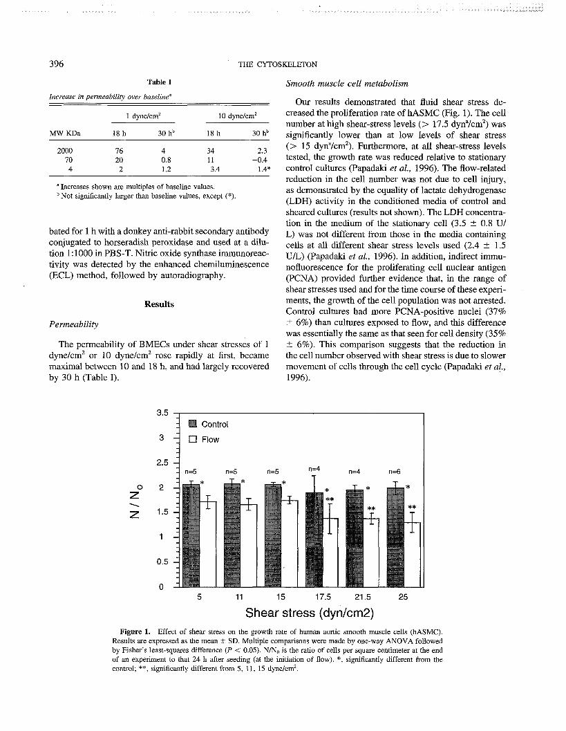
396 THE CYTOSKELETON
Table I Smooth muscle cell metabolism Increase in permeability over baseline"
" Increases shown are multiples of baseline values. Not significantly larger than baseline values, except (*).
bated for 1 h with a donkey anti-rabbit secondary antibody conjugated to horseradish peroxidase and used at a dilu- tion 1: 1000 in PBS-T. Nitric oxide synthase immunoreac- tivity was detected by the enhanced chemilurninescence (ECL) method, followed by autoradiography.
Results
Permeability
The permeability of BMECs under shear stresses of 1 dyne/cm2 or 10 dyne/cm2 rose rapidly at first, became maximal between 10 and 18 h, and had largely recovered by 30 h (Table I).
Our results demonstrated that fluid shear stress de- creased the proliferation rate of hASMC (Fig. 1). The cell number at high shear-stress levels (> 17.5 dyna/cm2) was significantly lower than at low levels of shear stress (> 15 dyna/cm2). Furthermore, at all shear-stress levels tested, the growth rate was reduced relative to stationary control cultures (Papadaki et al., 1996). The flow-related reduction in the cell number was not due to cell injury, as demonstrated by the equality of lactate dehydrogenase (LDH) activity in the conditioned media of control and sheared cultures (results not shown). The LDH concentra- tion in the medium of the stationary cell (3.5 t 0.8 U/ L) was not different from those in the media containing cells at all different shear stress levels used (2.4 +- 1.5 U/L) (Papadaki et al., 1996). In addition, indirect immu- nofluorescence for the proliferating cell nuclear antigen (PCNA) provided further evidence that, in the range of shear stresses used and for the time course of these experi- ments, the growth of the cell population was not arrested. Control cultures had more PCNA-positive nuclei (37% t 6%) than cultures exposed to flow, and this difference was essentially the same as that seen for cell density (35% 5 6%). This comparison suggests that the reduction in the cell number observed with shear stress is due to slower movement of cells through the cell cycle (Papadaki et al., 1996).
Shear stress (dynfcm2) Figure 1. Effect of shear stress on the growth rate of human aortic smooth muscle cells (hASMC).
Results are expressed as the mean t SD. Multiple comparisons were made by one-way ANOVA followed by Fisher's least-squares difference (P < 0.05). NIN, is the ratio of cells per square centimeter at the end of an experiment to that 24 h after seeding (at the initiation of flow). *, significantly different from the control; **, significantly different from 5, 11, 15 dyne/cm2.

BIOLOGICAL INTERACTIONS OF THE CYTOSKELETON
80
Time of flow exposure (hours)
Figure 2. Nitrite production in human aortic smooth muscle cells in the presence of shear stress. Results are expressed as mean SEM. Multiple comparisons were made by one-way ANOVA followed by Fisher's least-squares difference (P < 0.05); (n = 7-9 for control cultures). *, significantly different from the control; t, significantly different from 5 dyn/cm2.
The effects of flow on nitrite production by hASMC are shown on Figure 2. Shear stress significantly increased nitrite levels in the conditioned media, whereas the levels present in the stationary control cells were almost unde- tectable (Papadaki et al., 1998). Cumulative nitrite pro- duction in conditioned media increased with the duration of flow exposure and with shear. However, nitrite produc- tion rates were inversely correlated with time. An initial burst in nitrite production rate, detected as early as 1 h after flow exposure, was followed by a gradual decrease with time. Similar experiments with endothelial cells have shown similar trends in nitrite production rates (Kuchan and Frangos, 1994). This result was the first indication that the NOS isoform responsible for shear-induced NO production is not NOS 11; i.e., activation of NOS I1 results in sustained nitrite production rates for extended periods (up to 72 h after addition of stimulant). Treatment with 100 pM NG-amino-L-arginine (L-NAA) for 30 min before . and during exposure to shear stress (25 dynes/cm2) com- pletely abolished the flow-induced release of nitrite with- out affecting release from stationary cultures. L-NAA is a potent amino-substituted NOS inhibitor (Kuchan and Frangos, 1994). The complete inhibition of the nitrite signal in the presence of inhibitor provided evidence that the NO production in response to shear stress comes from the enzymatic reaction of L-arginine to L-citrulline, and is not a result of cell debris.
To identify the NOS isoform involved, cultures were incubated with 1 pM dexamethasone (DM) 24 h before and during exposure to shear stress (Papadaki et al., 1998). DM is a steroid that blocks transcription of NOS L1 by interfering with the binding of transcription factors to the promoter region of the gene (Rees et al., 1990). Dexamethasone had no effect on the nitrite levels in either the controls or the flow cultures. This result provided further evidence that NOS I1 plays no role in flow-induced nitrite production by hASMC. Monoclonal or polyclonal antibodies against NOS 11 showed no imrnunoreactivity with Western blot analysis (Fig. 3), which verified that the inducible isoform was not present, either in control or sheared hASMC cultures. On the other hand, polyclonal antibodies against the constitutively expressed isoform of neuronal NOS (NOS I) gave specific products in all stationary control and flow samples (Fig. 3). The intensi- ties of the control and shear NOS I bands are identical at 6 h, indicating that cultured hASMC express a constitu- tive NOS I protein whose enzymatic activity, rather than the amount of protein, is modulated by shear stress. Endo- thelial NOS (NOS 111) was found in neither control nor sheared hASMC (Fig. 3).
Summary and Conclusions
BMECs in static culture have a swirling spindoidal morphology. After exposure to flow for 10-18 h, BMECs

1) NOS ll
THE CYTOSKELETON
2) NOS I l I
8 \B 3 $4 3) NOS I 6"' .\" .$Q #a C \" ,+Q\~&
+o@ GoQ $6 , d) *b+ $3 **
Figure 3. Western blot of nitric oxide synthase (NOS) protein in total cell lysates of hASMC. (1) Blot was incubated with a monoclonal NOS II antibody. Lane 1, positive control for NOS II (human glioblastoma cell line, incubated with cytokines); lanes 2-4, representative samples of a 6-h experiment; lanes 5-7, representative samples of a 24-h experiment. (2) Blot was incubated with a monoclonal NOS III antibody. Lane 1, positive control for NOS Ill (human endothelial cells); lanes 2-4, representative samples of a 6- h experiment; lanes 5-7, representative samples of a 24-h experiment. (3) Blot was immunoblotted with a monoclonal NOS I antibody. Lane 1, positive control for NOS I (rat pituitary tumor cell line); lanes 2- 4, representative samples of a 6-h experiment; lanes 5-7, representative samples of a 24-h experiment; lane 8, human endothelial cell (EC) lysate used as a negative control.
appeared rounded with no preferred orientation. Further application of 10 dynes/cm2, but not 1 dyne/cm2, induced the cells to elongate in the direction of flow (data not shown). We demonstrated that BMECs initially respond to either of 1 dyne/cm2 or 10 dynes/cm2 shear stress with a dramatically increased macromolecular permeability. Maximum permeabilities were obtained between 10 and 18 h in the shear field for both shear rates, and these timepoints corresponded to the most rounded morphol- ogy. Continued application of the shear field led to a partial recovery in the permeability of the cerebral endo- thelial cells to macromolecules. The initial increase in permeability and the recovery was most dramatic for the higher molecular weight dextran markers.
We have also shown that the flow-induced shear
stress stimulates NO production in hASMC due to acti- vation of a constitutively expressed NOS I enzyme. The constitutive expression of NOS I in SMC, and its con- comitant activation by flow-induced shear stress, may play a regulatory role in the blood vessel wall in the 'absence of endothelium due to vascular injury. Shear- induced NO production from vascular SMC may inhibit excessive adhesion of platelets and other inflammatory molecules at the injury site, and may regulate the release of mitogenic factors by activated blood cells. In vascular wall homeostasis, constitutive NO production by under- lying SMC, modulated due to transmural flow, may act in concert with endothelial-cell-derived NO to regulate vascular tone and maintain a non-proliferative pheno- type for SMC.
BIOLOGICAL INTERACTIONS OF THE CYTOSKELETON 399
Literature Cited Casnocha, S. A., S. G. Eskin, E. R. Hall, and L. V. McIntire. 1989.
Permeability of human endothelial cell monolayers: effect of vaso- active agonists and CAMP. J. Appl. Physiol. 87: 1997-2005.
Kohler, T. R., and A. Jawien. 1992. Flow affects development of intimal hyperplasia after arterial injury in rats. Arterioscler. Z'hromb. 12: 963-971.
Kohler, T. R., T. R. Kirkman, L. W. Krais, B. K. Ziegler, and A. W. Clowes. 1991. Increased blood flow inhibits neointimal hyperpla- sia in endothelialized vascular grafts. Circ. Res. 69: 1557-156.
Koprowski, H., and H. Maeda. 1995. The Role of Nitric Oxide in Physiology and Pathophysiology. Springer-Verlag, New York.
Kuchan, M. J., and J. A. Frangos. 1994. Role of calcium and cal- modulin in flow-induced nitric oxide production in endothelial cells. Am. J. Physiol. 35: C628-C636.
McIntire, L. V. 1994. Bioengineering and vascular biology. Ann. Bio- med. Eng. 22: 2- 13.
Misko, T. P., R. J. Schilling, D. Salvemini, W. M. Moore, and M. G. Currie. 1993. A fluorometric assay for the measurement of nitrite in biological samples. Anal. Biochem. 214: 1-6.
Nollert, M. U., S. L. Diamond, and L. V. McIntire. 1991. Hydrody- namic shear stress and mass transport modulation of endothelial cell metabolism. Biotechnol. Bioeng. 38: 588-602.
Papadaki, M., and S. G. Eskin. 1997. Effects of fluid shear stress on gene replication in vascular cells. Biotechnol. Prog. 13: 209- 221.
Papadaki, M., L. V. McIntire, and S. G. Eskin. 1996. Effects of shear stress on the growth kinetics of human aortic smooth muscle cells in vitro. Biotechnol. Bioeng. 50: 555-561.
Papadaki, M., R. G. Tilton, S. G. Eskin, and L. V. McIntire. 1998. Effects of shear stress on nitric oxide production by human aortic smooth muscle cells. Am. J. Phys. 274: H616-H626.
Rees, D. D., R. M. J. Palmer, and S. Moncada. 1990. Dexametha- sone prevents the induction by endotoxin of a nitric oxide synthase and the associated effects on vascular tone. an insight into endotoxin shock. Biochem. Biophys. Res. Com. 173: 541-547.
Schwartz, R. S. 1993. Coronary Resfenosis. Blackwell Scientific, Cambridge, MA.
Sessa, W. C. 1994. The nitric oxide synthase family of proteins. J. Vas. Res. 31: 131-143.
Wagner, J. E., L. V. McIntire, and P. A. Whitson. 1997. Macro- molecular permeability of bovine brain endothelial cells monolayers subjected to shear stress in vitro. Am. J. Physiol. (in press).
Wang, D. M., and J. M. Tarbell. 1995. Modeling interstitial flow in an artery wall allows estimation of wall shear stress on smooth muscle cells. J. Biomech. Eng. 117: 358-363.
Discussion
LUNA: Did YOU say that there is a 36-fold increase for high molecular weight species and only a 2-fold increase for the low molecular weights?
MCINTIRE: Yes, 36- to 70-fold for high molecular weight species and 2- to 4-fold for low molecular weight species.
LUNA: Can you speculate on what kind of hole would be opening up that would allow the bigger molecules through, or is it some kind of transcytosis through the cell?
MCINTIRE: We think that there is a cytoskeletal rearrange- ment that leads to a change in the junction integrity. This would allow larger molecular weight species through, whereas the lower molecular weight species are going through relatively fast anyway.
FUJIWARA: DO YOU have any explanation for the fact that the mololayer with the round cell shape has increased permeability?
MCINTIRE: Those time points happen to coincide with the high flow. At the lower shear stress we see even bigger changes in permeability; but we don't see changes in gross cell shape because we don't see any realignment at very low stress. I assume this to mean there are various cytoskeletal rearrange- ments occurring even at the low stresses, but they don't lead to alignment in the direction of the flow, because the flow forces aren't forcing the cells to do that. I don't know why the maximum permeability occurs for that rounded shape at that particular time.
FUJIWARA: We are looking at the motility of cells in the monolayer, and when cells are round they move at lot more. Perhaps movement breaks the cell-cell adhesion more fre- quently than in the aligned area, contributing to the increased permeability.
MCINTIRE: Yes, that's possible.

Reference: Biol. Bull. 194: 400-401. (June, 1998)
Identification of a Functional Domain in Laminin-5
SCOTT E. BAKER* AND JONATHAN C. R. JONES?
Department of Cell and Molecular Biology, Northwestern University Medical School, 303 E. Chicago, Chicago, Illinois 60611
Laminin-5, a trimer composed of a3, P3, and y2 sub- units, is an important component of epithelial basement membranes and is spatially associated with cell-substrate adhesion structures called hemidesmosomes (Rouselle et al., 1991 ; Jones et al., 1994). Hemidesmosomes are asso- ciated with keratin filaments and are therefore considered linkers of the extracellular matrix and the intermediate filament cytoskeleton (Jones et al., 1994).
A laminin-5-rich matrix is capable of inducing rapid cell adhesion as well as hemidesmosome assembly in a squamous cell carcinoma line (SCC12) (Langhofer et al., 1993; Baker et al., 1996). Two members of the integrin family of cell matrix receptors, a3P1 and a6P4, bind laminin-5 and are involved in these processes (Carter et al., 1991; Langhofer et al., 1993). a301 integrin is in- volved in rapid cell adhesion to laminin-5, whereas a604 is involved in hemidesmosome formation and cell spread- ing on laminin-5-rich substrates (Jones et al., 1991; Lang- hofer et al., 1993; Baker et al., 1997). We have taken an immunological approach to defining the domains of laminin-5 involved in epithelial cell-substrate interac- tions.
A panel of monoclonal antibodies to the a3, 03, and y2 subunit chains of the laminin-5 heterotrimer was gen- erated (Langhofer et al., 1993; Plopper et al., 1996). Two of these antibodies, 5C5 and CM6, recognized the a 3
laminin-5 subunit (Langhofer et al., 1993; Plopper et al., 1996; Baker et al., 1996). We assessed, by electron mi- croscopy, the ability of these antibodies to block laminin- 5-induced hemidesmosome assembly in SCCl2 cells. When the laminin-5 matrix was treated with CM6 anti- body, SCC12 cells plated on this substrate lacked hemidesmosomes (Baker et al., 1996). This indicated that CM6 antibodies block the ability of laminin-5 to nucleate hemidesmosome formation in SCC12 cells. In contrast, when SCC12 cells were plated onto 5C5 antibody-treated matrix, there were many hemidesmosomes at the basal aspect of these cells (Baker et al., 1996). These results indicate that the epitope recognized by the CM6 antibod- ies plays an important role in hemidesmosome formation.
To characterize the epitopes of the CM6 and 5C5 anti- bodies, laminin-5 was purified from conditioned medium of epithelial cells, incubated with each antibody, and ana- lyzed by rotary shadowing. Purified non-antibody-treated laminin-5 is a Y-shaped molecule with a long (about 110 nm) arm (Baker et al., 1996). Rotary shadowed im- ages of laminin-5 incubated with the 5C5 antibody showed that this antibody bound to the long rod domain of laminin-5. Interestingly, the function-blocking antibody, CM6, localized to the globular or G domain of the laminin-5 heterotrimer (Baker et al., 1996).
Our results indicate that the G domain of laminin-5 appears to be essential for both epithelial cell interaction aid nucleation of hemidesmosome assembly. This con-
* Present address: Dept. Molecular and Cellular Biology, Life Sci- clusion is consistent with studies of laminin-1, whose G ences South Bldg, University of Arizona, Tucson AZ 85721.
t Author for correspondence. domain has also been shown to mediate a number of This paper was originally presented at a workshop titled The Cytoskel- important processes (Skubitz et al., 1991 ; Yurchenco et
eton: Mechanical, Physical, and Biological Interactions. The workshop, aA. 1993: Matter and Laurie. 1994). Our studv ~rovides e L
which was held at the Marine Biological Laboratory, Woods Hole, the first direct evidence that the G domain of laminin-5 Massachusetts, from 15-17 November 1996, was sponsored by the Center for Advanced Studies in the Space Life Sciences at MBL and is not only a structural component of the extracellular funded by the National Aeronautics and Space Administration under matrix-c~tOskeletOn link, but a player in its Cooperative Agreement NCC 2-896. formation.

BIOCHEMICAL PROPERTIES OF THE CYTOSKELETON 40 1
Acknowledgments
This work was supported by NIH and ACS.
Literature Cited
Baker, S. E., S. B. Hopkinson, M. Fitchmun, G. L. Andreason, F. Grasier, G. Plopper, V. Quaranta, and J. C. R. Jones. 1996. Laminin-5 and hemidesmosomes: role of the a 3 subunit in hemides- mosome stability and assembly. J. Cell Sci. 109: 2509-2520.
Baker, S. E., 0. Skalli, R. D. Goldman, and J. C. R. Jones. 1997. Laminin-5 and modulation of keratin cytoskeleton arrangement in FG pancreatic carcinoma cells: involvement of PAP300 and evi- dence that laminin-5Icell interactions correlate with a dephosphory- lation of a6A integrin. Cell Motil. Cytoskel. 37: 271 -286.
Carter, W. G., M. C. Ryan, and P. J. Gahr. 1991. Epiligrin, a new cell adhesion ligand for integrin a3p l in epithelial basement mem- branes. Cell 65: 599-610.
Jones, J. C. R., J. Asmuth, S. E. Baker, M. Langhofer, S. I. Roth, and S. B. Hopkinson. 1994. Hemidesmosomes: extracellular ma- trixlintermediate filament connectors. Exp. Cell Res. 213: 1 - l l .
Jones, J. C. R., M. A. Kurpakus, H. M. Cooper, and V. Quaranta.
1991. A function for the integrin alpha 6 beta 4 in the hemidesmo- some. Cell Reg. 2: 427-438.
Langhofer, M., S. B. Hopkinson, and J. C. R. Jones. 1993. The ma- trix secreted by 804G cells contains laminin-related components that participate in hemidesmosome assembly in vitro. J. Cell Sci. 105: 753-764.
Matter, M. L., and G. W. Larurie. 1994. A novel laminin E8 cell adhesion site required for lung alveolar formation in vitro. J. Cell Biol. 124: 1083- 1090.
Plopper, G., J. Falk-Marzillier, S. Glaser, M. Fitchmun, G. Gian- nelli, T. Romario, J. C. R. Jones, and V. Qnaranta. 1996. Changes in expression of monoclonal antibody epitopes on laminin- 5r induced by cell contact. J. Cell Sci. 109: 1965-1973.
Rousselle, P., G. P. Lunstrum, D. R. Keene, and R. E. Burgeson. 1991. Kalinin: an epithelium-specific basement membrane adhe- sion molecule that is a component of anchoring filaments. J. Cell Biol. 12: 205-214.
Skubitz, A. P., P. C. Letourneau, E. Wayner, and L. T. Furcht. 1991. Synthetic peptides from the carboxy-terminal domain of the A chain of laminin: their ability to promote cell adhesion and neurite out- growth, and interact with heparin and the beta 1 integrin subunit. J. Cell Biol. 115: 1137-1148.
Yurchenco, P. D., U. Sung, M. D. Ward, Y. Yamada, and J. J. O'Rear. 1993. Recombinant laminin G domain mediates myoblast adhesion and heparin binding. J. Biol. Chem. 268: 8356-8365.
Reference: Biol. Bull. 194: 402-405. (June, 1998)
Interaction of Virnentin With Actin and Phospholipids
JAGESH V. SHAH*,?, LOUISE Z. WANG?, PETER TRAUB*, AND PAUL A. JANMEY?
*Haward-MIT Division of Health Sciences and Technology, Division of Experimental Medicine, Brigham and Women's Hospital, Haward Medical School, Boston, Massachusetts;
and #Max-Planck Institute for Cell Biology, Heidelberg, Germany
Vimentin intermediate filaments are a major cytoskele- and the nucleus. Here we present in vitro studies indicat- tal constituent of cells of mesenchymal origin. They have ing that vimentin interacts directly with both F-actin and been colocalized with a variety of intracellular structures polyphosphoinositide Lipids. such as actin filaments and the plasma membrane. La- beled actin filaments, observed in vitro by fluorescence microscopy, break in the presence of polymerizing vimen- Materials and Methods
tin; the time course is consistent with stopped-flow mea- PuriJication of actin and vimentin
surements of vimentin polymerization. This breakage phenomenon appears to be specific for vimentin. Inhibi- tion of vimentin network formation was observed with phosphatidyl inositol phosphate (PI(4)P) and phosphati- dyl inositol bisphosphate (PI(4,5)P2), but not phosphatidyl choline (PC), phosphatidyl serine (PS), or phosphatidyl inositol (PI). Taken together, these results indicate a spe- cific interaction of vimentin with F-actin and polyphos- phoinositide lipids.
Introduction
Vimentin-type intermediate filaments are a major cy- toskeletal constituent of cells of mesenchymal origin. Theories as to their function vary from maintenance of cellular integrity (Lazarides, 1980) to gene regulation (Traub and Shoeman, 1994). There is in vivo evidence for vimentin colocalization with other cytoskeletal elements, such as actin (Brown and Binder, 1992; Cary et al., 1994; and Tint et al., 1991) and microtubules (Gurland and
Actin was purified by the method of Spudich and Watt (1971) with slight modifications. Actin was stored in G- buffer (2 mM Tris, 0.2 mM CaC12, 0.5 mM ATP, 0.5 mM DTT, pH 8.0) at -80°C; it was polymerized at a concentration of 5 pM by the addition of a ten-times concentrated solution of F-buffer (1X:20 mM Tris, 150 mM KC1, 2 mM MgC12, 0.2 mM CaC12, 0.5 mM ATP, 0.5 mM DTT, pH 7.4) and was stabilized by the addition of equimolar TRITC-phalloidin (Sigma Chemicals, St. Louis, MO). Vimentin was purified from Ehrlich ascites tumor cells by the method of Nelson et a1 (1982). Vimen- tin was extensively dialyzed against non-polymerizing buffer (10 mM Tris, 6 mM DTT, pH 7.6) to remove residual urea and was polymerized by the addition of KC1 to 150 mM. All reagents were purchased from Sigma Chemicals (St. Louis, MO).
Gunderson, 1995; Gyoeva and Gelfand, 1991), as well as F-actin-vimentin interactions
with cellular organelles, such as the plasma membrane
F-actin, under either polymerizing or non-polymerizing This paper was originally presented at a workshop titled The Cytoskel- conditions, and stabilized by TRITC-phalloidin (10 nM),
eton: Mechanical, Physical, and Biological Interactions. The workshop, Was visualized by fluorescence microscopy in the pres- which was held at the Marine Biological Laboratory, Woods Hole, ence of 20 p~ unpolymerized vimentin. A large number Massachusetts, from 15-17 November 1996, was sponsored by the Center for Advanced Studies in the Space Life Sciences at MBL and were recorded to videotape Over and the funded by the National Aeronautics and Space Administration under average length of F-actin-based on at least 200 f ih lent Cooperative Agreement NCC 2-896. traces-was calculated.

BIOCHEMICAL PROPERTIES OF THE CYTOSKELETON
Figure 1. With increasing time, the average length of F-actin decreases in the presence of polymerizing vimentin. Labeled, phalloidin-stabilized actin filaments (10 nM) are visible by fluorescence microscopy in a sample containing I8 pM (1 mglml) unlabeled vimentin before and after addition of KC1 to initiate vimentin polymerization. The dimension of each image is 50 pm in width, and the image in panel 4 was taken 25 min after polymerization.
Rheology of vimentin networks
Rheology of vimentin networks was carried out as de- scribed previously (Janmey et al., 1991). Oscillatory mea- surements of elastic modulus were made with a Rheome- trics RFS I1 fluids spectrometer (Rheometrics, Piscata- way, NJ) at a frequency of 1 radls and a strain amplitude of 1%.
Results
F-actin-vimentin interactions
Labelled actin filaments were examined in vitro by fluorescence microscopy and were seen to break in the presence of polymerizing vimentin. Figure 1 shows a panel of four video frames; the fluorescent filaments are F-actin in a matrix of polymerizing unlabeled vimentin filaments. Such breakage was not seen under non-poly- merizing conditions, indicating that polymerization is re- quired. The decrease in average F-actin length is shown
18 FM Polymerizing 43 7 pM Polymerizing m- 1 G O
7 pM Non-polymerizing 0 4
1-3 0.8
3; u 0 0.6
- 0 0 5 10 15 20 25 30 35 40
Time [min]
Figure 2. Average length of F-actin decreases in the presence of polymerizing vimentin filaments. Higher concentrations of vimentin re- sult in faster rate of length decrease. Unpolymerized vimentin does not cause a decrease in F-actin length.

THE CYTOSKELETON
0 500 1000 1500 2000 2500 3000
Time [see]
Figure 3. Rheology of vimentin networks disrupted by polyphosphoinositide lipids. Other phospholip- ids, such as phosphatidyl serine, phosphatidyl choline, and phosphatidyl inositol, had no significant effect on vimentin polymerization.
in Figure 2 as a function of vimentin polymerization and PIP and PIP, inhibit the polymerization of vimentin as concentration. The time course is consistent with stopped measured by rheological methods. This evidence points flow measurements of vimentin polymerization (data not to a specific interaction between vimentin and polyphos- shown). This breakage phenomenon appears to be specific phoinositide lipids. for vimentin, since no breakage is seen with microtubules in actin, or with actin in microtubules or fibrin (data not Acknowledgments shown).
Vimentin-phospholipid interactions
The interaction of vimentin and phospholipids was measured by rheological methods and showed that poly- phosphoinositide lipids inhibit the formation of an elastic network. Inhibition was observed with PI(4)P and PI(4,5)P2, and to a lesser extent with PI (Fig. 3). Inhibition was not observed with PC or PS. These results are consis- tent with early studies by Perides et al. (1986) showing . that phospholipid vesicles, especially those containing PIP and PIP,, inhibit vimentin polymerization and depo- lymerize preformed vimentin filaments.
Conclusions
Fluorescent actin filaments decrease in length in the presence of polymerizing vimentin. Filament breakage was not observed in other biopolymer systems, indicating that the interaction is specific.
The authors thank Jay Tang, Zeno Guttenburg, and Wolfgang Goldman for performing the kinetic stopped- flow measurements. This work was sponsored by the NIH grant AR38910 (P.A.J.) and the National Science and Engineering Research Council of Canada (J.V.S.).
Literature Cited
Brown, K. D., and L. I. Binder. 1992. Identification of the intermedi- ate filament-associated protein gyronemin as filarnin. Implications for a novel mechanism of cytoskeletal interaction. J. Cell Sci. 102: 19-30.
Cary, R. B., M. W. Klymkowsky, R. M. Evans, A. Domingo, J. A. Dent, and L. E. Backhus. 1994. Vimentin's tail interacts with actin-containing structures in vivo. J. Cell Sci. 107: 1609- 1622.
Gurland, G., and G. G. Gundersen. 1995. Stable, detyrosinated mi- crotubules function to localize vimentin intermediate filaments in fibroblasts. J. Cell Biol. 131: 1275 - 1290.
Gyoeva, F. K., and V. I. Gelfand. 1991. Coalignment of vimentin intermediate filaments with microtubules depends on kinesin. Nature 353: 445-448.
Janmey,P. A., U. Euteneuer,P. Traub,andM. Schliwa. 1991. Visco-

BIOCHEMICAL PROPERTIES OF THE CYTOSKELETON 405
elastic properties of vimentin compared with other filamentous bio- polymer networks. J. Cell Biol. 113: 155-160.
Lazarides, E. 1980. Intermediate filaments as mechanical integrators of cellular space. Nature 283: 249-256.
Nelson, W. J., C. E. Vorgias, and P. Traub. 1982. A rapid method for the large scale purification of the intermediate filament protein vimentin by single-stranded DNA-cellulose affinity chromatography. Biochem. Biophys. Res. Commun. 106: 1 141 - 1 147.
Perides, G., A. Scherbarth, and P. Traub. 1986. Influence of phos- pholipids on the formation and stability of vimentin-type intermedi- ate filaments. Eur. J. Cell Biol. 42: 268-280.
Spudich, J., and S. Watt. 1971. The regulation of rabbit skeletal muscle contraction. I. Biochemical studies of the interaction of the tropomyosin-troponin complex with actin and the proteolytic frag- ments of myosin. J. Biol. Chem. 246: 4866-4871.
Tint, I. S., P. J. Hollenbeck, A. B. Verkhovsky, I. G. Surgucheva, and A. D. Bershadsky. 1991. Evidence that intermediate filament reorganization is induced by ATP-dependent contraction of the acto- myosin cortex in permeabilized fibroblasts. J. Cell Sci. 98:375-384.
Traub, P., and R. L. Shoeman. 1994. Intermediate filament and re- lated proteins: potential activators of nucleosomes during transcrip- tion initiation and elongation? Bioessays 16: 349-355.

Reference: Biol. Bull. 194: 406-408. (June, 1998)
Two Distinct Mechanisms of Actin ,733 - 91 . Bundle Formation / i
JAY X. TANG AND PAUL A. JANMEY 3 /? L
Brigham and Women's Hospital, Haward Medical School, 221 Longwood Ave., LMRC 301, Boston, Massachusetts 021 1.5
Actin filaments (F-actin)-one of the major types of cytoskeletal filament-can be induced to form bundles by the addition of any of a number of polycations, including divalent metal ions, trivalent hexaminecobalt, or basic polypeptides. The general features of bundle formation, as detected by light scattering, centrifugation, optical and electron microscopy, are largely independent of the spe- cific structure of the bundling agent used. The formation of lateral aggregates of actin filaments in response to polycations begins at a threshold concentration that varies strongly with the valence of the cation and increases with the ionic strength of the solution. Polyanions, such as nucleoside phosphates and acidic polypeptides, disperse actin bundles into single filaments. These features are similar to those associated with DNA condensation and can be explained analogously by polyelectrolyte theory (Tang and Janmey, 1996; Tang et al., 1996). The general behavior is dictated by the polyelectrolyte nature of F- actin, which causes a class of nonspecific binding by ligands that carry several net, opposite charges. Such a bundling mechanism can be applied to a class of cationic actin-bundling proteins, including smooth muscle cal- ponin and the microtubule-associated proteins tau and Map2c.
One direct consequence of this model of bundling is that neither dual binding sites nor dimerization of a pro- tein with a single binding site is required to bundle F- actin (Tang et al., 1997b). This alternative and somewhat
This paper was originally presented at a workshop titled The Cytoskel- eton: Mechanical, Physical, and Biological Interactions. The workshop, which was held at the Marine Biological Laboratory, Woods Hole, Massachusetts, from 15-17 November 1996, was sponsored by the Center for Advanced Studies in the Space Life Sciences at MBL and funded by the National Aeronautics and Space Administration under Cooperative Agreement NCC 2-896.
counterintuitive concept may help explain why some ac- tin-bundling and cross-linking proteins have only a single identifiable actin-binding site, and the purified proteins exist in solution as monomers. Smooth muscle calponin and a 25-amino-acid actin-binding peptide (aa 151- 175) derived from the myristoylated, alanine-rich, C kinase substrate (MARCKS) are two examples among poly- cations that appear to induce actin-bundle formation mainly by an electrostatic mechanism (Tang and Janmey, 1996; Tang et al., 1997b).
A different type of actin bundle is formed by solution crowdedness, the thermodynamic basis of which is to maximize the entropy of the solution, including all solute and solvent molecules. Two equivalent terms-the ex- cluded volume effect (Onsager, 1949) and steric exclusion (Arakawa and Timasheff, 1985)-have been used in the literature to describe the restriction that two macromole- cules cannot overlap their positions in solution. At high concentrations, such a constraint may lead to various forms of self-assembly or other aggregation phenomena (Herzfeld, 1996). For the case of an F-actin solution, high concentrations of noninteracting proteins like ovalbumin, and inert polymers like polyethylene glycol, facilitate lat- eral aggregation of F-actin (Suzuki et al., 1989). This type of actin-bundle formation has different features than that induced by polycations, including an opposite depen- dence on the ionic strength of the solution (Tang et al., 1997a) and an opposite dependence on the concentration of actin (Suzuki et al., 1989; Tang et al., 1997a; Tang and Janmey, 1996), which by itself contributes to the solution crowdedness.
The optical images in Figure 1 show examples of actin bundles formed by the two different mechanisms. Panel A shows bundling induced by 50 mM MgC12, representa- tive of excess polycation concentration. Panel B shows bundles induced by 8% polyethylene glycol [molecular

BlOCHEMICAL PROPERTIES OF THE CYTOSKELETON 407
Figure 1. Comparison of actin bundles formed by excess polycations (A) and by solution crowdedness (B). Panel A: large bundles of F-actin induced by 50. mM MgCl,. Panel B: actin bundles formed by 8% (wt) polyethylene glycol of average molecular weight 8000 dalton (PEG-8000).
weight approximately 8000 (PEG-8000)], illustrating the only subtle difference is that the Mg-actin bundles tend effect of solution crowding. Bundles of both types were to reach a larger size than PEG-actin bundles, and the visualized by the fluorescent labeling of F-actin with rho- difference is more apparent by phase-contrast microscopy damine phalloidin. The pair of images illustrates the gen- (data not shown). Both types of actin bundles have also era1 observation that the two types of bundles are virtually been examined by electron microscopy, and the general identical in appearance at this level of resolution. The morphology is indistinguishable. In cells, various poly-

408 THE CYTOSKELETON
cations and cationic proteins are abundant, and the cyto- plasm is also crowded with other macromolecules that may or may not directly interact with F-actin. Therefore, the two distinct mechanisms may jointly play pivotal roles in forming the functional arrays of bundled actin filaments found in many cell types.
Literature Cited
Arakawa, T., and S. N. Timasheff. 1985. Mechanism of poly(ethy1- ene glycol) interaction with proteins. Biochemistry 24: 6756-6762.
Herzfeld, J. 1996. Entropically driven order in crowded solutions: from liquid crystals to cell biology. Accounts Chem. Res. 29: 31- 37.
Onsager, L. 1949. The effects of shape on the interaction of colloidal particles. Ann. NY Acad. Sci. 51: 627-659.
Suzuki, A., M. Yamazaki, and T. Ito. 1989. Osmoelastic coupling in biological structures: formation of parallel bundles of actin fila- ments in a crystalline-like structure caused by osmotic stress. Bio- chemistry 28: 6513-6518.
Tang, J. X., and P. A. Janmey. 1996. Polyelectrolyte nature of F- actin and mechanism of actin bundle formation. J. Biol. Chem. 271: 8556-8563.
Tang, J. X., S. Wong, P. T. Tran, and P. A. Janmey. 1996. Counter- ion induced bundle formation of rodlike polyelectrolytes. Ber. Bun- senges. Phys. Chem. 100: 796-806.
Tang, J. X., T. Ito, T. Tao, P. Traub, and P. A. Janmey. 1997a. Opposite effects of electrostatics and steric exclusion on bundle formation by F-actin and other filamentous polyelectrolytes. Bio- chemistry 36: 12600- 12607.
Tang, J. X., P. Szymanski, P. A. Janmey, and T. Tao. 1997b. Elec- trostatic effects of smooth muscle calponin on actin assembly. Eur. J. Biochem. 247: 432-440.

Reference: Biol. Bull. 194: 409-410. (June, 1998)
Plectin Sidearms Mediate Interactions of 7 -c>, Intermediate Filaments With Microtubules and
Other Components of the Cytoskeleton $ ) P
TATYANA M. SVITKINA, ALEXANDER B. VERKHOVSKY, AND GARY B. BORISY
Laboratory of Molecular Biology, University of Wisconsin, Madison, Wisconsin 53706
The IF-associated protein plectin has been proposed to be a cytoplasmic cross-linker (Foisner and Wiche, 1991). We recently reported unique "millipede-like" structures in mammalian cell cytoskeletons revealed after removal of actin by treatment with gelsolin (Svitkina et al., 1995, 1996). Here we demonstrate, by immunogold labeling, that the millipede structures are composed of cores of vimentin IFs with sidearms containing plectin. The di- mensions of the sidearms (up to 200 nm long and 2-3 nm wide) match the size of plectin molecules (Foisner and Wiche, 1987), suggesting that sidearms are made of indi- vidual plectin molecules, or a few of them in register. These plectin sidearms connect IFs to microtubules, the actin-based cytoskeleton, and possibly to membrane com- ponents. Plectin binding to microtubules is significantly increased in cells from transgenic mice lacking IFs (MFT- 16 cell line, courtesy of Dr. R. Evans). Numerous side- arms are associated with microtubules in these cells, giv- ing them an unusual "hairy" appearance. After microin- jection of exogenous vimentin in MFT-16 cells, IFs of variable length are formed within 3 h; the IFs have a very high density of associated plectin, whereas microtubules lose most of their plectin sidearms. These results suggest the existence of a pool of plectin that preferentially associ- ates with IFs, but that may also be competed for by micro- tubules. The association of IFs with microtubules shows
This paper was originally presented at a workshop titled The Cytoskel- eton: Mechanical, Physical, and Biological Interactions. The workshop, which was held at the Marine Biological Laboratory, Woods Hole, Massachusetts, from 15-17 November 1996, was sponsored by the Center for Advanced Studies in the Space Life Sciences at MBL and funded by the National Aeronautics and Space Administration under Cooperative Agreement NCC 2-896.
no preference for Glu-tubulin. Nor does it depend upon the presence of MAP4; indeed plectin links were retained after specific immunodepletion of MAP4.
The IF-plectin complexes also displayed extensive as- sociation with stress-fibers. The actin-binding domain identified in the plectin sequence (McLean et al., 1996) may play a role in this interaction. However, plectin bind- ing to stress-fibers survives actin depletion by gelsolin, revealing structural association with myosin 11 minifila- ments, the major non-actin component of stress-fibers. These results suggest that myosin I1 minifilaments, or components closely associated with them, may play a role as plectin targets. An additional association partner of plectin seen in our cytoskeleton preparations consists of material with irregular granular and fibrillar substructure. This material, as yet unidentified, may represent protein remnants of membrane structures, such as plasma mem- brane or membrane organelles.
In conclusion, our results provide direct structural evi- dence for the hypothesis that IF-plectin complexes com- prise an extensive cross-linking of cellular components (Fig. 1) and provide a structural framework for the inte- gration of cytoplasm. In this model, IFs provide the core while plectin forms peripheral linkers that connect to MTs, the actin-based cytoskeleton, and membrane struc- tures. However, IFs and plectin do not seem to be equal partners in performing their functions. The absence of obvious phenotype in vimentin-null mice (Colucci-Guyon et al., 1994) suggests that vimentin's role in cytoplasmic organization is not essential. In contrast, plectin seems to play a key role in maintaining tissue integrity, as has been demonstrated by analysis of a human hereditary disorder (epidermolysis bullosa with muscular dystrophy) showing plectin deficiency (McLean et al., 1996; Smith et al.,

THE CYTOSKELETON
1996). Though plectin prefers to associate with IFs, it can cross-link cytoskeletal structures in the absence of vimentin, Consequently, plectin may be able to maintain cytoplasmic integrity independently of IFs.
a
- - - - - -- - - INTERMEDIATE FILAMENTS
MICROTUBULES - ACTW FILAMENTS - MYOSIN FILAMENTS
Figure 1. Diagram illustrating integration of the cytoskeleton. Plec- tin sidearms on IFs link them to microtubules, myosin I1 minifilaments, actin filaments, and putative membrane components, thus mechanically connecting various cellular structures.
Literature Cited
Colucci-Guyon, E., M.-M. Porteir, I. Dunia, D. Paulin, S. Pourin, and C. Babinet. 1994. Mice lacking vimentin develop and repro- duce without an obvious phenotype. Cell 79: 679-694.
Foisner, R., and G. Wiche. 1987. Structure and hydrodynamic prop- erties of plectin molecules. J. Mol. Biol. 198: 515-531.
Foisner, R., and G. Wiche. 1991. Intermediate filament-associated proteins. Curr. Opin. Cell Biol. 3: 75-81.
McLean, W. H. I., L. Pulkkinen, F. J. D. Smith, E. L. Rugg, E. B. Lane, F. Bullrich, R.E. Burgeson, S. Amano, D. Hudson, K. Owaribe, J. A. McGrath, J. R. McMillan, R. A. J. Eady, I. M. Leigh, A.M. Christiano, and J. Uitto. 1996. Loss of plectin causes epidermolysis bullosa with muscular dystrophy: cDNA clon- ing and genomic organization. Genes Devel. 10: 1724-1735.
Smith, F. J. D., R. A. J. Eady, I. M. Leigh, J. R. McMillan, E. L. Rugg, D. P. Kelsell, S. P. Bryant, N. K. Spurr, J. F. Geddes, G. Kirtschig, G. Milana, A. G. de Bono, K. Owaribe, G. Wiche, L. Pulkkinen, J. Uitto, W. H. I. McLean, and E. B. Lane. 1996. Plectin deficiency results in muscular dystrophy with epidermolysis buIlosa. Nature Genet. 13: 450-457.
Svitkina, T. M., A.B. Verkhovsky, and G. G. Borisy. 1995. Im- proved procedures for electron microscopic visualization of the cy- toskeleton of cultured cells. J. Struct. Biol. 115: 290-303.
Svitkina, T. M., A. B. Verkovsky, and G. G. Borisy. 1996. Plectin sidearms mediate interaction of intermediate filaments with microtu- bules and other components of the cytoskeleton. J. Cell Biol. 135: 991-1007.

Reference: Biol. Bull. 194: 411. (June, 1998)
Concluding Remarks
This has been an interesting few days for many of us who have not had the opportunity to meet each other in this kind of context before. It has forced us to think of other areas that we normally don't study and the complex- ity of those different systems. With respect to complexity, some of the terms that have been used in this meeting demonstrate that communication is one of the major prob- lems we face. However, the important prospect for this meeting is that it is the beginning of a series of multidisci- plinary communications that we hope NASA will perpet- uate, because they have an interesting niche to fill in the life sciences. I would therefore like to thank NASA for supporting this workshop.
In concluding, I would like to present a typical molecu- lar biologist's view of signalling in the cell. A signal is depicted by a lightning bolt which interacts with a recep- tor and is followed by a response within the nucleus. A cell biologist who works on microtubules depicts the cell as consisting primarily of microtubules; we have seen this many times at this meeting. Then there is the way I look at the cell-as containing intermediate filaments. Someone working with actin portrays a cell with only actin. We all use different antibodies and different probes, which suggests to our audience that there is only one major component inside the cell, because each of our images is filled with our own protein. In turn, each of us tries to explain signal transduction in light of our own interests.
We know, however, that there is a lot of cross-talk amongst these different elements. One of the most intrigu- ing things that has come up several times at this meeting is that a cell can be stimulated in various ways if it is stressed. Some refer to this stimulation simply as stress, while others call it signaling. There are also many ways to describe the response. Whether stimulated by heat shock, mechanical stress, or some toxic compound, cells behave
This paper was originally presented at a workshop titled The Cytoskel- eton: Mechanical, Physical, and Biological Interactions. The workshop, which was held at the Marine Biological Laboratory, Woods Hole, Massachusetts, from 15-17 November 1996, was sponsored by the Center for Advanced Studies in the Space Life Sciences at MBL and funded by the National Aeronautics and Space Administration under Cooperative Agreement NCC 2-896.
in interesting ways, from the moment that some signal impinges on the cell surface. I will just remind you of what Rick Morimoto told us very briefly at this meeting. That is, if you stress a cell by using heat shock or some poisonous divalent cation, or any of a whole spectrum of different agents, they all produce this kind of response in which cytoskeletal systems such as intermediate filaments collapse back to the nucleus, or actin paracrystals form inside the nucleus. These responses happen in minutes, and it takes time for the cell to recover. At the same time, heat-shock transcription factors, which are normal constituents of the cytoplasm, enter the nucleus to form structures inside it which alter gene expression in some way. At this meeting we heard about the effect of shear stress on endothelial cells. Peter Davies has shown, and Keigi Fujiwara described to us this morning, that an endo- thelial cell exposed to shear elongates, and stress fibers proliferate-or at least they get longer, more abundant, and thicker.
Any drug that reacts with one of the cytoskeletal ele- ments will change the others. However, if experiments are narrowly focused on one cytoskeletal element, the action of the drug appears to be very specific. I think we are learning that there is a lot of cross-talk amongst the cytoskeletal systems which form extremely complex in- teracting networks. Nevertheless, although a very close look at just actin in solution, or even solubilized actin containing interdigitated intermediate filaments, may seem simplistic, in fact, this is probably the only way to begin learning how these systems interact.
I think that cell biologists must begin to communicate with physicists if we are to get a handle on the exquisite sensitivity of these systems to external perturbations. Per- haps everything starts as a mechanical stimulus at the cell surface and is then transmitted through these elements to the nucleus and back again to the surface of the cell. I hope that this meeting will be only the first exchange in a dialog addressing these issues. I thank NASA again for giving us the opportunity to get together.
ROBERT D. GOLDMAN Woods Hole, Massachusetts November 1996

Reference: Biol. Bull. 194: 412. (June, 1998)
Published by Title Only
ALBRECHT-BUEHLER, GUENTER The conceptual challenge of cellular gravi-sensing.
BORISY, GARY G., AND VLADIMIR I. RODIONOV Self organization of cytoplasm.
JANSEN, N., J. MASHL, W. GELBART, AND R. BRUINSMA Electrostatic interactions and collapse of biopolymers.
KOWALCZYK, A. P., 3. E. BORGWARDT, E. A. BORN- SLAEGER, H. L. PALKA, A. S. DHALIWAL, AND K. J. GREEN
The amino-terminal domain of desmoplakin clusters des- mosomal cadherin-plakoglobin complexes: implica- tions for desmosome assembly and intermediate fila- ment attachment.
MORIMOTO, RICHARD I. Effects of stress and protein damage on the mammalian
cell shape and function.
OSCHMAN, JAMES L. The nuclear, cytoskeletal, and extracellular matrices: a
continuous communication network.
PALACEK, SEAN P., JOSEPH C. LOFTUS, MARK H. GINS- BERG, ALAN F. HORWITZ, AND DOUGLAS A. LAUFFEN- BURGER
Changes in cell-substratum adhesiveness regulate cell mi- gration rate.
SCHWARTZ, MARTIN A. Tho and C-Abl as mediators of integrin signaling.

Reference: Biol. Bull. 194: 413-415. (June, 1998)
Chairs and Speakers
Workshop Chair ROBERT D. GOLDMAN
Department of Cell, Molecular and Structural Biology Northwestern University Medical School
303 E. Chicago Avenue Chicago, IL 606 1 1
UELI AEBI Am Biozentrum der Universitat Base1 Klingelbergstr 70 Basel, CH-4056 Switzerland E-mail: [email protected]
J. CHLOE BULINSKI Department of Anatomy and Cell Biology Columbia University College of Physicians and Surgeons 630 W. 168th Street New York, NY 10032-3702 E-mail: [email protected]
SHU CHIEN GUENTER ALBRECHT-BUEHLER Department of Bioengineering Department of Cell, Molecular and Structural Biology University of California Northwestern University Medical School 9500 Gilman Drive 303 E. Chicago Avenue La Jolla, CA 92093-0412 Chicago, IL 606 1 1 E-mail: [email protected] E-mail: [email protected]
DAVID BOAL Department of Physics Simon Fraser University Burnaby, British Columbia Canada V5A 1S6 E-mail: [email protected]
GARY G. BORISY Department of Molecular Biology and Zoology University of Wisconsin 1525 Linden Drive Madison, WI 53706-15 14 E-mail: [email protected]
ROBIJN BRUINSMA Department of Physics University of California Los Angeles, CA 90024 E-mail: [email protected]
PIERRE A. COULOMBE Department of Biological Chemistry The Johns Hopkins University School of Medicine 725 N. Wolfe Street Baltimore, MD 21205 E-mail: [email protected]
GABOR FORGACS Department of Physics Clarkson University Potsdarn, NY 13676
' E-mail: [email protected]
KEIGI FUJIWARA Department of Structural Analysis National Cardiovascular Center Research Inst. Fujishiro-dai, Suita Osaka 565 Japan E-mail: [email protected]

414 THE CYTOSKELETON
KATHLEEN J. GREEN Department of Pathology Northwestern University Medical School 303 E. Chicago Avenue Chicago, IL 606 1 1 E-mail: [email protected]
FREDERICK C. MACKINTOSH Department of Physics 2071 Randall Lab University of Michigan Ann Arbor, MI 48 109-1 120 E-mail: [email protected]
LARRY V. MCINTIRE GREGG G. GUNDERSEN Institute of Biosciences and Bioengineering Department of Anatomy and Cell Biology Rice University Columbia University College of Physicians and Surgeons Houston, TX 7725 1 - 1892 630 W. 168th Street E-mail: [email protected] New York, NY 10032-3702 E-mail: ggg 1 @columbia.edu
DONALD INGBER Department of Surgical Research Enders 1007 Children's Hospital 300 Longwood Avenue Boston, MA 021 16-5737 E-mail: ingber@al .tch.harvard.edu
WALLACE IP Department of Cell Biology, Neurobiology, and Anatomy University of Cincinnati College of Medicine 23 1 Bethesda Avenue P.O. Box 670521 Cincinnati, OH 45267-0521 E-mail: [email protected]
PAUL JANMEY Experimental Medicine Division Brigham and Women's Hospital LMRC #302 221 Longwood Avenue Boston, MA 021 15 E-mail: [email protected]
JONATHAN C. R. JONES Department of Cell and Molecular Biology Northwestern University Medical School 303 E. Chicago Avenue, Ward Building Chicago, IL 6061 1 E-mail: [email protected]
ELIZABETH LUNA University of Massachusetts Medical Center Worcester Foundation Campus 222 Maple Avenue Shrewsbury, MA 01545-2795 E-mail: [email protected]
RICHARD I. MORIMOTO Department of Biochemistry, Molecular, and Cell
Biology Northwestern University 2159 N. Campus Drive Evanston, IL 60208-3500 E-mail: [email protected]
CAROL A. OTEY Department of Anatomy and Cell Biology University of Virginia School of Medicine - Box 439 Charlottesville, VA 22908-0439 E-mail: [email protected]
MARTIN A. SCHWARTZ Department of Vascular Biology VB-4 Scripps Research Institute 10666 N. Torrey Pines Road La Jolla, CA 92037 E-mail: [email protected]
PETER M. STEINERT Laboratory of Skin Biology NIAMSINIH Building 6, Room 425 Bethesda, MD 20892-2755 E-mail: [email protected]
MURRAY STEWART Medical Research Council Laboratory of Molecular Biology MRC Centre Hills Road Cambridge United Kingdom CB2 2QH E-mail: [email protected]
EDWIN TAYLOR Dept. of Molec. Genetics and Cell Bio. University of Chicago 920 E 58th Street Chicago, IL 60637 E-mail: ewtl @midway.uchicago.edu

CHAIRS AND SPEAKERS
GERHARD WICHE Yu-LI WANG Institute of Biochemistry and Molecular Cell Biology University of Massachusetts Medical School University of Vienna Worcester Foundation Campus Dr. Bohrgasse 9 222 Maple Avenue A-1030 Wien Shrewsbury, MA 01545-2795 Austria E-mail: [email protected] E-mail: Wiche @ abc.univie.ac.at

Reference: Biol. Bull. 194: 416-418. (June, 1998)
Participants
PHILIP ALLEN Experimental Medicine Division Brigham and Women's Hospital LMRC #302 Boston, MA 02115
SCOTT BAKER Dept. of Cell and Molecular Biology Northwestern University Medical School 303 E. Chicago Avenue Ward Building Chicago, IL 6061 1
ABDUL BARAKAT Dept. of Mechanical and Aeronautical Eng. University of California Davis, CA 95616
JOSEPH D. CASSIDY The Priory of St. Thomas Aquinas Providence College Providence, RI 029 18-000 1
WINSTON CHANG Dept. of Anatomy and Cell Biology Columbia University College of Physicians and Surgeons 630 W. 168th Street New' York, NY 10032-3702
CHRIS CHEN Enders 1007-Surgical Research Children's Hospital 300 Longwood Avenue Boston, MA 021 15
REX CHISHOLM Dept. of Cell and Molecular Biology Northwestern University 303 E. Chicago Avenue Chicago, IL 6061 1
MAGNUS EDLUND Dept. of Anatomy and Cell Biology University of Virginia School of Medicine Box 439 Charlottesville, VA 22908-0439
SUZANNE ESKIN Cell Biology Department Texas Biotechnology Corporation Houston. TX 77030
ANDREW GILMORE Dept. of Cell Biology and Anatomy University of North Carolina Chapel Hill, NC 27599
DIANE HECK Dept. of Pharmacology and Toxicol. Rutgers University Piscataway, NJ 08855
ANNE HIP University of Massachusetts Medical Center Worcester Foundation Campus 222 Maple Ave Shrewsbury, MA 01545-2795
FRANCIS HOSKIN Marine Biological Laboratory Home address: 8 Meyer Terrace Canton, MA 02021
ANDREW KOWALCZYK Dept. of Dermatology Northwestern University Medical School 303 E. Chicago Avenue Chicago, IL 60611
LEONARD LASTER University of Massachusetts Medical Ctr. 120 Front St., Suite 800 Worcester, MA 01608
PAUL LETOURNEAU Dept. of Cell Biol. and Neuroanatomy 4-135 Jackson Hall University of Minnesota Medical School Minneapolis, MN 55455

PARTICIPANTS
GUOJUAN LIAO ROBERT PELHAM Dept. of Anatomy and Cell Biology University of Massachusetts Medical Center Columbia University College of Physicians and Surgeons Worcester Foundation Campus 630 W. 168th St. 222 Maple Ave New York, NY 10032 Shrewsbury, MA 01545-2795
EDUARDO MACAGNO Dept. of Biological Sciences 1003B Fairchild, MC 2442 Columbia University New York, NY 10027
ROBERT D. MACELROY NASA Ames Research Center 239-23 Moffett Field, CA 94035
ANTHONY MAHOWALD Dept. Mol. Genetics and Cell Biology University of Chicago 920 E. 58th Street Chicago, IL 60637
ANDREW MANIOTIS Enders 1007-Surgical Research Children's Hospital 300 Longwood Avenue Boston. MA 021 15
ALEXANDE MOGILNER Department of Mathematics University of California Davis, CA 95616
PAUL O'LAGUE Department of Biology UCLA Los Angeles, CA 90024
JAMES OSCHMAN Nature's Own Research Assoc. P.O. Box 5101 Dover, NH 03821-5101
SEAN PALACEK Dept. of Chem. Engineering and Center for Biomed.
Engineering MIT Cambridge, MA 02139
WAYNE PATTON Department of Biology Boston University 5 Cumrnington St. Boston, MA 02215-2406
NANCY RAFFERTY Marine Biological Laboratory 7 MBL St. Woods Hole, MA 02543
ROBERT RICE Box 219 Woods Hole, MA 02543
BIRGIT ROSE Lab. of Cell Communication Marine Biological Laboratory 7 MBL St. Woods Hole, MA 02543
TOM SCOTT Life and Biomed. Sci and Applications Div. NASA Headquarters Washington, DC 20546
NANCY SEARBY NASA Ames Research Center MS 236-5 Moffett Field, CA 94035
YINON SHAFRIR Department of Physics Clarkson University Potsdam. NY 13699
JAGESH SHAH Experimental Medicine Division Brigham and Women's Hospital 221 Longwood Ave. LMRC #301 Boston, MA 021 15
DAVID SHEPRO Microvascular Research Lab Boston University Medical School Boston, MA 02215
JOHN SHYY Department. of Bioengineering University of California at San Diego La Jolla, CA 92093-0412
MARVIN SOLIT 212 Concord Ave Cambridge, MA 02138

TIMOTHY SPANN Dept. of Cell and Mol. Biol. Northwestern University Medical School 303 Chicago Ave Chicago, IL 606 1 1
ALFRED STRACHER Dept. of Biochemistry SUNY Health Sci. Ctr at Brooklyn 450 Clarkson Ave Brooklyn, NY 11203
TATYANA SVITKINA ,
Lab. of Molecular Biology University of Wisconsin 1525 Linden Drive Madison, WI 53706- 1514
SIDNEY TAMM Department of Biology Boston University
THE CYTOSKELETON
5 Cummington St. Boston, MA 02215
JAY TANG Div. of Experimental Medicine Brigham and Women's Hospital 221 Longwood Ave. LMRC #301 Boston, MA 021 15
CHARLES WADE NASA Arnes Research Center Life Sciences MS-239-11 Moffett Field, CA 94965
JULIA WULFKUHLE University of Massachusetts Medical Center Worcester Foundation Campus 222 Maple Ave. Worcester, MA 01545-2795