Embed Size (px)
Citation preview
Achilles Ruptures
Selene G. Parekh, MD, MBAAssociate Professor of Surgery
Partner, North Carolina Orthopaedic Clinic
Department of Orthopaedic Surgery
Adjunct Faculty Fuqua Business School
Duke University
Durham, NC
919.471.9622
http://seleneparekhmd.com
Twitter: @seleneparekhmd
Controversies
• Treatment:
• Operative vs. non-operative
• If surgery:
• Open vs percutaneous
• Augmentation
• Post-op rehab
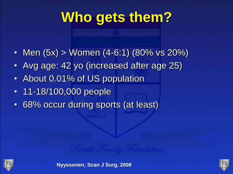
Who gets them?
• Men (5x) > Women (4-6:1) (80% vs 20%)
• Avg age: 42 yo (increased after age 25)
• About 0.01% of US population
• 11-18/100,000 people
• 68% occur during sports (at least)
Nyyssonen, Scan J Surg, 2008
Who gets them?
• Contributing factors:
• Age: >40yo
• Blood flow decreases
• Stiffness of tendon increases
• Prior rupture: 6-8%
• Steroid injections into tendon
• Medications: fluoroquinolones, statins
Vosseller, FAI, 2013
Kujala, Clin J Sports Med, 2005
Hess, Foot Ankle Spec, 2009
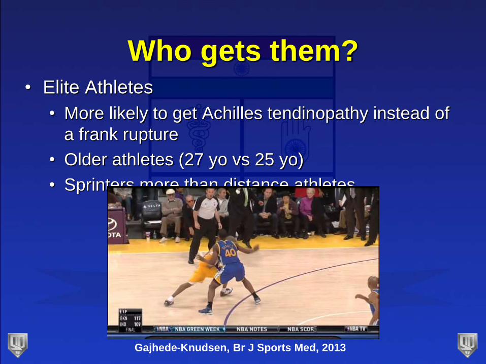
Who gets them?• Elite Athletes
• More likely to get Achilles tendinopathy instead of
a frank rupture
• Older athletes (27 yo vs 25 yo)
• Sprinters more than distance athletes
Gajhede-Knudsen, Br J Sports Med, 2013
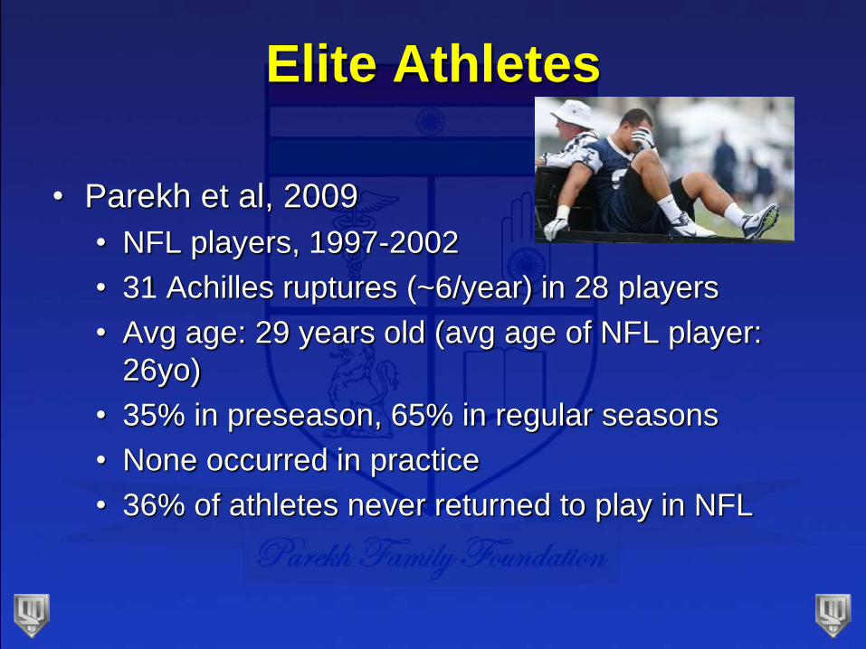
Elite Athletes
• Parekh et al, 2009
• NFL players, 1997-2002
• 31 Achilles ruptures (~6/year) in 28 players
• Avg age: 29 years old (avg age of NFL player:
26yo)
• 35% in preseason, 65% in regular seasons
• None occurred in practice
• 36% of athletes never returned to play in NFL
Fluoroquinolone Use
• Exposure increases risk tendon injuries
• Achilles tendon more than others
• Increased risk:
• First month of use (even first 7 days)
• Combined with oral corticosteroids
• >60 yo
• Renal disease
• Type of fluoroquinolone
• Black Box warningStephenson, Drug Saf. 2013
Parmar, FAI, 2007
Corticosteroid Use
• No clear etiological role
• Injections:
• Animal studies:
• Necrosis at site of injection
• Delay in healing response
• Clinical studies: case reports
• 5 athletes after injection: residual corticosteroid steroid found at the site of injection
• Oral:
• Case reports suggest increased risk of rupture
Balasubramaniam, JBJS-Br, 1972
Unverferth, JBJS, 1973
Mechanism
• 53% at push off (eccentric contraction)
• Occurs during running or jumping
• Tears 2-6cm from insertion (80%)
Hess, Foot Ankle Spec, 2010
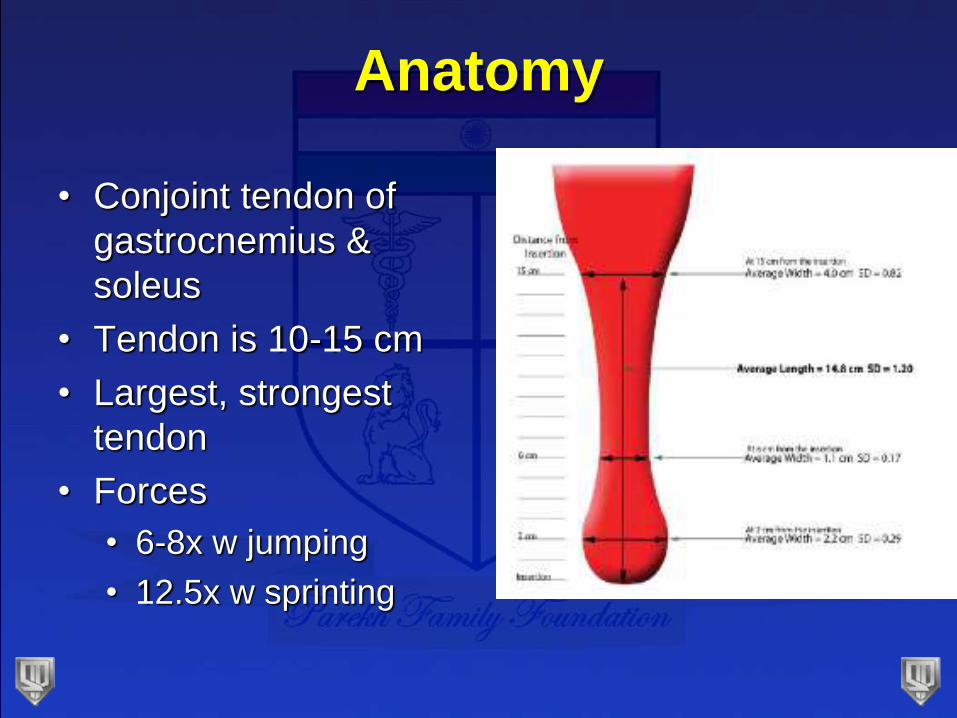
Anatomy
• Conjoint tendon of
gastrocnemius &
soleus
• Tendon is 10-15 cm
• Largest, strongest
tendon
• Forces
• 6-8x w jumping
• 12.5x w sprinting
Anatomy
• Contribution variable
• More from gastroc
• Fibers rotate 90o
• Gastroc contribution is lateral
• Maximum rotation of fibers is at 2-5cm proximal
to insertion
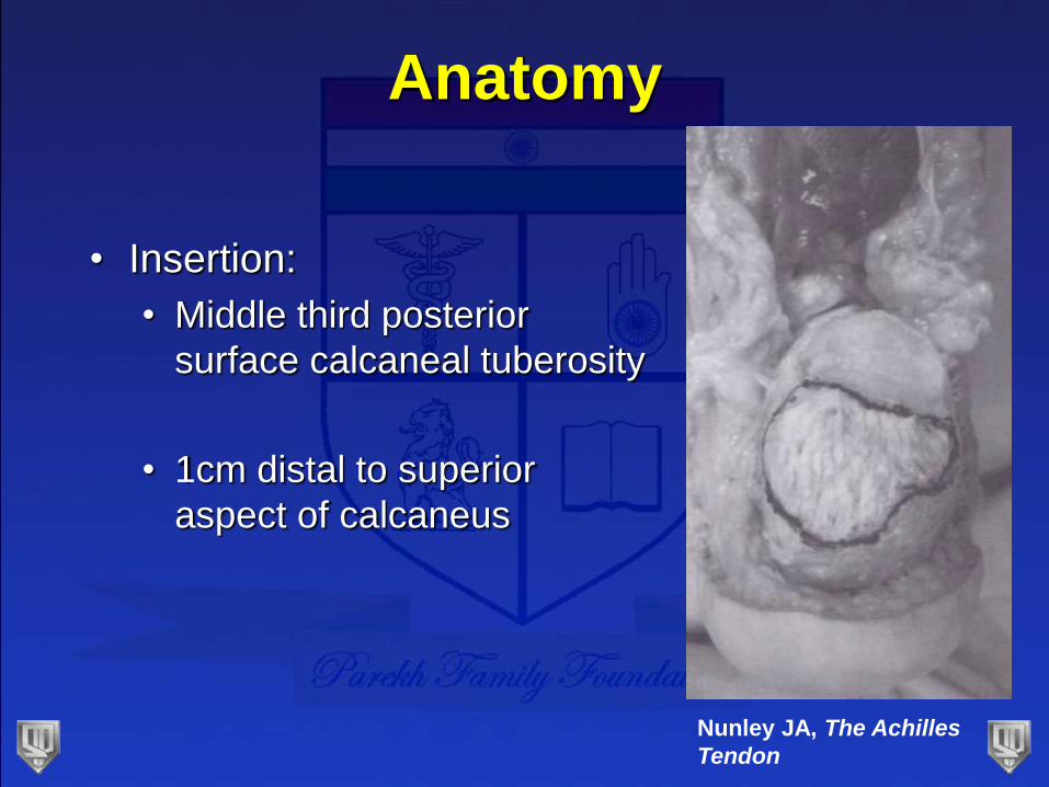
Anatomy
• Insertion:
• Middle third posterior
surface calcaneal tuberosity
• 1cm distal to superior
aspect of calcaneus
Nunley JA, The Achilles
Tendon
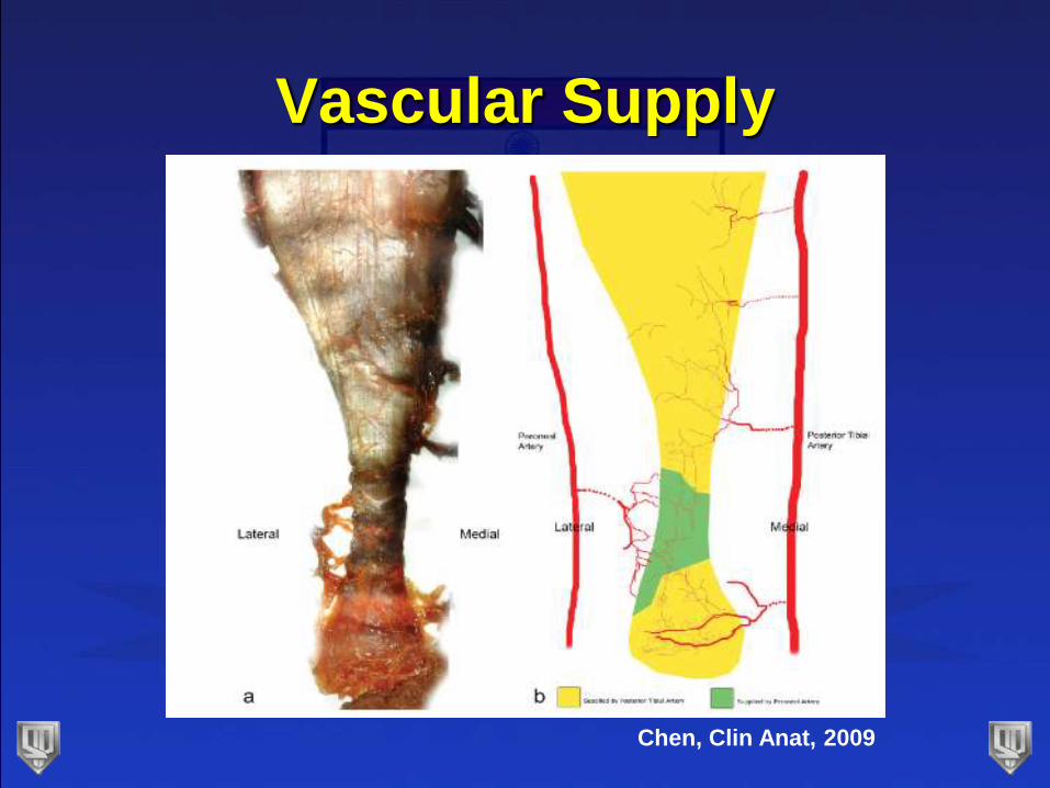
Vascular Supply
Chen, Clin Anat, 2009
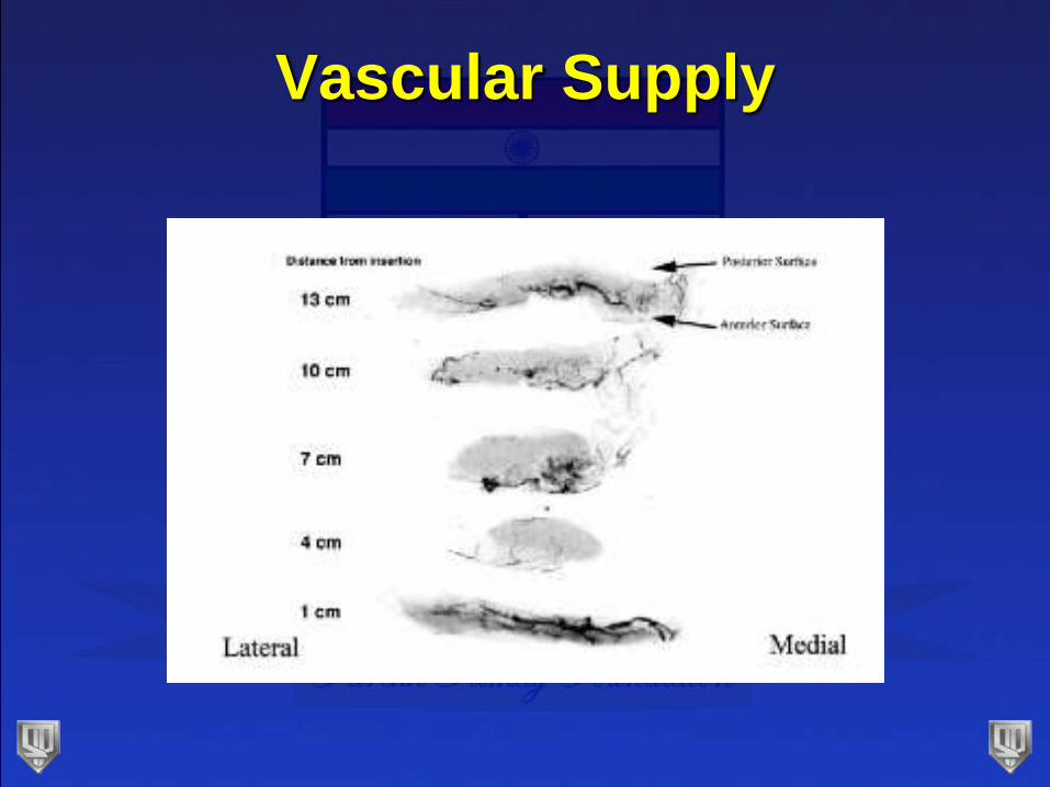
Vascular Supply
Vascular Supply
• Posterior longitudinal midline incision
• Least disruptive
• Close the paratenon and deep fascia
• Thought to help healing
• Skin perfusion
• Maximized at 20o plantar flexion
Poynton, FAI 2001
Presentation
• Sudden pain
• “kicked in the back of my calf”
• Audible snap
• Weakness in ankle
• Initial diagnosis missed as often as 25%
• Commonly diagnosed as ankle sprain
Kvist, Sports Med. 1994
Diagnosis
• “Should not pose a diagnostic problem”
• At least 2 positive physical exam tests
• Palpation: least sensitive
• Calf squeeze: most sensitive
• Maltes: knee flexion test (88% sensitive)
• Copeland: blood pressure cuff test
• O’Brien: needle test
Maffulli, Am J Sports Med. 1998
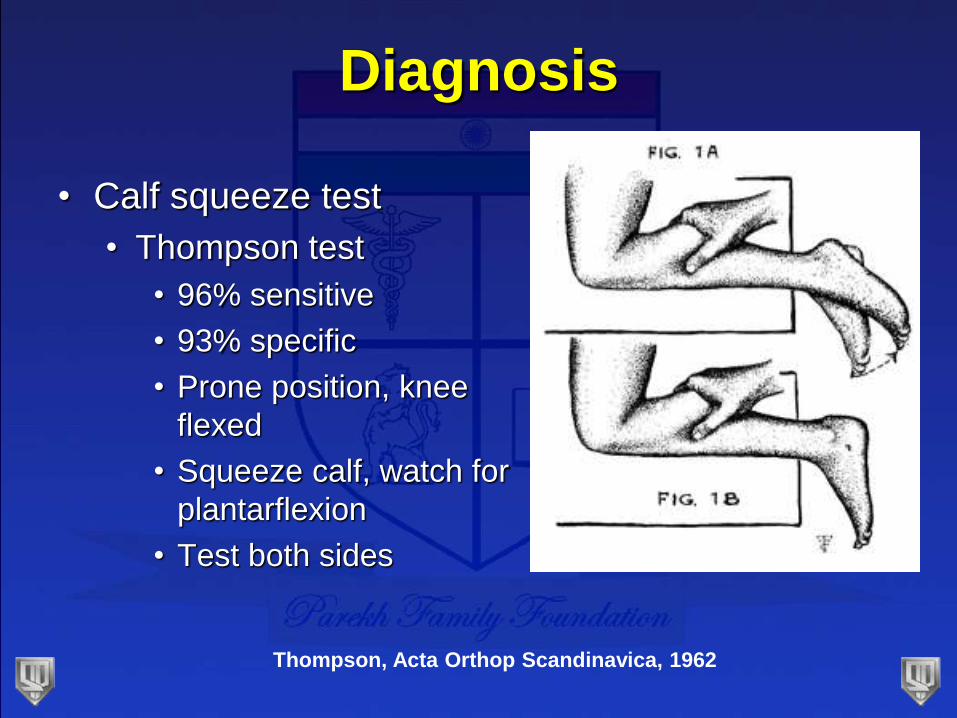
Diagnosis
• Calf squeeze test
• Thompson test
• 96% sensitive
• 93% specific
• Prone position, knee
flexed
• Squeeze calf, watch for
plantarflexion
• Test both sides
Thompson, Acta Orthop Scandinavica, 1962
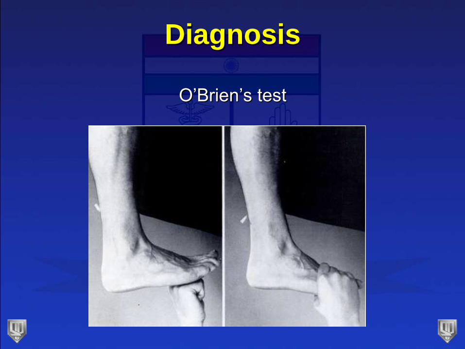
Diagnosis
O’Brien’s test
Imaging
• Should not rely on imaging
• Radiographs:
• Useful for distal avulsions
• Particularly with chronic insertional disease
• Loss of configuration of Kager’s triangle
• Toygar’s sign
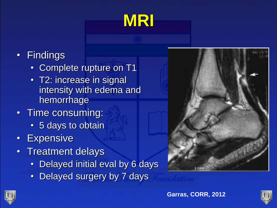
MRI
• Findings
• Complete rupture on T1
• T2: increase in signal intensity with edema and hemorrhage
• Time consuming:
• 5 days to obtain
• Expensive
• Treatment delays
• Delayed initial eval by 6 days
• Delayed surgery by 7 days
Garras, CORR, 2012
Ultrasound
• Performed in office
• Faster
• Cheaper
• Can examine healing or repair
• Best method to follow treatment
• Still not necessary
Maffulli, Internat J Sports Med, 1990
Treatment
• AAOS guidelines, published in 2009:
• Conflicting Evidence
• No definitive answer on operative vs
nonoperative treatment
• 16 recommendations:
• None graded as strong
• 2 consensus statements
• 2 moderate strength recommendations
AAOS Clinical Practice
Guidelines• Consensus:
• Detailed history & physical exam (2 physical exam
tests)
• Surgery is option
• Approach cautiously in pts >65, systemic issues,
obesity, or tobacco
• Moderate:
• Early protected weight bearing after surgical repair
• Protective device for mobilization b/t 2-4wk post-op
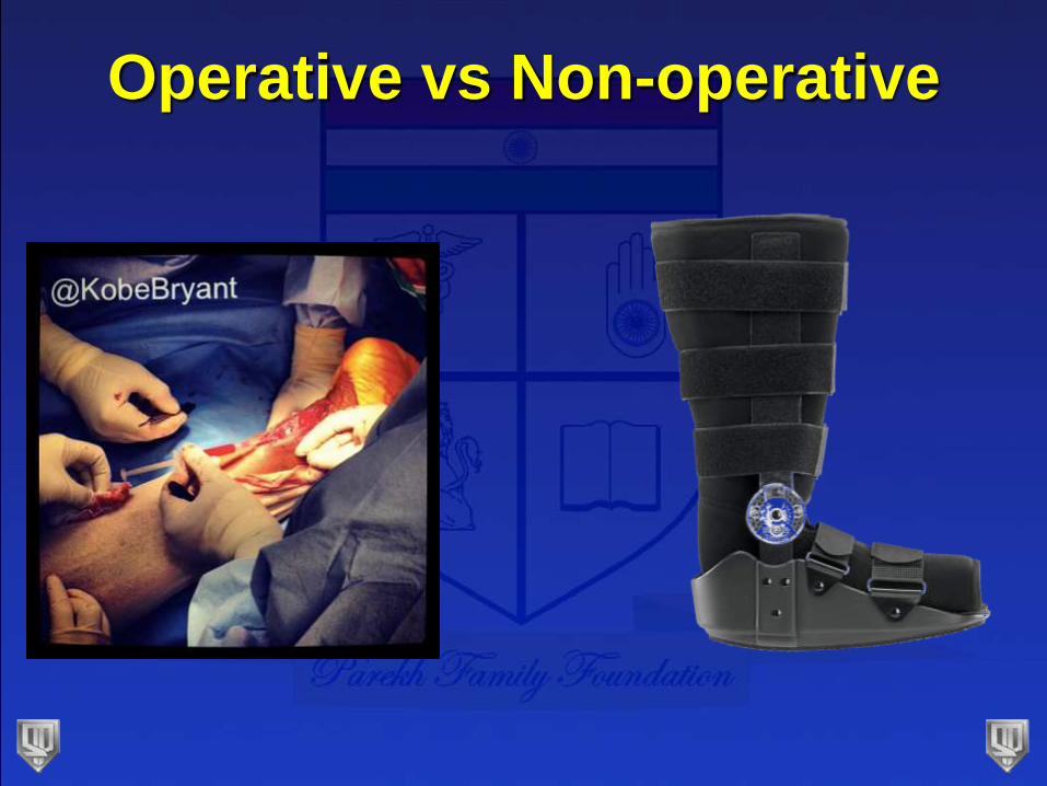
Operative vs Non-operative
Non-Operative Treatment
• Traditionally treated w immobilization
• 6-8wks cast
• High re-rupture rates
• Lee and smith, 1972: 13%
• Person and Wedmark, 1976: 32%
• Inglis, 1976: 39%
Non-Operative Treatment
• Functional rehab instead of cast immobilization
• Post-op: mobile cast is better than immobilization
Cetti, CORR 1994
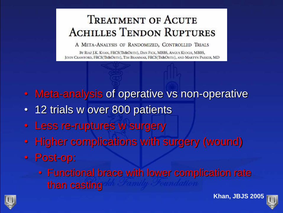
• Meta-analysis of operative vs non-operative
• 12 trials w over 800 patients
• Less re-ruptures w surgery
• Higher complications with surgery (wound)
• Post-op:
• Functional brace with lower complication rate
than castingKhan, JBJS 2005
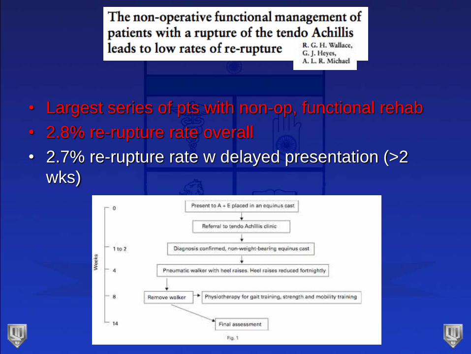
• Largest series of pts with non-op, functional rehab
• 2.8% re-rupture rate overall
• 2.7% re-rupture rate w delayed presentation (>2
wks)
Non-Operative Treatment
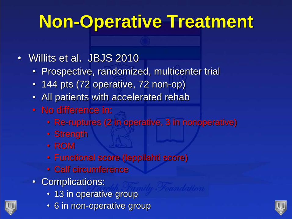
• Willits et al. JBJS 2010
• Prospective, randomized, multicenter trial
• 144 pts (72 operative, 72 non-op)
• All patients with accelerated rehab
• No difference in:
• Re-ruptures (2 in operative, 3 in nonoperative)
• Strength
• ROM
• Functional score (leppilahti score)
• Calf circumference
• Complications:
• 13 in operative group
• 6 in non-operative group
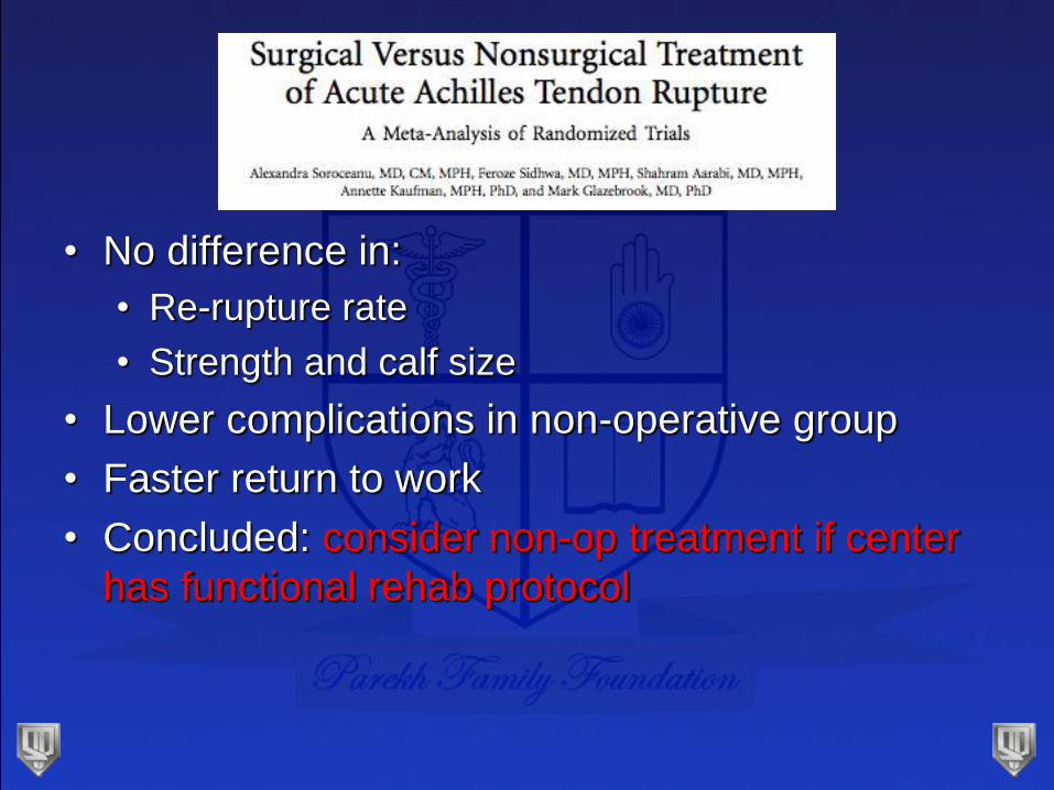
• No difference in:
• Re-rupture rate
• Strength and calf size
• Lower complications in non-operative group
• Faster return to work
• Concluded: consider non-op treatment if center
has functional rehab protocol
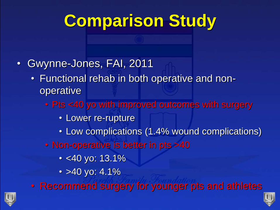
Comparison Study
• Gwynne-Jones, FAI, 2011
• Functional rehab in both operative and non-
operative
• Pts <40 yo with improved outcomes with surgery
• Lower re-rupture
• Low complications (1.4% wound complications)
• Non-operative is better in pts >40
• <40 yo: 13.1%
• >40 yo: 4.1%
• Recommend surgery for younger pts and athletes
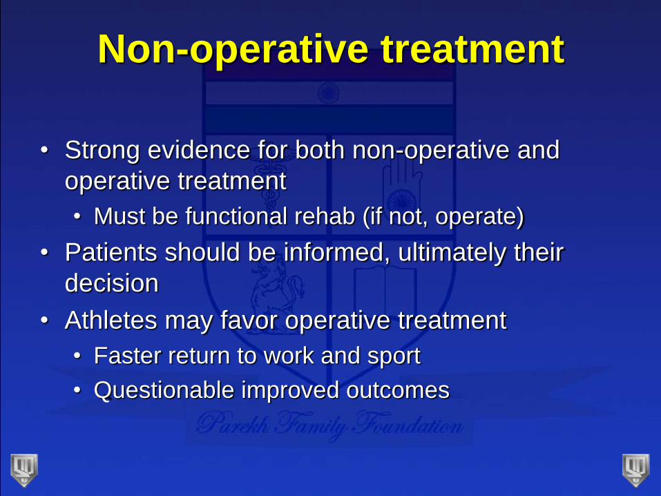
Non-operative treatment
• Strong evidence for both non-operative and
operative treatment
• Must be functional rehab (if not, operate)
• Patients should be informed, ultimately their
decision
• Athletes may favor operative treatment
• Faster return to work and sport
• Questionable improved outcomes
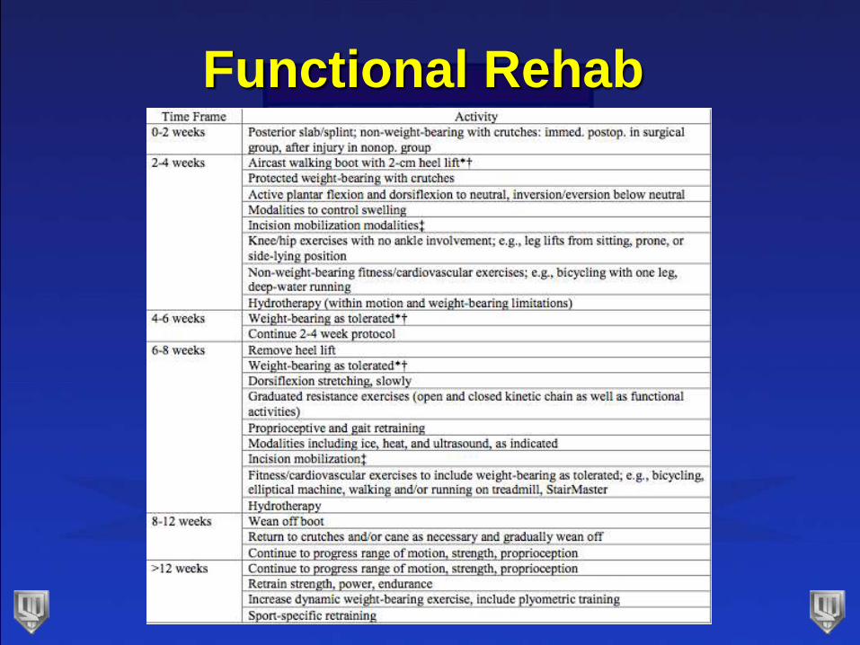
Functional Rehab
Who should have surgery
• Elite athletes
• Delayed presentation
• Inability for functional rehab
If surgery…
• More controversy
• How to repair
• Open vs percutaneous
• Post-op rehab
• Augment repair?
How to repair
• Watson, FAI, 1995
• Single Kessler
• Single Bunnel
• Double Krackow
• Double Krackow had double the strength
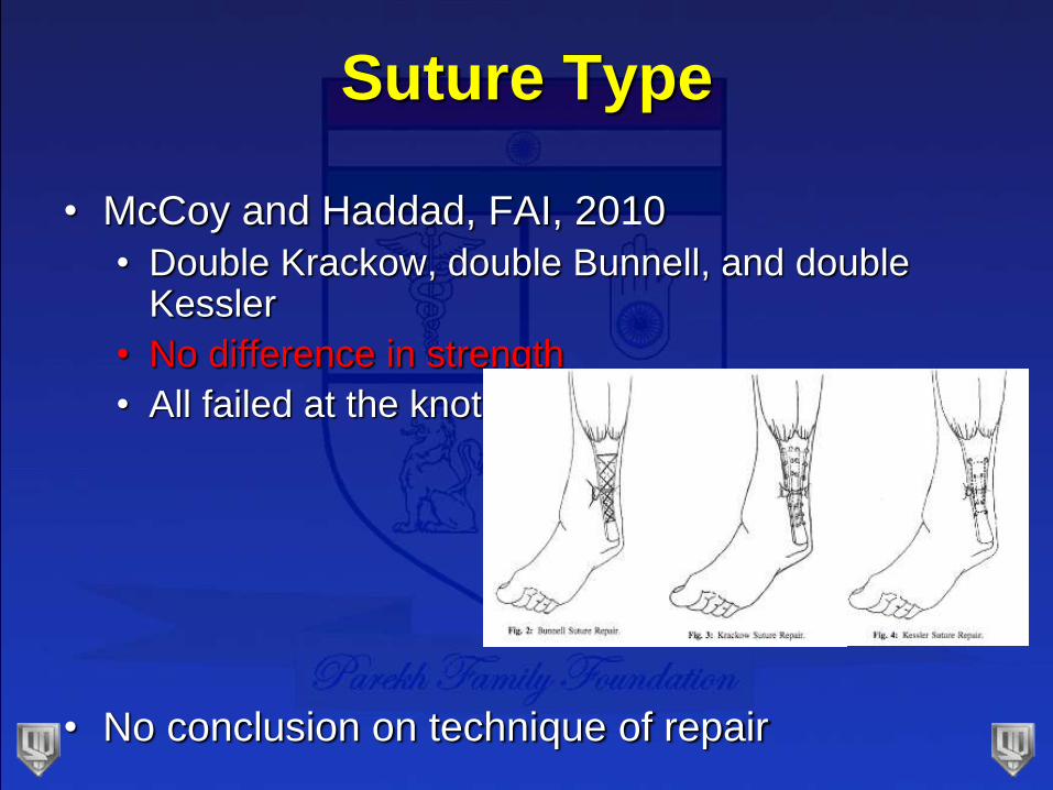
Suture Type
• McCoy and Haddad, FAI, 2010
• Double Krackow, double Bunnell, and double Kessler
• No difference in strength
• All failed at the knot
• No conclusion on technique of repair
Percutaneous Repair
• Minimizes trauma to tenuous skin
• Reduces surface area for adhesion formation
• Decreases possibility of contamination
• Minimal complications (11%)
• Skin dimple at operative site
• Tender nodule at operative site
Does Incision Size Matter?
• Cochrane review in 2010 (Khan et al)
• Percutaneous surgery vs open:
• Lower risk of infection
• Interpret with caution
• JBJS meta-anaylsis (Khan, 2005)
• Percutaneous with lower complication rate
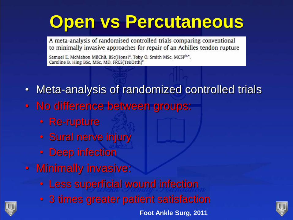
Open vs Percutaneous
• Meta-analysis of randomized controlled trials
• No difference between groups:
• Re-rupture
• Sural nerve injury
• Deep infection
• Minimally invasive:
• Less superficial wound infection
• 3 times greater patient satisfaction
Foot Ankle Surg, 2011
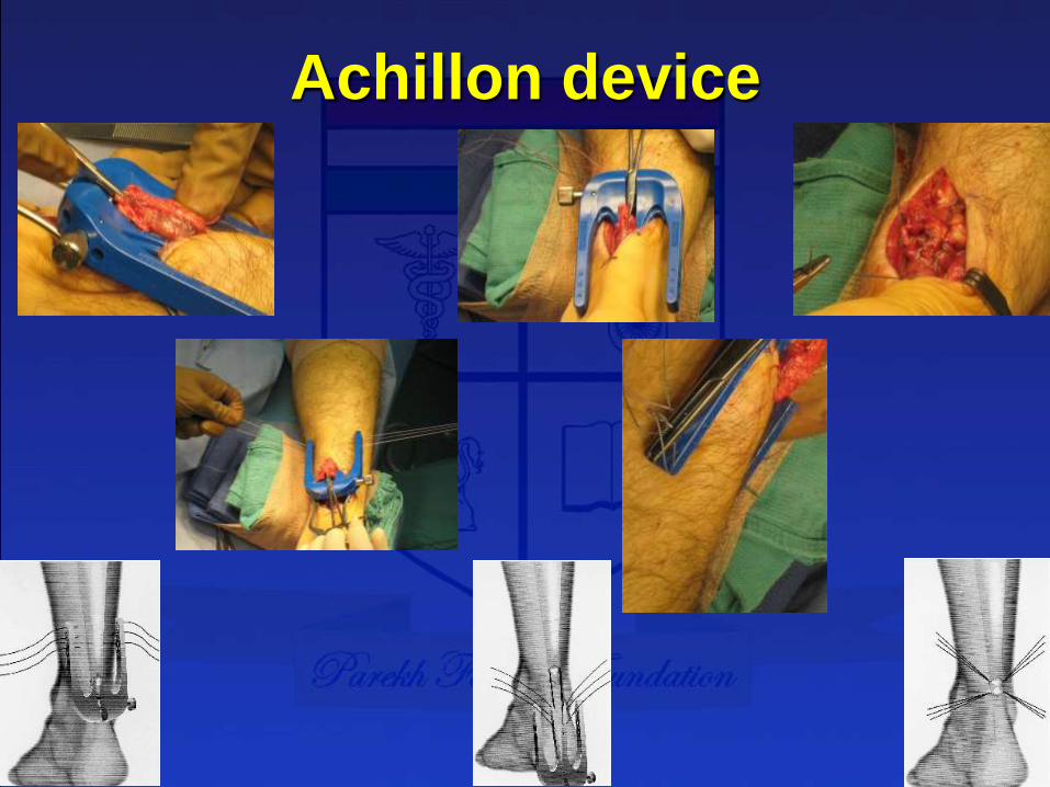
Achillon device
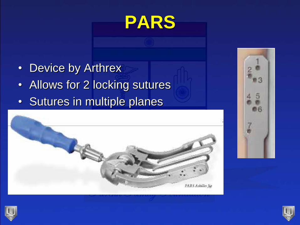
PARS
• Device by Arthrex
• Allows for 2 locking sutures
• Sutures in multiple planes
PARS
• Charlotte experience
• AOFAS 2012
• 46 pts
• AOFAS: 97
• 45/46 satisfied at 6 months
• No re-ruptures
• No sural nerve complications
• No wound healing issues
• Paid consultants
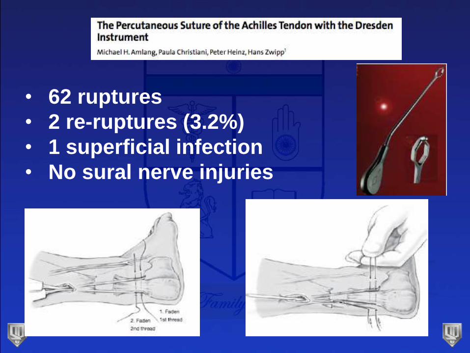
• 62 ruptures
• 2 re-ruptures (3.2%)
• 1 superficial infection
• No sural nerve injuries
Problems with Percutaneous
Repair?
• Aracil, FAI, 1992
• Sural nerve injury
• Taken back to OR for suture to be cut
• Re-rupture
• 33% re-rupture rate
• Didn’t limit dorsiflexion
• Hockenbury, Foot Ankle, 1990
• 60% sural nerve injury
• All within 2.5 cm from rupture site
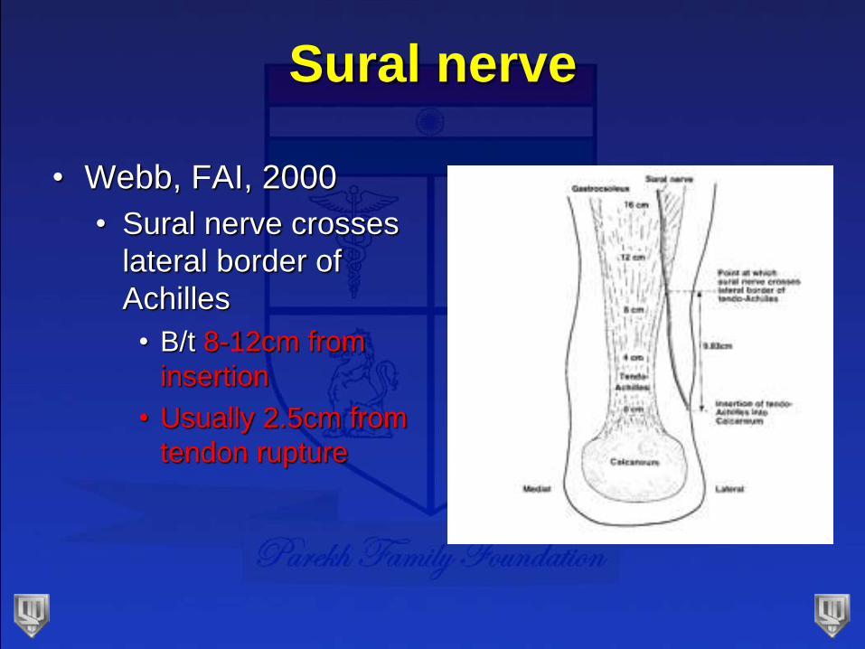
Sural nerve
• Webb, FAI, 2000
• Sural nerve crosses
lateral border of
Achilles
• B/t 8-12cm from
insertion
• Usually 2.5cm from
tendon rupture
Avoid Sural Nerve
• Don’t place percutaneous sutures in lateral half
of proximal tendon
• Make small proximal incision to find the nerve
(Webb, JBJS-Br, 1999, Klein, 1991)
• Place suture in medial half of proximal tendon
Post-op Rehab
• Maffulli, AJSM, 2003
• Prospective randomized study
• Early weight-bearing and ROM after open repair
• Fewer outpatient visits
• Discarded crutches early
• Higher satisfaction
• No difference in:
• Ultrasound appearance of tendon
• Isometric strength
Conclusions
• Increasing evidence for non-operative treatment
• Must be functional rehab
• Elite athletes still favor operative repair
• Safe, low re-rupture
• Best functional outcome (fastest)
• Pressure (athlete, coach, media)
• Maffulli, FAI, 2011
• Mini-open is a good option
• Risk of sural nerve injury
• Do what works in your hands
RE
ECTthe ankle
the foot