Embed Size (px)
Citation preview
1/08/2015
1
ASC171NERVOUS SYSTEM – MODULE 5
Part 3
Part 4
LEARNING OBJECTIVES: PART 3
8. List the major subdivisions and anatomical landmarks of the brain
9. List the meninges & the order in which they are found around the brain
10. Identify the cerebral ventricles, surrounding fluid and difference between white and grey matter
11. Identify the four main lobes of the cerebrum and their functions
12. Identify the main brain regions involved in: The control of movement
Receiving information on somatic sensation
Memory formation & storage and emotion
13. Describe the role of the hypothalamus, thalamus & cerebellum
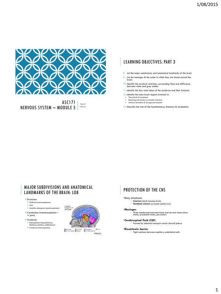
MAJOR SUBDIVISIONS AND ANATOMICAL LANDMARKS OF THE BRAIN: LO8
Brainstem
Midbrain (mesencephalon)
Pons
Medulla oblongata (myelencephalon)
Cerebellum (metaencephalon –w pons)
Forebrain
Diencephalon (hypothalamus, thalamus, pituitary, epithalamus)
Cerebrum (telencephalon)
PROTECTION OF THE CNS
Bony structures Cranium (skull) encases brain
Vertebral column surrounds spinal cord
Meninges Three membranes between bone and nervous tissue (dura
mater, arachnoid mater, pia mater)
Cerebrospinal fluid (CSF) Formed by selective transport across choroid plexus
Blood-brain barrier Tight junctions between capillary endothelial cells
1/08/2015
2
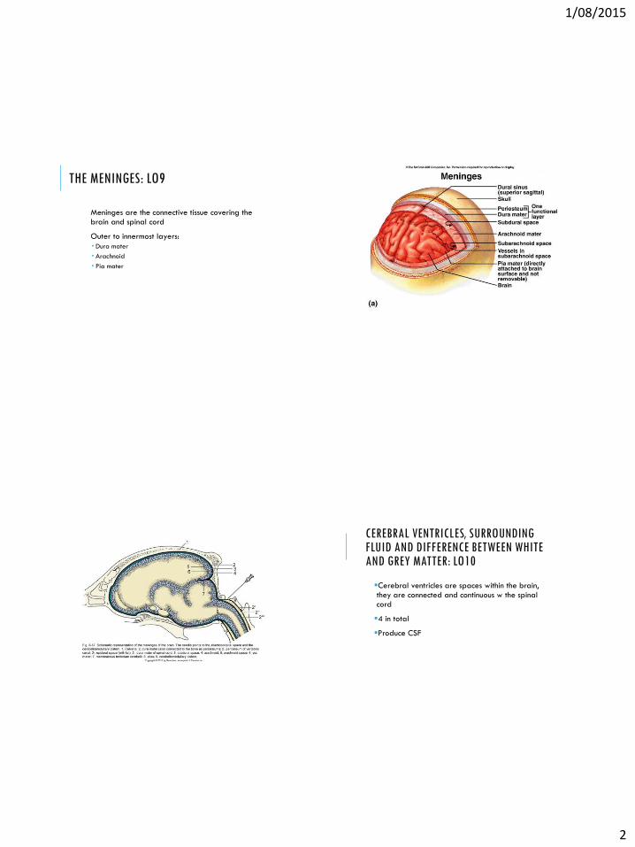
THE MENINGES: LO9
Meninges are the connective tissue covering the brain and spinal cord
Outer to innermost layers:
Dura mater
Arachnoid
Pia mater
CEREBRAL VENTRICLES, SURROUNDING FLUID AND DIFFERENCE BETWEEN WHITE AND GREY MATTER: LO10
Cerebral ventricles are spaces within the brain, they are connected and continuous w the spinal cord
4 in total
Produce CSF
1/08/2015
3
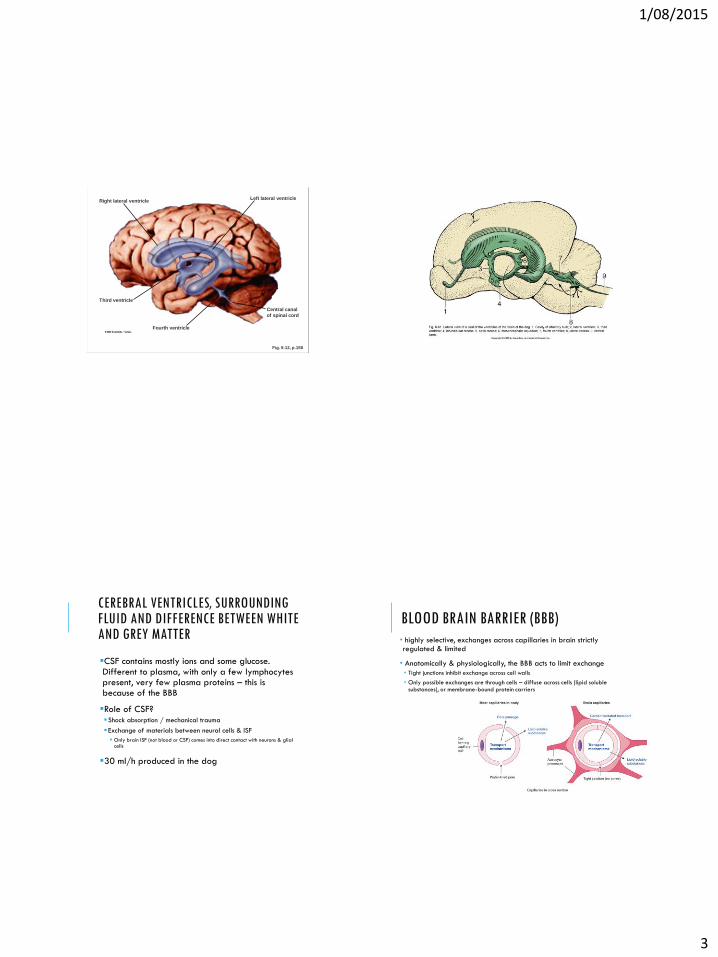
Fig. 5-12, p.158
Right lateral ventricle
Third ventricle
Fourth ventricle
Left lateral ventricle
Central canal
of spinal cord
CEREBRAL VENTRICLES, SURROUNDING FLUID AND DIFFERENCE BETWEEN WHITE AND GREY MATTER
CSF contains mostly ions and some glucose. Different to plasma, with only a few lymphocytes present, very few plasma proteins – this is because of the BBB
Role of CSF?Shock absorption / mechanical trauma
Exchange of materials between neural cells & ISF
Only brain ISF (not blood or CSF) comes into direct contact with neurons & glial cells
30 ml/h produced in the dog
BLOOD BRAIN BARRIER (BBB)
• highly selective, exchanges across capillaries in brain strictly regulated & limited
• Anatomically & physiologically, the BBB acts to limit exchange
• Tight junctions inhibit exchange across cell walls
• Only possible exchanges are through cells – diffuse across cells (lipid soluble substances), or membrane-bound protein carriers
1/08/2015
4
CEREBRAL VENTRICLES, SURROUNDING FLUID AND DIFFERENCE BETWEEN WHITE AND GREY MATTERWhite matter – dense collections of myelinatedfibres “Wires” connecting computers to each other
Grey matter – mostly cell bodies, dendrites and unmyelinated fibres “Computers” of the CNS
Integration of neural input & initiation of neural output synapses within grey matter
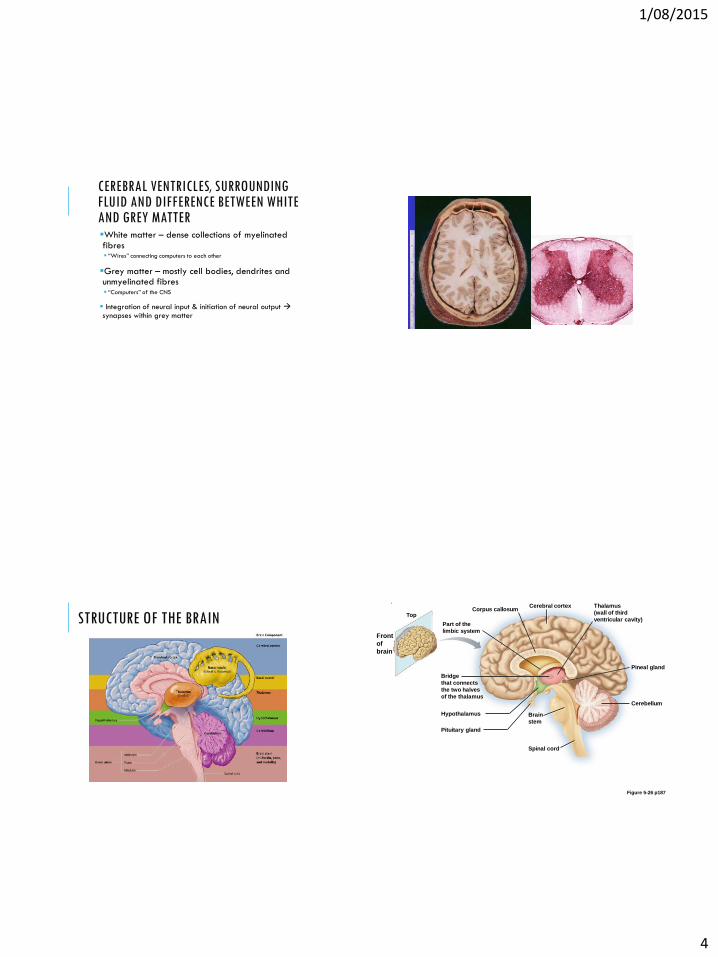
STRUCTURE OF THE BRAINCorpus callosum
Front
of
brain
Part of the
limbic system
Bridge
that connects
the two halves
of the thalamus
Spinal cord
Top
Hypothalamus
Pituitary gland
Brain
stem
Cerebellum
Pineal gland
Thalamus
(wall of third
ventricular cavity)
Cerebral cortex
Figure 5-26 p187
1/08/2015
5
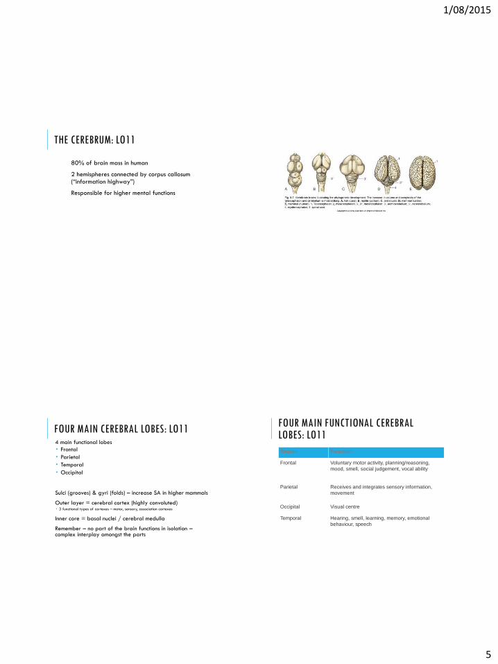
THE CEREBRUM: LO11
80% of brain mass in human
2 hemispheres connected by corpus callosum (“information highway”)
Responsible for higher mental functions
FOUR MAIN CEREBRAL LOBES: LO114 main functional lobes
Frontal
Parietal
Temporal
Occipital
Sulci (grooves) & gyri (folds) – increase SA in higher mammals
Outer layer = cerebral cortex (highly convoluted) 3 functional types of cortexes – motor, sensory, association cortexes
Inner core = basal nuclei / cerebral medulla
Remember – no part of the brain functions in isolation –complex interplay amongst the parts
FOUR MAIN FUNCTIONAL CEREBRAL LOBES: LO11
Region Function
Frontal Voluntary motor activity, planning/reasoning,
mood, smell, social judgement, vocal ability
Parietal Receives and integrates sensory information,
movement
Occipital Visual centre
Temporal Hearing, smell, learning, memory, emotional
behaviour, speech

1/08/2015
6
THE MAIN BRAIN REGIONS INVOLVED IN: CONTROL OF MOVEMENT (LO12)
Level Function Structures
High Strategy (goal of
the movement)
Neocortex, Motor
cortex
Middle Tactics (sequence
of muscle
contractions,
arrangement in
space)
Cerebellum
Low Execution Brain stem, spinal
cord
1/08/2015
7
Fig. 5-19a, p.169
Front
Top
view
Occipital lobe
Parietal
lobeSomato-
sensorycortex
Primary
motorcortex
Left
hemisphere
Central
sulcus
Right
hemisphere
Frontal
lobe
Back
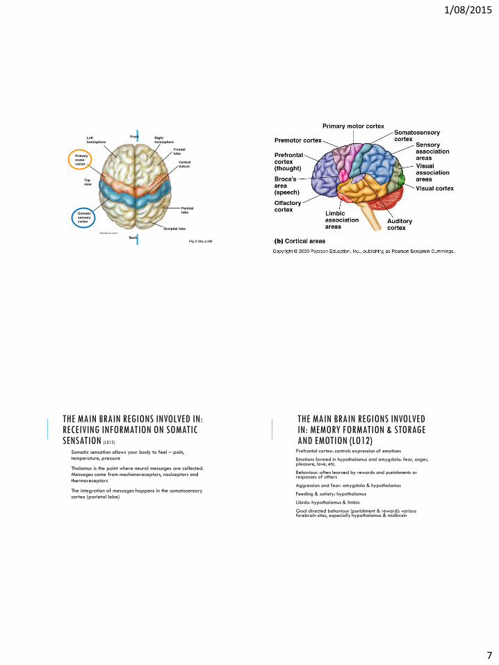
THE MAIN BRAIN REGIONS INVOLVED IN: RECEIVING INFORMATION ON SOMATIC SENSATION (LO12)
Somatic sensation allows your body to feel – pain, temperature, pressure
Thalamus is the point where neural messages are collected. Messages come from mechanoreceptors, nociceptors and thermoreceptors
The integration of messages happens in the somatosensory cortex (parietal lobe)
THE MAIN BRAIN REGIONS INVOLVED IN: MEMORY FORMATION & STORAGE AND EMOTION (LO12)
Prefrontal cortex: controls expression of emotions
Emotions formed in hypothalamus and amygdala: fear, anger, pleasure, love, etc.
Behaviour: often learned by rewards and punishments or responses of others
Aggression and fear: amygdala & hypothalamus
Feeding & satiety: hypothalamus
Libido: hypothalamus & limbic
Goal directed behaviour (punishment & reward): various forebrain sites, especially hypothalamus & midbrain

1/08/2015
8
Hypothalamus
THE MAIN BRAIN REGIONS INVOLVED IN MEMORY FORMATION & STORAGE AND EMOTION
THE HYPOTHALAMUS, THALAMUS & CEREBELLUM: LO13
Hypothalamus
Maintenance of homeostasis
Contains neural centres for hunger, thirst, and body temperature
Contributes to the regulation of sleep, wakefulness, emotions and sexual performance
Regulates hormone release from pituitary gland
Coordinates sympathetic and parasympathetic reflexes
THE HYPOTHALAMUS, THALAMUS & CEREBELLUM: LO13
Thalamus
Receives input from the limbic system and from all sensory angles apart from olfaction.
Acts as a ‘filter’ for information (selective hearing)
THE HYPOTHALAMUS, THALAMUS & CEREBELLUM: LO13
Cerebellum
Maintains body posture and balance, controls eye movements
Coordinates movement
Integrates information from proprioceptors

1/08/2015
9
LEARNING OBJECTIVES: PART 3
14. How many pairs of cranial nerves are there and which one innervates the viscera?
15. Describe the gross and microscopic structure of the spinal cord.
16. Describe the general components of a typical reflex arc
17. What are the main structural and functional features of the autonomic nervous system
18. How do the two divisions of the autonomic nervous system differ in their general function?
HOW MANY PAIRS OF CRANIAL NERVES ARE THERE AND WHICH ONE INNERVATES THE VISCERA? LO14
Cranial nerves come directly from the brain, as opposed to spinal nerves.
12 pairs
2 from forebrain, 10 from midbrain/hindbrain
Some are afferent, some efferent
Mostly for head except vagus (X) for viscera
Number Name Function
I Olfactory Olfaction
II Optic Vision
III Oculomotor Controls eye movement
IV Trochlear Eye movement
V Trigeminal Mouth movements
VI Abducens Eye movement
VII Facial Taste
VIII Vestibulocochlear Hearing and equilibrium
IX Glossopharyngeal Swallowing muscles
X Vagus Motor control of larynx, pharynx,
oesophagus, parasympathetic control of
heart/lungs, gut
XI Accessory Head movement
XII Hypoglossal Tongue movement
1/08/2015
10
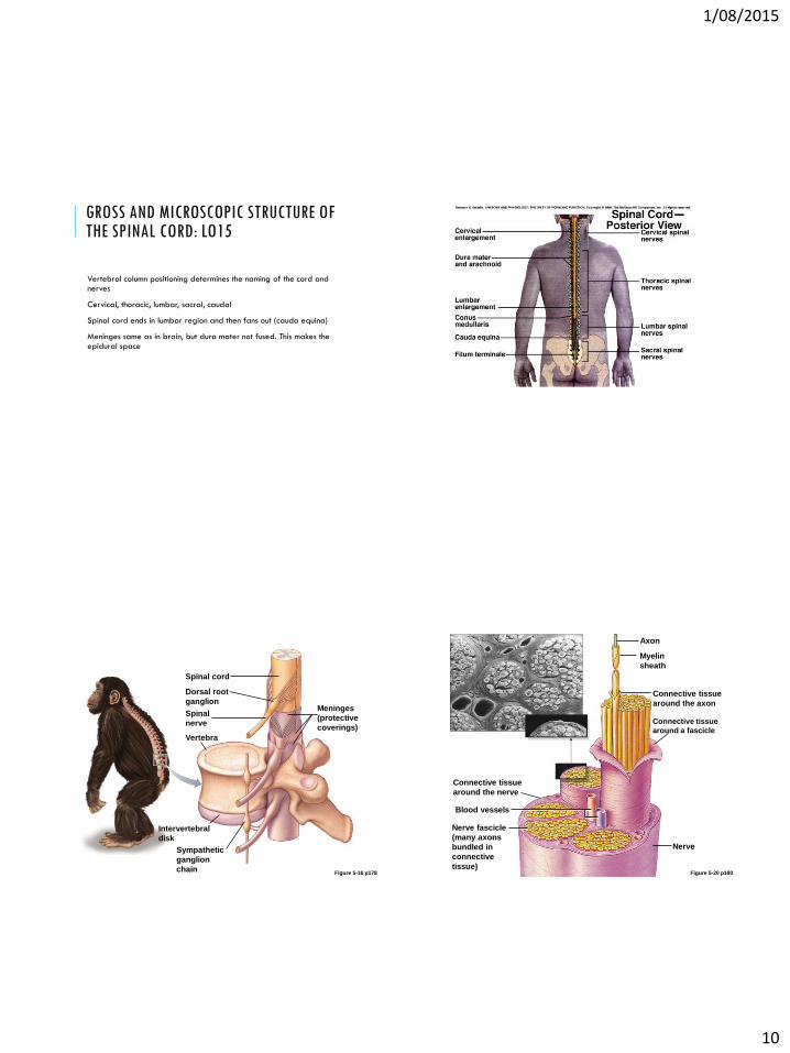
GROSS AND MICROSCOPIC STRUCTURE OF THE SPINAL CORD: LO15
Vertebral column positioning determines the naming of the cord and nerves
Cervical, thoracic, lumbar, sacral, caudal
Spinal cord ends in lumbar region and then fans out (cauda equina)
Meninges same as in brain, but dura mater not fused. This makes the epidural space
Figure 5-16 p178
Spinal cord
Dorsal root
ganglionMeninges
(protective
coverings)
Spinal
nerve
Vertebra
Intervertebral
disk
Sympathetic
ganglion
chainFigure 5-20 p180
Axon
Myelin
sheath
Connective tissue
around the axon
Connective tissue
around a fascicle
Connective tissue
around the nerve
Blood vessels
Nerve fascicle
(many axons
bundled in
connective
tissue)
Nerve

1/08/2015
11
CAUDA EQUINA (LATIN FOR HORSE’S TAIL)
GROSS AND MICROSCOPIC STRUCTURE OF THE SPINAL CORD: LO15
HORNS: grey matter Dorsal – interneuron cell bodies (sensory receiving)
Ventral – motor neuron cell bodies
Lateral – autonomic nervous system
COLUMNS: white matter Axons
Grouped into tracts of common locations and functions
ROOTS: connecting points between spinal nerves and spinal cord
Efferent fiber
White matter Gray matter
Afferent fiber
Dorsal root
Dorsal root
ganglion
Spinal nerve
Cell body of
efferent neuron
Interneuron
Cell body of
afferent neuron
To effectors
From receptors
Figure 5-17 p178
Ventral root
1/08/2015
12
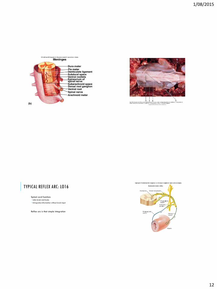
TYPICAL REFLEX ARC: LO16
Spinal cord function:
Links brain and body
Intregrates information without brain input
Reflex arc is that simple integration

1/08/2015
13
STIMULUS
Receptor
Motor (efferent) pathway
Interneuron (integration)
Sensory (afferent) pathway
Effector organ - Action response
Stimulates=
Stimulus
Triceps
(extensor) relaxes
Biceps
(flexor) contracts
Thermal pain
receptor in paw
Foot
withdrawn
Afferent
pathway
Efferent
pathway
Effector
organs
Response
Ascending pathway
to brain
Integrating center
(spinal cord)
Components
of a
reflex arc
Receptor
Afferent
pathway
Integrating
center
Efferent
pathway
Effector
organs
Inhibitory=
interneuron
Inhibits=
Synapse=
Excitatory interneuron=
Fig. 5-36, p.184
MAIN STRUCTURAL AND FUNCTIONAL FEATURES OF THE AUTONOMIC NERVOUS SYSTEM: LO17Innervate organs whose function is not normally under voluntary control (no brain input)
smooth muscles, cardiac muscle, glands
This includes most visceral organs and blood vessels
Therefore regulates all body systems
Including skeletal muscle since muscles contain blood vessels
AUTONOMIC NERVOUS SYSTEM
Sympathetic system Preparation for strenuous physical activity in emergency
situations (fight or flight)
Heart rate increases
Respiratory airways open
Glycogen and fat stores are broken down
Blood vessels supplying skeletal muscle dilate
Pupils dilate
Parasympathetic system General housekeeping activities in relaxed situations
Digestion
Emptying the urinary bladder

1/08/2015
14
Figure 5-7 p160
Eye
Nasal mucosaLacrimal gland
Parotid gland
SympatheticTrachea
Parasympathetic
Salivary
glands
IXVII III
Lung
X Cranial nerves
Splanchnic nerves
S2
Gall
bladderStomach
Liver
Sympathetic trunk Heart
S3PancreasS4Spleen
Adrenal gland Spinal nerves
Kidney
Small
intestineColon
Rectum
Urinary bladder
Genitalia
Spinal nerves
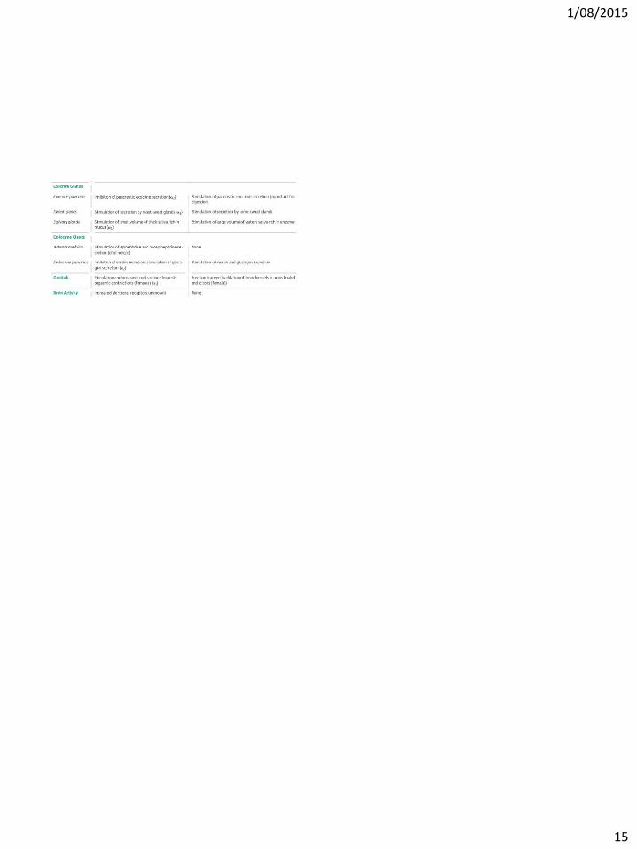
HOW DO THE TWO DIVISIONS OF THE AUTONOMIC NERVOUS SYSTEM DIFFER IN THEIR GENERAL FUNCTION? LO18
Sympathetic NS
Fight or flight
Parasympathetic NS
Rest or digest
VS
HOW DO THE TWO DIVISIONS OF THE AUTONOMIC NERVOUS SYSTEM DIFFER IN THEIR GENERAL FUNCTION? LO18
Sympathetic
Organ SNS PSNS
Heart rate Increase Decrease
Airways Dilate Constrict
Digestive tract Inhibit Activate
Skeletal blood vessels Dilate Contract
Preipheral blood vessels Contract No change
Energy stores Release No change
Parasympathetic
1/08/2015
15