Embed Size (px)
Citation preview

Fundamentals of the Nervous System and Nervous Tissue: Part B

Neurons are highly irritable Respond to adequate stimulus by
generating an action potential (nerve impulse)
Impulse is always the same regardless of stimulus
Opposite charges attract each other Energy is required to separate opposite
charges across a membrane Energy is liberated when the charges
move toward one another If opposite charges are separated, the
system has potential energy
Voltage (V): measure of potential energy generated by separated charge
Potential difference: voltage measured between two points
Current (I): the flow of electrical charge (ions) between two points
Resistance (R): hindrance to charge flow (provided by the plasma membrane)
Insulator: substance with high electrical resistance
Conductor: substance with low electrical resistance
Proteins serve as membrane ion channels
Two main types of ion channels1. Leakage (nongated) channels—always
open

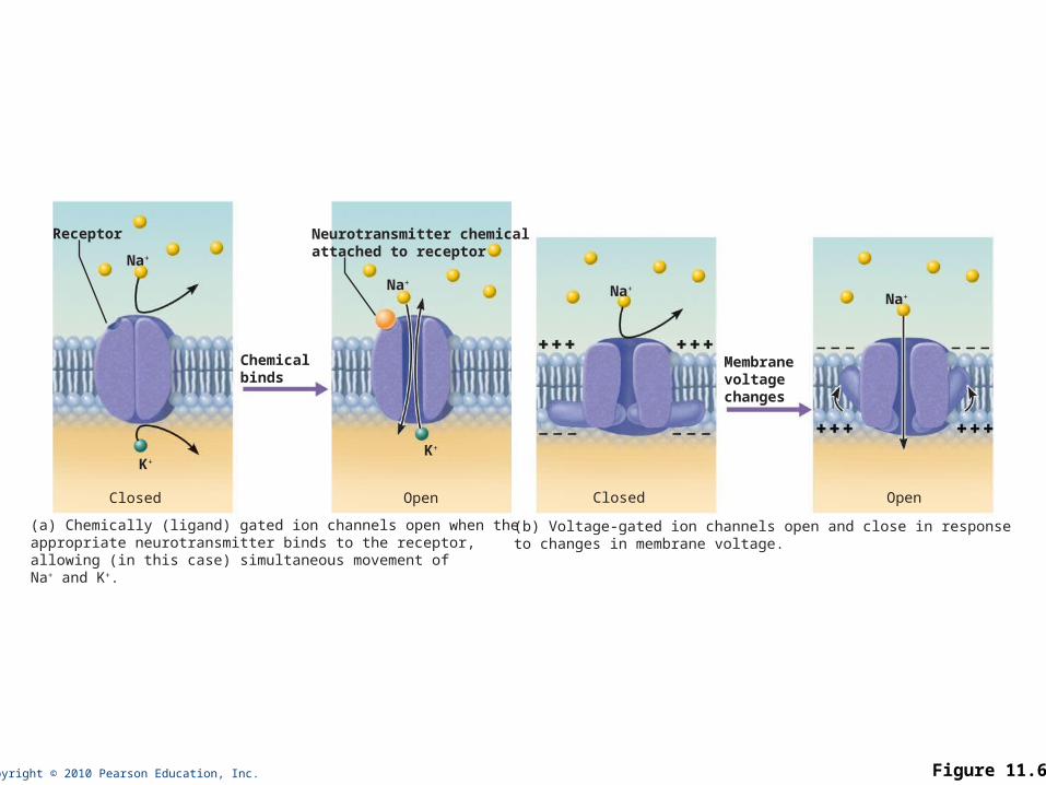
2. Gated channels (three types): Chemically gated (ligand-gated) channels—open with
binding of a specific neurotransmitter Voltage-gated channels—open and close in response
to changes in membrane potential Mechanically gated channels—open and close in
response to physical deformation of receptors
Copyright © 2010 Pearson Education, Inc. Figure 11.6
(b) Voltage-gated ion channels open and close in responseto changes in membrane voltage.
Na+
Na+
Closed Open
Receptor
(a) Chemically (ligand) gated ion channels open when theappropriate neurotransmitter binds to the receptor,allowing (in this case) simultaneous movement of Na+ and K+.
Na+
K+
K+
Na+
Neurotransmitter chemicalattached to receptor
Chemicalbinds
Closed Open
Membranevoltagechanges
When gated channels are open: Ions diffuse quickly across the membrane
along their electrochemical gradients Along chemical concentration gradients from
higher concentration to lower concentration Along electrical gradients toward opposite
electrical charge Ion flow creates an electrical current and
voltage changes across the membrane

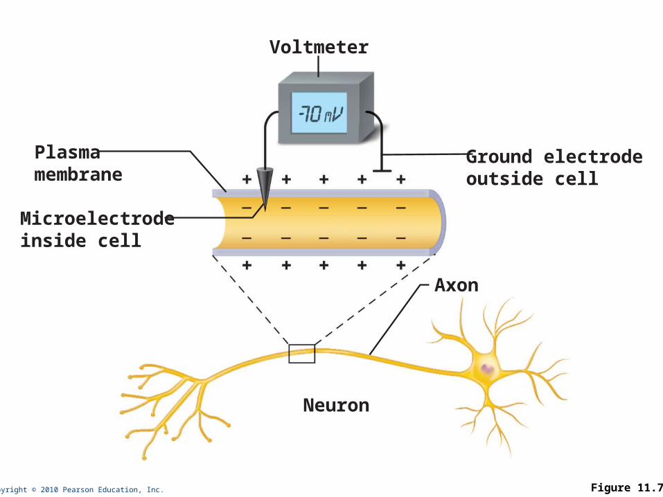
Potential difference across the membrane of a resting cell Approximately –70 mV in neurons
(cytoplasmic side of membrane is negatively charged relative to outside)
Generated by: Differences in ionic makeup of ICF and ECF Differential permeability of the plasma
membrane
Copyright © 2010 Pearson Education, Inc. Figure 11.7
Voltmeter
Microelectrodeinside cell
Plasmamembrane
Ground electrodeoutside cell
Neuron
Axon

Differences in ionic makeup ICF has lower concentration of Na+ and Cl–
than ECF ICF has higher concentration of K+ and
negatively charged proteins (A–) than ECF

Differential permeability of membrane Impermeable to A–
Slightly permeable to Na+ (through leakage channels)
75 times more permeable to K+ (more leakage channels)
Freely permeable to Cl–

Negative interior of the cell is due to much greater diffusion of K+ out of the cell than Na+ diffusion into the cell
Sodium-potassium pump stabilizes the resting membrane potential by maintaining the concentration gradients for Na+ and K+
Copyright © 2010 Pearson Education, Inc. Figure 11.8
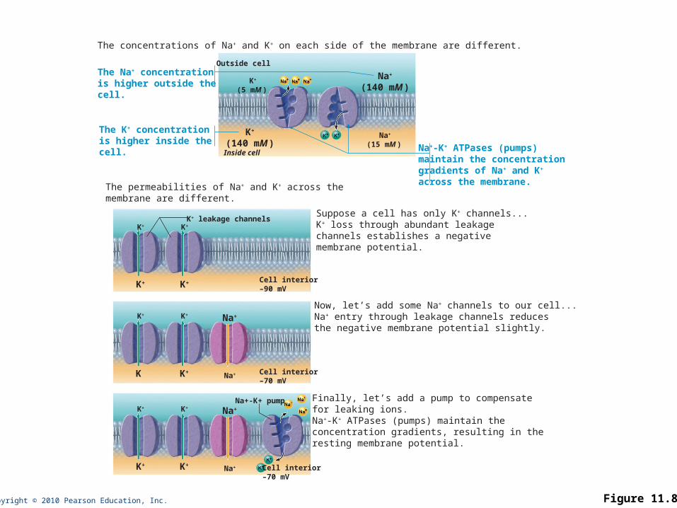
Finally, let’s add a pump to compensate for leaking ions.Na+-K+ ATPases (pumps) maintain the concentration gradients, resulting in the resting membrane potential.
Suppose a cell has only K+ channels...K+ loss through abundant leakagechannels establishes a negativemembrane potential.
Now, let’s add some Na+ channels to our cell...Na+ entry through leakage channels reducesthe negative membrane potential slightly.
The permeabilities of Na+ and K+ across the membrane are different.
The concentrations of Na+ and K+ on each side of the membrane are different.
Na+
(140 mM )K+
(5 mM )
K+ leakage channels
Cell interior–90 mV
Cell interior–70 mV
Cell interior–70 mV
K+
Na+
Na+-K+ pump
K+
K+K+
K+
Na+
K+
K+K
Na+
K+K+ Na+
K+K+
Outside cell
Inside cellNa+-K+ ATPases (pumps) maintain the concentration gradients of Na+ and K+
across the membrane.
The Na+ concentration is higher outside the cell.
The K+ concentration is higher inside the cell.
K+
(140 mM )Na+
(15 mM )

Membrane potential changes when: Concentrations of ions across the
membrane change Permeability of membrane to ions changes
Changes in membrane potential are signals used to receive, integrate and send information

Two types of signals Graded potentials
Incoming short-distance signals Action potentials
Long-distance signals of axons
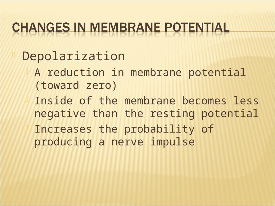
Depolarization A reduction in membrane potential (toward
zero) Inside of the membrane becomes less
negative than the resting potential Increases the probability of producing a
nerve impulse
Copyright © 2010 Pearson Education, Inc. Figure 11.9a
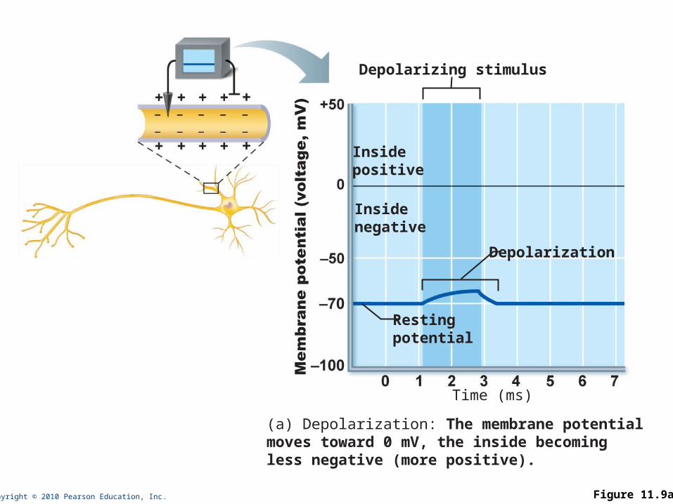
Depolarizing stimulus
Time (ms)
Insidepositive
Insidenegative
Restingpotential
Depolarization
(a) Depolarization: The membrane potentialmoves toward 0 mV, the inside becoming less negative (more positive).
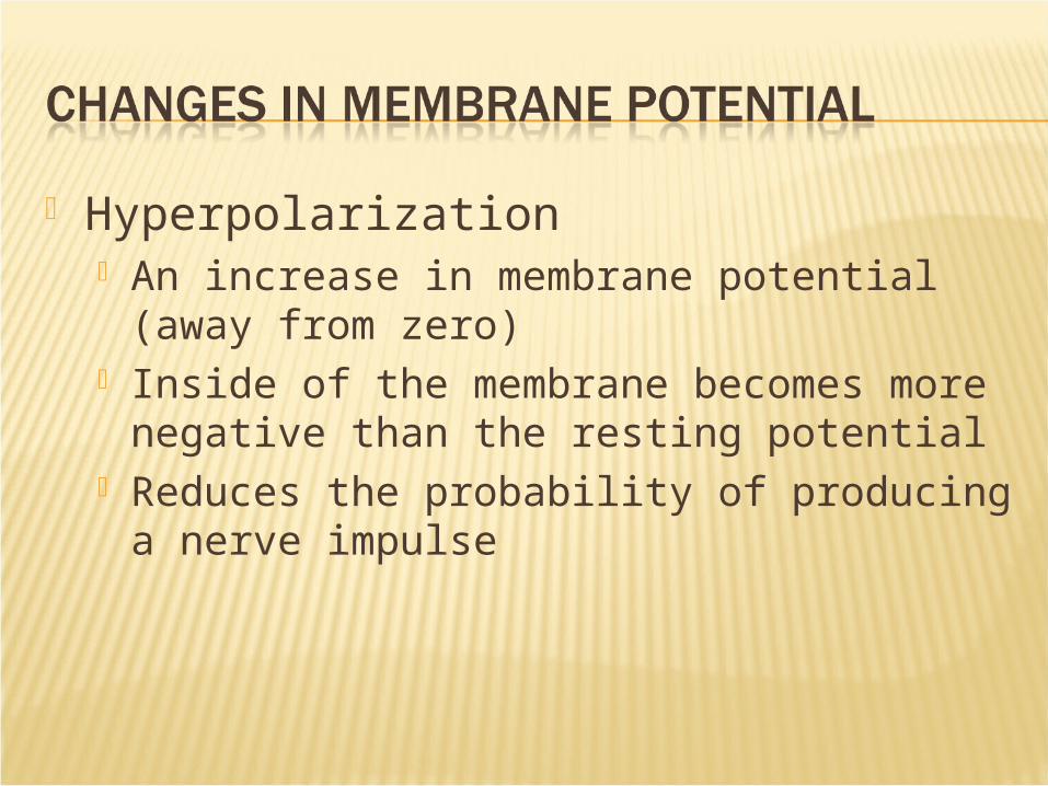
Hyperpolarization An increase in membrane potential (away
from zero) Inside of the membrane becomes more
negative than the resting potential Reduces the probability of producing a
nerve impulse
Copyright © 2010 Pearson Education, Inc. Figure 11.9b
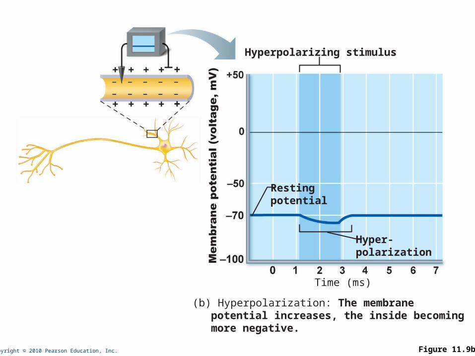
Hyperpolarizing stimulus
Time (ms)
Restingpotential
Hyper-polarization
(b) Hyperpolarization: The membranepotential increases, the inside becomingmore negative.

Short-lived, localized changes in membrane potential
Depolarizations or hyperpolarizations Graded potential spreads as local
currents change the membrane potential of adjacent regions
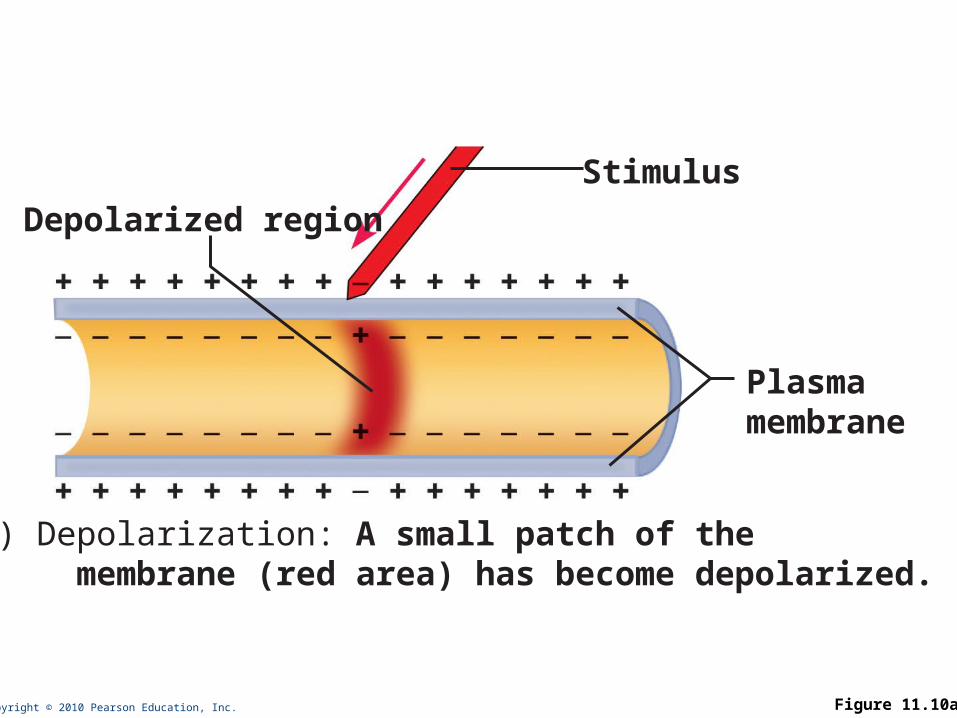
Copyright © 2010 Pearson Education, Inc. Figure 11.10a
Depolarized region
Stimulus
Plasmamembrane
(a) Depolarization: A small patch of the membrane (red area) has become depolarized.
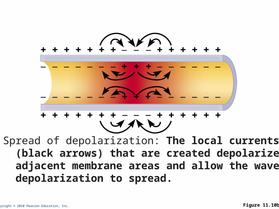
Copyright © 2010 Pearson Education, Inc. Figure 11.10b
(b) Spread of depolarization: The local currents (black arrows) that are created depolarize adjacent membrane areas and allow the wave of depolarization to spread.
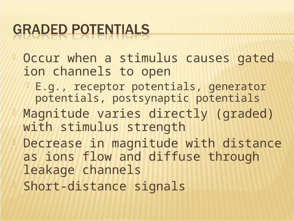
Occur when a stimulus causes gated ion channels to open E.g., receptor potentials, generator
potentials, postsynaptic potentials Magnitude varies directly (graded) with
stimulus strength Decrease in magnitude with distance as
ions flow and diffuse through leakage channels
Short-distance signals
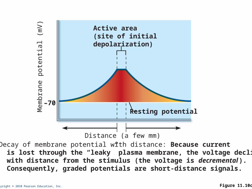
Copyright © 2010 Pearson Education, Inc. Figure 11.10c
Distance (a few mm)
–70Resting potential
Active area(site of initialdepolarization)
(c) Decay of membrane potential with distance: Because current is lost through the “leaky” plasma membrane, the voltage declines with distance from the stimulus (the voltage is decremental ). Consequently, graded potentials are short-distance signals.
Mem
bra
ne p
ote
nti
al (m
V)
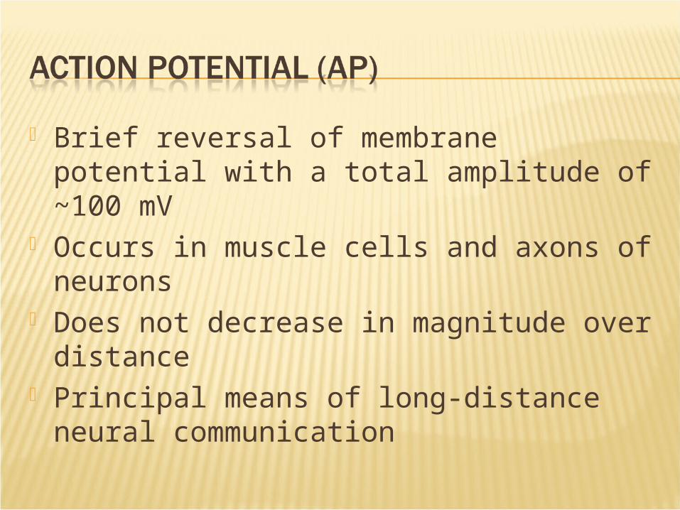
Brief reversal of membrane potential with a total amplitude of ~100 mV
Occurs in muscle cells and axons of neurons
Does not decrease in magnitude over distance
Principal means of long-distance neural communication

Copyright © 2010 Pearson Education, Inc.
Actionpotential
1 2 3
4
Resting state Depolarization Repolarization
Hyperpolarization
The big picture
1 1
2
3
4
Time (ms)
ThresholdMem
bra
ne p
ote
nti
al (m
V)
Figure 11.11 (1 of 5)
Resting state Only leakage channels for Na+ and K+ are
open All gated Na+ and K+ channels are closed

Properties of gated channels Each Na+ channel has two voltage-
sensitive gates Activation gates
Closed at rest; open with depolarization Inactivation gates
Open at rest; block channel once it is open
Each K+ channel has one voltage-sensitive gate
Closed at rest Opens slowly with depolarization

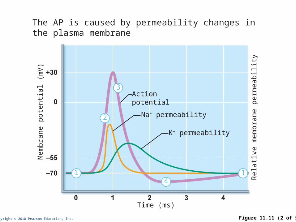
Depolarizing local currents open voltage-gated Na+ channels
Na+ influx causes more depolarization At threshold (–55 to –50 mV) positive
feedback leads to opening of all Na+ channels, and a reversal of membrane polarity to +30mV (spike of action potential)

Repolarizing phase Na+ channel slow inactivation gates close Membrane permeability to Na+ declines to
resting levels Slow voltage-sensitive K+ gates open K+ exits the cell and internal negativity is
restored
Hyperpolarization Some K+ channels remain open, allowing
excessive K+ efflux This causes after-hyperpolarization of the
membrane (undershoot)
Copyright © 2010 Pearson Education, Inc.
Actionpotential
Time (ms)
1 1
2
3
4
Na+ permeability
K+ permeability
The AP is caused by permeability changes inthe plasma membrane
Mem
bra
ne p
ote
nti
al (m
V)
Rela
tive m
em
bra
ne p
erm
eab
ility
Figure 11.11 (2 of 5)
Repolarization Restores the resting electrical conditions of
the neuron Does not restore the resting ionic
conditions Ionic redistribution back to resting
conditions is restored by the thousands of sodium-potassium pumps
Na+ influx causes a patch of the axonal membrane to depolarize
Local currents occur Na+ channels toward the point of origin
are inactivated and not affected by the local currents
Local currents affect adjacent areas in the forward direction
Depolarization opens voltage-gated channels and triggers an AP
Repolarization wave follows the depolarization wave
(Fig. 11.12 shows the propagation process in unmyelinated axons.)
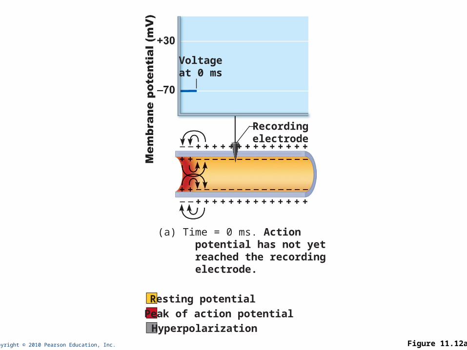
Copyright © 2010 Pearson Education, Inc. Figure 11.12a
Voltageat 0 ms
Recordingelectrode
(a) Time = 0 ms. Action potential has not yet reached the recording electrode.
Resting potential
Peak of action potential
Hyperpolarization

Copyright © 2010 Pearson Education, Inc. Figure 11.12b
Voltageat 2 ms
(b) Time = 2 ms. Action potential peak is at the recording electrode.

Copyright © 2010 Pearson Education, Inc. Figure 11.12c
Voltageat 4 ms
(c) Time = 4 ms. Action potential peak is past the recording electrode. Membrane at the recording electrode is still hyperpolarized.
At threshold: Membrane is depolarized by 15 to 20 mV Na+ permeability increases Na influx exceeds K+ efflux The positive feedback cycle begins

Subthreshold stimulus—weak local depolarization that does not reach threshold
Threshold stimulus—strong enough to push the membrane potential toward and beyond threshold
AP is an all-or-none phenomenon—action potentials either happen completely, or not at all

All action potentials are alike and are independent of stimulus intensity How does the CNS tell the difference between
a weak stimulus and a strong one? Strong stimuli can generate action
potentials more often than weaker stimuli The CNS determines stimulus intensity by
the frequency of impulses
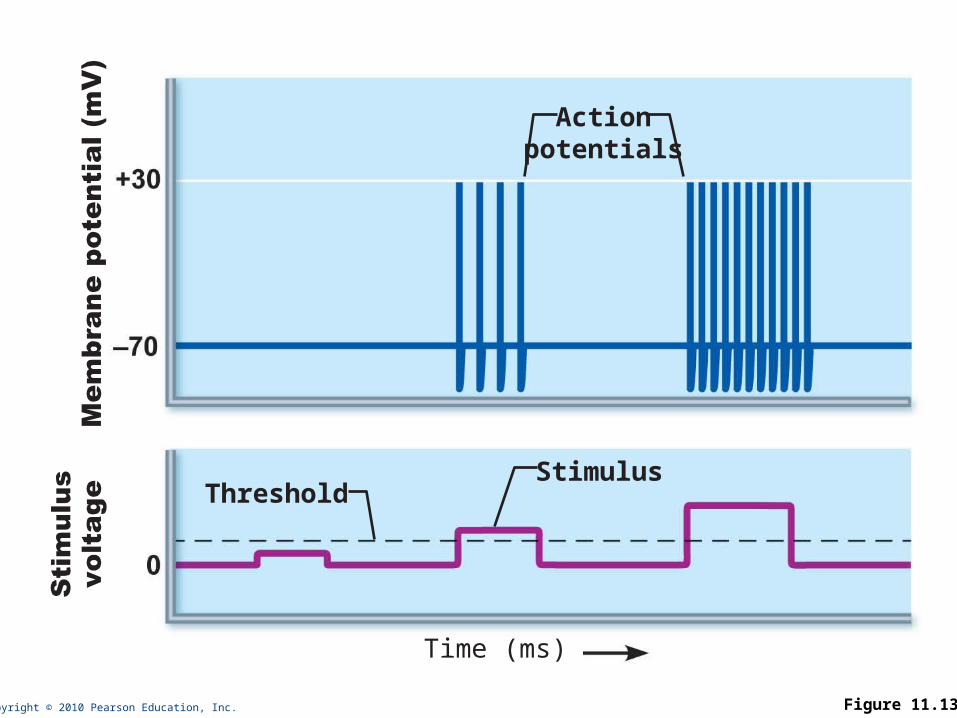
Copyright © 2010 Pearson Education, Inc. Figure 11.13
Threshold
Actionpotentials
Stimulus
Time (ms)
Time from the opening of the Na+ channels until the resetting of the channels
Ensures that each AP is an all-or-none event
Enforces one-way transmission of nerve impulses
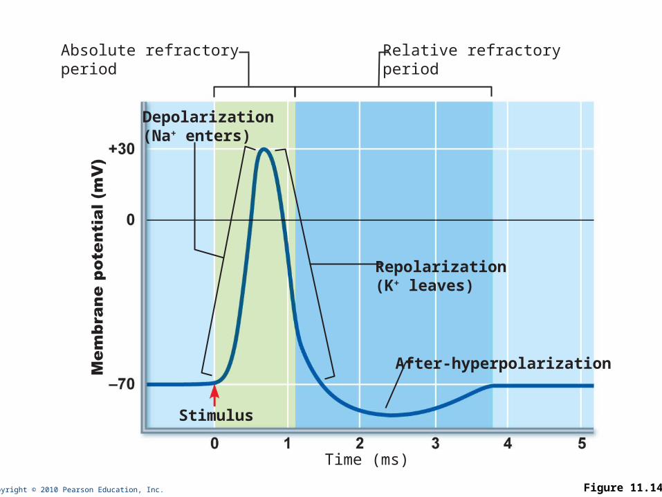
Copyright © 2010 Pearson Education, Inc. Figure 11.14
Stimulus
Absolute refractoryperiod
Relative refractoryperiod
Time (ms)
Depolarization(Na+ enters)
Repolarization(K+ leaves)
After-hyperpolarization
Follows the absolute refractory period Most Na+ channels have returned to their
resting state Some K+ channels are still open Repolarization is occurring
Threshold for AP generation is elevated Exceptionally strong stimulus may
generate an AP

Conduction velocities of neurons vary widely
Effect of axon diameter Larger diameter fibers have less resistance
to local current flow and have faster impulse conduction
Effect of myelination Continuous conduction in unmyelinated
axons is slower than saltatory conduction in myelinated axons
Effects of myelination Myelin sheaths insulate and prevent
leakage of charge Saltatory conduction in myelinated axons
is about 30 times faster Voltage-gated Na+ channels are located at the
nodes APs appear to jump rapidly from node to node
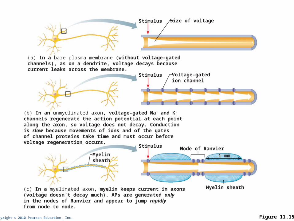
Copyright © 2010 Pearson Education, Inc. Figure 11.15
Size of voltage
Voltage-gatedion channel
Stimulus
Myelinsheath
Stimulus
Stimulus
Node of Ranvier
Myelin sheath
(a) In a bare plasma membrane (without voltage-gatedchannels), as on a dendrite, voltage decays becausecurrent leaks across the membrane.
(b) In an unmyelinated axon, voltage-gated Na+ and K+
channels regenerate the action potential at each pointalong the axon, so voltage does not decay. Conduction is slow because movements of ions and of the gatesof channel proteins take time and must occur beforevoltage regeneration occurs.
(c) In a myelinated axon, myelin keeps current in axons(voltage doesn’t decay much). APs are generated onlyin the nodes of Ranvier and appear to jump rapidlyfrom node to node.
1 mm
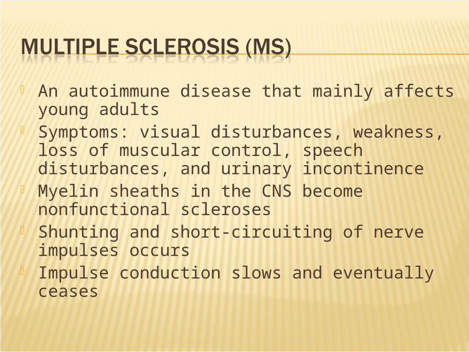
An autoimmune disease that mainly affects young adults
Symptoms: visual disturbances, weakness, loss of muscular control, speech disturbances, and urinary incontinence
Myelin sheaths in the CNS become nonfunctional scleroses
Shunting and short-circuiting of nerve impulses occurs
Impulse conduction slows and eventually ceases
Some immune system–modifying drugs, including interferons and Copazone: Hold symptoms at bay Reduce complications Reduce disability
Nerve fibers are classified according to: Diameter Degree of myelination Speed of conduction
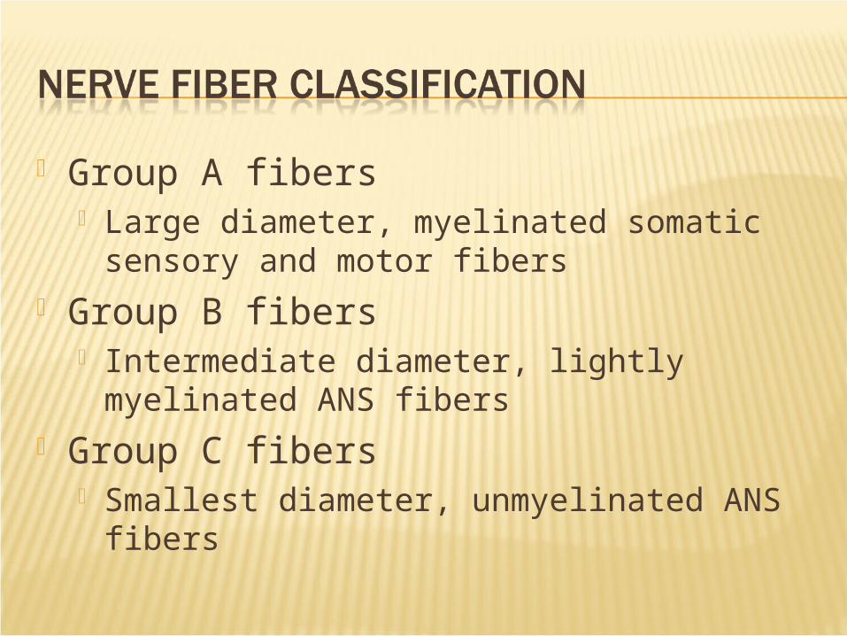
Group A fibers Large diameter, myelinated somatic
sensory and motor fibers Group B fibers
Intermediate diameter, lightly myelinated ANS fibers
Group C fibers Smallest diameter, unmyelinated ANS
fibers