Embed Size (px)
Citation preview

The Plant Cell, Vol. 15, 5–18, January 2003, www.plantcell.org © 2002 American Society of Plant Biologists
RESEARCH ARTICLE
LEAFY COTYLEDON1-LIKE Defines a Class of Regulators Essential for Embryo Development
Raymond W. Kwong,
a,b
Anhthu Q. Bui,
c,1
Hyeseung Lee,
a,b,1
Linda W. Kwong,
a
Robert L. Fischer,
d
Robert B. Goldberg,
c
and John J. Harada
a,b,2
a
Section of Plant Biology, Division of Biological Sciences, One Shields Avenue, University of California, Davis, California 95616
b
Graduate Group in Plant Biology, University of California, Davis, California 95616
c
Department of Molecular, Cell, and Developmental Biology, University of California, Los Angeles, California 90024-1606
d
Department of Plant and Microbial Biology, University of California, Berkeley, California 94720
Arabidopsis LEAFY COTYLEDON1 (LEC1) is a critical regulator required for normal development during the early and
late phases of embryogenesis that is sufficient to induce embryonic development in vegetative cells.
LEC1
encodes aHAP3 subunit of the CCAAT binding transcription factor. We show that the 10 Arabidopsis HAP3 (AHAP3) subunits canbe divided into two classes based on sequence identity in their central, conserved B domain. LEC1 and its most closelyrelated subunit, LEC1-LIKE (L1L), constitute LEC1-type AHAP3 subunits, whereas the remaining AHAP3 subunits aredesignated non-LEC1-type. Similar to
LEC1
,
L1L
is expressed primarily during seed development. However, suppres-
sion of
L1L
gene expression induced defects in embryo development that differed from those of
lec1
mutants, suggestingthat LEC1 and L1L play unique roles in embryogenesis. We show that
L1L
expressed under the control of DNA se-quences flanking the
LEC1
gene suppressed genetically the
lec1
mutation, suggesting that the LEC1-type B domains ofL1L and LEC1 are critical for their function in embryogenesis. Our results also suggest that LEC1-type HAP3 subunitsarose from a common origin uniquely in plants. Thus, L1L, an essential regulator of embryo development, defines aunique class of plant HAP3 subunits.
INTRODUCTION
The single-celled zygote of a flowering plant undergoes aseries of controlled cell divisions and cell differentiationevents that lead to the formation of a mature, multicellularembryo that is metabolically quiescent and desiccated.Early in embryogenesis, during the morphogenesis phase,the plant body is formed through the establishment of theshoot-root axis and the formation of the embryonic tissueand organ systems (West and Harada, 1993; Goldberg etal., 1994; Laux and Jurgens, 1997; Jurgens, 2001). Later,during the seed maturation phase, the embryo acquires theability to withstand desiccation, accumulates storage mac-romolecules such as lipids and proteins, and becomes met-abolically quiescent as a result of desiccation (reviewed byBewley, 1997; Harada, 1997). Once environmental condi-
tions are favorable, the seed germinates and the vegetativephase of the life cycle begins.
Genetic studies have identified regulatory genes that playcritical roles in embryogenesis during either the morphogen-esis or the maturation phases. For example, genes such as
WUSCHEL
,
SHOOTMERISTEMLESS
,
SCARECROW
, and
SHORT ROOT
(Dilaurenzio et al., 1996; Long et al., 1996;Mayer et al., 1998; Helariutta et al., 2000) have been shownto be essential for the formation of the shoot and root apicalmeristems that define the embryonic axis of developing Ara-bidopsis embryos. A different class of genes, including
AB-SCISIC ACID INSENSITIVE3
(
ABI3
),
ABI4
, and
ABI5
, playimportant roles during the maturation phase of embryogen-esis (Giraudat et al., 1992; Finkelstein et al., 1998; Finkelsteinand Lynch, 2000), preparing the embryo for desiccation andpostgerminative growth.
Another set of genes encoding Arabidopsis LEAFY COT-YLEDON (LEC) proteins, LEC1, LEC2, and FUSCA3, areunique in that they are the only known embryonic regulatorsrequired for normal development during both the morpho-genesis and maturation phases (reviewed by Harada, 2001).For example, LEC1 is required to maintain suspensor cell
1
These two authors contributed equally to this work.
2
To whom correspondence should be addressed. E-mail [email protected]; fax 530-752-5410.Article, publication date, and citation information can be found atwww.plantcell.org/cgi/doi/10.1105/tpc.006973.

6 The Plant Cell
fate, to specify cotyledon identity in the early morphogenesisphase, and to initiate and/or maintain the maturation phaseand inhibit precocious germination late in embryogenesis(Meinke, 1992; Meinke et al., 1994; West et al., 1994; Parcyet al., 1997; Lotan et al., 1998; Vicient et al., 2000). Further-more, ectopic postembryonic expression of
LEC1
is suffi-cient to confer embryonic characteristics to seedlings andto induce somatic embryo formation from vegetative cells(Lotan et al., 1998). Because LEC1 is required for normaldevelopment both early and late during embryogenesisand is sufficient to confer embryogenic competence tovegetative cells, it is a central regulator that acts far up-stream in the regulatory hierarchy that controls embryo-genesis. We speculate that
LEC1
establishes a cellular en-vironment that promotes embryo development and thatthis environment coordinates the early and late phases ofembryogenesis in flowering plants (Lotan et al., 1998). Amajor goal of our research is to understand, at a mechanis-tic level, how LEC1 establishes competence to initiate em-bryo development.
Given the key role of LEC1 in the control of embryogene-sis, we asked if genes related to
LEC1
also encode embry-onic regulators. LEC1 shares significant sequence similar-ity with the HAP3 subunit of CCAAT binding factor (CBF,also known as NF-Y; Lotan et al., 1998). CBFs are eukary-otic transcriptional activators that serve diverse roles in dif-ferent organisms (Li et al., 1992). In yeast, CBF activates a setof genes involved in mitochondrial respiration (Guarente etal., 1984; Keng and Guarente, 1987; Trueblood et al., 1988;Schneider and Guarente, 1991), whereas mammalian CBFsare thought to act generally to enhance transcription rates,often in combination with other proteins (reviewed byMaity and De Crombrugghe, 1998; Mantovani, 1999). Thetranscription factor is a hetero-oligomeric complex con-sisting of at least three subunits, HAP2, HAP3, and HAP5,although yeast possesses a fourth subunit, HAP4 (re-viewed by Maity and De Crombrugghe, 1998; Mantovani,1999). HAP3 subunits are recognized by their central Bdomain, an
�
90–amino acid region of the protein that isconserved across eukaryotic organisms. For example, theLEC1 B domain has 57 and 62% sequence identity withHAP3 subunits from yeast and mammals, respectively.Thus, LEC1 appears to encode a subunit of a transcriptionfactor that regulates the expression of genes required forembryo development.
We used the LEC1 polypeptide sequence to identify othergenes encoding Arabidopsis HAP3 (AHAP3) subunits. Weshow that the subunit most closely related to LEC1, desig-nated LEC1-LIKE (L1L), is required for normal embryo de-velopment. L1L and LEC1 have distinct functions in em-bryogenesis, but L1L can substitute functionally for LEC1when expressed ectopically. Comparison of the deducedamino acid sequences of L1L and LEC1 identified specificamino acid sequences that appear to be required for thefunction of these proteins in regulating embryo identity anddevelopment.
RESULTS
Arabidopsis HAP3 Proteins Are Encoded by a Gene Family That Can Be Divided into Two Classes
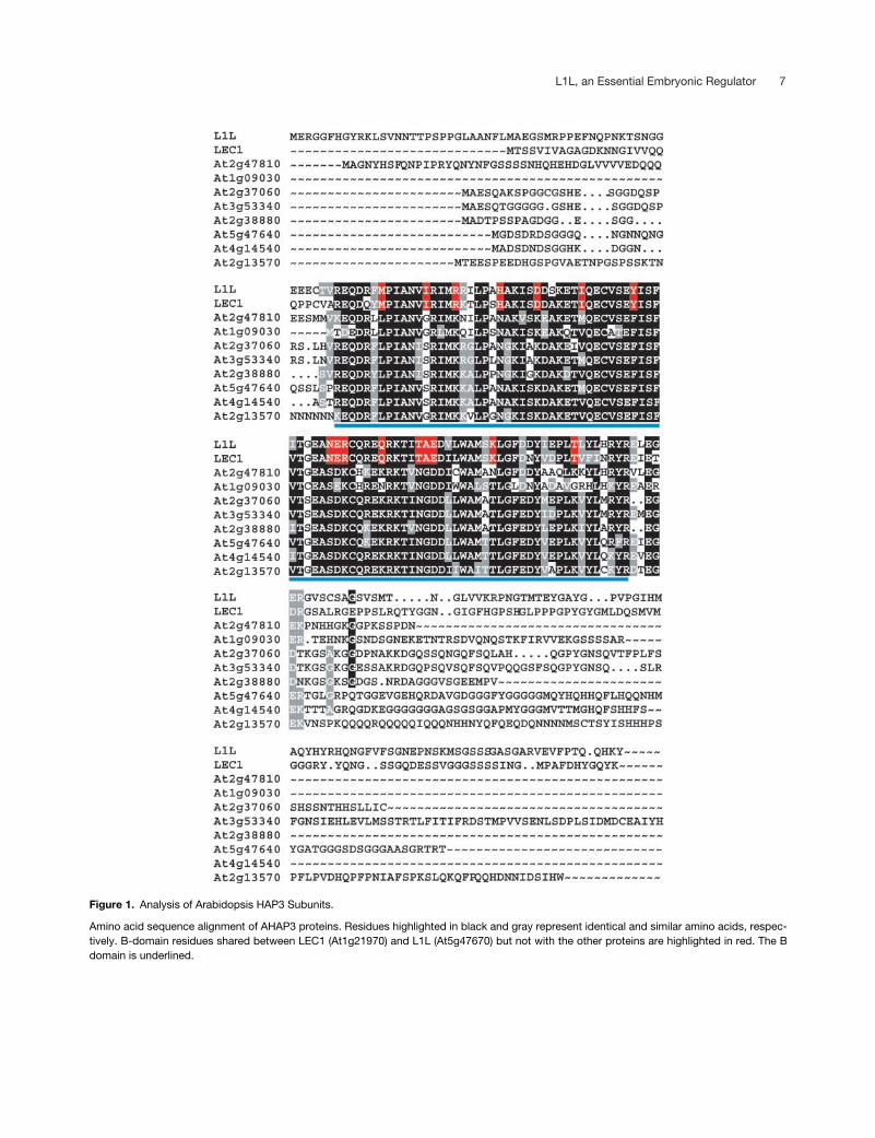
We used the amino acid sequence of LEC1 as a query toidentify related Arabidopsis polypeptides. Database searchesof the sequenced Arabidopsis genome showed that thereare nine genes encoding proteins that share significant se-quence identity and that the gene encoding L1L (At5g47670) isrelated most closely to LEC1. As shown in Figure 1, se-quence similarity among the 10 proteins is limited primarilyto the central B domain, consistent with comparisons ofHAP3 subunits from other organisms (Li et al., 1992). Onlytwo of these putative proteins, At2g37060 and At3g53340,display sequence identity with each other in the N-terminalA domain or the C-terminal C domain. Because the B do-main has been shown to underlie HAP3 function in other or-ganisms (Xing et al., 1993; Kim et al., 1996; Sinha et al.,1996) and because all of these Arabidopsis proteins pos-sess residues that are conserved among HAP3 proteins, weconclude that there are 10 AHAP3 subunits.
Close examination of B-domain sequence alignmentsshowed that L1L and LEC1 define a distinct class of AHAP3subunits. The two proteins share 83% sequence identitywith each other but only 52 to 71% identity with the othereight AHAP3 subunits. Furthermore, L1L and LEC1 sharethe amino acid residues highlighted in red in Figure 1 thatdiffer from the residues that are conserved in the other eightAHAP3 subunits. On the basis of sequence identity withinthe B domain, we define two classes of AHAP3 subunits:the LEC1-type and the non-LEC1-type. Thus, L1L is theAHAP3 most closely related to LEC1, opening the possibilitythat L1L also may be an embryonic regulator.
L1L
RNA Accumulates Primarily in Developing Embryos
We analyzed
L1L
gene expression to obtain clues about itsrole during development.
L1L
RNA was detected in RNA gelblot hybridization experiments in developing siliques but notin vegetative organs or in flowers, as shown in Figure 2A.This pattern of RNA accumulation closely resembled that of
LEC1
, which accumulates specifically in seeds and differedsubstantially from those of non-LEC1-type AHAP3 subunits,whose RNAs are not limited to seed development (Edwardset al., 1998; Lotan et al., 1998; Gusmaroli et al., 2001; M.Kim and J.J. Harada, unpublished results). However, differ-ences between
L1L
and
LEC1
RNA accumulation patternswere observed. Figure 2A shows that
L1L
RNA levelspeaked at a later stage of embryogenesis than did
LEC1
lev-els. Furthermore, sensitive reverse transcriptase–mediated(RT) PCR amplification experiments indicated that
L1L
RNAis present in all vegetative organs, although presumably atlow levels, as shown in Figure 2B. By contrast,
LEC1
RNA
L1L, an Essential Embryonic Regulator 7
Figure 1. Analysis of Arabidopsis HAP3 Subunits.
Amino acid sequence alignment of AHAP3 proteins. Residues highlighted in black and gray represent identical and similar amino acids, respec-tively. B-domain residues shared between LEC1 (At1g21970) and L1L (At5g47670) but not with the other proteins are highlighted in red. The Bdomain is underlined.

8 The Plant Cell
was not detected in vegetative organs in parallel experi-ments. These results suggest that L1L is likely to functionprimarily during seed development and that it is expresseddifferently from
LEC1
.We localized
L1L
RNA using in situ hybridization experi-ments to determine where it functions in developing si-liques. As shown in Figures 3A to 3C and 3E to 3G,
L1L
RNAwas detected at low but statistically significant levels in thedeveloping embryo proper, suspensor, and endosperm atearly stages, including zygotes (data not shown). During thetorpedo stage (Figures 3C and 3G) and the linear cotyledonstage (Figures 3D and 3H),
L1L
RNA became prevalent pri-marily in the outer cell layers of the embryo, similar to thedistribution of
LEC1
RNA (Lotan et al., 1998).
L1L
RNA be-came evenly distributed throughout the embryo at a highlevel by the bent-cotyledon stage (Figures 3I, 3J, 3M, and3N) and was present at low levels in mature-stage embryos(Figures 3K, 3L, 3O, and 3P). This temporal pattern of RNAaccumulation corresponds with the results from RNA gelblot analyses. Sense RNA did not bind appreciably with thesections, showing the specificity of the hybridization reac-tions (data not shown). Together, the RNA accumulationpatterns suggest a role for
L1L
in embryogenesis.
L1L
Is Required for Embryogenesis
Given that
L1L
is expressed primarily during embryogene-sis, we used RNA interference (RNAi) experiments to deter-mine if the suppression of
L1L
RNA levels affected embryodevelopment. Because
L1L
shares substantial identity withother AHAP3 subunits in the central B domain,
L1L
-specificnucleotide sequences encoding the C domain were used fortargeted suppression (see Methods). Wild-type plants weretransformed with the
L1L
RNAi construct under the controlof the
35S
promoter, and transgenic plants were recovered.Thirteen of 172 T1 transgenic lines produced defective T2seeds. More specifically, T1 plants from three independentlyderived lines segregated 30.1% (
n
�
1372), 31.9% (
n
�
1156), and 21.7% (
n
�
1327) defective T2 seeds. Althoughthe RNAi construct was incompletely penetrant, these re-sults suggest that L1L is required for embryo development.We also found that 4 of 15 lines containing a
35S
:
L1L
trans-gene segregated defective seeds. Together, these resultssuggest that cosuppression of
L1L
gene expression inducesdefects in embryogenesis (see below).
As shown in Figures 4B to 4D, RNAi mutants arrested at anumber of different embryonic stages with a range of mor-phological phenotypes. Some embryos arrested at the glob-ular stage (Figures 4B and 4C) but had extra cells in the sus-pensor. Other mutants arrested at later embryonic stagesand had reduced cotyledons (Figure 4D). Seeds containingthese defective embryos did not germinate, nor did imma-ture seeds collected before desiccation germinate in cul-ture. However, the effects appeared to be limited to seeddevelopment, because no defects in the vegetative devel-opment of the RNAi transgenic lines were detected. Sup-pression of
L1L
gene expression induced embryonic de-fects that differed from those caused by the
lec1
mutation(Lotan et al., 1998).
To confirm that defects in embryo development resultedfrom the silencing of the
L1L
gene, we analyzed
L1L
RNAlevels in transgenic lines using in situ hybridization experi-ments with
L1L
-specific probes that excluded sequencesencoding the C domain.
L1L
RNA was not detected at sig-nificant levels in 60 of 62 embryos with a mutant phenotype(Figures 4E and 4F) from four independent transgenic linesand was present only at a low level compared with the wildtype in the 2 other mutant embryos. Of embryos that segre-gated with a wild-type phenotype, 31% (
n
�
352) pos-sessed high levels of
L1L
RNA, similar to embryos with awild-type genotype (Figures 4G and 4H), whereas the re-mainder had only intermediate levels. We interpret these re-sults to indicate that very low levels of
L1L
RNA do not sup-port embryo development but intermediate levels aresufficient for normal embryogenesis. The incomplete pene-trance and variable expressivity of RNAi suppression of
L1L
gene expression probably allowed us to recover viableprogeny containing the transgene. We also demonstratedthe specificity of gene silencing by showing that
LEC1
, ole-osin, and cruciferin storage protein RNAs were detected in
Figure 2. L1L RNA Is Detected Predominantly in Developing Si-liques.
(A) Analysis of L1L and LEC1 RNA levels with RNA gel blot hybrid-ization experiments. Each lane contained 1 �g of poly(A) RNA fromsiliques with zygote- to early-globular-stage seeds (S1), siliques withglobular- to heart-stage seeds (S2), siliques with torpedo- to bent-cotyledon-stage seeds (S3), siliques with mature green seeds (S4),2-day-old seedlings (Sl), mature rosette leaves (Le), 3-week-oldseedling roots (Ro), stems (St), and unopened floral buds and inflo-rescences (Fl). Control represents the accumulation of a ribosomalprotein RNA.(B) RT-PCR amplification of L1L RNA. Abbreviations are as in (A)with the following additions: ND, no DNA; GD, wild-type genomicDNA; and lec1-1, mutant siliques with torpedo- to bent-cotyledon-stage embryos.

L1L, an Essential Embryonic Regulator 9
Figure 3. In Situ Detection of L1L RNA in Developing Embryos.
Wild-type seed sections were hybridized with an L1L-specific antisense probe. All sections were exposed for 10 days. (A) to (D) and (I) to (L)show bright-field micrographs, and (E) to (H) and (M) to (P) show dark-field micrographs. The sense RNA control did not bind appreciably withthe sections. Bars � 50 �m.(A) and (E) Globular-stage embryo.(B) and (F) Heart-stage embryo.(C) and (G) Linear cotyledon-stage embryo.(D) and (H) Early bent-cotyledon-stage embryo.(I) and (M) Bent-cotyledon-stage embryo.(J) and (N) Late bent-cotyledon-stage embryo.(K) and (O) Mature green-stage embryo.(L) and (P) Mature yellowing-stage embryo.

10 The Plant Cell
RNAi embryos exhibiting a mutant phenotype as they were inwild-type embryos (data not shown). Together, these data sug-gest strongly that
L1L is essential for embryo development.
Ectopically Expressed L1L Can Function in Placeof LEC1
To examine the functional relationship between L1L andLEC1, we asked if L1L could suppress the lec1 mutationwhen expressed ectopically. L1L RNA was detected in lec1-1null mutants (Figure 2B), indicating that the endogenous L1Lgene cannot substitute completely for the LEC1 gene. Al-though the spatial distribution of L1L RNA was similar tothat of LEC1, there were temporal differences in accumula-tion during embryogenesis (Figures 2 and 3) (Lotan et al.,1998). Therefore, we fused the L1L coding region with 1997and 774 bp of sequence 5� and 3�, respectively, of the LEC1coding region and transferred the chimeric gene into lec1-1null mutants. The lec1 mutation causes embryos to becomeintolerant of desiccation, and no mutant seeds germinate
(Meinke, 1992; West et al., 1994; Lotan et al., 1998). Asshown in Table 1 and Figure 5A, transgenic lec1-1 mutantseeds that were dried extensively (see Methods) producedviable seedlings, indicating that L1L expressed under thecontrol of LEC1 flanking DNA sequences was able to rescuethe desiccation intolerance of lec1 mutants. Moreover, noembryonic or postembryonic abnormalities were detected inlec1 mutant plants containing the L1L transgene, and T1plants segregated progeny with wild-type and mutant phe-notypes at ratios indicating the presence of one, two, ormultiple transgenes (data not shown). By contrast, the ex-pression of two genes encoding non-LEC1-type AHAP3subunits, At4g14540 and At3g53340, under the control ofLEC1 5� and 3� flanking sequences did not rescue the des-iccation intolerance of the lec1 mutants significantly (Table1). Together, these results suggest that L1L but not non-LEC1-type AHAP3 genes can function in place of LEC1when expressed ectopically.
We also showed that L1L reproduced effects caused byLEC1 when both were expressed postembryonically. Wefused the L1L coding region with the 35S promoter from
Figure 4. RNAi Suppression of L1L Gene Expression Induces Embryo Defects.
(A) Seed with a wild-type embryo at the bent-cotyledon stage. The seed was cleared and viewed with Nomarski optics.(B) to (D) Cleared seeds containing defective embryos from lines containing the L1L RNAi constructs. Progeny segregating with a wild-type phe-notype in the same silique were at the bent-cotyledon stage.(E) to (H) L1L RNA accumulation in defective embryos. Sections were hybridized with an antisense L1L probe and exposed for 10 days.(E) and (F) Bright- and dark-field micrographs of a defective embryo from a line containing the L1L RNAi construct.(G) and (H) Bright- and dark-field micrographs of a wild-type embryo at the mature green stage.Bars � 50 �m in (A), (D), (E), and (H) and 25 �m in (B) and (C).
L1L, an Essential Embryonic Regulator 11
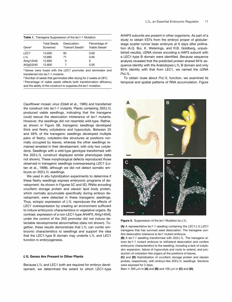
Cauliflower mosaic virus (Odell et al., 1985) and transferredthe construct into lec1-1 mutants. Plants containing 35S:L1Lproduced viable seedlings, indicating that the transgenecould rescue the desiccation intolerance of lec1 mutants.However, the seedlings did not resemble wild-type. Rather,as shown in Figure 5B, transgenic seedlings developedthick and fleshy cotyledons and hypocotyls. Between 25and 59% of the transgenic seedlings developed multiplepairs of fleshy, cotyledon-like structures at positions nor-mally occupied by leaves, whereas the other seedlings re-mained arrested in their development, with only two cotyle-dons. Seedlings with a wild-type genotype transformed withthe 35S:L1L construct displayed similar phenotypes (datanot shown). These morphological defects reproduced thoseobserved in transgenic seedlings overexpressing LEC1 (Lo-tan et al., 1998), although we did not detect somatic em-bryos on 35S:L1L seedlings.
We used in situ hybridization experiments to determine ifthese fleshy seedlings express embryonic programs of de-velopment. As shown in Figures 5C and 5D, RNAs encodingcruciferin storage protein and oleosin lipid body protein,which normally accumulate specifically during embryo de-velopment, were detected in these transgenic seedlings.Thus, ectopic expression of L1L reproduces the effects ofLEC1 overexpression by creating an environment sufficientto induce embryonic characteristics in vegetative organs. Bycontrast, expression of a non-LEC1-type AHAP3, At4g14540,under the control of the 35S promoter did not induce de-tectable developmental abnormalities (data not shown). To-gether, these results demonstrate that L1L can confer em-bryonic characteristics to seedlings and support the ideathat the LEC1-type B domain may underlie L1L and LEC1function in embryogenesis.
L1L Genes Are Present in Other Plants
Because L1L and LEC1 both are required for embryo devel-opment, we determined the extent to which LEC1-type
AHAP3 subunits are present in other organisms. As part of astudy to obtain ESTs from the embryo proper of globular-stage scarlet runner bean embryos at 6 days after pollina-tion (A.Q. Bui, K. Weterings, and R.B. Goldberg, unpub-lished results), cDNA clones encoding a HAP3 subunit witha LEC1-type B domain were identified. Because sequenceanalysis revealed that the predicted protein shared 94% se-quence identity with the Arabidopsis L1L B domain and only85% identity with that from LEC1, we named the cDNAPcL1L.
To obtain clues about PcL1L function, we examined itstemporal and spatial patterns of RNA accumulation. Figure
Table 1. Transgene Suppression of the lec1-1 Mutation
Genea
Total SeedsScreened
Desiccation-Tolerant Seedsb
Percentage ofViable Seedsc
LEC1 13,600 82 0.60L1L 10,885 71 0.65At4g14540 12,800 0 0At3g53340 12,800 7 0.05
a Genes were fused with the LEC1 promoter and terminator andtransferred into lec1-1 mutants.b Number of seeds that germinated after drying for 2 weeks at 28�C.c Percentage of viable seeds reflects both transformation efficiencyand the ability of the construct to suppress the lec1 mutation.
Figure 5. Suppression of the lec1 Mutation by L1L.
(A) A representative lec1-1 seedling containing the LEC1:L1L:LEC1transgene that has survived seed desiccation. The transgene con-fers desiccation tolerance to lec1 mutant embryos.(B) A lec1-1 seedling transformed with 35S:L1L. The transgene al-lows lec1-1 mutant embryos to withstand desiccation and confersembryonic characteristics to the seedling, including a lack of cotyle-don expansion, failure of hypocotyls and roots to extend, and pro-duction of cotyledon-like organs at the positions of leaves.(C) and (D) Hybridization of cruciferin storage protein and oleosinprobes, respectively, with embryo-like 35S:L1L seedlings. Sectionswere exposed for 2 days.Bars � 300 �m in (A) and (B) and 100 �m in (C) and (D).

12 The Plant Cell
6A shows that PcL1L RNA was detected only in developingembryos by RNA gel blot analysis. However, RT-PCR analy-sis showed that PcL1L RNA was present, presumably at lowlevels, at all developmental stages tested, including in vege-tative organs, inflorescences, and unfertilized ovules (Figure6B). Thus, the pattern of PcL1L RNA accumulation is moresimilar to that of L1L than that of LEC1 in Arabidopsis (Fig-ure 2). In situ hybridization analysis showed that PcL1L RNAaccumulated at high levels in the embryo proper and sus-pensor of preglobular-stage (5 days after pollination [DAP])and globular-stage (7 DAP) embryos but at very low levels inthe endosperm of the seeds and integuments of the unfertil-ized ovules (Figures 6C to 6E). PcL1L RNA was present atthe highest levels in the epidermal and subepidermal layersof the embryo proper at the globular stage (Figure 6D). Thispattern is similar to the distribution of Arabidopsis L1L RNAobserved at the torpedo and bent-cotyledon stages (Figures3C and 3D). The finding that scarlet runner bean appears topossess a protein with a LEC1-type B domain and that RNAencoding this protein accumulates with a pattern similar tothat of Arabidopsis L1L suggests that the two L1L genes areorthologous.
Through database searches, we extended this analysis byidentifying 12 other HAP3 subunits from other plants withconserved amino acid residues characteristic of the LEC1-type B domain. Most were identified from embryo or seed-derived cDNA libraries, although sequences from pine pollencone and lotus root nodule cDNA libraries were identified.As shown in Figure 7A, alignment of the B domains of these15 plant HAP3 subunits revealed 17 amino acid residuesthat are shared between LEC1-type B domains but that dif-fer from residues conserved in non-LEC1-type B domains.By contrast, no conserved amino acid residues were de-tected in the A and C domains of these 15 proteins, al-though some similarities were observed in these regions be-tween L1L, PcL1L, and some of the other plant L1L proteins(data not shown). The phylogenetic tree obtained by maxi-mum parsimony analysis (PAUP 4.0) shown in Figure 7B in-dicates that the L1L proteins constitute a well-supported,monophyletic clade. Based on their sequence identity withinthe B domain, their origin in seed RNA populations, and theability of L1L to suppress the lec1 mutation when expressedectopically, we speculate that some of these L1L genes alsomay play roles in embryogenesis.
DISCUSSION
L1L and LEC1 Constitute a Subclass of HAP3 Subunits
L1L and LEC1 display substantial sequence identity withHAP3 subunits of CBFs. Although a CBF from plants hasnot been isolated, several lines of evidence suggest that L1Land LEC1 function as part of a CBF that regulates embryo-genesis. First, in addition to HAP3, paralogs of the other two
Figure 6. PcL1L RNA Is Present Primarily in Developing Seeds.
(A) Gel blot analysis of PcL1L RNA accumulation. Twenty-five micro-grams of total RNA was analyzed from leaves (Le), stems (St), 2-week-old seedling leaves (Sl), 2-week-old seedling roots (SlRo), 2-week-oldseedling stems (SlSt), inflorescences (In), ovules (Ov), 2-DAP seeds(Se I), 4- to 5-DAP seeds (Se II), 6-DAP seeds (Se III), 12- to 14-DAPembryos (Em I), and 19- to 21-DAP embryos (Em II).(B) RT-PCR analysis of PcL1L RNA accumulation. Each lane corre-sponds to the RNA gel blot sample in (A).(C) to (F) Distribution of PcL1L RNA. Sections were hybridized with aPcL1L antisense probe (C) to (E) or a sense RNA control (F). (C) and(D) were exposed for 4 days, whereas (E) was exposed for 47 days.(C) Preglobular-stage seed. PcL1L RNA is high in the embryo properand suspensor.(D) Globular-stage seed. PcL1L RNA is at its highest levels in outertissue layers of the embryo.(E) Unfertilized ovule. PcL1L RNA is present at low levels throughoutthe ovule.(F) Unfertilized ovule that does not bind sense RNA probe.Bars � 100 �m.

L1L, an Essential Embryonic Regulator 13
CBF subunits required for DNA binding activity, HAP2 andHAP5, have been identified in plants (Li et al., 1992; Albaniand Robert, 1995; Edwards et al., 1998; Kusnetsov et al.,1999; Gusmaroli et al., 2001). Unlike yeast and mammals,which possess single genes for each subunit, plants pos-sess families of subunits. For example, there are 6, 10, and8 genes encoding the AHAP2, AHAP3, and AHAP5 sub-units, respectively, in Arabidopsis, opening the possibilitythat different combinations of subunits may regulate diverseprocesses (Edwards et al., 1998; Gusmaroli et al., 2001; M.Kim, H. Lee, R.W. Kwong, and J.J. Harada, unpublished re-sults). Second, an AHAP2 gene has been shown to comple-ment a yeast HAP2 mutation, indicating that the Arabidopsisprotein can function in a CBF (Edwards et al., 1998). Third,an AHAP5 subunit has been shown to interact with othernuclear proteins, presumably AHAP2 and AHAP3, to form acomplex that binds a double-stranded oligonucleotide con-taining a CAAT box (Kusnetsov et al., 1999). Fourth, loss-of-function mutations of L1L and LEC1, two HAP3 paralogs,have severe consequences on plant development, suggest-ing that these subunits play essential roles.
Our results show that there are at least two distinctclasses of AHAP3 subunits that differ in several respects.LEC1-type and non-LEC1-type HAP3 subunits differ by 16amino acid residues that serve as signatures of their B do-mains (Figure 1) (Gusmaroli et al., 2001). Phylogenetic anal-ysis suggests that HAP3 subunits possessing these signa-ture residues have a common evolutionary origin (Figure 7).Residues at corresponding positions of yeast and mamma-lian HAP3 subunits are more similar to non-LEC1-type thanto LEC1-type AHAP3 subunits. This finding opens the pos-sibility that L1L and LEC1 represent novel HAP3 subunits ofCBF. Next, sequence diversity between the two types ofsubunits appears to underlie the functional differences, be-cause L1L but not two other genes that encode non-LEC1-type AHAP3 subunits suppressed the lec1 mutation whenexpressed under the control of LEC1 flanking sequences(Table 1). Similarly, a non-LEC1-type AHAP3 did not induceembryonic characteristics in seedlings when fused with the35S promoter, as did L1L and LEC1 (Table 1, Figure 5). Fi-nally, genes encoding L1L and LEC1 are expressed pre-dominately or exclusively during seed development (Figure2) (Lotan et al., 1998), whereas the non-LEC1-type AHAP3genes generally are expressed at high levels in nonembry-onic tissues (Edwards et al., 1998; Gusmaroli et al., 2001; M.Kim and J.J. Harada, unpublished results). In this regard,LEC1, L1L, and PcL1L exhibit similar spatial patterns ofRNA accumulation in developing embryos (Figures 3 and 6)(Lotan et al., 1998).
The B Domain Underlies the Function of LEC1-Type HAP3 Subunits in Embryogenesis
We present strong evidence that the B domain of LEC1-typeHAP3 subunits underlies their function in embryogenesis. Se-
quence similarity between L1L and LEC1 was observed ex-clusively in the B domain and not in the A and C domains(Figure 1). Suppression of the lec1 mutation by L1L (Figure5, Table 1) suggests that some or all of the 16 residuesunique to LEC1-type B domains account for the ability ofL1L to substitute functionally for LEC1 when expressed ec-topically. Most HAP3 subunits from other plants that pos-sess LEC1-type B domains (Figure 7) are present in em-bryos or seeds, consistent with the expression patterns ofL1L and LEC1. This finding opens the possibility that otherL1Ls play important roles in seed development. However,two L1Ls are present in pollen cones and root nodules, sug-gesting that the LEC1-type HAP3 subunit may function atother developmental stages. This class of HAP3 subunit ispresent in gymnosperms and monocotyledonous and dicot-yledonous angiosperms, but it has not been detected innonplant organisms, suggesting that the LEC1-type B do-main evolved uniquely in plants.
The central B domain of HAP3 subunits serves criticalroles in CBF function. Studies of yeast and mammalianHAP3s show that the B domain contains amino acid resi-dues that account for its ability to interact with HAP2 andHAP5 subunits and for the CBF to bind DNA (Xing et al.,1993; Kim et al., 1996; Sinha et al., 1996). One explanationfor the differences in the activities of L1L and LEC1 versusnon-LEC1-type AHAP3 subunits is that LEC1-type B do-mains may mediate interactions with specific AHAP2 andAHAP5 subunits to form a CBF that activates the genesrequired for embryo development. Because AHAP2 andAHAP5 are encoded by six and eight genes, respectively,and most are expressed in nonseed tissues (Gusmaroli etal., 2001; H. Lee, M. Kim, and J.J. Harada, unpublishedresults), the possibility exists that defined combinations ofAHAP subunits confer specific functions to the transcrip-tion complex. However, this interpretation requires thatthe specific AHAP2 and AHAP5 subunits present in em-bryos also are present in vegetative tissues, because ec-topic expression of L1L and LEC1 confers embryoniccharacteristics to vegetative tissues (Figure 5) (Lotan etal., 1998).
A second possibility is that the LEC1-type B domain mayrecruit other transcription factors to the CBF that conferunique specificity to the complex. CBFs have been shownto interact with other transcription factors to activate spe-cific sets of genes. For example, activation of the MHC classII gene promoter requires the binding of CBF and an X-boxbinding factor, and activation of the 3-hydroxy-3-methylglu-taryl-CoA synthase gene requires the binding of both CBFand sterol regulatory element binding proteins (Wright et al.,1994; Linhoff et al., 1997; Dooley et al., 1998). In addition,no Arabidopsis paralog of the HAP4 subunit that provides atranscriptional activation domain to the yeast CBF has beenidentified, and transcriptional activation domains are not ap-parent in the HAP2 and HAP5 subunits as they are in theirmammalian counterparts (Forsburg and Guarente, 1989;Coustry et al., 1996). Thus, a protein with transcriptional

14 The Plant Cell
activation function may be recruited to the complex by L1Land LEC1.
A final alternative is that B domain residues unique toLEC1-type HAP3 subunits may confer a novel DNA bindingspecificity to the CBF that differs from that afforded by non-LEC1-type AHAP3 subunits. Thus, CBFs containing L1L andLEC1 would bind and modulate the transcription of genesrequired for embryo development, whereas non-LEC1-typeAHAP3s would not. This is the simplest alternative, becausethere is no need to invoke novel interactions with other pro-teins. However, to our knowledge, no HAP3 subunit hasbeen identified that alters the binding specificity of CBFs inother organisms.
L1L and LEC1 Have Distinct Functionsduring Embryogenesis
Although our studies have shown that L1L can substitute forLEC1 if expressed ectopically (Table 1, Figure 5), other evi-dence suggests that L1L and LEC1 normally have distinctfunctions during embryogenesis. The first and most compel-ling argument is that monogenic, loss-of-function mutationsin either L1L or LEC1 cause defects in embryo develop-ment. These results show that the endogenous genes can-not substitute for one another, although we cannot excludethe possibility that the genes have partial overlaps in func-tion. Consistent with this interpretation is the finding that thesuppression of L1L and LEC1 gene expression induces dif-ferent embryonic phenotypes. lec1 mutants arrest at a latestage of embryo development, with complete though mis-shapen cotyledons and embryonic axes, and mutant em-bryos can be rescued before desiccation to produce viableseedlings (reviewed by Harada, 2001). By contrast, RNAisuppression of L1L caused embryos to arrest in their devel-opment as early as the globular stage, and mutant embryoscannot be rescued to produce postembryonic plants (Figure4). We conclude that although L1L clearly is required for em-bryo development, it appears to play a fundamentally differ-ent role in embryogenesis than LEC1.
With regard to the L1L mutant phenotype, RNAi suppres-sion of L1L gene expression is characterized by incompletepenetrance and variable expressivity. Not all embryos con-taining the construct exhibit a mutant phenotype, and thosethat do arrest at a number of different embryonic stageswith a variety of defects (Figure 4). However, transgenic em-bryos displaying a mutant phenotype possessed low to un-detectable levels of L1L RNA, whereas those with a wild-
Figure 7. Identification of L1L Proteins from Other Plants.
(A) Amino acid sequence alignment of the B domains of plant L1Lproteins. Conserved amino acid residues are highlighted in gray,and residues unique to L1L proteins are highlighted in black. Acces-sion numbers are given at the end of Methods.(B) Phylogenetic relationships between L1L and non-LEC1-type-HAP3 subunits. The cladogram illustrates the most parsimoniousconsensus pattern of relationships obtained using maximum parsi-
mony analysis. Bootstrap values generated with 1000 replicates areindicated before the nodes. Nodes with bootstrap scores of �50%are not shown. The high bootstrap values provide strong support forthe monophyletic L1L clade.

L1L, an Essential Embryonic Regulator 15
type phenotype had intermediate to high RNA levels (Figure4). Therefore, defects in embryo development appear to re-sult from the suppression of L1L expression. Although wehave not yet identified an insertional mutation of L1L, wenote the possibility that the RNAi suppression of L1L maynot produce a mutant phenotype as severe as that of a ge-netic null mutation. The RNAi construct is controlled by the35S promoter, and we have shown that this promoter doesnot become active detectably during embryogenesis untilthe globular stage (J. Pelletier and J.J. Harada, unpublishedresults). Thus, L1L RNA may accumulate early during em-bryogenesis in RNAi lines, albeit at a very low level, and de-cline only after the globular stage. Despite these qualifica-tions, it is unlikely that a null l1l mutant would share similarcharacteristics with the lec class of mutants, because l1lmutant embryos arrest earlier in embryogenesis than dolec1 mutants.
Three other lines of evidence support the conclusion thatL1L and LEC1 have distinct endogenous functions. First,L1L RNA accumulates later in embryogenesis than doesLEC1 RNA (Figure 2). Second, L1L RNA is present in devel-oping seeds and at low levels in vegetative organs, whereasLEC1 RNA is detected only in developing seeds (Lotan etal., 1998) (Figure 2). Third, lec1 mutants display an abnormalphenotype even though L1L RNA is detected in the mutantseeds, indicating that the endogenous L1L gene is not suffi-cient to completely prevent defects induced by the lec1 mu-tation (Figure 2).
There are several potential explanations to reconcile thefindings that the endogenous L1L and LEC1 genes do notact redundantly, yet L1L can be made to substitute func-tionally for LEC1. One hypothesis is that the specific patternof LEC1 gene expression is critical for its function. Althoughthe distribution of LEC1 and L1L RNAs in embryos is similar,there are differences in the timing of their accumulation (Fig-ures 2 and 3) (Lotan et al., 1998). Similar situations havebeen described for two Arabidopsis MYB genes, WERE-WOLF and GLABROUS1. Genes encoding these function-ally equivalent proteins play different roles in plant develop-ment because they are transcribed in distinct cell types (Leeand Schiefelbein, 2001). Alternatively, increased dosage ofthe L1L gene and, by inference, increased L1L RNA levelsin transgenic lec1 mutants containing the LEC1:L1L:LEC1transgene may account for the suppression of the mutation.Dosage suppression has been described in microorganisms(Puziss et al., 1994). A third possibility is that because theaccumulation of LEC1 and L1L RNA does not differ sub-stantially, RNA sequences in the LEC1 5� and/or 3� untrans-lated regions, which are included in the LEC1:L1L:LEC1gene (see Methods), may regulate LEC1 function at thetranslational level. Additional information is needed to distin-guish between these possibilities.
In conclusion, we have shown that L1L, the AHAP3 sub-unit most closely related to LEC1, is a regulator of embryodevelopment. L1L is expressed predominately during em-bryo development, and it is required for the completion of
embryogenesis. The ability of L1L but not non-LEC1-typeAHAP3 subunits to function in place of LEC1 when ex-pressed ectopically implicates the B domain as the region ofL1L and LEC1 that is critical for their function. Mutagenesisstudies are needed to define which of the 16 amino acid res-idues of LEC1-type AHAP3 subunits differentiate their func-tions from non-LEC1-type subunits. Although L1L can func-tion redundantly with LEC1 when expressed ectopically, thetwo subunits have distinct functions during embryogenesis.Thus, L1L is a novel regulatory protein that plays an essen-tial role during embryogenesis.
METHODS
Plant Materials and Manipulations
lec1-1 mutants and wild-type plants of Arabidopsis thaliana (ecotypeWassilewskija) were grown as described previously (West et al.,1994). Seeds of the day-neutral scarlet runner bean (Phaseolus coc-cineus cv Hammond’s Dwarf Red Flower) were grown in the green-house as described by Weterings et al. (2001). Seeds were germi-nated in vermiculite to obtain seedlings. Flowers were pollinated andcollected at specific days after pollination (DAP) as described previ-ously (Weterings et al., 2001).
Approximately 500 unfertilized runner bean ovules were collectedfrom young, open flowers. Approximately 90, 66, and 50 seeds werecollected from 2-DAP, 4- to 5-DAP, and 6-DAP pods, respectively.Approximately 100 cotyledon-stage embryos were isolated fromseeds of 12- to 14-DAP and 19- to 21-DAP pods. Seed and embryostages were according to Weterings et al. (2001). Small young leaves,stems, and inflorescences were collected from lateral branches offlowering plants. True leaves, roots, and stems were collected from2-week-old seedlings. Upon collection, tissues were frozen immedi-ately in liquid nitrogen and stored at �80�C until use.
Agrobacterium tumefaciens strain GV3101 containing transfor-mation constructs was infiltrated into lec1-1 and wild-type plants(Bechtold et al., 1993). Seeds from T0 plants were germinated onmedium containing 60 �g/mL glufosinate ammonium to select fortransgenic plants (Finale; AgrEvo Environmental Health, Montvale,NJ). Plant genotypes were verified in PCR amplification experiments.
Isolation and Preparation of cDNA and Genomic Clones
PCR was used to amplify the genomic fragments containing AHAP3genes. Primers that flanked the putative L1L open reading frame withthe addition of BamHI and XbaI sites for subcloning purposes wereused (BAMMNJ7-5, 5�-AGGATCCATGGAACGTGGAGGCTTCCAT-3�;and 3-MNJ7XBA, 5�-ATCTAGATCAGTACTTATGTTGTTGAGTCG-3�).The AHAP3 genes At4g14540 (3-224) and At3g53340 (3-180) were am-plified using primer combinations 3-224-F/3-224-R (5�-CCTATC-TCGAGATGGCGGATTCGGACAACGATTC-3�/5�-CCCGGTCTAGAT-TAAGAAAAATGATGGGAAAATTGATGTCC-3�) and AH3-180-F/AH3-180-R (5�-CCCGGGGAGATCTATGGCGGATACGCCTTCGAGCCC-AGC-3�/5�-GGGCCCCTAGGCTTTTACCAGCTCGGCATTTCTTCA-CC-3�), respectively. Nucleotide sequences of the genomic cloneswere verified.
L1L, At4g14540, and At3g53340 genomic clones were inserted

16 The Plant Cell
between the LEC1 promoter and terminator within the plant transfor-mation vector BJ49 (Gleave, 1992). The LEC1 promoter/terminatorcassette consists of 1992 bp of DNA 5� of the LEC1 translation startcodon plus 770 bp 3� of the LEC1 stop codon (H. Lee and J.J.Harada, unpublished results). The L1L gene was fused with the 35Spromoter from Cauliflower mosaic virus and the octopine synthaseterminator of the plasmid pART7 and transferred into the binarytransformation vector pMLBART (Gleave, 1992).
cDNA clone pPCEP112 was identified from a scarlet runner beancDNA library by EST sequencing analysis. This cDNA library wasconstructed with total RNA isolated from the embryo proper of 6-DAPseeds using the SMART PCR cDNA Library Construction Kit (Clon-tech, Palo Alto, CA) (A.Q. Bui, K. Weterings, and R.B. Goldberg, un-published results).
Protein Sequence Analysis
Amino acid sequences were aligned with the PileUp program (Seq-web version 2.0.2; Accelrys, Burlington, MA), and alignments wereprepared with BOXSHADE (ch.EMBnet.org). Database searcheswere performed with the LEC1 protein sequence as a query (http://www.ncbi.nlm.nih.gov/blast/ and http://www.arabidopsis.org/Blast/[as of August 5, 2002]). The analysis identified the following AHAP3genes: At2g47810, At1g09030, At2g37060, At3g53340, At2g38880,At5g47640, At4g14540, At2g13570, and At5g47670 [the last ofwhich we renamed LEC1-LIKE].
Parsimony trees of the B domains of HAP3 subunits were gener-ated with CLUSTAL X (version 1.8; Thompson et al., 1997) and theheuristic search algorithm of the PAUP program (version 4.0 beta;Swofford et al., 1996). One hundred replicates were used forweighted analysis in generated consensus parsimony trees. Formaximum parsimony analysis, 1000 iterations were used to createbootstrap percentages.
RNA Analyses
Arabidopsis RNA was isolated as described previously (Stone et al.,2001). Total RNA from scarlet runner bean was isolated using theRNeasy Plant Mini Kit (Qiagen, Valencia, CA) and treated withRNase-free DNase I (Boehringer Mannheim, Indianapolis, IN) ac-cording to the protocol of Ausubel et al. (1995). Approximately 25 �gof RNA was loaded on a formaldehyde gel, and RNA gel blot analysiswas performed as described previously (Harada et al., 1988). In situhybridization experiments with Arabidopsis and scarlet runner beantissues were performed as described previously (Dietrich et al., 1989;Weterings et al., 2001).
The presence of L1L RNA in an organ system was assessed usingnonquantitative reverse transcriptase–mediated PCR analysis. ForArabidopsis, first-strand cDNA was generated from 5 �g of eachRNA in a 20-�L reaction volume using the Thermal Script reversetranscriptase system (Invitrogen, Carlsbad, CA). One microliter ofeach reaction was amplified in a 20-�L reaction volume according tothe manufacturer’s specifications using primers for L1L (see above),LEC1 (LP/UP, 5�-GACATACAACACTTTTCCTTAAAG-3�/5�-CAGCAA-CAACCCACCCCCAATG-3�), and a ribosomal protein gene (TIN1/TIN2, 5�-TTTGGTGGATGCCCCTGATA-3�/5�-TAATTTCCGAATCCA-AAATC-3�) (T. Lotan and J.J. Harada, unpublished results). Amplifi-cation products were fractionated by agarose gel electrophoresis.
For scarlet runner bean RNA, first-strand cDNA was generatedfrom 2 �g of each total RNA in a 20-�L reaction using Superscript II
Reverse Transcriptase according to the manufacturer’s specifica-tions (Gibco BRL, Rockville, MD). PCR amplification was performedusing the primers PcL1L-F (5�-AGATTCTTCCTCCACATGCCAA-GAT-3�) and PcL1L-R (5�-CCTTAATCCCATCCATCCCCTTAAT-3�)with 2 �L of each reverse transcriptase reaction in a 50-�L reactionvolume.
RNA Interference Suppression of L1L
The primer combination 3LEFTXX (5�-TCTAGACTCGAGCTTAGCT-GCAGTGCTGGG-3�) and 3RIGHTBAM (5�-GGATCCTTGAACCAA-GACGCATTACG-3�) was used to amplify a 500-bp fragment uniqueto the C domain of L1L. The fragment was placed in both orientationsinto the RNA interference vector pRNA69, which contains the 35Spromoter (J.F. Emery and J.L. Bowman, unpublished results). Thisconstruct then was placed into the pMLBART binary vector.
Upon request, all novel materials described in this article will bemade available in a timely manner for noncommercial research pur-poses.
Accession Numbers
Accession numbers for the CCAAT binding factor HAP3 subunitsshown in Figure 7 are as follows: LEC1, AF036684; L1L, AY138461;PcL1L, AF533650; barley (A), AL506199 and AL509098; wheat,AY058921; pine A, AW754604; pine B, AW981729; Argemone,AY058920; rice, AU088581; maize, AF410176; barley B, BE603222;Vernonia, AY058919; soybean A, AY058917; soybean B, AY058918;and lotus, AW719547 and AW720671.
ACKNOWLEDGMENTS
We thank John Emery and John Bowman for the gift of pRNA69,Keith Lowe and Bill Gordon-Kamm for sharing unpublished data,Kook-Hyun Chung and Neelima Sinha for help with the phylogeneticanalysis, the ABRC for seeds, and Abeba Kiros, Diana Lee, Julie Pelletier,and Kelly Matsudaira Yee for technical assistance. This work wassupported by grants to R.B.G. and J.J.H. from the U.S. Departmentof Energy and Ceres, Inc.
Received August 7, 2002; accepted October 13, 2002.
REFERENCES
Albani, D., and Robert, L.S. (1995). Cloning and characterization ofa Brassica napus gene encoding a homologue of the B subunit ofa heteromeric CCAAT-binding factor. Gene 167, 209–213.
Ausubel, F.M., Brent, R., Kingston, R.E., Moore, D.D., Seidman,J.G., and Smith, J.A. (1995). Short Protocols in Molecular Biol-ogy, 3rd ed. (New York: John Wiley & Sons).
Bechtold, N., Ellis, J., and Pelletier, G. (1993). In planta Agrobacte-

L1L, an Essential Embryonic Regulator 17
rium mediated gene transfer by infiltration of adult Arabidopsisthaliana plants. C. R. Acad. Sci. Paris 316, 1194–1199.
Bewley, J.D. (1997). Seed germination and dormancy. Plant Cell 9,1055–1066.
Coustry, F., Maity, S.N., Sinha, S., and de Crombrugghe, B.(1996). The transcriptional activity of the CCAAT-binding factorCBF is mediated by two distinct activation domains, one in theCBF-B subunit and the other in the CBF-C subunit. J. Biol. Chem.272, 14485–14491.
Dietrich, R.A., Maslyar, D.J., Heupel, R.C., and Harada, J.J.(1989). Spatial patterns of gene expression in Brassica napusseedlings: Identification of a cortex-specific gene and localizationof messenger RNA encoding isocitrate lyase and a polypeptidehomologous to proteinases. Plant Cell 1, 73–80.
Dilaurenzio, L., Wysocka-Diller, J., Malamy, J.E., Pysh, L.,Helariutta, Y., Freshour, G., Hahn, M.G., Feldmann, K.A., andBenfey, P.N. (1996). The SCARECROW gene regulates an asym-metric cell division that is essential for generating the radial orga-nization of the Arabidopsis root. Cell 86, 423–433.
Dooley, K.A., Millinder, S., and Osborne, T.F. (1998). Sterol regula-tion of 3-hydroxy-3-methylglutaryl-coenzyme A synthase genethrough a direct interaction between sterol regulatory elementbinding protein and the trimeric CCAAT-binding factor/nuclearfactor Y. J. Biol. Chem. 273, 1349–1356.
Edwards, D., Murray, J.A.H., and Smith, A.G. (1998). Multiplegenes encoding the conserved CCAAT-box transcription factorcomplex are expressed in Arabidopsis. Plant Physiol. 117, 1015–1022.
Finkelstein, R.R., and Lynch, T.J. (2000). The Arabidopsis abscisicacid response gene ABI5 encodes a basic leucine zipper tran-scription factor. Plant Cell 12, 599–609.
Finkelstein, R.R., Wang, M.L., Lynch, T.J., Rao, S., and Goodman,H.M. (1998). The Arabidopsis abscisic acid response locus ABI4encodes an APETALA 2 domain protein. Plant Cell 10, 1043–1054.
Forsburg, S.L., and Guarente, L. (1989). Identification and charac-terization of HAP4: A third component of the CCAAT-boundHAP2/HAP3 heteromer. Genes Dev. 3, 1166–1178.
Giraudat, J., Hauge, B.M., Valon, C., Smalle, J., Parcy, F., andGoodman, H.M. (1992). Isolation of the Arabidopsis ABI3 gene bypositional cloning. Plant Cell 4, 1251–1261.
Gleave, A.P. (1992). A versatile binary vector system with a T-DNAorganisational structure conducive to efficient integration ofcloned DNA into the plant genome. Plant Mol. Biol. 20, 1203–1207.
Goldberg, R.B., de Paiva, G., and Yadegari, R. (1994). Plantembryogenesis: Zygote to seed. Science 266, 605–614.
Guarente, L., Lalonde, B., Gifford, P., and Alani, E. (1984). Dis-tinctly regulated tandem upstream activation sites mediate cataboliterepression of the CYC1 genes of S. cerevisiae. Cell 32, 317–321.
Gusmaroli, G., Tonelli, C., and Mantovani, R. (2001). Regulation ofthe CCAAT-binding NF-Y subunits in Arabidopsis thaliana. Gene264, 173–185.
Harada, J.J. (1997). Seed maturation and control of germination. InAdvances in Cellular and Molecular Biology of Plants, Vol. 4, Cel-lular and Molecular Biology of Seed Development, B.A. Larkinsand I.K. Vasil, eds (Dordrecht, The Netherlands: Kluwer AcademicPublishers), pp. 545–592.
Harada, J.J. (2001). Role of Arabidopsis LEAFY COTYLEDONgenes in seed development. J. Plant Physiol. 158, 405–409.
Harada, J.J., Baden, C.S., and Comai, L. (1988). Spatially regu-
lated genes expressed during seed germination and postgermina-tive development are activated during embryogeny. Mol. Gen.Genet. 212, 466–473.
Helariutta, Y., Fukaki, H., Wysocka-Diller, J., Nakajima, K., Jung,J., Sena, G., Hauser, M.T., and Benfey, P.N. (2000). TheSHORT-ROOT gene controls radial patterning of the Arabidopsisroot through radial signaling. Cell 101, 555–567.
Jurgens, G. (2001). Apical-basal pattern formation in Arabidopsisembryogenesis. EMBO J. 20, 3609–3616.
Keng, T., and Guarente, L. (1987). Constitutive expression of theyeast HEM1 gene is actually a composite of activation and repres-sion. Proc. Natl. Acad. Sci. USA 84, 9113–9117.
Kim, I.S., Sinha, S., de Crombrugghe, B., and Maity, S.N. (1996).Determination of functional domains in the C subunit of theCCAAT-binding factor (CBF) necessary for formation of a CBF-DNA complex: CBF-B interacts simultaneously with both theCBF-A and CBF-C subunits to form a heterotrimeric CBF mole-cule. Mol. Cell. Biol. 16, 4003–4013.
Kusnetsov, V., Landsberger, M., Meurer, J., and Oelmuller, R.(1999). The assembly of the CAAT-box binding complex at a pho-tosynthesis gene promoter is regulated by light, cytokinin, and thestage of the plastids. J. Biol. Chem. 274, 36009–36014.
Laux, T., and Jurgens, G. (1997). Embryogenesis: A new start inlife. Plant Cell 9, 989–1000.
Lee, M.M., and Schiefelbein, J. (2001). Developmentally distinctMYB genes encode functionally equivalent proteins in Arabidop-sis. Development 128, 1539–1546.
Li, X.-Y., Mantovani, R., Hooft Van Huijsduijnen, R., Andre, I.,Benoist, C., and Mathis, D. (1992). Evolutionary variation of theCCAAT-binding transcription factor NF-Y. Nucleic Acids Res. 20,1087–1091.
Linhoff, M.W., Wright, K.L., and Ting, J.P.-Y. (1997). CCAAT-bind-ing factor NF-Y and RFX are required for in vivo assembly of anucleoprotein complex that spans 250 base pairs: The invariantchain promoter as a model. Mol. Cell. Biol. 17, 4589–4596.
Long, J.A., Moan, E.I., Medford, J.I., and Barton, M.K. (1996). Amember of the knotted class of homeodomain proteins encodedby the STM gene of Arabidopsis. Nature 379, 66–69.
Lotan, T., Ohto, M., Matsudaira Yee, K., West, M.A.L., Lo, R.,Kwong, R.W., Yamagishi, K., Fischer, R.L., Goldberg, R.B.,and Harada, J.J. (1998). Arabidopsis LEAFY COTYLEDON1 issufficient to induce embryo development in vegetative cells. Cell93, 1195–1205.
Maity, S.N., and De Crombrugghe, B. (1998). Role of the CCAAT-binding protein CBF/NF-Y in transcription. Trends Biochem. Sci.23, 174–178.
Mantovani, R. (1999). The molecular biology of the CCAAT-bindingfactor NF-Y. Gene 239, 15–27.
Mayer, K.F.X., Schoof, H., Haecker, A., Lenhard, M., Juergens,G., and Laux, T. (1998). Role of WUSCHEL in regulating stem cellfate in the Arabidopsis shoot meristem. Cell 95, 805–815.
Meinke, D.W. (1992). A homoeotic mutant of Arabidopsis thalianawith leafy cotyledons. Science 258, 1647–1650.
Meinke, D.W., Franzmann, L.H., Nickle, T.C., and Yeung, E.C.(1994). leafy cotyledon mutants of Arabidopsis. Plant Cell 6,1049–1064.
Odell, J.T., Nagy, F., and Chua, N.-H. (1985). Identification of DNAsequences required for activity of cauliflower mosaic virus 35Spromoter. Nature 313, 810–812.
Parcy, F., Valon, C., Kohara, A., Misera, S., and Giraudat, J.(1997). The ABSCISIC ACID-INSENSITIVE3, FUSCA3, and LEAFY

18 The Plant Cell
COTYLEDON1 loci act in concert to control multiple aspects ofArabidopsis seed development. Plant Cell 9, 1265–1277.
Puziss, J.W., Hardy, T.A., Johnson, R.B., Roach, P.J., and Hieter,P. (1994). MDS1, a dosage suppressor of an mck1 mutant,encodes a putative yeast homolog of glycogen synthase kinase 3.Mol. Cell. Biol. 14, 831–839.
Schneider, J.C., and Guarente, L. (1991). Regulation of the yeastCYT1 gene encoding cytochrome c1 by HAP1 and HAP2/3/4. Mol.Cell. Biol. 11, 4934–4942.
Sinha, S., Kim, I.S., Sohn, K.-Y., de Crombrugghe, B., and Maity,S.N. (1996). Three classes of mutations in the A subunit of theCCAAT-binding factor CBF delineate functional domains involvedin the three-step assembly of the CBF-DNA complex. Mol. Cell.Biol. 16, 328–337.
Stone, S.L., Kwong, L.W., Yee, K.M., Pelletier, J., Lepiniec, L.,Fischer, R.L., Goldberg, R.B., and Harada, J.J. (2001). LEAFYCOTYLEDON2 encodes a B3 domain transcription factor thatinduces embryo development. Proc. Natl. Acad. Sci. USA 98,11806–11811.
Swofford, D.L., Olsen, G.J., Waddell, P.J., and Hills, D. (1996).Phylogenic inference. In Molecular Systematics, 2nd ed, C.Moritz, D.M. Hills, and B.K. Mable, eds (Sunderland, MA: Sinauerand Associates), pp. 407–514.
Thompson, J.D., Gibson, T.J., Plewniak, F., Jeanmougin, F., andGiggins, D.G. (1997). The CLUSTALX Windows interface: Flexible
strategies for multiple sequence alignment aided by quality analy-sis tools. Nucleic Acids Res. 25, 4876–4882.
Trueblood, C.E., Wright, R.M., and Poyton, R.O. (1988). Differen-tial regulation of the two genes encoding Saccharomyces cerevi-siae cytochrome oxidase subunit V by heme and HAP2 and REO1genes. Mol. Cell. Biol. 8, 4537–4540.
Vicient, C.M., Bies-Etheve, N., and Delseny, M. (2000). Changes ingene expression in the leafy cotyledon1 (lec1) and fusca3 (fus3)mutants of Arabidopsis thaliana L. J. Exp. Bot. 51, 995–1003.
West, M.A.L., and Harada, J.J. (1993). Embryogenesis in higherplants: An overview. Plant Cell 5, 1361–1369.
West, M.A.L., Matsudaira Yee, K.L., Danao, J., Zimmerman, J.L.,Fischer, R.L., Goldberg, R.B., and Harada, J.J. (1994). LEAFYCOTYLEDON1 is an essential regulator of late embryogenesis andcotyledon identity in Arabidopsis. Plant Cell 6, 1731–1745.
Weterings, K., Apuya, N.R., Bi, Y., Fischer, R.L., Harada, J.J., andGoldberg, R.B. (2001). Regional localization of suspensor mRNAsduring early embryo development. Plant Cell 13, 2409–2425.
Wright, K.L., Vilen, B.J., Itoh-Lindstrom, Y., Moore, T.L., Li, G.,Criscitiello, M., Cogswell, P., Clarke, J.B., and Ting, J.P.-Y. (1994).CCAAT box binding protein NF-Y facilitates in vivo recruitment ofupstream DNA binding transcription factors. EMBO J. 13, 4042–4053.
Xing, Y., Fikes, J.D., and Guarente, L. (1993). Mutations in yeastHAP/HAP3 define a hybrid CCAAT box binding domain. EMBO J.12, 4647–4655.

DOI 10.1105/tpc.006973; originally published online December 19, 2002; 2003;15;5-18Plant Cell
Goldberg and John J. HaradaRaymond W. Kwong, Anhthu Q. Bui, Hyeseung Lee, Linda W. Kwong, Robert L. Fischer, Robert B.
LEAFY COTYLEDON1-LIKE Defines a Class of Regulators Essential for Embryo Development
This information is current as of April 10, 2020
References /content/15/1/5.full.html#ref-list-1
This article cites 48 articles, 27 of which can be accessed free at:
Permissions https://www.copyright.com/ccc/openurl.do?sid=pd_hw1532298X&issn=1532298X&WT.mc_id=pd_hw1532298X
eTOCs http://www.plantcell.org/cgi/alerts/ctmain
Sign up for eTOCs at:
CiteTrack Alerts http://www.plantcell.org/cgi/alerts/ctmain
Sign up for CiteTrack Alerts at:
Subscription Information http://www.aspb.org/publications/subscriptions.cfm
is available at:Plant Physiology and The Plant CellSubscription Information for
ADVANCING THE SCIENCE OF PLANT BIOLOGY © American Society of Plant Biologists
![Truncation of LEAFY COTYLEDON1 Protein Is Required for ... · Truncation of LEAFY COTYLEDON1 Protein Is Required for Asexual Reproduction in Kalanchoë daigremontiana1[OPEN] Helena](https://img.pdfslide.us/doc/110x75/5f64373416450f40ab502c31/truncation-of-leafy-cotyledon1-protein-is-required-for-truncation-of-leafy-cotyledon1.jpg)