Embed Size (px)
DESCRIPTION
Clinical Review
Citation preview
!142" THE JOURNAL OF MANUAL & MANIPULATIVE THERAPY ■ VOLUME 16 ■ NUMBER 3
The relationship between the sacroil-iac joint (SIJ) and low back pain has been a subject of debate with some
researchers regarding SIJ pain as a major contributor to the low back pain prob-lem1 with others regarding it as unim-portant or irrelevant2. It is now generally accepted that about 13% (95% CI: 9-26%) of patients with persistent low back pain have the origin of pain con!rmed as the SIJ3. Movement and positional abnor-malities of the SIJ and their treatments have appeared in the manual therapy,
manual medicine, osteopathic, and chi-ropractic literatures from the 19th century onwards4-7. "e prevalence of these dis-orders is reported as being about 20% in college students8 and between 8 and 16% in asymptomatic individuals9. "e rela-tionship between perceived motion and positional abnormalities remains un-clear8,10, and it is claimed that every pa-tient with low back pain has these abnor-malities, e.g., a perceivable anterior rotary subluxation of the ilium, and that the great majority can be made rapidly pain-
free by its manual correction11. "e pur-pose of this commentary is to clarify the conceptual distinction between these perceived anatomical and biomechanical abnormalities, i.e., SIJ dysfunction, and pain arising from the SIJ, and its relation to the common complaint of low back and referred pain into the buttock, pelvis, and lower extremity. In addition, fruitful directions for future research are dis-cussed in some detail.
"ere are two clinical perspectives to consider: the SIJ as a load-transferring mechanical junction between the pelvis and the spine that may cause either the SIJ or other structures to produce painful stimuli, and the SIJ as a source of pain. "e !rst perspective proposes that the joint is malfunctioning in some manner and the word dysfunction is commonly used to encapsulate the complexity of ab-errations believed to occur. Unfortu-nately, the terms SIJ dysfunction and SIJ pain are commonly used interchangeably as though they have the same meaning. In this paper, these two terms will be clearly di#erentiated.
Sacroiliac Joint Dysfunction"e evidence favoring the perspective that mechanical SIJ dysfunctions are re-lated to the experience of back and re-ferred pain is less than convincing, de-spite the volume of papers published on the subject12,13. "e range of motion in the SIJ is small, less than 4° of rotation and up
CLINICAL PERSPECTIVE
Evidence-Based Diagnosis and Treatment of the Painful Sacroiliac Joint
MARK LASLETT, FNZCP, PhD, Dip MT, Dip MDT
ABSTRACT: Sacroiliac joint (SIJ) pain refers to the pain arising from the SIJ joint struc-tures. SIJ dysfunction generally refers to aberrant position or movement of SIJ structures that may or may not result in pain. "is paper aims to clarify the di#erence between these clinical concepts and present current available evidence regarding diagnosis and treatment of SIJ disorders. Tests for SIJ dysfunction generally have poor inter-examiner reliability. A reference standard for SIJ dysfunction is not readily available, so validity of the tests for this disorder is unknown. Tests that stress the SIJ in order to provoke familiar pain have accept-able inter-examiner reliability and have clinically useful validity against an acceptable refer-ence standard. It is unknown if provocation tests can reliably identify extra-articular SIJ sources of pain. "ree or more positive pain provocation SIJ tests have sensitivity and speci-!city of 91% and 78%, respectively. Speci!city of three or more positive tests increases to 87% in patients whose symptoms cannot be made to move towards the spinal midline, i.e., centralize. In chronic back pain populations, patients who have three or more positive prov-ocation SIJ tests and whose symptoms cannot be made to centralize have a probability of having SIJ pain of 77%, and in pregnant populations with back pain, a probability of 89%. "is combination of test !ndings could be used in research to evaluate the e$cacy of spe-ci!c treatments for SIJ pain. Treatments most likely to be e#ective are speci!c lumbopelvic stabilization training and injections of corticosteroid into the intra-articular space.
KEYWORDS: Corticosteroid Injection, Diagnostic Accuracy, Intra-Articular Injection, Lumbopelvic Stabilization Training, Pregnancy-Related Pelvic Girdle Pain, Sacroiliac Joint Dysfunction, Sacroiliac Joint Pain
Senior Research Fellow Auckland University of Technology, Auckland, New Zealand; Director of Clinical Services and Clinical Expert, PhysioSouth Ltd, Christchurch, New Zealand. Address all correspondence to Dr Mark Laslett, [email protected].
THE JOURNAL OF MANUAL & MANIPULATIVE THERAPY ■ VOLUME 16 ■ NUMBER 3 [143]
to 1.6 mm of translation14,15. Addition-ally, in patients presumed to have an SIJ source of pain, Sturesson16 found no dif-ference in range of motion between the symptomatic and asymptomatic sides.
Reliability of Palpation SIJ Tests Aimed at Identifying Dysfunction
A large number of clinical tests have been proposed to assess movement or asym-metry of the SIJ. "ese tests have been examined for intra- and inter-examiner reliability in studies of varying quality. In general, inter-examiner reliability of in-dividual tests is poor13,17-25, but some tests have shown adequate reliability26,27. "ere is also evidence that greater expe-rience in using these tests results in poorer inter-examiner reliability com-pared to the reliability of novices24,28.
A number of studies have addressed the problem of poor reliability of indi-vidual palpation SIJ tests by assessing groups or clusters of tests with some success29-32. While this may provide some encouragement to those accus-tomed to using these tests, it is hard to see how this can be of real value. Clus-tering individually unreliable tests may improve reliability but still lacks face va-lidity.
Diagnostic Accuracy of Palpation SIJ Tests Aimed at Identifying
Dysfunction
Diagnostic accuracy is determined by comparing the results of a test with the results of a reference standard deemed to be superior in making the diagnosis. Sensitivity and speci!city are the key statistical measures used to estimate di-agnostic accuracy and to calculate the likelihood ratios of a positive or negative test. Sensitivity is the proportion of pa-tients with the disease in question who have positive tests. Speci!city is the pro-portion of patients without the disease in question who have negative tests. In musculoskeletal medicine, individual tests generally have either high sensitiv-ity or high speci!city, but not both.
A test with high sensitivity and low speci!city cannot be used to make a di-agnosis because of the high proportion of cases with positive tests but negative
to the reference standard; i.e., there is a high false positive rate. A test with high speci!city and low sensitivity is useful in making the diagnosis, but a large pro-portion of cases positive to the reference standard will have negative tests; i.e., there is a high false negative rate33,34. Consequently, if making the diagnosis of SIJ dysfunction is the objective, tests for dysfunction need to have high speci-!city with respect to an acceptable refer-ence standard.
"e problem is that there is no widely accepted reference standard for SIJ dysfunction. Any reference standard must measure or identify the same phe-nomenon as the tests. "e only credible developed reference standard for SIJ mobility so far utilized and studied is radiostereometric x-ray analysis during %exion/extension with metal markers imbedded into the sacrum and ilia14,15,35. Using a di#erent reference standard, Dreyfuss et al10 examined the diagnostic accuracy of commonly used palpation tests for position or mobility in relation to the results of diagnostic anesthetic injection into the SIJ. "ese researchers found that the sensitivity and speci!city of the Gillet, standing %exion, and mo-tion demand spring tests were poor. "is was an expected !nding given that the reference standard related to SIJ pain, not dysfunction. In an earlier study, the same authors found a prevalence of pos-itive Gillet, standing %exion, and sitting %exion tests of 16%, 13%, and 8%, re-spectively, in asymptomatic individu-als9.
Cibulka et al32 reported a sensitivity of 82% and speci!city of 88% for three of four palpation-based tests (standing %exion, PSIS position in sitting, supine long sitting, and prone knee %exion). "ese results are unconvincing for three reasons: the study used an inappropriate reference standard, i.e., the presence or absence of low back pain; there was in-adequate blinding in that the report does not use the word blinding nor describe a blinding procedure worthy of the name; and the study lacked face validity due to the use of a cluster of individually unre-liable tests. Overall, palpation tests for SIJ movement, position, and symmetry are compromised for a variety of rea-sons, not the least of which are the nor-
mal variations in form and the common !nding of natural fusion36-38.
Sacroiliac Joint PainStimulation of SIJ in asymptomatic vol-unteers produces pain39. Buttock and lower extremity pain can be ablated by the introduction of local anesthetic into the joint space under image intensi!er guidance40, and pain referral maps in symptomatic patients are available39,41. "ese facts provide a strong case for the SIJ as a potential and possibly sole source of pain in speci!c patients with buttock and lower extremity pain30,42,43.
SIJ pain cannot be diagnosed using nerve blocks because of its di#use inner-vation44. A reference standard for diag-nosing SIJ pain was recommended in 1994 by the International Association Society for the Study of Pain (IASP)45. IASP’s three diagnostic criteria were:
1. Pain is present in the region of the SIJ.
2. Stressing the SIJ by clinical tests that are selective for the joint repro-duces the patient’s pain.
3. Selectively in!ltrating the puta-tively symptomatic joint completely relieves the patient of the pain.
Based on recent research, the IASP criteria have been superseded for a vari-ety of reasons. Diagnostic injections must be performed under image intensi-!er control because blind injections rarely succeed in placing injectate within the SIJ cavity46,47. "e optimal technique of injection was established in 199248 and is described in the current edition of the practice guidelines issued by the In-ternational Spine Intervention Society42. Because false positive responses to sin-gle diagnostic blocks into synovial joints are common49, comparative or placebo-controlled blocks are now considered essential before a diagnosis of SIJ medi-ated pain is con!rmed42.
Clinical Pain Provocation Sacroiliac Joint Tests
Non-invasive clinical testing for SIJ pain rests on pain provocation tests that stress the SIJ structures and provoke the usual
EVIDENCE!BASED DIAGNOSIS AND TREATMENT OF THE PAINFUL SACROILIAC JOINT: A CLINICAL PERSPECTIVE
[144] THE JOURNAL OF MANUAL & MANIPULATIVE THERAPY ■ VOLUME 16 ■ NUMBER 3
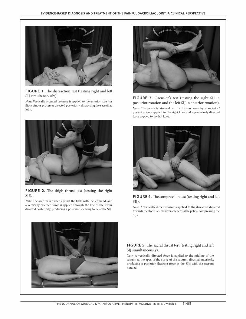
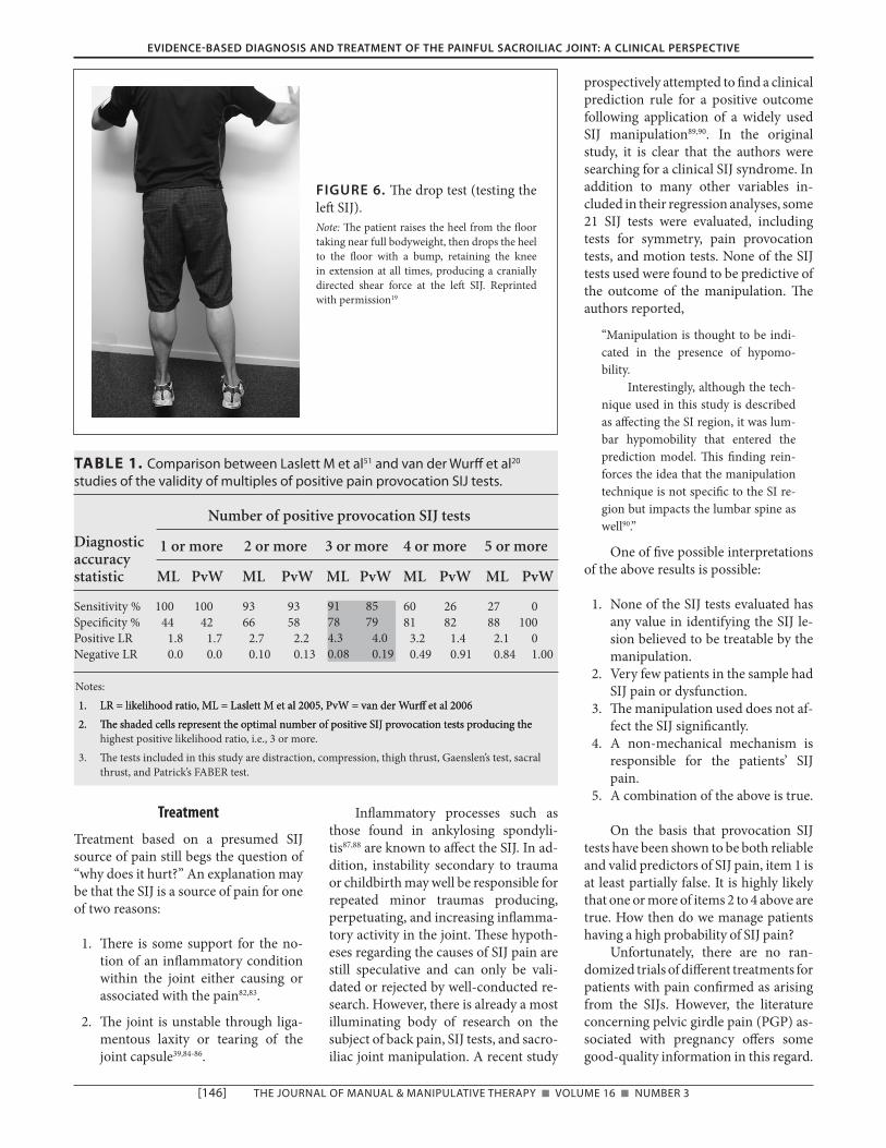
or familiar pain of which the patient complains. "e key tests (distraction, compression, thigh thrust, Gaenslen’s, and sacral thrust) have been described in detail in previous publications19,50-52 and are reproduced in Figures 1–5. "e Drop test (Figure 6) described by Rob-inson et al is reliable19 but has not yet been assessed for validity in a diagnostic accuracy study.
Reliability of Pain Provocation SIJ Tests
Early studies reported mixed results on the inter-examiner reliability of pain provocation tests17,25,53,54, but subse-quently these tests have been shown to possess acceptable levels of reliability provided that they are highly standard-ized12,13,19,50.
Validity of Pain Provocation SIJ Tests
A recent study con!rmed that three or more pain provocation SIJ tests have modest predictive power in relation to controlled comparative SIJ blocks. Sen-sitivity and speci!city were 91% and 78%, respectively52. In a second paper, the data were analyzed in more detail against a single block reference standard to report on the diagnostic accuracy of composites of pain provocation SIJ tests. It was found that the optimum number of positive tests is three or more positive tests51. Since that time, other researchers have replicated these !ndings against a double block standard20 in a di#erent and larger sample, using di#erent exam-iners and a di#erent physician perform-ing the diagnostic injection. "e results of the two studies are strikingly similar55 despite the use of a slightly di#erent mix of SIJ tests in each study. A comparison of results appears in Table 1.
SIJ pain and discogenic pain, as re-vealed by double SIJ blocks and provo-cation discography, rarely co-exist56,57. Anecdotal experience has indicated that provocation SIJ tests were commonly positive in those with nerve root pain secondary to a herniated lumbar disc and in those whose symptoms could be made to centralize during a McKenzie-type physical examination58. "e cen-
tralization phenomenon is a common clinical observation when low back pa-tients are examined using the standard-ized test movements and sustained pos-tures !rst described by McKenzie59. "e centralization phenomenon has been repeatedly described and evaluated for reliability and validity60-74. Subsequently, it has been found to be highly speci!c to discogenic pain and is not observed in patients with con!rmed SIJ pain or facet joint pain52,57,75-78. On this basis, it seems reasonable to assume that SIJ tests, pos-itive in the presence of the centralization phenomenon, are falsely positive.
Restricting the interpretation of the SIJ tests to non-centralization cases im-proves the speci!city of three or more positive pain provocation SIJ tests from 78% to 87% with the sensitivity remain-ing at 91%52. Patients satisfying these criteria have a high probability that SIJ pain will be con!rmed by diagnostic in-jection of local anesthetic. "is clinical reasoning process may be considered a clinical prediction rule for the identi!-cation of a subset of patients most likely to have pain of SIJ origin. For conve-nience, we may refer to this as the SIJCPR.
Likelihood ratios are summary sta-tistics derived from sensitivity and spec-i!city values. "e likelihood ratio for a positive test is an estimate of the proba-bility of the condition/disease. Random guessing will produce a positive likeli-hood ratio of 1.0. Values higher than 1.0 represent probability better than ran-dom chance. "e higher the value, the better the test. For example, a test with a positive likelihood ratio of 10 indicates that a positive test result is 10 times more likely in patients with the disease in question than in those known to be free of that disease. "e likelihood ratio of a negative test describes the test’s ability to rule out the disorder for which the test is applied. As the value of a negative like-lihood ratio approaches zero, the test’s power to rule out the disease in question approaches perfection. Conversely, as the value of the negative likelihood ratio increases towards 1.0, the test’s ability to rule out the disorder approaches ran-dom chance79. When both the preva-lence of the disorder and the results of a test are known, likelihood ratios permit
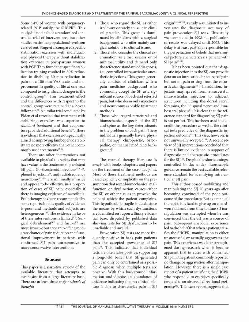
calculation of the change in odds and probability of a disorder being present or absent80. Prior to any examination, the probability of a given disorder being present is its prevalence. For example, if the prevalence of SIJ pain is 13%81, its pre-examination probability is 0.13. "e diagnostic value of a test is re%ected by how much the probability of the disor-der increases when the test is positive and by how much it falls when it is negative. "e diagnostic value of a given test can be depicted using Fagan’s nomogram (http://araw.mede.uic.edu/ cgi-bin/testcalc.pl) in which the pretest probability, prevalence, positive and negative likelihood ratios, and post-test probabilities are presented graphically. Figure 7 presents Fagan’s nomogram us-ing data from Laslett et al52 in which three or more positive SIJ tests are con-sidered positive for SIJ pain without consideration of the centralization phe-nomenon. "e likelihood ratio for a positive test (three or more SIJ tests pro-voke the patient’s familiar pain) is 4.16 so the probability of SIJ pain more than doubles from 26% to 59%. "e likeli-hood ratio of a negative test is 0.12 yield-ing a post-test probability of 4%.
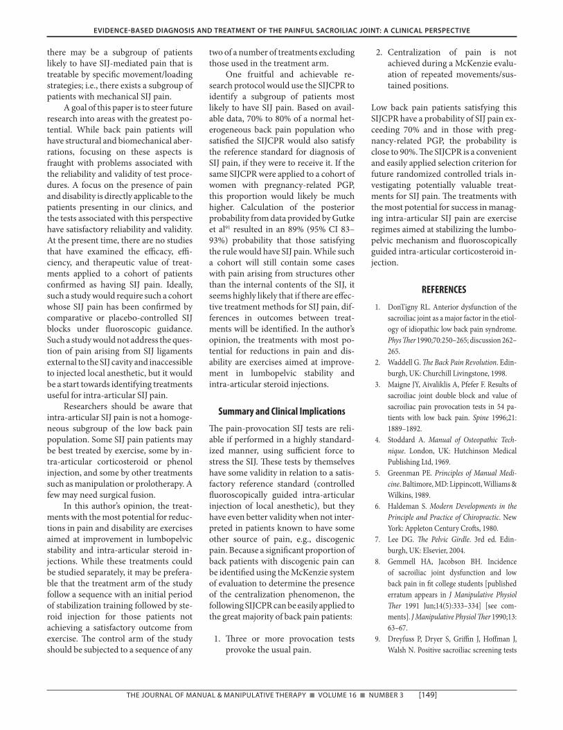
If the SIJCPR of three or more pos-itive provocation SIJ tests and the ab-sence of centralization are applied, the diagnostic performance is improved be-cause the false positive rate is decreased with proportionate improvement in speci!city from 78% to 87%. Fagan’s no-mogram created using the SIJCPR is presented in Figure 8. "e sample size is 34 as a result of removal of the 9 central-ization cases from the calculation and the prevalence is higher at 32%. "e positive likelihood ratio is 7.0, yielding a post-test probability of 77%. "e nega-tive likelihood ratio is 0.10, yielding a post-test probability of about 5%.
"e practical value of this data is as follows. If about 30% of patients with low back pain have pain of SIJ origin, and an individual patient has three or more positive provocation SIJ tests, there is a 59% chance that this patient will have SIJ pain. If a McKenzie assess-ment of repeated movements fails to re-veal the centralization phenomenon, there is a 77% chance that the pain is of SIJ origin.
EVIDENCE!BASED DIAGNOSIS AND TREATMENT OF THE PAINFUL SACROILIAC JOINT: A CLINICAL PERSPECTIVE
THE JOURNAL OF MANUAL & MANIPULATIVE THERAPY ■ VOLUME 16 ■ NUMBER 3 [145]
FIGURE 5. "e sacral thrust test (testing right and le& SIJ simultaneously).Note: A vertically directed force is applied to the midline of the sacrum at the apex of the curve of the sacrum, directed anteriorly, producing a posterior shearing force at the SIJs with the sacrum nutated.
EVIDENCE!BASED DIAGNOSIS AND TREATMENT OF THE PAINFUL SACROILIAC JOINT: A CLINICAL PERSPECTIVE
FIGURE 1. "e distraction test (testing right and le& SIJ simultaneously).Note: Vertically oriented pressure is applied to the anterior superior iliac spinous processes directed posteriorly, distracting the sacroiliac joint.
FIGURE 2. "e thigh thrust test (testing the right SIJ).Note: "e sacrum is !xated against the table with the le& hand, and a vertically oriented force is applied through the line of the femur directed posteriorly, producing a posterior shearing force at the SIJ.
FIGURE 3. Gaenslen’s test (testing the right SIJ in posterior rotation and the le& SIJ in anterior rotation).Note: "e pelvis is stressed with a torsion force by a superior/posterior force applied to the right knee and a posteriorly directed force applied to the le& knee.
FIGURE 4. "e compression test (testing right and le& SIJ). Note: A vertically directed force is applied to the iliac crest directed towards the %oor, i.e., transversely across the pelvis, compressing the SIJs.
[146] THE JOURNAL OF MANUAL & MANIPULATIVE THERAPY ■ VOLUME 16 ■ NUMBER 3
TreatmentTreatment based on a presumed SIJ source of pain still begs the question of “why does it hurt?” An explanation may be that the SIJ is a source of pain for one of two reasons:
1. "ere is some support for the no-tion of an in%ammatory condition within the joint either causing or associated with the pain82,83.
2. "e joint is unstable through liga-mentous laxity or tearing of the joint capsule39,84-86.
In%ammatory processes such as those found in ankylosing spondyli-tis87,88 are known to a#ect the SIJ. In ad-dition, instability secondary to trauma or childbirth may well be responsible for repeated minor traumas producing, perpetuating, and increasing in%amma-tory activity in the joint. "ese hypoth-eses regarding the causes of SIJ pain are still speculative and can only be vali-dated or rejected by well-conducted re-search. However, there is already a most illuminating body of research on the subject of back pain, SIJ tests, and sacro-iliac joint manipulation. A recent study
FIGURE 6. "e drop test (testing the le& SIJ). Note: "e patient raises the heel from the %oor taking near full bodyweight, then drops the heel to the %oor with a bump, retaining the knee in extension at all times, producing a cranially directed shear force at the le& SIJ. Reprinted with permission19
prospectively attempted to !nd a clinical prediction rule for a positive outcome following application of a widely used SIJ manipulation89,90. In the original study, it is clear that the authors were searching for a clinical SIJ syndrome. In addition to many other variables in-cluded in their regression analyses, some 21 SIJ tests were evaluated, including tests for symmetry, pain provocation tests, and motion tests. None of the SIJ tests used were found to be predictive of the outcome of the manipulation. "e authors reported,
“Manipulation is thought to be indi-cated in the presence of hypomo-bility.
Interestingly, although the tech-nique used in this study is described as a#ecting the SI region, it was lum-bar hypomobility that entered the prediction model. "is !nding rein-forces the idea that the manipulation technique is not speci!c to the SI re-gion but impacts the lumbar spine as well90.”
One of !ve possible interpretations of the above results is possible:
1. None of the SIJ tests evaluated has any value in identifying the SIJ le-sion believed to be treatable by the manipulation.
2. Very few patients in the sample had SIJ pain or dysfunction.
3. "e manipulation used does not af-fect the SIJ signi!cantly.
4. A non-mechanical mechanism is responsible for the patients’ SIJ pain.
5. A combination of the above is true.
On the basis that provocation SIJ tests have been shown to be both reliable and valid predictors of SIJ pain, item 1 is at least partially false. It is highly likely that one or more of items 2 to 4 above are true. How then do we manage patients having a high probability of SIJ pain?
Unfortunately, there are no ran-domized trials of di#erent treatments for patients with pain con!rmed as arising from the SIJs. However, the literature concerning pelvic girdle pain (PGP) as-sociated with pregnancy o#ers some good-quality information in this regard.
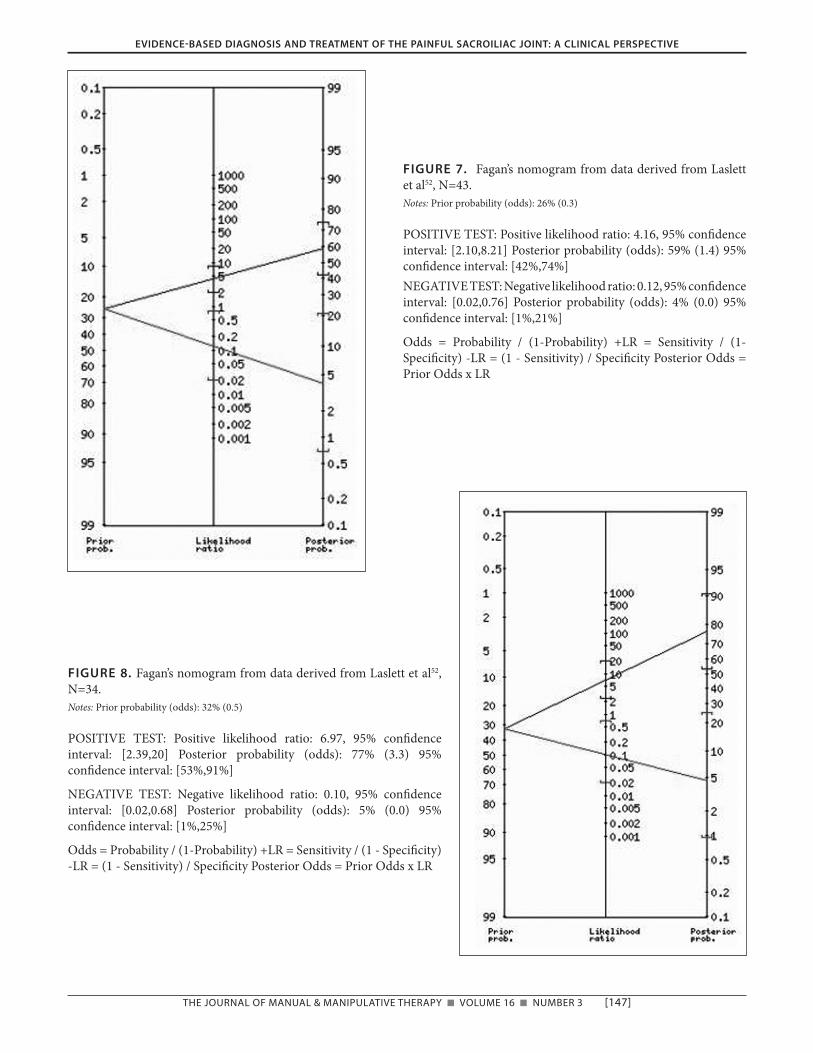
TABLE 1. Comparison between Laslett M et al51 and van der Wur# et al20 studies of the validity of multiples of positive pain provocation SIJ tests.
Diagnostic
Number of positive provocation SIJ tests
accuracy 1 or more 2 or more 3 or more 4 or more 5 or more
statistic ML PvW ML PvW ML PvW ML PvW ML PvW
Sensitivity % 100 100 93 93 91 85 60 26 27 0Speci!city % 44 42 66 58 78 79 81 82 88 100Positive LR 1.8 1.7 2.7 2.2 4.3 4.0 3.2 1.4 2.1 0Negative LR 0.0 0.0 0.10 0.13 0.08 0.19 0.49 0.91 0.84 1.00
Notes: 1. LR = likelihood ratio, ML = Laslett M et al 2005, PvW = van der Wur# et al 20061. LR = likelihood ratio, ML = Laslett M et al 2005, PvW = van der Wur# et al 2006 2. "e shaded cells represent the optimal number of positive SIJ provocation tests producing the2. "e shaded cells represent the optimal number of positive SIJ provocation tests producing the
highest positive likelihood ratio, i.e., 3 or more. 3. "e tests included in this study are distraction, compression, thigh thrust, Gaenslen’s test, sacral
thrust, and Patrick’s FABER test.
EVIDENCE!BASED DIAGNOSIS AND TREATMENT OF THE PAINFUL SACROILIAC JOINT: A CLINICAL PERSPECTIVE
91 8578 794.3 4.00.08 0.19
THE JOURNAL OF MANUAL & MANIPULATIVE THERAPY ■ VOLUME 16 ■ NUMBER 3 [147]
FIGURE 8. Fagan’s nomogram from data derived from Laslett et al52, N=34.Notes: Prior probability (odds): 32% (0.5)
POSITIVE TEST: Positive likelihood ratio: 6.97, 95% con!dence interval: [2.39,20] Posterior probability (odds): 77% (3.3) 95% con!dence interval: [53%,91%]
NEGATIVE TEST: Negative likelihood ratio: 0.10, 95% con!dence interval: [0.02,0.68] Posterior probability (odds): 5% (0.0) 95% con!dence interval: [1%,25%]
Odds = Probability / (1-Probability) +LR = Sensitivity / (1 - Speci!city) -LR = (1 - Sensitivity) / Speci!city Posterior Odds = Prior Odds x LR
FIGURE 7. Fagan’s nomogram from data derived from Laslett et al52, N=43. Notes: Prior probability (odds): 26% (0.3)
POSITIVE TEST: Positive likelihood ratio: 4.16, 95% con!dence interval: [2.10,8.21] Posterior probability (odds): 59% (1.4) 95% con!dence interval: [42%,74%] NEGATIVE TEST: Negative likelihood ratio: 0.12, 95% con!dence interval: [0.02,0.76] Posterior probability (odds): 4% (0.0) 95% con!dence interval: [1%,21%]
Odds = Probability / (1-Probability) +LR = Sensitivity / (1- Speci!city) -LR = (1 - Sensitivity) / Speci!city Posterior Odds = Prior Odds x LR
EVIDENCE!BASED DIAGNOSIS AND TREATMENT OF THE PAINFUL SACROILIAC JOINT: A CLINICAL PERSPECTIVE
[148] THE JOURNAL OF MANUAL & MANIPULATIVE THERAPY ■ VOLUME 16 ■ NUMBER 3
Some 54% of women with pregnancy-related PGP satisfy the SIJCPR91. "is study did not include a randomized con-trolled trial of interventions, but other studies on similar populations have been carried out. Stuge et al compared speci!c stabilization exercises with individual-ized physical therapy without stabiliza-tion exercises in post-partum women with PGP. "ey found that speci!c stabi-lization training resulted in 50% reduc-tion in disability, 30 mm reduction in pain on a 100 mm VAS scale, and im-provement in quality of life at one year compared to insigni!cant changes in the control group92. "is treatment e#ect and the di#erences with respect to the control group were retained at a 2-year follow-up93. A similar trial conducted by Elden et al revealed that treatment with stabilizing exercises was superior to standard treatment and that acupunc-ture provided additional bene!t94. "ere is evidence that exercises not speci!cally aimed at improving lumbopelvic stabil-ity are no more e#ective than other com-monly used treatments95,96.
"ere are other interventions not available to physical therapists that may have value in the treatment of persistent SIJ pain. Corticosteroid injections88,97,98, phenol injections99, and radiofrequency neurotomy100-104 are minimally invasive and appear to be e#ective in a propor-tion of cases of SIJ pain, especially if there is imaging evidence of sacroiliitis. Prolotherapy has been recommended by some reports, but the quality of evidence is poor, and methods and subjects are heterogeneous105. "e evidence in favor of these interventions is limited106. Sur-gical debridement107 and fusion108 are more invasive but appear to o#er a mod-erate chance of pain reduction and func-tional improvement in patients with con!rmed SIJ pain unresponsive to more conservative interventions.
Discussion"is paper is a narrative review of the available literature that attempts to synthesize from a large literature base. "ere are at least three major schools of thought:
1. "ose who regard the SIJ as either irrelevant or rarely an issue in clini-cal practice. "is group is domi-nated by clinicians with a surgical background who o#er mainly sur-gical solutions to clinical issues.
2. "ose who consider the clinical ex-amination as either useless or of minimal utility and demand only the reference standard of diagnosis, i.e., controlled intra-articular anes-thetic injections. "is group gener-ally consists of clinicians with a pain medicine background who commonly accept the SIJ as a sig-ni!cant source of back and referred pain, but who deem only injections and neurotomy as viable treatment methods.
3. "ose who regard structural and biomechanical aspects of the SIJ and spine as the key determinants in the problem of back pain. "ese individuals generally have a physi-cal therapy, chiropractic, osteo-pathic, or manual medicine back-ground.
"e manual therapy literature is awash with books, chapters, and papers on the treatment of the sacroiliac joint. Most of these treatment methods are based explicitly or implicitly on the pre-sumption that some biomechanical mal-function or dysfunction causes either the SIJ or other tissues to provoke the pain of which the patient complains. "is hypothesis is fragile indeed, since the means by which such dysfunctions are identi!ed rest upon a %imsy eviden-tial base, disputed by published data showing tests for SIJ dysfunction to be unreliable and invalid.
Provocation SIJ tests are more fre-quently positive in back pain patients than the accepted prevalence of SIJ pain58. "is indicates that individual tests are o&en false-positive, supporting a long-held belief that SIJ-generated pain can only be entertained as a possi-ble diagnosis when multiple tests are positive. With this background infor-mation and despite an abundance of evidence indicating that no clinical pic-ture is able to characterize pain of SIJ
origin3,10,40,109, a study was initiated to in-vestigate the diagnostic accuracy of pain-provocation SIJ tests. "is study was completed in 1998 but publication of results was delayed until 2003. "is delay is at least partially responsible for the perpetuation of beliefs that no clini-cal picture characterizes a patient with SIJ pain42,110.
It has been pointed out that diag-nostic injection into the SIJ can provide data on an intra-articular source of pain but not on pain arising from the extra-articular ligaments3,51. In addition, in-jectate may spread from a successful intra-articular injection to adjacent structures including the dorsal sacral foramina, the L5 spinal nerve and lum-bosacral plexus84. It is clear that the ref-erence standard for diagnosing SIJ pain is not perfect. "is has been used to dis-credit the procedure as well as the clini-cal tests predictive of the diagnostic in-jection outcome85. "is view, however, is not universally accepted111. A recent re-view of SIJ interventions concluded that there is limited evidence in support of diagnostic and therapeutic procedures for the SIJ106. Despite the shortcomings, controlled blocks under %uoroscopic guidance remain the best available refer-ence standard for identifying intra-ar-ticular SIJ pain.
"is author ceased mobilizing and manipulating the SIJ 20 years ago a&er becoming convinced of the poor out-come of the procedures. But as a manual therapist, it is hard to give up on a hard-won skill, and from time to time SIJ ma-nipulation was attempted when he was convinced that the SIJ was a source of pain. Subsequent anecdotal experience led to the belief that when a patient satis-!es the SIJCPR, manipulation is either unsuccessful or actually aggravates the pain. "is experience was later strength-ened during research when it became apparent that in cases with con!rmed SIJ pain, the patient commonly reported no change or aggravation a&er manipu-lation. However, there is a single case report of a patient satisfying the SIJCPR who responded to exercises speci!cally targeted to an observed directional pref-erence112. "is case report suggests that
EVIDENCE!BASED DIAGNOSIS AND TREATMENT OF THE PAINFUL SACROILIAC JOINT: A CLINICAL PERSPECTIVE
THE JOURNAL OF MANUAL & MANIPULATIVE THERAPY ■ VOLUME 16 ■ NUMBER 3 [149]
there may be a subgroup of patients likely to have SIJ-mediated pain that is treatable by speci!c movement/loading strategies; i.e., there exists a subgroup of patients with mechanical SIJ pain.
A goal of this paper is to steer future research into areas with the greatest po-tential. While back pain patients will have structural and biomechanical aber-rations, focusing on these aspects is fraught with problems associated with the reliability and validity of test proce-dures. A focus on the presence of pain and disability is directly applicable to the patients presenting in our clinics, and the tests associated with this perspective have satisfactory reliability and validity. At the present time, there are no studies that have examined the e$cacy, e$-ciency, and therapeutic value of treat-ments applied to a cohort of patients con!rmed as having SIJ pain. Ideally, such a study would require such a cohort whose SIJ pain has been con!rmed by comparative or placebo-controlled SIJ blocks under %uoroscopic guidance. Such a study would not address the ques-tion of pain arising from SIJ ligaments external to the SIJ cavity and inaccessible to injected local anesthetic, but it would be a start towards identifying treatments useful for intra-articular SIJ pain.
Researchers should be aware that intra-articular SIJ pain is not a homoge-neous subgroup of the low back pain population. Some SIJ pain patients may be best treated by exercise, some by in-tra-articular corticosteroid or phenol injection, and some by other treatments such as manipulation or prolotherapy. A few may need surgical fusion.
In this author’s opinion, the treat-ments with the most potential for reduc-tions in pain and disability are exercises aimed at improvement in lumbopelvic stability and intra-articular steroid in-jections. While these treatments could be studied separately, it may be prefera-ble that the treatment arm of the study follow a sequence with an initial period of stabilization training followed by ste-roid injection for those patients not achieving a satisfactory outcome from exercise. "e control arm of the study should be subjected to a sequence of any
two of a number of treatments excluding those used in the treatment arm.
One fruitful and achievable re-search protocol would use the SIJCPR to identify a subgroup of patients most likely to have SIJ pain. Based on avail-able data, 70% to 80% of a normal het-erogeneous back pain population who satis!ed the SIJCPR would also satisfy the reference standard for diagnosis of SIJ pain, if they were to receive it. If the same SIJCPR were applied to a cohort of women with pregnancy-related PGP, this proportion would likely be much higher. Calculation of the posterior probability from data provided by Gutke et al91 resulted in an 89% (95% CI 83–93%) probability that those satisfying the rule would have SIJ pain. While such a cohort will still contain some cases with pain arising from structures other than the internal contents of the SIJ, it seems highly likely that if there are e#ec-tive treatment methods for SIJ pain, dif-ferences in outcomes between treat-ments will be identi!ed. In the author’s opinion, the treatments with most po-tential for reductions in pain and dis-ability are exercises aimed at improve-ment in lumbopelvic stability and intra-articular steroid injections.
Summary and Clinical Implications"e pain-provocation SIJ tests are reli-able if performed in a highly standard-ized manner, using su$cient force to stress the SIJ. "ese tests by themselves have some validity in relation to a satis-factory reference standard (controlled %uoroscopically guided intra-articular injection of local anesthetic), but they have even better validity when not inter-preted in patients known to have some other source of pain, e.g., discogenic pain. Because a signi!cant proportion of back patients with discogenic pain can be identi!ed using the McKenzie system of evaluation to determine the presence of the centralization phenomenon, the following SIJCPR can be easily applied to the great majority of back pain patients:
1. "ree or more provocation tests provoke the usual pain.
2. Centralization of pain is not achieved during a McKenzie evalu-ation of repeated movements/sus-tained positions.
Low back pain patients satisfying this SIJCPR have a probability of SIJ pain ex-ceeding 70% and in those with preg-nancy-related PGP, the probability is close to 90%. "e SIJCPR is a convenient and easily applied selection criterion for future randomized controlled trials in-vestigating potentially valuable treat-ments for SIJ pain. "e treatments with the most potential for success in manag-ing intra-articular SIJ pain are exercise regimes aimed at stabilizing the lumbo-pelvic mechanism and %uoroscopically guided intra-articular corticosteroid in-jection.
REFERENCES 1. DonTigny RL. Anterior dysfunction of the
sacroiliac joint as a major factor in the etiol-ogy of idiopathic low back pain syndrome. Phys !er 1990;70:250–265; discussion 262–265.
2. Waddell G. !e Back Pain Revolution. Edin-burgh, UK: Churchill Livingstone, 1998.
3. Maigne JY, Aivaliklis A, Pfefer F. Results of sacroiliac joint double block and value of sacroiliac pain provocation tests in 54 pa-tients with low back pain. Spine 1996;21: 1889–1892.
4. Stoddard A. Manual of Osteopathic Tech-nique. London, UK: Hutchinson Medical Publishing Ltd, 1969.
5. Greenman PE. Principles of Manual Medi-cine. Baltimore, MD: Lippincott, Williams & Wilkins, 1989.
6. Haldeman S. Modern Developments in the Principle and Practice of Chiropractic. New York: Appleton Century Cro&s, 1980.
7. Lee DG. !e Pelvic Girdle. 3rd ed. Edin-burgh, UK: Elsevier, 2004.
8. Gemmell HA, Jacobson BH. Incidence of sacroiliac joint dysfunction and low back pain in !t college students [published erratum appears in J Manipulative Physiol !er 1991 Jun;14(5):333–334] [see com-ments]. J Manipulative Physiol !er 1990;13: 63–67.
9. Dreyfuss P, Dryer S, Gri$n J, Ho#man J, Walsh N. Positive sacroiliac screening tests
EVIDENCE!BASED DIAGNOSIS AND TREATMENT OF THE PAINFUL SACROILIAC JOINT: A CLINICAL PERSPECTIVE
[150] THE JOURNAL OF MANUAL & MANIPULATIVE THERAPY ■ VOLUME 16 ■ NUMBER 3
in asymptomatic adults. Spine 1994;19:1138–1143.
10. Dreyfuss PH, Michaelsen M, Pauza K, McLarty J, Bogduk N. "e value of history and physical examination in diagnosing sac-roiliac joint pain. Spine 1996;21:2594–2602.
11. DonTigny RL. A detailed and critical biome-chanical analysis of the sacroiliac joints and relevant kinesiology. "e implications for lumbopelvic function and dysfunction. In Vleeming A, Mooney V, and Stoeckart R, eds, 2nd ed. Movement, Stability and Lumbo-pelvic Pain: Integration of Research and !erapy. Philadelphia, PA: Churchill Living-stone, 2007.
12. Kokmeyer DJ, van der Wur# P, Aufdem-kampe G, Fickenscher TCM. "e reliability"e reliability of multi-test regimens with sacroiliac pain provocation tests. J Manipulative Physiol !er 2002;25:42–48.
13. van der Wur# P, Hagmeijer RH, Meyne W. Clinical tests of the sacroiliac joint: A sys-tematic methodological review. Part 1: Reli-ability. Man !er 2000;5:30–36.
14. Sturesson B, Selvik G, Uden A. Movements of the sacroiliac joints: A roentgnen stereo-photogrammetric analysis. Spine 1989;14: 162–165.
15. Sturesson B, Uden A, Vleeming A. A radio-stereometric analysis of the movements of the sacroiliac joints in the reciprocal strad-dle position. Spine 2000;25:214–217.
16. Sturesson B. Load and movement of the sac-roiliac joint. PhD thesis, Lund University, Malmo, Sweden,1999;29–35.
17. Potter NA, Rothstein JM. Intertester reli-ability for selected clinical tests of the sacro-iliac joint. Phys !er 1985;65:1671–1675.
18. Freburger JK, Riddle DL. Measurement of sacroiliac joint dysfunction: A multicenter intertester reliability study. Phys !er 1999; 79:1134–1141.
19. Robinson HS, Brox JI, Robinson R, Bjelland E, Solem S, Telje T. "e reliability of selected motion and pain provocation tests for the sacroiliac joint. Man !er 2007;12:72–79.
20. van der Wur# P, Buijs EJ, Groen GJ. A multi-A multi-test regimen of pain provocation tests as an aid to reduce unnecessary minimally inva-sive sacroiliac joint procedures. Arch Phys Med Rehabil 2006;87:10–14.
21. O’Haire C, Gibbons P. Inter-examiner and intra-examiner agreement for assessing sac-roiliac anatomical landmarks using palpa-tion and observation: A pilot study. Man !er 2000;5:13–20.
22. Vincent-Smith B, Gibbons P. Inter-examiner and intra-examiner reliability of the stand-ing %exion test. Man !er 1999;4:87–93.
23. Meijne W, van Neerbos K, Aufdemkampe G, van der Wur# P. Intraexaminer and interex-aminer reliability of the Gillet test. J Manipu-lative Physiol !er 1999;22:4–9.
24. Herzog W, Read LJ, Conway PJ, Shaw LD, McEwen DC. Reliability of motion palpa-tion procedures to detect sacroiliac joint !xations. J Manipulative Physiol !er 1989; 12:86–92.
25. Carmichael JP. Inter- and intra-examiner reliability of palpation for sacroiliac joint dysfunction. J Manipulative Physiol !er 1987;10:164–171.
26. Hungerford BA, Gilleard W, Moran M, Em-merson C. Evaluation of the ability of physi-cal therapists to palpate intrapelvic motion with the Stork test on the support side. Phys !er 2007;87:879–887.
27. Bussey MD, Yanai T, Milburn P. A non-inva-sive technique for assessing innominate bone motion. Clin Biomech (Bristol, Avon) 2004;19:85–90.
28. Mior SA, McGregor M, Schut B. "e role of experience in clinical accuracy. J Manipula-tive Physiol !er 1990;13:68–71.
29. Tong HC, Heyman OG, Lado DA, Isser MM. Interexaminer reliability of three methods of combining test results to determine side of sacral restriction, sacral base position, and innominate bone position. J Am Osteo-path Assoc 2006;106:464–468.
30. Foley BS, Buschbacher RM. Sacroiliac joint pain: Anatomy, biomechanics, diagnosis, and treatment. Am J Phys Med Rehabil 2006;85:997–1006.
31. Riddle DL, Freburger JK. Evaluation of the presence of sacroiliac joint region dysfunc-tion using a combination of tests: A multi-center intertester reliability study. Phys !er 2002;82:772–781.
32. Cibulka MT, Koldeho# R. Clinical useful-ness of a cluster of sacroiliac joint tests in patients with and without low back pain. J Orthop Sports Phys !er 1999;29:83–99.
33. Altman DG, Machin D, Bryant TN, Gardner MJ. Statistics with Con"dence. 2nd ed. Bris-tol, UK: British Medical Journal, 2000.
34. Sackett DL, Haynes RB, Guyatt GH, Tugwell P. Clinical Epidemiology: A Basic Science for Clinical Medicine. 2nd ed. Boston: Little, Brown and Company, 1991.
35. Sturesson B, Uden A, Vleeming A. A radio-stereometric analysis of movements of the
sacroiliac joints during the standing hip %exion test. Spine 2000;25:364–368.
36. Dar G, Peleg S, Masharawi Y, Steinberg N, Rothschild BM, Hershkovitz I. "e associa-tion of sacroiliac joint bridging with other enthesopathies in the human body. Spine 2007;32:E303–E308.
37. Dar G, Khamis S, Peleg S, et al. Sacroiliac joint fusion and the implications for manual therapy diagnosis and treatment. Man !er 2008;13:155–158.
38. Waldron T, Rogers J. An epidemiologic study of sacroiliac fusion in some human skeletal remains. Am J Phys Anthropol 1990;83:123–127.
39. Fortin JD, Aprill C, Pontieux RT, Pier J. Sac-roiliac joint: Pain referral maps upon apply-ing a new injection/arthrography technique. Part II: Clinical evaluation. Spine 1994;19: 1483–1489.
40. Schwarzer AC, Aprill C, Bogduk N. "e sac-roiliac joint in chronic low back pain. Spine 1995;20:31–37.
41. Fortin JD, Dwyer AP, West S, Pier J. Sacro-iliac joint: Pain referral maps upon applying a new injection/arthrography technique. Part I: Asymptomatic volunteers. Spine 1994;19:1475–1482.
42. Bogduk N. Practice Guidelines: Spinal Diag-nostic and Treatment Procedures. San Fran-cisco: International Spine Intervention Soci-ety, 2004.
43. Forst SL, Wheeler MT, Fortin JD, Vilensky JA. "e sacroiliac joint: Anatomy, physiol-ogy and clinical signi!cance. Pain Physician 2006;9:61–67.
44. Ikeda R. Innervation of the sacroiliac joint: Macroscopic and histological studies. J Nip-pon Med School 1991;58:587–596.
45. Merskey H, Bogduk N. Classi"cation of Chronic Pain: Descriptions of Chronic Pain Syndromes and De"nitions of Pain Terms. 2nd ed. Seattle, WA: IASP Press, 1994.
46. Rosenberg JM, Quint TJ, de Rosayro AM. Computerized tomographic localization of clinically-guided sacroiliac joint injections. Clin J Pain 2000;16:18–21.
47. Hansen HC. Is %uoroscopy necessary for sacroiliac joint injections? Pain Physician 2003;6:155–158.
48. Aprill CN. !e Role of Anatomically Speci"c Injections into the Sacroiliac Joint. In: Vleem-ing A. et al. 1st Interdisciplinary World Con-gress on Low Back Pain and Its Relation to the S.I. Joint. Rotterdam ECO. 1992;373–380.
EVIDENCE!BASED DIAGNOSIS AND TREATMENT OF THE PAINFUL SACROILIAC JOINT: A CLINICAL PERSPECTIVE
THE JOURNAL OF MANUAL & MANIPULATIVE THERAPY ■ VOLUME 16 ■ NUMBER 3 [151]
49. Schwarzer AC, Aprill CN, Derby R, Fortin J, Kine G, Bogduk N. "e false-positive rate of uncontrolled diagnostic blocks of the lum-bar zygapophysial joints. Pain 1994;58:195–200.
50. Laslett M, Williams M. "e reliability of selected pain provocation tests for sacro- iliac joint pathology. Spine 1994;19:1243–1249.
51. Laslett M, Aprill CN, McDonald B, Young SB. Diagnosis of sacroiliac joint pain: Validity of individual provocation tests and composites of tests. Man !er 2005;10:207–218.
52. Laslett M, Young SB, Aprill CN, McDonald B. Diagnosing painful sacroiliac joints: A validity study of a McKenzie evaluation and sacroiliac joint provocation tests. Aust J Physiother 2003;49:89–97.
53. McCombe PF, Fairbank JCT, Cockersole BC, Pynsent PB. Reproducibility of physical signs in low back pain. Spine 1989;14:908–918.
54. van Deursen LLJM, Patijn J, Ockhuysen AL, Vortman BJ. "e value of some clinical tests"e value of some clinical tests of the sacroiliac joint. J Manual Med 1990; 5:96–99.
55. Laslett M, Aprill CN, McDonald B. Provoca-tion sacroiliac joint tests have validity in the diagnosis of sacroiliac joint pain. Arch Phys Med Rehab 2006;87:874–875.
56. Schwarzer AC, Aprill CN, Derby R, Fortin J, Kine G, Bogduk N. "e relative contribu-tions of the disc and zygapophyseal joint in chronic low back pain. Spine 1994;19:801–806.
57. Laslett M, McDonald B, Tropp H, Aprill CN, Oberg B. Agreement between diagnoses reached by clinical examination and avail-able reference standards: A prospective study of 216 patients with lumbopelvic pain. BMC Musculoskelet Disord 2005;6:28.
58. Laslett M. Pain provocation sacroiliac joint tests: Reliability and prevalence. In Vleem-ing A, Mooney V, Snijders CJ, Dormann TA, Stoeckart R, eds. Movement, Stability and Low Back Pain: !e Essential Role of the Pel-vis. 1st ed. New York: Churchill Livingstone, 1997.
59. McKenzie RA. !e Lumbar Spine: Mechani-cal Diagnosis and !erapy. Waikanae, NZ: Spinal Publications Ltd, 1981.
60. Razmjou H, Kramer JF, Yamada R. Inter-tes-ter reliability of the McKenzie evaluation in mechanical low back pain. J Orthop Sports Phys !er 2000;30:368–383.
61. Kilpikoski S, Airaksinen O, Kankaanpaa M, Leminen P, Videman T, Alen M. Interexam-iner reliability of low back pain assessment using the McKenzie method. Spine 2002;27:E207–E214.
62. Aina A, May S, Clare H. "e centralization phenomenon of spinal symptoms: A sys-tematic review. Man !er 2004;9:134–143.
63. Clare HA, Adams R, Maher CG. Reliability of McKenzie classi!cation of patients with cervical or lumbar pain. J Manipulative Physiol !er 2005;28:122–127.
64. Donelson R, Silva G, Murphy K. Centralisa-tion phenomenon: Its usefulness in evaluat-ing and treating referred pain. Spine 1990;15: 211–213.
65. Donelson R, Grant W, Kamps C, Medcalf R. Pain response to sagittal end range spinal motion: A multi-centered, prospective, ran-domized trial. Spine 1991;16:S206–S212.
66. Donelson R, Aprill C, Medcalf R, Grant W. A prospective study of centralization of lum-bar and referred pain: A predictor of symp-tomatic discs and annular competence. Spine 1997;22:1115–1122.
67. Wetzel FT, Donelson R. "e role of repeated end-range/pain response assessment in the management of symptomatic lumbar discs. Spine J 2003;3:146–154.
68. Long A, Donelson R, Fung T. Does it matter which exercise? A randomized control trial of exercise for low back pain. Spine 2004;29: 2593–2602.
69. Donelson R. Rapidly Reversible Low Back Pain: An Evidence-Based Pathway to Wide-spread Recoveries and Savings. Hanover, NH: Selfcare First LLC, 2007.
70. Werneke MW, Hart DL. Centralization: As-sociation between repeated end-range pain responses and behavioral signs in patients with acute non-speci!c low back pain. J Re-habil Med 2005;37:286–290.
71. Werneke M, Hart DL. Centralization phe-nomenon as a prognostic factor for chronic low back pain and disability. Spine 2001;26: 758–765.
72. Werneke M, Hart DL. Discriminant validity and relative precision for classifying patients with non-speci!c neck and low back pain by anatomic pain patterns. Spine 2003;28:161–166.
73. Werneke M, Hart DL, Cook D. A descriptive study of the centralization phenomenon: A prospective analysis. Spine 1999;24:676–683.
74. Werneke M, May S. "e centralization phe-
nomenon and fear-avoidance beliefs as prognostic factors for acute low back pain. J Orthop Sports Phys !er 2005;35:844–845.
75. Laslett M, Oberg B, Aprill CN, McDonald B. Centralization as a predictor of provocation discography results in chronic low back pain, and the in%uence of disability and dis-tress on diagnostic power. Spine J 2005;5: 370–380.
76. Laslett M, McDonald B, Aprill CN, Tropp H, Oberg B. Clinical predictors of screening lumbar zygapophysial joint blocks: Devel-opment of clinical prediction rules. Spine J 2006;6:370–379.
77. Laslett M, Oberg B, Aprill CN, McDonald B. A study of clinical predictors of lumbar dis-cogenic pain as determined by provocation discography. Eur Spine J 2006;15:1473–1484.
78. Young SB, Aprill CN, Laslett M. Correlation of clinical examination characteristics with three sources of chronic low back pain. Spine J 2003;3:460–465.
79. Sackett DL, Straus SE, Richardson WS, Rosenberg W, Haynes RB. Evidence-Based Medicine: How to Practice and Teach EBM. Edinburgh, UK: Churchill Livingstone, 2000.
80. Knottnerus A. !e Evidence Base of Clinical Diagnosis. London, UK: BMJ Books, 2002.
81. Bogduk N. "e anatomical basis for spinal pain syndromes. J Manipulative Physiol !er 1995;18:603–605.
82. Heu&-Dorenbosch L, Weijers R, Landewe R, van der Linden S, van der Heijde D. Mag-Mag-netic resonance imaging changes of sacroil-iac joints in patients with recent-onset in-%ammatory back pain: Inter-reader reliabil-ity and prevalence of abnormalities. Arthritis Res !er 2006;8:R11.
83. Slipman CW, Sterenfeld EB, Chou LH, Her-zog R, Vresilovic E. "e value of radionu-clide imaging in the diagnosis of sacroiliac joint syndrome. Spine 1996;21:2251–2254.
84. Fortin JD, Washington WJ, Falco FJE. "ree pathways between the sacro-iliac joint and neural structures. AJNR 1999;20:1429–1434.
85. Berthelot JM, Labat JJ, Le Go# B, Gouin F, Maugers Y. Provocative sacroiliac joint ma-neuvers and sacroiliac joint block are unreli-able for diagnosing sacroiliac joint pain. Joint Bone Spine 2006;73:17–23.
86. van Wingerden JP, Vleeming A, Buyruk HM, Raissadat K. Stabilization of the sacro-iliac joint in vivo: Veri!cation of muscular
EVIDENCE!BASED DIAGNOSIS AND TREATMENT OF THE PAINFUL SACROILIAC JOINT: A CLINICAL PERSPECTIVE
[152] THE JOURNAL OF MANUAL & MANIPULATIVE THERAPY ■ VOLUME 16 ■ NUMBER 3
contribution to force closure of the pelvis. Eur Spine J 2004;13:199–205.
87. Gunaydin I, Pereira PL, Fritz J, Konig C, Kotter I. Magnetic resonance imaging guided corticosteroid injection of sacroiliac joints in patients with spondylarthropathy. Are multiple injections more bene!cial? Rheumatol Int 2006;26:396–400.
88. Pereira PL, Gunaydin I, Trubenbach J, et al. Interventional MR imaging for injection of sacroiliac joints in patients with sacroili-itis. AJR Am J Roentgenol 2000;175:265–266.
89. Childs JD, Fritz JM, Flynn TW, et al. A clin-ical prediction rule to identify patients with low back pain most likely to bene!t from spinal manipulation: A validation study. Ann Intern Med 2004;141:920–928.
90. Flynn T, Fritz JM, Whitman J, et al. A clinical prediction rule for classifying patients with low back pain who demonstrate short-term improvement with spinal manipulation. Spine 2003;27:2835–2843.
91. Gutke A, Ostgaard HC, Oberg B. Pelvic gir-dle pain and lumbar pain in pregnancy: A cohort study of the consequences in terms of health and functioning. Spine 2006;31:E149–E155.
92. Stuge B, Laerum E, Kirkesola G, Vollestad N. "e e$cacy of a treatment program fo-cusing on speci!c stabilizing exercises for pelvic girdle pain a&er pregnancy: A ran-domized controlled trial. Spine 2004;29:351–359.
93. Stuge B, Veierod MB, Laerum E, Vollestad N. "e e$cacy of a treatment program fo-cusing on speci!c stabilizing exercises for pelvic girdle pain a&er pregnancy: A two-year follow-up of a randomized clinical trial. Spine 2004;29:E197–E203.
94. Elden H, Ladfors L, Olsen MF, Ostgaard HC, Hagberg H. E#ects of acupuncture and sta-bilising exercises as adjunct to standard treatment in pregnant women with pelvic
girdle pain: Randomised single blind con-trolled trial. BMJ 2005;330:761.
95. Nilsson-Wikmar L, Holm K, Oijerstedt R, Harms-Ringdahl K. E#ect of three di#erent physical therapy treatments on pain and ac-tivity in pregnant women with pelvic girdle pain: A randomized clinical trial with 3, 6, and 12 months follow-up postpartum. Spine 2005;30:850–856.
96. Mens JM, Snijders CJ, Stam HJ. Diagonal trunk muscle exercises in peripartum pelvic pain: A randomized clinical trial. Phys !er 2000;80:1164–1173.
97. Maugars Y, Mathis C, Berthelot JM, Charlier C, Prost A. Assessment of the e$cacy of sac-roiliac corticosteroid injections in spondy-larthropathies: A double-blind study. Br J Rheumatol 1996;35:767–770.
98. Slipman CW, Lipetz JS, Plastaras CT, et al. Fluoroscopically guided therapeutic sacro-iliac joint injections for sacroiliac joint syn-drome. Am J Phys Med Rehabil 2001;80:425–432.
99. Ward S, Jenson M, Royal MA, Movva V, Bhakta B, Gunyea I. Fluoroscopy-guided sacroiliac joint injections with phenol abla-tion for persistent sacroiliitis: A case series. Pain Pract 2002;2:332–335.
100. Burnham RS, Yasui Y. An alternate method of radiofrequency neurotomy of the sacro-iliac joint: A pilot study of the e#ect on pain, function, and satisfaction. Reg Anesth Pain Med 2007;32:12–19.
101. Vallejo R, Benyamin RM, Kramer J, Stanton G, Joseph NJ. Pulsed radiofrequency dener-vation for the treatment of sacroiliac joint syndrome. Pain Med 2006;7:429–434.
102. Ferrante FM, King LF, Roche EA, et al. Ra-diofrequency sacroiliac joint denervation for sacroiliac syndrome. Regl Anesth Pain Med 2001;26:137–142.
103. Yin W, Willard F, Carreiro J, Dreyfuss P. Sen-sory stimulation-guided sacroiliac joint ra-diofrequency neurotomy: Technique based
on neuroanatomy of the dorsal sacral plexus. Spine 2003;28:2419–2425.
104. Cohen SP, Abdi S. Lateral branch blocks as a treatment for sacroiliac joint pain: A pilot study. Reg Anesth Pain Med 2003;28:113–119.
105. Dagenais S, Haldeman S, Wooley JR. Intra-ligamentous injection of sclerosing solu-tions (prolotherapy) for spinal pain: A criti-cal review of the literature. Spine J 2005;5: 310–328.
106. Hansen HC, Kenzie-Brown AM, Cohen SP, Swicegood JR, Colson JD, Manchikanti L. Sacroiliac joint interventions: A systematic review. Pain Physician 2007;10:165–184.
107. Haufe SM, Mork AR. Sacroiliac joint de-bridement: A novel technique for the treat-ment of sacroiliac joint pain. Photomed La-ser Surg 2005;23:596–598.
108. Buchowski JM, Kebaish KM, Sinkov V, Co-hen DB, Sieber AN, Kostuik JP. Functional and radiographic outcome of sacroiliac ar-throdesis for the disorders of the sacroiliac joint. Spine J 2005;5:520–528.
109. Slipman CW, Sterenfeld EB, Chou LH, Her-zog R, Vresilovic E. "e predictive value of provocative sacroiliac joint stress maneu-vers in the diagnosis of sacroiliac joint syn-drome. Arch Phys Med Rehabil 1998;79:288–292.
110. Dreyfuss P, Dreyer SJ, Cole A, Mayo K. Sac-roiliac joint pain. J Am Acad Orthop Surg 2004;12:255–265.
111. Laslett M, van der Wur# P, Buijs EJ, Aprill C. Comments on Berthelot et al review “Pro-vocative sacroiliac joint maneuvers and sac-roiliac joint block are unreliable for diagnos-ing sacroiliac joint pain.” Joint Bone Spine 2007; 74:306–307.
112. Horton SJ, Franz A. Mechanical diagnosis and therapy approach to assessment and treatment of derangement of the sacro-iliac joint. Man !er 2007;12:126–132.
EVIDENCE!BASED DIAGNOSIS AND TREATMENT OF THE PAINFUL SACROILIAC JOINT: A CLINICAL PERSPECTIVE