Embed Size (px)
Citation preview

Rev. Inst. Med. trop. S. Paulo
45(3):167-171, May-June, 2003
(1) Department of Internal Medicine, Faculdade de Ciências Médicas, Universidade Estadual de Campinas, FCM/Unicamp, Campinas, SP, Brazil.(2) Department of Pathological Anatomy, FCM/Unicamp, Campinas, SP, Brazil.Correspondence to: Paulo Velho, Department of Internal Medicine, Faculdade de Ciências Médicas, Universidade Estadual de Campinas, FCM/Unicamp, Campinas, SP, Brazil. Cidade
Universitária Zeferino Vaz, s/n, 13081-970 Campinas, SP, Brazil. Fone/Fax: 55-19-3289-4107. E-mail: [email protected]
Larva migrans: A CASE REPORT AND REVIEW
Paulo Eduardo Neves Ferreira VELHO(1), Andreia Vasconcellos FARIA(1), Maria Letícia CINTRA(2),Elemir Macedo de SOUZA(1) & Aparecida Machado de MORAES(1)
SUMMARY
A case of massive Ancylostoma sp. larval infestation is presented in a patient who had received systemic corticosteroid therapy.What attracts attention in this case is the exuberance and rarity of clinical manifestation. Based on the pertinent literature, we discussthe mechanisms of parasital infection, the natural history of the disease and its treatment.
KEYWORDS: Creeping eruption; Larva migrans; Skin.
INTRODUCTION
Creeping eruption, as it is also called, was described by Lee in 1874.Subsequently, the term larva migrans was linked to the Ancylostoma sp.larva by White and Dove in 19289,10. It is one of the most frequentcutaneous infestations in warm countries (Latin America, the Caribbean,Africa, Southeast Asia and South-Western United States)4. It is also themost common travel-associated dermatoses in tourists who travel totropical areas2.
With a predominantly clinical diagnosis in the most prevalent areas,it is usually under-diagnosed in temperate countries. This fact leads to adelay in starting treatment and, not infrequently, to the exacerbation ofthe clinical manifestation, often aided by the mistaken prescription ofcorticosteroid.
CASE REPORT
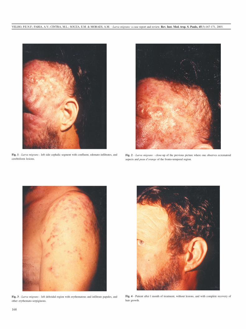
A 44-year-old Caucasian man, truck driver, came to the StateUniversity of Campinas (Unicamp), Dermatology Clinic stating that threemonths before he had been victim of assault and battery, which left himunconscious on the ground for several hours. He had been hospitalizedin coma and given intravenous therapy with corticoid for cerebral edema.Upon regaining consciousness days later, he noted erythematous,edematous and intensely pruritic lesions on the left frontal region andon the left parietotemporal, preauricular, orbicular and malar ones (Figs.l and 2). At the same time, similar lesions appeared in the deltoidal region(Fig. 3) and arm, and other sparse lesions on the abdomen and upperthigh, all on the left side.
He also reported having been subsequently seen by several doctorswho prescribed corticoid of diverse strengths.
When examined, he had confluent, eczematoid, edemato-infiltrative,cerebriform and peau d’orange-looking lesions on the cephalic segmentin the aforementioned regions. There were small papules of up to 5 mm,palpable further beyond. On the left arm and deltoidal region, there wereerythematous infiltrated papules of 5 to 10 mm, and other erythematousserpiginous ones. There were also isolated lesions on the abdomen andthigh. Most of these were follicular.
The skin biopsy performed on the fronto-parietal and deltoidalregions showed dermoepidermitis, both subacute and chronic, superficialand deep, with multiple foci of eosinophilic folliculites (Fig. 5). Focally,in a superficial vein, a circular structure could be seen, with an externallining of keratin, suggesting Ancylostoma sp. larva (Fig. 6).
The patient was treated with albendazole, 400 mg per day for threedays. Due to the severity of the lesions, a second cycle was prescribedafter 21 days. At the end of the treatment, there was a complete regressionof the manifestations, with recovery of hair growth of the alopecic areasassociated with the lesions (Fig. 4). After 24 months, the patient statedthat he had not had any more cutaneous lesions.
REVIEW OF THE LITERATURE
The causal agents of larva migrans are the A. braziliense and the A.caninum, whose eggs are found in dog and cat faeces. Some authorsalso consider the Uncinaria stenocephala, the Gnathostoma spinigerumand the Bunostomun phlebotomum as causal agents9. Others, however,indicate subtle differences in the appearance of the lesions, which leadsthem to characterize them as distinct dermatoses4,12. Man is an accidentalhost to these hookworms. Cats and dogs are definitive hosts4.
The larvae in the soil, in their third stage, penetrate the organism
168
VELHO, P.E.N.F.; FARIA, A.V.; CINTRA, M.L.; SOUZA, E.M. & MORAES, A.M. - Larva migrans: a case report and review. Rev. Inst. Med. trop. S. Paulo, 45(3):167-171, 2003.
Fig. 1 - Larva migrans - left side cephalic segment with confluent, edemato-infiltrates, and
cerebriform lesions.
Fig. 4 - Patient after l month of treatment, without lesions, and with complete recovery of
hair growth.Fig. 3 - Larva migrans - left deltoidal region with erythematous and infiltrate papules, and
other erythemato-serpiginous.
Fig. 2 - Larva migrans - close-up of the previous picture where one observes eczematoid
aspects and peau d’orange of the fronto-temporal region.

VELHO, P.E.N.F.; FARIA, A.V.; CINTRA, M.L.; SOUZA, E.M. & MORAES, A.M. - Larva migrans: a case report and review. Rev. Inst. Med. trop. S. Paulo, 45(3):167-171, 2003.
169
through the skin, reaching the veins in the dermis of these animals. Theymigrate to the lungs and then the trachea, being degluted. The adultworms reproduce in the intestine and the eggs are eliminated in the faeces4.
The larvae are wrapped in a protective pellicle, permeable only bygases. Attracted by the high concentration of carbon dioxide in the skin,they lose their carapaces, releasing proteases. These enzymes will enablethem to migrate through the epidermis. Some authors consider the A.braziliense incapable of reaching the dermis since it does not have specificcollagenases9.
HOLTEZ et al. demonstrated in 1992 that the larval forms of thisnemathelmintes, as well as the A. caninum, produce two enzymesresponsible for the tissue invasion: the proteases and the hialuronidases6.The latter have similar characteristics to those found in mammalspermatozoids.
The hialuronidases of these invertebrates are said to facilitate themigration through the skin and detach the cells of the basal membraneof the epidermis, strongly joined by the hialuronic acid. This enzyme inthe A. braziliense in vitro is more active than the A. caninum, and bothof them are more active than that of other ancylostomas. Whence theease with which these two species lead to cutaneous infestation, unlikeother Ancylostoma sp.6.
Eosinophil-derived major basic protein and eosinophil cationicprotein are the main means by which the host overcomes the energyreserves of the larva, resulting in its incapacitation and elimination9.
The incubation period is usually short and vaguely established, oneor two weeks being the estimate. Experimental studies show that thelarvae penetrate the pillous follicules and the orifices of the sebaceousglands. They begin migration four days after penetration and are moreactive at night2,4,5,12 .
The main affected areas are the dorsum and sole of the feet (uni andbilateral), buttocks, pelvic waist, legs and shoulders. More than one lesionis compatible with more than one entry point4.
The main signs and symptoms are linear and/or serpiginous lesions(which progress from 2 - 3 mm to 2 - 3 cm per day) and the pruritus.This is intensified after some days by the inflammatory reaction of thehost and may even interfere with sleep. Pain may occur in papulovesicularlesions2,4.
Infestation is usually self-limited, as the larva resists for anythingbetween two days and a few weeks, although it may occasionally persistfor more than a year3,4.
Fig. 5 - Larva migrans – a rich in eosinophils, chronic inflammatory process, with epidermic
hyperplasia, fibrosis, hypervascularization, and edema in the dermis. In the center of the
fragment, presence of the follicular eosinophilic abscess. (HE, original magnification x100).
Fig. 6 - Larva migrans – small vein in the papillar dermis containing traces of larva. Exuberant
peri-vascular inflammatory exudates rich in eosinophils (HE, original magnification x400).

170
VELHO, P.E.N.F.; FARIA, A.V.; CINTRA, M.L.; SOUZA, E.M. & MORAES, A.M. - Larva migrans: a case report and review. Rev. Inst. Med. trop. S. Paulo, 45(3):167-171, 2003.
Infestation with A. caninum usually spreads more, with systemicmanifestations, folliculitis and migration to the muscles of the region9.
The diagnosis is essentially clinical and the histological featuresusually contribute very little to it. Wilson evaluated 300 biopsies of larvamigrans lesions in 1951 and found larval structures in only eight, asreported by MILLER et al., 19919. The latter authors describe the findingof a A. caninum structure, and eosinophilic folliculitis in a patient witha clinical diagnosis and histological confirmation. They mention onlyone previous description of folliculitis associated clinically with larvamigrans. CAUMES et al.,1995, clinically associate follicular lesionswith serpiginous dermatitis in two out of 67 patients with this diagnosis2.
The main differential diagnosis should be made with the larvacurrens, caused by Strongyloides stercoralis, whose lesions, linear andurticated, grow on average 10 cm per day12. There may also be similaritieswith the chronic migratory erythema of Lyme’s disease4.
The main therapeutic schemes are thiabendazole, 15% cream, applied2 to 3 times on the affected areas for 5 days. Symptoms should ceasewithin 48 hours. Albendazole can be administered in a single dose of400 mg or in the same daily dose for 3 to 5 days4,7,8,9.
Ivermectin may be administered in a single dose of 12 mg withexcellent results, according to CAUMES & GENTILINI, 19933.
Thiabendazole, taken orally, is not recommended because it is poorlyeffective in comparison with the other therapeutic options, especially ina single dose1. One reason may be that the protective pellicle of the thirdstage of the larva is impermeable to thiabendazole9. In addition, its sideeffects such as nauseas, vomiting, vertigo and chronic headache alsolimit its use3.
JONES, 1994, described the recrudescence of lesions after 3 days ofalbendazole, although he still considers it the best therapeutic option.This may have happened, according to him, because of the variableabsorption of the drug, or because the infestation was massive. Herecommends a later evaluation of patients with this condition7.
DISCUSSION
We have related a case of larva migrans with an unusualmanifestation.
With regard to symptoms, the patient mentioned pain, which occursin only 10% of cases4. In the literature, this symptom is more associatedwith vesiculous lesions, and in this patient an eczematous aspect in thecephalic segment was observed.
The histological finding of rich eosinophilic infiltrate, added to theinformation about anamnesis, led to the diagnosis. This was confirmedby finding the larval structure after exhaustive search. Besides identifyingthe keratin structure inside the blood vessel, which shows larva invasion,the anatomopathological exam showed various areas of folliculites withrich infiltration of eosinophils, as in the case described by MILLER etal., 19919.
Clinical and histological finding of folliculitis and the linking of
larval structure to histology, as in this case, are described in the literatureas rare and more usually associated with A. caninum.
Repetition of treatment with albendazole is supported by the case ofrecrudescence already cited7, and by the fact that this was a case ofmassive infestation. Therapeutic success was confirmed by reevaluationafter a prolonged period of treatment.
It is thought that prolonged contact of the broad affected areas withthe sun may have allowed entry of multiple larvae. The presence of manyspots of folliculites may be due to larval penetration through orifices ofthe pillo-sebaceous apparatus, as suggested by experimental studiesalready mentioned and observed by GUIMARÃES et al., 19995.
The clinical exuberance may have been aided by transitoryimmunodeficiency caused by corticosteroid. This diminishes thecutaneous inflammatory response in many ways, such as the inhibitionof phospholipase A
2, of the action of cytokines, of the expression of
ELAM-1 and ICAM-1, and of the liberation of histamine. In this way, itbecomes efficient in suppression of the eosinophilic activity and mayhave increased susceptibility to this infestation11.
RESUMO
Larva migrans: relato de caso e revisão
É apresentado um caso de infestação maciça por Ancylostoma sp.em paciente que recebeu terapia sistêmica com corticosteroide. O quechama a atenção neste caso é a exuberância e a raridade da manifestaçãoclínica. Com base na literatura pertinente, discutimos os mecanismos dainfecção parasitária, a história natural da doença e seu tratamento.
REFERENCES
1. CAUMES, E.; CARRIÈRE, J.; DATRY, A. et al. - A randomized trial of ivermectinversus albendazole for the treatment of cutaneous larva migrans. Amer. J. trop.Med. Hyg., 49: 641-644, 1993.
2. CAUMES, E.; CARRIÈRE, J.; GUERMONPREZ, G. et al. - Dermatoses associatedwith travel to tropical countries: a prospective study of the diagnosis and managementof 269 patients presenting to a tropical disease unit. Clin. infect. Dis., 20: 542-548,1995.
3. CAUMES, E. & GENTILINI, M. - Traitement de la larva migrans cutanéeankylostomienne. Ann. Derm. Vénéreol., 120: 571-573, 1993.
4. DAVIES, H.D.; SAKULS, P. & KEYSTONE, J.S. - Creeping eruption. A review of clinicalpresentation and management of 60 cases presenting to a tropical disease unit. Arch.Derm., 129: 588-591, 1993.
5. GUIMARÃES, L.C.; SILVA, J.H.; SAAD, K.; LOPES, E.R. & MENESES, A.C.O. -Larva migrans within scalp sebaceous gland. Rev. Soc. bras. Med. trop., 32: 187-189, 1999.
6. HOLTEZ, P.J.; NARASHIMAN, S.; HAGGERTY, J. et al. - Hyaluronidase from infectiveAncylostoma hookworm larvae and its possible function as virulence factor in tissueinvasion and in cutaneous larva migrans. Infec. Immun., 60: 1018-1023, 1992.
7. JONES, S.K. - Cutaneous larva migrans - ‘recurrens’. Brit. J. Derm., 130: 546, 1994.
8. KIRTCHEN, L.W. - Case studies in international travelers. Amer. Fam. Phycn., 60:471-474, 1999.

VELHO, P.E.N.F.; FARIA, A.V.; CINTRA, M.L.; SOUZA, E.M. & MORAES, A.M. - Larva migrans: a case report and review. Rev. Inst. Med. trop. S. Paulo, 45(3):167-171, 2003.
171
9. MILLER, A.C.; WALKER, J.; JAWORSKI, R.; LAUNEY, W. & PAVER, R. - Hookwormfolliculitis. Arch. Derm., 127: 547-549, 1991.
10. NICHOLS, R. - The etiology of visceral larva migrans. II. Comparative larval morphologyof Ascaris lumbricoides, Necator americanus, Strongyloides stercoralis andAncylostoma caninum. J. Parasit., 42: 363-399, 1956.
11. SCHIMMER, P.B. & PARKER, K.L. - Adrenocorticotropic hormone, adrenocorticalsteroids and their synthetic analogs; inhibitors of the synthesis and actions ofadrenocortical hormones. In: HARDMAN, J.G.; GILMAN, A.G. & LIMBIRD, L.E.,ed. The pharmacological basis of therapeutics. Nashville, Goodman & Gilman,1996. p. 1471.
12. STONE, O.J.; NEWELL, G.B. & MULLINS, J.F. – Cutaneous strongyloidiasis: larvacurrens. Arch. Derm., 106: 734-736, 1972.
Received: 20 February 2003Accepted: 19 May 2003