Embed Size (px)
Citation preview
HAL Id: pasteur-01429562https://hal-pasteur.archives-ouvertes.fr/pasteur-01429562
Submitted on 8 Jan 2017
HAL is a multi-disciplinary open accessarchive for the deposit and dissemination of sci-entific research documents, whether they are pub-lished or not. The documents may come fromteaching and research institutions in France orabroad, or from public or private research centers.
L’archive ouverte pluridisciplinaire HAL, estdestinée au dépôt et à la diffusion de documentsscientifiques de niveau recherche, publiés ou non,émanant des établissements d’enseignement et derecherche français ou étrangers, des laboratoirespublics ou privés.
Distributed under a Creative Commons Attribution| 4.0 International License
Large-Scale Phylogenomic Analysis Reveals theComplex Evolutionary History of Rabies Virus in
Multiple Carnivore Hosts.Cécile Troupin, Laurent Dacheux, Marion Tanguy, Claude Sabeta, Hervé
Blanc, Christiane Bouchier, Marco Vignuzzi, Sebastián Duchene, Edward CHolmes, Hervé Bourhy
To cite this version:Cécile Troupin, Laurent Dacheux, Marion Tanguy, Claude Sabeta, Hervé Blanc, et al.. Large-ScalePhylogenomic Analysis Reveals the Complex Evolutionary History of Rabies Virus in Multiple Car-nivore Hosts.. PLoS Pathogens, Public Library of Science, 2016, 12 (12), pp.e1006041. �10.1371/jour-nal.ppat.1006041.s014�. �pasteur-01429562�
RESEARCH ARTICLE
Large-Scale Phylogenomic Analysis Reveals
the Complex Evolutionary History of Rabies
Virus in Multiple Carnivore Hosts
Cecile Troupin1, Laurent Dacheux1, Marion Tanguy1,2, Claude Sabeta3, Herve Blanc4,
Christiane Bouchier2, Marco Vignuzzi4, Sebastian Duchene5,6, Edward C. Holmes5,
Herve Bourhy1*
1 Institut Pasteur, Unit Lyssavirus Dynamics and Host Adaptation, WHO Collaborating Centre for Reference
and Research on Rabies, Paris, France, 2 Institut Pasteur, Genomics Platform, Paris, France, 3 Agricultural
Research Council, Onderstepoort Veterinary Institute, OIE Rabies Reference Laboratory, Pretoria, South
Africa, 4 Institut Pasteur, Centre National de la Recherche Scientifique UMR 3569, Viral Populations and
Pathogenesis Unit, Paris, France, 5 Marie Bashir Institute for Infectious Diseases and Biosecurity, Charles
Perkins Centre, School of Life and Environmental Sciences and Sydney Medical School, The University of
Sydney, Sydney, Australia, 6 Centre for Systems Genomics, University of Melbourne, Parkville, Victoria,
Australia
Abstract
The natural evolution of rabies virus (RABV) provides a potent example of multiple host
shifts and an important opportunity to determine the mechanisms that underpin viral emer-
gence. Using 321 genome sequences spanning an unprecedented diversity of RABV, we
compared evolutionary rates and selection pressures in viruses sampled from multiple pri-
mary host shifts that occurred on various continents. Two major phylogenetic groups, bat-
related RABV and dog-related RABV, experiencing markedly different evolutionary dynam-
ics were identified. While no correlation between time and genetic divergence was found in
bat-related RABV, the evolution of dog-related RABV followed a generally clock-like struc-
ture, although with a relatively low evolutionary rate. Subsequent molecular clock dating
indicated that dog-related RABV likely underwent a rapid global spread following the intensi-
fication of intercontinental trade starting in the 15th century. Strikingly, although dog RABV
has jumped to various wildlife species from the order Carnivora, we found no clear evidence
that these host-jumping events involved adaptive evolution, with RABV instead character-
ized by strong purifying selection, suggesting that ecological processes also play an impor-
tant role in shaping patterns of emergence. However, specific amino acid changes were
associated with the parallel emergence of RABV in ferret-badgers in Asia, and some host
shifts were associated with increases in evolutionary rate, particularly in the ferret-badger
and mongoose, implying that changes in host species can have important impacts on evolu-
tionary dynamics.
PLOS Pathogens | DOI:10.1371/journal.ppat.1006041 December 15, 2016 1 / 20
a11111
OPENACCESS
Citation: Troupin C, Dacheux L, Tanguy M, Sabeta
C, Blanc H, Bouchier C, et al. (2016) Large-Scale
Phylogenomic Analysis Reveals the Complex
Evolutionary History of Rabies Virus in Multiple
Carnivore Hosts. PLoS Pathog 12(12): e1006041.
doi:10.1371/journal.ppat.1006041
Editor: Colin Parrish, Cornell University, UNITED
STATES
Received: June 3, 2016
Accepted: November 3, 2016
Published: December 15, 2016
Copyright: © 2016 Troupin et al. This is an open
access article distributed under the terms of the
Creative Commons Attribution License, which
permits unrestricted use, distribution, and
reproduction in any medium, provided the original
author and source are credited.
Data Availability Statement: All sequence files are
available from the GenBank database under
accession numbers KX148100-KX148269, and
accession numbers of each sample are available in
S1 Table. All other relevant data are within the
paper and its supporting information files.
Funding: This work was supported by European
Union Seventh Framework Programme
PREDEMICS (grant number 278433) and by the
Agence Nationale de la Recherche (grant number
BSV3-0019). The Genomics Platform is member of
“France Genomique” consortium (ANR10-INBS-
Author Summary
Zoonoses account for most recently emerged infectious diseases of humans, although little
is known about the evolutionary mechanisms involved in cross-species virus transmis-
sion. Understanding the evolutionary patterns and processes that underpin such cross-
species transmission is of importance for predicting the spread of zoonotic infections, and
hence to their ultimate control. We present a large-scale and detailed reconstruction of
the evolutionary history of rabies virus (RABV) in domestic and wildlife animal species.
RABV is of particular interest as it is capable of infecting many mammals but, paradoxi-
cally, is only maintained in distinct epidemiological cycles associated with animal species
from the orders Carnivora and Chiroptera. We show that bat-related RABV and dog-
related RABV have experienced very different evolutionary dynamics, and that host
jumps are sometimes characterized by significant increases in evolutionary rate. Among
Carnivora, the association between RABV and particular host species most likely arose
from a combination of the historical human-mediated spread of the virus and jumps into
new primary host species. In addition, we show that changes in host species are associated
with multiple evolutionary pathways including the occurrence of host-specific parallel
evolution. Overall, our data indicate that the establishment of dog-related RABV in new
carnivore hosts may only require subtle adaptive evolution.
Introduction
Revealing how viruses jump species boundaries and establish productive infections in new
hosts is key to understanding disease emergence. As most recent emerging and re-emerging
viruses have RNA genomes [1], it is of central importance to understand the drivers of RNA
virus evolution, diversification and cross-species transmission. Clearly, successful virus emer-
gence has diverse causes, likely involving anthropogenic, social and environmental factors [2].
However, the capacity of the viral genome to vary and generate advantageous mutations is also
an important element, enabling RNA viruses to exploit new niches, including novel host spe-
cies, often more rapidly than DNA-based organisms [1, 3, 4]. One important manifestation of
RNA virus evolution and diversification is the rate of evolutionary change (i.e. nucleotide sub-
stitution), with analyses of how this parameter varies by host species providing important
information on the nature of virus-host interactions.
Disease emergence results from complex mechanisms that shape the ability of a virus to be
maintained within its primary host species, then be serially transmitted to a new host species
and initiate a pathologic process to cause disease [5]. As such, lyssaviruses (family Rhabdoviri-dae), the causative agents of rabies–an acute and almost invariably fatal encephalomyelitis in
humans–represent an informative case study to examine the relationship between virus
genetic diversity and disease emergence. In particular, the natural history of these zoonotic
viruses provides an excellent model to study how replication in different host species alters the
selection pressures that act on virus genomes. Lyssaviruses are single-stranded, negative-sense
RNA viruses with a genome size of approximately 12 kb that encodes five proteins: the nucleo-
protein (N), the phosphoprotein (P), the matrix protein (M), the glycoprotein (G) and the
Large protein or polymerase (L). Currently, the lyssaviruses are classified into 14 species and
one tentative species [6]. Like other RNA viruses, lyssaviruses exhibit high rates of mutation
due to a lack of proofreading activity in the L protein [7]. Notably, although many mammalian
species appear to be susceptible to lyssavirus infection, the virus is only able to establish
Evolutionary History of Rabies Virus and Host Jumps
PLOS Pathogens | DOI:10.1371/journal.ppat.1006041 December 15, 2016 2 / 20
09-08). SD is supported by a University of
Melbourne McKenzie fellowship. ECH is supported
by an NHMRC Australia Fellowship. HB and CS
were supported by the European Virus Archive
goes Global (EVAg) project that has received
funding from the European Union’s Horizon 2020
research and innovation programme under grant
agreement No 653316. The funders had no role in
study design, data collection and analysis, decision
to publish, or preparation of the manuscript.
Competing Interests: The authors have declared
that no competing interests exist.
sustained transmission networks in a relatively small number, indicating that there are major
barriers to successful cross-species transmission [8–11].
One species of lyssavirus, rabies virus (RABV), is present worldwide and circulates in a
diverse set of reservoir hosts among the mammalian orders Chiroptera and Carnivora [12]. Its
natural evolution provides an illustrative example of multiple host switches, in turn enabling
comparative studies of the evolutionary patterns, processes and dynamics associated with host
adaptation. Previous studies demonstrated that RABV isolates fall into two major phylogenetic
groups; the bat- and the dog-related RABV groups [8, 13, 14]. The ‘bat-related’ RABV group is
confined to New World viruses circulating mainly among bats, as well as in some terrestrial
carnivores such as skunks and raccoons [14–17]. In contrast, the ‘dog-related’ RABV group
contains viruses circulating worldwide in dogs, as well as in wildlife carnivores in specific geo-
graphic areas such as foxes and raccoon dogs in Europe, foxes in the Middle East, raccoon
dogs and ferret-badgers in Asia, skunks, foxes, coyotes and mongooses in the Americas, and
mongooses in Africa [14, 16, 18–22]. Importantly, dogs are responsible for more than 99% of
the human rabies cases worldwide [23] and are likely the main vector for the inter-species
transmission of dog-related RABV.
Previous phylogenetic analyses have largely been performed on individual genes [13–19,
21, 24–29] with a few assessing the full-length viral genome [20, 30, 31]. In addition, most of
these phylogenetic studies were performed on relatively small numbers of sequences originat-
ing from one specific geographical area and/or associated with a specific animal host [20, 22,
30, 32, 33]. Despite these limitations, these studies are consistent in showing that RABV is sub-
ject to strong purifying selection [10] coupled to geographical clustering that is occasionally
disrupted by human mediated dispersion [13, 34, 35]. Recently, it was shown that nucleotide
substitution rates in RABV vary markedly among those viruses infecting bats, such that rates
in tropical and subtropical species were markedly higher than those from temporal bat species,
perhaps reflecting a combination of host and environmental factors [36]. However, equivalent
data for dog-related RABV are lacking. In addition, whether evolutionary rates in RABV vary
among wild carnivores and domestic dogs is unknown, although studies in other systems have
revealed that rates of RNA virus evolution may differ between wild and domestic animals [37].
Clearly, the large-scale analysis of RABV, particularly comprising full-length genome
sequences, is needed to reveal the nature of the selection pressures associated with host switch-
ing. That the RABV genome encodes a limited number of proteins that necessarily have multi-
functional roles [38], and hence potentially large-scale epistasis, also means that these selection
pressures may be complex.
Herein we present the first phylogenetic study of RABV on a genome-wide and global scale,
utilizing a data set of 321 whole-genome sequences sampled from 66 countries over a time
period of 65 years, with the aim of inferring those evolutionary patterns and processes associ-
ated with host-switching. In particular, we compared RABV from wild carnivores and in
domestic dogs with respect to selection pressures, evolutionary rates, and the time-scale of
their evolutionary history. Importantly, the size of the data set allowed us to reveal any hetero-
geneity in evolutionary rates among RABV adapted to different primary hosts, and determine
the complex evolutionary dynamics of RABV as it adapts to new hosts.
Results
Host and geographical clustering of RABV
A phylogenetic analysis was performed on the (99%) full-length genome sequences of 321
RABV sequences sampled from 66 countries (S1 Fig, S1 Table). Of these viruses, 170 were
newly sequenced as part of this study. As expected given the low levels of recombination in
Evolutionary History of Rabies Virus and Host Jumps
PLOS Pathogens | DOI:10.1371/journal.ppat.1006041 December 15, 2016 3 / 20
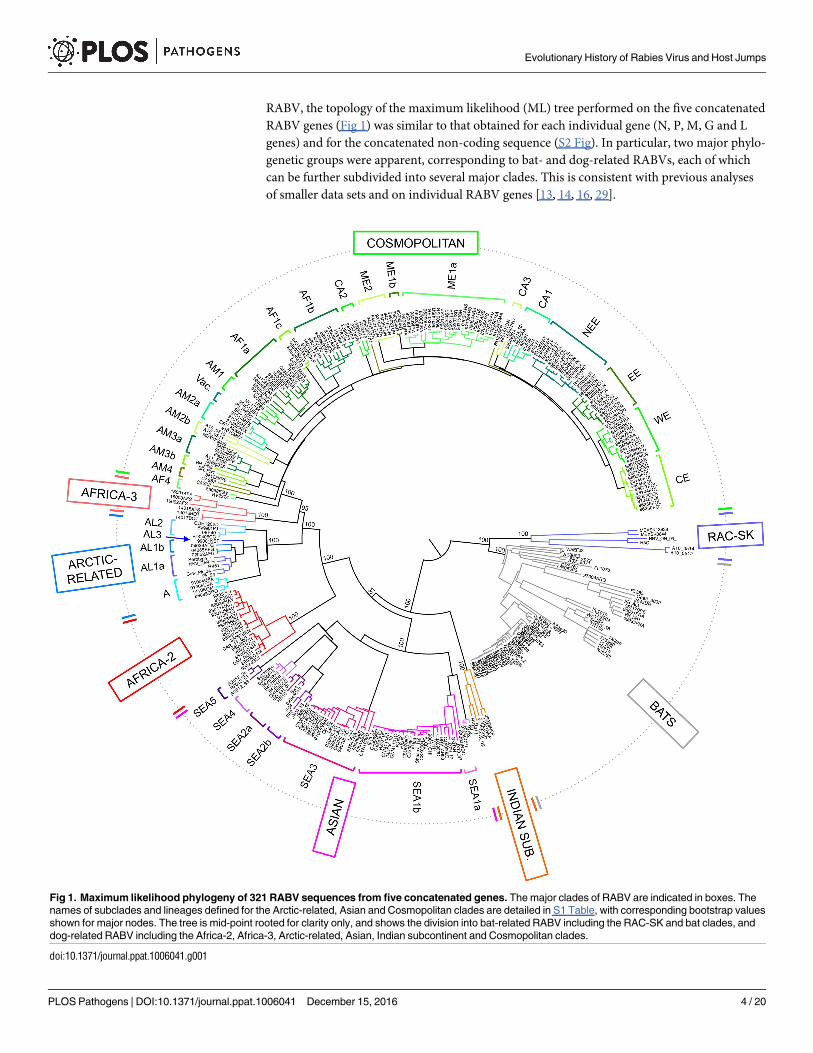
RABV, the topology of the maximum likelihood (ML) tree performed on the five concatenated
RABV genes (Fig 1) was similar to that obtained for each individual gene (N, P, M, G and L
genes) and for the concatenated non-coding sequence (S2 Fig). In particular, two major phylo-
genetic groups were apparent, corresponding to bat- and dog-related RABVs, each of which
can be further subdivided into several major clades. This is consistent with previous analyses
of smaller data sets and on individual RABV genes [13, 14, 16, 29].
Fig 1. Maximum likelihood phylogeny of 321 RABV sequences from five concatenated genes. The major clades of RABV are indicated in boxes. The
names of subclades and lineages defined for the Arctic-related, Asian and Cosmopolitan clades are detailed in S1 Table, with corresponding bootstrap values
shown for major nodes. The tree is mid-point rooted for clarity only, and shows the division into bat-related RABV including the RAC-SK and bat clades, and
dog-related RABV including the Africa-2, Africa-3, Arctic-related, Asian, Indian subcontinent and Cosmopolitan clades.
doi:10.1371/journal.ppat.1006041.g001
Evolutionary History of Rabies Virus and Host Jumps
PLOS Pathogens | DOI:10.1371/journal.ppat.1006041 December 15, 2016 4 / 20
The bat-related group contained two major clades, one including the bat RABVs circulating
in the Americas, and the other (RAC-SK) comprising viruses from American skunks and rac-
coons (Fig 1). In turn, the RAC-SK group contained a number of ‘subclades’ corresponding to
Mexican skunks (MeSK-1), North American raccoons (RAC) and South-Central skunks
(SCSK) as previously described (S3 Fig) [14, 16, 17, 39].
Similarly, the dog-related group includes six major clades supported by high bootstrap val-
ues (S2 Table and Fig 1), and previously identified as the Africa-2, Africa-3, Arctic-related,
Asian, Cosmopolitan and Indian subcontinent clades [13]. The phylogenetic analysis based on
the five concatenated genes was particularly informative, allowing us to distinguish various
subclades and lineages among these six major clades, with some of which are characterized for
the first time here (Fig 1 and supplementary text).
Some of these clades and subclades are of particular interest. The SEA2 subclade contains
viruses from China and is divided into two lineages, SEA2a and SEA2b, corresponding to iso-
lates from dogs and ferret-badgers, respectively. Subclade SEA5 appears to be specific to
RABV circulating in ferret-badgers in Taiwan, an epidemiological cycle that was only identi-
fied recently [21, 40, 41]. For the first time, we were also able to fully characterize full-length
genome sequences of RABV isolates belonging to the Africa-3 clade (n = 6). These viruses cir-
culating in Southern Africa are monophyletic and phylogenetically distinct from the other
major RABV clades, particularly those circulating in Africa [19, 42, 43].
Temporal dynamics and spread of the dog-related RABV
To determine the evolutionary dynamics of RABV, we first determined whether individual
data sets contained sufficient temporal structure to undertake detailed molecular clock analy-
ses by performing a regression of root-to-tip genetic distance against the year of sampling.
Notably, no correlation between time and genetic divergence was found when the sequences
of both bat- and dog-related RABV groups were analyzed together, indicating that there is
extensive variation in the rate of RABV evolution among these taxa (and hence that they
should not be combined in molecular clock studies) (S4A Fig). In addition, no temporal struc-
ture was observed when the sequences of bat-related RABV were analyzed separately, indicat-
ing that this subset of viruses is not evolving uniformly (S4B Fig) as noted previously [36].
However, a clear association between genetic distance and time (i.e. a molecular clock) was
observed for the dog-related group alone (S4C Fig), allowing us to estimate substitution rates,
and hence times to common ancestry, more precisely in this cluster using a Bayesian
approach.
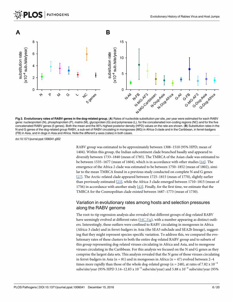
The mean rate of evolutionary change in the dog-related RABV was estimated to be 2.44 x
10−4 subs/site/year (95% HPDs of 2.10–2.80 x 10⁻⁴ subs/site/year) for the five concatenated
genes. Importantly, we were also able to compare the substitution rate of each RABV gene and
of the concatenated non-coding regions from the same genomic sequence data set. These esti-
mates varied in the following ascending order: N, L, G, M and P (Fig 2A). However, only the P
gene had a nucleotide substitution rate considerably higher than those of N and L genes. As
expected, the evolutionary rate in the non-coding regions was significantly higher than those
of the coding regions, indicative of weaker selective constraints.
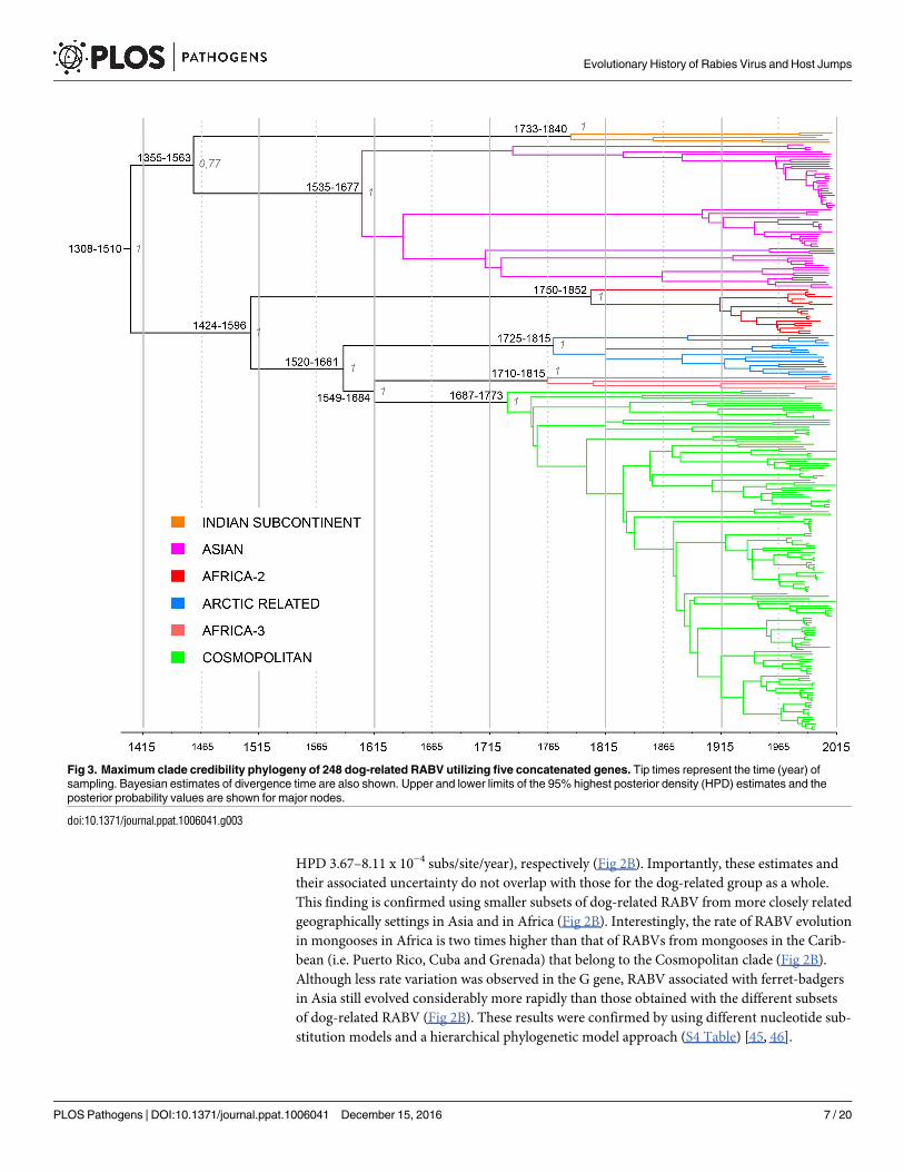
The estimation of a reliable substitution rate allowed us to determine the mean times to
common ancestry (TMRCA) for each RABV clade, subclade and lineage defined above (Fig 3;
estimates for different sub-clades shown in S3 Table and discussed in the Supplementary text).
For this analysis we utilized the concatenated coding genes as these had the lowest variance.
Notably, these TMRCA estimates exhibited less uncertainty than previous studies performed
on N and/or G genes alone [13, 25, 27, 29, 43, 44]. Briefly, the TMRCA of the dog-related
Evolutionary History of Rabies Virus and Host Jumps
PLOS Pathogens | DOI:10.1371/journal.ppat.1006041 December 15, 2016 5 / 20
RABV group was estimated to be approximately between 1308–1510 (95% HPD; mean of
1404). Within this group, the Indian subcontinent clade branched basally and appeared to
diversify between 1733–1840 (mean of 1785). The TMRCA of the Asian clade was estimated to
be between 1535–1677 (mean of 1604), which is in accordance with other studies [44]. The
emergence of the Africa 2 clade was estimated to be between 1750–1852 (mean of 1802), simi-
lar to the mean TMRCA found in a previous study conducted on complete N and G genes
[27]. The Arctic-related clade appeared between 1725–1815 (mean of 1770), slightly earlier
than previously estimated [25], while the Africa-3 clade emerged between 1710–1815 (mean of
1756) in accordance with another study [43]. Finally, for the first time, we estimate that the
TMRCA for the Cosmopolitan clade existed between 1687–1773 (mean of 1730).
Variation in evolutionary rates among hosts and selection pressures
along the RABV genome
The root-to-tip regression analysis also revealed that different groups of dog-related RABV
have seemingly evolved at different rates (S4C Fig), with a number appearing as distinct outli-
ers. Interestingly, these outliers were confined to RABV circulating in mongooses in Africa
(Africa-3 clade) and in ferret-badgers in Asia (the SEA5 subclade and SEA2b lineage), suggest-
ing that they might represent species-specific variation. To address this, we compared the evo-
lutionary rates of these clusters to both the entire dog-related RABV group and to subsets of
this group representing dog-related viruses circulating in Africa and Asia, and to mongoose
viruses circulating in the Caribbean. For this analysis we focused on the N and G genes as they
comprise the largest data sets. This analysis revealed that the N gene of those viruses circulating
in ferret-badgers in Asia (n = 81) and in mongooses in Africa (n = 47) evolved between 2–4
times more rapidly than those of the whole dog-related group (n = 248), at rates of 7.82 x 10⁻⁴
subs/site/year (95% HPD 3.14–12.83 x 10⁻⁴ subs/site/year) and 5.88 x 10⁻⁴ subs/site/year (95%
Fig 2. Evolutionary rates of RABV genes in the dog-related group. (A) Rates of nucleotide substitution per site, per year were estimated for each RABV
gene: nucleoprotein (N), phosphoprotein (P), matrix (M), glycoprotein (G) and polymerase (L), for the concatenated non-coding regions (NC) and for the five
concatenated RABV genes (5 genes). Both the mean and the 95% highest posterior density (HPD) values on the rate are shown. (B) Substitution rates in the
N and G genes of the dog-related group RABV, a sub-set of RABV circulating in mongooses (MG) in Africa-3 clade and in the Caribbean, in ferret-badgers
(FB) in Asia, and in dogs in Asia and Africa. Note the different y-axes (rates) in both cases.
doi:10.1371/journal.ppat.1006041.g002
Evolutionary History of Rabies Virus and Host Jumps
PLOS Pathogens | DOI:10.1371/journal.ppat.1006041 December 15, 2016 6 / 20
HPD 3.67–8.11 x 10⁻⁴ subs/site/year), respectively (Fig 2B). Importantly, these estimates and
their associated uncertainty do not overlap with those for the dog-related group as a whole.
This finding is confirmed using smaller subsets of dog-related RABV from more closely related
geographically settings in Asia and in Africa (Fig 2B). Interestingly, the rate of RABV evolution
in mongooses in Africa is two times higher than that of RABVs from mongooses in the Carib-
bean (i.e. Puerto Rico, Cuba and Grenada) that belong to the Cosmopolitan clade (Fig 2B).
Although less rate variation was observed in the G gene, RABV associated with ferret-badgers
in Asia still evolved considerably more rapidly than those obtained with the different subsets
of dog-related RABV (Fig 2B). These results were confirmed by using different nucleotide sub-
stitution models and a hierarchical phylogenetic model approach (S4 Table) [45, 46].
Fig 3. Maximum clade credibility phylogeny of 248 dog-related RABV utilizing five concatenated genes. Tip times represent the time (year) of
sampling. Bayesian estimates of divergence time are also shown. Upper and lower limits of the 95% highest posterior density (HPD) estimates and the
posterior probability values are shown for major nodes.
doi:10.1371/journal.ppat.1006041.g003
Evolutionary History of Rabies Virus and Host Jumps
PLOS Pathogens | DOI:10.1371/journal.ppat.1006041 December 15, 2016 7 / 20
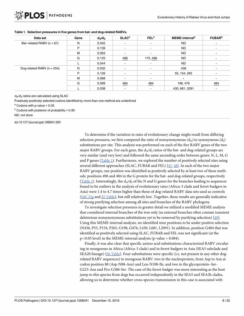
To determine if the variation in rates of evolutionary change might result from differing
selection pressures, we first compared the ratio of nonsynonymous (dN) to synonymous (dS)substitutions per site. This analysis was performed on each of the five RABV genes of the two
major RABV groups. For each gene, the dN/dS ratios of the bat- and dog-related groups are
very similar (and very low) and followed the same ascending order between genes: N, L, M, G
and P genes (Table 1). Furthermore, we explored the number of positively selected sites using
several different approaches (SLAC, FUBAR and FEL) [47, 48]. In each of the two major
RABV groups, one position was identified as positively selected by at least two of these meth-
ods: positions 496 and 484 in the G protein for the bat- and dog-related groups, respectively
(Table 1). Interestingly, the dN/dS of the N and G genes for the branches leading to sequences
found to be outliers in the analysis of evolutionary rates (Africa-3 clade and ferret-badgers in
Asia) were 1.4 to 4.7 times higher than those of dog-related RABV data sets used as controls
(S4C Fig and S5 Table), but still relatively low. Together, these results are generally indicative
of strong purifying selection among all sites and branches of the RABV phylogeny.
To investigate selection pressures in greater detail we utilized a modified MEME analysis
that considered internal branches of the tree only (as external branches often contain transient
deleterious nonsynonymous substitutions yet to be removed by purifying selection) [49].
Using this MEME-internal analysis, we identified nine positions to be under positive selection
(N436, P55, P154, P265, G198, G476, L430, L681, L2091). In addition, position G484 that was
identified as positively selected using SLAC, FUBAR and FEL was not significant (at the
p<0.05 level) in the MEME-internal analysis (p-value = 0.084).
Finally, it was also clear that specific amino acid substitutions characterized RABV circulat-
ing in mongooses in Africa (Africa-3 clade) and in ferret-badgers in Asia (SEA5 subclade and
SEA2b lineage) (S6 Table). Four substitutions were specific (i.e. not present in any other dog-
related RABV sequences) to mongoose RABV: two in the nucleoprotein, from Asp to Asn at
codon position 88 (Asp-N88-Asn) and Leu-N108-Ile, and two in the glycoprotein–Ser-
G223-Asn and Pro-G386-Ser. The case of the ferret-badger was more interesting as the host
jump to this species from dogs has occurred independently in the SEA5 and SEA2b clades,
allowing us to determine whether cross-species transmission in this case is associated with
Table 1. Selection pressures in five genes from bat- and dog-related RABVs.
Data set Gene dN/dS SLACa FELa MEME-internala FUBARb
Bat–related RABV (n = 67) N 0.045 - - ND -
P 0.139 - - ND -
M 0.063 - - ND -
G 0.133 496 175, 496 ND -
L 0.044 - - ND -
Dog-related RABV (n = 254) N 0.032 - - 436 -
P 0.126 - - 55, 154, 265 -
M 0.068 - - - -
G 0.099 484 484 198, 476 484
L 0.038 - - 430, 681, 2091 -
dN/dS ratios are calculated using SLAC
Putatively positively selected codons identified by more than one method are underlineda Codons with p-value < 0.05b Codons with posterior of probability > 0.95
ND: not done
doi:10.1371/journal.ppat.1006041.t001
Evolutionary History of Rabies Virus and Host Jumps
PLOS Pathogens | DOI:10.1371/journal.ppat.1006041 December 15, 2016 8 / 20
parallel viral evolution. This analysis revealed that two amino acid substitutions were common
to all ferret-badger viruses across both clades: Leu-N374-Ser and Lys-L200-Arg. The Leu-
N374-Ser substitution is particularly noteworthy as it only occurs in the ferret-badger, this res-
idue is normally highly conserved in RABV, and Leu-to-Ser is a non-conservative amino acid
change. Hence, we suspect that Leu-N374-Ser, and perhaps Lys-L200-Arg, facilitate RABV
adaptation to ferret-badgers. Notably, neither of these sites was found to be subject to positive
selection using the methods employed here (Table 1).
Discussion
The central aim of this study was to determine whether the patterns and processes of RABV
evolution vary between viruses sampled from different host species reflect the impact of cross-
species transmission. To that end we present the largest phylogenomic analysis of RABVs cir-
culating worldwide performed to date. Although the topology of the RABV phylogeny is simi-
lar to those obtained previously [13, 14, 16, 29], it clearly presents a more comprehensive and
precise reconstruction of evolutionary history of this virus. In particular, the analysis of the
five concatenated genes allowed us to obtain a finer-scale dating of the emergence of the major
clades with narrower confidence intervals than obtained previously [13, 25, 27, 29, 43, 44].
RABV undergoes relatively frequent cross-species transmission [8, 11, 13, 18], which pro-
vides an opportunity to determine whether host jumping impacts rates of evolutionary change.
Notably, we found no correlation between root-to-tip genetic distance and sampling time in
the bat-related RABV group, nor when combined with dog-related RABV group, indicating
that these viruses have not evolved in a clock-like manner, with substantial rate variation
already observed in bat-associated RABV [36]. In contrast, a strong association between
genetic divergence and time (i.e. a molecular clock) was observed within the dog-related
RABV group, with a mean evolutionary rate of 2.44 x 10⁻⁴ subs/site/year (95% HPDs of 2.10–
2.80 x 10⁻⁴ subs/site/year) for the five concatenated genes. This estimate is evidently more pre-
cise than those determined previously [13, 25, 27, 30, 44, 50–53].
Despite the relative rate constancy in the dog-related RABV, it was striking that some of the
clades or sub-clades have experienced substantially higher rates of nucleotide substitution. In
particular, viruses circulating in ferret-badgers in Asia (mainland China and Taiwan) and in
mongooses in Africa have evolved at least twice as rapidly as those of the dog-related group.
Although there is some uncertainty in these rate estimates, they do not overlap with the esti-
mates for the entire dog-related RABV group. Determining the evolutionary basis to this rate
variation is more complex. Changes in evolutionary rate could only be driven either by
changes in background mutation rate (which we consider unlikely to differ between dog-
related RABV) or, more likely, by changes in the population size and/or incubation time that
may vary among different animal hosts [36]. It is also possible that the evolutionary rates esti-
mated here have been impacted by time-dependency, such that they are elevated toward the
present (i.e. in closely related sequences sampled recently) due to the presence of transient del-
eterious mutations that have yet to be removed by purifying selection [54]. However, while
this may in part explain the high rate in the recently sampled RABV from ferret-badgers, it is
unlikely to explain the higher evolutionary rate in mongoose RABV whose evolutionary his-
tory sampled here covers a longer time period. In the case of the ferret-badgers, two amino
acid changes (Leu-N374-Ser and Lys-L200-Arg) have evolved in parallel in the two clades asso-
ciated which is compatible with the occurrence of adaptive evolution, and which have in turn
elevated the nucleotide substitution rate. That these two sites were not detected in analyses of
dN/dS suggests that these methods may have limitations when identifying adaptive evolution
involving limited amounts of amino acid change.
Evolutionary History of Rabies Virus and Host Jumps
PLOS Pathogens | DOI:10.1371/journal.ppat.1006041 December 15, 2016 9 / 20
Our analysis also showed that the nucleotide substitution rate varied markedly according to
the gene analyzed in the ascending order: N, L, G, M and P. As expected, the two proteins
often described as more conserved for RABV—N and L—exhibited the lowest rates, as well as
the lowest dN/dS ratios, indicating that they are subject to the strongest purifying selection.
Notably, the highest substitution rate and dN/dS was observed in the P protein, perhaps reflect-
ing the weak structural organization of the C-term part of this protein [55, 56].
The presence of relatively constant molecular clock also enabled us to provide a more
robust time-scale for the evolution of the principal geographical clusters of dog-related RABV
(Fig 3, S3 Table). Accordingly, we estimate that the most recent ancestor of all dog-related
RABV dates to between 1308 and 1510. Consequently, any older canid RABV lineages, pro-
posed to have circulated in the Middle-East more than 2000 years ago [57, 58], have not sur-
vived to be sampled in the current study. Interestingly, the timing of the most recent ancestor
of all dog-related RABV circulating to date coincides with the development of the world’s first
truly global trade network following the explorations of Columbus, Vasco da Gama and Zheng
He, commissioned by the Spanish, the Portuguese and the Chinese Ming Dynasty, respec-
tively. This age of exploration and colonization contributed to the establishment of new long
distance commercial practices and transoceanic shipping services between 1450 and 1750 [59].
The concomitant dissemination of RABV during this period, probably by dogs travelling by
boats with their owners, therefore provides a powerful example of the early human-mediated
dissemination of a zoonotic disease. In addition, all the ancestors of the major clades found cir-
culating today in North and South America, Africa, Asia and Europe originated between 1687
and 1840 at the apogee of this international trade and colonization process [59]. This is further
exemplified by the global spread of the Cosmopolitan clade.
A fundamental question in evolutionary virology is how and why some viruses are seem-
ingly better able to jump species boundaries than others. A compelling theory is that the more
closely related the host species in questions, the greater the chance of successful transmission
[9, 60, 61]. However, it is unclear how strictly this theory holds for RABV [11], and our results
confirm species jumps of RABV among animal species of the order Chiroptera, and from bats
to striped skunks (Mephitis mephitis) [14, 62, 63]. In addition, there is also clearly a geographic
component to cross-species transmission as bat-related RABVs are only found in the Ameri-
cas. More notably, our study clearly confirms that although spill-over infections from wildlife
species to dog take place, species jumps involving dog-related RABVs generally occur fromdogs to wildlife species of the order Carnivora; not only to the family Canidae (dog, red fox,
raccoon dog), but also to more distant species belonging to the families Mustelidae (ferret-bad-
ger), Herpestidae (mongoose) and Mephetidae (skunk) (S5 Fig) [13, 18, 42, 43]. These changes
in primary animal host species have occurred independently in different localities and at dif-
ferent times during RABV evolution. Further, some carnivore species, notably skunks, are
infected by RABV of both dog and bat origin [14, 16, 39].
Revealing the respective roles of genetic drift and the selection of advantageous mutations
in shaping the genetic diversity of RABV, particularly during host shifts, is a central evolution-
ary question. There is currently no definitive data on whether dog-related RABV emergence
requires active adaptive evolution (i.e. positive selection) to in a new host species, or whether it
is largely a chance process involving ecological factors facilitating the transmission of a viral
strain with the pre-existing necessary genetic characteristics [64]; the latter has been proposed
for the repeated outbreaks of bat-related RABV in striped skunks and gray foxes in Arizona
[14] and of gray foxes due to skunk-associated RABV in California [65]. Our analysis showed
that the dog-related RABV group is subject to strong purifying selection, and when positive
selection did occur on internal branches of the phylogenetic tree it was not obviously associ-
ated with host jumping. As noted above, however, the failure to detect positive selection in the
Evolutionary History of Rabies Virus and Host Jumps
PLOS Pathogens | DOI:10.1371/journal.ppat.1006041 December 15, 2016 10 / 20
case of ferret-badger RABV despite the occurrence of parallel evolution suggests that these
methods may suffer from false-negatives.
Successful cross-species transmission is a complex ecological and evolutionary process,
beginning with exposure and contact between the two species, followed by the successful infec-
tion of the new host species, and potentially host-adaptive evolution to enable long-term sus-
tained transmission [66, 67]. However, due to complex interactions among the five viral
proteins and with their cellular counterparts, including epistasis [68], it is often difficult to
clearly determine which mutations are advantageous or fixed by genetic drift. Moreover, some
mutations in the RABV P protein can improve the modulation of the innate immune response
of the host but reduce replication efficiency [69]. That two amino acid changes have evolved in
parallel in the ferret-badger alone suggests that they have played a role in host adaptation. Fur-
ther, it is possible that some of the other amino acid substitutions that define individual viral
clades associated with different host species represent host-adaptive sites that have not been
identified as positively selected through simple analyses of dN/dS. Clearly, additional large-scale
analyses of RABV based on full-length genome sequences, extending that presented here, fol-
lowed by linked experimental studies including generation of mutant RABVs by reverse genet-
ics and phenotypic testing, are needed to reveal the nature of complex evolutionary processes
that occur during host switching.
In conclusion, RABV is capable of infecting many mammals but paradoxically is main-
tained in distinct epidemiological cycles associated with animals almost exclusively from the
orders Carnivora and Chiroptera. This strict association between RABV and host-species most
likely arose from a combination of historical human-mediated spread of RABV and jumps
into new primary host species. These data also suggest that the establishment of dog-related
RABV in new carnivore hosts may only require subtle adaptive evolution as demonstrated by
parallel evolution in the ferret-badger. Evidently, along with more defined analyses of individ-
ual mutations, additional studies are needed to determine the role played by the frequency of
exposure, animal host behavior, density of the recipient species, duration of incubation and
optimum infectious doses in cross-species transmission.
Materials & Methods
Samples
A total of 321 complete genome sequences of RABV isolates were analysed, originating from a
wide variety of host species and collected in 66 countries between 1950 and 2015. Details of
these isolates are described in S1 Table and S1 Fig. Among these genome sequences, 170 came
from the archives of the World Health Organization Collaborative Center for Reference and
Research on Rabies, or from the National Reference Centre for Rabies, both located at Institut
Pasteur, Paris, France. These samples were newly sequenced as part of this study. These data
were combined with 151 full-length genome sequences extracted from GenBank and selected
to be representative of the overall phylogenetic diversity of RABV.
RNA extraction and next-generation sequencing
Total RNA was extracted using Trizol (Ambion) according to the manufacturer’s instructions
from primary brain samples or after an amplification passage on suckling mouse brain. RNA
was then reverse transcribed using Superscript III reverse transcriptase with random hexamers
(Invitrogen) according to manufacturer’s instructions. The complete viral genome (excluding
the 3’ and 5’ extremities, corresponding to the leader and the trailer regions, respectively) of
160 isolates was amplified with six overlapping PCR fragments by using the Phusion polymer-
ase (ThermoFisher). Details of primers are given in S7 Table. After electrophoresis, each PCR
Evolutionary History of Rabies Virus and Host Jumps
PLOS Pathogens | DOI:10.1371/journal.ppat.1006041 December 15, 2016 11 / 20
fragment was independently purified using the NucleoSpin Gel and PCR clean-up kit
(Macherey-Nagel) and quantified using Picogreen dsDNA quantification kit (Invitrogen). For
each sample, all six PCR fragments were pooled with equimolar proportions to obtain 500 ng
of dsDNA.
Different protocols were used for the preparation of libraries and next-generation sequenc-
ing on Illumina platforms (NextSeq 500, HiSeq2000, HiSeq2500 or MiSeq platforms), depend-
ing on the isolates considered (details provided in S1 Table). Briefly, three different protocols
were used: (i) dsDNA was fragmented by ultrasound with Bioruptor (Diagenode), libraries
were prepared using NEXTflex PCR-Free DNA-Seq kit (Bioo Scientific), and then sequenced
using an 100 or 150 nucleotides single-end strategy on the HiSeq2500 platform or a 2 x 300
nucleotides paired-end strategy on the MiSeq platform, (ii) dsDNA was fragmented by NEB-
Next dsDNA fragmentase (New England Biolabs), libraries were prepared using NEBNext
Ultra DNA Library Prep kit (New England Biolabs) and sequenced using an 100 nucleotides
single-end strategy on the NextSeq500 platform, and (iii) dsDNA libraries were constructed
using Nextera XT kit (Illumina) and sequenced using a 2 x 150 nucleotides paired-end strategy
on the NextSeq500 platform. For nine remaining isolates (S1 Table), the viral RNAs were
reverse transcribed using Superscript III reverse transcriptase (Invitrogen) and then amplified
using the whole-transcription amplification (WTA) protocol (QuantiTect Whole Transcrip-
tome kit; Qiagen) as previously described [70]. dsDNA was fragmented by ultrasound, librar-
ies were prepared using TruSeq protocol (Illumina) and sequenced using an 100 nucleotides
single-end strategy on the HiSeq2000 platform. Finally, the sequence of 09035FRA was deter-
mined using a shotgun base approach [31].
Genome sequence analyses
All reads were pre-processed to remove low-quality or artifactual bases. Library adapters, PCR
primers used for amplification of the genome, and base pairs occurring at 5’ and 3’ ends with a
Phred quality score <25 were trimmed using AlienTrimmer as implemented in Galaxy [71–
74] (https://research.pasteur.fr/en/tool/pasteur-galaxy-platform/). Reads with lengths of less
than half of the original read after these pre-processing steps or those containing >20% of bp
with a Phred score of<25 were discarded. The filtered reads were then mapped to complete
genome sequences specific for each RABV clade obtained from GenBank using the CLC Geno-
mics Assembly Cell (http://www.clcbio.com/products/clc-assembly-cell/) implemented in Gal-
axy. The majority nucleotide (>50%) at each position with a minimum of coverage of 200 was
used to generate the consensus sequence.
All consensus sequences were manually inspected for accuracy, such as the presence of
intact open reading frames, using BioEdit (http://www.mbio.ncsu.edu/bioedit/bioedit.html).
A sequence alignment of the 170 newly sequenced genomes combined with the 151 complete
genome sequences from GenBank was constructed using ClustalW2 with default parameters
[75] (http://www.ebi.ac.uk/Tools/msa/clustalw2/) implemented in Galaxy and manually
adjusted when necessary. Sequence alignments of individual RABV genes (N, P, M, G and L
genes) and concatenated non-coding regions (from the stop codon in N to the initiation
codon of L) were also generated. All the full-length genome sequences generated in the present
study have been submitted to GenBank (S1 Table).
Phylogenetic analysis
We used jModelTest2 [76, 77] to determine the best-fit model of nucleotide substitution
according to the Bayesian Information Criterion. This revealed that the general time reversible
model with proportion of invariable sites plus gamma-distributed rate heterogeneity (GTR+I
Evolutionary History of Rabies Virus and Host Jumps
PLOS Pathogens | DOI:10.1371/journal.ppat.1006041 December 15, 2016 12 / 20
+Γ4) was optimal for all the RABV data sets compiled here. Phylogenetic trees using the differ-
ent data sets (i.e. individual genes, concatenated genes or non-coding regions) were then esti-
mated using the maximum likelihood (ML) method available in PhyML 3.0 [78] utilizing SPR
branch-swapping. The robustness of individual nodes on the phylogeny was estimated using
1,000 bootstrap replicates for the five concatenated gene data set, and using the approximate
likelihood ratio test (aLRT) with SH-like supports for each individual RABV gene as well as
the concatenated non-coding region data set [79].
Estimates of RABV evolutionary dynamics and time-scale
To determine the degree of clock-like structure in each data set we employed root-to-tip linear
regression as available in the TempEst program [80]. For those data sets with sufficient phylo-
genetic structure we then inferred a maximum clade credibility (MCC) tree using the Bayesian
Markov chain Monte Carlo (MCMC) method available in the BEAST v1.8 package [81] by
incorporating information on sampling time (year) of the dog-related RABV group (isolates
for which the date of sampling was unavailable and vaccine strains were excluded). Posterior
probability values provided an assessment of the degree of support for each node on the tree.
This analysis utilized the GTR+I+Γ4 model of nucleotide substitution, a relaxed (uncorrelated
log-normal) molecular clock and the constant population size model as a coalescent prior. Ten
independent MCMC analyses were run for 100 million steps and sampled every 10,000 states.
The log and tree files of each MCMC chains were combined using Logcombiner v1.8.2 (http://
tree.bio.ed.ac.uk/software/beast/), with a burn-in of 10%. The convergence of each parameter
in this combined file was checked using TRACER v1.6 (http://tree.bio.ed.ac.uk/software/
tracer/) and indicated by an effective sample size >200. The MCC tree was obtained using
TreeAnnotator v1.8.2 (http://tree.bio.ed.ac.uk/software/beast/). Additional analyses were per-
formed utilizing the GMRF Bayesian Skyride [82] and Bayesian SkyGrid [83] demographic
models, and gave similar results.
Based on the BEAST analysis, we also estimated the rate of nucleotide substitution per site,
per year (see below) and the time of most recent common ancestor (TRMCA) for host-specific
clusters of sequences. The degree of statistical uncertainty in each parameter estimate was
given by the 95% highest posterior density (HPD) values.
The root-to-tip regression analysis performed on the 248 sequences of the dog-related
RABV group revealed a number of clear outlier taxa characterized by anomalously high evo-
lutionary rates (S4C Fig). These outliers belong to three clades or sub-clades: the Africa-3
clade that is specific to mongooses in Southern Africa, and the SEA5 and SEA2b subclades
that are confined to viruses from ferret-badgers in Taiwan and China, respectively. To fur-
ther assess if there are considerable differences in evolutionary rate in these clades, we per-
formed additional analyses on the N and the G proteins for which relatively large numbers of
sequences were available on GenBank. We therefore collected from GenBank an additional
41 N and 26 G sequences from the Africa-3 clade, and an additional 72 N and 71 G sequences
from the ferret-badger in Taiwan and China (S8 Table). These data sets were compared to
the N and G sequences of the dog-related RABV group (n = 248) and two RABV subsets cor-
responding to viruses circulating in dogs in Asia (n = 51) and in Africa (n = 46). As the
Africa-3 clade is specific to the mongoose, we also estimated the evolutionary rate of RABV
circulating in mongooses in the Caribbean region, for which we constructed a data set of 64
N sequences (no G sequences were available). As the ferret-badger data set was small, cov-
ered a relatively short time-range, and comprised two groups sampled during different time
periods, it was unfortunately impossible to analyse the evolutionary dynamics in these two
groups separately.
Evolutionary History of Rabies Virus and Host Jumps
PLOS Pathogens | DOI:10.1371/journal.ppat.1006041 December 15, 2016 13 / 20
Estimates of nucleotide substitution rate of each data set were performed using BEAST as
described above. Preliminary analysis on the N and G gene data sets using different nucleotide
substitutions models (GTR+I+Γ4 or GTR+I), strict or relaxed (uncorrelated log-normal)
molecular clocks, constant population size or Bayesian skyline coalescent priors gave similar
results. Therefore, all analyses were performed using the GTR+I+Γ4 substitution model, a
relaxed (uncorrelated log-normal) molecular clock, and a constant population size.
Finally, to assess the robustness of our rate estimates we also utilized and hierarchical phylo-
genetic models [46]. This analysis considered the lineages of the N and G genes defined previ-
ously (i.e. those viruses circulating in ferret-badgers, in mongooses in Southern Africa and the
Caribbean, and in dogs in Africa and Asia) which we treated as data partitions. To be as robust
as possible we used two substitution models–SRD06 [45] and GTR+I+Γ4. For the SRD06
model we specified hyperprior distributions to govern κ (the relative rate of transitions to
transversions) and the shape parameter, α, of the Γ-distribution among the first and second,
and third codon positions. In the case of the GTR+I+Γ4 model we linked each of the six rate
parameters of the substitution matrix, α, and the proportion of invariable sites (I). Impor-
tantly, for both substitution models we set separate uncorrelated lognormal relaxed clock mod-
els and constant-size coalescent tree priors for each of the partitions, which is appropriate
because they involve different taxa.
Analysis of selection pressures
To reveal the selection pressures acting on the RABV genome we compared the numbers of
nonsynonymous (dN) and synonymous (dS) substitutions per site for the different RABV genes
and phylogenetic clusters using the Single Likelihood Ancestor Counting (SLAC), Fixed Effect
Likelihood (FEL), the internal branch Mixed Effects of Model Evolution (MEME-internal;
Kosakovsky Pond SL, personal communication) and the Fast Unbiased Bayesian Approximation
(FUBAR) models [47–49]. Only codon positions with a p-value < 0.05 for the SLAC, FEL and
MEME models and with a posterior of probability > 0.95 for the FUBAR method were consid-
ered as containing evidence for positive selection. For each data set and gene, the best-fit
model of nucleotide substitution model was determined using the model selection tool avail-
able on the DATAMONKEY server [84, 85].
Supporting Information
S1 Text. Description of each dog-RABV clade, subclade and lineage identified in Fig 1 and
their TRMCA estimates.
(DOCX)
S1 Fig. Geographic distribution of the 321 RABV isolates analyzed in the study. (A) Trian-
gles and dots represent the bat- and dog-related RABV, respectively, with sizes proportional to
the number of isolates as indicated in the legend. (B) The different colors represent the major
clades in the bat- and dog-related RABV groups.
(PDF)
S2 Fig. Comparison of the maximum likelihood phylogenies of 321 RABV sequences rep-
resenting the N, P, M, G, L genes and concatenated non-coding regions. The ML trees are
mid-point rooted and aLRT values are shown for each clade, subclade and lineage named
according to Fig 1 and the nucleoprotein (A), phosphoprotein (B), matrix (C), glycoprotein
(D), polymerase (E) genes and concatenated non-coding regions (F) are shown separately.
(PDF)
Evolutionary History of Rabies Virus and Host Jumps
PLOS Pathogens | DOI:10.1371/journal.ppat.1006041 December 15, 2016 14 / 20
S3 Fig. Maximum likelihood phylogeny of 67 bat-related RABV sequences representing
five concatenated genes. The tree is mid-point rooted with bootstrap values shown for each
major subclades. The subclade names are given according to Kuzmin et al., (2012) [14]. Sub-
clade abbreviations: SCSK–South-Central skunk; RAC–North-American Raccoon; MexSK-1 –
Mexican skunk, variant 1; EF-W1 and EF-W2 –Eptesicus fuscus, in western USA; MYu–Myotisyumanensis; LX–Lasiurus xanthinus; LS–Lasiurus seminolus; LC–Lasiurus cinereus; LB–
Lasiurus borealis; PS–Perimyotis subflavus; LN–Lasionycteris noctivagans; LI–Lasiurus interme-dius; TB–Tadarida brasiliensis; DR–Desmodus rotundus; MYsp–Myotis spp; PH–Parastrellushesperus; AP–Antrozous pallidus; EF–Eptesicus fuscus, in eastern and central USA.
(PDF)
S4 Fig. Root-to-tip regression of genetic distances against the year of sampling for five
concatenated RABV genes. The root-to-tip regressions were obtained using TempEst [80], on
(A) a combined bat- and dog-related RABV data set (n = 315), (B) bat-related RABV data set
(n = 67), and (C) dog-related RABV data set (n = 248) (isolates for which the date of sampling
were unavailable and vaccine strains were excluded). The red circle indicates a number of out-
lier strains characterized by anomalously high rates (employed an arbitrary cut-off). The
inferred rate of nucleotide substitution corresponding to the slope and the correlation coeffi-
cient are also indicated.
(PDF)
S5 Fig. Maximum likelihood tree of 321 RABV from the five concatenated genes labelled
by host species. Tip names are colored according to the isolation species of each virus, dog in
black, bat in grey, ferret-badger in magenta, mongoose in orange, human in red, other carni-
vores in green, herbivores and/or omnivores in blue and vaccine strains in purple. The major
clades of RABV are indicated in boxes like in Fig 1. The names of subclades and lineages
defined for the Arctic-related, Asian and Cosmopolitan clades are detailed in S1 Table. The
tree is mid-point rooted for clarity only.
(PDF)
S1 Table. List of viruses used in the full-length genome analyses.
(DOCX)
S2 Table. Bootstrap values of nodes corresponding to all clades, subclades and lineages of
the dog-related group defined in Fig 1.
(DOCX)
S3 Table. Evolutionary characteristics of dog-related RABV group clades, subclades and
lineages.
(DOCX)
S4 Table. Substitution rates of the N and G genes among different host species in the dog-
related RABV group.
(DOCX)
S5 Table. Selection pressures in the N and G genes among different host species in the dog-
related RABV group.
(DOCX)
S6 Table. Amino acid substitutions specific to mongoose-related RABV (Africa-3 clade) or
ferret-badger-related RABV (SEA5 subclade and the SEA2b lineage).
(DOCX)
Evolutionary History of Rabies Virus and Host Jumps
PLOS Pathogens | DOI:10.1371/journal.ppat.1006041 December 15, 2016 15 / 20
S7 Table. List of primers used in this study.
(DOCX)
S8 Table. List of additional nucleotide and glycoprotein sequences used to estimate the
evolution rates among different hosts.
(DOCX)
Acknowledgments
We thank Laurence Ma and Magali Tichit from the Genomics Platform and Andreea Alexan-
dru from the Mutualized Platfom for Microbiology for technical assistance on the NGS
sequencing. We thank Alexis Criscuolo and Julien Guglielmini from the Mutualized Platfom
for Microbiology of the Pasteur International Bioresources Network (PIBnet) and Nizar Fawal
for their advices in bio-informatics analyses. We thank Sergei L. Kosakovsky Pond for his help
on the MEME-internal analysis.
Author Contributions
Conceived and designed the experiments: CT LD ECH HBo.
Performed the experiments: CT MT HBl CB ECH.
Analyzed the data: CT LD MT SD ECH HBo.
Contributed reagents/materials/analysis tools: CT LD CS HBl CB MV HBo.
Wrote the paper: CT LD CS CB MV SD ECH HBo.
References1. Holmes EC. Evolutionary history and phylogeography of human viruses. Annual review of microbiology.
2008; 62:307–28. doi: 10.1146/annurev.micro.62.081307.162912 PMID: 18785840
2. Kuiken T, Fouchier R, Rimmelzwaan G, Osterhaus A. Emerging viral infections in a rapidly changing
world. Current opinion in biotechnology. 2003; 14(6):641–6. PMID: 14662395
3. Cleaveland S, Haydon DT, Taylor L. Overviews of pathogen emergence: which pathogens emerge,
when and why? Current topics in microbiology and immunology. 2007; 315:85–111. PMID: 17848062
4. Cleaveland S, Laurenson MK, Taylor LH. Diseases of humans and their domestic mammals: pathogen
characteristics, host range and the risk of emergence. Philosophical transactions of the Royal Society
of London Series B, Biological sciences. 2001; 356(1411):991–9. doi: 10.1098/rstb.2001.0889 PMID:
11516377
5. Childs JE, Richt JA, Mackenzie JS. Introduction: conceptualizing and partitioning the emergence pro-
cess of zoonotic viruses from wildlife to humans. Current topics in microbiology and immunology. 2007;
315:1–31. PMID: 17848058
6. (ICTV) ICoToV. Virus Taxonomy: 2015 Release 2016. http://www.ictvonline.org/virustaxonomy.asp.
7. Steinhauer DA, Holland JJ. Rapid evolution of RNA viruses. Annual review of microbiology. 1987;
41:409–33. doi: 10.1146/annurev.mi.41.100187.002205 PMID: 3318675
8. Badrane H, Tordo N. Host switching in Lyssavirus history from the Chiroptera to the Carnivora orders.
Journal of virology. 2001; 75(17):8096–104. doi: 10.1128/JVI.75.17.8096-8104.2001 PMID: 11483755
9. Faria NR, Suchard MA, Rambaut A, Streicker DG, Lemey P. Simultaneously reconstructing viral cross-
species transmission history and identifying the underlying constraints. Philosophical transactions of
the Royal Society of London Series B, Biological sciences. 2013; 368(1614):20120196. doi: 10.1098/
rstb.2012.0196 PMID: 23382420
10. Holmes EC, Woelk CH, Kassis R, Bourhy H. Genetic constraints and the adaptive evolution of rabies
virus in nature. Virology. 2002; 292(2):247–57. doi: 10.1006/viro.2001.1271 PMID: 11878928
11. Rupprecht CE, Turmelle A, Kuzmin IV. A perspective on lyssavirus emergence and perpetuation. Curr
Opin Virol. 2011; 1(6):662–70. doi: 10.1016/j.coviro.2011.10.014 PMID: 22440925
Evolutionary History of Rabies Virus and Host Jumps
PLOS Pathogens | DOI:10.1371/journal.ppat.1006041 December 15, 2016 16 / 20
12. Nel LH, Markotter W. Lyssaviruses. Critical reviews in microbiology. 2007; 33(4):301–24. doi: 10.1080/
10408410701647602 PMID: 18033596
13. Bourhy H, Reynes JM, Dunham EJ, Dacheux L, Larrous F, Huong VT, et al. The origin and phylogeo-
graphy of dog rabies virus. The Journal of general virology. 2008; 89(Pt 11):2673–81. doi: 10.1099/vir.
0.2008/003913-0 PMID: 18931062
14. Kuzmin IV, Shi M, Orciari LA, Yager PA, Velasco-Villa A, Kuzmina NA, et al. Molecular inferences sug-
gest multiple host shifts of rabies viruses from bats to mesocarnivores in Arizona during 2001–2009.
PLoS pathogens. 2012; 8(6):e1002786. doi: 10.1371/journal.ppat.1002786 PMID: 22737076
15. Biek R, Henderson JC, Waller LA, Rupprecht CE, Real LA. A high-resolution genetic signature of demo-
graphic and spatial expansion in epizootic rabies virus. Proceedings of the National Academy of Sci-
ences of the United States of America. 2007; 104(19):7993–8. doi: 10.1073/pnas.0700741104 PMID:
17470818
16. Davis R, Nadin-Davis SA, Moore M, Hanlon C. Genetic characterization and phylogenetic analysis of
skunk-associated rabies viruses in North America with special emphasis on the central plains. Virus
research. 2013; 174(1–2):27–36. doi: 10.1016/j.virusres.2013.02.008 PMID: 23524137
17. Kuzmina NA, Lemey P, Kuzmin IV, Mayes BC, Ellison JA, Orciari LA, et al. The phylogeography and
spatiotemporal spread of south-central skunk rabies virus. PloS one. 2013; 8(12):e82348. doi: 10.1371/
journal.pone.0082348 PMID: 24312657
18. Bourhy H, Kissi B, Audry L, Smreczak M, Sadkowska-Todys M, Kulonen K, et al. Ecology and evolution
of rabies virus in Europe. The Journal of general virology. 1999; 80 (Pt 10):2545–57.
19. Nel LH, Sabeta CT, von Teichman B, Jaftha JB, Rupprecht CE, Bingham J. Mongoose rabies in south-
ern Africa: a re-evaluation based on molecular epidemiology. Virus research. 2005; 109(2):165–73. doi:
10.1016/j.virusres.2004.12.003 PMID: 15763147
20. Oem JK, Kim SH, Kim YH, Lee MH, Lee KK. Complete genome sequences of three rabies viruses iso-
lated from rabid raccoon dogs and a cow in Korea. Virus genes. 2013; 47(3):563–8. doi: 10.1007/
s11262-013-0923-1 PMID: 23975690
21. Tsai KJ, Hsu WC, Chuang WC, Chang JC, Tu YC, Tsai HJ, et al. Emergence of a sylvatic enzootic for-
mosan ferret badger-associated rabies in Taiwan and the geographical separation of two phylogenetic
groups of rabies viruses. Veterinary microbiology. 2016; 182:28–34. doi: 10.1016/j.vetmic.2015.10.030
PMID: 26711025
22. Zhao J, Liu Y, Zhang S, Zhang F, Wang Y, Mi L, et al. Molecular characterization of three ferret badger
(Melogale moschata) rabies virus isolates from Jiangxi province, China. Archives of virology. 2014; 159
(8):2059–67. doi: 10.1007/s00705-014-2044-0 PMID: 24643334
23. Hampson K, Coudeville L, Lembo T, Sambo M, Kieffer A, Attlan M, et al. Estimating the global burden of
endemic canine rabies. PLoS neglected tropical diseases. 2015; 9(4):e0003709. doi: 10.1371/journal.
pntd.0003709 PMID: 25881058
24. Nadin-Davis SA, Abdel-Malik M, Armstrong J, Wandeler AI. Lyssavirus P gene characterisation pro-
vides insights into the phylogeny of the genus and identifies structural similarities and diversity within
the encoded phosphoprotein. Virology. 2002; 298(2):286–305. PMID: 12127791
25. Pant GR, Lavenir R, Wong FY, Certoma A, Larrous F, Bhatta DR, et al. Recent emergence and spread
of an Arctic-related phylogenetic lineage of rabies virus in Nepal. PLoS neglected tropical diseases.
2013; 7(11):e2560. doi: 10.1371/journal.pntd.0002560 PMID: 24278494
26. Streicker DG, Altizer SM, Velasco-Villa A, Rupprecht CE. Variable evolutionary routes to host establish-
ment across repeated rabies virus host shifts among bats. Proceedings of the National Academy of Sci-
ences of the United States of America. 2012; 109(48):19715–20. doi: 10.1073/pnas.1203456109
PMID: 23150575
27. Talbi C, Holmes EC, de Benedictis P, Faye O, Nakoune E, Gamatie D, et al. Evolutionary history and
dynamics of dog rabies virus in western and central Africa. The Journal of general virology. 2009; 90(Pt
4):783–91. doi: 10.1099/vir.0.007765-0 PMID: 19264663
28. Zhao J, Zhang S, Liu Y, Zhang F, Hu R. Complete Genome Sequence of a Rabies Virus Isolate from a
Ferret Badger (Melogale moschata) in Jiangxi, China. Genome Announc. 2013; 1(3).
29. Zieger U, Marston DA, Sharma R, Chikweto A, Tiwari K, Sayyid M, et al. The phylogeography of rabies
in Grenada, West Indies, and implications for control. PLoS neglected tropical diseases. 2014; 8(10):
e3251. doi: 10.1371/journal.pntd.0003251 PMID: 25330178
30. Brunker K, Marston DA, Horton DL, Cleaveland S, Fooks AR, Kazwala R, et al. Elucidating the phylogy-
namics of endemic rabies virus in eastern Africa using whole-genome sequencing. Virus Evolution.
2015; 1(1):vev011. doi: 10.1093/ve/vev011 PMID: 27774283
Evolutionary History of Rabies Virus and Host Jumps
PLOS Pathogens | DOI:10.1371/journal.ppat.1006041 December 15, 2016 17 / 20
31. Delmas O, Holmes EC, Talbi C, Larrous F, Dacheux L, Bouchier C, et al. Genomic diversity and evolu-
tion of the lyssaviruses. PloS one. 2008; 3(4):e2057. doi: 10.1371/journal.pone.0002057 PMID:
18446239
32. Tang HB, Lu ZL, Zhong YZ, He XX, Zhong TZ, Pan Y, et al. Characterization of the biological properties
and complete genome sequence analysis of a cattle-derived rabies virus isolate from the Guangxi prov-
ince of southern China. Virus genes. 2014; 49(3):417–27. doi: 10.1007/s11262-014-1108-2 PMID:
25142164
33. Zhang J, Zhang HL, Tao XY, Li H, Tang Q, Jiang XY, et al. The full-length genome analysis of a street
rabies virus strain isolated in Yunnan province of China. Virologica Sinica. 2012; 27(3):204–13. doi: 10.
1007/s12250-012-3251-z PMID: 22684475
34. Talbi C, Lemey P, Suchard MA, Abdelatif E, Elharrak M, Nourlil J, et al. Phylodynamics and human-
mediated dispersal of a zoonotic virus. PLoS pathogens. 2010; 6(10):e1001166. doi: 10.1371/journal.
ppat.1001166 PMID: 21060816
35. Bourhy H, Nakoune E, Hall M, Nouvellet P, Lepelletier A, Talbi C, et al. Revealing the Micro-scale Sig-
nature of Endemic Zoonotic Disease Transmission in an African Urban Setting. PLoS pathogens. 2016;
12(4):e1005525. doi: 10.1371/journal.ppat.1005525 PMID: 27058957
36. Streicker DG, Lemey P, Velasco-Villa A, Rupprecht CE. Rates of viral evolution are linked to host geog-
raphy in bat rabies. PLoS pathogens. 2012; 8(5):e1002720. doi: 10.1371/journal.ppat.1002720 PMID:
22615575
37. Fourment M, Holmes EC. Avian influenza virus exhibits distinct evolutionary dynamics in wild birds and
poultry. BMC evolutionary biology. 2015; 15:120. doi: 10.1186/s12862-015-0410-5 PMID: 26111936
38. Fooks AR, Banyard AC, Horton DL, Johnson N, McElhinney LM, Jackson AC. Current status of rabies
and prospects for elimination. Lancet. 2014; 384(9951):1389–99. doi: 10.1016/S0140-6736(13)62707-
5 PMID: 24828901
39. Velasco-Villa A, Orciari LA, Souza V, Juarez-Islas V, Gomez-Sierra M, Castillo A, et al. Molecular epizo-
otiology of rabies associated with terrestrial carnivores in Mexico. Virus research. 2005; 111(1):13–27.
doi: 10.1016/j.virusres.2005.03.007 PMID: 15896399
40. Chang JC, Tsai KJ, Hsu WC, Tu YC, Chuang WC, Chang CY, et al. Rabies Virus Infection in Ferret
Badgers (Melogale moschata subaurantiaca) in Taiwan: A Retrospective Study. Journal of wildlife dis-
eases. 2015; 51(4):923–8. doi: 10.7589/2015-04-090 PMID: 26267459
41. Chiou HY, Hsieh CH, Jeng CR, Chan FT, Wang HY, Pang VF. Molecular characterization of cryptically
circulating rabies virus from ferret badgers, Taiwan. Emerging infectious diseases. 2014; 20(5):790–8.
doi: 10.3201/eid2005.131389 PMID: 24751120
42. Davis PL, Rambaut A, Bourhy H, Holmes EC. The evolutionary dynamics of canid and mongoose rabies
virus in Southern Africa. Archives of virology. 2007; 152(7):1251–8. doi: 10.1007/s00705-007-0962-9
PMID: 17401615
43. Van Zyl N, Markotter W, Nel LH. Evolutionary history of African mongoose rabies. Virus research. 2010;
150(1–2):93–102. doi: 10.1016/j.virusres.2010.02.018 PMID: 20214938
44. Gong W, Jiang Y, Za Y, Zeng Z, Shao M, Fan J, et al. Temporal and spatial dynamics of rabies viruses
in China and Southeast Asia. Virus research. 2010; 150(1–2):111–8. doi: 10.1016/j.virusres.2010.02.
019 PMID: 20214936
45. Shapiro B, Rambaut A, Drummond AJ. Choosing appropriate substitution models for the phylogenetic
analysis of protein-coding sequences. Molecular biology and evolution. 2006; 23(1):7–9. doi: 10.1093/
molbev/msj021 PMID: 16177232
46. Suchard MA, Kitchen CM, Sinsheimer JS, Weiss RE. Hierarchical phylogenetic models for analyzing
multipartite sequence data. Systematic biology. 2003; 52(5):649–64. PMID: 14530132
47. Kosakovsky Pond SL, Frost SD. Not so different after all: a comparison of methods for detecting amino
acid sites under selection. Molecular biology and evolution. 2005; 22(5):1208–22. doi: 10.1093/molbev/
msi105 PMID: 15703242
48. Murrell B, Moola S, Mabona A, Weighill T, Sheward D, Kosakovsky Pond SL, et al. FUBAR: a fast,
unconstrained bayesian approximation for inferring selection. Molecular biology and evolution. 2013; 30
(5):1196–205. doi: 10.1093/molbev/mst030 PMID: 23420840
49. Murrell B, Wertheim JO, Moola S, Weighill T, Scheffler K, Kosakovsky Pond SL. Detecting individual
sites subject to episodic diversifying selection. PLoS genetics. 2012; 8(7):e1002764. doi: 10.1371/
journal.pgen.1002764 PMID: 22807683
50. Guo Z, Tao X, Yin C, Han N, Yu J, Li H, et al. National borders effectively halt the spread of rabies: the
current rabies epidemic in China is dislocated from cases in neighboring countries. PLoS neglected
tropical diseases. 2013; 7(1):e2039. doi: 10.1371/journal.pntd.0002039 PMID: 23383359
Evolutionary History of Rabies Virus and Host Jumps
PLOS Pathogens | DOI:10.1371/journal.ppat.1006041 December 15, 2016 18 / 20
51. Meng S, Sun Y, Wu X, Tang J, Xu G, Lei Y, et al. Evolutionary dynamics of rabies viruses highlights the
importance of China rabies transmission in Asia. Virology. 2011; 410(2):403–9. doi: 10.1016/j.virol.
2010.12.011 PMID: 21195445
52. Ming P, Du J, Tang Q, Yan J, Nadin-Davis SA, Li H, et al. Molecular characterization of the complete
genome of a street rabies virus isolated in China. Virus research. 2009; 143(1):6–14. doi: 10.1016/j.
virusres.2009.02.014 PMID: 19463716
53. Wu H, Wang L, Tao X, Li H, Rayner S, Liang G, et al. Genetic diversity and molecular evolution of the
rabies virus matrix protein gene in China. Infection, genetics and evolution: journal of molecular epide-
miology and evolutionary genetics in infectious diseases. 2013; 16:248–53. doi: 10.1016/j.meegid.
2013.02.009 PMID: 23453987
54. Wertheim JO, Kosakovsky Pond SL. Purifying selection can obscure the ancient age of viral lineages.
Molecular biology and evolution. 2011; 28(12):3355–65. doi: 10.1093/molbev/msr170 PMID: 21705379
55. Assenberg R, Delmas O, Ren J, Vidalain PO, Verma A, Larrous F, et al. Structure of the nucleoprotein
binding domain of Mokola virus phosphoprotein. Journal of virology. 2010; 84(2):1089–96. doi: 10.
1128/JVI.01520-09 PMID: 19906936
56. Mavrakis M, McCarthy AA, Roche S, Blondel D, Ruigrok RW. Structure and function of the C-terminal
domain of the polymerase cofactor of rabies virus. J Mol Biol. 2004; 343(4):819–31. doi: 10.1016/j.jmb.
2004.08.071 PMID: 15476803
57. Steele JH, Fernandez PJ. History of rabies and global aspects. In: Baer GM, editor. The Natural History
of Rabies. 2. Boca Raton, USA: CRC Press; 1991. p. 1–26.
58. Theodorides J. Histoire de la Rage. Paris: Masson; 1986. 289 p.
59. History APW. Key Concept 4.1 Globalizing Networks of Communication and Exchange. http://
apworldipedia.com/index.php?title=Key_Concept_4.1_Globalizing_Networks_of_Communication_
and_Exchange.
60. Streicker DG, Turmelle AS, Vonhof MJ, Kuzmin IV, McCracken GF, Rupprecht CE. Host phylogeny
constrains cross-species emergence and establishment of rabies virus in bats. Science. 2010; 329
(5992):676–9. doi: 10.1126/science.1188836 PMID: 20689015
61. Villarreal LP, Defilippis VR, Gottlieb KA. Acute and persistent viral life strategies and their relationship to
emerging diseases. Virology. 2000; 272(1):1–6. doi: 10.1006/viro.2000.0381 PMID: 10873743
62. Engeman RM, Christensen KL, Pipas MJ, Bergman DL. Population monitoring in support of a rabies
vaccination program for skunks in Arizona. Journal of wildlife diseases. 2003; 39(3):746–50. doi: 10.
7589/0090-3558-39.3.746 PMID: 14567243
63. Leslie MJ, Messenger S, Rohde RE, Smith J, Cheshier R, Hanlon C, et al. Bat-associated rabies virus
in Skunks. Emerging infectious diseases. 2006; 12(8):1274–7. doi: 10.3201/eid1208.051526 PMID:
16965714
64. Mollentze N, Biek R, Streicker DG. The role of viral evolution in rabies host shifts and emergence. Curr
Opin Virol. 2014; 8:68–72. doi: 10.1016/j.coviro.2014.07.004 PMID: 25064563
65. Borucki MK, Chen-Harris H, Lao V, Vanier G, Wadford DA, Messenger S, et al. Ultra-deep sequencing
of intra-host rabies virus populations during cross-species transmission. PLoS neglected tropical dis-
eases. 2013; 7(11):e2555. doi: 10.1371/journal.pntd.0002555 PMID: 24278493
66. Parrish CR, Holmes EC, Morens DM, Park EC, Burke DS, Calisher CH, et al. Cross-species virus trans-
mission and the emergence of new epidemic diseases. Microbiol Mol Biol Rev. 2008; 72(3):457–70.
doi: 10.1128/MMBR.00004-08 PMID: 18772285
67. Woolhouse ME, Haydon DT, Antia R. Emerging pathogens: the epidemiology and evolution of species
jumps. Trends Ecol Evol. 2005; 20(5):238–44. doi: 10.1016/j.tree.2005.02.009 PMID: 16701375
68. Sanjuan R, Cuevas JM, Moya A, Elena SF. Epistasis and the adaptability of an RNA virus. Genetics.
2005; 170(3):1001–8. doi: 10.1534/genetics.105.040741 PMID: 15879507
69. Wiltzer L, Okada K, Yamaoka S, Larrous F, Kuusisto HV, Sugiyama M, et al. Interaction of rabies virus
P-protein with STAT proteins is critical to lethal rabies disease. J Infect Dis. 2014; 209(11):1744–53.
doi: 10.1093/infdis/jit829 PMID: 24367042
70. Dacheux L, Berthet N, Dissard G, Holmes EC, Delmas O, Larrous F, et al. Application of broad-spec-
trum resequencing microarray for genotyping rhabdoviruses. Journal of virology. 2010; 84(18):9557–
74. doi: 10.1128/JVI.00771-10 PMID: 20610710
71. Criscuolo A, Brisse S. AlienTrimmer: a tool to quickly and accurately trim off multiple short contaminant
sequences from high-throughput sequencing reads. Genomics. 2013; 102(5–6):500–6. doi: 10.1016/j.
ygeno.2013.07.011 PMID: 23912058
72. Goecks J, Nekrutenko A, Taylor J, Galaxy T. Galaxy: a comprehensive approach for supporting acces-
sible, reproducible, and transparent computational research in the life sciences. Genome biology. 2010;
11(8):R86. doi: 10.1186/gb-2010-11-8-r86 PMID: 20738864
Evolutionary History of Rabies Virus and Host Jumps
PLOS Pathogens | DOI:10.1371/journal.ppat.1006041 December 15, 2016 19 / 20
73. Blankenberg D, Von Kuster G, Coraor N, Ananda G, Lazarus R, Mangan M, et al. Galaxy: a web-based
genome analysis tool for experimentalists. Current protocols in molecular biology / edited by Ausubel
Frederick M [et al]. 2010; Chapter 19:Unit 19 0 1–21.
74. Giardine B, Riemer C, Hardison RC, Burhans R, Elnitski L, Shah P, et al. Galaxy: a platform for interac-
tive large-scale genome analysis. Genome research. 2005; 15(10):1451–5. doi: 10.1101/gr.4086505
PMID: 16169926
75. Larkin MA, Blackshields G, Brown NP, Chenna R, McGettigan PA, McWilliam H, et al. Clustal W and
Clustal X version 2.0. Bioinformatics. 2007; 23(21):2947–8. doi: 10.1093/bioinformatics/btm404 PMID:
17846036
76. Darriba D, Taboada GL, Doallo R, Posada D. jModelTest 2: more models, new heuristics and parallel
computing. Nat Methods. 2012; 9(8):772.
77. Guindon S, Gascuel O. A simple, fast, and accurate algorithm to estimate large phylogenies by maxi-
mum likelihood. Systematic biology. 2003; 52(5):696–704. PMID: 14530136
78. Guindon S, Dufayard JF, Lefort V, Anisimova M, Hordijk W, Gascuel O. New algorithms and methods to
estimate maximum-likelihood phylogenies: assessing the performance of PhyML 3.0. Systematic biol-
ogy. 2010; 59(3):307–21. doi: 10.1093/sysbio/syq010 PMID: 20525638
79. Anisimova M, Gascuel O. Approximate likelihood-ratio test for branches: A fast, accurate, and powerful
alternative. Systematic biology. 2006; 55(4):539–52. doi: 10.1080/10635150600755453 PMID:
16785212
80. Rambaut A, Lam TT, L.M. C, Pybus OG. Exploring the temporal structure of heterochronous sequences
using TempEst (formerly Path-O-Gen). Virus Evolution. 2016; 2.
81. Drummond AJ, Rambaut A. BEAST: Bayesian evolutionary analysis by sampling trees. BMC evolution-
ary biology. 2007; 7:214. doi: 10.1186/1471-2148-7-214 PMID: 17996036
82. Minin VN, Bloomquist EW, Suchard MA. Smooth skyride through a rough skyline: Bayesian coalescent-
based inference of population dynamics. Molecular biology and evolution. 2008; 25(7):1459–71. doi:
10.1093/molbev/msn090 PMID: 18408232
83. Gill MS, Lemey P, Faria NR, Rambaut A, Shapiro B, Suchard MA. Improving Bayesian population
dynamics inference: a coalescent-based model for multiple loci. Molecular biology and evolution. 2013;
30(3):713–24. doi: 10.1093/molbev/mss265 PMID: 23180580
84. Delport W, Poon AF, Frost SD, Kosakovsky Pond SL. Datamonkey 2010: a suite of phylogenetic analy-
sis tools for evolutionary biology. Bioinformatics. 2010; 26(19):2455–7. doi: 10.1093/bioinformatics/
btq429 PMID: 20671151
85. Pond SL, Frost SD. Datamonkey: rapid detection of selective pressure on individual sites of codon
alignments. Bioinformatics. 2005; 21(10):2531–3. doi: 10.1093/bioinformatics/bti320 PMID: 15713735
Evolutionary History of Rabies Virus and Host Jumps
PLOS Pathogens | DOI:10.1371/journal.ppat.1006041 December 15, 2016 20 / 20