Embed Size (px)
Citation preview
48 Original article
Large oroantral fistula repair using combined buccaland palatal flaps: a case seriesAhmed N. Abdelhamid, Tamer Youssef
Department of Otorhinolaryngology, Faculty of
Medicine, Ain Shams University, Cairo, Egypt
Correspondence to Ahmed N. Abdelhamid,
MD, 6th Nile Valley Street, Hadayek Alkoba,
Cairo 11331, Egypt.
Tel: +201012053054; fax: 0224343309;
e-mail: [email protected]/
Received 18 July 2017
Accepted 20 November 2017
The Egyptian Journal of Otolaryngology2018, 34:48–54
© 2018 The Egyptian Journal of Otolaryngology | Publish
AimThe aim was to detect the efficacy of combined buccal advancement andpalatal rotational flaps in closure of large oroantral fistulas (OAFs) after dentalextraction.Materials and methodsA 3-year prospective study was conducted between February 2014 and May 2017.A total of 11 patients with large OAF after dental extraction were included in thestudy. Seven patients developed OAF after dental extraction of the maxillary firstmolar teeth, whereas two patients developed an OAF after dental extraction of thesecond maxillary premolars. The last two patients developed an OAF after dentalextraction of the second maxillary molars.ResultsClosure of the defect was achieved in 10 cases, whereas only one case had failure.In addition to postoperative pain, swelling, and reduction of the vestibular sulcus,one patient experienced postoperative nasal adhesions between the nasal septumand inferior turbinate.ConclusionA combined buccal and palatal flap is efficient in closure of large delayed OAFsecondary to dental extraction. Further study is required to assess new boneformation after repair of large OAF using this technique.
Keywords:buccal advancement flap, combined flaps, maxillary tooth extraction, oroantral fistula, palatalrotational flap
Egypt J Otolaryngol 34:48–54
© 2018 The Egyptian Journal of Otolaryngology
1012-5574
This is an open access article distributed under the terms of the Creative
Commons Attribution-NonCommercial-ShareAlike 3.0 License, which
allows others to remix, tweak, and build upon the work
noncommercially, as long as the author is credited and the new
creations are licensed under the identical terms.
IntroductionThe oroantral fistula (OAF) is a pathologicalcommunication between the oral cavity and themaxillary sinus. Like any fistula, it is lined byepithelium arising from the oral mucosa and/or fromthe antral sinus mucosa, which, if not removed, couldinhibit spontaneous healing [1].
OAF is a common complication following posteriormaxillary dental extraction owing to the closerelationship between the floor of the maxillary sinusand the root apices of the molar teeth and premolars.The incidence of OAF after dental extraction variesfrom 0.3 to 3.8% [2].
OAF must be closed as it causes contamination ofthe maxillary sinus from the oral cavity resultingin sinusitis, in addition to communication ofthe oral cavity squamous epithelium with thepseudostratified columnar ciliated respiratory cellsof the maxillary sinus [3]. Many options for theclosure of the fistula exist including soft tissuewith or without bony closure. In this study, weassessed the closure of large OAF using combinedbuccal and palatal flaps.
ed by Wolters Kluwer - Med
Materials and methodsPatientsThis is a prospective study. A total of 11patients experienced delayed large OAF afterdental extraction. This study was conducted inthe Otorhinolaryngology Department, Faculty ofMedicine, Ain Shams University Hospitals, betweenFebruary 2014 and May 2017. They were operatedunder general anesthesia. Preoperative computerizedtomography (CT) of the paranasal sinuses (coronal andaxial thin cuts) was done for all patients. The studyprotocol was explained to the patients in detail, and aninformed written consent was obtained from all thepatients involved in this study, including the use oftheir lesion photographs, CT photographs, operativevideo recordings, and follow-up photographs.Preoperative treatment of sinus infection andperioperative strict control of blood glucose level fordiabetic cases were ensured.
know DOI: 10.4103/ejo.ejo_59_17
Large oroantral fistula repair Abdelhamid and Youssef 49
Ethical approvalAll procedures performed in studies involving humanparticipants were in accordance with the ethicalstandards of the institutional and the national researchcommittee andwith the 1964Helsinki declaration and itslater amendments. This study did not have any influenceon patient management. Application of the classificationsystem in each casewas by formal recognition ofwhatwasalready implemented regularly in clinical practice.
Inclusion criteriaThe study included patients with a large OAF (>5mmin diameter) with the following criteria:
(1)
Figu
(A, B
After dental extraction for dental caries.
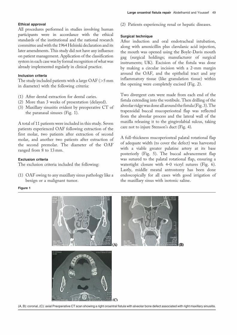
(2) More than 3 weeks of presentation (delayed). (3) Maxillary sinusitis evident by preoperative CT ofthe paranasal sinuses (Fig. 1).
A total of 11 patients were included in this study. Sevenpatients experienced OAF following extraction of thefirst molar, two patients after extraction of secondmolar, and another two patients after extraction ofthe second premolar. The diameter of the OAFranged from 8 to 13mm.
Exclusion criteriaThe exclusion criteria included the following:
(1)
OAF owing to any maxillary sinus pathology like abenign or a malignant tumor.re 1
): coronal, (C): axial Preoperative CT scan showing a right oroantral fis
(2)
tula w
Patients experiencing renal or hepatic diseases.
Surgical techniqueAfter induction and oral endotracheal intubation,along with amoxicillin plus clavulanic acid injection,the mouth was opened using the Boyle-Davis mouthgag (surgical holdings; manufacturer of surgicalinstruments; UK). Excision of the fistula was doneby making a circular incision with a 2-mm marginaround the OAF, and the epithelial tract and anyinflammatory tissue (like granulation tissue) withinthe opening were completely excised (Fig. 2).
Two divergent cuts were made from each end of thefistula extending into the vestibule. Then drilling of thealveolar ridgewasdoneall aroundthe fistula (Fig.3).Thetrapezoidal buccal mucoperiosteal flap was reflectedfrom the alveolar process and the lateral wall of themaxilla releasing it to the gingivolabial sulcus, takingcare not to injure Stenson’s duct (Fig. 4).
A full-thickness mucoperiosteal palatal rotational flapof adequate width (to cover the defect) was harvestedwith a viable greater palatine artery at its baseposteriorly (Fig. 5). The buccal advancement flapwas sutured to the palatal rotational flap, ensuring awatertight closure with 4-0 vicryl sutures (Fig. 6).Lastly, middle meatal antrostomy has been doneendoscopically for all cases with good irrigation ofthe maxillary sinus with isotonic saline.
ith alveolar bone defect associated with right maxillary sinusitis.
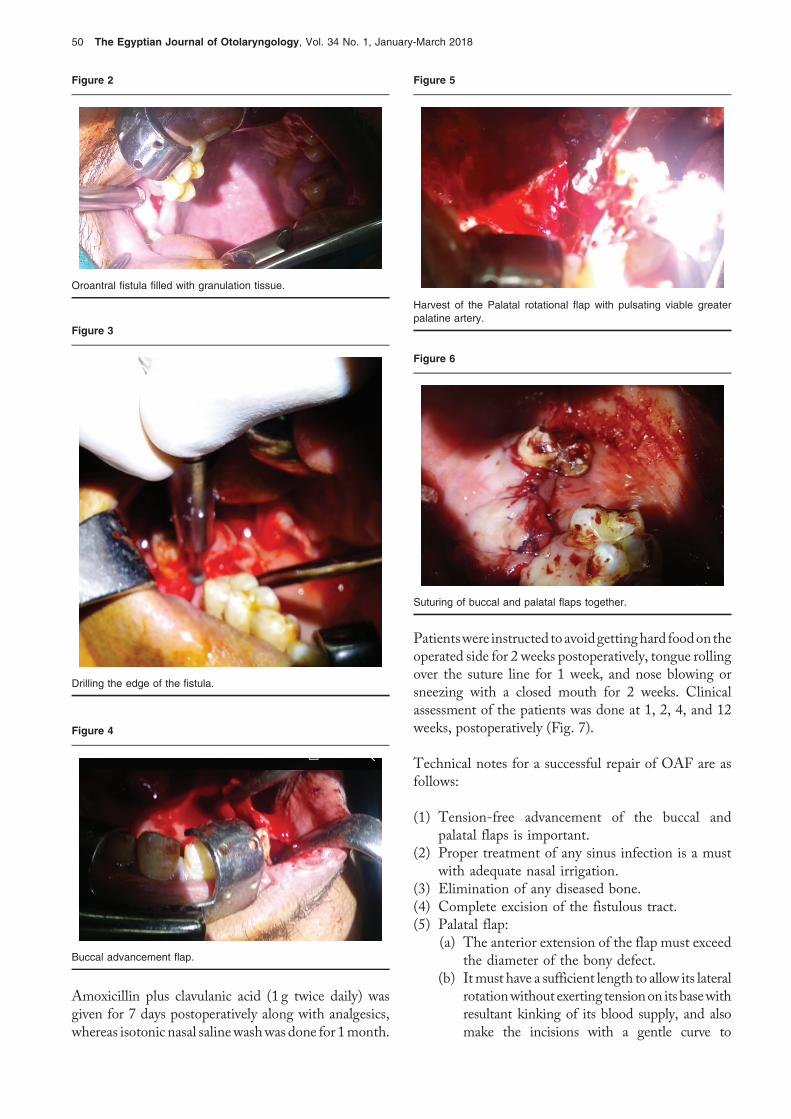
Figure 4
Buccal advancement flap.
Figure 5
Harvest of the Palatal rotational flap with pulsating viable greaterpalatine artery.
Figure 2
Oroantral fistula filled with granulation tissue.
Figure 3
Drilling the edge of the fistula.
Figure 6
Suturing of buccal and palatal flaps together.
50 The Egyptian Journal of Otolaryngology, Vol. 34 No. 1, January-March 2018
Amoxicillin plus clavulanic acid (1 g twice daily) wasgiven for 7 days postoperatively along with analgesics,whereas isotonic nasal salinewashwas done for 1month.
Patientswere instructed toavoidgettinghard foodontheoperated side for 2 weeks postoperatively, tongue rollingover the suture line for 1 week, and nose blowing orsneezing with a closed mouth for 2 weeks. Clinicalassessment of the patients was done at 1, 2, 4, and 12weeks, postoperatively (Fig. 7).
Technical notes for a successful repair of OAF are asfollows:
(1)
Tension-free advancement of the buccal andpalatal flaps is important.(2)
Proper treatment of any sinus infection is a mustwith adequate nasal irrigation.(3)
Elimination of any diseased bone. (4) Complete excision of the fistulous tract. (5) Palatal flap:(a) The anterior extension of the flap must exceedthe diameter of the bony defect.
(b) Itmust have a sufficient length to allow its lateralrotationwithout exerting tensionon its basewithresultant kinking of its blood supply, and alsomake the incisions with a gentle curve to
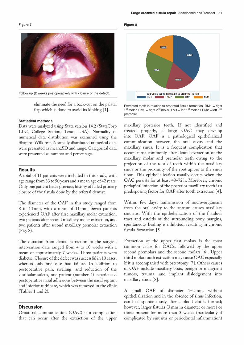
Figure 7
Follow up (2 weeks postoperatively with closure of the defect).
Figure 8
Extracted tooth in relation to oroantral fistula formation. RM1 = right1st molar; RM2 = right 2nd molar; LM1 = left 1st molar; LPM2 = left 2nd
Large oroantral fistula repair Abdelhamid and Youssef 51
eliminate the need for a back-cut on the palatalflap which is done to avoid its kinking [1].
premolar.
Statistical methodsData were analyzed using Stata version 14.2 (StataCorpLLC, College Station, Texas, USA). Normality ofnumerical data distribution was examined using theShapiro–Wilk test. Normally distributed numerical datawere presented as mean±SD and range. Categorical datawere presented as number and percentage.
ResultsA total of 11 patients were included in this study, withage range from33 to 50 years and amean age of 42 years.Only one patient had a previous history of failed primaryclosure of the fistula done by the referral dentist.
The diameter of the OAF in this study ranged from8 to 13mm, with a mean of 11mm. Seven patientsexperienced OAF after first maxillary molar extraction,two patients after second maxillary molar extraction, andtwo patients after second maxillary premolar extraction(Fig. 8).
The duration from dental extraction to the surgicalintervention date ranged from 4 to 10 weeks with amean of approximately 7 weeks. Three patients werediabetic. Closure of the defect was successful in 10 cases,whereas only one case had failure. In addition topostoperative pain, swelling, and reduction of thevestibular sulcus, one patient (number 4) experiencedpostoperative nasal adhesions between the nasal septumand inferior turbinate, which was removed in the clinic(Tables 1 and 2).
DiscussionOroantral communication (OAC) is a complicationthat can occur after the extraction of the upper
maxillary posterior teeth. If not identified andtreated properly, a large OAC may developinto OAF. OAF is a pathological epithelializedcommunication between the oral cavity and themaxillary sinus. It is a frequent complication thatoccurs most commonly after dental extraction of themaxillary molar and premolar teeth owing to theprojection of the root of teeth within the maxillarysinus or the proximity of the root apices to the sinusfloor. This epithelialization usually occurs when theOAC persists for at least 48–72 h. Moreover, chronicperiapical infection of the posterior maxillary teeth is apredisposing factor for OAF after tooth extraction [4].
Within few days, transmission of micro-organismsfrom the oral cavity to the antrum causes maxillarysinusitis. With the epithelialization of the fistuloustract and osteitis of the surrounding bony margins,spontaneous healing is inhibited, resulting in chronicfistula formation [5].
Extraction of the upper first molars is the mostcommon cause for OACs, followed by the uppersecond premolars and the second molars [6]. Upperthird molar tooth extraction may cause OAC especiallyif it is accompanied with osteotomy [7]. Others causesof OAF include maxillary cysts, benign or malignanttumors, trauma, and implant dislodgement intomaxillary sinus [8].
A small OAF of diameter 1–2mm, withoutepithelialization and in the absence of sinus infection,can heal spontaneously after a blood clot is formed;however, larger fistulas (3mm in diameter or more) orthose present for more than 3 weeks (particularly ifcomplicated by sinusitis or periodontal inflammation)
Table 1 Summary of cases
Caseno.
Age(years)
Sex DM Extractedtooth
Fistula size(mm)
Duration of fistula(weeks)
Previous trial ofclosure
Successfulclosure
1 42 Male Positive RM1 13 4 Positive Successful
2 35 Male Negative RM2 12 9 Negative Successful
3 44 Female Negative LM1 11 8 Negative Successful
4 41 Male Negative LM1 10 7 Negative Successful
5 45 Female Negative LM1 10 8 Negative Successful
6 43 Female Positive RM1 12 6 Negative Failed
7 38 Female Negative LPM2 8 10 Negative Successful
8 47 Female Negative LPM2 9 7 Negative Successful
9 50 Female Positive RM1 12 5 Negative Successful
10 40 Female Negative RM2 13 5 Negative Successful
11 33 Male Negative LM1 11 6 Negative Successful
DM, diabetes mellitus; LM1, left first molar; LPM2, left second premolar; RM1, right first molar; RM2, right second molar.
Table 2 Descriptive statistics for the studied series
Variables Values
Age (years) 41.6±5.0 (33–50)
Sex (male/female) 4/7
DM 3 (27.3)
Extracted tooth
LM1 4 (36.3)
LPM2 2 (18.2)
RM1 3 (27.3)
RM2 2 (18.2)
Fistula size (mm) 11±1.6 (8–13)
Duration of fistula (weeks) 6.8±1.8 (4–10)
Previous trial at closure 1 (9.1)
Outcome of procedure
Successful closure of fistula 10 (90.9)
Failed closure of fistula 1 (9.1)
Data are represented as mean±SD (range) or number (%).
52 The Egyptian Journal of Otolaryngology, Vol. 34 No. 1, January-March 2018
will persist requiring early surgical closure. OACs widerthan 5mm require the use of flaps for closure [9,10].
Acute sinusitis can occur in approximately 90% ofpatients experiencing untreated OAF for 2 weeksowing to contamination by food or saliva [11].
Probing (the introduction of a probe through the fistulainto the antrum) should never be attempted because itmay lead to sinusitis or widening of the fistula owing tothe pushing of foreign bodies or oral flora into themaxillary sinus. Large OAF are clinically seen oninspection and are evident by CT of the paranasalsinuses which gives an accurate estimate of the bonydefect of the fistula and also reveals the presence andlocation of any dental roots, implants, or any foreignbody that may have been dislodged into the sinus. CTalso detects presence of maxillary sinusitis or associatedperiodontal disease [1,11].
Immediate closure of the OAF has a high success rate(approaching 95%), which is significantly higher than
for the closure of chronic fistulae [1]. Options for therepair of the OAF include palatal rotational, buccaladvancement flaps, and buccal pad fat flap [12].
Advantages of the palatal flap include the following:
(1)
Good vascularization (the greater palatine artery). (2) No lowering of the vestibule (like with the use ofbuccal flaps), so can be used in patients wearingdentures.
(3)
Moreover, palatal flap is firmer and more resistantto trauma and infection than buccal flap [2].Disadvantages of palatal flaps are the denudation ofthe palatal surface that requires secondary healing,pain, and the roughness and deepening of this areaowing to secondary epithelialization over two to 3months. The unpleasant complication is necrosis ofthe palatal flap if its blood supply (greater palatineartery) is jeopardized because of kinking along its archof rotation or owing to the back-cut used to eliminatethis kinking [1,2].
Borgonovo et al. [1] mentioned that palatal flap isfeasible only in closing fistulas in the premolar regionbecause excessive tension in the molar regionmay causeischemia of the flap owing to occlusion of greaterpalatine artery. However, we think that it can beused in fistula of the molar region if good releasewas done with a gentle curve incision, eliminatingthe need for a back-cut on the palatal flap requiredto avoid its kinking.
Borgonovo et al. [1] also concluded that buccaladvancement flaps alone are best for small OAF, andthat they should not be used alone in largeOAF, insteadbuccal flap must be combined with palatal flaps to givethe best results. Disadvantages of the buccal flap includelowering of the vestibulum representing a serious
Large oroantral fistula repair Abdelhamid and Youssef 53
problem to patients wearing removable dentures andrequiring a second procedure (vestibuloplasty) to releasethe gingivolabial sulcus [1].
Yilmaz et al. [13] showed that blood supply of palatalflap is better than buccal flap and hence it is preferred inlarge and recurrent OAF.
Buccal fat pad can be used in a fistula of 8–20mm indiameter. Over a period of 3 weeks, the fatty tissueconverts into granulation tissue and epithelizes [9].
Risk factors for failure of closure of OAF include thesize of the fistula, sinus infection not properly treatedpreoperatively, osteitis of the fistula margins (hence thedrilling of the bony margins of OAF in all cases in thisstudy), epithelialization of the fistulous tract, andexcessive tension on the flap impairing blood supplyfor healing. The most common reported cause ofchronicity of the OAF and also failure of repair isthe insufficient treatment of the sinusitis, hence theneed for endoscopic middle meatal antrostomy in allcases in this study [14,15].
Endoscopic sinus surgery can be used successfully totreat sinus infection associated withOAF instead of theCaldwell–Luc procedure to decrease morbidity andcomplications [16].
In large oroantral fistulas with a diameter more than5mm, failure rate increases owing to the large defect inthe underlying bone that supports the overlying flap.Many options for the reconstruction of this bony defectexist, including autologous bone graft [17], nonporoushydroxyapatite blocks, and titanium plate with wiring.However, these materials are not widely accepted inroutine surgical closure of OAF owing to cost, difficulthandling, increased rate of infection, and exfoliation[18–20].
Auricular cartilagegraft is anewtechnique for theclosureofOAF. It is biocompatible, highly resistant to infection,easy to harvest and manipulate, nonresorbable, and costeffective. It does not require vascularization for theintegration to the recipient site decreasing the failurerate of the graft. Additionally, it acts as a barrier betweenthe sinus membrane and the oral mucosa, which allowssuccessful healing. A disadvantage of this technique isdefect formation at the donor site occurs [21].
Sandwich technique is another new technique for theclosure of OAF, in which both hard tissue (bone)and soft tissue closure is achieved. It uses Bio-Oss,which is a bone grafting material similar to human
bone and highly successful in new bone formation,sandwiched between two sheaths of Biogide(a resorbable synthesized collagen membrane). Theporous surface facing the bone allows the ingrowth ofbone-forming cells. Owing to its high purity, noallergic reaction or infection is observed. It is asimple and excellent technique, especially whensubsequent placement of endosseous dental implantis considered without the need of donor site surgeryfor bone grafting [22].
Although new techniques (like the sandwichtechnique) avoid the disadvantages of the palataland buccal flaps, they are not used in our instituteowing to cost issues. In this study, new bone formationhas not been assessed. The study focused on whetherthis technique is suitable for the soft tissue closure oflarge OAF. Further study is required to detect long-term new bone formation using this technique, whichis an important issue now for the subsequentplacement of endosseous dental implants.Previousstudies showed that the frequency of occurrence ofOAF is nearly the same in both sexes [6,23]. Femalesexhibit larger sinuses than males and should,therefore, be at a greater risk of OAF [24]. In thisstudy, the frequency of occurrence of OAF is more infemales than males noting that this study group issmall.
Mean age of patients in our study was 42 years,knowing that the incidence of OAC is high afterthe third decade as the maxillary sinus reaches itsgreatest size [6,12].
Although Hassan et al. [25] mentioned that diabetescould be a risk factor for failure of OAF closure, thethree diabetic patients in this study had successfulclosure of the OAF using this technique with goodhealing (noting that strict perioperative control ofblood glucose in diabetic patients was ensured inthis study).
ConclusionCombined buccal advancement and palatal rotationalflaps procedure is efficient in closure of large OAF.Further study is required to assess new bone formationafter repair of the OAF using this technique.
Financial support and sponsorshipNil.
Conflicts of interestThere are no conflicts of interest.
54 The Egyptian Journal of Otolaryngology, Vol. 34 No. 1, January-March 2018
References1 Borgonovo AE, Berardinelli FV, Favale M, Maiorana C. Surgical options in
oroantral fistula treatment. Open Dent J 2012; 6:94–98.
2 Solker K, Vuksan V, Lauc T. Treatment of oroantral fistula. Acta StomatolCroat 2002; 36:135–140.
3 Guven O. A clinical study on oroantral fistulae. J Craniomaxillofac Surg1998; 26:267–271.
4 De Biasi M, Maglione M, Angerame D. The effectiveness of surgicalmanagement of oroantral communications: a systematic review of theliterature. Eur J Oral Implantol 2014; 7:347–357.
5 Yalcin S, Oncü B, Emes Y, Atalay B, Aktas ̧ I. Surgical treatment of oroantralfistulas: a clinical study of 23 cases. J Oral Maxillofac Surg 2011; 69:333–339.
6 Punwutukorn C, Waikakul A, Pairuchvej V. Clinically significant oroantralcommunications − a study of incidence and site. Int J Oral Maxillofac Surg1994; 23:19–21.
7 Del Rey-Santamaría M, Valmaseda-Castellón E, Berini-Aytés L, Gay-Escoda C. Incidence of oral sinus communications in 389 upperthirmolar extraction. Med Oral Patol Oral Cir Bucal 2006; 11:E334–E338.
8 Awang MN. Closure of oroantral fistula. Int J Oral Maxillofac Surg 1988;17:110–115.
9 Hanazawa Y, Itoh K, Mabashi T, Sato K. Closure of oroantralcommunications using a pedicled buccal fat pad graft. J Oral MaxillofacSurg 1995; 53:771–776.
10 Liversedge RL, Wong K. Use of the buccal fat pad in maxillary and sinusgrafting of the severely atrophic maxilla preparatory to implantreconstruction of the partially or completely edentulous patient: technicalnote. Int J Oral Maxillofac Implants 2002; 17:424–428.
11 Scattarella A, Ballini A, Grassi FR, Carbonara A, Ciccolella F, Dituri A, et al.Treatment of oroantral fistula with autologous bone graft and application ofa non-reabsorbable membrane. Int J Med Sci 2010; 7:267–271.
12 Anavi Y, Gal G, Silfen R, Calderon S. Palatal rotation-advancement flap fordelayed repair of oroantral fistula: a retrospective evaluation of 63 cases.Oral Surg Oral Med Oral Pathol Oral Radiol Endod 2003; 96:527–534.
13 Yilmaz T, Suslu AE, Gursel B. Treatment of oroantral fistula:experiencewith 27 cases. Am J Otolaryngol 2003; 24:221–223.
14 Hernando J, Gallego L, Junguera L, Villarreal P. Oroantral communications.A retrospective analysis. Med Oral Patol Oral Cir Bucal 2010; 15:e499–e503.
15 Haanaes HR, Pederson KN. Treatment of oroantral communication. Int JOral Surg 1974; 3:124–132.
16 Hajiioannou J, Koudounarakis E, Alexopoulos K, Kotsani A, Kyrmizakis DE.Maxillary sinusitis of dental origin due to oroantral fistula, treated byendoscopic sinus surgery and primary fistula closure. J Laryngol Otol2010; 124:986–989.
17 Proctor B. Bone graft closure of large or persistent oromaxillary fistula.Laryngoscope 1969; 79:822–826.
18 Ahmed WM. Closure of oroantral fistula using titanium plate withtransalveolar wiring. J Maxillofac Oral Surg 2015; 14:121–125.
19 GoldmanEH, Stratigos GT, Arthur AL. Treatment of oroantral fistula by goldfoil closure: report of case. J Oral Surg 1969; 27:875–877.
20 Zide MF, Karas ND. Hydroxylapatite block closure of oroantral fistulas:report of cases. J Oral Maxillofac Surg 1992; 50:71–75.
21 Isler SC, Demircan S, Cansiz E. Closure of oroantral fistula using auricularcartilage: a new method to repair an oroantral fistula. Br J Oral MaxillofacSurg 2011; 49:e86–e87.
22 Ogunsalu C. A new surgical management for oro-antralcommunication: the resorbable guided tissue regenerationmembrane-bone substitute sandwich technique. West Indian Med J2005; 54:261–263.
23 Skoglund LA, Pedersen SS, Holst E. Surgical management of 85perforations to the maxillary sinus. Int J Oral Surg 1983; 12:1–5.
24 Lin PT, Bukachevsky R, Blake M. Management of odontogenicsinusitis with persistent oro-antral fistula. Ear Nose Throat J 1991;70:488–490.
25 Hassan O, Shoukry T, Raouf AA, Wahba H. Combined palatal and buccalflaps in oroantral fistula repair. Egypt J Ear Nose Throat Allied Sci 2012;13:77–81.
![Prosthodontic Management of Oroantral Fistula: A Case Report · prosthodontic management of oroantral fistula. Case Series Abstract Oroantral fistula (oroantral communications [OACs])](https://img.pdfslide.us/doc/110x75/5e7beca2e72ed6083b54888d/prosthodontic-management-of-oroantral-fistula-a-case-report-prosthodontic-management.jpg)