Embed Size (px)
Citation preview

Current Neurobiology 2012 Volume 3 Issue 2 85
Current Neurobiology 2012 (2): 85-89 ISSN 0975-9042 Laminar cytoarchitecture of cerebral cortex in Cav2.1 mutant, rolling mouse Nagoya Miwa Horiuchi-Hirose 1, Kazuhiko Sawada2 and Yoshihiro Fukui3 1 Department of Nutrition, and 2 Department of Physical Therapy, Faculty of Medical and Health Sciences, Tsukuba International University, Tsuchiura, Ibaraki, Japan 3 Department of Anatomy and Developmental Neurobiology, University of Tokushima Graduate School Institute of Health Biosciences, Tokushima, Japan Running title: Cortical cytoarchitecture in rolling mice
Abstract
The present study quantitatively examined the laminarcytoarchitecture of functional corti-cal areas of the cerebrum in Cav2.1 mutant, rolling mouse Nagoya. Brain weights were not significantly different between rolling and wild-type mice. Klüver Barrera’s staining of the cerebral cortex revealed no obvious changes in ctytoarchitecture of three functional cortical areas, i.e.,primary motor (M1), somatosensory (S1) and primary visual (V1) areas,in rolling mice.The cortical thickness and the thickness of the cortical layers (I-VI) of those three ar-eas were not significantly different between rolling and wild-type mice.The results suggest that the cytoarchitectural organization in the functional cortical areas of the cerebral cortex is not altered by a Cav2.1 gene mutation. Accepted September 11 2012
Introduction Many neuronal processes such as membrane excitability and neurotransmitter release are regulated by calcium influx through the voltage-gated Ca2+channels, which areclassified intoT-, L-, N-, P/Q- and R-types bypharma-cological and electrophysiological characteristics of the pore-forming a1 subunit [1-3]. The P/Q-type Ca2+ channel has theα1A subunit (Cav2.1),which is produced by alter-nating the splicing of an a1 subunit gene family [4], an-dexpresses prominentlythroughout the brain[5]. Rolling mouse Nagoya carries a recessive mutant allele of the tottering locus (tgrol) on chromosome 8 [6], whichen-codes a Cav2.1 gene [7], and is known as a model for hu-man Ca2+ channelopathies such as episodic ataxia type 2 and familial hemiplegic migraine [8]. This mutant mouse is characterized by a severe ataxic gait and abnormal hindlimb extension, but does not exhibit epilepsy [9;10] as seen in allelic mutants, tottering, leaner [11] and rock-ermice [12]. Those phenotypes areinvolved in a selective reduction of Ca2+currents through the P/Q-type chan-nel[7], which is highly expressed in cerebellar Purkinje cells not only in normal mice [5] but also in Cav2.1 mu-tants [12;13], including rolling mice [14]. On the other hand, theCav2.1is known to be expressed in the cerebral cortex[5;15], andmediates Ca2+ inflow in pyramidal cells of layersII/IIIof the cerebral cortex [15;17].Considering the pivotal roles of the Cav2.1 channel, the presence of the
mutated Cav2.1 channel in pyramidal neurons in the su-perficial pyramidal layer predicts dysfunction of cortico-cortical neuronal networks involved in abnormal synaptic signaling.The present study aimed to clarify whether fun-damental laminar cytoarchitecture of the cerebral cortex was altered in rolling mice. The thicknessof cortical lam-ina was measured in threedifferentcortical areas, i.e., pri-mary motor (M1), somatosensory (S1) and primary visual areas (V1)inrolling mice. Materials and Methods Animals All experimental procedures were conducted in accor-dance with the guidelines of the NationalInstitutes of Health (NIH) for the Care and Use of Laboratory Animals (No. 80-23, revised 1996). The Institutional Animal Care and Use Committee of the University of Tokushima ap-provedthe procedures, and all efforts were made to mini-mize the number of animals used and their attendant suf-fering. Rolling mice were raised on a C3Hf/Nga back-ground. Homozygous rolling mice (tgrol/tgrol), raised by intercrossing heterozygous pairs, were readily identifiable by their ataxiclocomotion between postnatal days 10 and14. Wild-type (+/+) mice were used as controls. Tissue preparation A total of 5 male rolling and 5 male wild-type mice at 2 months of age were used. Animals were perfused with
Horiuchi-Hirose/Sawada/Fukui
Current Neurobiology 2012 Volume 3 Issue 2 86
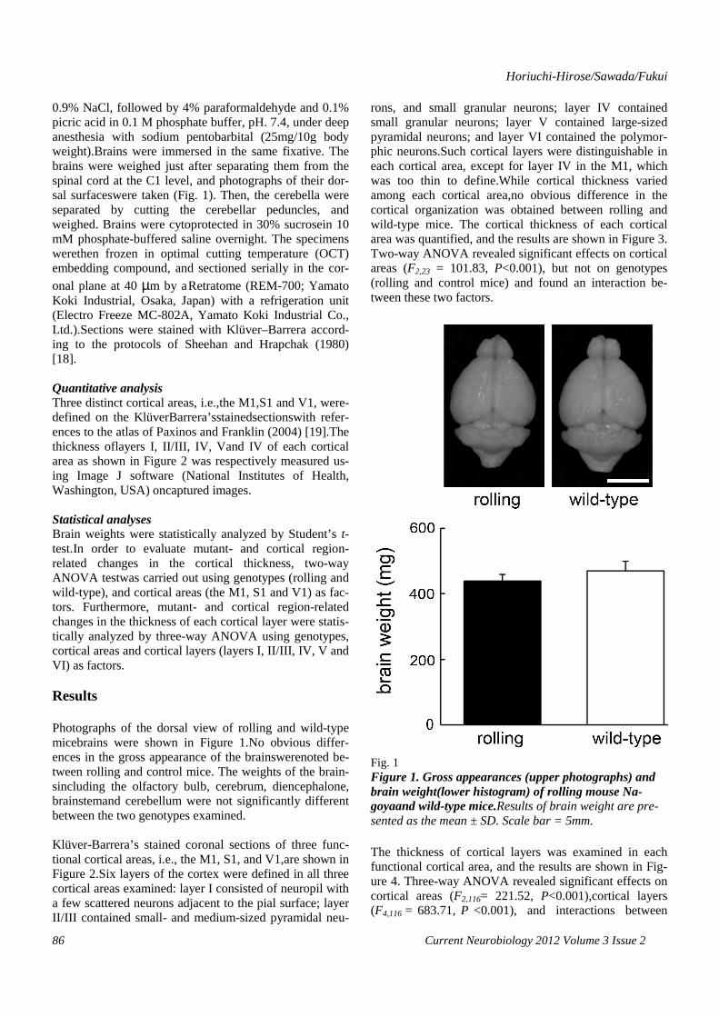
0.9% NaCl, followed by 4% paraformaldehyde and 0.1% picric acid in 0.1 M phosphate buffer, pH. 7.4, under deep anesthesia with sodium pentobarbital (25mg/10g body weight).Brains were immersed in the same fixative. The brains were weighed just after separating them from the spinal cord at the C1 level, and photographs of their dor-sal surfaceswere taken (Fig. 1). Then, the cerebella were separated by cutting the cerebellar peduncles, and weighed. Brains were cytoprotected in 30% sucrosein 10 mM phosphate-buffered saline overnight. The specimens werethen frozen in optimal cutting temperature (OCT) embedding compound, and sectioned serially in the cor-onal plane at 40 µm by aRetratome (REM-700; Yamato Koki Industrial, Osaka, Japan) with a refrigeration unit (Electro Freeze MC-802A, Yamato Koki Industrial Co., Ltd.).Sections were stained with Klüver–Barrera accord-ing to the protocols of Sheehan and Hrapchak (1980) [18]. Quantitative analysis Three distinct cortical areas, i.e.,the M1,S1 and V1, were-defined on the KlüverBarrera’sstainedsectionswith refer-ences to the atlas of Paxinos and Franklin (2004) [19].The thickness oflayers I, II/III, IV, Vand IV of each cortical area as shown in Figure 2 was respectively measured us-ing Image J software (National Institutes of Health, Washington, USA) oncaptured images. Statistical analyses Brain weights were statistically analyzed by Student’s t-test.In order to evaluate mutant- and cortical region-related changes in the cortical thickness, two-way ANOVA testwas carried out using genotypes (rolling and wild-type), and cortical areas (the M1, S1 and V1) as fac-tors. Furthermore, mutant- and cortical region-related changes in the thickness of each cortical layer were statis-tically analyzed by three-way ANOVA using genotypes, cortical areas and cortical layers (layers I, II/III, IV, V and VI) as factors. Results Photographs of the dorsal view of rolling and wild-type micebrains were shown in Figure 1.No obvious differ-ences in the gross appearance of the brainswerenoted be-tween rolling and control mice. The weights of the brain-sincluding the olfactory bulb, cerebrum, diencephalone, brainstemand cerebellum were not significantly different between the two genotypes examined. Klüver-Barrera’s stained coronal sections of three func-tional cortical areas, i.e., the M1, S1, and V1,are shown in Figure 2.Six layers of the cortex were defined in all three cortical areas examined: layer I consisted of neuropil with a few scattered neurons adjacent to the pial surface; layer II/III contained small- and medium-sized pyramidal neu-
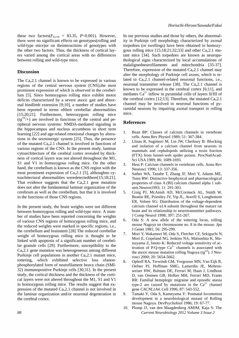
rons, and small granular neurons; layer IV contained small granular neurons; layer V contained large-sized pyramidal neurons; and layer VI contained the polymor-phic neurons.Such cortical layers were distinguishable in each cortical area, except for layer IV in the M1, which was too thin to define.While cortical thickness varied among each cortical area,no obvious difference in the cortical organization was obtained between rolling and wild-type mice. The cortical thickness of each cortical area was quantified, and the results are shown in Figure 3. Two-way ANOVA revealed significant effects on cortical areas (F2,23 = 101.83, P<0.001), but not on genotypes (rolling and control mice) and found an interaction be-tween these two factors.
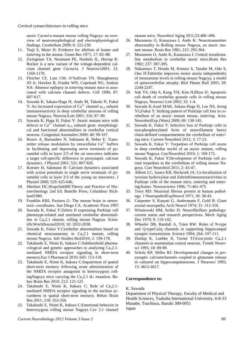
Fig. 1 Figure 1. Gross appearances (upper photographs) and brain weight(lower histogram) of rolling mouse Na-goyaand wild-type mice.Results of brain weight are pre-sented as the mean ± SD. Scale bar = 5mm. The thickness of cortical layers was examined in each functional cortical area, and the results are shown in Fig-ure 4. Three-way ANOVA revealed significant effects on cortical areas (F2,116= 221.52, P<0.001),cortical layers (F4,116 = 683.71, P <0.001), and interactions between

Cortical cytoarchitecture in rolling mice
Current Neurobiology 2012 Volume 3 Issue 2 87
Fig. 2 Figure 2. Klüver Barrera’s stainedcoronal sections of primary motor (M1), primary sensory (S1) and primary visual (V1) areas of cerebral cortex of rolling and wild-type mice. The layer IV in the M1 was too thin to be distinguishable in either rolling or wild-type mice. Scale bar = 10µm.
Fig. 3 Figure 3. Histogram showing cortical thickness of pri-mary motor (M1), somatosensory (S1) and primary vis-ual (V1) areas of cerebrum in rolling and wild-type mice. Results are presented as the mean ± SD. No signifi-cant effect on the cortical thickness of three cortical areas between rolling and wild-type mice was noted by three-way ANOVA.
Figure 4. Histogram showing thickness of cortical lay-ers I-VI in primary motor (M1), somatosensory (S1) and primary visual (V1) areas of cerebrum of rolling and wild-type mice. The layer IV in the M1 was too thin to be distinguishable in either rolling or wild-type mice. Re-sults are presented as the mean ± SD. No significant ef-fect on the thickness of each layer between rolling and wild-type mice was noted in three cortical areas by three-way ANOVA.
Fig. 4
Horiuchi-Hirose/Sawada/Fukui
Current Neurobiology 2012 Volume 3 Issue 2 88
these two factors(F8,116 = 83.35, P<0.001). However, there were no significant effects on genotypes(rolling and wild-type mice)or on theinteractions of genotypes with the other two factors. Thus, the thickness of cortical lay-ers varied among the cortical areas with no differences between rolling and wild-type mice.
Discussion
The Cav2.1 channel is known to be expressed in various regions of the central nervous system (CNS),the most prominent expression of which is observed in the cerebel-lum [5]. Since homozygous rolling mice exhibit motor deficits characterized by a severe ataxic gait and abnor-mal hindlimb extension [9;10], a number of studies have been reported in terms oftheir cerebellar abnormalities [15;20;21]. Furthermore, heterozygous rolling mice (tgrol/+) are involved in functions of the central and pe-ripheral nervous systems: NMDA-mediated signaling in the hippocampus and nucleus accumbens in short term learning [22] and age-related emotional changes by altera-tions in the serotonergic system [25]. Thus, the presence of the mutated Cav2.1 channel is involved in functions of various regions of the CNS. In the present study, laminar cytoarchitecture of the cerebral cortex revealed by thick-ness of cortical layers was not altered throughout the M1, S1 and V1 in homozygous rolling mice. On the other hand, the cerebellum is known as the CNS region with the most prominent expression of Cav2.1 [5], althoughno cy-toachitectural abnormalities weredetectedthere[15;18;21]. That evidence suggests that the Cav2.1 gene mutation does not alter the fundamental laminar organization of the cerebrum as well as the cerebellum, but that it is involved in the functions of those CNS regions. In the present study, the brain weights were not different between homozygous rolling and wild-type mice. A num-ber of studies have been reported concerning the weights of various CNS regions of homozygous rolling mice, and the reduced weights were marked in specific regions, i.e., the cerebellum and brainstem [28] The reduced cerebellar weight of homozygous rolling mice is thought to be linked with apoptosis of a significant number of cerebel-lar granule cells [29]. Furthermore, susceptibility to the Cav2.1 gene mutation was heterogeneous among different Purkinje cell populations in another Cav2.1 mutant mice, tottering, which exhibited selective loss ofanon-phosphorylated form of neurofilament heavy chain (SMI-32) immunopositive Purkinje cells [30;31]. In the present study, the cortical thickness and the thickness of the corti-cal layers were not altered throughout the M1, S1 and V1 in homozygous rolling mice. The results suggest that ex-pression of the mutated Cav2.1 channel is not involved in the laminar organization and/or neuronal degeneration in the cerebral cortex.
In our previous studies and those by others, the abnormal-ity in Purkinje cell morphology characterized by axonal torpedoes (or swellings) have been obtained in homozy-gous rolling mice [15;18;21;32;33] and other Cav2.1 mu-tant mice [34]. Such torpedoes are known as neuropa-thological signs characterized by local accumulations of malalignedneurofilaments and mitochondria [35-37]. Therefore, expression of the mutated Cav2.1 channel may alter the morphology of Purkinje cell axons, which is re-lated to Cav2.1 channel-related neuronal functions, i.e., neuronal transmitter release [38]. The Cav2.1 channel is known to be expressed in the cerebral cortex [6;11], and mediates Ca2+ inflow in pyramidal cells of layers II/III of the cerebral cortex [12;13]. Therefore, the mutated Cav2.1 channel may be involved in neuronal functions of py-ramidal neurons by impairing axonal transport in rolling mice.
References 1. Bean BP: Classes of calcium channels in vertebrate
cells. Annu Rev Physiol 1989; 51: 367-384. 2. Llinas R, Sugimori M, Lin JW, Cherksey B: Blocking
and isolation of a calcium channel from neurons in mammals and cephalopods utilizing a toxin fraction (FTX) from funnel-web spider poison. ProcNatlAcad-Sci USA 1989; 86: 1689-1693.
3. Hess P: Calcium channels in vertebrate cells. Annu Rev Neurosci 1990; 13: 337-356.
4. Sather WA, Tanabe T, Zhang JF, Mori Y, Adams ME, Tsien RW: Distinctive biophysical and pharmacological properties of class A (BI) calcium channel alpha 1 sub-unit.Neuron1993; 11: 291-303.
5. Craig PJ, McAinsh AD, McCormack AL, Smith W, Beattie RE, Priestley JV, Yip JL, Averill S, Longbottom ER, Volsen SG: Distribution of the voltage-dependent calcium channel α1A subunit throughout the mature rat brain and its relationship to neurotransmitter pathways. J Comp Neurol 1998; 397: 251-267.
6. Oda S: A new allele of the tottering locus, rolling mouse Nagoya on chromosome no. 8 in the mouse. Jpn J Genet 1981; 56: 295-299.
7. Mori Y, Wakamori M, Oda S, Fletcher CF, Sekiguchi N, Mori E, Copeland NG, Jenkins NA, Matsushita K, Ma-tsuyama Z, Imoto K: Reduced voltage sensitivity of ac-tivation of P/Q-type Ca2+ channels is associated with the ataxic mouse mutation rolling Nagoya (tgrol). J Neu-rosci 2000; 20: 5654-5662.
8. Ophoff RA, Terwindt GM, Vergouwe MN, Van Eijk R, Oefner PJ, Hoffman SMG, Lamerdin JE, Mohren-weiser HW, Bulman DE, Ferrari M, Haan J, Lindhout D, van Ommen GB, Hofker MH, Ferrari MD, Frants RR: Familial hemiplegic migraine and episodic ataxia type-2 are caused by mutations in the Ca2+ channel gene CACNL1A4. Cell 1996; 87: 543-552.
9. Tamaki Y, Oda S, Kameyama Y: Postnatal locomotion development in a neurobiological mutant of Rolling mouse Nagoya. DevPsychobiol 1986; 19: 67-77.
10. Plomp JJ, van den Maagdenberg AMJM, Kaja S: The
Cortical cytoarchitecture in rolling mice
Current Neurobiology 2012 Volume 3 Issue 2 89
ataxic Cacna1a-mutant mouse rolling Nagoya: an over-view of neuromorphological and electrophysiological findings. Cerebellum 2009; 8: 222-230.
11. Tsuji S, Meier H: Evidence for allelism of leaner and tottering in the mouse. Genet Res 1971; 17: 83–88.
12. Zwingman TA, Neumann PE, Noebels JL, Herrup K: Rocker is a new variant of the voltage-dependent cal-cium channel gene Cacna1a. J Neurosci2001; 21: 1169-1178.
13. Fletcher CF, Lutz CM, O’Sullivan TN, Shaughnessy JD Jr, Hawkes R, Franke WN, Copeland NG, Jenkins NA: Absence epilepsy in tottering mutant mice is asso-ciated with calcium channel defects. Cell 1996; 87: 607-617.
14. Sawada K, Sakata-Haga H, Ando M, Takeda N, Fukui Y: An increased expression of Ca2+ channel α1A subunit immunoreactivity in deep cerebellar neurons of rolling mouse Nagoya. NeurosciLett 2001; 316: 87–90.
15. Sawada K, Haga H, Fukui Y: Ataxic mutant mice with defects in Ca2+ channel α1A subunit gene: morphologi-cal and functional abnormalities in cerebellar cortical neurons. Congenital Anomalies 2000; 40: 99-107.
16. Rozov A, Burnashev N, Sakmann B, Neher E: Trans-mitter release modulation by intracellular Ca2+ buffers in facilitating and depressing nerve terminals of py-ramidal cells in layer 2/3 of the rat neocortex indicates a target cell-specific difference in presynaptic calcium dynamics. J Physiol 2001; 531: 807-826.
17. Koester H, Sakmann B: Calcium dynamics associated with action potentials in single nerve terminals of py-ramidal cells in layer 2/3 of the young rat neocortex. J Physiol 2000; 529: 625-646.
18. Sheehan DC,HrapchakBB:Theory and Practice of His-totechnology 2nd Ed. Battelle Press, Columbus/ Rich-land1980.
19. Franklin KBJ, Paxinos G: The mouse brain in stereo-taxic coordinates. San Diego CA. Academic Press 1997.
20. Sawada K, Fukui Y:Zebrin II expressing Purkinje cell phenotype-related and unrelated cerebellar abnormali-ties in Cav2.1 mutant, rolling mouse Nagoya. Scien-tificWorldJournal2010; 10: 2032-2038.
21. Sawada K, Fukui Y:Cerebellar abnormalities based on chemical neuroanatomy in Cav2.1 mutant, rolling mouse Nagoya. Adv Studies Biol2010; 2: 159-178.
22. Takahashi E, Niimi K, Itakura C:Subthreshold pharma-cological and genetic approaches to analyzing Cav2.1-mediated NMDA receptor signaling in short-term memory.Eur J Pharmacol 2010; 645: 113-118.
23. Takahashi E, Niimi K, Itakura C:Impairment of spatial short-term memory following acute administration of the NMDA receptor antagonist in heterozygous roll-ingNagoya mice carrying the Cav2.1 α1 mutation. Be-hav Brain Res 2010; 213: 121-125
24. Takahashi E, Niimi K, Itakura C: Role of Cav2.1-mediated NMDA receptor signaling in the nucleus ac-cumbens in spatial short-term memory. Behav Brain Res 2011; 218: 353-356.
25. Takahashi E, Niimi K, Itakura C:Emotional behavior in heterozygous rolling mouse Nagoya Cav 2.1 channel
mutant mice. Neurobiol Aging 2011;32:486–496. 26. Muramoto O, Kanazawa I, Ando K: Neurotransmitter
abnormality in Rolling mouse Nagoya, an ataxic mu-tant mouse. Brain Res 1981; 215: 295-304.
27. Muramoto O, Ando K, Kanazawa I: Central noradrena-line metabolism in cerebellar ataxic mice.Brain Res 1982; 237: 387-395.
28. Nakamura T, Honda M, Kimura S, Tanabe M, Oda S, Ono H:Taltirelin improves motor ataxia independently of monoamine levels in rolling mouse Nagoya, a model of spinocerebellar atrophy. Biol Pharm Bull 2005; 28: 2244-2247.
29. Suh YS, Oda S, Kang YH, Kim H,Rhyu IJ: Apoptotic cell death of cerebellar granule cells in rolling mouse Nagoya, Neurosci Lett 2002; 32: 1-4.
30. Sawada K,Azad MAK, Sakata-Haga H, Lee NS, Jeong YG,Fukui Y: Striking pattern of Purkinje cell loss in ce-rebellum of an ataxic mutant mouse, tottering. Acta-NeurobiolExp (Wars) 2009; 69: 138-145.
31. Sawada K, Fukui Y: Selective loss of Purkinje cells in non-phosphorylated form of neurofilament heavy chain-defined compartmentsin the cerebellum of totter-ing mice. Current Neurobiol 2012; 3: 17-23.
32. Sawada K, Fukui Y: Torpedoes of Purkinje cell axons in deep cerebellar nuclei of an ataxic mutant, rolling mouse Nagoya. CurrNeurobiol 2010; 1: 169-172.
33. Sawada K, Fukui Y:Development of Purkinje cell ax-onal torpedoes in the cerebellum of rolling mouse Na-goya. Curr Neurobiol 2011; 2: 43-47.
34. Abbott LC, Isaacs KR, Heckroth JA: Co-localization of tyrosine hydroxylase and ZebrinIIimmunoreactivities in Purkinje cells of the mutant mice, tottering and totter-ing/leaner. Neuroscience 1996; 71:461-475.
35. Terry RD: Neuronal fibrous protein in human pathol-ogy. J NeuropatholExpNeurol 1971; 30: 38-43.
36. Carpenter S, Karpati G, Andermann F, Gold R: Giant axonal neuropathy.Arch Neurol 1974; 31: 312-316.
37. Wisniewski HM, Soifer D: Neurofibrillary pathology: current status and research perspectives. Mech Aging Dev 1979; 9: 119-142.
38. Wheeler DB, Randall A, Tsien RW: Roles of N-type and Q-typeCa2q channels in supporting hippocampal synaptic transmission. Science 1994; 264: 107-111.
39. Dunlap K, Luebke JI, Turner TJ:Exocytotic Cav2.1 channels in mammalian central neurons. Trends Neuro-sci 1995; 18: 89-98.
40. Scholz KP, Miller RJ: Developmental changes in pre-synaptic calciumchannels coupled to glutamate release in cultured rat hippocampalneurons. J Neurosci 1995; 15: 4612-4617.
Correspondence to: K. Sawada Department of Physical Therapy, Faculty of Medical and Health Sciences, Tsukuba International University, 6-8-33 Manabe, Tsuchiura, Ibaraki 300-0051 Japan